Submitted:
27 August 2025
Posted:
01 September 2025
You are already at the latest version
Abstract
Bristletails (Microcoryphia) represent a relatively understudied group of class Insecta. These wingless hexapods, which retain numerous plesiomorphic structures, remain less well understood compared to most other insect lineages. Through detailed morphological examination of specimens collected from southern China, we describe six new species (Pedetontus (Verhoeffilis) elegans sp. n. from Zhejiang, P. (V.) hezhouensis sp. n., P. (V.) jinxiuensis sp. n. and P. (V.)nanningensissp. n. from Guangxi, P. (V.)shenzhenensis sp. n. and P. (V.)xanthospilus sp. n. form Guangdong) of subgenus Verhoeffilis Paclt, 1972, this subgenus distinguished from other members of the Petrobiinae by several diagnostic features: paired ocelli shoe-shaped (fusiform-shaped) and submedian; two pairs of retractile vesicles on abdominal segments II–V; parameres restricted to coxite IX; penis opening small and apical; male genitalia not exceeding coxite IX; ovipositor of primary type. The new species can be distinguished from known species and each other by the morphology of the compound eyes, maxilla, labial palps, legs, and genitalia, here, we provide detailed illustrations, tables, and descriptions. Furthermore, we sequenced the COX1 gene to construct a phylogenetic tree of Microcoryphia. This contribution increases the total number of reported Pedetontus species in China to seventeen.
Keywords:
Microcoryphia
; Archaeognatha
; Pedetontus
; China
; taxonomy
; morphology
; COX1
1. Introduction
Insects represent the most widespread and adaptable group of animals on Earth, primarily due to their well-developed wings and diverse modes of metamorphosis. However, bristletails (Microcoryphia, also referred to as Archaeognatha) differ significantly from the majority of insects: they are wingless and thus unable to fly, and their juveniles and adults differ only in body size and sexual maturity—they moult throughout their entire lives [1,2]. With cryptic habitats and lifestyles, bristletails retain numerous plesiomorphic characteristics of ancestral insects, such as monocondylous mandibles, abdominal appendages not fully reduced on each segment, and well-developed caudal filaments and cerci. As the sister group to all other extant insect lineages, bristletails hold significant value in understanding the evolutionary origins of insects [3,4]. Nevertheless, knowledge regarding bristletails remain limited compared to that of other insect groups. Many species have yet to be discovered, and numerous regions remain taxonomically unexplored or poorly understood, often described as "black holes" in entomological research [5].
Genus Pedetontus Silvestri, 1911 is classified under the family Machilidae, subfamily Petrobiinae, and has a distribution extending along North America, the Pacific coast of North Asia, East Asia, and Southeast Asia [6,7]. The genus can be distinguished from other genera based on the following characteristics: antennae scaled except for the flagellum; shoe-shaped (fusiform or dumbbell-shaped) ocelli located subinferior to the compound eyes; mandibles with four typical apical teeth; maxillary palps and all thoracic legs scaled; fore femora not swollen; meso- and metacoxae bearing styli; abdominal segments II–V or VI possessing two pairs of retractive vesicles; penis shorter than the coxite IX; parameres on coxite IX, coxite XIII without parameres; ovipositor primary type, without fossil claw.
Pedetontus was originally established as a subgenus of Petrobius Leach, 1809 by the distinguished Italian entomologist Filippo Silvestri in 1911, who described 18 new species [8,9,10,11,12,13]. Subsequently in 1936, Silvestri treated Pedetontus as a genus in his paper "Descripzione di alcuni Machilidae (Thysanura) della Cina". In 1972, Pedetontus was subdivided by Paclt (1972) into two subgenera: Pedetontus sensu stricto Silvestri, 1911 (= Pedetontus s. str. = Pedetontus (P.)) and Verhoeffilis Paclt, 1972. Subgenus Pedetontus Verhoeffilis is characterized by the presence of two pairs of retractile vesicles restricted to coxites II–V and posterior angles of abdominal sternites ≤90°. In contrast, the subgenus Pedetontus s. str. possesses retractile vesicles on coxites II–VI and exhibits obtuse posterior angles of sternites[14]. Following 1960, an additional 17 species were formally described by subsequent taxonomists [6,14,15,16,17,18,19,20,21,22,23,24,25,26]. The three Chinese Pedetontus species—P. (Verhoeffilis) savioi, P. (V.) bianchii, and P. (V.) fukiensis—were described by Silvestri [12]. In 1943, Silvestri described three more species: P. (V.) formosanus, P. (V.) issikii, and P. (V.) sauterii [13]. In 1965, the Japanese scholar Uchida described a new species, P. (V.) uariensis, based on specimens collected in Taiwan, China by the Japan Lepidoptera Society [22]. In 1991, Xue & Yin described P. (V.) zhejiangensis from Tianmu Mountain [24]. In 2008, based on specimens collected in Beijing, Zhang reported the complete mitochondrial genome sequence of P. (V.) silvestrii, the type locality of which is Korea [19,26]. Two years later, the new species P. (V.) hainanensis and P. (V.) zhoui were described from Hainan and Fujian, respectively [25]. To date, a total of 11 species of Pedetontus have been documented in China, along with a cumulative count of 35 extant species of bristletails reported nationwide [9,12,13,22,24,25,27,28,29,30,31,32,33,34,35,36,37,38,39,40,41,42].
Southern China is characterized by complex terrain, diverse climatic conditions, and abundant biological resources; however, research on bristletail groups remains comparatively limited. Through the dissection and morphological examination of bristletail specimens collected in southern China, this study has identified and formally described six new species of Pedetontus, and presents the first comprehensive set of detailed color photographs for species within this genus.
2. Materials and Methods
2.1. Specimen Collection and Morphological Examination
After capture, some specimens were immediately preserved in 70% ethanol for subsequent dissection and morphological examination, while the remaining individuals were reared on a diet of moss and algae. Following molting and the complete development of scales, whole-body images were captured. Specimens were dissected and observed using an Olympus SZX16 stereomicroscope (Olympus Corporation, Tokyo, Japan) and a Novel N-117M microscope (Novel Corporation, Ningbo, China). Morphological structures were mounted on temporary slide and imaged with an Olympus DP73 camera (Olympus Corporation, Tokyo, Japan). The resulting images were post-processed and assembled into figure plates using Affinity Photo 2 (Serif Ltd., Europe, available from: https://affinity.serif.com/zh-cn/ (accessed 12 December 2023)).
2.2. DNA Extraction, PCR, DNA Sequencing and Phylogenetic Tree.
The muscle tissue dissected and isolated from the examined specimens was utilized for DNA extraction. Total genomic DNA extraction was conducted using the Ezup Column Animal Genomic DNA Purification Kit (Sangon Biotech Company, Shanghai, China), following the manufacturer’s standard operating procedures.
This study utilized Sanger sequencing to obtain COX1 gene sequences. Following DNA extraction, the COX1 gene was amplified using universal primers LCO1490 (5′-GGTCAACAAATCATAAAGATATTGG-3′) and HCO2198 (5′-TAAACTTCAGGGTGACCAAAAATCA-3′)[43].Eleven PCR products were submitted to Zhejiang Youkang Biotechnology Co., Ltd. for Sanger sequencing. The concatenated maximum matrix was subjected to best-fit model selection through PartitionFinder 2.2.1 [44] in Python, which identified "GTR+I+G" as the optimal evolutionary model.
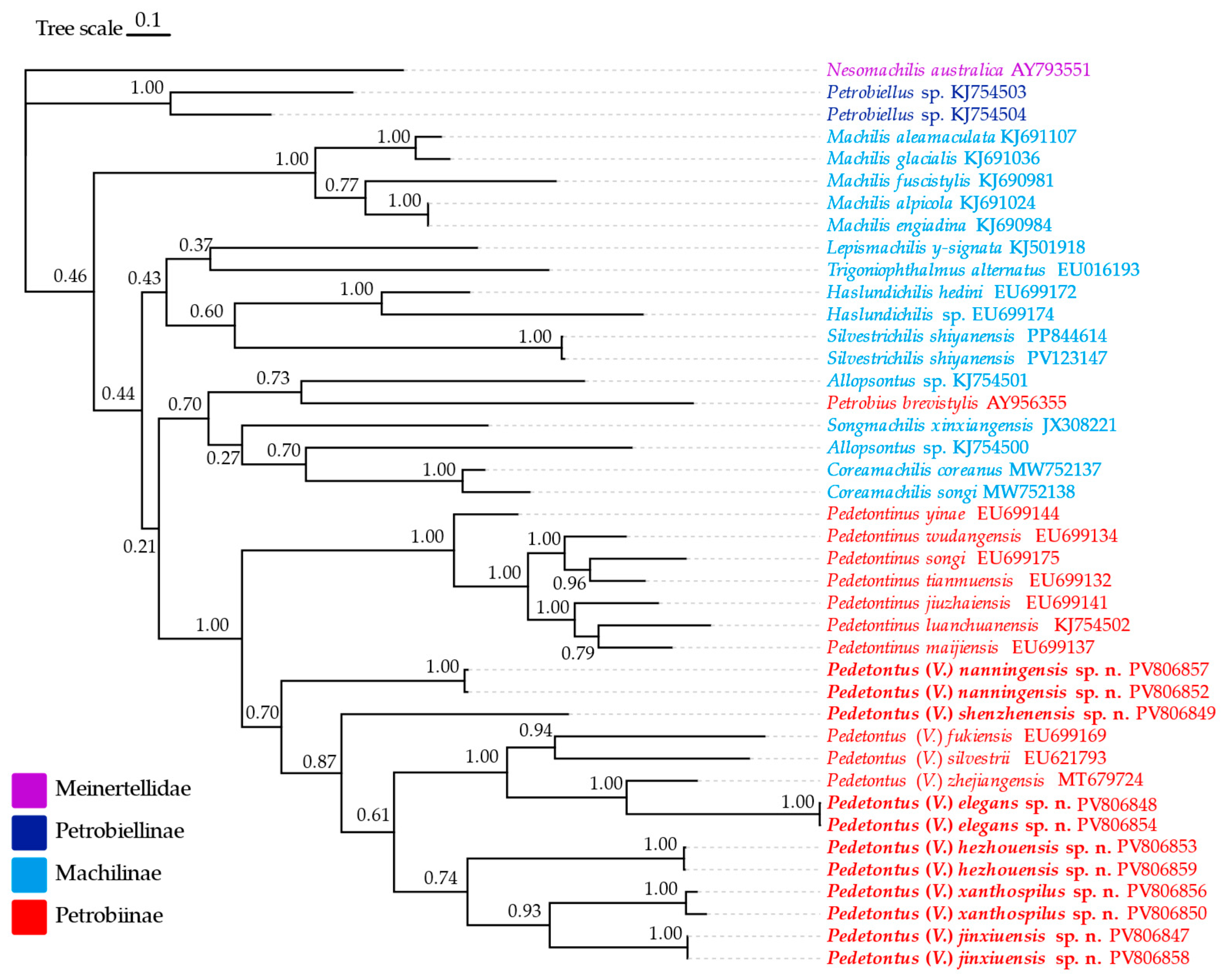
Phylogenetic construction was performed using 41 COX1 sequences, including 11 newly sequenced COX1 genes from this study and 30 Microcoryphia COX1 sequences (comprised of Meinertellidae and Machilidae) downloaded from NCBI [27,36,42,45,46,47,48,49,50,51]. Nesomachilis australica (AY793551) from Meinertellidae served as the outgroup[52]. Detailed COX1 accession numbers are provided in Table S1. Bayesian Inference (BI) tree was constructed using MrBayes 3.2 [53] with 10 million generations of Markov chain Monte Carlo (MCMC) simulations, sampling every 1000 generations (25% initial samples discarded as burn-in). Final phylogenetic tree was visualized and refined using ChiPlot [54] (https://www.chiplot.online, accessed on 26 June 2025).
3. Results
3.1. Taxonomy
Family Machilidae Lubbock, 1873
Subfamily Petrobiinae Kaplin 1985
Genus Pedetontus Silvestri, 1911
Subgenus Pedetontus (Verhoeffilis) Paclt, 1972
Type species: Petrobius (Pedetontus) calcaratus (Silvestri, 1911)
Subgeneric diagnosis. Flagellum of antenna devoided of scales, maxilla, legs provided of scales. Paired ocelli shoe-shaped (dumbbell-shaped), submedian, relatively close each other. Mandibles typical, with four apical teeth. Thorax normal. Coxal stylets on midlegs and hindlegs. Forelegs of male not swollen, without sensory fields. Two pairs of retactile vesicles on the abdominal segments II–V. Paramera restricted to the IXth. Penis opening little, apical. Male genitalia not exceeding IXth coxites. Ovipositor of the primary type.
Distribution. Russia Primorskii Territory, East Asia, Southeast Asia.
3.2. Keys
Key to subgenre of Pedetontus worldwide
|
| …………………………………………………………………Pedetontus (Verhoeffilis) Paclt, 1972 |
|
| Pedetontus sensu stricto Silvestri, 1911 |
| Key to known species of Pedetontus in China |
|
|
|
|
|
| P. (Verhoeffilis) silvestrii Mendes, 1991 |
|
|
| P. (V.) formosanus Silvestri 1943 |
|
| VII)………………………………………………………………………………………………………5 |
|
|
|
|
|
| ………………………………………………………………………………………………………………… |
| ……………………………………………………………………P. (V.) zhoui Yu, Zhang & Zhang 2010 |
|
|
|
|
| P. (V.) uraiensis Uchida 1965 |
|
|
|
|
|
|
|
|
|
|
| ……………………………………………………………………………………………………………… |
| ……………………………………P. (V.) shenzhenensis Shen, Yang, Ji & Zhang sp. n. |
|
|
|
| P. (V.) jinxiuensis Shen, Yang, Ji & Zhang sp. n. |
3.3. Description
Pedetontus (Verhoeffilis) elegans Shen, Yang, Ji & Zhang sp. n. (Figure 1.1, Figure 1.2, Figure 1.3, Figure 1.4, Figure 1.5 and Figure 1.6)
Zoobank: urn:lsid:zoobank.org:act:98F98919-E673-4C0A-8FE7-11CCCD2AF8BE
Type specimens & type locality. ♂ – holotype (in 70% ethanol); 3♂4♀ – paratypes (in 70% ethanol). Fengya Valley, Pan'an County, Jinhua City, Zhejiang Province, China (29°04'45"N 120°37'39"E, 542m), in leaf litter and on rocks near signal station (Figure 1.1), 17.VII.2024, coll. Jia-Yong Zhang, Chen-Yang Shen, Zhi-Qiang Guo, Jie-Hong Ji. All type specimens deposited in the Animal Herbarium, Zhejiang Normal University, Jinhua, Chi
Description. Male body length 10.4–11.2 mm, female 11.9 mm. Body covered with scales, light brown, with several white longitudinal stripes, black markings, and a few green markings; caudal filament and cerci with several well-developed white annulate setae. Antennae and median caudal filament approximately 1.6 times body length; lateral cerci length approximately two-thirds of body length (Figure 1.1A, B, Figure 1.2A, Figure 1.5A).
Head. Compound eyes well-developed, strongly convex, bright yellowish-green (Figure 1.2D–G, Figure 1.5B, C). Ratios of eye length to width, contact line length to eye length given in Table 1. Paired ocelli subinferior to compound eyes, reddish-brown, dumbbell-shaped. Ratios of ocellus length to width, distance between inner margins of ocelli to combined width of eyes given in Table 1. Vertex straight, with deep chestnut markings. Frons convex, bearing fine setae, without long bristles. Genae bearing black setae. Clypeus and labrum bearing transparent long setae (Figure 1.2D,E, Figure 1.5B,C).
Antennae scaled except on flagellum; flagellum extremely long, with dark annulations and well-developed annulate setae; terminal chains divided into 21–22 segments. Scape length-to-width ratio 2.2–2.33 (Figure 1.2B).
Mandibles typical, apex with four distal teeth (Figure 1.3D).
Maxilla basal segment with one processus basalis and one inner protuberance (Figure 1.3F). Male maxillary palp articles III–VII with well-developed long brush-like setae ventrally; female with long brush-like setae only on article 2. Ultimate article of maxillary palp subequal in length to penultimate article (Figure 1.3E, Figure 1.5D,E). Maxillary palp articles V–VII bearing transparent spines dorsally (Figure 1.3E Figure 1.5E), numbers given in Table 2.
Labial palp third article not modified, clavate, apex bearing sensory cone setae, numbers given in Table 2. Male labial palp II and III bearing long hair-like setae; female lacking these (Figure 1.3G,H, Figure 1.5F,G).
Thorax. Thorax normal. Foreleg femur not expanded, without sensory field; mid- and hindlegs with coxal styli. Coxae, trochanters, femora, and tibiae with long bristles ventrally; tarsi and hind tibiae bearing transparent needle-shaped setae ventrally, numbers given in Table 3. Tibiae bearing black setae dorsally. Pretarsal claw structure normal (Figure 1.3A–C, Figure 1.6A–D).
Abdomen. Abdominal coxites I, VI, and VII with one pair of retractile vesicles; coxites II–V with two pairs of retractile vesicles (Figure 1.4A–G, Figure 1.6E–F). Female coxite VII with inner lobes fused and extending posteriorly (Figure 1.6F). Angles of posterior angles of abdominal sternites given in Table 4. Ratios of styli length to coxites length on abdominal segment V, styli length to supporting spines length, and basal width to length of sternites given in Table 5. Coxite IX with 2–4+2–4 transparent macrochaetae internally (Figure 1.4I, Figure 1.6H). Caudal filament laterally and cerci internally bearing deciduous long setae and supporting spines (Figure 1.4K).
Coxite VIII without parameres. Coxite IX with penis and parameres (Figure 1.4I–J). Penis bearing black setae, opening apically, surpassing parameres; parameres 1+7 segmented (Figure 1.4J). Ovipositor of primary type, surpassing styli (including supporting spines) of coxite IX (Figure 1.6G,H). Number of segments of anterior gonapophyses and posterior gonapophyses given in Table 6.
Differential diagnosis. Pedetontus (Verhoeffilis) elegans sp. n. is most striking due to its extremely large compound eyes and the well-developed ultimate article of the maxillary palp – subequal in length to the penultimate article. P. (V.) formosanus Silvestri, 1943, which also has large eyes and slender maxillary palps, is morphologically the closest species, but the two can be distinguished by the shape of the lateral ocelli and the length of the article IV of maxillary palp. When viewed laterally from the front, the lateral ocelli of P. (V.) elegans sp. n. are constricted medially, presenting a dumbbell shape, whereas those of P. (V.) formosanus are almost clavate. The maxillary palp IV of P. (V.) elegans sp. n. is subequal in length to palp V, while that of P. (V.) formosanus is distinctly shorter than the palp V. P. (V.) diversicornis Silvestri, 1943 is also similar to P. (V.) elegans sp. n. Key distinguishing characters are antennae and penis length: the former has antennae shorter than the body and a penis subequal in length to the parameres, while the latter has antennae much longer than the body and a penis longer than the parameres.
Etymology. The specific epithet “elegans” (Latin adjective meaning “slender” or “graceful”) refers to the species' slender antennae, elegant markings, and fascinating green eyes. The Chinese name for P. (V.) elegans sp. n. is 秀丽跳蛃.
Distribution. Zhejiang Province, China.
Pedetontus (Verhoeffilis) hezhouensis Shen, Yang, Ji & Zhang sp. n. (Figure 2.1, Figure 2.2, Figure 2.3, Figure 2.4, Figure 2.5 and Figure 2.6)
Zoobank: urn:lsid:zoobank.org:act:7B7D8D89-1579-40FA-A1EB-04C3B6DEB877
Type Specimens & Type Locality. Holotype, 1♀ (in 70% ethanol), Paratypes: 1♂6♀ (in 70% ethanol), Huangyao Town, Hezhou City, Guangxi Zhuang Autonomous Region, China (24°14'N 111°13E), 18.XII.2024, collected by Jia-Yong Zhang. All type specimens deposited in the Animal Herbarium, Zhejiang Normal University, Jinhua, China.
Description. Male body length 7.9 mm, female 7.6–8.9 mm. Body covered with grayish-green scales; posterior part of mesonotum with a dark arched marking; tergites of subsequent segments bearing variously sized paired black spots. Caudal filament and cerci with white annulations. Antennae slightly longer than body length; caudal filament nearly 1.5 times body length; cerci longer than half of body length (Figure 2.1A,B).
Head. Compound eyes small, smooth, dark-colored, with indistinct yellow markings internally (Figure 2.1C, Figure 2.2C–F, Figure 2.5B,C); ratios of eye length to width and contact line length to eye length given in Table 1. Paired ocelli subinferior to compound eyes, black (fading to dark gray when preserved in 70% ethanol), with white borders, shoe-shaped. Ratios of ocellus length to width and distance between inner margins of ocelli to combined width of eyes given in Table 1. Vertex without markings. Frons smooth, with long transparent bristles, covered with scales . Genae without setae. Clypeus and labrum with transparent bristles (Figure 2.2C–E, Figure 2.5B,C).
Antennae scaled except on flagellum; flagellum without annulations, bearing well-developed annulate setae; terminal chain divided into 10–13 segments. Scape length-to-width ratio 1.73–1.84 (Figure 2.2B).
Mandibles typical, apex with four distal teeth (Figure 2.3D).
Maxillary palp I with one processus basalis and one inner protuberance (Figure 2.3E, F, Figure 2.5E). Male maxillary palp articles II–VII with well-developed short brush-like setae ventrally; female without. Ratio of ultimate article to penultimate article length 0.57–0.62 (Figure 2.3E, Figure 2.5E,F). Dorsal surface of maxillary palp articles V–VII with transparent spines (Figure 2.3E, Figure 2.5E,F); numbers given in Table 2.
Male labial palp yellow, third segment modified, laterally expanded, apex with sensory cones; numbers given in Table 2; ultimate article of female less swollen, clavate. Labial palp without long hair-like setae (Figure 2.3G,H, Figure 2.5G,H).
Hypopharynx structure typical (Figure 2.3I)
Thorax. Thorax normal. Foreleg femur not expanded, without sensory field; mid- and hind legs with coxal styli. Ventral surface of legs with long bristles, without needle-shaped setae. Pretarsal claw structure normal (Figure 2.3A–C, Figure 2.6A–C).
Abdomen. Abdominal coxites I, VI, and VII with one pair of retractile vesicles; coxites II–V with two pairs of retractile vesicles (Figure 2.4A–G, Figure 2.6D,E); inner lobes of female coxite VII fused and posteriorly extended (Figure 2.4G). Ratios of styli length to coxites length on abdominal segment V, styli length to supporting spines length, and basal width to length of sternites given in Table 5. Coxite IX medially with 4–8+4–8 transparent macrochaetae (Figure 2.4I, Figure 2.6G). Caudal filament laterally and cerci medially with deciduous long hairs and supporting spines (Figure 2.4J).
Coxite VIII without parameres, coxite IX with penis and parameres (Figure 2.6F,G). Penis opening apically, slightly shorter than parameres; parameres 1+8 segmented (Figure 2.6H). Ovipositor of primary type, surpassing styli (including supporting spines) of coxite IX (Figure 2.4H,I). Number of segments of anterior gonapophyses and posterior gonapophyses given in Table 6.
Differential diagnosis. Pedetontus (Verhoeffilis) hezhouensis sp. n. is morphologically most similar to P. (V.) sauterii Silvestri, 1943 from Taiwan, China. They differ primarily in the length of the contact line of the eyes: in the former, the ratio of this structure's length to the length of the eye is < 0.4, while in the latter it is > 0.4.
Etymology. The species is named after its type locality, Hezhou City. The Chinese name for P. (Verhoeffilis) hezhouensis sp. n. is 贺州跳蛃.
Distribution. Hezhou City, Guangxi Zhuang Autonomous Region, China.
Pedetontus (Verhoeffilis) jinxiuensis Shen, Yang, Ji & Zhang sp. n. (Figure 3.1, Figure 3.2, Figure 3.3, Figure 3.4, Figure 3.5 and Figure 3.6)
Zoobank: urn:lsid:zoobank.org:act:4C21B2F2-5BD0-4D46-BD78-7A78A941DCA0
Type Specimens & Type Locality. Holotype, 1♂ (in 70% ethanol), Paratypes: 15♂2♀ (in 70% ethanol), China, Guangxi Zhuang Autonomous Region, Jinxiu County, Dayao Mountain (24°6′4.88″N, 110°9′37.69″E, 947m), 1.V.2023, collected by Wen-Yong Feng. All type specimens deposited in the Animal Herbarium, Zhejiang Normal University, Jinhua, China.
Description. Male body length 11.2 mm, female 9.7 mm. Body covered with dark brown scales; mesonotum with an arched dark marking; subsequent nota bearing paired dark patches; abdominal nota patterned with three longitudinal pale diamond-shaped markings. Antennae equal to or slightly longer than body length; caudal filament nearly 1.5 times body length; cerci about half as long as body length (Figure 3.1A,B).
Head. Compound eyes smooth, deep reddish-brown, with indistinct yellow blotches (Figure 3.2C–F, Figure 3.5B,C); ratios of eye length to width and contact line length to eye length given in Table 1. Ocelli dark brown with white borders, dumbbell-shaped. Ratios of ocellus length to width and distance between inner margins of ocelli to combined width of eyes given in Table 1. Vertex without markings. Frons convex, with long transparent bristles, scaled. Genae without setae. Clypeus and labrum with transparent bristles (Figure 3.2C–F, Figure 3.5B,C).
Antennae scaled except on flagellum; flagellum with pale annulations, bearing well-developed annulate setae; terminal chain divided into 8–10 segments. Scape length-to-width ratio 1.86–1.91 (Figure 3.2B).
Mandibles typical, apex with four distal teeth (Figure 3.3D).
Maxillary palp I with one processus basalis and one inner protuberance (Figure 3.3E, G, Figure 3.5D). Male maxillary palp articles II–VII with well-developed short brush-like setae ventrally; female without. Ratio of ultimate article to penultimate article length 0.69–0.85. Dorsal surface of maxillary palp articles V–VII with transparent spines (Figure 3.3E,F, Figure 3.5D); numbers given in Table 2.
Labial palp third segment modified, extremely laterally expanded, apex with sensory cones; numbers given in Table 2; ultimate article of female less swollen, apex without sensory cones. Labial palp without long hair-like setae (Figure 3.3H, Figure 3.5F,G).
Hypopharynx structure typical (Figure 3.5E).
Thorax. Thorax normal. Foreleg femur not expanded, without sensory field; mid- and hind legs with coxal styli, apex of coxal styli yellow. Ventral surface of legs with long bristles, without needle-shaped setae. Pretarsal claw structure normal (Figure 3.3A–C, Figure 3.6A–C).
Abdomen. Abdominal coxites I, VI, and VII with one pair of retractile vesicles; coxites II–V with two pairs of retractile vesicles; coxites I–IX each with a pair of yellow styli (Figure 3.4A–I, Figure 3.6D–G); inner lobes of female coxite VII fused and posteriorly extended (Figure 3.6E). Ratios of styli length to coxites length on abdominal segment V, styli length to supporting spines length, and basal width to length of sternites given in Table 5. Coxite IX medially with 3–4+3–4 transparent macrochaetae (Figure 3.4I, Figure 3.6G). Caudal filament laterally and cerci medially with deciduous long hairs and supporting spines (Figure 3.1B).
Coxite VIII without parameres. Coxite IX with penis and parameres (Figure 3.4I, J). Penis opening apically, slightly shorter than parameres; parameres 1+7 segmented (Figure 3.4J). Ovipositor of primary type, surpassing styli (including supporting spines) of coxite IX (Figure 3.6G). Number of segments of anterior gonapophyses and posterior gonapophyses given in Table 6.
Differential diagnosis. Pedetontus (Verhoeffilis) jinxiuensis sp. n. is morphologically very similar to P. (V.) xanthospilus sp. n. from Zhaoqing, Guangdong Province, but can be distinguished by the form of the labial palps: the former has a highly modified ultimate article (width significantly greater than length), with the anterolateral part of article I in males also laterally expanded. Additionally, the apex of the female ultimate article lacks sensory cones—an extremely rare feature. In contrast, although the labial palps of P. (V.) xanthospilus sp. n. are also swollen, they are far less modified than in P. (V.) jinxiuensis sp. n., article I lacks specialization, and both sexes possess sensory cones at the apex of the ultimate article.
Etymology. The species is named after its type locality, Jinxiu County. The Chinese name for P. (V.) jinxiuensis is 金秀跳蛃.
Distribution. Jinxiu County, Guangxi Zhuang Autonomous Region, China.
Pedetontus (Verhoeffilis) nanningensis Shen, Yang, Ji & Zhang sp. n. (Figure 4.1, Figure 4.2, Figure 4.3, Figure 4.4, Figure 4.5 and Figure 4.6)
Zoobank: urn:lsid:zoobank.org:act:4D83A441-4098-4384-B529-75E5B3D303A4
Type Specimens & Type Locality. Holotype, 1♂ (in 70% ethanol), Paratypes: 4♂ 6♀ (in 70% ethanol), collected from Jiangnan District, Nanning City, Guangxi Zhuang Autonomous Region, China, 18 II.2025, by Jia-Yong Zhang. All type specimens deposited in the Animal Herbarium, Zhejiang Normal University, Jinhua, China.
Description. Male body length 10.6 mm, female 11.6 mm. Body covered with grayish-green scales. Antennae slightly shorter than body length; caudal filament slightly longer than body length; cerci about three-quarters as long as body length (Figure 4.1A, C).
Head. Compound eyes smooth, black, without markings (Figure 4.1B, Figure 4.2C–F, Figure 4.5C); ratios of eye length to width and contact line length to eye length given in Table 1. Ocelli dark brown with white borders, dumbbell-shaped. Ratios of ocellus length to width and distance between inner margins of ocelli to combined width of eyes given in Table 1. Vertex without markings. Frons convex, with long transparent bristles, scaled. Genae without bristles. Clypeus and labrum with transparent bristles (Figure 4.2C–F, Figure 4.5C).
Antennae scaled except on flagellum; flagellum almost without dark annulations, bearing well-developed annulate setae; terminal chain divided into 14 segments. Scape length-to-width ratio 1.97–2.11 (Figure 4.2B, Figure 4.5B).
Mandibles typical, apex with four distal teeth (Figure 4.3D).
Maxillary palp I with one processus basalis and one inner protuberance (Figure 4.3D, E, Figure 4.6C). Maxillary palp articles without short brush-like setae ventrally. Ratio of ultimate article to penultimate article length 0.45–0.52. Dorsal surface of maxillary palp articles V–VII with transparent spines (Figure 4.3D, Figure 4.6C); numbers given in Table 2.
Hypopharynx structure typical (Figure 4.3F).
Male labial palp third segment modified, laterally expanded, apex with sensory cones; numbers given in Table 2; ultimate article of female almost unswollen, clavate. Labial palp without long hair-like setae (Figure 4.3B, C, Figure 4.6A,B).
Thorax. Thorax normal. Foreleg without sensory field or coxal styli; mid- and hind legs with coxal styli. Ventral surface of legs with long bristles, without needle-shaped setae. Pretarsal claw structure normal (Figure 4.3G–I, Figure 4.5D–F).
Abdomen. Abdominal coxites I, VI, and VII with one pair of retractile vesicles; coxites II–V with two pairs of retractile vesicles (Figure 4.4A–G, Figure 4.6D,E); inner lobes of female coxite VII fused and posteriorly extended (Figure 4.6E). Ratios of styli length to coxites length on abdominal segment V, styli length to supporting spines length, and basal width to length of sternites given in Table 5. Coxite IX medially with 6+6 transparent macrochaetae (Figure 4.4I, Figure 4.6G). Caudal filament laterally and cerci medially with deciduous long hairs and supporting spines (Figure 4.4J).
Coxite VIII without parameres. Coxite IX with penis and parameres (Figure 4.4H, I). Penis opening apically, slightly shorter than parameres; parameres 1+7 segmented (Figure 4.4K). Ovipositor of primary type, narrowly exceeding styli (including supporting spines) of coxite IX (Figure 4.6F, G). Number of segments of anterior gonapophyses and posterior gonapophyses given in Table 6.
Differential diagnosis. Pedetontus (Verhoeffilis) nanningensis sp. n. is morphologically most similar to P. (V.) gershneri Allen, 1995 from Arkansas, USA. They can be distinguished by the maxillary palps: the former lacks brush-like setae ventrally on the male maxillary palps, whereas the latter possesses brush-like setae ventrally on articles V–VII. Additionally, the ultimate article of the maxillary palp in P. (V.) nanningensis sp. n. is only half the length of the penultimate article, while in P. (V.) gershneri it exceeds three-quarters the length of the penultimate article.
Etymology. The species is named after its type locality, Nanning City. The Chinese name for P. (V.) nanningensis sp. n. is 南宁跳蛃.
Distribution. Guangxi Zhuang Autonomous Region, China.
Zoobank: urn:lsid:zoobank.org:act:D809BC47-4BED-4B63-993D-31267071EE31
Type Specimens & Type Locality. Holotype, 1♀ (in 70% ethanol), Paratype, 1♀ collected from Yangtai Mountain, Shenzhen City, Guangdong Province, China (22°39'57"N 113°58'29"E, 129m), 3.IV.2023, by Yong-Ying Ruan All type specimens deposited in the Animal Herbarium, Zhejiang Normal University, Jinhua, China.
Description. Female 11.2 mm. Body covered with brown scales (in 70% ethanol). Antennae longer than body length; caudal filament approximately 1.5 times body length; cerci slightly shorter than body length (Figure 5.1A).
Head. Compound eyes smooth, yellowish-green with maroon borders (Figure 5.1C–F); ratios of eye length to width and contact line length to eye length given in Table 1. Ocelli dark brown with white borders, dumbbell-shaped. Ratios of ocellus length to width and distance between inner margins of ocelli to combined width of eyes given in Table 1. Vertex without markings, bearing minute black setae arranged longitudinally. Frons convex, with long transparent bristles and minute black setae, scaled. Genae with black setae near compound eyes. Clypeus and labrum with transparent setae (Figure 5.1C–F).
Antennae scaled except on flagellum; flagellum with distinct pale annulations, bearing well-developed annulate setae; terminal chain divided into 10 segments. Scape length-to-width ratio 1.90 (Figure 5.1B).
Mandibles typical, apex with four distal teeth (Figure 5.2A).
Maxillary palp I with one processus basalis and one inner protuberance (Figure 5.2D, G). Maxillary palp articles without short brush-like setae ventrally. Ratio of ultimate article to penultimate article length 0.62. Dorsal surface of maxillary palp articles V–VII with transparent spines (Figure 5.2D, E); numbers given in Table 2.
Labial palp third segment slightly swollen apically, with sensory cones; numbers given in Table 2. Labial palp without long hair-like setae (Figure 5.2B, C).
Thorax. Thorax normal. Foreleg without sensory field or coxal styli; mid- and hind legs with coxal styli. Ventral surface of legs with long bristles, without needle-shaped setae. Pretarsal claw structure normal (Figure 5.2H–M).
Abdomen. Abdominal coxites I, VI, and VII with one pair of retractile vesicles; coxites II–V with two pairs of retractile vesicles (Figure 5.3A–G); inner lobes of female coxite VII fused and posteriorly extended (Figure 5.3G). Ratios of styli length to coxites length on abdominal segment V, styli length to supporting spines length, and basal width to length of sternites given in Table 5. Coxite IX medially with 4–5+4–5 transparent macrochaetae (Figure 5.3I). Caudal filament laterally and cerci medially with deciduous long hairs and supporting spines (Figure 5.3J).
Ovipositor of primary type, slightly shorter than styli (including supporting spines) of coxite IX (Figure 5.3H, I). Number of segments of anterior gonapophyses and posterior gonapophyses given in Table 6.
Differential Diagnosis. Pedetontus (Verhoeffilis) shenzhenensis sp. n. is morphologically closest to P. (V.) bianchii Silvestri, 1936 from Hong Kong. The abdominal segment IX provides a key distinction: in P. (V.) shenzhengensis sp. n., the styli (including supporting spines) of coxite IX are longer than coxite IX itself, while the posterior gonapophyses are slightly shorter than these styli. Conversely, in P. (V.) bianchii, the styli (including supporting spines) of coxite IX are shorter than coxite IX, and the posterior gonapophyses exceed their length. Additionally, the scape of P. (V.) shenzhenensis sp. n. is slender than that of P. (V.) bianchii, with length-to-width ratios of 1.90 and 1.42, respectively.
Etymology. The species is named after its type locality, Shenzhen City. The Chinese name for P. (V.) shenzhenensis sp. n. is 深圳跳蛃.
Distribution. Guangdong Province, China.
Pedetontus (Verhoeffilis) xanthospilus Shen, Yang, Ji & Zhang sp. n. (Figure 6.1, Figure 6.2, Figure 6.3, Figure 6.4, Figure 6.5 and Figure 6.6)
Zoobank: urn:lsid:zoobank.org:act:8C07BFC0-2BB3-4053-A2AB-23B08DA13180
Type Specimens & Type Locality. Holotype, 1♂ (in 70% ethanol), Paratypes: 11♂7♀ (in 70% ethanol), China, Guangdong Province, Zhaoqing City, Duanzhou District, Beiling Mountain Forest Park (23°09'N 112°30'E), from rocks along a stream, 20.XI.2024–3.I.2025, collected by Han-sheng Ou. All type specimens deposited in the Animal Herbarium, Zhejiang Normal University, Jinhua, China.
Description. Male body length 10.2 mm, female 9.2 mm. Body covered with dark brown scales; nota with a pale longitudinal stripe extending from the pronotum to the base of the caudal filament, terminating in two large and one small diamond-shaped markings. Antennae slightly shorter than body length; caudal filament slightly longer than body length; cerci about half as long as body length (Figure 6.1A–C, Figure 6.2A, Figure 6.5A).
Head. Compound eyes smooth, dark brown, with a large bright yellow blotch internally and a smaller one inferiorly (Figure 6.2D–G, Figure 6.5B); ratios of eye length to width and contact line length to eye length given in Table 1. Ocelli dark brown, dumbbell-shaped. Ratios of ocellus length to width and distance between inner margins of ocelli to combined width of eyes given in Table 1. Vertex without markings or setae. Frons convex, with long transparent bristles, scaled. Genae without markings or setae. Clypeus and labrum with transparent setae (Figure 6.2D–G, Figure 6.5B).
Antennae scaled except on flagellum; flagellum with distinct pale annulations, bearing well-developed annulate setae; terminal chain divided into 11–12 segments. Scape length-to-width ratio 1.74–1.91 (Figure 6.2B).
Mandibles typical, apex with four distal teeth (Figure 6.3A).
Maxillary palp I with one processus basalis and one inner protuberance (Figure 6.3D, F, Figure 6.5E). Maxillary palp articles II–VII with brush-like setae ventrally, setae on articles IV–V longer than others. Ratio of ultimate article to penultimate article length 0.45–0.52. Dorsal surface of maxillary palp articles V–VII with transparent spines (Figure 6.3D, Figure 6.5E); numbers given in Table 2.
Male labial palp third segment modified, laterally expanded, apex with sensory cones; numbers given in Table 2; ultimate article of female almost unswollen, clavate. Labial palp without long hair-like setae (Figure 6.3B,C, Figure 6.5D).
Thorax. Thorax normal. Foreleg without sensory field or coxal styli; mid- and hind legs with coxal styli, apex of coxal styli bright yellow. Ventral surface of legs with long bristles, without needle-shaped setae. Pretarsal claw structure normal (Figure 6.3G–I, Figure 6.6A–C).
Abdomen. Abdominal coxites I, VI, and VII with one pair of retractile vesicles; coxites II–V with two pairs of retractile vesicles; coxites I–IX each with a pair of yellow styli (Figure 6.4A–I, Figure 6.6D–G); inner lobes of female coxite VII fused and posteriorly extended (Figure 6.6E). Ratios of styli length to coxites length on abdominal segment V, styli length to supporting spines length, and basal width to length of sternites given in Table 5. Coxite IX medially with 4–5+4–5 transparent macrochaetae (Figure 6.4I, Figure 6.6G). Caudal filament laterally and cerci medially with deciduous long hairs and supporting spines (Figure 6.4J).
Coxite VIII without parameres. Coxite IX with penis and parameres (Figure 6.4I, K). Penis opening apically, shorter than parameres; parameres 1+7 segmented (Figure 6.4K). Ovipositor of primary type, not exceeding styli (including supporting spines) of coxite IX (Figure 6.6F, G). Number of segments of anterior gonapophyses and posterior gonapophyses given in Table 6.
Differential Diagnosis. See corresponding section of Pedetontus (Verhoeffilis) jinxiuensis sp. n.
Etymology. The specific epithet "xanthospilus" is a Latinized adjective derived from: Greek xanthos (ξανθός), meaning "yellow", Greek spilos (σπίλος), meaning "blotch" or "stain". It refers to the species' conspicuous yellow blotches on the compound eyes, a defining diagnostic trait that distinguishes this taxon from congeners. The Chinese name for P. (V.) xanthospilus sp. n. is 黄斑跳蛃.
Distribution. Guangdong Province, China.
3.4. Phylogenetic Tree of Machilidae Based on COX1
Based on the phylogenetic tree, Machilinae is a paraphyletic group, also the monophyly of Petrobiinae is refuted by the inclusion of Petrobius brevistylis within Machilinae, but the monophyly of the Pedetontus group is supported (Pedetontus + Pedetontinus). Within the genus Pedetontus, P. (V.) nanningensis sp. n. occupies the basal position, P. (V.) elegans sp. n. and P. (V.) zhejiangensis form a sister clade, while P. (V.) xanthospilus sp. n. and P. (V.) jinxiuensis sp. n. constitute another sister clade.
Figure 7.
Bayesian inference phylogenetic tree based on a tandem dataset of 41 COX1 genes. Posterior probabilities are shown at the nodes.
Figure 7.
Bayesian inference phylogenetic tree based on a tandem dataset of 41 COX1 genes. Posterior probabilities are shown at the nodes.
4. Discussion
Based on morphological observations and COX1 gene analysis, this study reports six new species of genus Pedetontus from southern China: P. (V.) elegans sp. n. from Zhejiang Province; P. (V.) shenzhenensis sp. n. and P. (V.) xanthospilus sp. n. from Guangdong Province; P. (V.) nanningensis sp. n., P. (V.) hezhouensis sp. n., and P. (V.) jinxiuensis sp. n. from Guangxi Zhuang Autonomous Region. All these species belong to the subgenus Verhoeffilis, which is characterized by the presence of two pairs of retractile vesicles restricted to coxites II–V and posterior angles of abdominal sternites ≤90°. P. (V.) elegans sp. n. and P. (V.) zhejiangensis form a sister clade, both species originate from Zhejiang Province and share some morphological similarities, but they can be distinguished by the following characteristics: The compound eyes of P. (V.) zhejiangensis are reddish-brown, while those of P. elegans sp. n. are yellowish-green. P(V.). zhejiangensis lacks stout setae on the frons, whereas P. (V.) elegans sp. n. possesses well-developed stout setae. The male labial palps of P. (V.) zhejiangensis bear a few long filamentous setae laterally, which are absent in P. (V.) elegans sp. n. The ultimate segment of the maxillary palps is distinctly shorter than the penultimate segment in P. (V.) zhejiangensis, whereas in P. (V.) elegans sp. n., the ultimate and penultimate segments are equal in length. The parameres of P. (V.) zhejiangensis exhibit 1+8 segmentation, and the penis is shorter than the parameres; in contrast, P. (V.) elegans sp. n. has 1+7 segmented parameres, and the penis is longer than the parameres.
The subgenus Verhoeffilis is believed to have originated in the southeastern Palaearctic Realm. Its northeastward migration led to the emergence of the American s. stricto subgenus, whereas its southward dispersal resulted in the current Indo-Malayan Pedetontus species [6]. Southern China is situated at the transitional zone between the Palaearctic and Oriental Realms. Among the new species described, P. (V.) elegans sp. n. (Zhejiang) exhibits morphological traits most consistent with the Palaearctic fauna: Compound eyes longer than wide; ventral surfaces of thoracic legs bearing subspine-form setae. In contrast, the remaining five species (from Guangdong and Guangxi) possess compound eyes that are wider than long and lack subspine-form setae on thoracic legs. Phylogenetic analysis indicates that P. (V.) elegans sp. n. is closely related to the Palaearctic species P. silvestrii.
P. (V.) xanthospilus sp. n. and P. (V.) jinxiuensis sp. n. form sister clades on the phylogenetic tree and exhibit morphological similarities. However, the genetic distance based on COX1 sequences and differences in the labial palpal provide evidence that they represent distinct species. These observations suggest the subgenus is undergoing a process of divergence.
Petrobius brevistylis is clustered within the Allopsontus group—an unexpected placement that highlighting its taxonomic distinctness. This is consistent with its possesion of non-segmented parameres and a penis that is significantly longer than the coxite IX, in contrast to most Petrobiinae species, which typically exhibit segmented parameres and a penis shorter than the coxite IX [55]. The genus Allopsontus appears to be non-monophyletic according to the phylogenetic tree, potentially due to the presence of seven morphologically distinct subgenera distributed across Northwest China, Central Asia, Mongolia, and the southern Himalayas. Key morphological differences include differences include variations in ocellar shape/position, the number of coxal vesicles, and penis morphology[56], suggesting a lack of monophyly that warrants further investigation through molecular and detailed morphological study.
Supplementary Materials
The following supporting information can be downloaded at the website of this paper posted on Preprints.org. S1: Information about samples used in this study and their NCBI GenBank accession numbers.
Author Contributions
Conceptualization, C.-Y.S., T.Y. and J.-Y.Z.; methodology, C.-Y.S. and J.-Y.Z.; software, C.-Y.S. and T.Y.; validation, C.-Y.S. and J.-Y.Z.; formal analysis, C.-Y.S., T.Y. and J.-Y.Z.; investigation, C.-Y.S., J.-H.J. and J.-Y.Z.; resources, C.-Y.S., J.-H.J. and J.-Y.Z.; data curation, C.-Y.S. and T.Y.; writing—original draft preparation, C.-Y.S. and T.Y.; writing—review and editing, C.-Y.S., T.Y., J.-H.J. and J.-Y.Z.; visualization, C.-Y.S. and T.Y.; supervision, J.-Y.Z.; project administration, J.-Y.Z.; All authors have read and agreed to the published version of the manuscript.
Funding
This work was supported by the Natural Science Foundation of Zhejiang Province (LY23C040002) and by the Natural Science Foundation of China (32470475).
Data Availability Statement
Supporting data for this study are available from the National Center for Biotechnology Information (https://www.ncbi.nlm.nih.gov) (accessed on 10 June 2025). The GenBank numbers, see Figure 7, Table S1.
Acknowledgments
We are deeply in debt to Zhi-Qiang Guo, Yong-Ying Ruan, Han-Sheng Qu, Wen-Yong Feng and the Institute of Zoology of the Guangdong Academy of Sciences for their assistance in sample collection during this study. Additionally, the authors express their gratitude to Hui-Yuan Wu for assistance in constructing the phylogenetic trees. Finally, special thanks are extended to Professor Shodo Mtow of Meijo University for generously providing literature, which significantly facilitated our study on the genus Pedetontus and other bristletail species.
Conflicts of Interest
The authors declare no conflicts of interest.
References
- Bach de Roca, C.; Baltanas, R.M.; Gaju Ricart, M. Orden Microcoryphia. Ibero Diversidad Entomológica 2015, 38, 1–12.
- Sturm, H.; Machida, R. Archaeognatha. In Handbook of Zoology; Walter de Gruyter: Berlin, New York, 2001; Vol. 4.
- Yanoviak, S.P.; Kaspari, M.; Dudley, R. Gliding Hexapods and the Origins of Insect Aerial Behaviour. Biol. Lett. 2009, 5, 510–512. [CrossRef]
- Grimaldi, D.A.; Engel, M.S. Evolution of the Insects; Repr.; Cambridge Univ. Press: Cambridge, 2006; ISBN 978-0-521-82149-0.
- Mendes, L.F. Taxonomy of Zygentoma and Microcoryphia: Historical Overview, Present Status and Goals for the New Millennium. Pedobiologia 2002, 46, 225–233.
- Kaplin, V.G. A New Species of the Bristletail Genus Pedetontus Silv. (Thysanura, Machilidae) from Thailand. Entmol. Rev. 2013, 93, 887–892. [CrossRef]
- Mendes, L.F. An Annotated List of Generic and Specific Names of Machilidae (Microcoryphia, Insecta) with Identification Keys for the Genera and Geographical Notes; Ministerio do Planeamento e da Administração do Território, Secretaria de Estado da Ciência e Tecnologia, Instituto de Investigação Científica Tropical: Lisbon, 1990; Vol. 155;.
- Silvestri, F. Quelques Formes Nouvelles de La Famille Des Machilides. Annales des Sciences Naturelles (Zool)(9) 1907, 6, 361–370.
- Silvestri, F. Note sui Machilidae. III. Descrizione di un nuovo genere e di sei nuove specie. Redia (Firenze) 1906, 3, 325–335.
- Silvestri, F. Machilidae (Thysanura). Entomologische Ergebnisse der schwedischen Kamtchatka-Expedition 1920-1922. Arkiv för Zoologi 1925, 17, 1–5.
- Silvestri, F. Contributo Alla Conoscenza Dei Machilidae Dell’America Settentrionale. Bolletino del Laboratorio de Zoologia Generale e Agraria della R. Scuola Superiore d’Agricoltura in Portici 1911, 5, 324–350.
- Silvestri, F. Descripzione Di Alcuni Machilidae (Thysanura) Della Cina. Notes d’Entomologie Chinoise 1936, 3, 103–115.
- Silvestri, F. Contributto Alla Conoscenza Dei Machilidae (Insecta, Thysanura) Del Giappone. Bolletino del Laboratorio di Zoologia generale i agraria di Portici 1943, 32, 283–306.
- Sturm, H. Possibilities and Problems of Morphological Taxonomy Shown by North American Representatives of the Subgenus Pedetontus s. str. and Petridiobius Canadensis (Archaeognatha, Machilidae, Petrobiinae). Deutsche Entomologische Zeitschrift 2001, 48, 3–21. [CrossRef]
- Allen, R.T. Pedetontus Gershneri, a New Species of Machilidae from the Interior Highlands of North America (Insecta: Microcoryphia). Entomological News 1995, 106, 195–198.
- Kaplin, V.G. New species of bristletails (Microcoryphia, Machilidae) from the Kuril Islands and Primorsky Kari. Txonomy of Insects of Far East 1980, 3–9.
- Lee, B.H.; Choe, G.H. Two New Species of Microcoryphia (Insecta) from Korea. The Korean journal of systematic zoology 1992, 8, 19–34.
- Machida, R. A New Species of the Genus Pedetontus from Japan (Insecta, Thysanura). Annotationes zoologicae Japonenses 1980, 53, 220–225.
- Mendes, L.F. Sur Quelques Microcoryphia de l’Asie Orientale: Notes et Descriptions (Insecta; Apterygota). Nouvelle revue d’entomologie 1981, 11, 15–28.
- Mendes, L. New Contribution towards the Knowledge of the Northern Korean Thysanurans (Microcoryphia and Zygentoma: Insecta). Garcia Orta 1991, 18, 67–78.
- Uchida, H. Two Species of Pedetontus from Amami-o-Sima (Thysanura: Machilidae). The entomological society of Japan 1960, 28, 247–250.
- Uchida, H. Pedetontus from Formosa ( Thysanura: Machilidae). Special Bulletin of Lepidopterological Society of Japan 1965, 1, 249–252.
- Wygodzinsky, P.W. Survey of the Microcoryphia (Insecta) of the Northeastern United States and Adjacent Provinces of Canada.
- Xue, L.Z.; Yin, W.Y. Two new species of bristletail from the Tianmu Mountain (Microcoryphia, Machilidae). Contributions from Shanghai Institute of Entomology 1991, 10, 77–86.
- Yu, D.N.; Zhang, W.W.; Zhang, J.Y. Two New Species of the Genus Pedetontus (Microcoryphia, Machilidae) from China. Acta Zootaxonomica Sinica 2010, 35, 444–450.
- Kaplin, V.G. НОВЫЕ ВИДЫ ЩЕТИНОХВОСТОК РОДА PEDETONTUS SILVESTRI, 1911 (MICROCORYPHIA, MACHILIDAE) C ДАЛЬНЕГО ВОСТОКА. ЗООЛОГИЧЕСКИЙ ЖУРНАЛ 2025, 104, 36–46. [CrossRef]
- Zhang, J.Y.; Song, D.X.; Zhou, K.Y. The Complete Mitochondrial Genome of the Bristletail Pedetontus Silvestrii (Archaeognatha: Machilidae) and an Examination of Mitochondrial Gene Variability within Four Bristletails. Annals of the Entomological Society of America 2008, 101, 1131–1136. [CrossRef]
- Chen, H.Y.; Yu, D.N.; Zhang, J.Y. A new species of the genus Pedetontinus (Microcoryphia, Machilidae) from China. Acta Zootaxonomica Sinica 2011, 036, 36–39.
- Deng, K.Z.; Zhang, J.Y.; Yu, D.N. A New Species of the Genus Haslundichilis (Microcoryphia, Machilidae) from China and Redescription of Haslundichilis Hedini (Silvestri). Acta Zootaxonomica Sinica 2011, 36, 882–887.
- Huang, F.S.; Song, Z.S.; Liang, A.P. A New Bristletail Species of the Genus Allopsontus Silvestri (Microcoryphia: Machilidae) from Shaanxi, China. Oriental Insects 2006, 40, 267–272. [CrossRef]
- Kaplin, V.G. Description of a New Species of the Bristletail Genus Pedetontinus Silv. (Thysanura, Machilidae) from China with a Review of Species of This Genus. Entmol. Rev. 2015, 95, 73–86. [CrossRef]
- Kaplin, V.G. A New Species of the Genus Allopsontus Silv. (Microcoryphia, Machilidae) from Northwestern China. Entomological Review 2016, 96, 126–131. [CrossRef]
- Kaplin, V.G. A New Species of Bristletails of the Genus Silvestrichilis Wygodzinsky, 1950 (Archaeognatha: Machilidae) from South China. Far East. entomol. 2019, 23–28. [CrossRef]
- Li, P.; Yu, D.N.; Zhang, J.Y. A New Species of the Genus Pedetontinus (Microcoryphia, Machilidae) from China. Acta Zootaxonomica Sinica 2012, 37, 740–746.
- Mendes, L.F.; Gaju-Ricart, M.; Bach de Roca, C.; Molero-Baltanás, R. On Some Silvestri Species of Machilidae (Microcoryphia, Insecta) Which Types Are in the Muséum National d’Histoire Naturelle, Paris. Pedobiologia 2000, 44, 268–284. [CrossRef]
- Shen, C.Y.; Wang, L.Y.; Wu, H.Y.; Zhang, J.Y. A New Species of Silvestrichilis Wygodzinsky, 1950 (Insecta: Microcoryphia) from Wudang Mountain, Hubei, China, with the Description of Both Sexes. Zootaxa 2025, 5621, 437–452. [CrossRef]
- Silvestri, F. Schwedisch-chinesiche wissenschaftliche Expedition nach den nordwestlichen Provinzen Chinas (38. Thysanura, Machilidae). Arkiv for Zoologi 1934, 27, 1–7.
- Song, Z.S.; Huang, F.S.; Liang, A.P. Machilontus (s. str.) Medogensis Song & Huang, Sp. n. from Tibet, the Northernmost Record of the Genus Machilontus Silvestri, 1912 and the First Record of the Family Meinertellidae (Insecta: Microcoryphia: Machiloidea) in China. Zootaxa 2011, 2822, 61–68. [CrossRef]
- Zhang, J.Y. A new record genus and species of Machilinae from China (Microcoryphia: Machilidae). Jiangxi Plant Protection 2010, 33, 57–59.
- Zhang, J.Y.; Li, T. A New Bristletail Species of the Genus Pedetontinus (Microcoryphia, Machilidae) from China. Acta Zootaxonomica Sinica 2009, 34, 203–206.
- Zhang, J.Y.; Song, D.X.; Zhou, K.Y. A new species of the genus Pedetontinus (Microcoryphia, Machilidae) from China. Acta Zootaxonomica Sinica 2005, 30, 549–554.
- Zhang, J.Y.; Zhou, K.Y. Descriptions of One New Genus and Six New Species of Machilidae (Insecta: Archaeognatha) from China: Morphological and Molecular Data. Journal of Natural History 2011, 45, 1131–1164. [CrossRef]
- Folmer, O.; Black, M.; Hoeh, W.; Lutz, R.; Vrijenhoek, R. DNA Primers for Amplification of Mitochondrial Cytochrome c Oxidase Subunit I from Diverse Metazoan Invertebrates. Mol. Mar. Biol. Biotechnol. Molecular Marine Biology and Biotechnology 1994, 3, 294–299.
- Lanfear, R.; Calcott, B.; Ho, S.Y.W.; Guindon, S. PartitionFinder: Combined Selection of Partitioning Schemes and Substitution Models for Phylogenetic Analyses. Mol. Biol. Evol. 2012, 29, 1695–1701. [CrossRef]
- Guan, J.Y.; Shen, S.Q.; Zhang, Z.Y.; Xu, X.D.; Storey, K.B.; Yu, D.N.; Zhang, J.Y. Comparative Mitogenomes of Two Coreamachilis Species (Microcoryphia: Machilidae) along with Phylogenetic Analyses of Microcoryphia. Insects 2021, 12, 795. [CrossRef]
- Gassner, M.; Dejaco, T.; Schönswetter, P.; Marec, F.; Arthofer, W.; Schlick-Steiner, B.C.; Steiner, F.M. Extensive Variation in Chromosome Number and Genome Size in Sexual and Parthenogenetic Species of the Jumping-Bristletail Genus Machilis (Archaeognatha). Ecology and Evolution 2014, 4, 4093–4105. [CrossRef]
- Shen, S.Q.; Cai, Y.Y.; Xu, K.K.; Chen, Q.P.; Cao, S.S.; Yu, D.N.; Zhang, J.Y. The Complete Mitochondrial Genome of Pedetontus Zhejiangensis (Microcoryphia: Machilidae) and Its Phylogeny. Mitochondrial DNA Part B 2020, 5, 3143–3145. [CrossRef]
- He, K.; Zhang, J.Y.; Deng, K.Z.; Chen, Z. The Complete Mitochondrial Genome of the Bristletail Songmachilis Xinxiangensis (Archaeognatha: Machilidae). Mitochondrial DNA 2013, 24, 99–101. [CrossRef]
- Ma, Y.; He, K.; Yu, P.P.; Yu, D.N.; Cheng, X.F.; Zhang, J.Y. The Complete Mitochondrial Genomes of Three Bristletails (Insecta: Archaeognatha): The Paraphyly of Machilidae and Insights into Archaeognathan Phylogeny. PLoS One 2015, 10, e0117669. [CrossRef]
- Podsiadlowski, L. The Mitochondrial Genome of the Bristletail Petrobius Brevistylis (Archaeognatha: Machilidae). Insect Molecular Biology 2006, 15, 253–258. [CrossRef]
- Carapelli, A.; Liò, P.; Nardi, F.; Van der Wath, E.; Frati, F. Phylogenetic Analysis of Mitochondrial Protein Coding Genes Confirms the Reciprocal Paraphyly of Hexapoda and Crustacea. BMC evolutionary biology 2007, 7, S8. [CrossRef]
- Cameron, S.L.; Miller, K.B.; D’Haese, C.A.; Whiting, M.F.; Barker, S.C. Mitochondrial Genome Data Alone Are Not Enough to Unambiguously Resolve the Relationships of Entognatha, Insecta and Crustacea Sensu Lato (Arthropoda). Cladistics 2004, 20, 534–557. [CrossRef]
- Ronquist, F.; Teslenko, M.; Van Der Mark, P.; Ayres, D.L.; Darling, A.; Höhna, S.; Larget, B.; Liu, L.; Suchard, M.A.; Huelsenbeck, J.P. MrBayes 3.2: Efficient Bayesian Phylogenetic Inference and Model Choice across a Large Model Space. Systematic biology 2012, 61, 539–542. [CrossRef]
- Xie, J.; Chen, Y.R.; Cai, G.J.; Cai, R.L.; Hu, Z.; Wang, H. Tree Visualization By One Table (tvBOT): A Web Application for Visualizing, Modifying and Annotating Phylogenetic Trees. Nucleic acids research 2023, 51, W587–W592. [CrossRef]
- Kaplin, V.G. On the Fauna of Bristletails of the Genera Petrobius and Trigoniophthalmus (Thysanura, Machilidae) from the Caucasus. Entmol. Rev. 2010, 90, 387–404. [CrossRef]
- Kaplin, V.G. On the Fauna of the Bristletail Family Machilidae (Thysanura) of the Caucasus and Southern Kazakhstan. Entmol. Rev. 2012, 92, 951–965. [CrossRef]
Figure 1.1.
Pedetontus (Verhoeffilis) elegans Shen, Yang, Ji & Zhang sp. n. (A) P. (V.) elegans sp. n. in situ. (B) Ditto. (C) Habitat.
Figure 1.1.
Pedetontus (Verhoeffilis) elegans Shen, Yang, Ji & Zhang sp. n. (A) P. (V.) elegans sp. n. in situ. (B) Ditto. (C) Habitat.
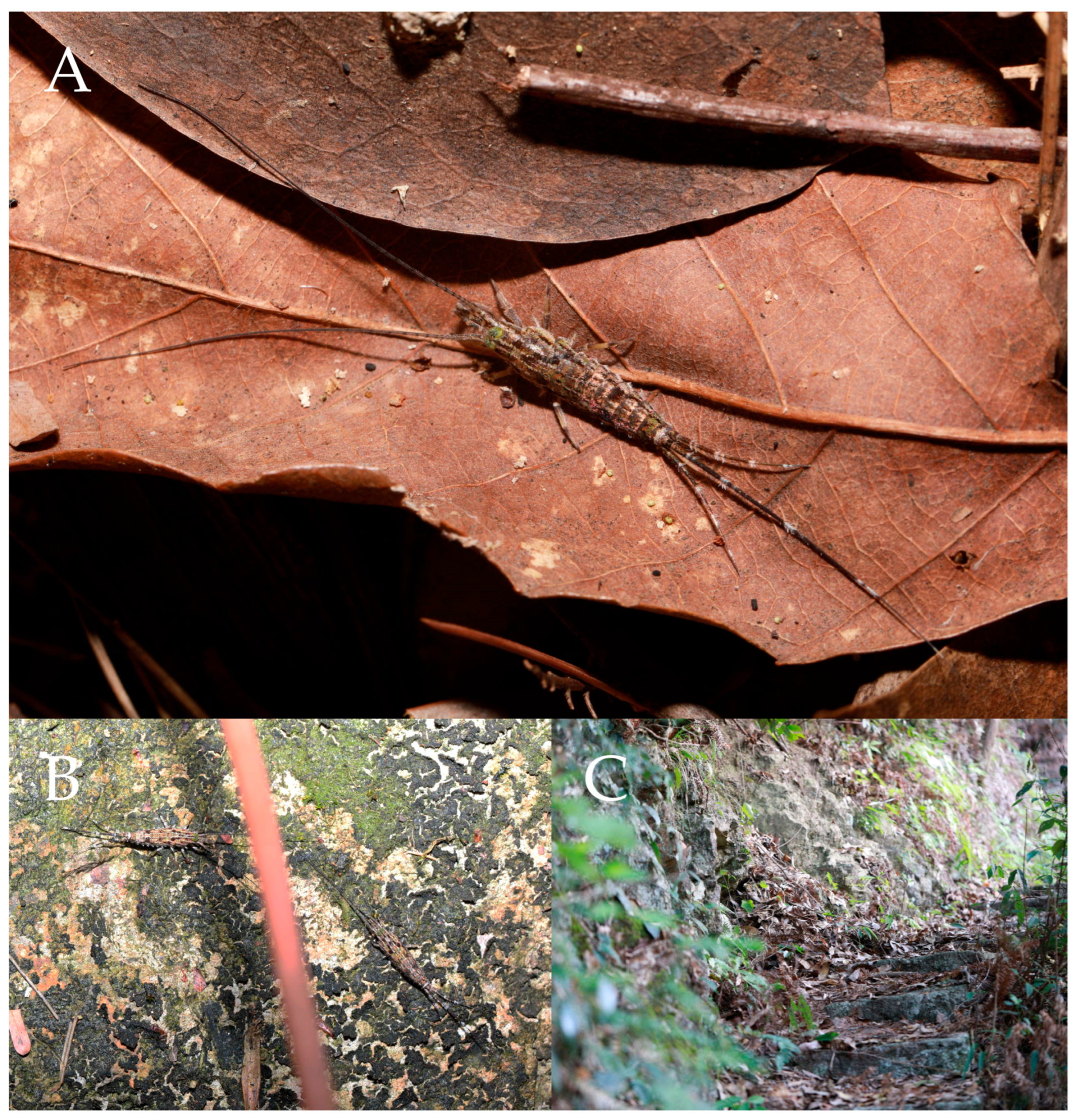
Figure 1.2.
Pedetontus (Verhoeffilis) elegans Shen, Yang, Ji & Zhang sp. n., holotype, male. (A) Habitus, lateral view. (B) Antenna. (C) Terminal chain of flagellum. (D) Head, frontal view. (E) Ditto, lateral view. (F, G) Compound eyes & paired ocelli. Scale bars: 500 μm.
Figure 1.2.
Pedetontus (Verhoeffilis) elegans Shen, Yang, Ji & Zhang sp. n., holotype, male. (A) Habitus, lateral view. (B) Antenna. (C) Terminal chain of flagellum. (D) Head, frontal view. (E) Ditto, lateral view. (F, G) Compound eyes & paired ocelli. Scale bars: 500 μm.
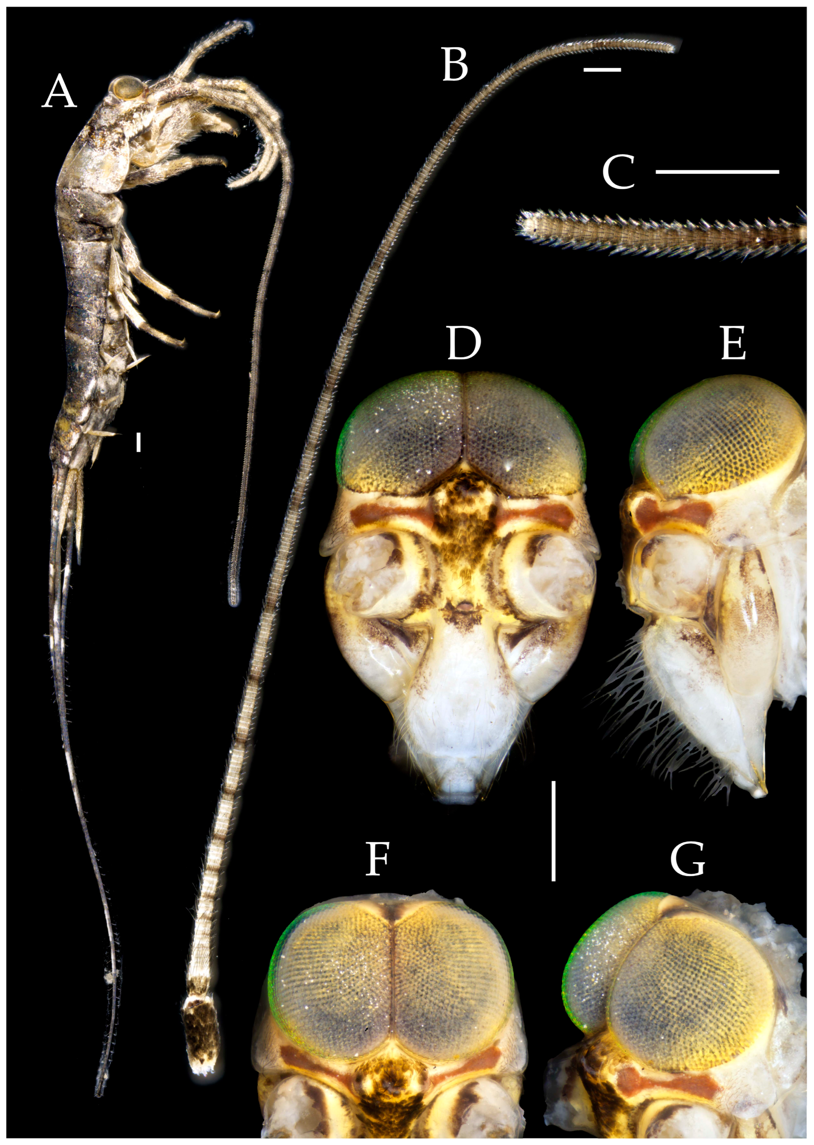
Figure 1.3.
Pedetontus (Verhoeffilis) elegans Shen, Yang, Ji & Zhang sp. n., holotype, male. (A–C) Foreleg, midleg & hindleg. (D) Mandible. (E) Maxilla. (F) Maxillary inner protuberance. (G) Labium. (H) Labial palp. Scale bars: 500 μm.
Figure 1.3.
Pedetontus (Verhoeffilis) elegans Shen, Yang, Ji & Zhang sp. n., holotype, male. (A–C) Foreleg, midleg & hindleg. (D) Mandible. (E) Maxilla. (F) Maxillary inner protuberance. (G) Labium. (H) Labial palp. Scale bars: 500 μm.
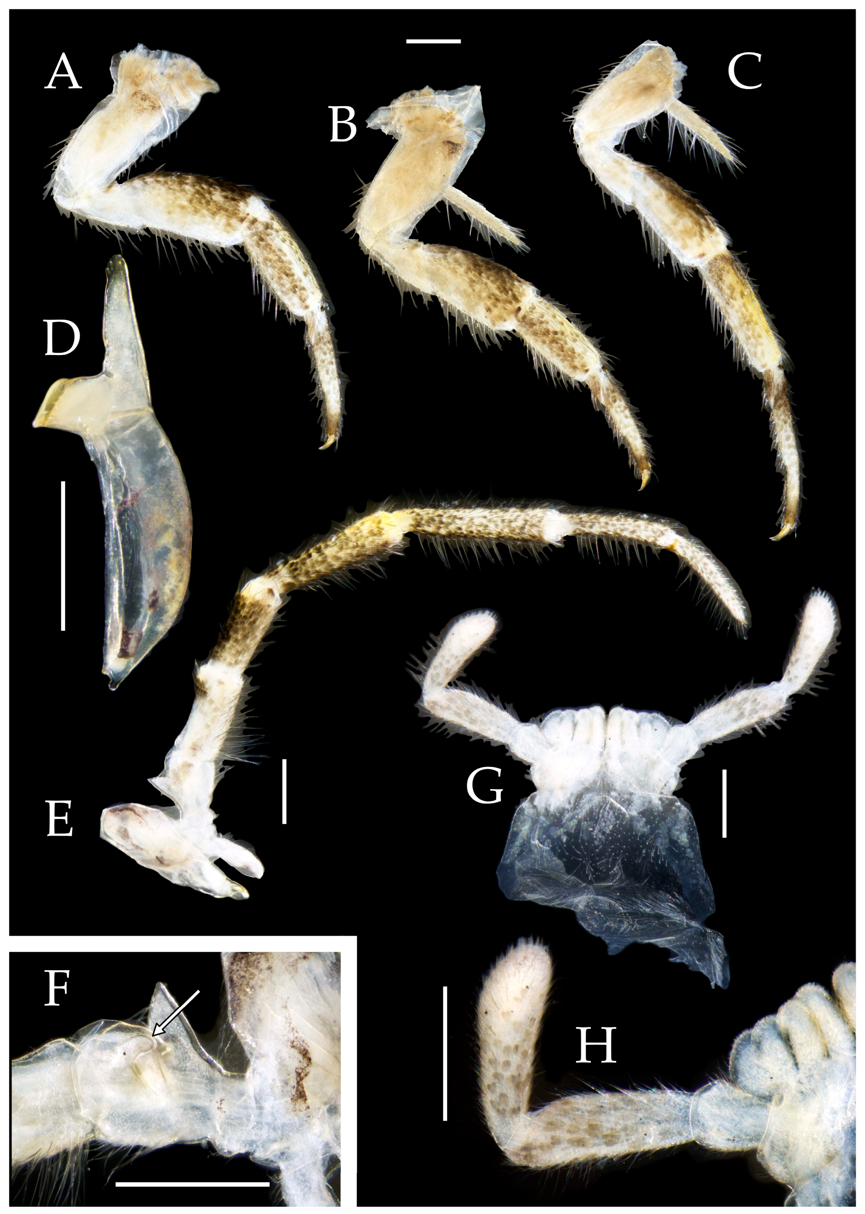
Figure 1.4.
Pedetontus (Verhoeffilis) elegans Shen, Yang, Ji & Zhang sp. n., holotype, male. (A–I) Abdominal sternites and coxites I–IX. (J) Penis & parameres. (K) Caudal filament & cerci. Scale bars: 500 μm.
Figure 1.4.
Pedetontus (Verhoeffilis) elegans Shen, Yang, Ji & Zhang sp. n., holotype, male. (A–I) Abdominal sternites and coxites I–IX. (J) Penis & parameres. (K) Caudal filament & cerci. Scale bars: 500 μm.
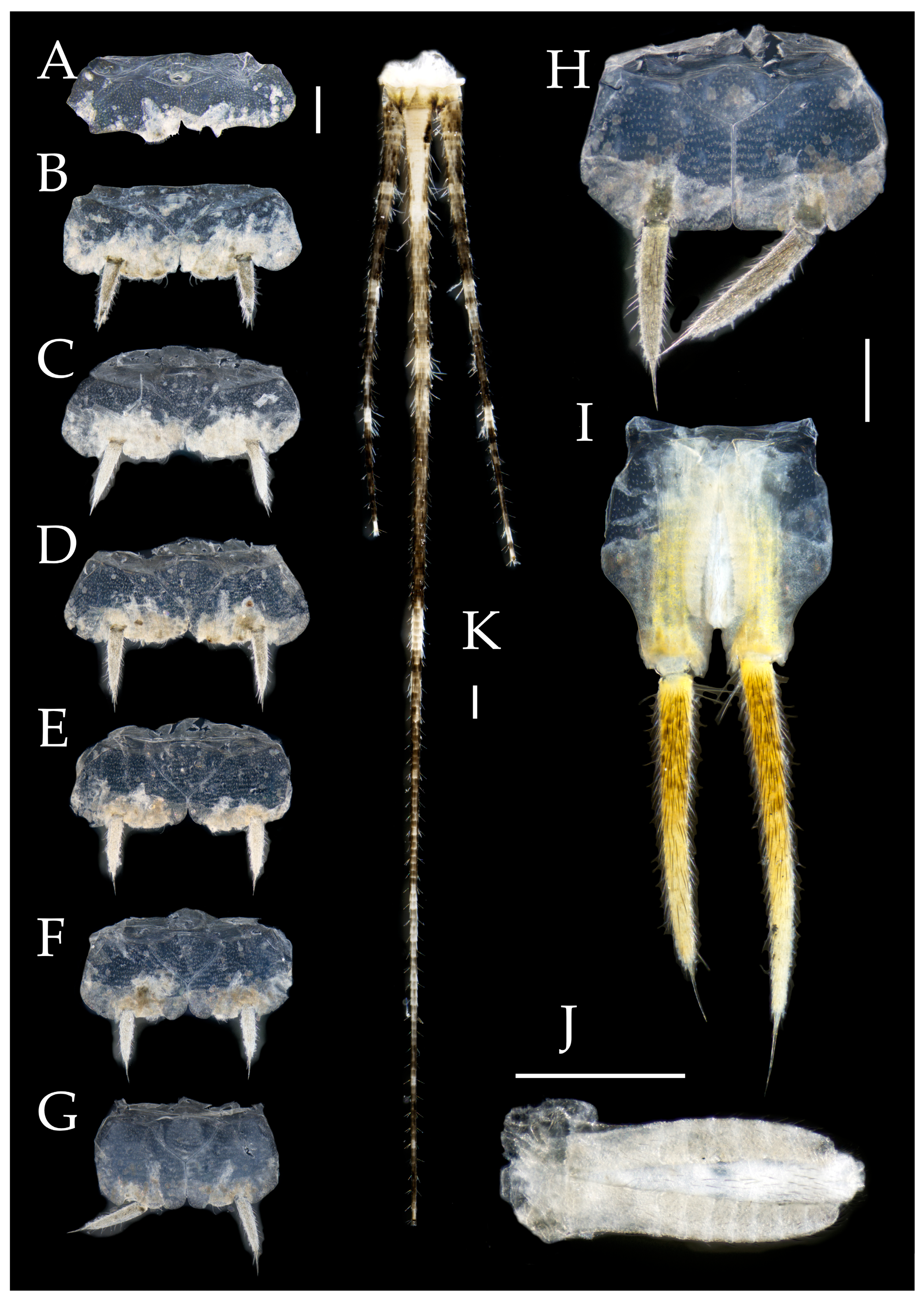
Figure 1.5.
Pedetontus (Verhoeffilis) elegans Shen, Yang, Ji & Zhang sp. n., paratype, female. (A) Habitus, lateral view. (B) Head, frontal view. (C) Ditto, lateral view. (D) Maxilla. (E) Maxillary palp article V–VII. (F) Labium. (G) Labium palp. Scale bars: 500 μm.
Figure 1.5.
Pedetontus (Verhoeffilis) elegans Shen, Yang, Ji & Zhang sp. n., paratype, female. (A) Habitus, lateral view. (B) Head, frontal view. (C) Ditto, lateral view. (D) Maxilla. (E) Maxillary palp article V–VII. (F) Labium. (G) Labium palp. Scale bars: 500 μm.
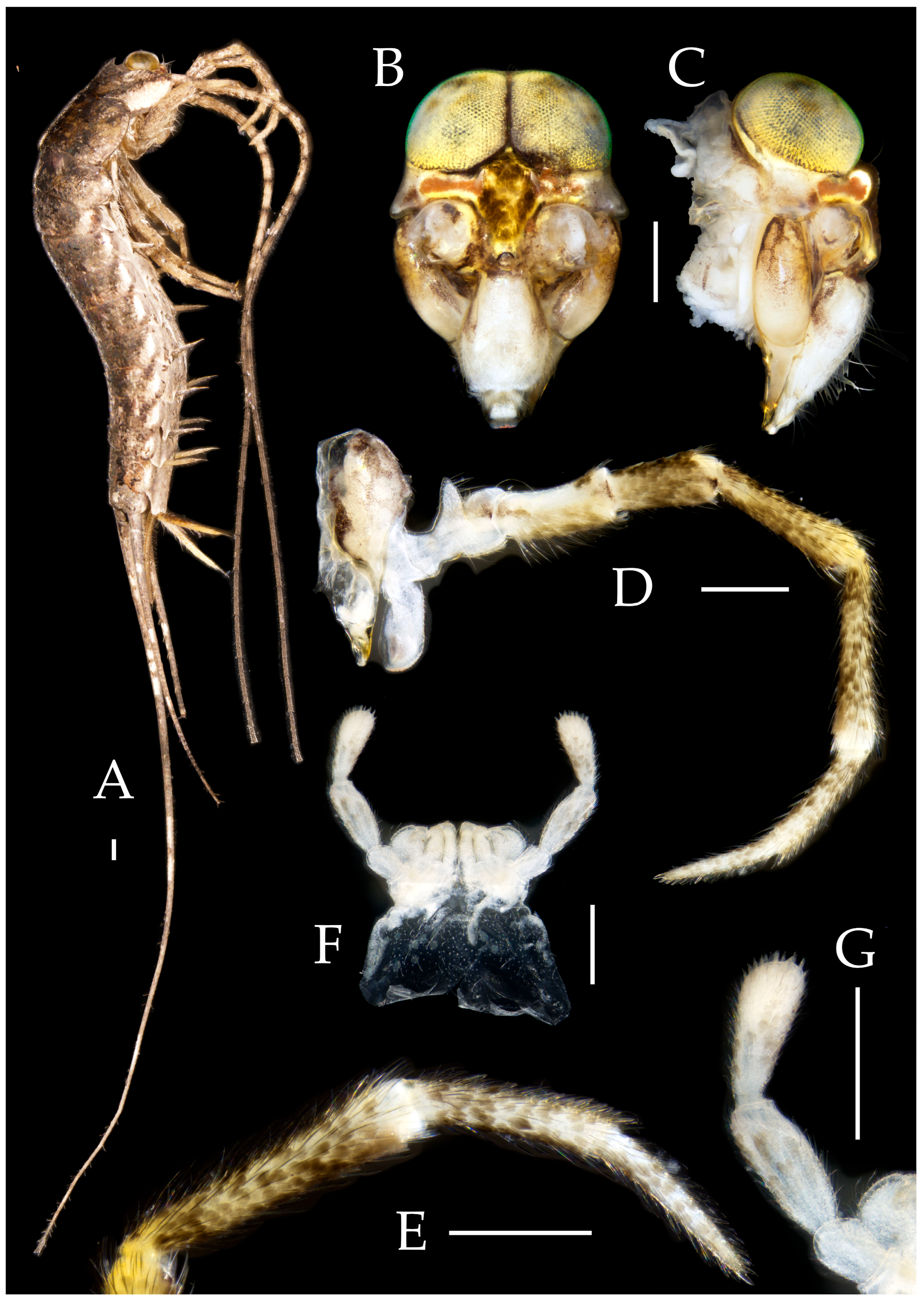
Figure 1.6.
Pedetontus (Verhoeffilis) elegans Shen, Yang, Ji & Zhang sp. n., paratype, female. (A–C) Foreleg, midleg & hindleg. (D) Foreleg tarsus. (E–H) Abdominal sternites and coxites V, VII–IX. Scale bars: 500 μm.
Figure 1.6.
Pedetontus (Verhoeffilis) elegans Shen, Yang, Ji & Zhang sp. n., paratype, female. (A–C) Foreleg, midleg & hindleg. (D) Foreleg tarsus. (E–H) Abdominal sternites and coxites V, VII–IX. Scale bars: 500 μm.
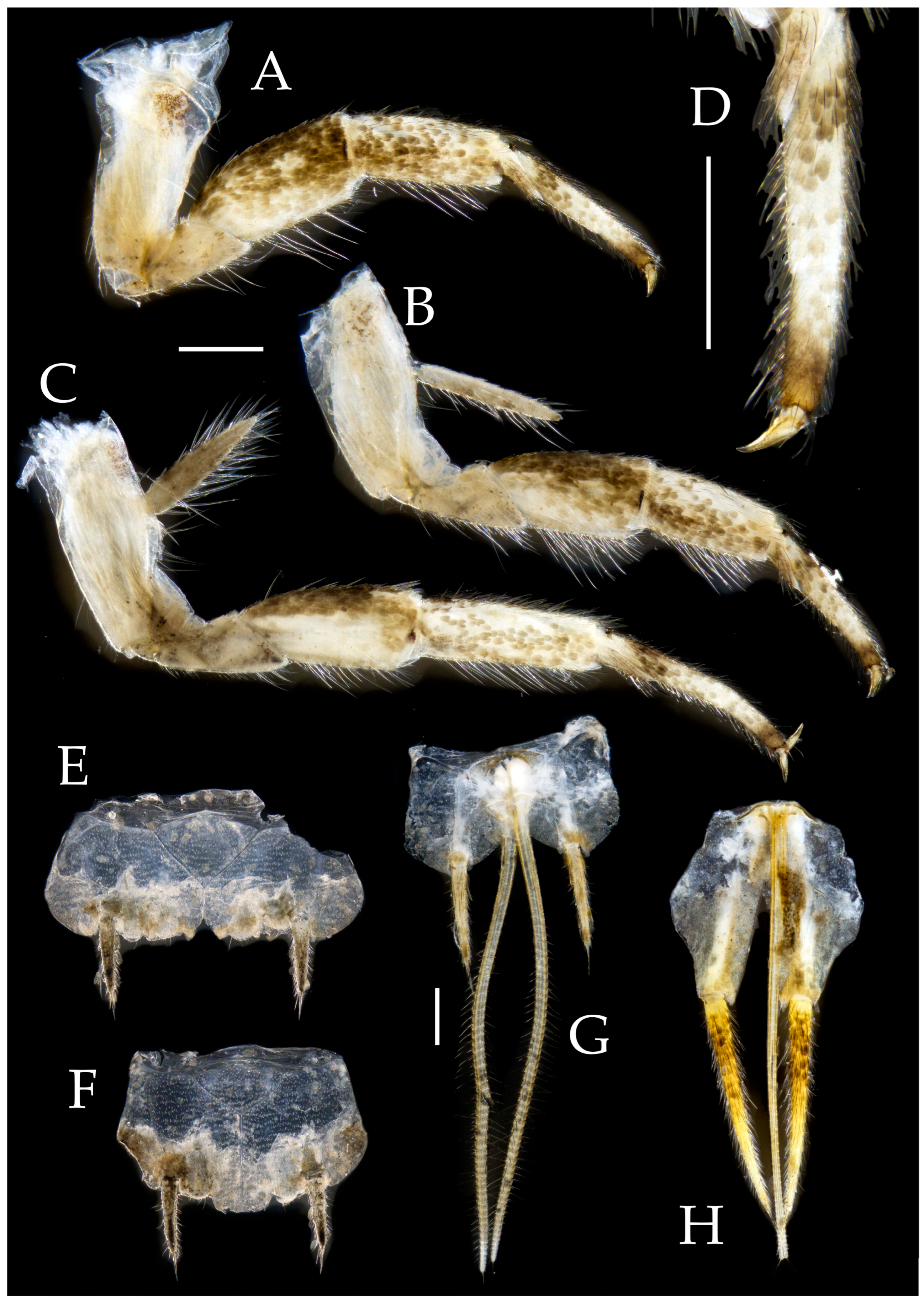
Figure 2.1.
Pedetontus (Verhoeffilis) hezhouensis Shen, Yang, Ji & Zhang sp. n., holotype, female. (A–C) Dorsal, lateral and anterolateral view.
Figure 2.1.
Pedetontus (Verhoeffilis) hezhouensis Shen, Yang, Ji & Zhang sp. n., holotype, female. (A–C) Dorsal, lateral and anterolateral view.
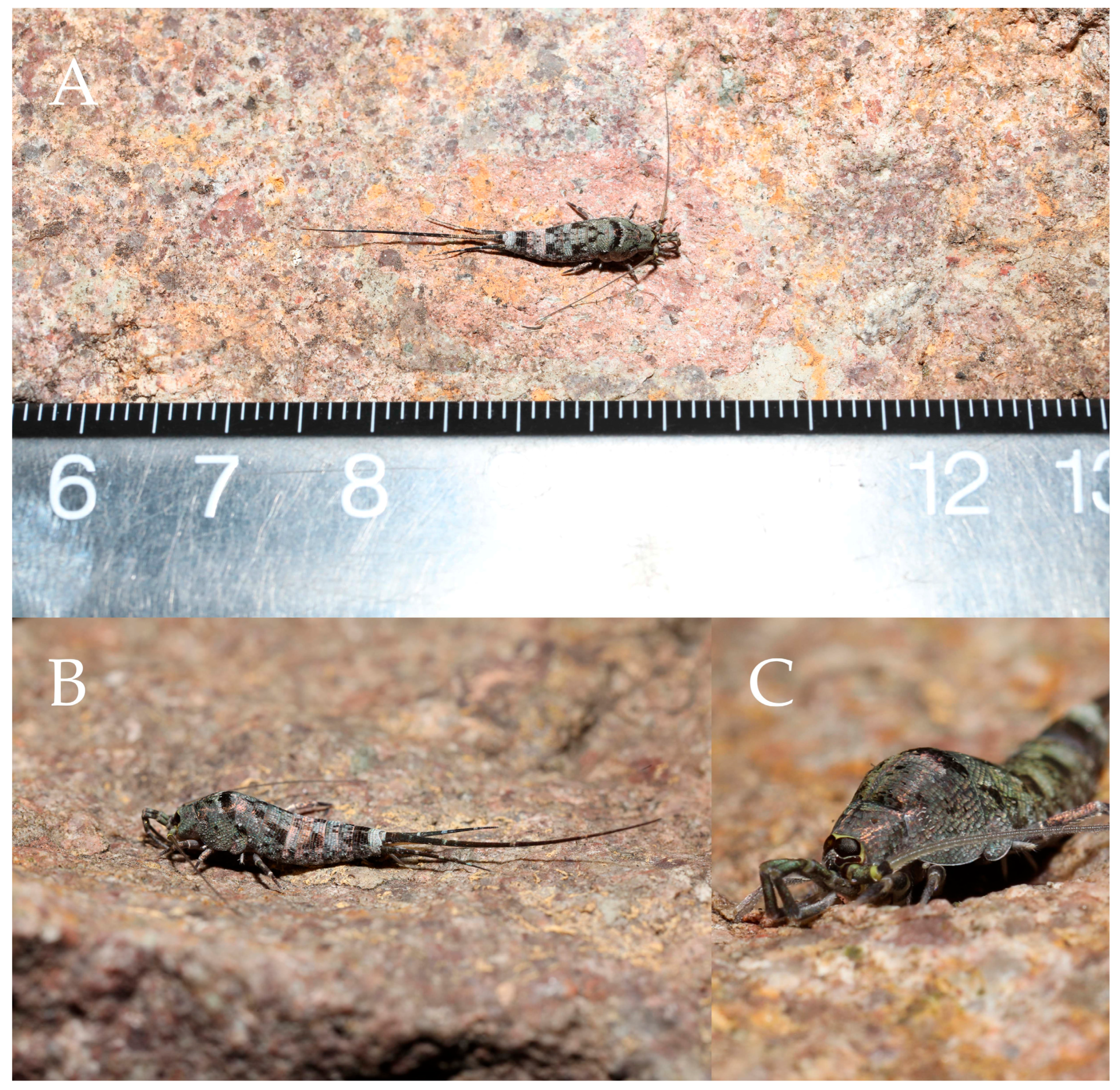
Figure 2.2.
Pedetontus (Verhoeffilis) hezhouensis Shen, Yang, Ji & Zhang sp. n., holotype, female. (A) Habitus, lateral view. (B) Antenna. (C) Head, frontal view. (D) Ditto, lateral view. (E, F) Compound eyes & paired ocelli Scale bars: 500 μm.
Figure 2.2.
Pedetontus (Verhoeffilis) hezhouensis Shen, Yang, Ji & Zhang sp. n., holotype, female. (A) Habitus, lateral view. (B) Antenna. (C) Head, frontal view. (D) Ditto, lateral view. (E, F) Compound eyes & paired ocelli Scale bars: 500 μm.
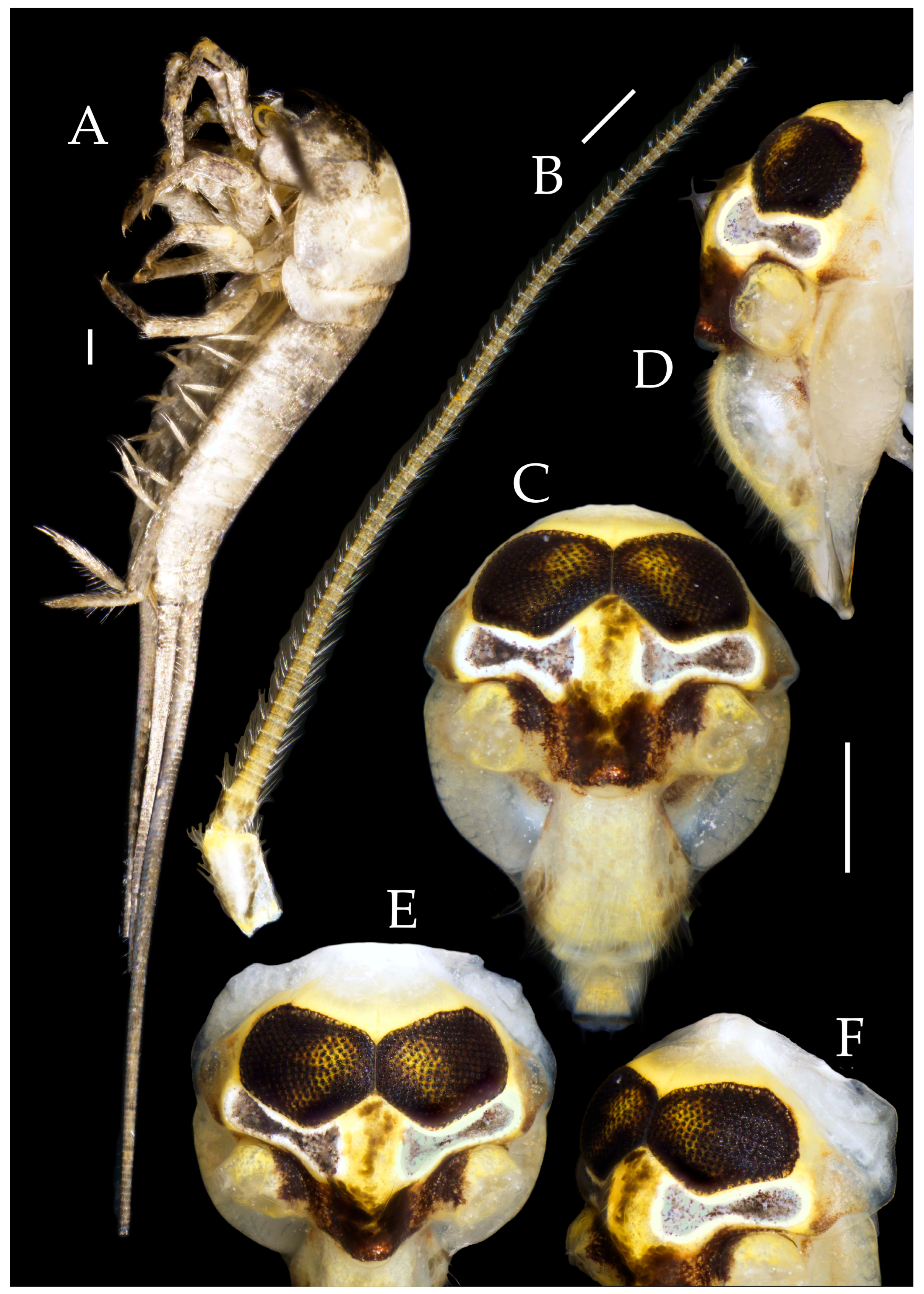
Figure 2.3.
Pedetontus (Verhoeffilis) hezhouensis Shen, Yang, Ji & Zhang sp. n., holotype, female. (A–C) Foreleg, midleg & hindleg. (D) Mandible. (E) Maxilla. (F) Maxillary inner protuberance. (G) Labium. (H) Labial palp. (I) Hypopharynx. Scale bars: 500 μm.
Figure 2.3.
Pedetontus (Verhoeffilis) hezhouensis Shen, Yang, Ji & Zhang sp. n., holotype, female. (A–C) Foreleg, midleg & hindleg. (D) Mandible. (E) Maxilla. (F) Maxillary inner protuberance. (G) Labium. (H) Labial palp. (I) Hypopharynx. Scale bars: 500 μm.
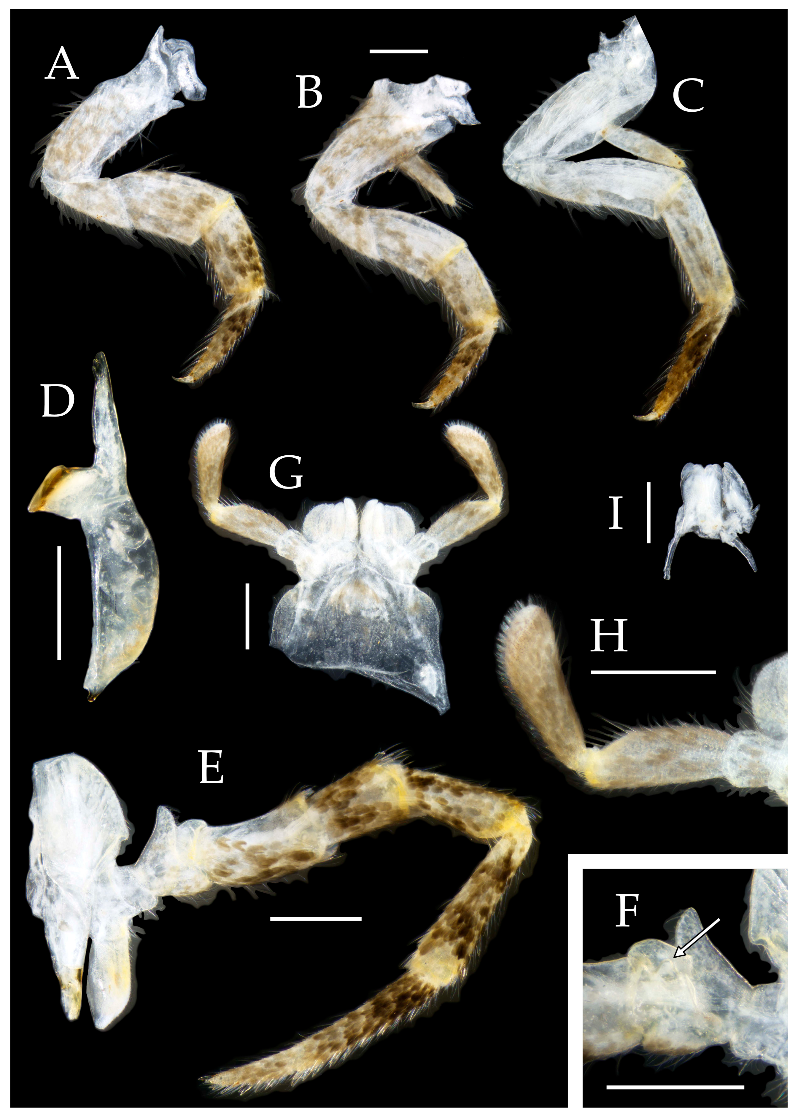
Figure 2.4.
Pedetontus (Verhoeffilis) hezhouensis Shen, Yang, Ji & Zhang sp. n., holotype, female. (A–I) Abdominal sternites and coxites I–IX. (J) Caudal filament & cerci. Scale bars: 500 μm.
Figure 2.4.
Pedetontus (Verhoeffilis) hezhouensis Shen, Yang, Ji & Zhang sp. n., holotype, female. (A–I) Abdominal sternites and coxites I–IX. (J) Caudal filament & cerci. Scale bars: 500 μm.
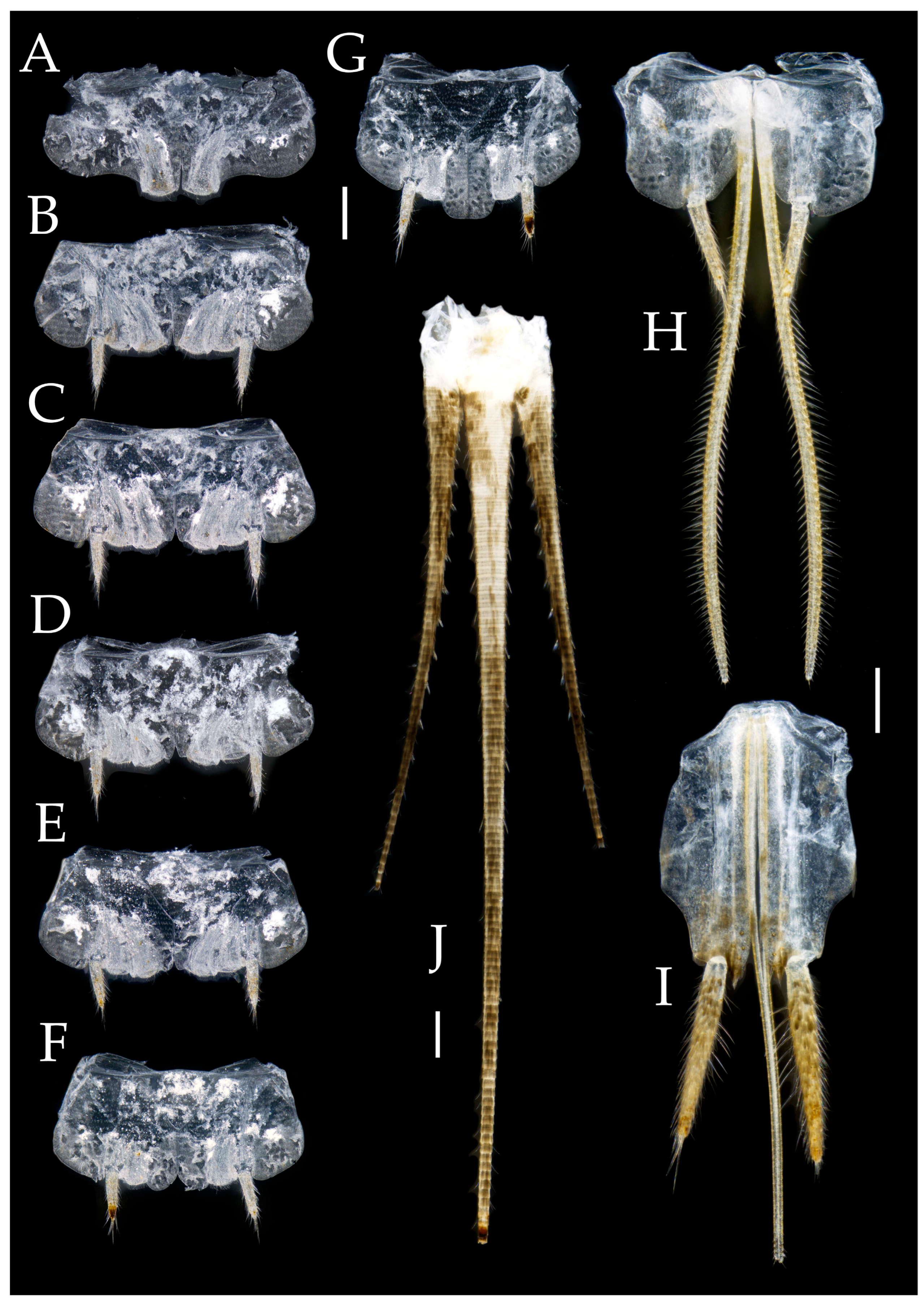
Figure 2.5.
Pedetontus (Verhoeffilis) hezhouensis Shen, Yang, Ji & Zhang sp. n., paratype, male. (A) Habitus, lateral view. (B) Head, frontal view. (C) Ditto, lateral view. (D) Mandible. (E) Maxilla. (F) Maxillary palp article V–VII. (G) Labium. (H) Labium palp. Scale bars: 500 μm.
Figure 2.5.
Pedetontus (Verhoeffilis) hezhouensis Shen, Yang, Ji & Zhang sp. n., paratype, male. (A) Habitus, lateral view. (B) Head, frontal view. (C) Ditto, lateral view. (D) Mandible. (E) Maxilla. (F) Maxillary palp article V–VII. (G) Labium. (H) Labium palp. Scale bars: 500 μm.
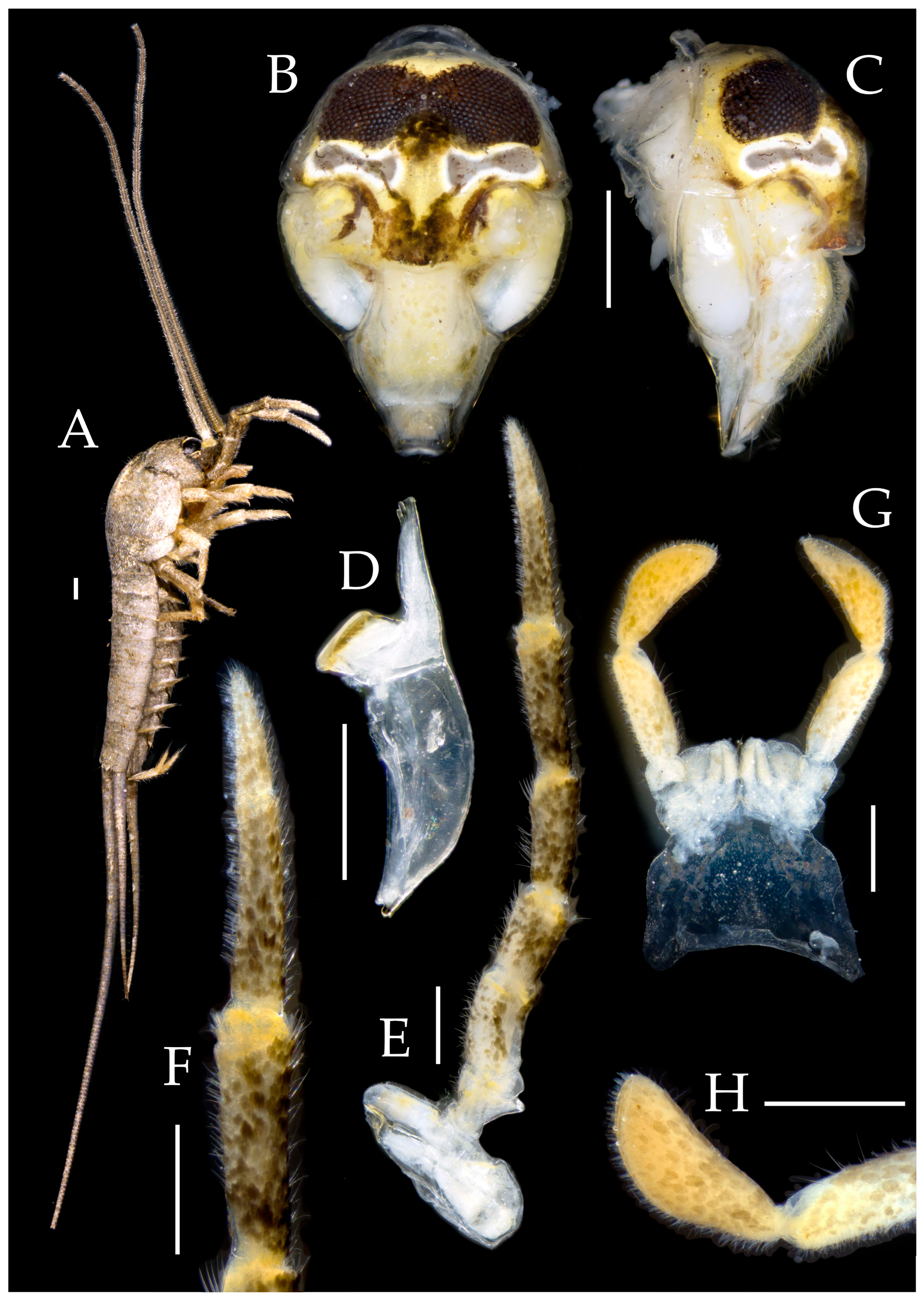
Figure 2.6.
Pedetontus (Verhoeffilis) hezhouensis Shen, Yang, Ji & Zhang sp. n., paratype, male. (A–C) Foreleg, midleg & hindleg. (D–G) Abdominal sternites and coxites V, VII–IX. (H) Penis & parameres. Scale bars: 500 μm.
Figure 2.6.
Pedetontus (Verhoeffilis) hezhouensis Shen, Yang, Ji & Zhang sp. n., paratype, male. (A–C) Foreleg, midleg & hindleg. (D–G) Abdominal sternites and coxites V, VII–IX. (H) Penis & parameres. Scale bars: 500 μm.
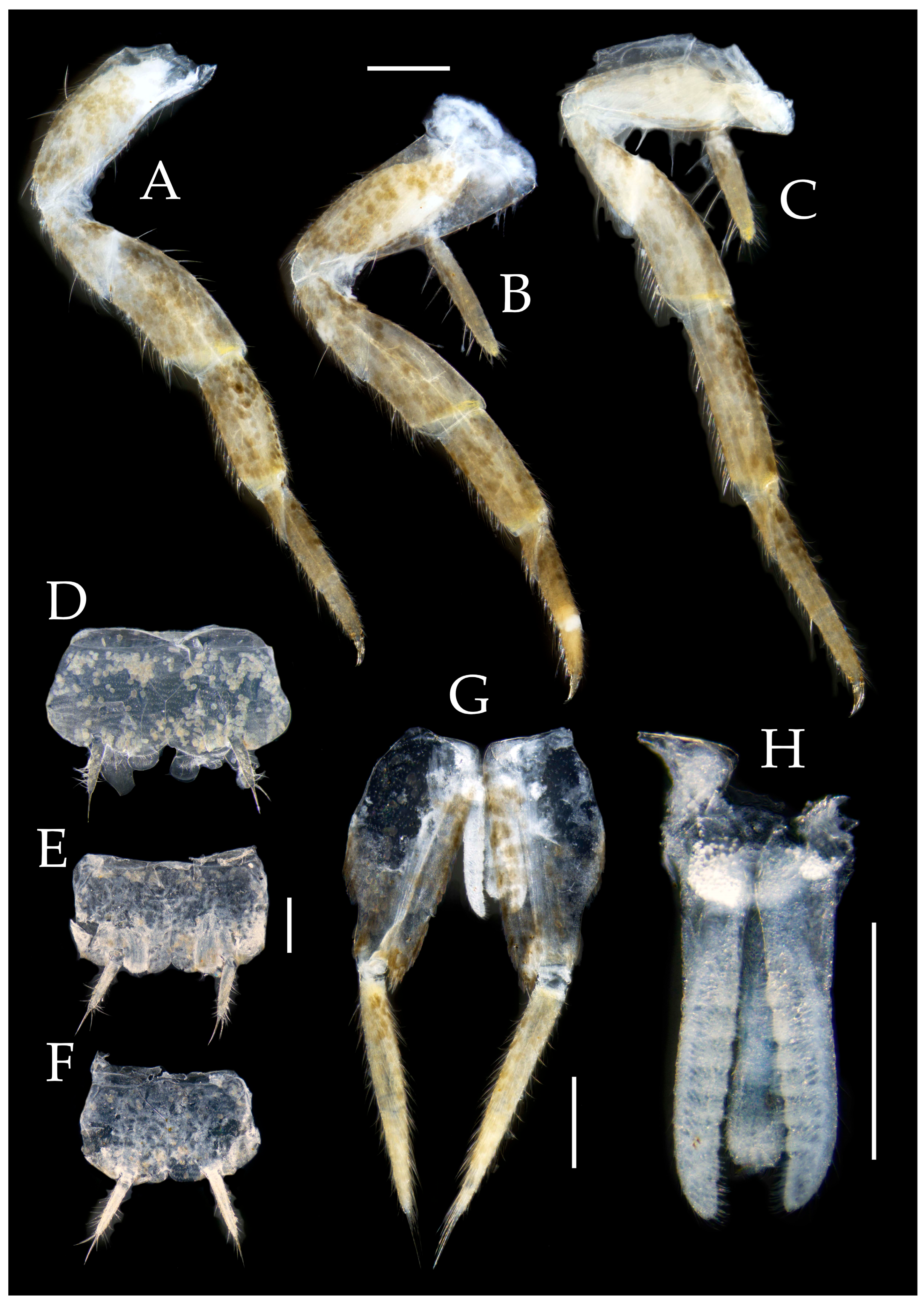
Figure 3.1.
Pedetontus (Verhoeffilis) jinxiuensis Shen, Yang, Ji & Zhang sp. n. (A–C) Lateral, dorsal and anterior view.
Figure 3.1.
Pedetontus (Verhoeffilis) jinxiuensis Shen, Yang, Ji & Zhang sp. n. (A–C) Lateral, dorsal and anterior view.
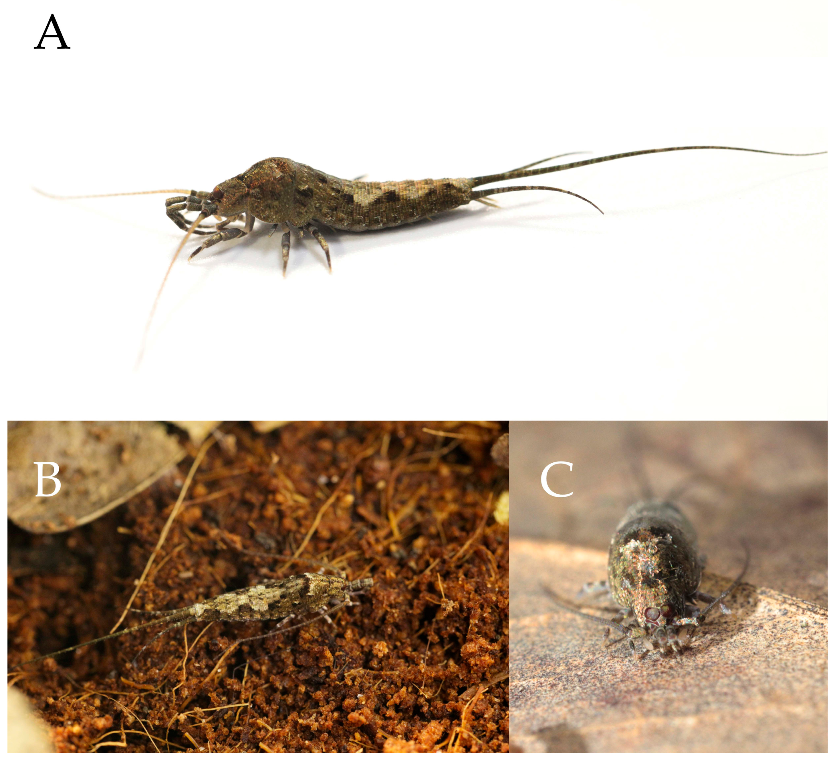
Figure 3.2.
Pedetontus (Verhoeffilis) jinxiuensis Shen, Yang, Ji & Zhang sp. n., holotype, male. (A) Habitus, lateral view. (B) Antenna. (C) Head, frontal view. (D) Ditto, lateral view. (E, F) Compound eyes & paired ocelli. Scale bars: 500 μm.
Figure 3.2.
Pedetontus (Verhoeffilis) jinxiuensis Shen, Yang, Ji & Zhang sp. n., holotype, male. (A) Habitus, lateral view. (B) Antenna. (C) Head, frontal view. (D) Ditto, lateral view. (E, F) Compound eyes & paired ocelli. Scale bars: 500 μm.
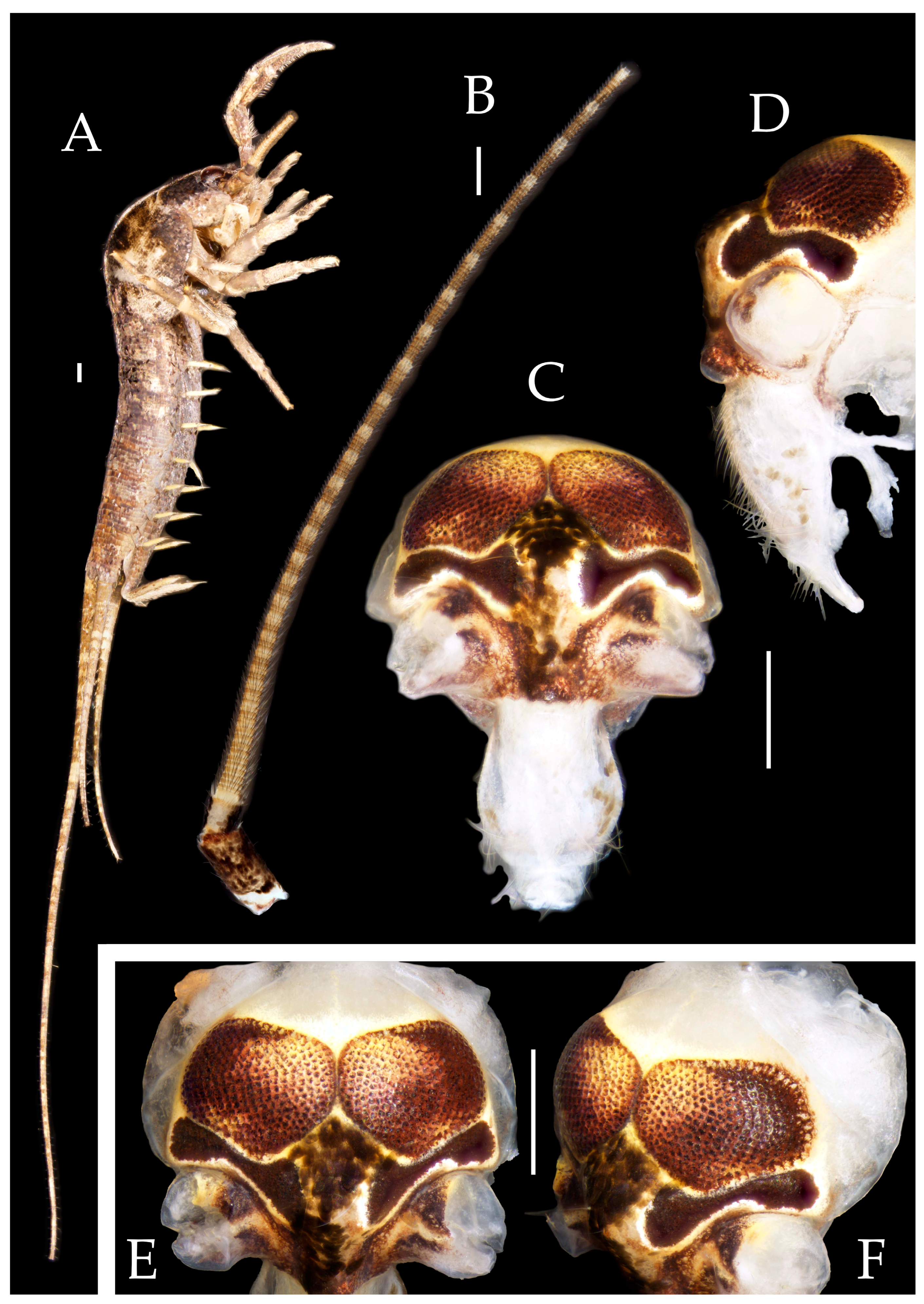
Figure 3.3.
Pedetontus (Verhoeffilis) jinxiuensis Shen, Yang, Ji & Zhang sp. n., holotype, male. (A–C) Foreleg, midleg & hindleg. (D) Mandible. (E) Maxilla. (F) Maxillary palp article VI & VII. (G) Maxillary inner protuberance. (H) Labium. Scale bars: 500 μm.
Figure 3.3.
Pedetontus (Verhoeffilis) jinxiuensis Shen, Yang, Ji & Zhang sp. n., holotype, male. (A–C) Foreleg, midleg & hindleg. (D) Mandible. (E) Maxilla. (F) Maxillary palp article VI & VII. (G) Maxillary inner protuberance. (H) Labium. Scale bars: 500 μm.
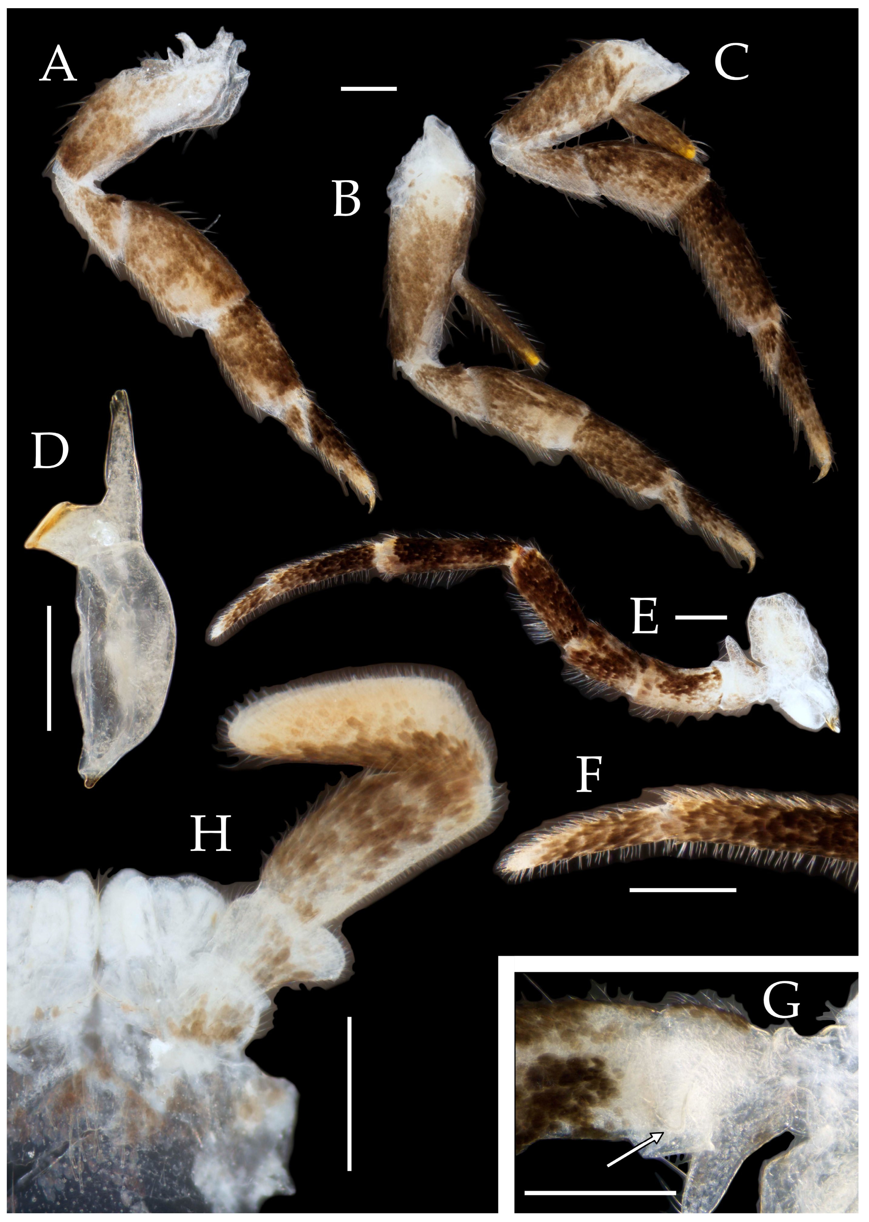
Figure 3.4.
Pedetontus (Verhoeffilis) jinxiuensis Shen, Yang, Ji & Zhang sp. n., holotype, male. (A–I) Abdominal sternites and coxites I–IX. (J) Penis & parameres. Scale bars: 500 μm.
Figure 3.4.
Pedetontus (Verhoeffilis) jinxiuensis Shen, Yang, Ji & Zhang sp. n., holotype, male. (A–I) Abdominal sternites and coxites I–IX. (J) Penis & parameres. Scale bars: 500 μm.
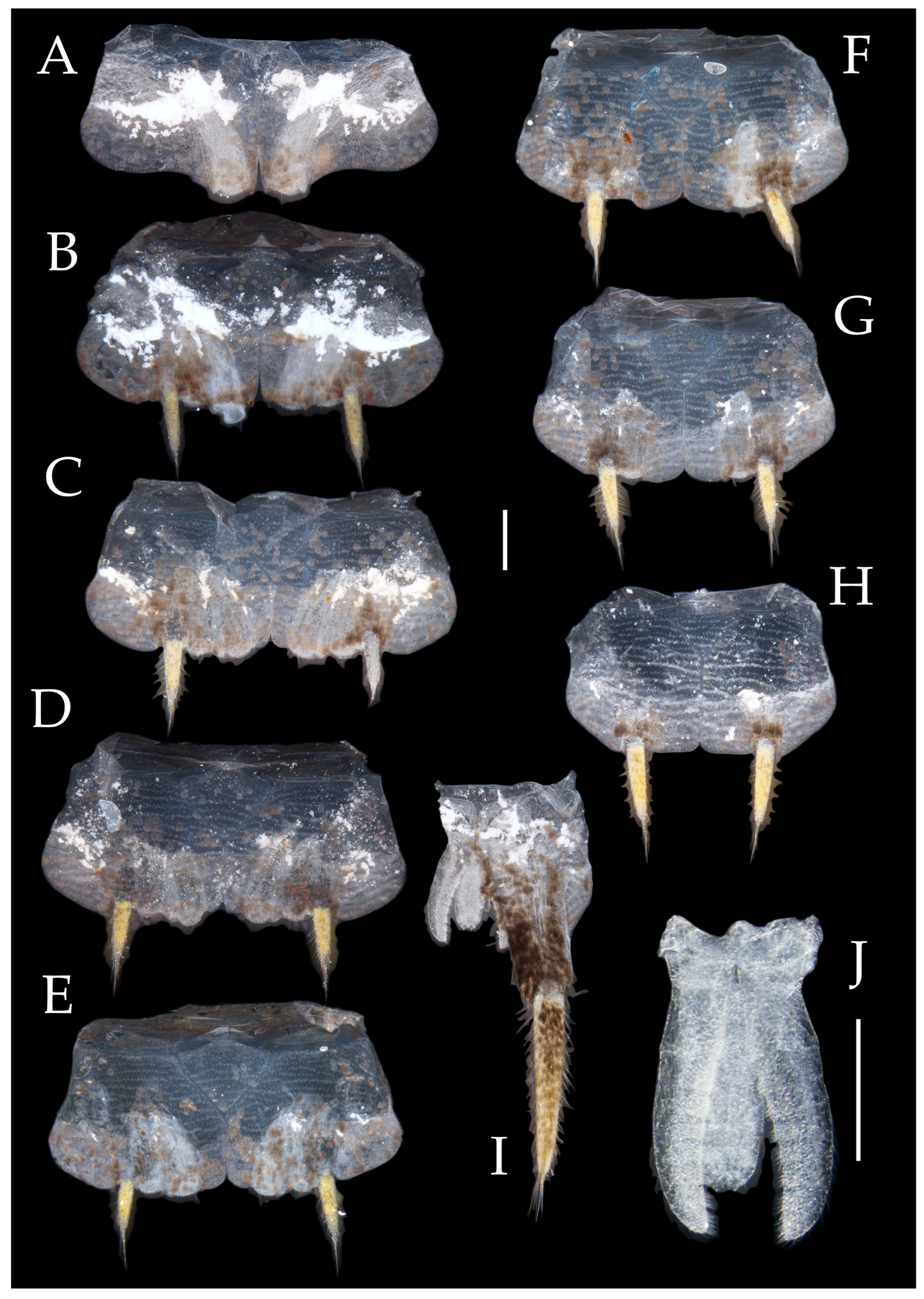
Figure 3.5.
Pedetontus (Verhoeffilis) jinxiuensis Shen, Yang, Ji & Zhang sp. n., paratype, female. (A) Habitus, lateral view. (B) Head, frontal view. (C) Ditto, lateral view. (D) Maxilla. (E) Hypophraynx. (F) Labium. (G) Labium palp. Scale bars: 500 μm.
Figure 3.5.
Pedetontus (Verhoeffilis) jinxiuensis Shen, Yang, Ji & Zhang sp. n., paratype, female. (A) Habitus, lateral view. (B) Head, frontal view. (C) Ditto, lateral view. (D) Maxilla. (E) Hypophraynx. (F) Labium. (G) Labium palp. Scale bars: 500 μm.
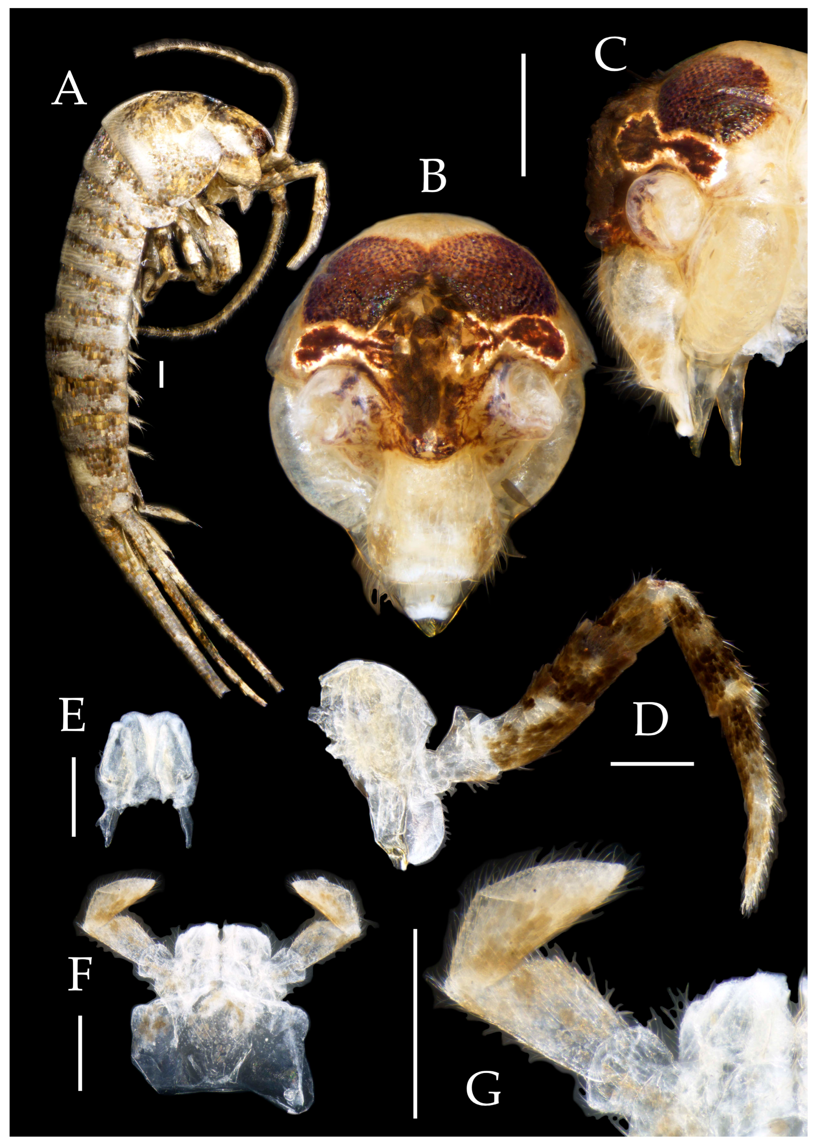
Figure 3.6.
Pedetontus (Verhoeffilis) jinxiuensis Shen, Yang, Ji & Zhang sp. n., paratype, female. (A–C) Foreleg, midleg & hindleg. (D–G) Abdominal sternites and coxites V, VII–IX. Scale bars: 500 μm.
Figure 3.6.
Pedetontus (Verhoeffilis) jinxiuensis Shen, Yang, Ji & Zhang sp. n., paratype, female. (A–C) Foreleg, midleg & hindleg. (D–G) Abdominal sternites and coxites V, VII–IX. Scale bars: 500 μm.
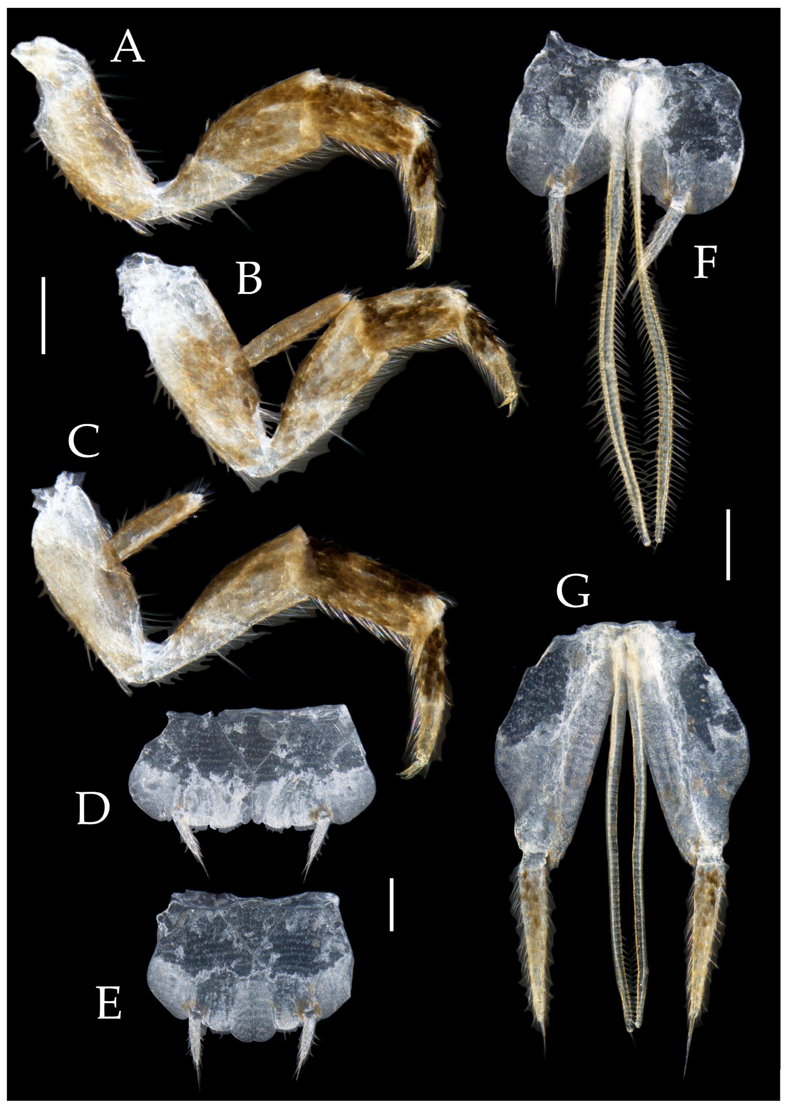
Figure 4.1.
Pedetontus (Verhoeffilis) nanningensis Shen, Yang, Ji & Zhang sp. n. (A–C) Dorsal, anterolateral and lateral view.
Figure 4.1.
Pedetontus (Verhoeffilis) nanningensis Shen, Yang, Ji & Zhang sp. n. (A–C) Dorsal, anterolateral and lateral view.
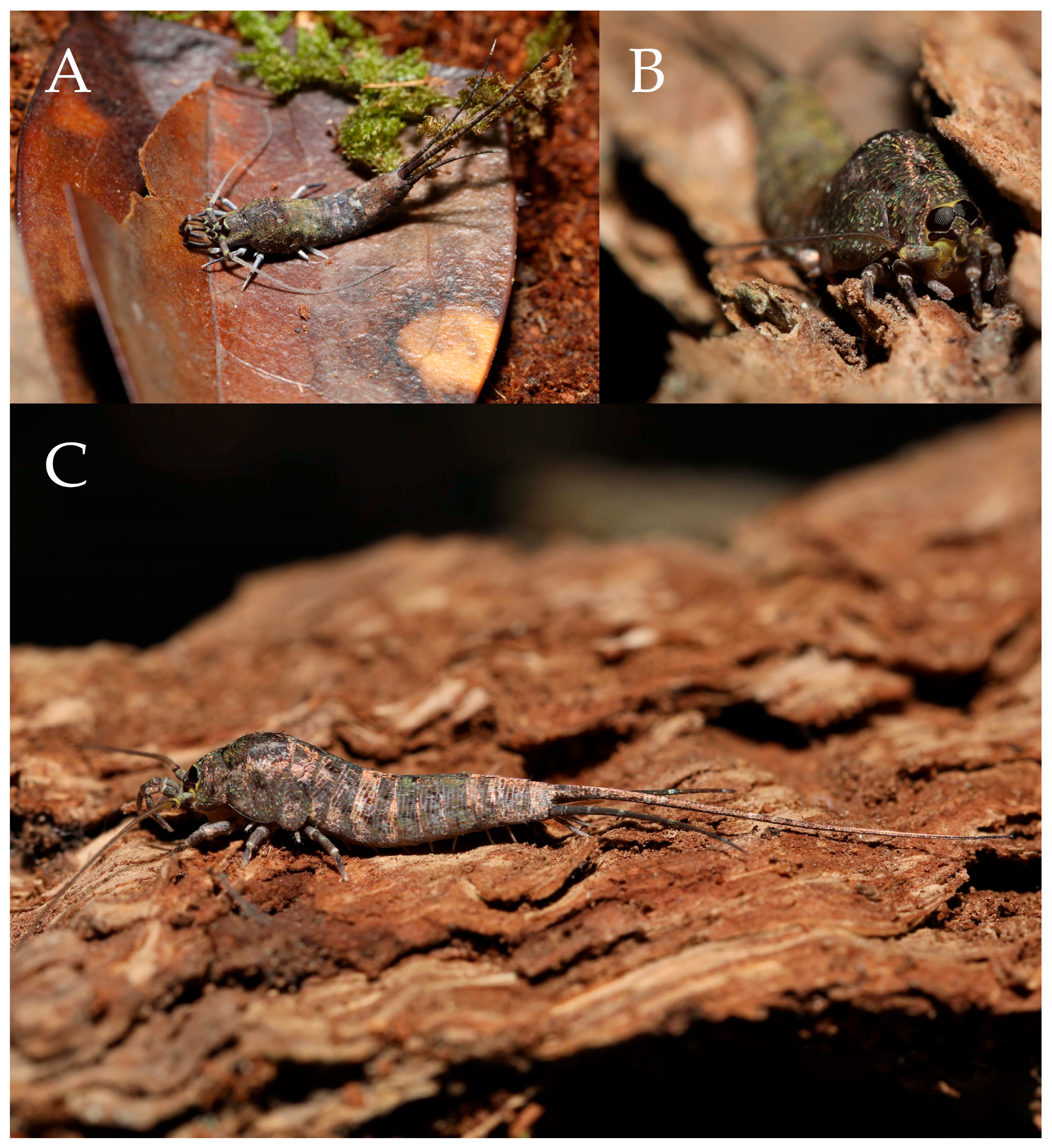
Figure 4.2.
Pedetontus (Verhoeffilis) nanningensis Shen, Yang, Ji & Zhang sp. n., holotype, male. (A) Habitus, lateral view. (B) Antenna. (C) Head, frontal view. (D) Ditto, lateral view. (E, F) Compound eyes & paired ocelli. Scale bars: 500 μm.
Figure 4.2.
Pedetontus (Verhoeffilis) nanningensis Shen, Yang, Ji & Zhang sp. n., holotype, male. (A) Habitus, lateral view. (B) Antenna. (C) Head, frontal view. (D) Ditto, lateral view. (E, F) Compound eyes & paired ocelli. Scale bars: 500 μm.
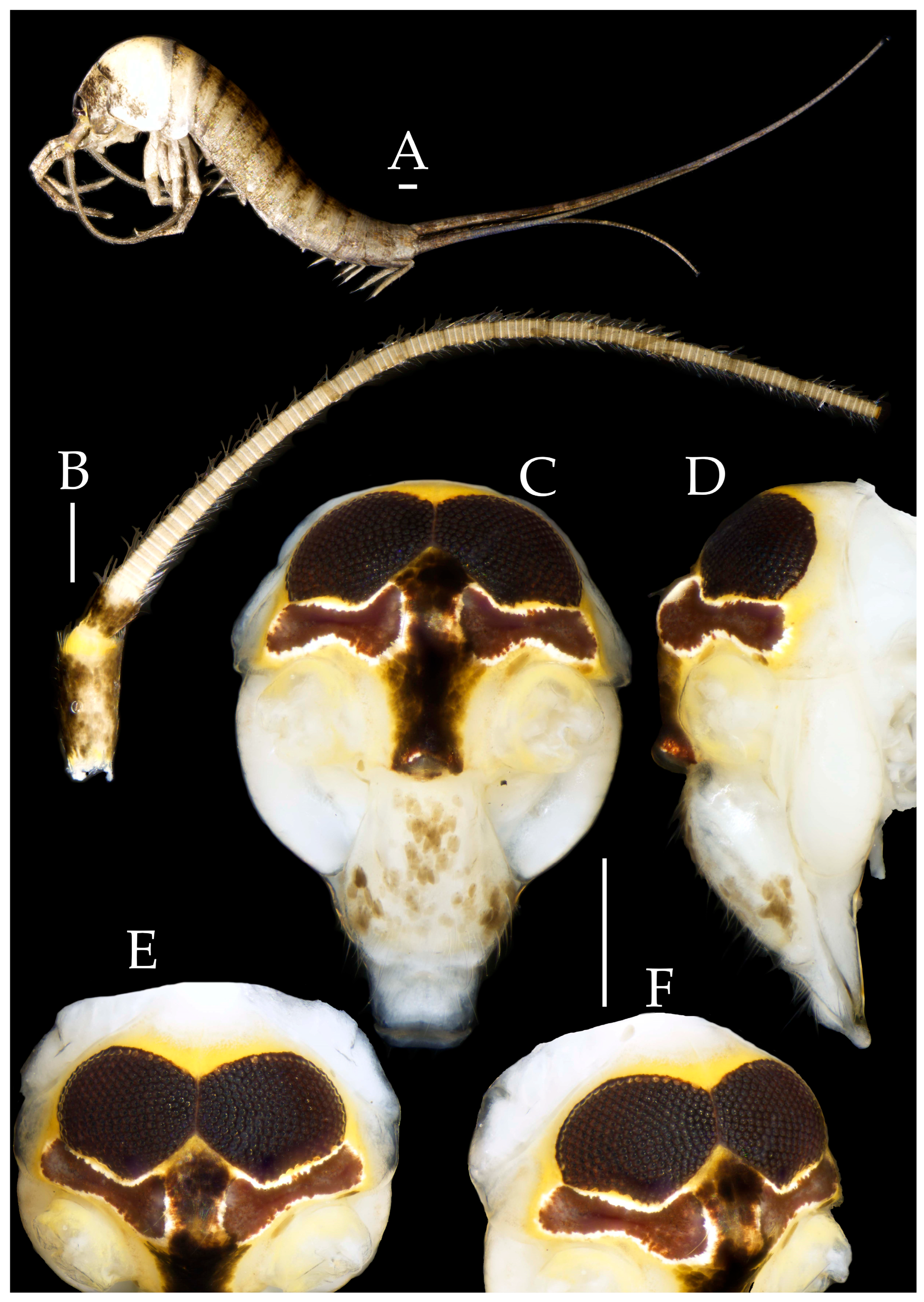
Figure 4.3.
Pedetontus (Verhoeffilis) nanningensis Shen, Yang, Ji & Zhang sp. n., holotype, male. (A) Mandible (B) Labium. (C) Labial palp (D) Maxilla. (E) Maxillary inner protuberance. (F) Hypopharynx. (G–I) Foreleg, midleg & hindleg. Scale bars: 500 μm.
Figure 4.3.
Pedetontus (Verhoeffilis) nanningensis Shen, Yang, Ji & Zhang sp. n., holotype, male. (A) Mandible (B) Labium. (C) Labial palp (D) Maxilla. (E) Maxillary inner protuberance. (F) Hypopharynx. (G–I) Foreleg, midleg & hindleg. Scale bars: 500 μm.
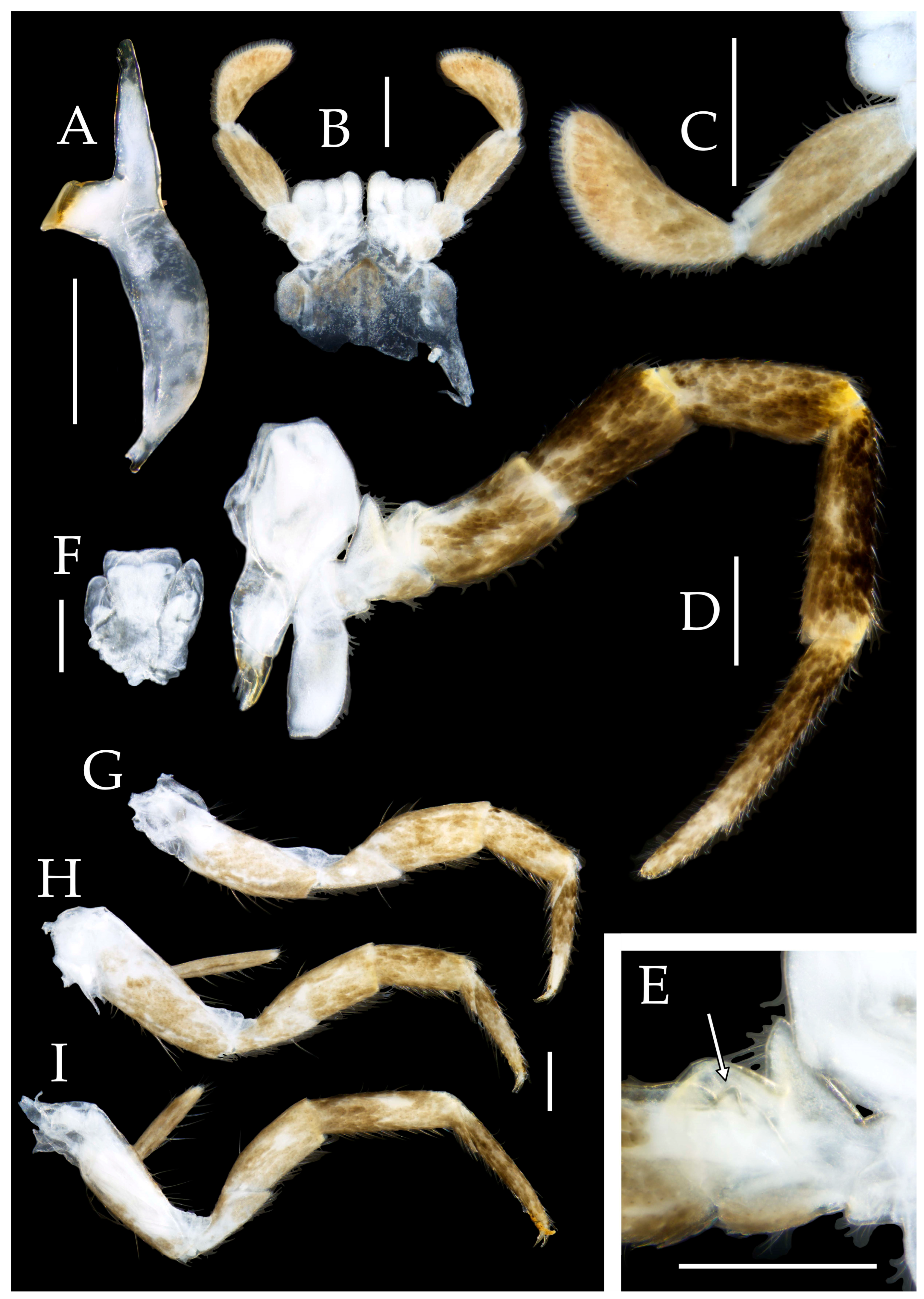
Figure 4.4.
Pedetontus (Verhoeffilis) nanningensis Shen, Yang, Ji & Zhang sp. n., holotype, male. (A–I) Abdominal sternites and coxites I–IX. (J) Caudal filament & cerci. (K) Penis & parameres. Scale bars: 500 μm.
Figure 4.4.
Pedetontus (Verhoeffilis) nanningensis Shen, Yang, Ji & Zhang sp. n., holotype, male. (A–I) Abdominal sternites and coxites I–IX. (J) Caudal filament & cerci. (K) Penis & parameres. Scale bars: 500 μm.
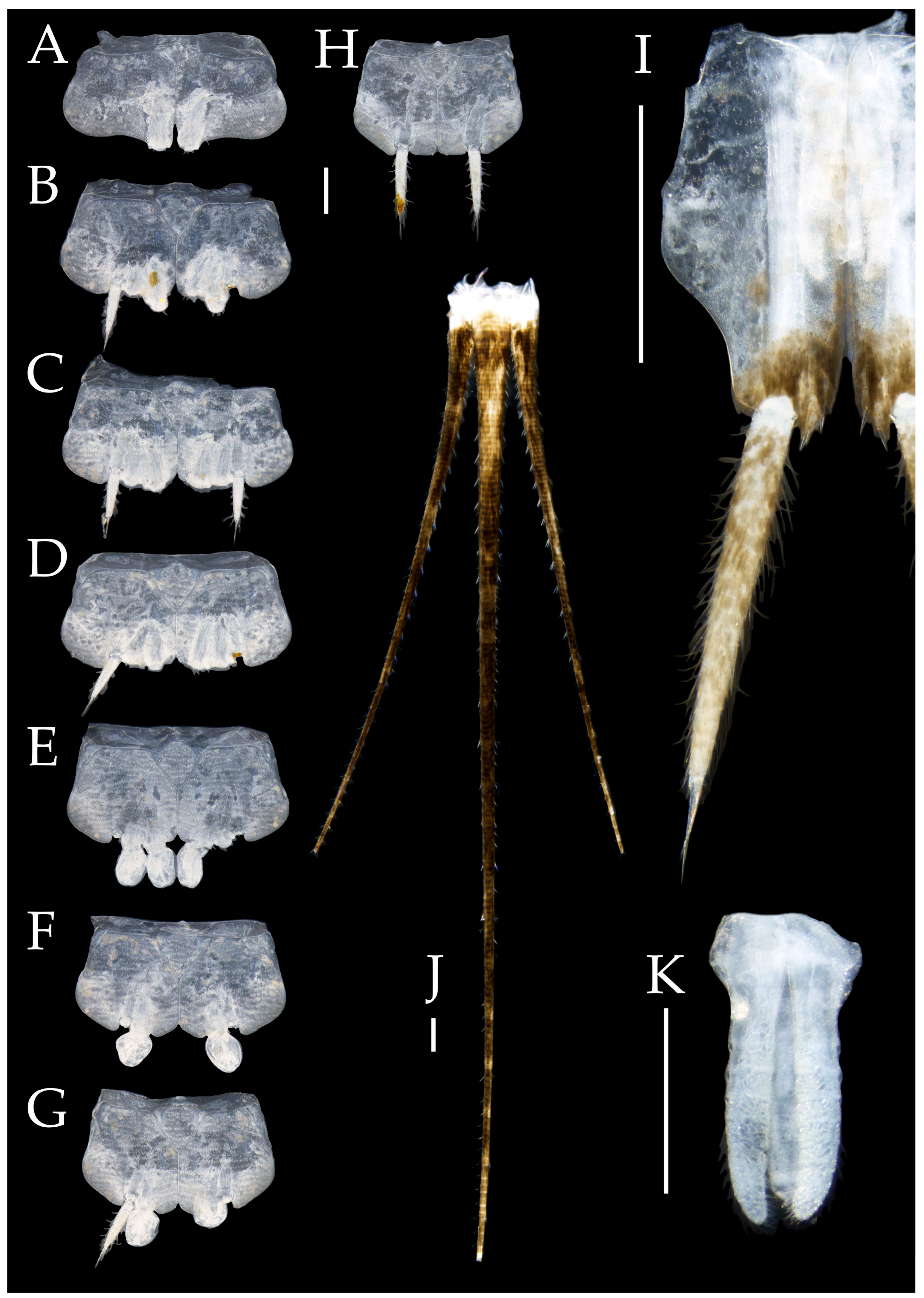
Figure 4.5.
Pedetontus (Verhoeffilis) nanningensis Shen, Yang, Ji & Zhang sp. n., paratype, female. (A) Habitus, lateral view. (B) Antenna. (C) Head, frontal view. (D–F) Foreleg, midleg & hindleg. Scale bars: 500 μm.
Figure 4.5.
Pedetontus (Verhoeffilis) nanningensis Shen, Yang, Ji & Zhang sp. n., paratype, female. (A) Habitus, lateral view. (B) Antenna. (C) Head, frontal view. (D–F) Foreleg, midleg & hindleg. Scale bars: 500 μm.
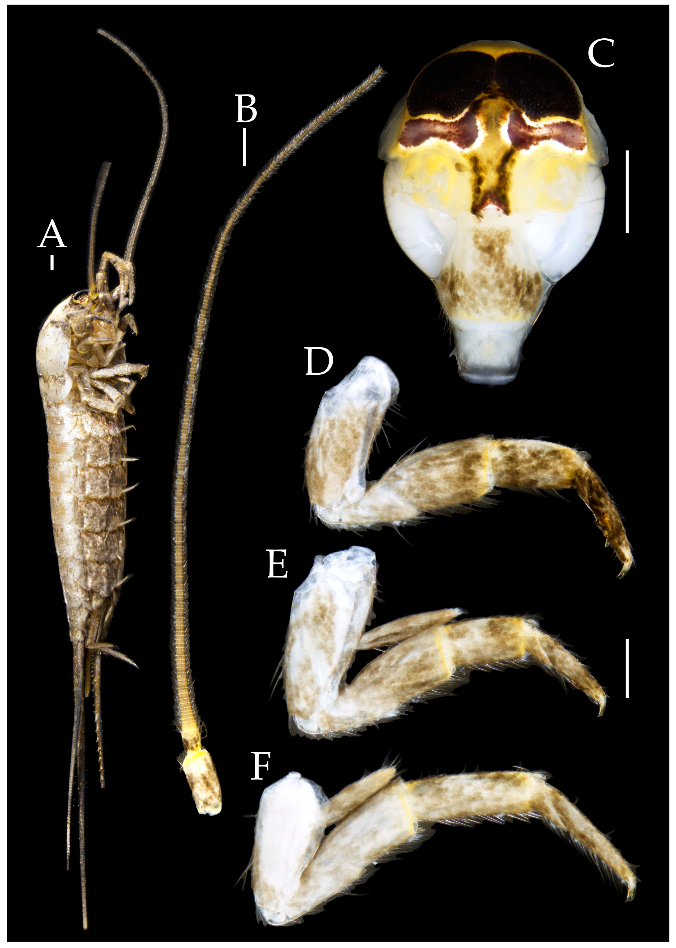
Figure 4.6.
Pedetontus (Verhoeffilis) nanningensis Shen, Yang, Ji & Zhang sp. n., paratype, female. (A) Labium. (B) Labial palp. (C) Maxilla. (D–G) Abdominal sternites and coxites V, VII–IX. Scale bars: 500 μm.
Figure 4.6.
Pedetontus (Verhoeffilis) nanningensis Shen, Yang, Ji & Zhang sp. n., paratype, female. (A) Labium. (B) Labial palp. (C) Maxilla. (D–G) Abdominal sternites and coxites V, VII–IX. Scale bars: 500 μm.
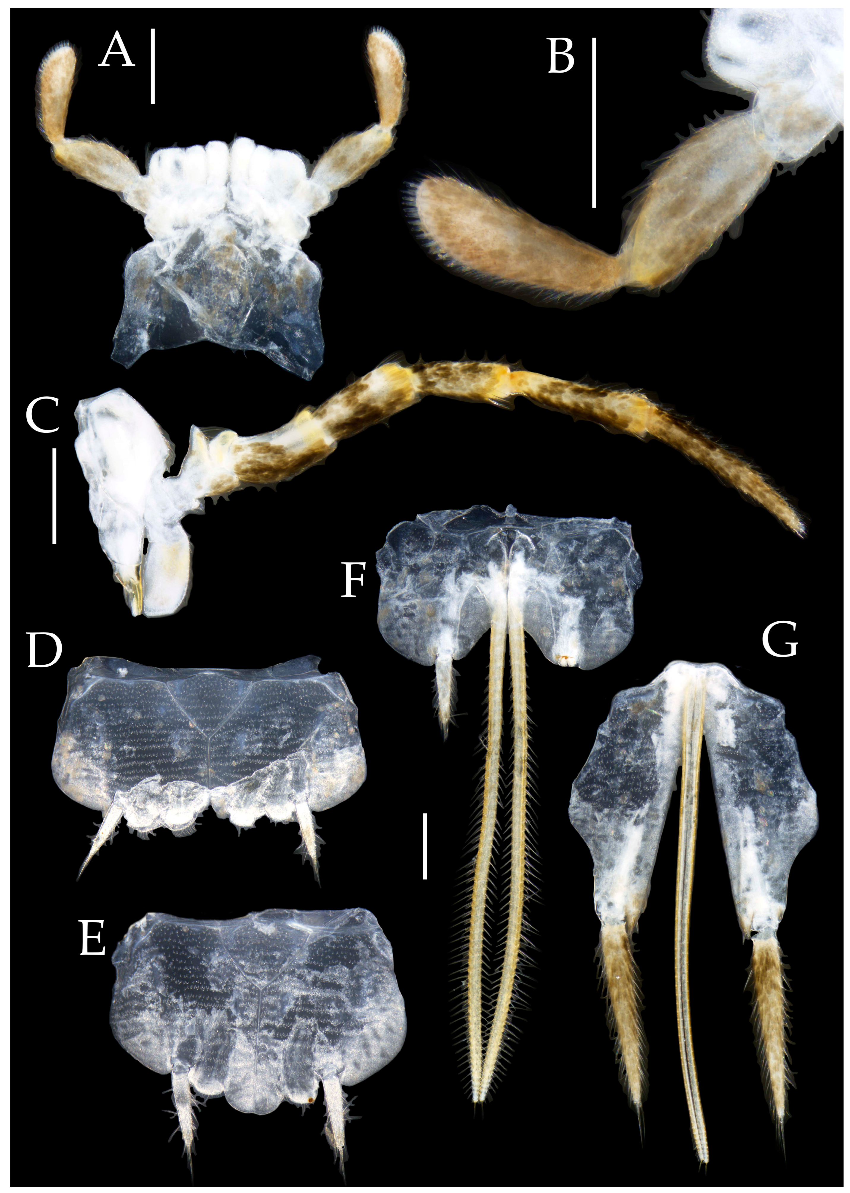
Figure 5.1.
Pedetontus (Verhoeffilis) shenzhenensis Shen, Yang, Ji & Zhang sp. n., holotype, female. (A) Habitus, lateral view. (B) Antenna. (C) Head, frontal view. (D) Ditto, lateral view. (E, F) Compound eyes & paired ocelli. (G) Excrement. Scale bars: 500 μm.
Figure 5.1.
Pedetontus (Verhoeffilis) shenzhenensis Shen, Yang, Ji & Zhang sp. n., holotype, female. (A) Habitus, lateral view. (B) Antenna. (C) Head, frontal view. (D) Ditto, lateral view. (E, F) Compound eyes & paired ocelli. (G) Excrement. Scale bars: 500 μm.
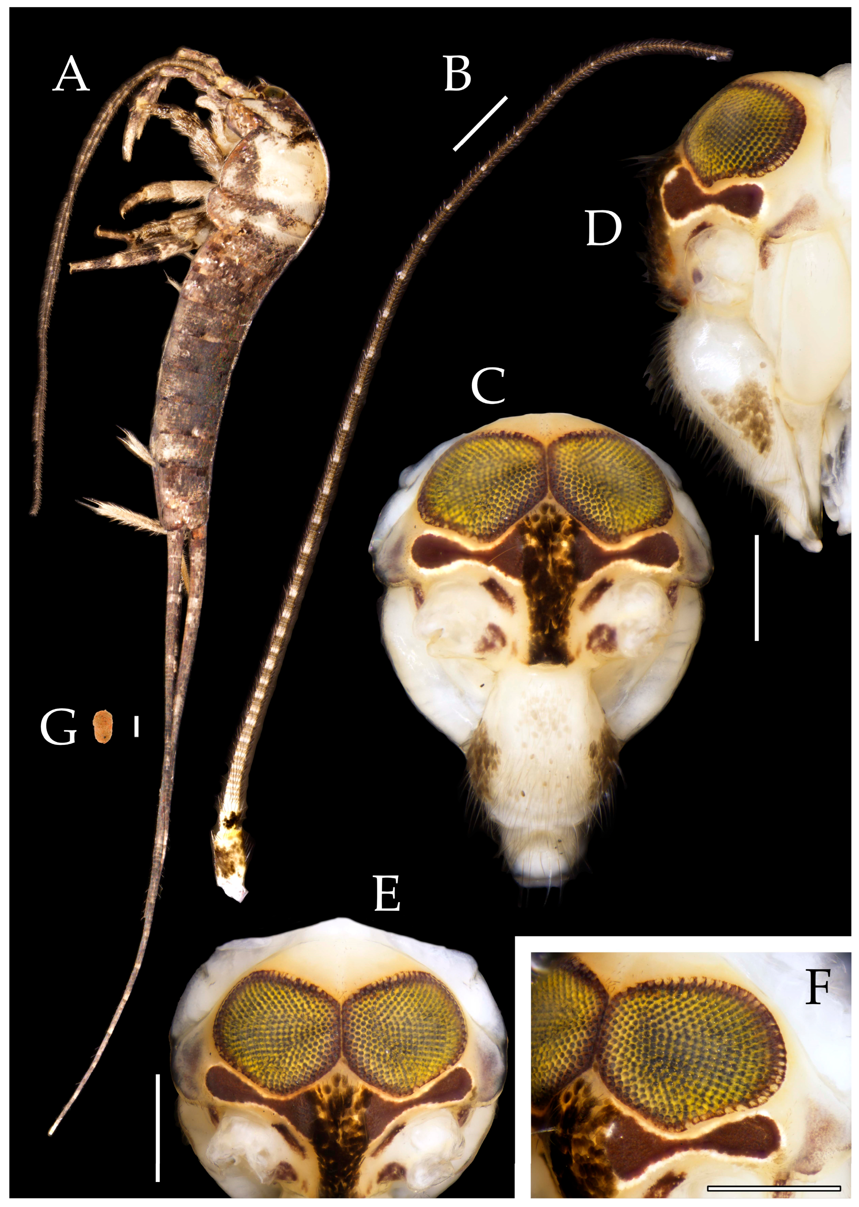
Figure 5.2.
Pedetontus (Verhoeffilis) shenzhenensis Shen, Yang, Ji & Zhang sp. n., holotype, female. (A) Mandible. (B) Labium. (C) Labial palp. (D) Maxilla. (E) Maxillary palp article VI & VII. (F) Lacinia & galea. (G) Maxillary inner protuberance. (G) Labium. (H–J) Foreleg, midleg & hindleg. (K–M) Foreclaw, midclaw & hindclaw. Scale bars: 500 μm.
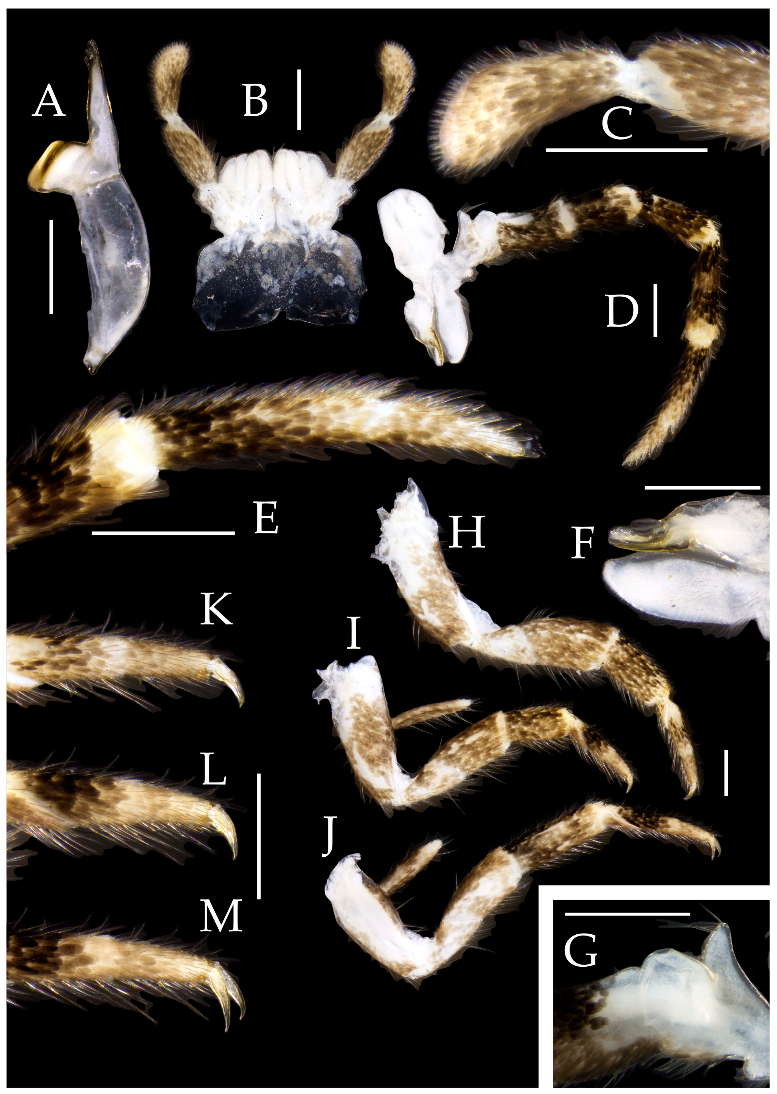
Figure 5.3.
Pedetontus (Verhoeffilis) shenzhenensis Shen, Yang, Ji & Zhang sp. n., holotype, female. (A–I) Abdominal sternites and coxites I–IX. (J) Caudal filament & cerci. (K) Apex of anterior gonapophyses. (L) Apex of posterior gonapophyses. Scale bars: 500 μm.
Figure 5.3.
Pedetontus (Verhoeffilis) shenzhenensis Shen, Yang, Ji & Zhang sp. n., holotype, female. (A–I) Abdominal sternites and coxites I–IX. (J) Caudal filament & cerci. (K) Apex of anterior gonapophyses. (L) Apex of posterior gonapophyses. Scale bars: 500 μm.
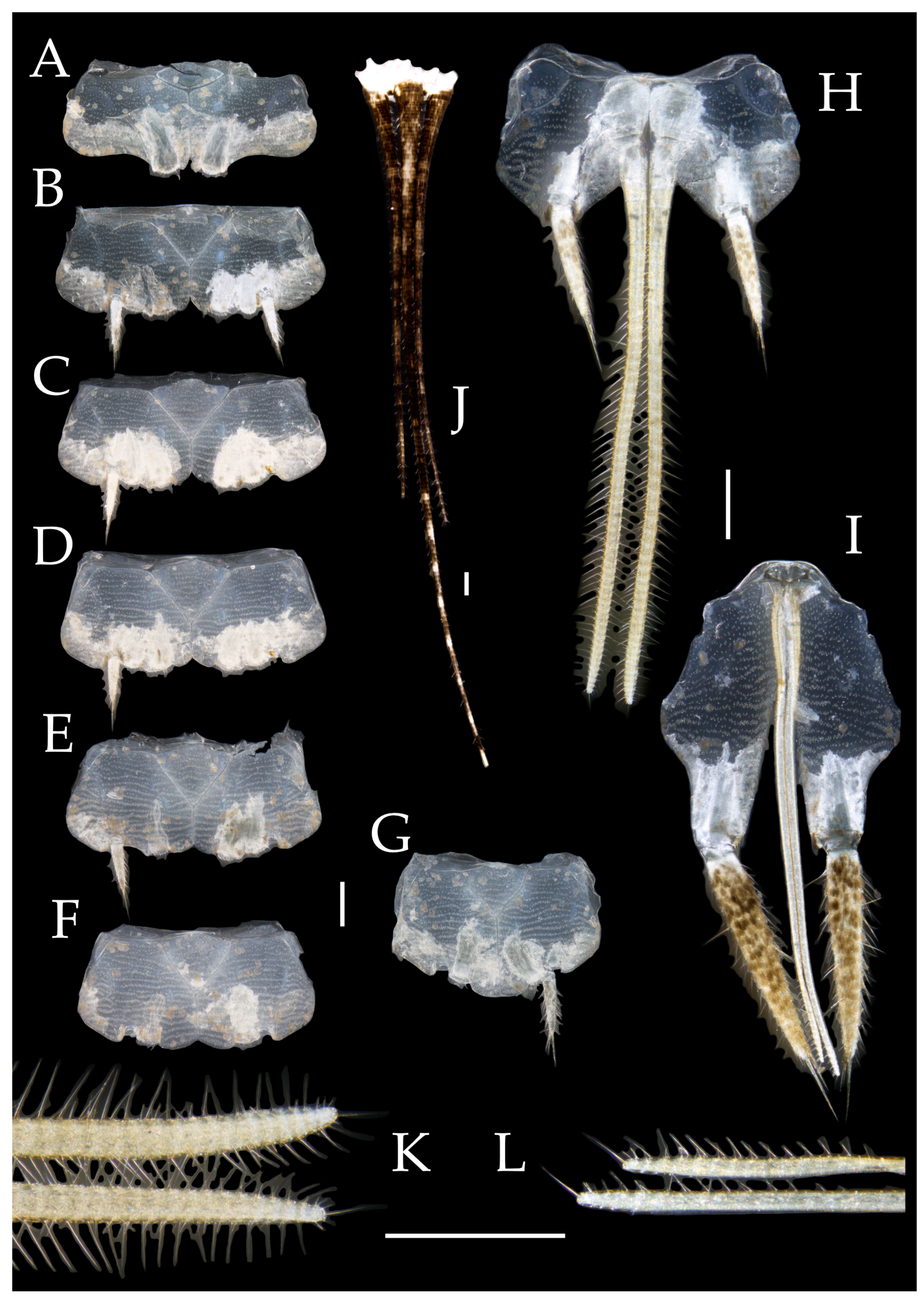
Figure 6.1.
Pedetontus (Verhoeffilis) xanthospilus Shen, Yang, Ji & Zhang sp. n. (A) The captured P. (V.) xanthospilus sp. n., arrow indicates a mite. (B) Ditto. (C) In situ. (D) Habitat.
Figure 6.1.
Pedetontus (Verhoeffilis) xanthospilus Shen, Yang, Ji & Zhang sp. n. (A) The captured P. (V.) xanthospilus sp. n., arrow indicates a mite. (B) Ditto. (C) In situ. (D) Habitat.
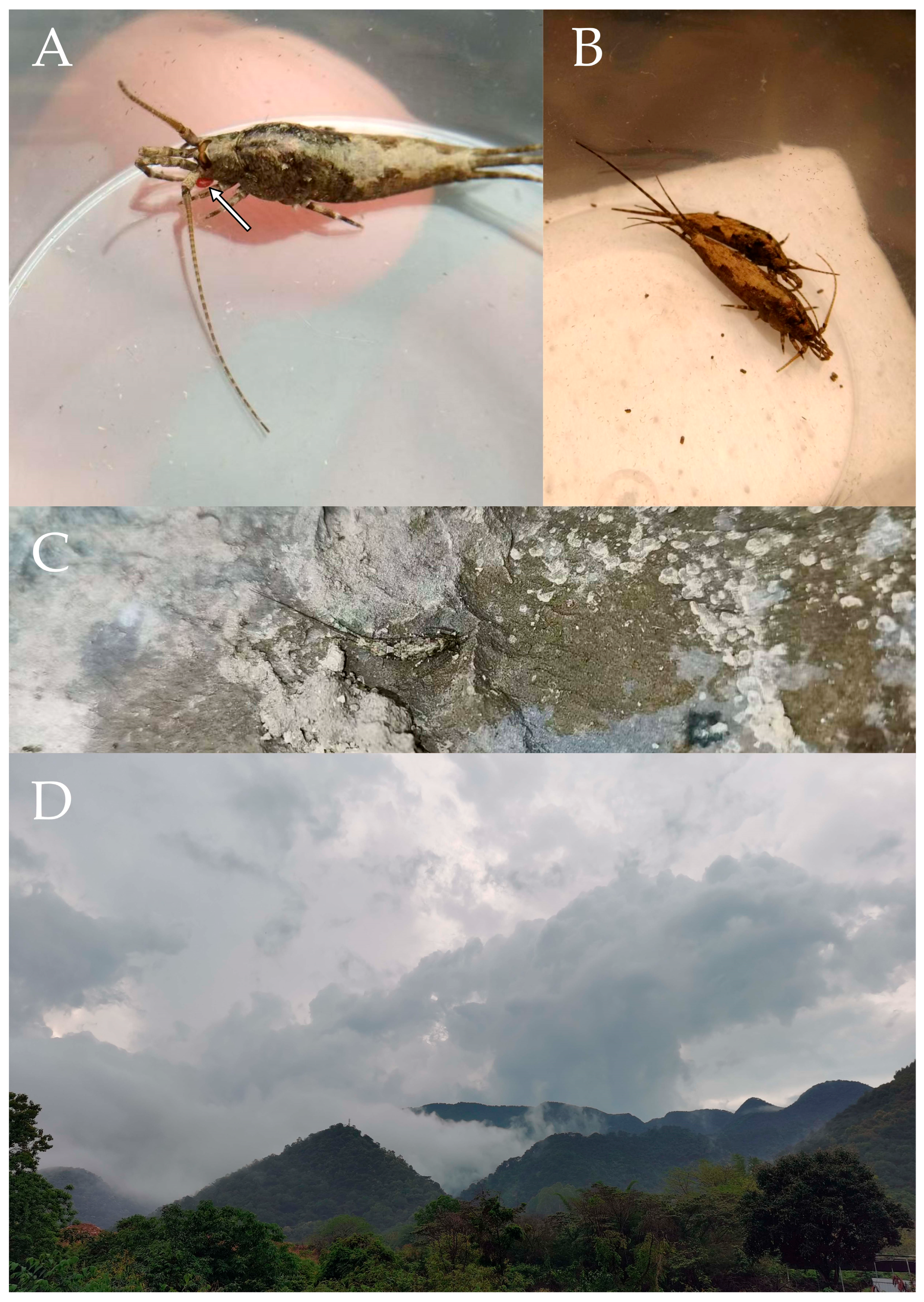
Figure 6.2.
Pedetontus (Verhoeffilis) xanthospilusShen, Yang, Ji & Zhang sp. n., holotype, male. (A) Habitus, lateral view, arrow indicates a mite attached to the maxilla. (B) Antenna. (C) Terminal chain of flagellum. (D) Head, frontal view. (E) Ditto, lateral view. (F, G) Compound eyes & paired ocelli. Scale bars: 500 μm.
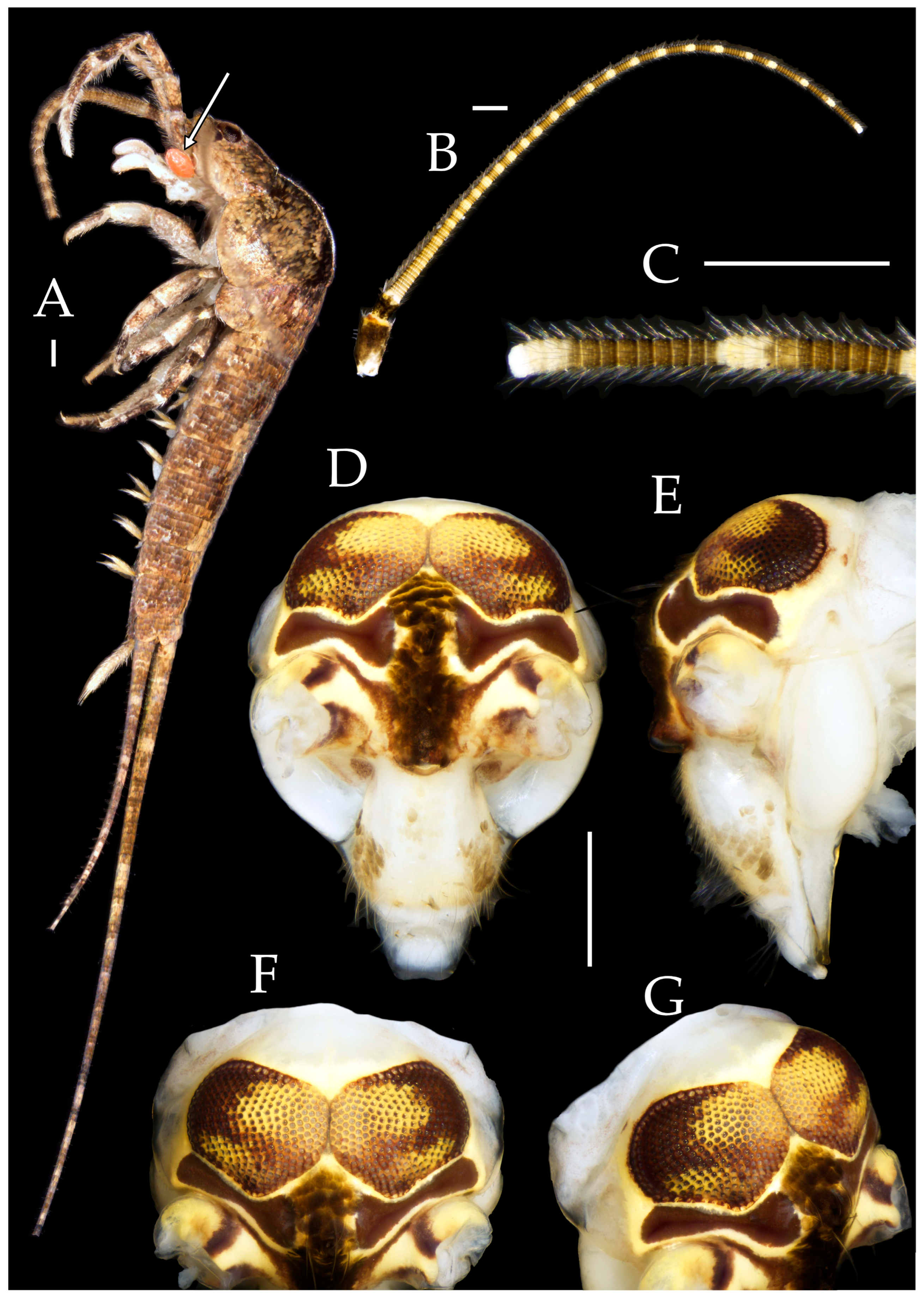
Figure 6.3.
Pedetontus (Verhoeffilis) xanthospilus Shen, Yang, Ji & Zhang sp. n., holotype, male. (A) Mandible. (B) Labium. (C) Labial palp. (D) Maxilla. (E) Maxillary palp article V–VII. (F) Maxillary inner protuberance. (G–I) Foreleg, midleg & hindleg. Scale bars: 500 μm.
Figure 6.3.
Pedetontus (Verhoeffilis) xanthospilus Shen, Yang, Ji & Zhang sp. n., holotype, male. (A) Mandible. (B) Labium. (C) Labial palp. (D) Maxilla. (E) Maxillary palp article V–VII. (F) Maxillary inner protuberance. (G–I) Foreleg, midleg & hindleg. Scale bars: 500 μm.
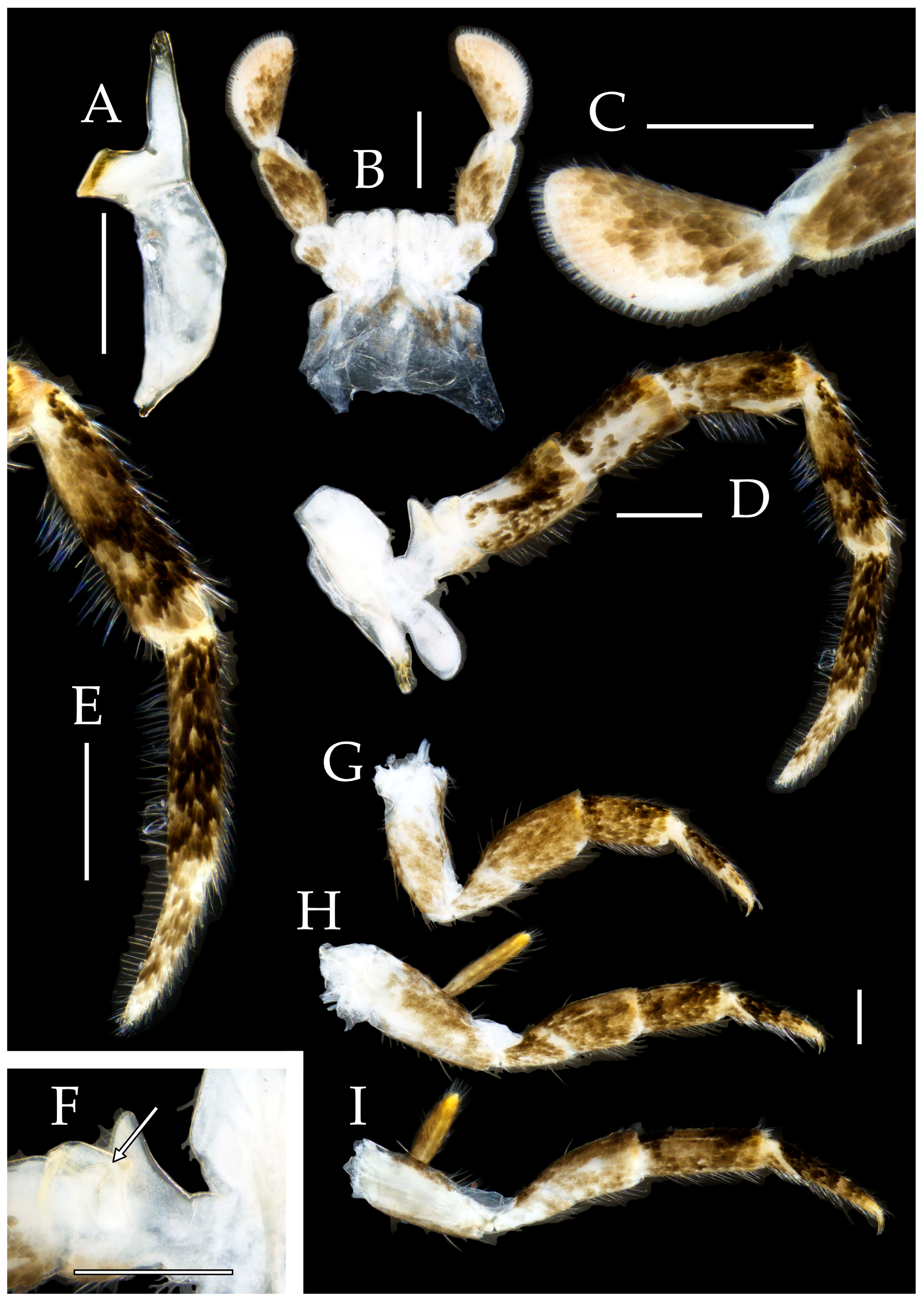
Figure 6.4.
Pedetontus (Verhoeffilis) xanthospilus Shen, Yang, Ji & Zhang sp. n., holotype, male. (A–I) Abdominal sternites and coxites I–IX. (J) Caudal filament & cerci. (K) Penis & paramere. Scale bars: 500 μm.
Figure 6.4.
Pedetontus (Verhoeffilis) xanthospilus Shen, Yang, Ji & Zhang sp. n., holotype, male. (A–I) Abdominal sternites and coxites I–IX. (J) Caudal filament & cerci. (K) Penis & paramere. Scale bars: 500 μm.
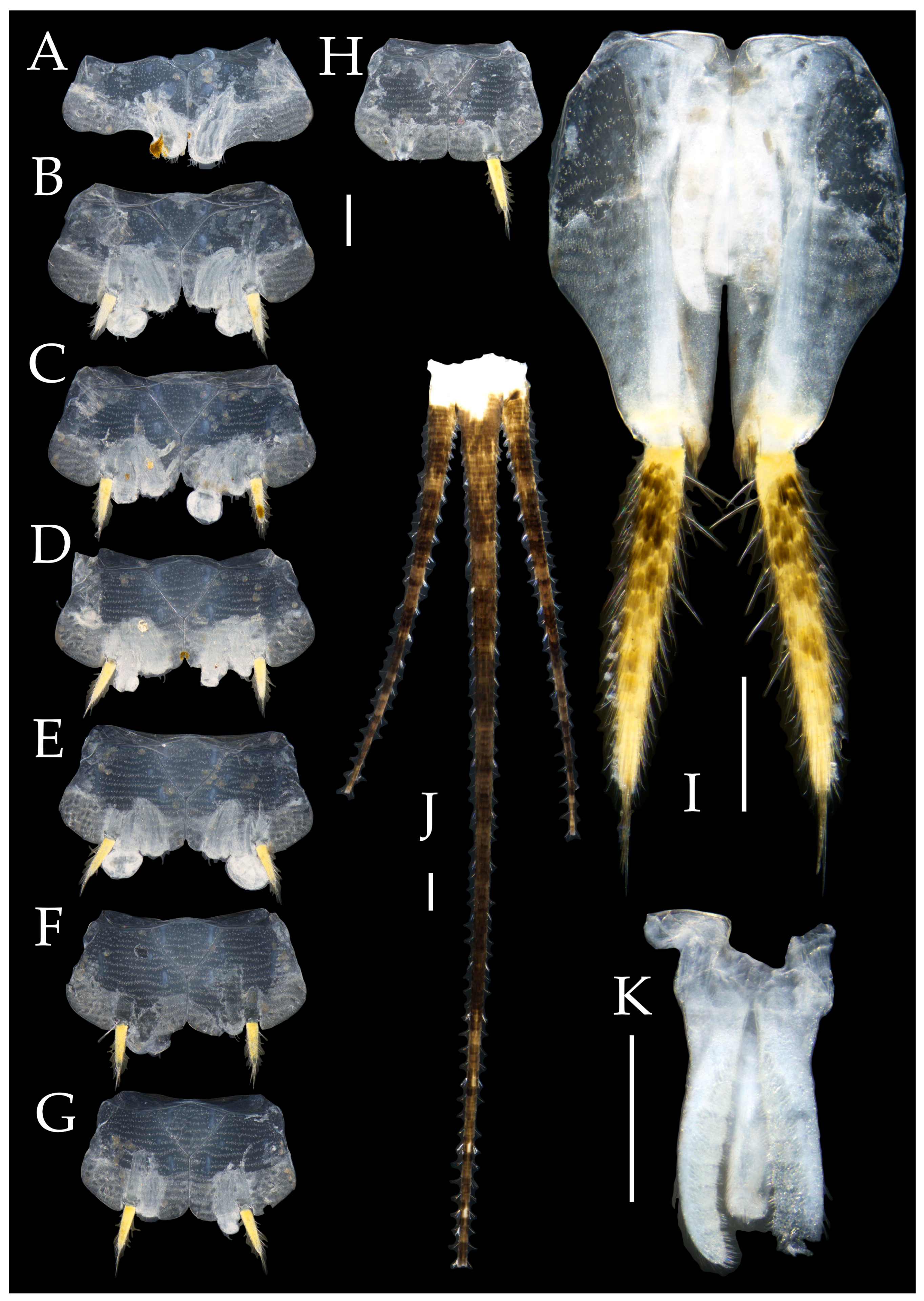
Figure 6.5.
Pedetontus (Verhoeffilis) xanthospilus Shen, Yang, Ji & Zhang sp. n., paratype, female. (A) Habitus, lateral view. (B) Head, frontal view. (C) Labium. (D) Labial palp. (E) Maxilla. Scale bars: 500 μm.
Figure 6.5.
Pedetontus (Verhoeffilis) xanthospilus Shen, Yang, Ji & Zhang sp. n., paratype, female. (A) Habitus, lateral view. (B) Head, frontal view. (C) Labium. (D) Labial palp. (E) Maxilla. Scale bars: 500 μm.
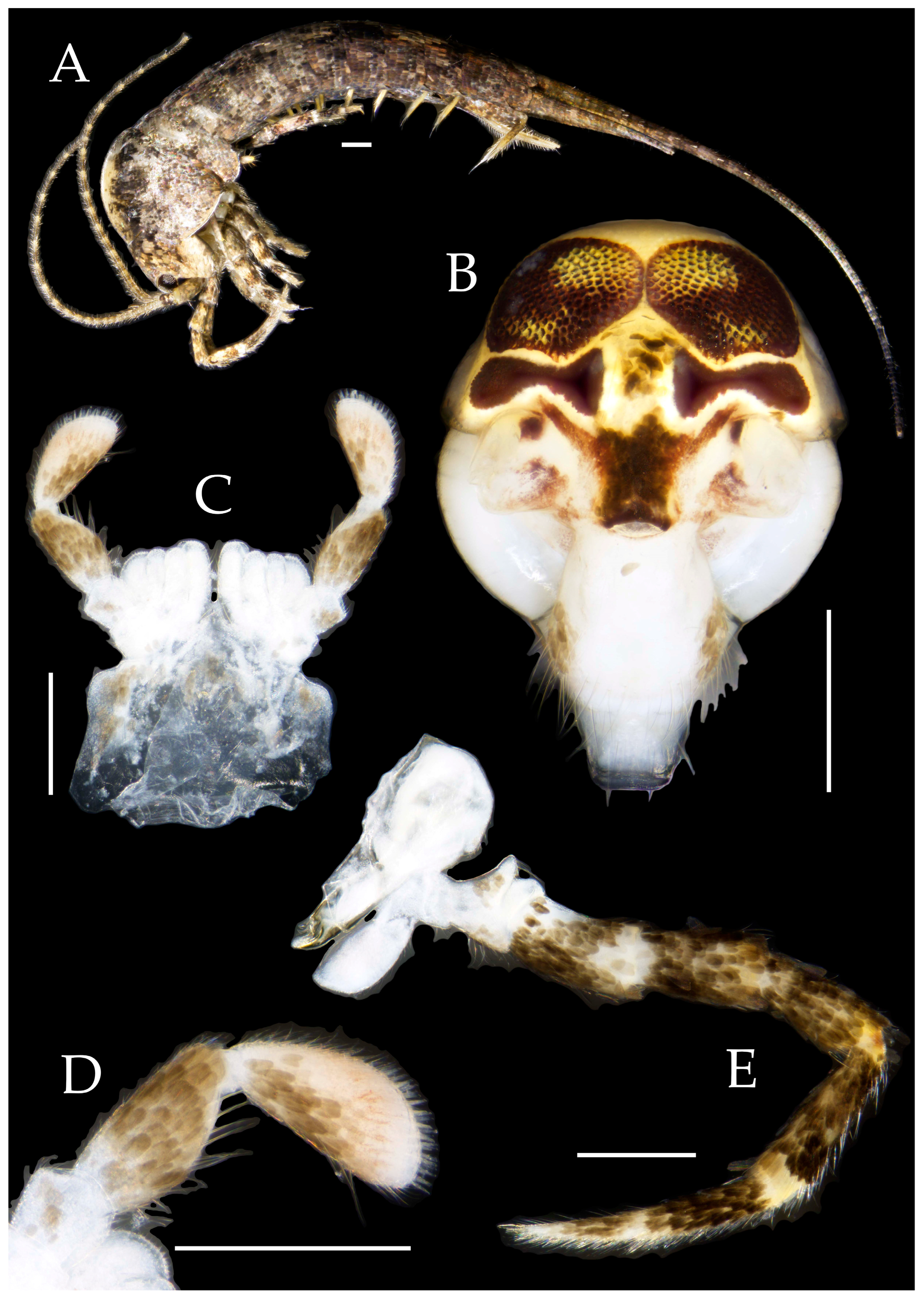
Figure 6.6.
Pedetontus (Verhoeffilis) xanthospilus Shen, Yang, Ji & Zhang sp. n., paratype, female. (A–C) Foreleg, midleg & hindleg. (D–G) Abdominal sternites and coxites V, VII–IX. (H) Apex of anterior gonapophyses. (I) Apex of posterior gonapophyses. Scale bars: 500 μm.
Figure 6.6.
Pedetontus (Verhoeffilis) xanthospilus Shen, Yang, Ji & Zhang sp. n., paratype, female. (A–C) Foreleg, midleg & hindleg. (D–G) Abdominal sternites and coxites V, VII–IX. (H) Apex of anterior gonapophyses. (I) Apex of posterior gonapophyses. Scale bars: 500 μm.
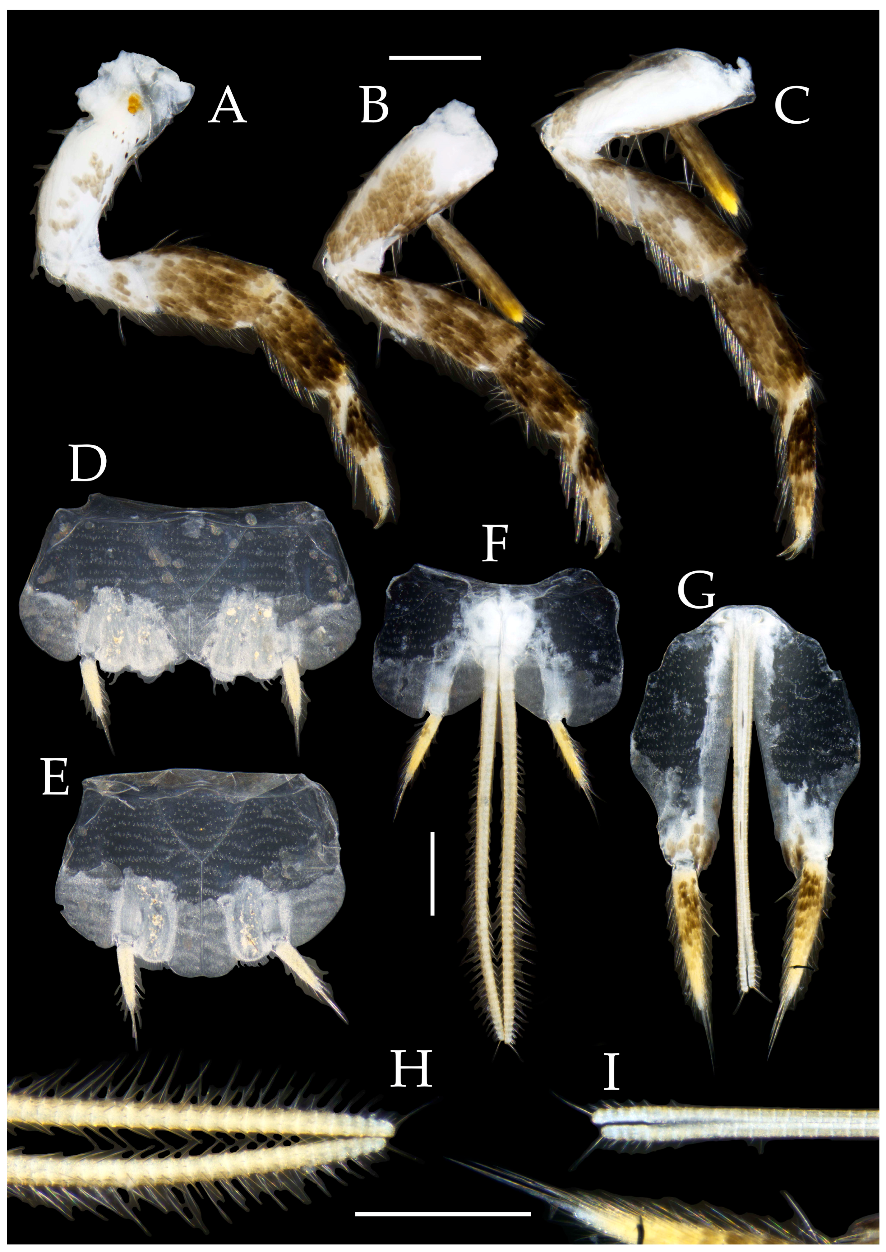

Table 1.
Ratio of compound eyes and ocelli of Pedetontus new species.
| Ratio | Eye length to width | Eye contact line length to eye length | Ocellus length to width | Distance between inner margins of ocelli to combined width of eyes |
|---|---|---|---|---|
| P.(V.) elegans sp. n. | 1.01–1.09 | 0.59–0.71 | 0.30–0.33 | 0.19–0.22 |
| P.(V.) hezhouensis sp. n. | 0.79–0.84 | 0.32–0.39 | 0.35–0.43 | 0.18 |
| P.(V.) jinxiuensis sp. n. | 0.81–0.85 | 0.20–0.25 | 0.33–0.36 | 0.15–0.16 |
| P.(V.)nanningensissp. n. | 0.82–0.84 | 0.31–0.37 | 0.38–0.40 | 0.10–0.15 |
| P.(V.) shenzhenensissp. n. | 0.84 | 0.41 | 0.40 | 0.19 |
| P.(V.) xanthospilus sp. n. | 0.83–0.84 | 0.27–0.28 | 0.36 | 0.21–0.22 |
Table 2.
Numbers of transparent spines on dorsal surfaces of maxillary palps & sensory cones on apex of labial palp III of Pedetontus new species.
Table 2.
Numbers of transparent spines on dorsal surfaces of maxillary palps & sensory cones on apex of labial palp III of Pedetontus new species.
| Species | Number of dorsal transparent spines on maxillary palps | Sensory cones on apex of labial palp III | |||
|---|---|---|---|---|---|
| V | VI | VII | |||
| P.(V.) elegans sp. n. | Male | 1–4 | 6–14 | 7–14 | 25–31 |
| Female | 2–3 | 10–16 | 10–14 | 23–26 | |
| P.(V.) hezhouensis sp. n. | Male | 2 | 7 | 6–7 | 50 |
| Female | 4–5 | 11–15 | 11–13 | 35–39 | |
| P.(V.) jinxiuensis sp. n. | Male | 0–1 | 11–12 | 8–9 | 0–43 |
| Female | 4 | 12 | 15 | 0 | |
| P.(V.)nanningensissp. n. | Male | 3 | 12 | 9–11 | 46–50 |
| Female | 6–7 | 16 | 15–16 | 36–40 | |
| P.(V.) shenzhenensissp. n. | Male | N/A | N/A | N/A | N/A |
| Female | 5–6 | 16–17 | 13 | 36–45 | |
| P.(V.) xanthospilus sp. n. | Male | 0–2 | 10 | 12–14 | 55–58 |
| Female | 2–3 | 12–14 | 11–12 | 36–38 | |
Table 3.
Number of needle-shaped setae on ventral surfaces of legs, Pedetontus (Verhoeffilis) elegans sp. n.
Table 3.
Number of needle-shaped setae on ventral surfaces of legs, Pedetontus (Verhoeffilis) elegans sp. n.
| Legs | Number of needle-shaped setae on ventral surfaces | ||
|---|---|---|---|
| Foreleg | Femur | 0 | |
| Tibia | 0 | ||
| Tarsi | Tarsomere I | 2 | |
| Tarsomere II | 5 | ||
| Tarsomere III | 0 | ||
| Midleg | Femur | 0 | |
| Tibia | 0 | ||
| Tarsi | Tarsomere I | 3 | |
| Tarsomere II | 6 | ||
| Tarsomere III | 1 | ||
| Hindleg | Femur | 0 | |
| Tibia | 4 | ||
| Tarsi | Tarsomere I | 3 | |
| Tarsomere II | 10 | ||
| Tarsomere III | |||
Table 4.
Angles of posterior angles (°) of abdominal sternites of Pedetontus new species.
| Species | Urosternite I | Urosternite II |
Urosternite III | Urosternite IV | Urosternite V |
Urosternite VI | Urosternite VII | Urosternite VIII (male) |
|---|---|---|---|---|---|---|---|---|
| P. elegans (V.)sp. n. | 129–138 | 78–92 | 88–93 | 92–94 | 86–95 | 74–92 | 99–113 | 123 |
| P. hezhouensis (V.)sp. n. | 129–131 | 78–87 | 78–83 | 82–83 | 81–84 | 91–96 | 89–100 | 91 |
| P. jinxiuensis (V.)sp. n. | 129–141 | 68–76 | 75–77 | 79–82 | 75–84 | 79–95 | 79–97 | 95 |
| P. nanningensis (V.) sp. n. | 123–129 | 67–88 | 68–82 | 78–86 | 83–88 | 79–88 | 80–101 | 84 |
| P. shenzhenensis (V.) sp. n. | 140 | 80 | 73 | 77 | 77 | 77 | 81 | N/A |
| P. xanthospilus (V.)sp. n. | 123–129 | 76–83 | 73–80 | 78–80 | 84–86 | 82 - 83 | 83 - 89 | 80 |
Table 5.
Ratios of styli length to coxites length on abdominal segment V, styli length to supporting spines length, and basal width to length of sternite.
Table 5.
Ratios of styli length to coxites length on abdominal segment V, styli length to supporting spines length, and basal width to length of sternite.
| Ratio | Styli (without supporting spines) length to coxites length of abdominal segment V | Styli (without supporting spines) length to supporting spines length of abdominal segment V | Basal width to length of sternite of abdominal segment V |
|---|---|---|---|
| P. (V.) elegans sp. n. | 0.58–0.64 | 0.40–0.43 | 1.33–1.34 |
| P. (V.)hezhouensis sp. n. | 0.42–0.48 | 0.53–0.58 | 1.08–1.24 |
| P. (V.)jinxiuensis sp. n. | 0.37–0.43 | 0.65–0.71 | 1.18–1.21 |
| P.(V.) nanningensis sp. n. | 0.43 | 0.52 | 1.06–1.36 |
| P.(V.)shenzhenensissp. n. | 0.47 | 0.60 | 1.20 |
| P. (V.)xanthospilus sp. n. | 0.38–0.41 | 0.55–0.68 | 1.19–1.25 |
Table 6.
Number of segments of anterior gonapophyses and posterior gonapophyses of Pedetontus new species.
Table 6.
Number of segments of anterior gonapophyses and posterior gonapophyses of Pedetontus new species.
| Species | Number of segments of anterior gonapophyses | Number of segments of posterior gonapophyses |
|---|---|---|
| P. (V.) elegans sp. n. | 56–59 | 58–63 |
| P. (V.)hezhouensis sp. n. | 60–67 | 53–55 |
| P. (V.)jinxiuensis sp. n. | 60 | 56–58 |
| P.(V.) nanningensis sp. n. | 59 | 50–55 |
| P.(V.)shenzhenensissp. n. | 42–54 | 48–49 |
| P. (V.)xanthospilus sp. n. | 50–51 | 49 |
Disclaimer/Publisher’s Note: The statements, opinions and data contained in all publications are solely those of the individual author(s) and contributor(s) and not of MDPI and/or the editor(s). MDPI and/or the editor(s) disclaim responsibility for any injury to people or property resulting from any ideas, methods, instructions or products referred to in the content. |
© 2025 by the authors. Licensee MDPI, Basel, Switzerland. This article is an open access article distributed under the terms and conditions of the Creative Commons Attribution (CC BY) license (http://creativecommons.org/licenses/by/4.0/).
Copyright: This open access article is published under a Creative Commons CC BY 4.0 license, which permit the free download, distribution, and reuse, provided that the author and preprint are cited in any reuse.


