Preprint
Case Report

This version is not peer-reviewed.

Life-Threatening Posterior Epistaxis Masquerading as Upper Gastrointestinal Bleeding Treated with Endoscopic Sphenopalatine Artery Ligation

Submitted:

26 August 2025

Posted:

27 August 2025

You are already at the latest version

Abstract

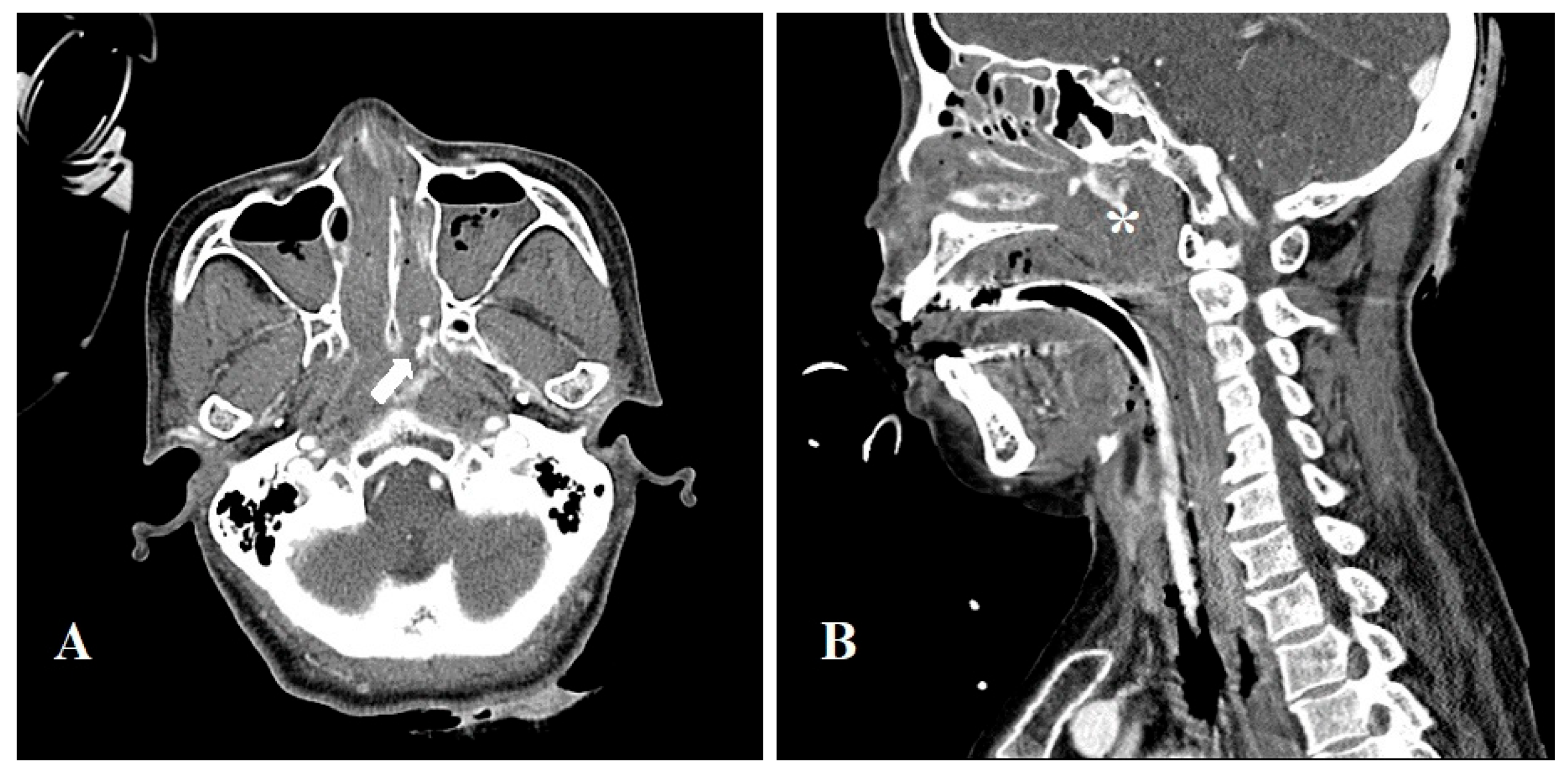
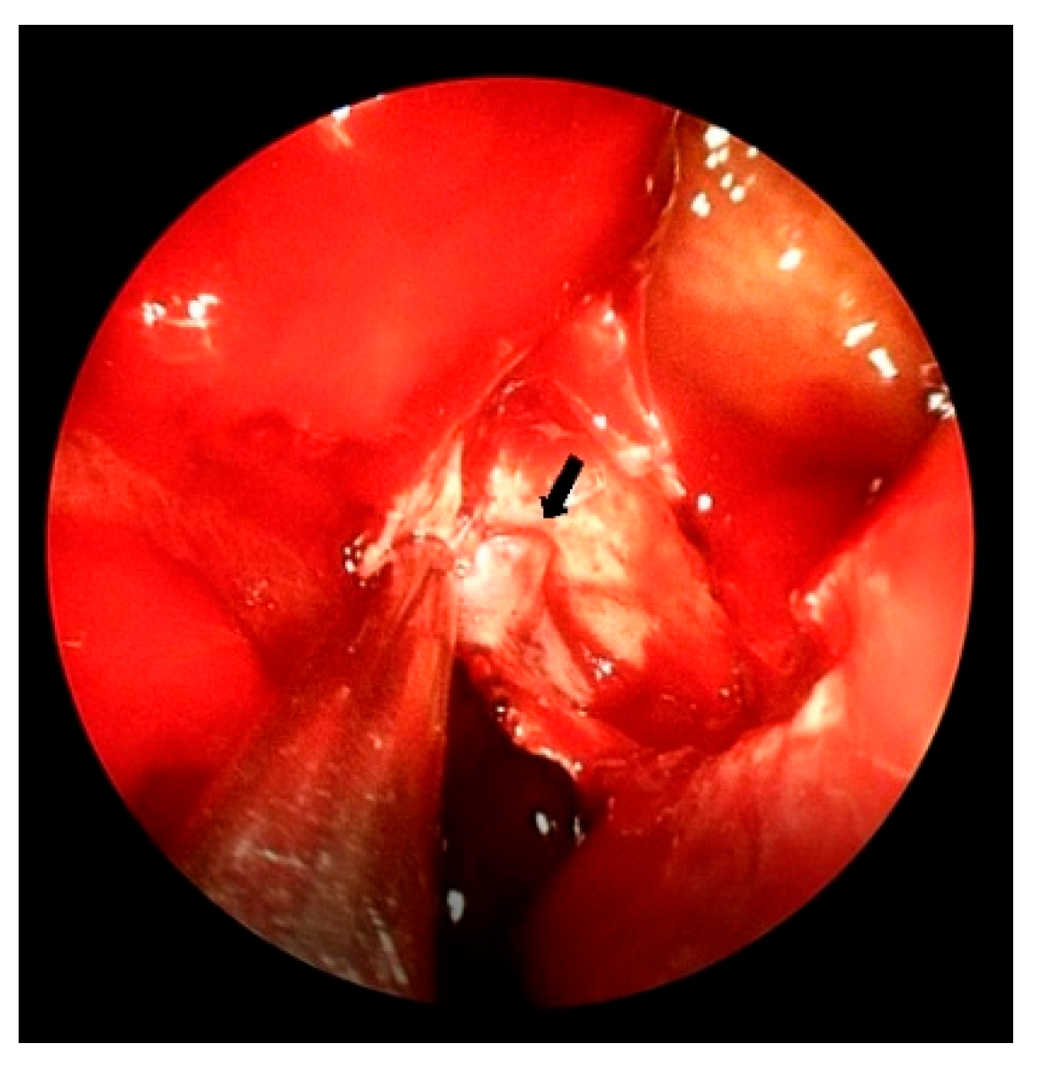
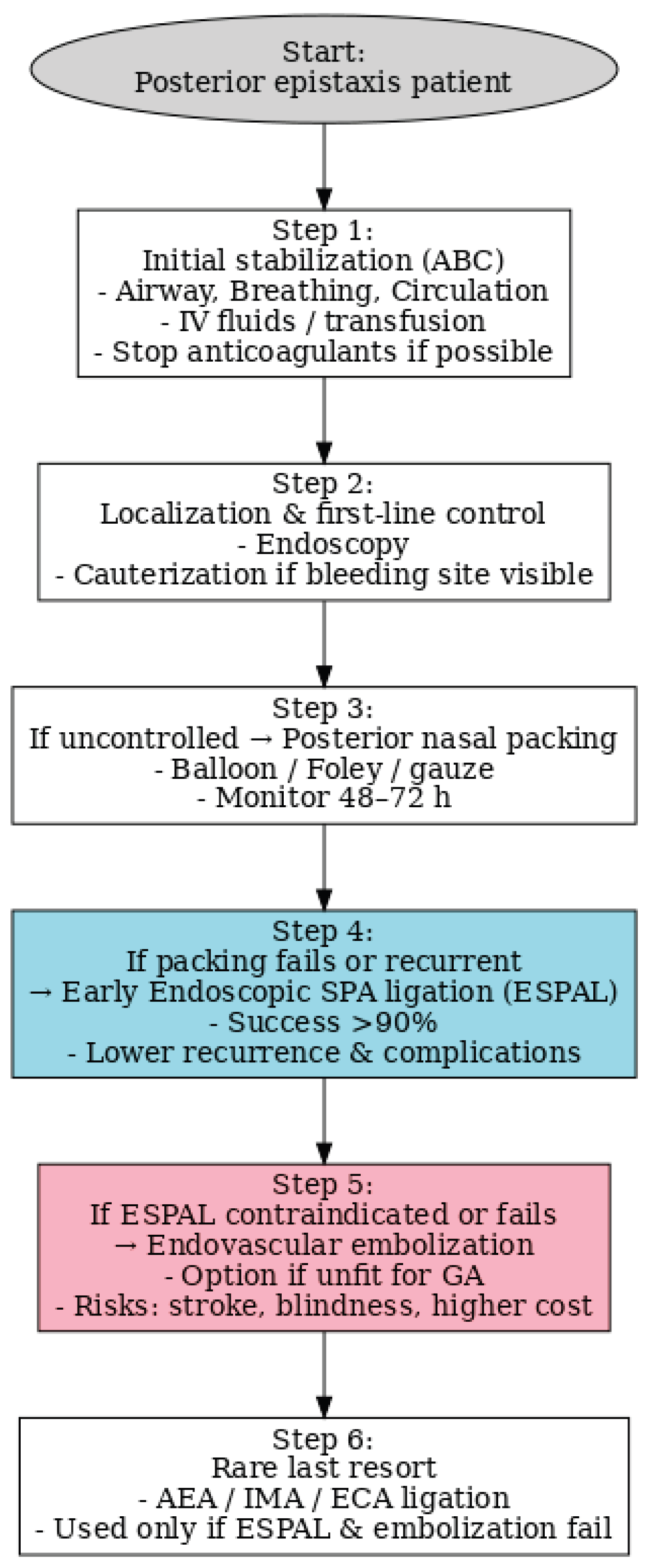
Background: Posterior epistaxis is a rare but potentially life-threatening condition that may be misdiagnosed as upper gastrointestinal bleeding due to similar clinical manifestations such as hematemesis. Case Presentation: We report a diagnostically challenging case of a 67-year-old woman presenting with hematemesis, initially suspected as gastrointestinal bleeding. Despite multiple interventions, the source of bleeding remained unclear until contrast-enhanced CT and otorhinolaryngology consultation revealed posterior nasal bleeding. The patient was successfully treated with endoscopic sphenopalatine artery ligation (ESPAL). Conclusion: This case highlights the importance of considering posterior epistaxis in the differential diagnosis of hematemesis and demonstrates the effectiveness of ESPAL in managing life-threatening epistaxis.

Keywords: 
;  ;  

1. Introduction

Posterior epistaxis is particularly relevant in elderly populations and those with systemic comorbidities such as hypertension or cardiovascular disease, conditions that are strongly associated with an increased risk of recurrent or severe nasal bleeding [1,2]. In contrast to anterior epistaxis, which is often managed conservatively in outpatient settings, posterior bleeds frequently present with hemodynamic instability and necessitate urgent hospital-based interventions [3,4]. The diagnostic complexity is further compounded by the possibility of misinterpreting swallowed blood as hematemesis or melena, leading to inappropriate initial consultations with gastroenterology services rather than otolaryngology specialists [5,6,7,8].
Several epidemiological analyses underscore the burden of epistaxis on emergency medical services. In the United States alone, tens of thousands of annual emergency department visits are attributable to epistaxis, with posterior bleeds representing the minority but disproportionately consuming resources due to the need for advanced interventions [9]. Long-term studies also reveal that posterior epistaxis, even when treated definitively, carries a substantial risk of recurrence, which can further complicate patient management and quality of life [1,4]. Therefore, it is essential to recognize posterior epistaxis not simply as a variant of anterior bleeding, but as a distinct clinical entity with unique diagnostic challenges and therapeutic considerations [10,11].
While epistaxis is one of the most common otorhinolaryngological emergencies, most cases can be treated with nasal packing or cauterization, without the need for hospitalization. Posterior epistaxis, however, can cause heavy bleeding along with refractory & intractable bleeding [3,12].
The sphenopalatine artery (SPA) and its branches are associated with most cases of posterior epistasis. Endoscopic evaluation may fail to diagnose and localize the site of bleeding due to the severity of the bleeding and difficulties in approaching the bleeding site [3]. When posterior epistaxis does not respond to nasal packing or cautery, it may lead to massive blood loss, resulting in life-threatening complications such as anemia, aspiration, and hypotension. Such cases require surgical arterial ligation or radiologic arterial embolization. Posterior epistaxis, although less common than anterior epistaxis, poses a significant diagnostic challenge due to its frequent presentation with atypical symptoms such as hematemesis or melena. Recent studies have demonstrated that up to 15–20% of severe posterior epistaxis cases are initially misdiagnosed as upper gastrointestinal bleeding, particularly in elderly patients with hemodynamic instability [5,6,12]. Early recognition of posterior nasal bleeding and prompt otorhinolaryngologic evaluation are therefore critical to avoid unnecessary gastrointestinal interventions and to prevent delays in definitive treatment, such as endoscopic sphenopalatine artery ligation (ESPAL) [5]. These findings highlight the importance of considering posterior epistaxis in the differential diagnosis of massive hematemesis of unclear origin. [4,6,7,10,13,14]
A patient may present with upper gastrointestinal bleeding instead of epistaxis after swallowing a large amount of blood from a posterior nasal bleeding source. This results in a delayed diagnosis, especially in older patients, which can lead to a fatal outcome. [15]
In this case study, a 67-year-old woman suffering from hematemesis, who was initially misdiagnosed with upper gastrointestinal bleeding(GI), is presented. She was eventually diagnosed with posterior epistaxis after medical and radiologic intervention failed. She received endoscopic SPA ligation (ESPAL) treatment on the 7th day of admission due to the delayed diagnosis [7,16].

2. Case Presentation

A 67-year-old woman visited the emergency room (ER) due to sudden hematemesis. She had 3-4 bleeding episodes before arriving at the ER, loosing approximately 500 cc of blood each time. As an upper GI bleed was suspected, a Sengstaken-Blakemore tube was inserted however, hematemesis persisted. Urgent embolization was necessary; however, there was a workforce shortage. The patient was intubated, and jugular catheter was inserted in order to facilitate intravenous assessment assurance before transfer to our institute.
Upon arrival, the patient’s hemoglobin (Hb) level had dropped from >11 g/dL to 8.3 g/dL. An urgent aortic CT was performed, which estimated the esophagus as the bleeding focus, thus, an embolization was planned. While preparing for embolization, her blood pressure dropped to 76/47 mmHg and hemodynamic instability was shown. The patient was transferred to the intensive care unit (ICU) for vital management where her arterial blood pressure (ABP) decreased to 60/49 mmHg prompting the administration of norpin, vasopressin, dopamine, and dobutamine in their maximum dosages. As a result, systolic BP (SBP) returned to 90 mmHg facilitating an emergency angiography of the left gastric artery and celiac artery. The extravasating focus was not clear, thus the procedure was terminated following empirical embolization of the esophageal branches of the left gastric artery after which SBP and Hb decreased to 65 mmHg and 6.5 g/dL, respectively. The patient continued to vomit large quantities of blood and nasal bleeding also began, thus, transfusion was continuously administered. A collection of fresh blood was observed in the oropharynx, prompting an urgent angiography; however, there was no definite evidence of extravasation. The otorhinolaryngology department were consulted and nasopharyngeal bleeding was confirmed, 13 hours after the patient suffered her first bleed and 8.5 hours after arriving at the ER. The patient was then sedated and her nasal cavity, mouth and pharynx were found to be filled with a large hematoma. Following the removal of the hematoma, an endoscopic examination was performed. A McIvor mouth gag was placed, and a sterile round adenoid sponge was inserted into the nasopharynx to prevent blood from running down to the pharynx. Saline irrigation was performed to confirm that there was no bleeding in the oral cavity, pharynx, and larynx. While suctioning the nasal cavity, a portable rigid endoscope examination was performed to confirm left intranasal bleeding. Despite this, the exact bleeding point was not identified, so nasal packing with an inflatable tamponade (Rapid Rhino™) and a merocel sponge was carried out to arrest bleeding. Hemodynamic instability increased and SBP was reported as 50 - 60 mmHg. Arterial blood gas analysis (ABGA) revealed a pH range of 7.1 to 7.3 points, prolonged prothrombin time (PT) and activated partial thromboplastin time (aPTT). The patient’s platelet levels dropped to <8 k/µL, electrolytes were imbalanced, and an increased level of aspartate aminotransferase (AST) and alanine aminotransferase (ALT) were recorded (> 1800 IU/L and 2100 IU/L respectively). These readings indicated multiorgan failure, secondary to blood loss and transfusion. This increased the risk of postoperative mortality; thus, conservative treatment was prioritized. A contrast-enhanced neck CT scan showed enhancement from the left posterior nasal cavity to the left lateral nasopharynx with an inferior extension, suggesting the possibility of active bleeding (Figure 1). Following the stabilization of the patient’s BP and serum creatinine level, endoscopic sinus surgery was performed on the 6th day of hospitalization.
All operative procedures were performed under general anesthesia. In this patient, septoplasty was performed in order to help identify the bleeding focus. The nasal septum was deviated and an uncinectomy and middle meatal antrostomy were conducted in order to remove the hematoma on the left maxillary sinus. Anterior ethmoidectomy was performed and a large hematoma was observed. Bleeding along the left Fossa of Rosenmüller in the nasopharynx to the pharynx was also noted. Following the incision over the posterior fontanelle, the mucoperiosteal flap was elevated and dissected with a freer elevator just posterior to the vertical ridge of the middle meatal antrostomy. Crista ethmoidalis was noted anterior to the upper section of the flap, and the SPA main trunk exiting at the sphenopalatine foramen (SPF) was identified posterior to the crista ethmoidalis (Figure 2); electrocautery was performed. The posterior nasal artery (PNA) and septal artery branching from the main trunk were also identified and cauterized together. After arresting the bleeding, a nasal pack with NasoPore® and a carbosymethyl cellulose sheet (Rhinocel®) were inserted. The nasal packing was removed on the first postoperative day and no active bleeding was observed. Postoperatively, the patient suffered from; aspiration pneumonia, pleural effusion, lower extremity edema, ear fullness, and nasal crust for several days. There was no postoperative bleeding noted and her hemodynamic state was stable. After medical treatment, the patient was discharged. One month postoperatively, there was no recurrence of epistaxis. As illustrated in Figure 3, the patient’s clinical course progressed from initial misdiagnosis as upper gastrointestinal bleeding to definitive diagnosis of posterior epistaxis, followed by successful ESPAL.

3. Discussion

A critical comparison of treatment modalities highlights the evolution of practice in managing intractable posterior epistaxis. Traditional nasal packing, once the cornerstone of therapy, is now recognized as suboptimal due to poor patient tolerance, increased risk of infection, and prolonged hospitalization [17]. In contrast, surgical approaches such as endoscopic sphenopalatine artery ligation (ESPAL) have demonstrated high success rates and are increasingly advocated as first-line definitive treatment [16,18,19]. This paradigm shift reflects not only improved surgical techniques but also the broader availability of endoscopic expertise within otolaryngology departments worldwide [14,20].
Embolization remains an important option in selected cases; however, multiple studies have cautioned against its liberal use due to the high cost, dependence on specialized radiologic expertise, and the potential for devastating complications such as cerebrovascular accidents or blindness [15,21,22]. Cost-effectiveness analyses clearly favor ESPAL over embolization for most patients, further supporting its adoption as the standard of care [21]. Nevertheless, embolization retains a role in centers with interventional radiology readily available, particularly in patients unfit for general anesthesia or in those with atypical vascular anatomy [23,24]. As shown in Figure 4, the management of posterior epistaxis should follow a structured stepwise approach. Initial stabilization with airway protection, circulatory support, and correction of coagulopathy is mandatory. Endoscopic examination is crucial for localization of the bleeding point and allows for immediate cauterization if feasible. When bleeding persists, posterior nasal packing can be attempted as a temporizing measure, but prolonged packing is associated with discomfort, infection risk, and high recurrence rates. For patients with persistent or recurrent hemorrhage after packing, ESPAL is recommended as the first-line definitive therapy, as it provides high success rates with low morbidity. Endovascular embolization is generally reserved for patients who are poor candidates for general anesthesia or for those in whom ESPAL fails, given its higher cost and risk of serious complications such as stroke or blindness. Only rarely, in refractory cases, should ligation of other branches such as the anterior ethmoidal or external carotid artery be considered. This algorithm underscores the need for early surgical intervention, particularly ESPAL, to improve patient outcomes and reduce recurrence.
Systematic reviews and meta-analyses further confirm the effectiveness of ESPAL, reporting success rates exceeding 90% with minimal morbidity [20,25,26]. Importantly, these outcomes are consistent across diverse populations and clinical settings, underscoring the robustness of this approach. Hypertension has been repeatedly identified as a significant risk factor for both the occurrence and recurrence of epistaxis, suggesting that optimal medical control of blood pressure may complement surgical strategies in reducing long-term morbidity [1,2]. Moreover, elderly patients with comorbidities such as diabetes or cardiovascular disease present unique perioperative challenges, emphasizing the need for individualized, multidisciplinary management [27].
Our case contributes to this body of knowledge by illustrating the potentially life-threatening consequences of delayed recognition and misdiagnosis of posterior epistaxis. It reinforces the imperative for early otolaryngology consultation in cases of unexplained hematemesis, the consideration of posterior nasal sources in differential diagnosis, and the role of ESPAL as a safe, effective, and cost-efficient intervention [3,5,6,7,8,11,12,20,25]. Taken together, the growing evidence base strongly supports ESPAL as the preferred intervention in appropriately selected patients, while highlighting the ongoing need for heightened clinical vigilance and tailored treatment strategies. A case of a patient with epistaxis mimicking massive upper gastrointestinal bleeding treated with endoscopic sphenopalatine artery ligation was reported. The patient did not experience any reoccurrence of epistaxis at one month postoperative [5,7]. The management of nasal bleeding begins with locating the bleeding site [18,28].
Posterior epistaxis is often clinically significant due to its propensity for massive bleeding and diagnostic challenges, particularly when it presents with atypical symptoms such as hematemesis or melena. While anterior epistaxis accounts for the majority of cases, posterior bleeding tends to be more severe and frequently requires advanced interventions, including balloon tamponade, arterial ligation, or angiographic embolization [18]. In a comprehensive review, Kucik and Clenney demonstrated that simple compression, topical vasoconstrictors, and chemical cautery remain the first-line approaches for anterior epistaxis, whereas refractory or posterior cases often necessitate hospital admission and multidisciplinary management involving otorhinolaryngologists [29]. These findings support our observation that patients with recurrent or severe epistaxis frequently require escalation to more invasive procedures, emphasizing the need for individualized treatment protocols based on bleeding patterns and patient comorbidities [1,6,7,29]
During a physical examination, the patient is kept in a seated position. The examination begins by checking for bleeding in the Kiesselbach’s plexus. If the patient complains of swallowing a large amount of blood, cotton may be inserted to the posterior nasal cavity to prevent blood flow into the pharynx. Nasal cavity exploration using a 0-degree and/or 70-degree rigid endoscope is carried out from the top to bottom, and from the anterior to the posterior nasal cavity. If bleeding point identification fails, anterior and/or posterior nasal packing is performed in order to arrest active bleeding. This patient was intubated in a supine position due to unstable BP and the upper airway was found to be filled with a hematoma during evaluation of the nasopharyngeal bleeding. The patient was sedated with no expectation of cooperation during physical examination. Her nasal septum was deviated to the left side and bleeding was suspected to originate from there. The otolaryngologist played an important role in the identification of the bleeding focus; and an enhanced neck CT contributed to the diagnosis. Nasal packing was applied for several days prior to the operation because of vital instability and continuous bleeding. Nasal packing causes serious discomfort to the patient and serious complications in the respiratory and cardiovascular system, especially in the elderly [27]. Therefore, if bleeding is unresponsive to nasal packing, surgical or radiologic intervention should be considered when the reevaluation of bleeding focus also fails. [11,27]
Recent literature highlights that posterior epistaxis can frequently present with atypical symptoms such as hematemesis and melena, leading to diagnostic delays when upper gastrointestinal bleeding is initially suspected [5,12]. A comprehensive review reported that up to 15–20% of severe posterior epistaxis cases are initially misdiagnosed as upper gastrointestinal bleeding, particularly when nasopharyngeal bleeding is not clinically evident [30]. According to this review, early otorhinolaryngology consultation and contrast-enhanced CT imaging are essential for differentiating nasal from gastrointestinal sources of bleeding. In pooled analyses, the success rate of endoscopic sphenopalatine artery ligation (ESPAL) exceeded 90%, demonstrating superior outcomes compared to posterior nasal packing alone and comparable efficacy to transarterial embolization but with fewer complications [5,12]. These findings support our management approach in this case, where prompt CT evaluation and early ESPAL led to rapid hemodynamic stabilization and favorable recovery. Similar observations have been reported by Yano et al., in which repeated hematemesis and melena were eventually attributed to posterior epistaxis [16], and by Wolf et al., who described delayed diagnosis in both elderly and pediatric patients with atypical presentations [5]. Incorporating these strategies into clinical practice may help reduce unnecessary gastrointestinal interventions and improve patient outcomes in patients presenting with massive hematemesis of unclear origin. [7,8]
Additionally, multiple recent studies emphasize the importance of differentiating posterior epistaxis from upper gastrointestinal bleeding in patients presenting with massive hematemesis or melena. Incorporating early otorhinolaryngology consultation and contrast-enhanced imaging has been shown to significantly improve diagnostic accuracy, avoiding unnecessary GI procedures and facilitating timely surgical intervention such as ESPAL. These findings align with current evidence-based recommendations for managing severe posterior epistaxis. [17,21]
Treatment options for posterior epistaxis include embolization and ESPAL [3,4]. Embolization is not a first-line treatment due to serious side effects such as; cerebrovascular accident, facial paralysis, internal carotid artery (ICA) dissection, blindness, trismus, facial pain, high cost, and the need of an expert with radiologic intervention skills [15,16]. ESPAL is, however, the preferred first line intervention for recalcitrant posterior epistaxis. ESPAL, described by Budrovich et al in 1992 [23], has a success rate of 85–98%, and it shortens the hospitalization time when performed early [16,28]. Failure occurs when surgical clips slip, branches of the SPA are not all identified, and bleeding from ICA branches such as the anterior ethmoidal artery (AEA) and/or posterior ethmoidal artery (PEA) occurs [16,29]. A Septoplasty and cautery on the PNA, septal artery and SPA were reported to improve the success rate. [2,9,24,25,26]
In this case, a detailed history of the patient was unavailable. The patient experienced intermittent nasal bleeding one month before hospitalization; however, this information was obtained late. The elderly and male are at a higher risk of recurrent epistasis and elderly patients are vulnerable to complications such as aspiration, angina, hypovolemia, and myocardial infarction [19]. Septal deviation, diabetes mellitus, congestive heart failure, and OSA [1,27] are also risk factors. The patient examined in this case study had associated risk factors including age, diabetes mellitus, and septal deviation as observed in the physical exam; she received inadequate treatment for recurrent nasal bleeding. Upper airway bleeding should have been considered when the cause of hematemesis was unclear. [20,22,23]
Future directions in the management of posterior epistaxis should aim not only at effective hemostasis but also at preventing recurrence and improving long-term outcomes. One promising strategy is the development of standardized clinical pathways that integrate early recognition, risk stratification, and timely referral for endoscopic evaluation. Incorporating routine use of nasal endoscopy in patients with unexplained hematemesis or recurrent anemia could significantly reduce diagnostic delays and misclassification [3,23,25]. Another area of potential progress lies in the refinement of perioperative care for high-risk populations, particularly elderly patients with multiple comorbidities. Comprehensive management involving cardiology, anesthesiology, and internal medicine teams may mitigate perioperative risks and optimize surgical outcomes [1,2,27].
In addition, further multicenter prospective studies are warranted to clarify the comparative efficacy and safety of ESPAL versus embolization, especially in resource-limited settings where access to interventional radiology is restricted [15,21,22]. Cost-effectiveness analyses should also be extended to account for quality-of-life outcomes and indirect costs, thereby providing a more comprehensive assessment of treatment value [21]. Finally, there remains a need for consensus-driven guidelines that clearly delineate diagnostic algorithms and therapeutic hierarchies for posterior epistaxis, bridging the gap between heterogeneous practices currently employed worldwide [11,12,20]. Such guidelines could enhance uniformity of care, facilitate training of younger otolaryngologists, and ultimately improve patient safety and outcomes on a global scale.

4. Conclusions

For extreme hematemesis, posterior epistaxis should be considered as the patient swallows and vomits blood, mimicking upper GI bleeding. If bleeding point identification fails following otorhinolaryngological examination, CT with contrast can be helpful. If hemostasis is not achieved, effective packing and ESPAL is the recommended choice for treatment. Early identification of posterior epistaxis is essential, particularly in patients presenting with massive hematemesis or melena of unclear origin. Prompt recognition of a nasal source can help avoid unnecessary gastrointestinal evaluations and allow earlier intervention. In clinical practice, rapid collaboration with otorhinolaryngologists and the use of contrast-enhanced imaging significantly improve diagnostic accuracy. Among the available treatment options, early application of endoscopic sphenopalatine artery ligation (ESPAL) has been shown to achieve stable hemostasis, reduce hospital stay, and minimize procedure-related complications compared to prolonged packing or embolization. These findings highlight the importance of integrating early diagnostic strategies and minimally invasive surgical techniques into the standard management pathway for severe posterior epistaxis.
In summary, implementing a structured diagnostic strategy that includes prompt ENT involvement and early imaging can markedly improve patient outcomes in cases of unexplained massive hematemesis. Minimally invasive techniques like ESPAL remain the cornerstone of effective management and should be considered early in the treatment pathway to prevent complications and shorten hospital stay.

Funding

The author(s) received no financial support for the research, authorship, and/or publication of this article.

Institutional Review Board Statement

The study was conducted in accordance with the Declaration of Helsinki and approved by the Institutional Review Board of Ewha Womans University Mokdong Hospital, 13 August 2021 (IRB No. 202108032).

Informed Consent Statement

Written informed consent was obtained from the patient for publication of this case report and any accompanying images.

Data Availability Statement

No new data were created or analyzed in this study. Data sharing is not applicable to this article.

Conflicts of Interest

The author(s) declare no potential conflicts of interest with respect to the research, authorship, and/or publication of this article.

References

  1. Rudmik L, Smith TL. Management of intractable spontaneous epistaxis. Am J Rhinol Allergy. 2012;26(1):55-60. [CrossRef]
  2. Janapala RN, et al. Efficacy of topical tranexamic acid in epistaxis: a systematic review and meta-analysis. Am J Emerg Med. 2022;51:169-175. [CrossRef]
  3. Gifford, T.O.; Orlandi, R.R. Epistaxis. Otolaryngol Clin North America 2008, 41, 525–536. [Google Scholar] [CrossRef] [PubMed]
  4. Pallin DJ, et al. Epidemiology of epistaxis in US emergency departments, 1992 to 2001. Ann Emerg Med. 2005;46(1):77-81. [CrossRef]
  5. Wolf S, et al. Epistaxis mimicking upper gastrointestinal bleeding. J Fam Pract. 1990. 1990.
  6. Rudmik L, Leung R. Cost-effectiveness analysis of endoscopic sphenopalatine artery ligation vs arterial embolization for intractable epistaxis. JAMA Otolaryngol Head Neck Surg. 2014;140(9):802-808. [CrossRef]
  7. Nouraei SAR, et al. Outcome of endoscopic sphenopalatine artery occlusion for intractable epistaxis. Laryngoscope. 2007;117(8):1452-1456. [CrossRef]
  8. Kindler, R.M.; Holzmann, D.; Landis, B.S.; Ditzen, B.; Soyka, M.B. The high rate of long-term recurrences and sequelae after epistaxis treatment. Auris Nasus Larynx 2016, 43, 412–417. [Google Scholar] [CrossRef] [PubMed]
  9. McDermott, A.M.; O’Cathain, E.; Carey, B.W.; O’Sullivan, P.; Sheahan, P. Sphenopalatine artery ligation for epistaxis: factors influencing outcome and impact of timing of surgery. Otolaryngol Head Neck Surg 2016, 154, 547–552. [Google Scholar] [CrossRef] [PubMed]
  10. Barnes ML, Spielmann PM, White PS. Epistaxis: a contemporary evidence based approach. Otolaryngol Clin North Am. 2012;45(5):1005-1017. [CrossRef]
  11. Badran K, et al. Randomized controlled trial comparing Merocel® and RapidRhino® packing in the management of anterior epistaxis. Clin Otolaryngol. 2005;30(4):333-337. [CrossRef]
  12. Brescia G, et al. A novel nasal endoscopic approach for removing displaced dental implants from the maxillary sinus. Am J Otolaryngol. 2017;38(1):92-95. [CrossRef]
  13. Koskinas, I.; Timoleon, T.; Christos, G.; Georgios, C. Posterior epistaxis management: review of the literature and proposed guidelines of the hellenic rhinological-facial plastic surgery society. European Archives of Oto-Rhino-Laryngology, 2024, 281.4: 1613-1627.
  14. Villwock JA, Jones K. Recent trends in epistaxis management in the United States: 2008-2010. JAMA Otolaryngol Head Neck Surg. 2013;139(12):1279-1284. [CrossRef]
  15. Aref, A.; Bara, M.A.; Amal, M.S.; Anwar, Y.J.; Sami, D.J.; Kareem, I. Posterior epistaxis presenting as upper gastrointestinal bleeding: A case report. Cureus. 2025.
  16. Hutchison, Stephen M.W. M.B.; Finlayson, Niall D.C.M.B. Epistaxis as a cause of hematemesis and melena. Journal of clinical gastroenterology, 1987, 9.3: 283-285.
  17. Christensen, N.P.; Smith, D.S.; Barnwell, S.L.; Wax, M. Arterial embolization in the management of posterior epistaxis. Otolaryngol Head Neck Surg 2005, 133, 748–753. [Google Scholar] [CrossRef] [PubMed]
  18. Chin-Lung, K.; Updates on the Management of Epistaxis. Clinical Medicine and Therapeutics (CMT), 2019,.
  19. Chaaban, M.R.; Zhang, D.; Resto, V.; Goodwin, J.S. Factors influencing recurrent emergency department visits for epistaxis in the elderly. Auris Nasus Larynx 2018, 45, 760–764. [Google Scholar] [CrossRef] [PubMed]
  20. Min HJ, et al. Association between hypertension and epistaxis: systematic review and meta-analysis. Otolaryngol Head Neck Surg. 2017;157(6):921-927. [CrossRef]
  21. Dispenza F, et al. Management of uncontrolled/recurrent epistaxis by ligation or cauterization of the sphenopalatine artery: a scoping review. Eur Arch Otorhinolaryngol. 2024;281(12):6229-6238. [CrossRef]
  22. Vizzuso, A.; Giraudo, A.; Martucci, M.; Baracchini, C.; Terranova, C.; Vitiello, C.; Cester, G.; Padoan, R.; Palumbo, V.; Masotto, B.; et al. Superselective Unilateral Embolization of the Sphenopalatine Artery for Severe Posterior Epistaxis: A Safe and Effective First-Line Treatment. J. Clin. Med. 2025, 14(14), 4864. [Google Scholar] [CrossRef] [PubMed]
  23. Budrovich, R.; Saetti, R. Microscopic and endoscopic ligature of the sphenopalatine artery. Laryngoscope 1992, 102, 1391–1394. [Google Scholar] [CrossRef] [PubMed]
  24. Nouraei, S.A.R.; Maani, T.; Hajioff, D.; Saleh, H.A.; Mackay, I.S. Outcome of endoscopic sphenopalatine artery occlusion for intractable epistaxis: a 10-year experience. Laryngoscope. 2007, 117(8), 1452–1456. [Google Scholar] [CrossRef] [PubMed]
  25. El N., K.; AbiChedid, J.; Safa, R.; Ballout, F.; Makki, M.; Chami, H.; Nasser, S.; Nader, M.E. Endovascular Embolization for Epistaxis: A Single Center Institutional Experience and Meta-Analysis. J. Clin. Med. 2023, 12(22), 6958. [Google Scholar] [CrossRef]
  26. Dispenza, F.; Maceri, A.; Martines, F.; Marchese, D.; Galletti, B.; Maniaci, A. Management of Uncontrolled/Recurrent Epistaxis by Ligation or Cauterization of the Sphenopalatine Artery: A Scoping Review. Eur. Arch. Otorhinolaryngol. 2024, 281(12), 6229–6238. [Google Scholar] [CrossRef] [PubMed]
  27. Traboulsi, H.; Alam, E.; Hadi, U. Changing trends in the management of epistaxis. Int J Otolaryngol 2015, 2015, 263987. [Google Scholar] [CrossRef] [PubMed]
  28. Chiu TW, McGarry GW. Prospective clinical study of bleeding sites in idiopathic adult posterior epistaxis. Otolaryngol Head Neck Surg. 2007;137(3):390-393. [CrossRef]
  29. Kucik CJ, Clenney T. Management of Epistaxis. Am Fam Physician. 2005; 71(2):305–311. https://www.aafp.org/pubs/afp/issues/2005/0115/p305.html.
  30. Soyka, M.B.; Nikolaou, G.; Rufibach, K.; Holzmann, D. On the effectiveness of treatment options in epistaxis: an analysis of 678 interventions. Rhinology 2011, 49, 474–478. [Google Scholar] [CrossRef] [PubMed]
Figure 1. Contrast-enhanced Neck CT (A) Axial image; extravasation of contrast dye from the left posterior nasal cavity (arrow) (B) Extravasation of contrast dye to the left lateral nasopharynx with an inferior extension indicating the possibility of active bleeding.
Preprints 174014 g001
Figure 2. Endoscopic exposure of the left sphenopalatine artery (arrow) exiting from the sphenopalatine foramen.
Preprints 174014 g002
Figure 3. Timeline of the reported case, from initial misdiagnosis to successful ESPAL and follow-up.
Preprints 174014 g003
Figure 4. Management algorithm for posterior epistaxis, highlighting the role of early endoscopic sphenopalatine artery ligation (ESPAL) as the preferred definitive treatment.
Preprints 174014 g004
Disclaimer/Publisher’s Note: The statements, opinions and data contained in all publications are solely those of the individual author(s) and contributor(s) and not of MDPI and/or the editor(s). MDPI and/or the editor(s) disclaim responsibility for any injury to people or property resulting from any ideas, methods, instructions or products referred to in the content.
Copyright: This open access article is published under a Creative Commons CC BY 4.0 license, which permit the free download, distribution, and reuse, provided that the author and preprint are cited in any reuse.
Prerpints.org logo

Preprints.org is a free preprint server supported by MDPI in Basel, Switzerland.

Subscribe

Disclaimer

Terms of Use

Privacy Policy

Privacy Settings

© 2026 MDPI (Basel, Switzerland) unless otherwise stated