
Submitted:
24 August 2025
Posted:
25 August 2025
You are already at the latest version
Abstract
Aflatoxin aldehyde reductase (AFAR, also known as AKR7A2) is a key member of the aldo-keto reductase superfamily and plays an essential role in cellular defense by reducing toxic aldehyde intermediates generated during aflatoxin B1 metabolism. Targeting the NADPH-binding pocket of AFAR may represent a novel strategy to modulate its detoxification capacity. In this study, we employed molecular docking to evaluate the binding potential of 50 selected natural compounds, including hesperidin, curcumin, rhaponticin, folic acid, and astringin. All tested ligands exhibited strong binding affinities (ΔG ≈ –9.6 to –11.0 kcal/mol), comparable to the native cofactor NADPH (–12.7 kcal/mol). Interaction analysis revealed multiple hydrogen bonds, hydrophobic contacts, and π–π stacking stabilizing the ligand–enzyme complexes. These findings suggest that natural polyphenols and vitamins may effectively compete with NADPH at the AFAR active site, thereby acting as potential modulators of the enzyme’s function. This work offers a computational foundation for future biochemical investigations and underscores the possible therapeutic relevance of these compounds in liver protection and cancer prevention.
Keywords:
AFAR
; AKR7A2
; Aflatoxin B1
; Molecular Docking
; Natural Compounds
; NADPH
; Enzyme Inhibition
1. Introduction
Aflatoxins are toxic secondary metabolites produced primarily by Aspergillus flavus and Aspergillus parasiticus, which are potent hepatocarcinogens and pose a significant threat to human and animal health [1,2,3]. Among these, aflatoxin B1 (AFB1) is the most potent toxin and is designated as a Group I human carcinogen by the International Agency for Research on Cancer (IARC). Once metabolically activated in the liver, AFB1 is transformed into highly reactive aldehyde derivatives that can bind to DNA and proteins, leading to mutations, liver toxicity, and the onset of hepatocellular carcinoma[4,5,6] Aflatoxin aldehyde reductase (AFAR, also referred to as aldo-keto reductase family 7 member A2, AKR7A2) is a cytosolic enzyme that plays a critical role in the detoxification of these reactive aldehydes. This enzymatic activity is a key defense mechanism against oxidative stress and chemical-induced hepatotoxicity [1,2,3]. The NADPH-binding site is essential for AFAR activity, and competition for this pocket can directly influence the enzyme’s catalytic efficiency.This enzymatic activity is a key defense mechanism against oxidative stress and chemical-induced hepatotoxicity. The NADPH-binding site is essential for AFAR activity, and competition for this pocket can directly influence the enzyme’s catalytic efficiency[1,2,3].
The present study aimed to explore the binding affinities of selected natural compounds—including hesperidin, curcumin, rhaponticin, folic acid, and astringin—towards the NADPH-binding site of AFAR. These molecules were chosen due to their reported pharmacological activities and structural features compatible with hydrogen bonding and π–π stacking interactions. By comparing their binding energies with that of the native cofactor NADPH, we sought to identify potential natural inhibitors or modulators of AFAR and provide a mechanistic rationale for their hepatoprotective and anticancer properties.
Natural compounds, particularly flavonoids, stilbenes, and other polyphenols, have been extensively studied for their antioxidant, hepatoprotective, and anticancer properties. Many of these compounds are abundant in various dietary sources, including fruits, vegetables, and medicinal plants [7,8,9,10].
Recent evidence suggests that their beneficial effects may be partially mediated by interactions with metabolic and detoxification enzymes, including the aldo-keto reductase superfamily [11,12]. However, the direct interaction of natural bioactive molecules with AFAR has not been systematically investigated.
Computational approaches, including molecular docking, provide an efficient strategy to predict the binding potential of natural compounds toward specific protein targets. Docking simulations of ligand–protein interactions can reveal key structural features involved in binding, propose potential mechanisms of inhibition, and help prioritize compounds for subsequent experimental testing [13,14]. This study highlights the molecular basis of AFB1 toxicity and demonstrates how in silico screening can guide the prioritization of compounds for experimental validation, ultimately contributing to strategies for preventing aflatoxin-induced liver damage and carcinogenes
2. Computational Methods
Protein Targets
Catalytic domain: 2BP1
Proteins were prepared by removing water molecules, adding hydrogens, and assigning Gasteiger charges.
Docking Protocol
AutoDock Vina was employed for docking. Exhaustiveness was set to 8, and grid boxes were centered on the active or binding sites of each protein. Binding energies (kcal/mol) were recorded, and top poses were analyzed for hydrogen bonds, π-π stacking, and hydrophobic interactions. Docking analysis was performed using the AutoDock Vina software [15], with a grid box centered on the NADPH binding site. The ligands were docked into the binding pocket, and the binding energies were calculated based on the ligand-protein interactions. A threshold of -10 kcal/mol was used to identify ligands with favorable binding affinities.
2.1. Protein Preparation
Aflatoxin B1 Aldehyde Reductase Member 2 ( pdb code 2BP1): Only the A-chain was retained, while all other crystallized chains, ligands, and water molecules were removed by Chimera program [16] . The complete structure was energy-minimized using Swiss PDB Viewer [17] and prepared for docking with AutoDock Vina via PyRx [18]. Docking simulations were focused on the NADPH active site, screening approximately 50 natural compounds to identify the one with the lowest binding energy (kcal/mol).
2.1.1. Validation of Docking Protocol
To validate the docking procedure, NADPH was redocked into the active site of aflatoxin aldehyde reductase (PDB ID: 2BPQ). As shown in the figure, the docked conformation of NADPH (green) closely overlaps with the crystallized NADPH molecule (yellow) ( See below Figure 1). This high degree of structural superposition confirms that the docking coordinates were accurately defined and that the protocol is capable of reliably reproducing the experimentally observed binding mode. Consequently, the validated workflow provides confidence in the subsequent docking results obtained for the library of natural compounds.
2.2. Ligand Preparation
A collection of 50 natural compounds, including flavonoids, stilbenoids, polyphenols, and vitamins, was compiled from the PubChem database (https://pubchem.ncbi.nlm.nih.gov/) in 2D SDF format. Each compound was geometrically optimized using PyRx [18], applying the MMFF94 force field with a decreasing optimization algorithm. After minimization, hydrogen atoms and Gasteiger charges were added via AutoDock Tools. The ligands were then converted to PDBQT format, making them ready for docking simulations with AutoDock Vina [15].
2.2.1. Center Grid Box Settings for AutoDock Vina Using PyRx
PDB: 2BP1 – Blind docking
- Center Coordinates:X = -12.210730083, Y = 31.9687218707, Z = 0.537193501922
- Grid Box Size:X = 20.1269507745, Y = 20.1269507745, Z = 20.1269507745
- Exhaustiveness:8
3. Results and Discussion
In this study, molecular docking simulations revealed several natural compounds with strong binding affinities for the NADPH-binding site of aflatoxin aldehyde reductase (AFAR, AKR7A2) Aflatoxin aldehyde reductase (AFAR) is an essential enzyme in the detoxification of aflatoxins [1,2,3]. The goal of this study is to identify and evaluate ligands that interact with the NDP binding site of AFAR using molecular docking techniques [13,14,15]. The computational analysis focuses on determining the binding energies of various ligands, exploring their interactions with key amino acids in the active site, and predicting their potential to inhibit AFAR.The docking protocol was validated using NADPH as a positive control, which displayed a high binding energy of –12.7 kcal/mol. Among the tested ligands, hesperidin showed the highest affinity (–11.0 kcal/mol), approaching that of NADPH. Other compounds, including curcumin, rhaponticin, folic acid, and astringin, also exhibited favorable interactions (–9.6 to –9.8 kcal/mol), indicating their potential to effectively compete with the native cofactor at the enzyme’s active site ( See below Table 1).
The strong binding observed for flavonoids such as hesperidin, quercetin, silibinin, silymarin, scutellarin, and taxifolin highlights the potential of polyphenolic structures to modulate AFAR activity. These compounds typically engage in multiple hydrogen bonds and π–π stacking interactions with aromatic residues in the NADPH pocket, stabilizing their binding. Interestingly, despite belonging to the same class, some polyphenols such as rutin and myricitrin displayed lower affinities, suggesting that glycosylation patterns and steric hindrance may influence their binding efficiency.
From a pharmacological perspective, modulation of AFAR is a double-edged sword. On one hand, inhibition of AFAR may reduce the detoxification of aflatoxin-derived aldehydes, potentially increasing cellular susceptibility to oxidative stress. This could be leveraged in oncology, where enhanced oxidative damage can sensitize tumor cells to chemotherapeutic agents. On the other hand, selective activation or partial inhibition of AFAR may provide hepatoprotective benefits by reducing the accumulation of toxic aldehyde intermediates, particularly in the context of aflatoxin exposure and liver disease.
Notably, hesperidin and curcumin have already been widely reported as hepatoprotective, antioxidant, and anticancer agents [19,20]. The strong binding observed here suggests that AFAR modulation may be one of their underlying mechanisms of action.
This study explores the interaction between various ligands and aflatoxin aldehyde reductase (AFAR), focusing on the NADPH (NDP) binding site. Computational docking simulations reveal that the binding energies of these ligands fall within the range of -10 kcal/mol, indicating strong binding affinities. The analysis of the 2D interaction diagrams and 3D protein-ligand complexes highlights critical interactions, including hydrogen bonding, hydrophobic interactions, and π-π stacking, which contribute to the stability and specificity of ligand binding. ( See below Figure 2, Figure 3, Figure 4, Figure 5, Figure 6 and Figure 7).
The docking results indicate that all ligands tested exhibit binding energies around -10 kcal/mol, suggesting that these compounds bind strongly to the AFAR-NDP binding site (see Table 1 and Figure 2, Figure 3, Figure 4, Figure 5, Figure 6 and Figure 7). Among the ligands, Curcumin, Astringin, folic acid, Hesperdin and Rhaponticin show particularly favorable interactions, with high binding energies of about -10.00 kcal/mol. These binding energies suggest that the ligands are likely to form stable complexes with AFAR, potentially competing with NADPH for the binding site. Moreover, the strong binding energies imply that these ligands could act as competitive inhibitors, effectively blocking the NADPH-dependent activity of AFAR.
The 2D interaction diagrams for each ligand reveal several key features of their binding interactions. For example:
- Curcumin interacts with residues such as TYR (A226), ASN (A227), ARG (A251), GLY (A231), and MET (A318). These residues participate in various types of interactions, including hydrogen bonds, hydrophobic contacts, and electrostatic interactions.
The types of interactions observed include:
- Conventional hydrogen bonds (green),
- π-π stacking interactions (purple),
- Van der Waals interactions (light green), and
- Electrostatic interactions (red).
These findings align with the established understanding of protein-ligand interactions, where a combination of hydrophobic and hydrophilic interactions governs binding strength. The presence of π-π stacking interactions between the ligand and aromatic residues further enhances the binding affinity, suggesting that aromatic ligands are particularly well-suited for targeting AFAR.
These results warrant further biochemical validation to determine whether AFAR inhibition occurs in vitro and whether these findings translate into potential therapeutic benefits or risks in vivo. A limitation of this study is the reliance on computational docking, which does not account for dynamic conformational changes, metabolic stability, or bioavailability of the tested compounds.
To sum up, molecular docking simulations were performed for a panel of natural compounds targeting the NADPH-binding pocket of aflatoxin aldehyde reductase (AFAR, PDB ID: 2BP1). The calculated binding energies (ΔG, kcal/mol) are summarized in Table 1.
NADPH, used as the reference ligand, exhibited the strongest binding affinity (–12.7 kcal/mol), confirming the accuracy of the docking protocol. Among the screened compounds, hesperidin displayed the most favorable binding energy (–11.0 kcal/mol), closely approaching the affinity of NADPH. Other compounds with high docking scores included curcumin (–9.8 kcal/mol), rhaponticin (–9.6 kcal/mol), folic acid (–9.6 kcal/mol), and astringin (–9.6 kcal/mol). Several flavonoids such as quercetin, silibinin, silymarin, scutellarin, and taxifolin also showed strong binding (–9.1 to –9.4 kcal/mol), suggesting a potential class effect.
Conversely, some ligands such as hypericin (+6.7 kcal/mol) and icariin (–2.4 kcal/mol) exhibited unfavorable interactions, indicating poor binding within the NADPH site. These results highlight structural selectivity within flavonoid subclasses and emphasize that not all polyphenols are suitable AFAR binders.
Overall, the docking analysis suggests that hesperidin, curcumin, rhaponticin, folic acid, and astringin are the most promising natural inhibitors of AFAR, showing strong competition with the cofactor NADPH.
These findings open new avenues for understanding the molecular mechanisms underlying the health benefits attributed to these compounds and offer a novel strategy for targeting AFAR in liver protection and cancer therapy.
4. Conclusion
In this study, we have investigated the binding interactions between various ligands and aflatoxin aldehyde reductase (AFAR), focusing on the NADPH binding site. Computational docking results reveal that all tested ligands exhibit strong binding affinities, with energies around -10 kcal/mol, suggesting that these compounds are likely to form stable complexes within the active site of AFAR. Among the ligands, Curcumin and Astringin showed particularly favorable binding, with energies of -10.2 kcal/mol and -9.8 kcal/mol, respectively, highlighting their potential as competitive inhibitors.
The analysis of 2D interaction diagrams and the underlying binding modes demonstrates several key interactions, including hydrogen bonds, π-π stacking, hydrophobic contacts, and electrostatic interactions. These interactions, particularly with aromatic residues, significantly enhance the binding affinity, suggesting that aromatic compounds are especially well-suited to target AFAR.
While the computational results offer compelling evidence for potential AFAR inhibition, further biochemical validation is necessary to assess the real-world effectiveness of these ligands in inhibiting AFAR activity. The promising results suggest that natural polyphenols, such as Curcumin, Astringin, Rhaponticin, and others, are strong candidates for AFAR modulation, providing new insights into potential therapeutic strategies for liver protection and cancer treatment.
Natural compounds like hesperidin, curcumin, rhaponticin, folic acid, and astringin exhibit robust binding affinities (≈ –10 kcal/mol) to the NADPH binding site of AFAR. These results underscore their potential as natural modulators of AFAR activity and warrant further biochemical and pharmacological evaluation to explore their therapeutic potential
Author contributions
Ivan Vito Ferrari conceived the idea, designed the studies, carried out the research, interpreted the results, and wrote the manuscript.
References
- Smith, J. E. (2020). Aflatoxins. In Handbook of plant and fungal toxicants (pp. 269-285). CRC Press.
- Wu, Q., Jezkova, A., Yuan, Z., Pavlikova, L., Dohnal, V., & Kuca, K. (2009). Biological degradation of aflatoxins. Drug metabolism reviews, 41(1), 1-7.
- Wogan, G. N. (1966). Chemical nature and biological effects of the aflatoxins. Bacteriological reviews, 30(2), 460-470. [CrossRef]
- Martínez, J., Hernández-Rodríguez, M., Méndez-Albores, A., Téllez-Isaías, G., Mera Jiménez, E., Nicolás-Vázquez, M. I., & Miranda Ruvalcaba, R. (2023). Computational studies of aflatoxin B1 (AFB1): A review. Toxins, 15(2), 135.
- Bedard, L. L., & Massey, T. E. (2006). Aflatoxin B1-induced DNA damage and its repair. Cancer letters, 241(2), 174-183. [CrossRef]
- B Rawal, S., Kim, J. E., & Coulombe Jr, R. (2010). Aflatoxin B1 in poultry: Toxicology, metabolism and prevention. Research in veterinary science, 89(3), 325-331.
- Ncube, N. S., Afolayan, A. J., & Okoh, A. I. (2008). Assessment techniques of antimicrobial properties of natural compounds of plant origin: current methods and future trends. African journal of biotechnology, 7(12). [CrossRef]
- Repetto, M. G., & Llesuy, S. F. (2002). Antioxidant properties of natural compounds used in popular medicine for gastric ulcers. Brazilian journal of medical and biological research, 35, 523-534. [CrossRef]
- Kubczak, M., Szustka, A., & Rogalińska, M. (2021). Molecular targets of natural compounds with anti-cancer properties. International Journal of Molecular Sciences, 22(24), 13659. [CrossRef]
- Liu, Y., Zhu, J., Liu, Z., Zhi, Y., Mei, C., & Wang, H. (2025). Flavonoids as promising natural compounds for combating bacterial infections. International Journal of Molecular Sciences, 26(6), 2455. [CrossRef]
- Kipkoech, G., Jepkorir, M., Kamau, S., Wanyoko, A., Kibunja, S., Jeremiah, R. A., ... & Mwitari, P. (2025). Immunomodulatory effects of aflatoxin B1 (AFB1) and the use of natural products to ameliorate its immunotoxic effects: A review. Open Research Africa, 6, 22.
- Bayomy, N. A., Elbakary, R. H., Gouda, N. S., Badawi, M. S., Elshafey, S., Elgendy, H. A., ... & Mokhtar, N. (2025). Resveratrol Alleviates Aflatoxin B1-induced Renal Cortex Oxidative Stress and Apoptosis in Adult Male Albino Rats. International Journal of Applied and Basic Medical Research, 15(3), 206-212.
- Pagadala, N. S., Syed, K., & Tuszynski, J. (2017). Software for molecular docking: a review. Biophysical reviews, 9(2), 91-102. [CrossRef]
- Butt, S. S., Badshah, Y., Shabbir, M., & Rafiq, M. (2020). Molecular docking using chimera and autodock vina software for nonbioinformaticians. JMIR Bioinformatics and Biotechnology, 1(1), e14232. [CrossRef]
- Trott, O., & Olson, A. J. (2010). AutoDock Vina: improving the speed and accuracy of docking with a new scoring function, efficient optimization, and multithreading. Journal of computational chemistry, 31(2), 455-461. [CrossRef]
- Pettersen, E. F., Goddard, T. D., Huang, C. C., Couch, G. S., Greenblatt, D. M., Meng, E. C., & Ferrin, T. E. (2004). UCSF Chimera—a visualization system for exploratory research and analysis. Journal of computational chemistry, 25(13), 1605-1612. [CrossRef]
- Guex, N., & Peitsch, M. C. (1997). SWISS-MODEL and the Swiss-Pdb Viewer: an environment for comparative protein modeling. electrophoresis, 18(15), 2714-2723. [CrossRef]
- Dallakyan, S., & Olson, A. J. (2014). Small-molecule library screening by docking with PyRx. In Chemical biology: methods and protocols (pp. 243-250). New York, NY: Springer New York.
- Tabeshpour, J., Hosseinzadeh, H., Hashemzaei, M., & Karimi, G. (2020). A review of the hepatoprotective effects of hesperidin, a flavanon glycoside in citrus fruits, against natural and chemical toxicities. DARU Journal of Pharmaceutical Sciences, 28(1), 305-317. [CrossRef]
- Elmansi, A. M., El-Karef, A. A., El-Shishtawy, M. M., & Eissa, L. A. (2017). Hepatoprotective effect of curcumin on hepatocellular carcinoma through autophagic and apoptic. [CrossRef]
Figure 1.
Superposition of NADPH within the active site of aflatoxin aldehyde reductase (PDB ID: 2BPQ). The docked conformation of NADPH (green) overlaps with the crystallized NADPH (DIHYDRO-NICOTINAMIDE-ADENINE-DINUCLEOTIDE PHOSPHATE) molecule (yellow), confirming that the docking protocol successfully reproduced the experimentally observed binding mode. The figure was generated using UCSF Chimera.
Figure 1.
Superposition of NADPH within the active site of aflatoxin aldehyde reductase (PDB ID: 2BPQ). The docked conformation of NADPH (green) overlaps with the crystallized NADPH (DIHYDRO-NICOTINAMIDE-ADENINE-DINUCLEOTIDE PHOSPHATE) molecule (yellow), confirming that the docking protocol successfully reproduced the experimentally observed binding mode. The figure was generated using UCSF Chimera.
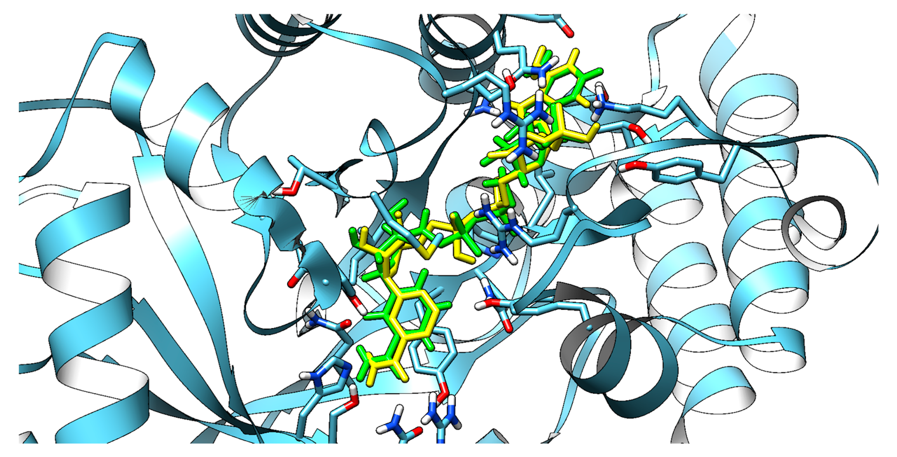
Figure 2.
Three-dimensional structure of aflatoxin aldehyde reductase (AFAR) in complex with docked astringin (left) and the corresponding two-dimensional interaction diagram (right). The figure was generated using the Discovery Studio Biovia program.
Figure 2.
Three-dimensional structure of aflatoxin aldehyde reductase (AFAR) in complex with docked astringin (left) and the corresponding two-dimensional interaction diagram (right). The figure was generated using the Discovery Studio Biovia program.
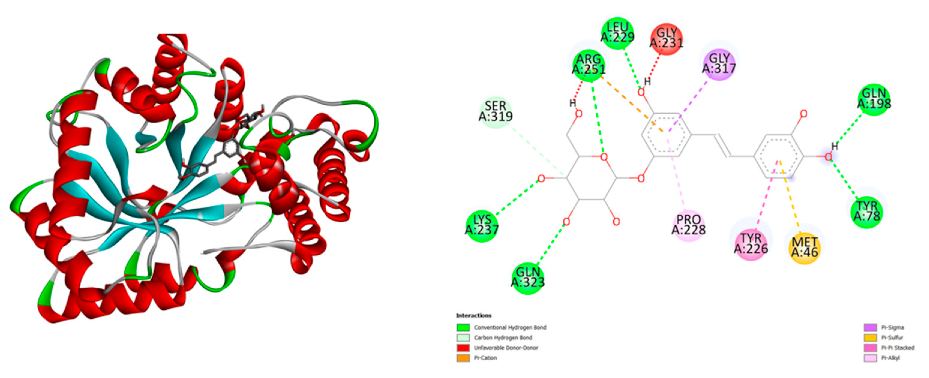
Figure 3.
Three-dimensional structure of aflatoxin aldehyde reductase (AFAR) in complex with docked Rhaponticin (left) and the corresponding two-dimensional interaction diagram (right). The figure was generated using the Discovery Studio Biovia program.
Figure 3.
Three-dimensional structure of aflatoxin aldehyde reductase (AFAR) in complex with docked Rhaponticin (left) and the corresponding two-dimensional interaction diagram (right). The figure was generated using the Discovery Studio Biovia program.
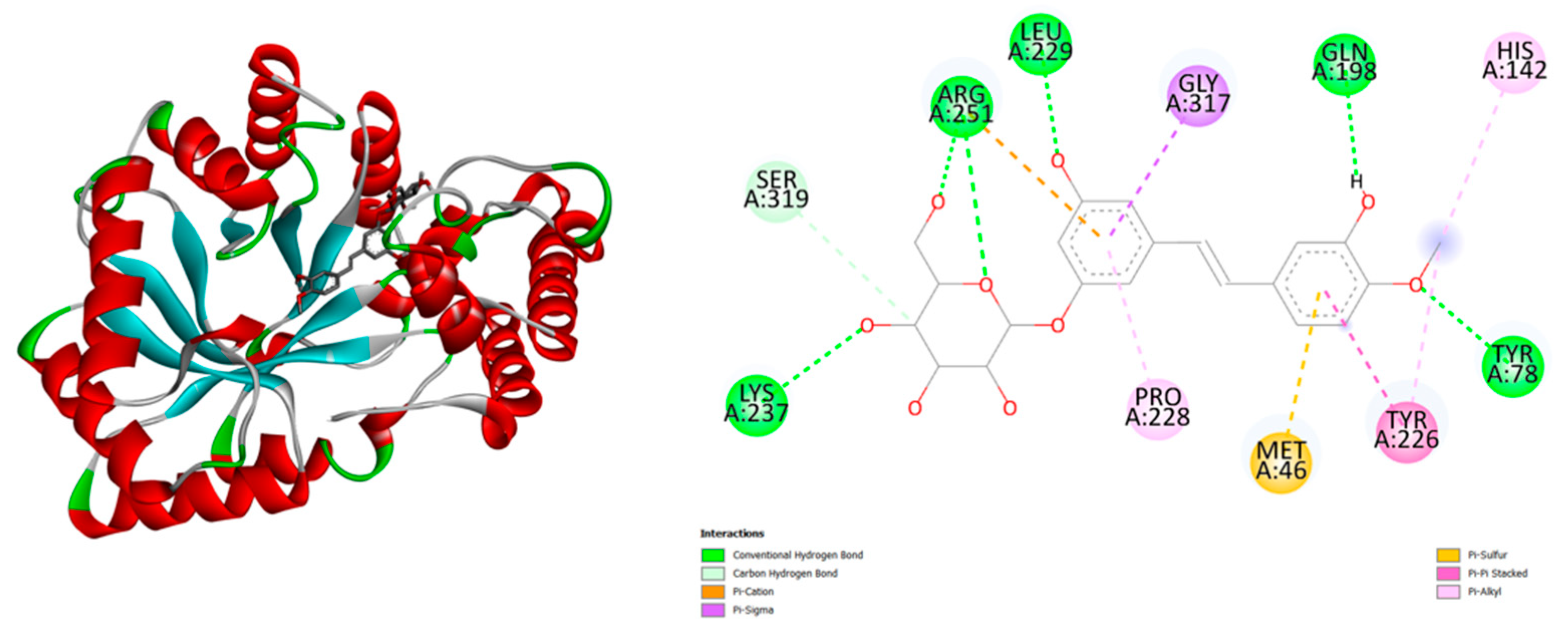
Figure 4.
Three-dimensional structure of aflatoxin aldehyde reductase (AFAR) in complex with docked folic acid (left) and the corresponding two-dimensional interaction diagram (right). The figure was generated using the Discovery Studio Biovia program.
Figure 4.
Three-dimensional structure of aflatoxin aldehyde reductase (AFAR) in complex with docked folic acid (left) and the corresponding two-dimensional interaction diagram (right). The figure was generated using the Discovery Studio Biovia program.
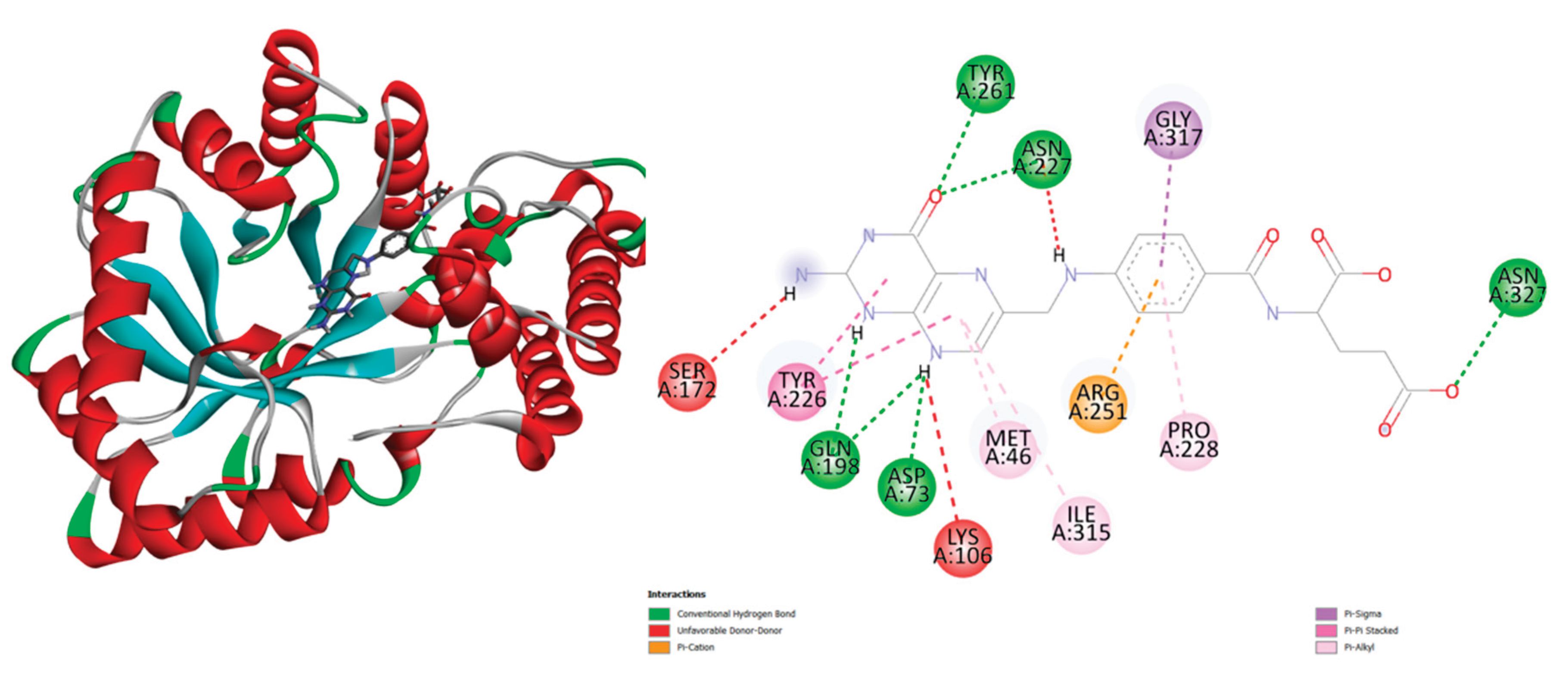
Figure 5.
Three-dimensional structure of aflatoxin aldehyde reductase (AFAR) in complex with docked Curcumin (left) and the corresponding two-dimensional interaction diagram (right). The figure was generated using the Discovery Studio Biovia program.
Figure 5.
Three-dimensional structure of aflatoxin aldehyde reductase (AFAR) in complex with docked Curcumin (left) and the corresponding two-dimensional interaction diagram (right). The figure was generated using the Discovery Studio Biovia program.
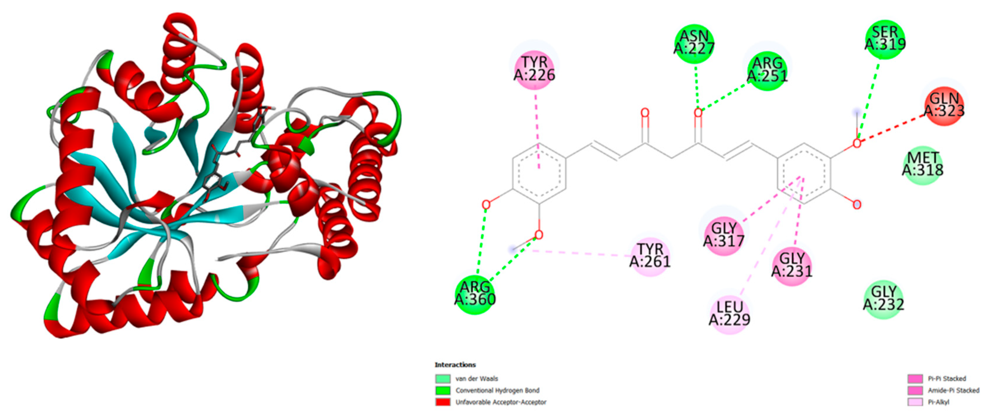
Figure 6.
Three-dimensional structure of aflatoxin aldehyde reductase (AFAR) in complex with docked Hesperidin (left) and the corresponding two-dimensional interaction diagram (right). The figure was generated using the Discovery Studio Biovia program.
Figure 6.
Three-dimensional structure of aflatoxin aldehyde reductase (AFAR) in complex with docked Hesperidin (left) and the corresponding two-dimensional interaction diagram (right). The figure was generated using the Discovery Studio Biovia program.
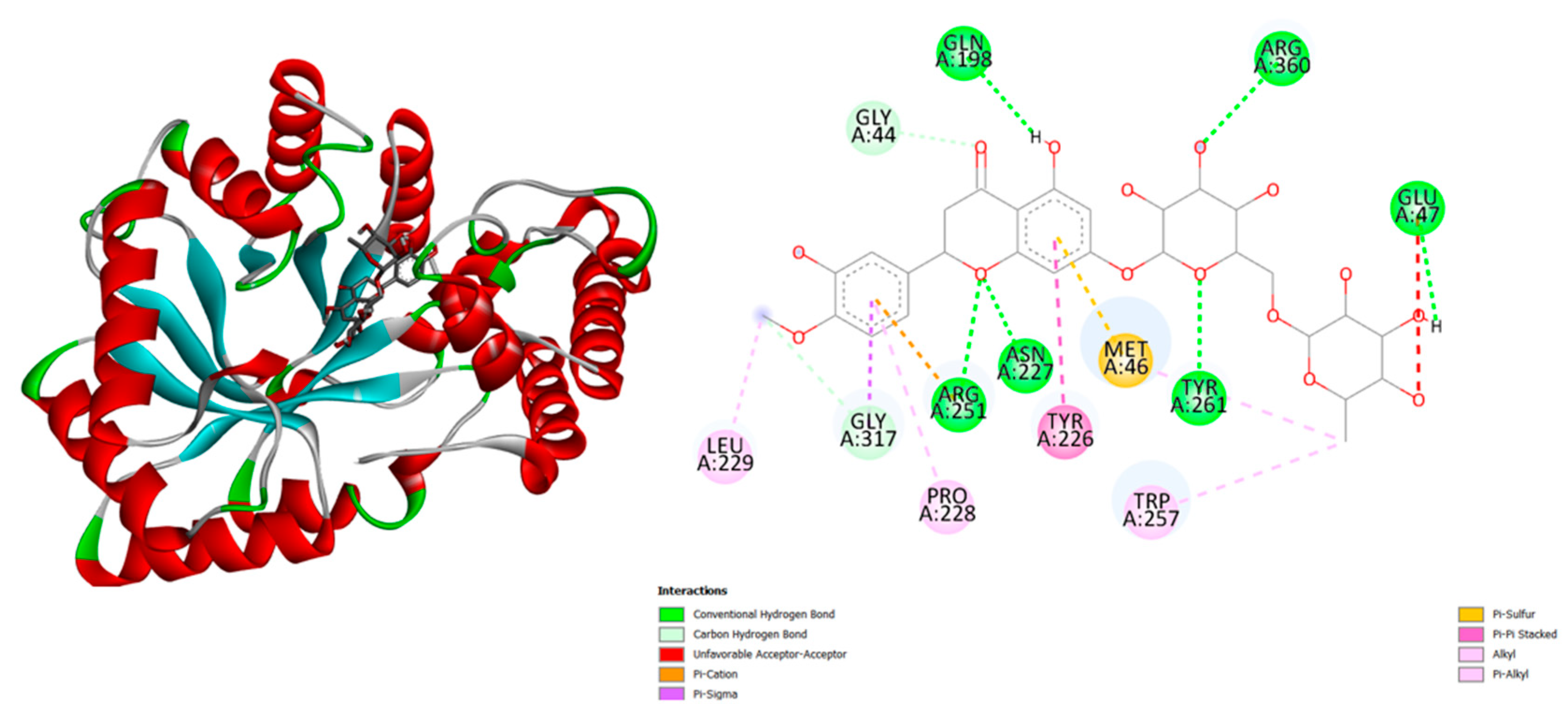
Figure 7.
Three-dimensional structure of aflatoxin aldehyde reductase (AFAR) in complex with docked NDP ( NADPH DIHYDRO-NICOTINAMIDE-ADENINE-DINUCLEOTIDE PHOSPHATE) (left) and the corresponding two-dimensional interaction diagram (right). The figure was generated using the Discovery Studio Biovia program.
Figure 7.
Three-dimensional structure of aflatoxin aldehyde reductase (AFAR) in complex with docked NDP ( NADPH DIHYDRO-NICOTINAMIDE-ADENINE-DINUCLEOTIDE PHOSPHATE) (left) and the corresponding two-dimensional interaction diagram (right). The figure was generated using the Discovery Studio Biovia program.
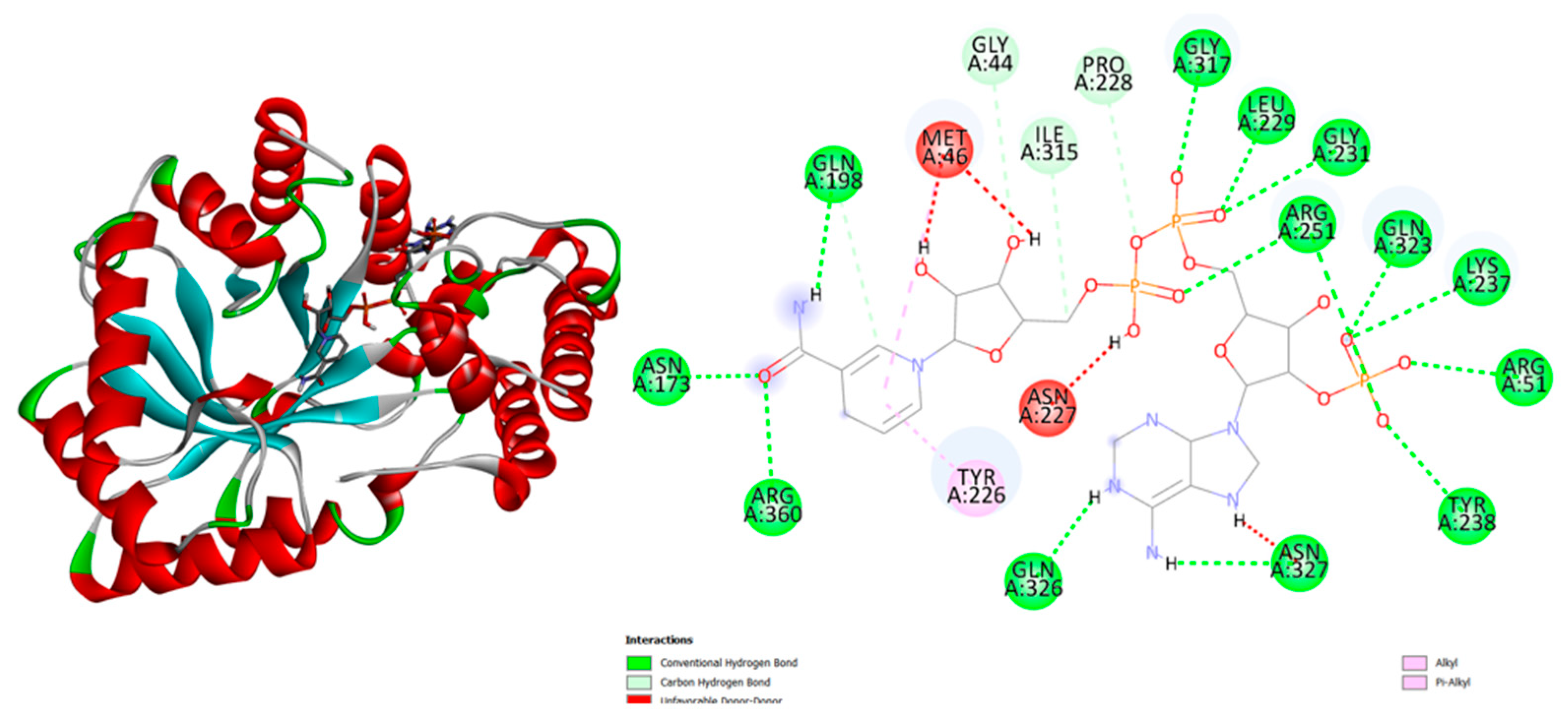

Table 1.
Docking binding energies (ΔG, kcal/mol) of selected natural compounds against AFAR (PDB: 2BP1).
Table 1.
Docking binding energies (ΔG, kcal/mol) of selected natural compounds against AFAR (PDB: 2BP1).
| Ligand | Binding Energy (kcal/mol) |
| NADPH (control) | –12.7 |
| Hesperidin | –11.0 |
| Curcumin | –9.8 |
| Rhaponticin | –9.6 |
| Folic Acid | –9.6 |
| Astringin | –9.6 |
| Taxifolin | –9.4 |
| Quercetin | –9.4 |
| Silibinin | –9.4 |
| Silymarin | –9.3 |
| Scutellarin | –9.1 |
| Kaempferol | –9.1 |
| Luteolin | –9.0 |
| Fisetin | –9.0 |
| Polydatin | –8.9 |
| Genistin | –8.9 |
| Baicalin | –8.7 |
| Diosmetin | –8.7 |
| Naringin | –8.7 |
| Apigenin | –8.6 |
| Xanthone | –8.5 |
| Hyperoside | –8.4 |
| Epicatechin Gallate | –8.4 |
| Daidzin | –8.3 |
| Capsaicin | –8.2 |
| Resveratrol | –7.6 |
| Syringin | –7.2 |
| Biotin | –7.1 |
| Sterculic Acid | –6.9 |
| Quercitrin | –6.6 |
| Rutin | –6.4 |
| Myricitrin | –5.3 |
| Allicin | –4.4 |
| Icariin | –2.4 |
| Hypericin | +6.7 |
Disclaimer/Publisher’s Note: The statements, opinions and data contained in all publications are solely those of the individual author(s) and contributor(s) and not of MDPI and/or the editor(s). MDPI and/or the editor(s) disclaim responsibility for any injury to people or property resulting from any ideas, methods, instructions or products referred to in the content. |
© 2025 by the authors. Licensee MDPI, Basel, Switzerland. This article is an open access article distributed under the terms and conditions of the Creative Commons Attribution (CC BY) license (http://creativecommons.org/licenses/by/4.0/).
Copyright: This open access article is published under a Creative Commons CC BY 4.0 license, which permit the free download, distribution, and reuse, provided that the author and preprint are cited in any reuse.