Submitted:
20 August 2025
Posted:
21 August 2025
You are already at the latest version
Abstract
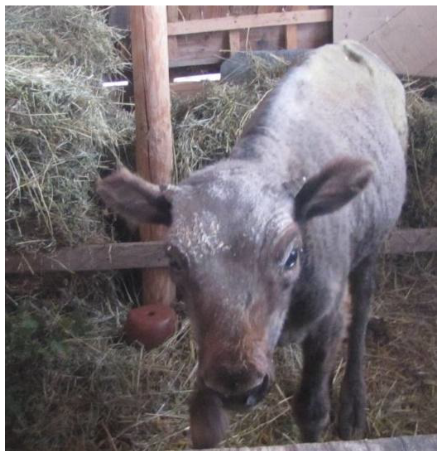
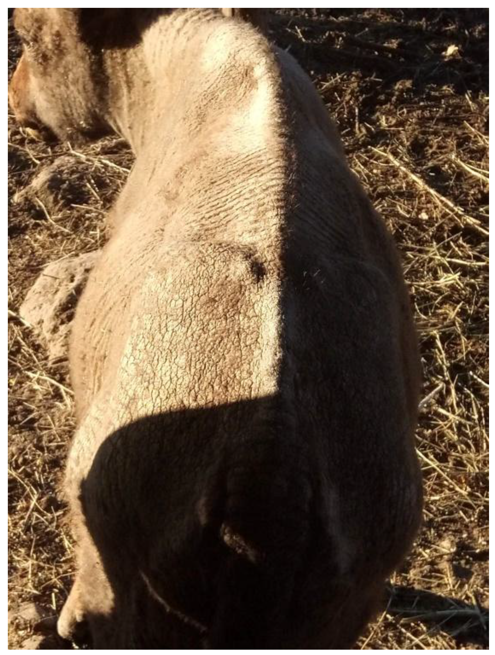
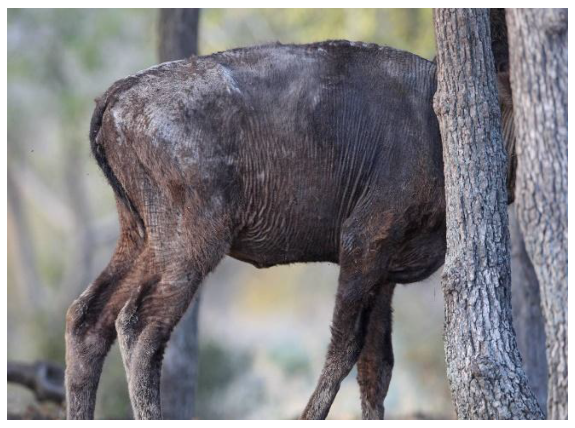
Bacterial dermatitis in animals caused by staphylococci is considered secondary and often results from prior conditions related to environmental factors (like ectoparasites, skin injuries, temperature, humidity) and internal factors (such as deficiency conditions, immunological dysfunction, and underlying diseases). The case represents a generalized dermatitis caused by a mixed infection of Staphylococcus aureus and Staphylococcus epidermidis in a female bison calf. Clinically, dermatitis with symptoms of alopecia and crusting was observed. The skin appeared scaly, dry, and rough, covered with thick crusts and showed no signs of itching. Upon removal of the crusts, inflamed, swollen, and oozing lesions were revealed underneath. Based on the laboratory results, the definitive therapy was initiated. After a lengthy treatment course, the bison’s condition improved, and new fur covered its body. This case emphasizes the necessity of targeted measures for timely etiological diagnosis. Furthermore, careful health monitoring and proactive disease management are essential for wildlife species, which rely directly on human actions for their preservation.
Keywords:
1. Introduction
2. Diagnostic Methods, and Laboratory Findings
3. Discussion
4. Conclusions
Author Contributions
Funding
Institutional Review Board Statement
Informed Consent Statement
Data Availability Statement
Conflicts of Interest
Abbreviations
| GP ID | Gramm positive identification card |
| GN ID | Gramm negative identification card |
References
- Mala, L.; Lalouckova, K.; Skrivanova, E. Bacterial skin infections in livestock and plant-based alternatives to their antibiotic treatment. Animals 2021, 11, 2473. [Google Scholar] [CrossRef] [PubMed]
- San Miguel, J. M.; Álvarez, G.; Luzón, M. Hypodermosis of red deer in Spain. Journal of Wildlife Diseases 2001, 37, 342–346. [Google Scholar] [CrossRef] [PubMed]
- Grice, E.A.; Segre, J.A. The skin microbiome. Nat. Rev. Microbiol. 2011, 9, 244–253. [Google Scholar] [CrossRef] [PubMed]
- Sullivan, L. E.; Evans, N. J.; Blowey, R. W.; Grove-White, D. H.; Clegg, S. R.; Duncan, J. S.; Carter, S. D. A molecular epidemiology of treponemes in beef cattle digital dermatitis lesions and comparative analyses with sheep contagious ovine digital dermatitis and dairy cattle digital dermatitis lesions. Veterinary microbiology 2015, 178, 77–87. [Google Scholar] [CrossRef] [PubMed]
- Ugochukwu, I. C. I.; Aneke, C. I.; Idoko, I. S.; Sani, N. A.; Amoche, A. J.; Mshiela, W. P.; Sackey, A. K. B. Bovine papilloma: Aetiology, pathology, immunology, disease status, diagnosis, control, prevention and treatment: A review. Comparative Clinical Pathology 2019, 28, 737–745. [Google Scholar] [CrossRef]
- Literák, I.; Tomita, Y.; Ogawa, T.; Shirasawa, H.; Šmid, B.; Novotný, L.; Adamec, M. Papillomatosis in a European bison. Journal of Wildlife Diseases 2006, 42, 149–153. [Google Scholar] [CrossRef] [PubMed]
- Jucker, S.; Alsaaod, M.; Steiner, A.; Zingre, T.; Kaessmeyer, S.; Gurtner, C.; Hoby, S. Treatment of digital dermatitis using salicylic acid in European bison (Bison bonasus) reveals promising results. Frontiers in veterinary science 2022, 9, 1012226. [Google Scholar] [CrossRef] [PubMed]
- Foster, A.P. Staphylococcal skin disease in livestock. Vet. Dermatol, 3: 23. [CrossRef]
- MacInnes, J. I.; Van Immerseel, F.; Boyce, J. D.; Rycroft, A. N.; Vázquez-Boland, J. A. Pathogenesis of bacterial infections in animals. 2022, J. F. Prescott (Ed.). John Wiley & Sons, Incorporated.
- Abrahamian, F.M.; Goldstein, E.J. Microbiology of animal bite wound infections. Clin. Microbiol. Rev. 2011, 24, 231–246. [Google Scholar] [CrossRef] [PubMed]
- Monecke, S.; Gavier-Widen, D.; Hotzel, H.; Peters, M.; Guenther, S.; Lazaris, A.; Ehricht, R. Diversity of Staphylococcus aureus isolates in European wildlife. PloS one, 2016, 11, e0168433. [Google Scholar] [CrossRef] [PubMed]
- Ringwaldt, E. M.; Brook, B. W.; Carver, S.; Buettel, J. C. The patterns and causes of dermatitis in terrestrial and semi-aquatic mammalian wildlife. Animals 2021, 11, 1691. [Google Scholar] [CrossRef] [PubMed]
- Fong, I.W. Animals and Mechanisms of Disease Transmission. Emerging Zoonoses, 1: 8. [CrossRef]
- Hoby, S.; Jensen, T. K.; Brodard, I.; Gurtner, C.; Eicher, R.; Steiner, A.; Alsaaod, M. Detection of treponemes in digital dermatitis lesions of captive European bison (Bison bonasus). PLoS One, 2021, 16, e0255921. [Google Scholar] [CrossRef] [PubMed]
- Roccaro, M.; Piva, S.; Scagliarini, A.; Giacometti, F.; Serraino, A.; Merialdi, G.; Frasnelli, M. ; Romano A,.; Bellio A., Ed.; Decastelli L.; Peli A. Case report of a pustular dermatitis outbreak in sheep: Clinical and food safety considerations. Ital J Food Saf. 2018 Apr 11, 7:6980. [Google Scholar] [CrossRef] [PubMed] [PubMed Central]
- Severn M., M.; Horswill A., R. Staphylococcus epidermidis and its dual lifestyle in skin health and infection. Nat Rev Microbiol, 9: 21. [CrossRef] [PubMed]
- Faccin, M.; Wiener D., J.; Rech R., R.; Santoro, D.; Rodrigues Hoffmann, A. ; Common superficial and deep cutaneous bacterial infections in domestic animals: A review. Veterinary Pathology, 7: 60. [CrossRef]
- Diegel, K. L.; Andrews-Jones, L.; Wojcinski, Z. W. Integument. Haschek and Rousseaux’s Handbook of Toxicologic Pathology Volume 5: Toxicologic Pathology of Organ Systems 2025, 505-582.
- Kocoń, A.; Nowak-Chmura, M. Skin ectoparasites of domestic animals. Annales Universitatis Paedagogicae Cracoviensis Studia Naturae 2017, (2), 137–158. [Google Scholar] [CrossRef]
- Oleński, K.; Kamiński, S.; Tokarska, M.; et al. Subset of SNPs for parental identification in European bison Lowland-Białowieża line (Bison bonasus bonasus). Conservation Genet Resour. 2018, 10, 73–78. [Google Scholar] [CrossRef]
- Bhopal, R. S. Concepts of Epidemiology: Integrating the Ideas, Theories, Principles, and Methods of Epidemiology. Oxford University Press, 2016.
- Feßler, A. T.; Wang, Y.; Burbick, C. R.; Diaz-Campos, D.; Fajt, V. R.; Lawhon, S. D.; Schwarz, S. Antimicrobial susceptibility testing in veterinary medicine: performance, interpretation of results, best practices and pitfalls. One Health Advances 2023, 1, 26. [Google Scholar] [CrossRef]
- Beco, L.; Guaguère, E.; Lorente Méndez, C.; Noli, C.; Nuttall, T.; Vroom, M. Suggested guidelines for using systemic antimicrobials in bacterial skin infections: part 2-- antimicrobial choice, treatment regimens and compliance. Vet Rec. 1: 9; 172. [CrossRef] [PubMed]
- Paterson, S. Rational use of antibiotics in skin disease. Companion Animal 2017, 22, 632–639. [Google Scholar] [CrossRef]
- Wirt, K. M.; Young, J. M.; Cramer, G.; Wagner, S. A. Topical salicylic acid treatment of digital dermatitis in dairy cows: Drug resides in milk and clinical efficacy. The Bovine Practitioner 2021, 45–51. [Google Scholar] [CrossRef]
- Popova, T. P.; Ignatov, I.; Petrova, T. E.; Kaleva, M. D.; Huether, F.; Karadzhov, S. D. Antimicrobial activity in vitro of cream from plant extracts and nanosilver, and clinical research in vivo on veterinary clinical cases. Cosmetics 2022, 9, 122. [Google Scholar] [CrossRef]
- Hoang, T. P. N.; Ghori, M. U.; Conway, B. R. Topical antiseptic formulations for skin and soft tissue infections. Pharmaceutics 2021, 13, 558. [Google Scholar] [CrossRef] [PubMed]
- Kaur, R.; Rathore, C.; Barik, B. B.; Samanta, A.; Rahamanulla, A.; Jafar, M.; Krishna, K. V. Development and Characterization of Calendula officinalis Extract-Loaded Topical Gels For Use In Inflammatory Skin Conditions Associated With Staphylococcus aureus and Pseudomonas aeruginosa in Diabetic Foot Ulcers. Journal of Neonatal Surgery.
Disclaimer/Publisher’s Note: The statements, opinions and data contained in all publications are solely those of the individual author(s) and contributor(s) and not of MDPI and/or the editor(s). MDPI and/or the editor(s) disclaim responsibility for any injury to people or property resulting from any ideas, methods, instructions or products referred to in the content. |
© 2025 by the authors. Licensee MDPI, Basel, Switzerland. This article is an open access article distributed under the terms and conditions of the Creative Commons Attribution (CC BY) license (https://creativecommons.org/licenses/by/4.0/).


