
Submitted:
15 August 2025
Posted:
18 August 2025
You are already at the latest version
Abstract
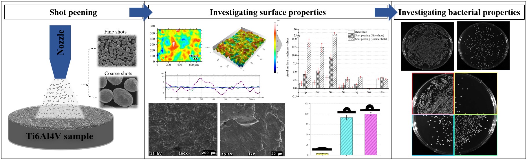
Interest in textured surfaces for biomaterials and implants is increasing, with shot peening emerging as a promising surface modification method. This study investigates the influence of conventional and fine shot peening on the surface morphology, topography, wettability, and antibacterial properties of biomedical-grade Ti6Al4V alloy. Peening was conducted using a custom-built, fully automated system, employing fine (100–200 µm) and coarse (700–1000 µm) shots using well-controlled sets of parameters. Both treatments introduced severe plastic deformation on the surface, resulting in increased roughness. Conventionally shot-peened samples exhibited deeper and wider dimples compared to finely peened ones. Surface wettability shifted from hydrophilic (contact angle: 4.1°, untreated) to hydrophobic, reaching contact angles of 91.13° and 99.76° for fine and conventional shot peening, respectively. Despite these topographical changes, both treatments preserved the intrinsic antibacterial activity of the alloy. Quantitative assays revealed high bacterial reduction rates after 6 hours of incubation: 99.58% (E. coli) and 92.54% (S. aureus) for fine shot peening, and 98.98% (E. coli) and 99.2% (S. aureus) for conventional shot peening. Both methods were effective in modifying the surface without compromising antibacterial performance. However, fine shot peening provided a smoother and more uniform surface topography, and thus potentially offers better control over bacterial adhesion and surface–cell interactions in biomedical applications.
Keywords:
antibacterial properties
; biomaterials
; implants
; surface metrology
; surface roughness
; surface wettability
; titanium alloys
1. Introduction
Titanium alloys have been preferred materials in various biomedical applications for a long time due to their good mechanical properties, including biocompatibility, strong acid and alkali resistance, toughness, and fatigue and yield strength [1,2,3,4,5,6]. However, these alloys are rarely employed in engineering disciplines particularly associated with tribology due to certain disadvantages that include low surface hardness, an elevated coefficient of friction, and inadequate wear resistance [3,4]. Their tribological, corrosion, and biological characteristics, including wear and corrosion resistance, antibacterial activity, osteointegration, are directly influenced by the surface properties of these materials [3]. Thus, there is a substantial demand for enhancements in their surface properties (such as roughness, wettability, and morphology), mechanical properties (including hardness, wear resistance, corrosion resistance, fatigue behavior, and toughness), and antibacterial properties of titanium alloys. Roughened surfaces featuring patterns are generally more useful than smooth, patternless surfaces in numerous respects, particularly when considered for biomedical properties [7]. Patterned surfaces are typically achieved through the deposition of coatings or by processing the material’s surface at the sub-micrometer or micrometer scale [8,9].
Methods employed for surface modifications of titanium alloys in biomedical applications include: laser surface texturing (LST) [10,11], laser peening (LSP) [12], electro-spark surface texturing (ESST) [13], electro-erosion (EDM), chemical reactive ion etching (RIE) [14], lithography and anisotropic etching (LIAE) [15], abrasive jet machining (AJM) [16], and physical vapor deposition (PVD) [17,18,19], among others [3,6]. The predominant technique for surface patterning seems to be LST. Liu et al. [4] found that laser processing of a titanium alloy to create cell-like patterns enhanced surface microhardness while decreasing the friction coefficient and wear volume. Milles et al. [20] generated patterns at varying depths by fabricating hexagonal honeycomb structures on the aluminum surface using a laser processing technique, concluding that these patterns enhanced surface wettability. Shivakoti et al. [3] developed pit and pillar surface patterns on titanium and zirconia alloys employed in the biomedical sector through laser processing, concluding that the corrosion and wear resistance, and cell adhesion, increased after the patterning process. Erdoğan et al. [9] employed a laser method to process dot and straight-line patterns on Ti6Al4V, concluding that the quality of cell adhesion improved following the patterning procedure. Numerous studies have demonstrated that laser oxidation can enhance the microhardness of titanium surfaces, refine grain size, improve micro morphology, and enhance corrosion resistance [6]. Nevertheless, the development of cracks on both the surface and subsurface of the material, with crack size directly proportional to the processing duration, has also been documented [21]. Even though laser treatment has proven to be an effective method for producing surface patterns on titanium alloys and improving the surface properties, it also often leads to undesirable surface and subsurface defects. Therefore, to present a relatively more robust method, researchers have recently focused on using mechanical surface treatment methods (e.g., shot peening) to modify surface properties of titanium alloys and to obtain specific surface patterns for biomedical applications [6,22,23,24]. Among these, there has been a particular interest in utilizing shot peening methods to tailor the surface properties of titanium alloys for biomedical applications.
In the shot peening process, peened material in the form of spheres impacts the surface at high velocities, resulting in plastic deformation and grain refinement in both the surface and subsurface regions [25]. Grain refinement enhances the mechanical properties of the material, including its hardness and toughness. Furthermore, shot peening notably modifies the surface characteristics of the treated alloy [26], such as topography, morphology, and wettability, particularly in relation to the applied shot peening parameters. Shot peening offers several potential advantages over other surface processing techniques including reduced equipment requirements, adjustable surface and subsurface characteristics based on adjustable process parameters, lower energy consumption, shorter processing durations, higher efficiency, cost-effectiveness, and the capability to treat more complex-shaped components with ease [27]. Nevertheless, suboptimal shot peening conditions may also result in detrimental surface effects including excessive roughening, microcrack initiation, and particle embedding [28], which can negatively influence mechanical integrity and in vivo performance. A rigorous optimization and comprehensive evaluation of shot peening parameters are imperative to maximize its functional benefits while mitigating potential surface damage. There are some well-documented mechanical benefits of shot peening; however, research is also required to elucidate the specific effects of both conventional and fine shot peening on surface roughness, wettability, and biological responses of titanium alloys. These surface-related attributes are critical particularly for biomedical applications where cell adhesion, protein adsorption, and antibacterial performance are highly sensitive to micro- and nano-scale surface features.
Recent studies addressing the influence of shot peening parameters, especially in relation to surface morphology, topography, and biofunctionality, are critically discussed as follows to provide a clearer understanding of current research trends and remaining knowledge gaps: Wang et al. [29] applied fine particle shot peening (FPSP) to Ti6Al4V, generating half-dimpled surfaces that improved tribological performance; however, assessment of wettability or antibacterial effects were not conducted. Zhu et al. [30] showed that ultrasonic shot peening (USP) on commercially pure Ti largely enhanced hydrophilicity, directly linking surface roughness modifications to greater antibacterial behavior. Hsiao et al. [31] combined conventional shot peening (CSP) with HF/HNO₃ etching to achieve Ra ≈ 0.9 µm on Ti6Al4V, promoting osteoblast adhesion and spreading. Uanlee & Khantachawana [32] applied FPSP to Ti6Al4V, resulting in higher surface hardness and improved fibroblast viability, though an assessment of wettability and antibacterial effect were not provided. Khandaker et al. [33] used laser peening to create microgrooves on Ti surfaces that notably boosts osteoblast adhesion, proliferation, and differentiation. Shen et al. [34] combined shot peening with Si/Cu-doped micro-arc oxidation coatings (MAO) coatings to achieve >95% antibacterial efficacy against S. aureus and stimulate osteogenic marker expression. Li et al. [35] implemented copper-powder ultrasonic shot peening (C-USSP) on cp-Ti, demonstrating important antibacterial effects directly due to peening-induced surface changes. Finally, Zheng et al. [36] applied shot peening and gas nitriding to TC4 and found that, although the mechanical properties were substantially improved, antibacterial effectiveness rates did not change much—highlighting the complexity of peening vs. antimicrobial surface behavior. Altogether, these studies confirm that shot peening, especially when combined with additional treatments or optimized as USP/C-USSP, has the potential to substantially modulate antibacterial responses and cell behavior via surface topography and wettability control. At the same time, not all peening variants largely enhance antibacterial performance, indicating the need for careful parameter optimization and comprehensive biological testing to identify protocols that consistently benefit implant integration and microbial resistance.
This study aims to systematically investigate the effects of conventional and fine shot peening techniques on the surface morphology, topography, wettability, and antibacterial performance of biomedical-grade Ti6Al4V alloy. Using a fully automated, custom-designed shot peening system, samples were treated under controlled parameters by using coarse and fine media to generate distinct surface textures. Through a comparative approach, the study mainly explores how shot size influences biologically relevant surface features and bacterial adhesion behavior. The outcomes are expected to advance the development of biofunctional titanium implant surfaces by promoting antibacterial efficacy while maintaining desirable wetting and morphological characteristics.
2. Materials and Methods
Conventional and fine shot peening were conducted on biomedical Ti6Al4V alloy samples utilizing fine (100-200 µm) and coarse shots (700-1000 µm) under regulated conditions with a custom-designed fully automated peening system. Subsequently, optical profilometry, scanning electron microscopy (SEM), and contact angle measurements were performed to characterize the surface properties of fine-peened and shot-peened samples. The antibacterial efficacy of the samples against S. aureus and E. coli was assessed.
2.1. Materials
In this study, Ti6Al4V alloy (grade 5) samples with equiaxed microstructure were commercially supplied in rod form (Ø20 mm x 1000 mm) by TIMET, Türkiye. These were cut into slices that were 10 mm thick using a semi-automatic band saw. Before shot peening, the samples were ground using sandpapers of 360, 600, 1000, and 2000 mesh and polished sequentially with diamond solutions of 9 µm, 3 µm, and 1 µm. The alloy’s nominal chemical composition, provided in the material certification by the supplier (TIMET, Türkiye), is presented in Table 1.
2.2. Shot Peening of Ti6Al4V Alloy
Shot peening treatments were carried out using a CNC-controlled, custom-designed shot peening system with the configuration and operating principles previously described in detail and illustrated schematically in our earlier work [24]. The samples subjected to shot peening at an applied pressure of 5 bar for 2.5 minutes from a distance of 40 mm, with the peening parameters detailed in Table 2. Two distinct sizes of stainless-steel shots were utilized in the shot peening processes. The morphology and chemical composition of the stainless-steel shots were examined by SEM coupled with energy-dispersive X-ray spectroscopy (EDS). As shown in Figure 1a,b, the coarse (700–1000 µm) and fine (100–200 µm) shots exhibited spherical morphology with surface texture features. EDS spectra revealed that both shot types primarily consisted of Fe, Cr, Ni, Mn, and Si, in agreement with their stainless-steel composition.
2.3. Surface Characterisation of Shot-Peened Alloy
Figure 2 illustrates the methodology employed for characterizing surface roughness of the peened samples. First, the three-dimensional topographies of the sample surfaces were acquired utilizing an optical profilometer (Huvitz, Gyeonggi, Republic of Korea). Then, the surface roughness characteristics (i.e., surface topography, areal roughness parameters, roughness profiles) of the samples were analyzed by Mountains® 9 software (Digital Surf, Besançon, France).
Subsequent to the characterization of surface roughness, the surface morphologies of the shot-peened samples were analyzed utilizing an SEM (Jeol JSM-6060, Tokyo, Japan) coupled with an energy dispersive spectroscopy (EDS) detector (Oxford Instruments, Oxford, UK). The wettability of the unpeened, fine-peened, and shot-peened samples was assessed using an optical contact angle measurement system (Kruess GmbH, Germany). Contact angle tests were conducted using distilled water, maintaining a constant droplet volume of 3 ml, with each specimen tested five times.
2.4. Antibacterial Activity of Shot-Peened Surface
To perform antibacterial tests of samples before and after shot peening, S. aureus strain ATCC 29213 and E. coli strain ATCC 25922 (Mueller-Hinton Agar-Merck, Germany) were commercially supplied as testing microorganisms. Nutrient broth was prepared by dissolving 34 g of Mueller Hinton Agar (MHA) in 1,000 ml of distilled water and then sterilized at 121 °C for 15 min. The resulting solution was spread into Petri dishes and then allowed to cool under sterile conditions at room temperature. S. aureus and E. coli were grown in the nutrient broth at 37 °C to a concentration of 108 CFU ml-1. Then, the broth was diluted 10-fold with phosphate buffered solution (PBS) to a concentration of 104 CFU ml-1 (bacterial suspension). 0.4 ml of bacterial suspension was dropped onto the samples and then the Petri dishes were incubated at 37 °C for 18-24 hours under 90% humidity. After incubation, the solutions on the surface of the samples were collected in sterile tubes containing 3.6 ml of PBS solution. The samples were also placed in the same tubes and then 0.02 ml of the solution was taken and cultured in MHA with glass rods and incubated at 37 °C for 18-24 hours. After incubation, the number of active bacteria was determined by manually counting the colony-forming units (CFUs) on each plate using a standard colony counter.
3. Results and Discussion
3.1. Surface Morphology After Shot Peening
Figure 3 and Figure 4 display low and high magnification SEM micrographs of the Ti6Al4V alloy surfaces subjected to conventional and fine shot peening, respectively. Both treatments induced severe plastic deformation, with characteristic surface features directly influenced by the size and kinetic energy of the impacting media. The surfaces exhibit dense impact craters and material pile-ups, typical of shot peening-induced topographical modifications [24,37,38]. In the conventionally shot-peened sample (Figure 3), the morphology is dominated by larger and deeper dimples, associated with high-energy impacts from coarse shots (700–1000 µm). The surface shows pronounced ridges and valleys with irregular crater geometry and evidence of micro-cutting or plowing, suggesting intense localized plastic deformation. Similar morphological features have been reported in Ti-based substrates processed by high-intensity peening, where the severity of plastic flow correlates with surface work hardening and increased roughness [24,39]. In contrast, the surface treated with fine shot peening (Figure 4) reveals more uniform and shallower dimples, attributed to the lower kinetic energy of fine shots (100–200 µm). The resulting surface layer thus exhibits a more homogeneous texture, with finer-scale peak-valley features and less severe surface distortion. This microtopography reflects a more controlled peening action, capable of refining the surface without extensive surface damage or excessive roughening, a behaviour that is consistent with prior observations on fine shot or ultrasonic peening of titanium alloys [32,40,41,42,43]. No significant cracks, microcracks, fractures, or particle dislodgement were seen on either surface, demonstrating the absence of harmful surface degradation. This observation corroborates the efficacy of the employed process parameters in attaining advantageous surface modification while preserving material integrity.
From a mechanistic standpoint, the microstructural refinement and surface texture variation stem from shot-induced strain localization. The higher dimple density and smoother contours in the fine shot-peened sample suggest a more uniform energy transfer, whereas coarse shot impacts lead to heterogeneous strain zones and larger deformation footprints. These distinct morphological features are known to critically influence surface properties such as protein adsorption, wettability, and cellular interaction, particularly in biomedical applications [32,33,34,35]. Moreover, the differences in morphology align with surface roughness measurements (Figure 5 and Figure 6), wherein conventionally peened surfaces exhibit greater roughness amplitudes. The fine peening technique offers a desirable balance between surface modification and preservation of topological integrity, supporting its suitability for biofunctional surface engineering of Ti6Al4V implants.
3.2. Surface Topography After Shot Peening
Surface topography plays a critical role in governing the mechanical and biological performance of titanium-based biomaterials. The results presented in Figure 5, Figure 6 and Figure 7 reveal the profound influence of shot peening parameters on the three-dimensional surface features of Ti6Al4V alloy. Conventional shot peening, characterized by larger shot sizes and higher kinetic energy, produced an irregular surface profile with pronounced peaks (Sp) and valleys (Sv), as indicated by increased areal roughness values—maximum height (Sz) ≈ 24.20 µm and arithmetic mean height (Sa) ≈ 2.78 µm. In contrast, fine shot peening generated a much smoother surface (Sz ≈ 11.20 µm, Sa ≈ 1.69 µm), while still inducing beneficial topographic features necessary for mechanical interlocking and cellular interaction [44,45,46,47]. These findings agree with the previously discussed SEM-based morphology results (Figure 3 and Figure 4), where larger and deeper impact craters were observed on the conventionally shot-peened surface, whereas the fine shot-peened surface exhibited more uniform and shallower surface features. These morphological observations support the interpretation that shot size plays a dominant role in determining local deformation mechanisms and surface reconstruction.
The data extracted from ISO 25178 parameters (Figures 6c) and comparative bar plots (Figure 7a) highlight a clear correlation between shot size and roughness amplitude. Sq (root mean square height) increased with shot size, confirming higher roughness amplitude for conventional shot peening. Both shot-peened surfaces showed slightly negative Ssk (skewness), indicating valley-dominated morphologies favorable for fluid retention and potential cell adhesion. Sku (kurtosis) values >3 reflected sharper peak–valley features, enhancing potential mechanical interlocking, which are critical for evaluating functional surfaces in biomedical implants [48]. In addition, roughness profiles (Figure 7b) demonstrate the amplitude modulation and wavelength distribution across the treated surfaces, with fine shot peening yielding a more periodic and less jagged profile than the conventional counterpart.
From a fundamental standpoint, the observed differences in roughness and topography arise from the interplay between shot diameter, kinetic energy per impact, and impact frequency. Larger shots used in conventional peening impart higher momentum and localized energy, leading to deeper plastic deformation zones, irregular asperity formation, and increased surface roughness. Fine shot peening, on the other hand, involves smaller media with lower energy per impact but a higher number of impacts per unit area. This results in more homogeneous plastic flow, reduced micro-cutting, and smoother surface reconstruction due to cumulative overlapping deformation events [49,50]. Such differences in surface characteristics directly impact the implant’s performance in vivo. Excessively rough surfaces may enhance mechanical anchorage but at the same time risk promoting bacterial colonization and debris entrapment [51]. Conversely, ultra-smooth surfaces may reduce biological fixation. Therefore, the moderately rough and homogenously peened surfaces achieved via fine shot peening represent an optimized compromise, ensuring both structural integration and, potentially, a reduced infection risk.
The present findings align with the literature on shot peening–induced surface texturing of Ti6Al4V alloys, where controlled roughness plays a pivotal role in modulating osteoblastic activity, corrosion behavior, and long-term implant performance. These morphological and topographical improvements serve as a mechanistic foundation for understanding the antibacterial responses discussed below.
3.3. Wettability After Shot Peening
Surface wettability plays a pivotal role in the biological performance of metallic implants by influencing protein adsorption, initial cell attachment, and bacterial adhesion. The results of the contact angle measurements presented in Figure 8 reveal a notable change in wettability following both fine and conventional shot peening treatments compared to the reference condition. The reference sample, which possessed an ultrafine and mirror-polished surface, exhibited a low contact angle (~15°), confirming its hydrophilic nature. This behavior is consistent with its smooth topography and minimal surface roughness, as quantified in Figure 5, Figure 6 and Figure 7. After shot peening, a clear transition toward hydrophobic behavior was observed. Specifically, fine shot peening increased the contact angle to ~90°, while conventional shot peening further elevated it to >100°, indicating strong water repellency. These wettability shifts are closely linked to the morphological and topographical alterations induced by the peening treatments. As shown in Figure 4 and Figure 6, the generation of micro-craters and overlapping deformation zones increased the surface roughness and introduced hierarchical surface features. These surface modifications reduce the real contact area and alter the solid–liquid interaction, resulting in an apparent increase in contact angle [52,53,54]. Such pronounced changes in wettability are expected to have direct implications for the biological response of the alloy surface. In particular, the transition from a hydrophilic to a hydrophobic state may influence subsequent protein adsorption, cell–material interactions, and bacterial adhesion, as will be discussed in the following sections.
Fine shot peening applied in the present study appears to provide an optimized surface condition by tuning wettability and surface texture simultaneously. The finer and more uniformly distributed dimples generated by fine shot peening produced a moderately rough but topographically ordered surface, supporting a transition to near-hydrophobic conditions while potentially maintaining favorable biological responses. In contrast, the irregular and sharper asperities caused by conventional shot peening likely reduce droplet spreading and enhance water repellency. From a functional perspective, the altered wettability is expected to influence subsequent biological interactions. Moderately hydrophobic surfaces can inhibit bacterial adhesion and delay biofilm formation, while still supporting protein anchorage and cell adhesion when combined with bioactive chemistry [44].
3.4. Antibacterial Activity After Shot Peening
Bacterial colony formation on Ti6Al4V was notably reduced compared to the control agar plate without material (Figure 9 and 10), confirming the alloy’s intrinsic antibacterial behavior [55]. Shot peening increased surface roughness, which in turn promoted bacterial growth relative to the very smooth reference surface. Nevertheless, both shot-peened surfaces still exhibited markedly lower bacterial proliferation compared to the control, with fine shot peening outperforming the conventional process. Reduced E. coli proliferation was observed on smoother, fine shot-peened surfaces (Figure 10), whereas S. aureus showed higher proliferation on fine shot-peened surfaces (Figure 11).
Consistent with earlier reports, Ti6Al4V demonstrates intrinsic antibacterial behavior [55]. Our findings confirm that antibacterial property is largely retained after shot peening i.e., not negatively affected by the applied surface modification methods (Figure 9 and Figure 10). Among the peening techniques employed, conventionally shot-peened samples exhibited a notably higher surface roughness compared to fine shot-peened ones. It is well established that surface topography and roughness critically affect bacterial adhesion and proliferation. Surfaces with higher roughness may serve as a scaffold for bacterial colonization, especially when the average roughness (Sa) exceeds 0.8 µm, facilitating bacterial attachment by providing protective niches [48]. In our study, both treatment methods yielded Sa > 0.8 µm, while conventionally shot-peened surfaces exhibited the higher roughness value (Sa > 2.775 µm). Additionally, the spacing of the surface grooves plays an important role in bacterial colonization. When the groove spacing is smaller than the bacterial cell size, adhesion becomes less favourable. In our case, the spacing formed by coarse shots (SZ ≈ 24.20 µm) was much larger than that of fine shots (SZ ≈ 9.528 µm), indicating higher potential bacterial adhesion on conventionally shot-peened surfaces. Priyanka et al. [48] reported greater bacterial adhesion on rougher TiN-Ag coated surfaces, which supports this claim.
Nishitani et al. [42] demonstrated that at lower surface roughness via fine particle bombardment, antibacterial activity improved, and particularly against both E. coli and S. aureus. Our results agree with these findings, as reduced E. coli proliferation was observed on the smoother, fine shot-peened surfaces (Figure 10). Moreover, the ~200 nm convex features formed during fine peening may have interacted with the E. coli cell membrane, causing membrane damage. However, to verify this, a further live/dead assay needs to be conducted. Interestingly, we observed a higher proliferation of S. aureus on fine shot-peened surfaces (SZ ≈ 9.528 µm), as seen in Figure 9. This result aligns with prior studies by Cunha et al. [56] and Truong et al. [57], which reported S. aureus adhesion to microgrooves (10–20 µm) created by femtosecond laser treatment on titanium.
The wettability of the surface is determined by its roughness, topography, and chemical composition. Untreated Ti6Al4V surfaces are typically hydrophilic (Figure 8), and both peening methods increased hydrophobicity. Trapped air within the hydrophobic interface can act as a barrier, preventing bacterial attachment and motility, thereby enhancing antibacterial resistance [58]. Bright et al. [59] also observed that S. aureus preferred proliferation on hydrophilic surfaces. Thus, the increased proliferation of S. aureus on the less hydrophobic, fine shot-peened surfaces in our study (Figure 9) is consistent with the literature. Biofilm formation typically begins with the weak, reversible attachment of bacterial cells to the surface. Environmental factors such as surface roughness, hydrophobicity, and surface charge, as well as bacterial traits and surrounding pH or flow conditions, strongly influence adhesion and biofilm development. Once adhered, microorganisms produce extracellular polymeric substances (EPS), forming a protective matrix that increases resistance to antibiotics and environmental stressors [60]. Biofilms are therefore a major concern in the context of chronic polymicrobial infections and medical device-related complications [58]. Wu et al. [61] reported that micro- and nanoscale surface roughness notably affect bacterial adhesion and early biofilm development. The observed proliferation of S. aureus on rougher surfaces may indicate a higher potential for biofilm formation. Additionally, the increased growth of E. coli on more hydrophobic regions (Figure 10) could also suggest potential biofilm development. However, to confirm these trends, time-dependent antibacterial testing at various incubation durations (e.g., 12, 18, and 24 hours) is needed. Quantitative reduction rates for both bacterial strains after 6 hours of incubation further support these observations, confirming that both shot peening techniques were effective in reducing bacterial activity (Figure 11). It should be noted that there are strain-specific differences in reduction levels: While reduction efficiency remains same for E. coli in untreated and treated samples, this is only true for S. aureus for unpeened and conventionally peened sample where there was some reduction in efficiency for fine shot-peened sample.
4. Conclusions
In this study, a comparative investigation was conducted to evaluate the effects of conventional and fine shot peening techniques on the surface morphology, roughness, wettability, and antibacterial activity of biomedical-grade Ti6Al4V alloy. Using a custom-designed automated shot peening system, surface modification was achieved through either coarse or fine stainless-steel media to explore their impact on biofunctional surface design. Critical findings of the study include:
- Surface characteristics were strongly influenced by shot size and energy. Conventional shot peening (700–1000 µm) induced rougher, irregular surfaces with higher peak-valley features (Sa ≈ 2.78 µm), while fine shot peening (100–200 µm) produced more homogeneous textures with smoother topographies (Sa ≈ 1.69 µm), minimizing potential surface damage.
- Wettability shifted from hydrophilic to hydrophobic after peening, with fine shot peening achieving a contact angle of ~91° and conventional peening exceeding 99°. These changes are attributed to the formation of micro-dimples and hierarchical textures, which reduce solid–liquid interaction and may favor antibacterial behavior.
- Both shot peening techniques largely preserved the intrinsic antibacterial activity of the Ti6Al4V alloy, with some strain-specific variation. The smoother fine-peened surfaces may offer an improved balance between reduced bacterial adhesion and topographical suitability for biomedical integration.
While these findings clearly demonstrate the potential of mechanical peening as an effective and scalable technique for biofunctional surface engineering, the study has certain limitations: Live/dead bacterial assays, protein adsorption studies, and biofilm formation analyses were not conducted and should be addressed in future work. In addition, long-term durability and corrosion resistance of the modified surfaces under physiological conditions remain to be evaluated. Future studies may also explore hybrid approaches combining shot peening with antibacterial coating or chemical functionalization to further enhance both antibacterial efficacy and osseointegration potential.
Author Contributions
Conceptualization, Eg.A., Y.Y.A., R.Y. and M.G.; methodology, Eg.A., Y.Y.A., H.U., M.S. and M.G.; software, Eg.A. and M.G.; validation, Eg.A., Y.Y.A., H.U. and R.Y.; formal analysis, Eg.A., H.U., M.S. and M.G.; investigation, Eg.A., Y.Y.A., H.U., M.I.B. and Er.A.; resources, Eg.A., H.U. and M.G.; data curation, Eg.A., M.I.B. and Er.A.; writing—original draft preparation, Eg.A., M.A., M.S. and M.G.; writing—review and editing, Eg.A., Y.Y.A., R.Y., C.E., M.F.-X.W. and M.G.; visualization, Eg.A., Er.A., M.A. and M.G.; supervision, Eg.A. and M.G.; project administration, Eg.A.; funding acquisition, Eg.A. and M.G. All authors have read and agreed to the published version of the manuscript.
Funding
The authors acknowledge the financial support provided by the Kocaeli University Scientific Research Projects Coordination Unit under project numbers FKA-2024-3749, FKA-2024-3934, and FBA-2025-4619.
Data Availability Statement
The original contributions presented in this study are included in the article Further inquiries can be directed to the corresponding author.
Acknowledgments
In this section, you can acknowledge any support given which is not covered by the author contribution or funding sections. This may include administrative and technical support, or donations in kind (e.g., materials used for experiments).
Conflicts of Interest
The authors declare no conflicts of interest. The funders had no role in the design of the study; in the collection, analyses, or interpretation of data; in the writing of the manuscript; or in the decision to publish the results.
References
- Shivakoti I., K.G., Cep R., Pradhan B. B., Sharma A. Laser Surface Texturing for Biomedical Applications: A Review. Coatings 2021.
- Sandoval-Robles J. A., R.C.A., García-López E. Laser Surface Texturing and Electropolishing of CoCr and Ti6Al4V-ELI Alloys for Biomedical Applications. Materials 2020.
- Lin N., L. D., Zou J., Xie R., Wang Z., Tang B. Surface Texture-Based Surface Treatments on Ti6Al4V Titanium Alloys for Tribological and Biological Applications: A Mini Review. Materials 2018. [Google Scholar]
- Liu Q., L.Y., Li X., Dong G. Pulse Laser-İnduced Cell-Like Texture On Surface Of Titanium Alloy For Tribological Properties İmprovement. Wear 2021.
- Shen X., S. P., Nath S., Lawrence J. Improvement in Mechanical Properties of Titanium Alloy (Ti-6Al-7Nb) Subject to Multiple Laser Shock Peening. Surf. Coat. 2017, 101–109. [Google Scholar]
- Abakay, E.; Armağan, M.; Yıldıran Avcu, Y.; Guney, M.; Yousif, B.F.; Avcu, E. Advances in improving tribological performance of titanium alloys and titanium matrix composites for biomedical applications: a critical review. Front. Mater. 2024, 11. [Google Scholar] [CrossRef]
- Gedikoğlu M., K.A., Tutuş H., Toker S. M. Current Approaches in Surface Processing of Biomedical Alloys; Laser Processes. J. Sci. 2021, 413-431.
- Günther D., S.D., Hess R., Wolf-Brandstetter C., Grosse Holthaus M., Lasagni A.F. High Precision Patterning of Biomaterials Using the Direct Laser İnterference Patterning Technology. Laser Surf. Modif. Biomater. 2016.
- Niemann, R.; Hahn, S.; Diestel, A.; Backen, A.; Schultz, L.; Nielsch, K.; Wagner, M.F.X.; Fähler, S. Reducing the nucleation barrier in magnetocaloric Heusler alloys by nanoindentation. APL Mater. 2016, 4. [Google Scholar] [CrossRef]
- Wu, S.; Wang, Y.; Jiang, X.; Hu, S.; Yao, C.; Dong, B.; Ma, C.; Lu, L.; Chen, J. Bioinspired femtosecond laser surface texturing for enhancing tribological properties of Ti6Al4V titanium alloy. Tribol. Int. 2026, 213. [Google Scholar] [CrossRef]
- Zhao, W.; Hu, J.; Yu, Z.; Pan, J. Effect of pulsed laser biomimetic patterns on surface properties and cell migration of titanium alloys. Surf. Interfaces 2025, 68. [Google Scholar] [CrossRef]
- Song, B.; Wang, X.; Xie, L.; Xiang, J.; Zou, X.; Zou, S. Effect of laser shock peening on the surface integrity and fretting fatigue properties of high-strength titanium alloy TC21. J. Mater. Res. Technol. 2024, 33, 4533–4547. [Google Scholar] [CrossRef]
- Puoza, J.C. Research progress of surface texturing and thermal diffusion technology to improve tribological properties of materials. Prog. Eng. Sci. 2025, 2. [Google Scholar] [CrossRef]
- Vlcak, P.; Fojt, J.; Koller, J.; Drahokoupil, J.; Smola, V. Surface pre-treatments of Ti-Nb-Zr-Ta beta titanium alloy: The effect of chemical, electrochemical and ion sputter etching on morphology, residual stress, corrosion stability and the MG-63 cell response. Results Phys. 2021, 28. [Google Scholar] [CrossRef]
- Löffler, R.; Fleischer, M.; Kern, D.P. An anisotropic dry etch process with fluorine chemistry to create well-defined titanium surfaces for biomedical studies. Microelectron. Eng. 2012, 97, 361–364. [Google Scholar] [CrossRef]
- Zhang, P.; Gao, Y.; Yu, Y.; Sun, Y.; Zhou, H.; Zhang, J. Effects of water-guided laser surface strengthening on surface properties and fatigue life of TC4 titanium alloy in tension-tension fatigue tests. Vacuum 2025, 232. [Google Scholar] [CrossRef]
- Ben Aissa, C.; Khlifi, K. CAE-PVD synthesis and characterization of titanium-based biocompatible coatings deposited on titanium alloy for biomedical application. Mater. Today: Proc. 2021, 42, A10–A17. [Google Scholar] [CrossRef]
- Bilen, M. Enhancing biomedical applications of Ti–6Al–4V alloys: The role of boron nitride and titanium diboride coatings in mechanical and radiation shielding performance. Radiat. Phys. Chem. 2025, 230. [Google Scholar] [CrossRef]
- Noori, M.; Atapour, M.; Ashrafizadeh, F.; Elmkhah, H.; di Confiengo, G.G.; Ferraris, S.; Perero, S.; Cardu, M.; Spriano, S. Nanostructured multilayer CAE-PVD coatings based on transition metal nitrides on Ti6Al4V alloy for biomedical applications. Ceram. Int. 2023, 49, 23367–23382. [Google Scholar] [CrossRef]
- Milles S., D.J., Soldera M., Lasagni A.F. Stable Superhydrophobic Aluminum Surfaces Based on Laser-Fabricated Hierarchical Textures. Materials 2021.
- Yao J., W.Y., Wu G., Sun M., Wang M., Zhang Q. Growth Characteristics and Properties of Micro-Arc Oxidation Coating on SLM-Produced TC4 Alloy for Biomedical Applications. Appl. Surf. Sci. 2019, 727-737.
- Avcu, Y.Y.; Iakovakis, E.; Guney, M.; Çalım, E.; Özkılınç, A.; Abakay, E.; Sönmez, F.; Koç, F.G.; Yamanoğlu, R.; Cengiz, A.; et al. Surface and Tribological Properties of Powder Metallurgical Cp-Ti Titanium Alloy Modified by Shot Peening. Coatings 2023, 13. [Google Scholar] [CrossRef]
- Avcu, E.; Abakay, E.; Yıldıran Avcu, Y.; Çalım, E.; Gökalp, İ.; Iakovakis, E.; Koç, F.G.; Yamanoglu, R.; Akıncı, A.; Guney, M. Corrosion Behavior of Shot-Peened Ti6Al4V Alloy Produced via Pressure-Assisted Sintering. Coatings 2023, 13. [Google Scholar] [CrossRef]
- Yildiran Avcu, Y.; Yetik, O.; Guney, M.; Iakovakis, E.; Sinmazcelik, T.; Avcu, E. Surface, Subsurface and Tribological Properties of Ti6Al4V Alloy Shot Peened under Different Parameters. Mater. (Basel) 2020, 13. [Google Scholar] [CrossRef]
- Žagar, S.a.J.G. Surface Modification Analysis after Shot Peening of AA 7075 in Different States. Mater. Sci. Forum 2013, 519–525. [Google Scholar] [CrossRef]
- Tini, S.C.; Zeren, A.; Avcu, Y.Y.; Abakay, E.; Iakovakis, E.; Sınmazçelik, T.; Guney, M.; Avcu, E. Effect of shot peening on the cavitation erosion of manganese aluminium bronze alloy: Microstructural and morphological insights. Mater. Lett. 2025, 394. [Google Scholar] [CrossRef]
- Lin Q., L.H., Zhu C., Chen D., Zhou S. Effects of Different Shot Peening Parameters on Residual Stress, Surface Roughness and Cell Size. Surf. Coat. Technol. 2020.
- Avcu, Y.Y.; Gonul, B.; Yetik, O.; Sonmez, F.; Cengiz, A.; Guney, M.; Avcu, E. Modification of Surface and Subsurface Properties of AA1050 Alloy by Shot Peening. Mater. (Basel) 2021, 14. [Google Scholar] [CrossRef] [PubMed]
- Wang, Y.M.; Guo, J.W.; Zhuang, J.P.; Jing, Y.B.; Shao, Z.K.; Jin, M.S.; Zhang, J.; Wei, D.Q.; Zhou, Y. Development and characterization of MAO bioactive ceramic coating grown on micro-patterned Ti6Al4V alloy surface. Appl. Surf. Sci. 2014, 299, 58–65. [Google Scholar] [CrossRef]
- Zhu, L.; Guan, Y.; Wang, Y.; Xie, Z.; Lin, J.; Zhai, J. Influence of process parameters of ultrasonic shot peening on surface roughness and hydrophilicity of pure titanium. Surf. Coat. Technol. 2017, 317, 38–53. [Google Scholar] [CrossRef]
- Hsiao, V.K.S.; Shih, M.-H.; Wu, H.-C.; Wu, T.-I. Comparative Study of Surface Modification Techniques for Enhancing Biocompatibility of Ti-6Al-4V Alloy in Dental Implants. Appl. Sci. 2024, 14. [Google Scholar] [CrossRef]
- Uanlee, T.; Khantachawana, A. Effect of fine shot peening on physical and biological properties of SUS316L and Ti-6Al-4V for medical applications. In Proceedings of the 2017 10th Biomedical Engineering International Conference (BMEiCON), 31 Aug.-2 Sept. 2017, 2017; pp. 1–4.
- Khandaker, M.; Riahinezhad, S.; Sultana, F.; Vaughan, M.B.; Knight, J.; Morris, T.L. Peen treatment on a titanium implant: effect of roughness, osteoblast cell functions, and bonding with bone cement. Int J Nanomed. 2016, 11, 585–594. [Google Scholar] [CrossRef] [PubMed]
- Shen, X.; Hu, W.; Ping, L.; Liu, C.; Yao, L.; Deng, Z.; Wu, G. Antibacterial and Osteogenic Functionalization of Titanium With Silicon/Copper-Doped High-Energy Shot Peening-Assisted Micro-Arc Oxidation Technique. Front Bioeng Biotechnol 2020, 8, 573464. [Google Scholar] [CrossRef] [PubMed]
- Li, J.; Chen, J.; Chen, Y.; Ren, J.; Li, J.; Wu, L.; Liu, H.; Cao, F.; Sun, Q. Copper powder ultrasonic shot peening and its application in antifouling of Ti alloy. Surf. Coat. Technol. 2025, 505. [Google Scholar] [CrossRef]
- Zheng, H.; You, X.; Zhang, J.; Liu, F.; Lou, C. Enhanced mechanical properties and antimicrobial performance of TC4 titanium alloy via surface nanocrystallization and gas nitriding. Dig. J. Nanomater. Biostructures 2025, 20, 545–558. [Google Scholar] [CrossRef]
- Linjee, S.; Bumrungpon, M.; Newyawong, P.; Wutikhun, T.; Songkuea, S.; Muengto, S.; Tange, M.; Suwanpreecha, C.; Manonukul, A. On the severe shot peening effect to generate nanocrystalline surface towards enhancing fatigue life of injection-moulded Ti-6Al-4V alloy. J. Mater. Sci. 2023, 58, 15513–15528. [Google Scholar] [CrossRef]
- Bagherifard, S.; Fernandez-Pariente, I.; Ghelichi, R.; Guagliano, M. Effect of severe shot peening on microstructure and fatigue strength of cast iron. Int. J. Fatigue 2014, 65, 64–70. [Google Scholar] [CrossRef]
- Yang, Q.; Cheng, J.-h.; Guan, H.-j.; Tan, W.-j.; Zhang, Y.-h. Investigation of wet shot peening on microstructural evolution and tensile-tensile fatigue properties of Ti-6Al-4V alloy. Mater. Chem. Phys. 2022, 291. [Google Scholar] [CrossRef]
- Zhang, Q.; Duan, B.; Zhang, Z.; Wang, J.; Si, C. Effect of ultrasonic shot peening on microstructure evolution and corrosion resistance of selective laser melted Ti–6Al–4V alloy. J. Mater. Res. Technol. 2021, 11, 1090–1099. [Google Scholar] [CrossRef]
- Nishitani, T.; Masuda, K.; Mimura, S.; Hirokawa, T.; Ishiguro, H.; Kumagai, M.; Ito, T. Antibacterial effect on microscale rough surface formed by fine particle bombarding. AMB Express 2022, 12, 9. [Google Scholar] [CrossRef]
- Nishitani, T.; Hirokawa, T.; Ishiguro, H.; Ito, T. Mechanism of antibacterial property of micro scale rough surface formed by fine-particle bombarding. Sci Technol Adv Mater 2024, 25, 2376522. [Google Scholar] [CrossRef]
- Agrawal, R.K.; Pandey, V.; Barhanpurkar-Naik, A.; Wani, M.R.; Chattopadhyay, K.; Singh, V. Effect of ultrasonic shot peening duration on microstructure, corrosion behavior and cell response of cp-Ti. Ultrasonics 2020, 104, 106110. [Google Scholar] [CrossRef]
- Avcu, E.; Yıldıran Avcu, Y.; Baştan, F.E.; Rehman, M.A.U.; Üstel, F.; Boccaccini, A.R. Tailoring the surface characteristics of electrophoretically deposited chitosan-based bioactive glass composite coatings on titanium implants via grit blasting. Prog. Org. Coat. 2018, 123, 362–373. [Google Scholar] [CrossRef]
- Vishnu, J.; Ansheed, A.R.; Hameed, P.; Praveenkumar, K.; Pilz, S.; Andrea Alberta, L.; Swaroop, S.; Calin, M.; Gebert, A.; Manivasagam, G. Insights into the surface and biocompatibility aspects of laser shock peened Ti-22Nb alloy for orthopedic implant applications. Appl. Surf. Sci. 2022, 586. [Google Scholar] [CrossRef]
- Chauhan, P.; Shadangi, Y.; Bhatnagar, A.; Singh, V.; Chattopadhyay, K. Influence of Surface Nano-Structuring on Microstructure, Corrosion Behavior and Osteoblast Response of Commercially Pure Titanium Treated Through Ultrasonic Shot Peening. Jom 2022, 74, 584–595. [Google Scholar] [CrossRef]
- Bagherifard, S.; Hickey, D.J.; de Luca, A.C.; Malheiro, V.N.; Markaki, A.E.; Guagliano, M.; Webster, T.J. The influence of nanostructured features on bacterial adhesion and bone cell functions on severely shot peened 316L stainless steel. Biomaterials 2015, 73, 185–197. [Google Scholar] [CrossRef]
- Priyanka, C.P.; Keerthi Krishnan, K.; Sudeep, U.; Ramachandran, K.K. Osteogenic and antibacterial properties of TiN-Ag coated Ti-6Al-4V bioimplants with polished and laser textured surface topography. Surf. Coat. Technol. 2023, 474. [Google Scholar] [CrossRef]
- Kikuchi, S.; Noguchi, S.; Mitake, K.; Kurosaka, S.; Doi, K.; Harada, H. Surface modification of low-alloy steels by fine particle peening using combination of iron sulfide and spring steel particles. Mater. Lett. 2025, 391. [Google Scholar] [CrossRef]
- Takesue, S.; Morita, T.; Misaka, Y.; Komotori, J. Rapid formation of titanium nitride coating by atmospheric-controlled induction-heating fine particle peening and investigation of its wear and corrosion resistance. Results Surf. Interfaces 2023, 11. [Google Scholar] [CrossRef]
- Huang, L.Z.Y.; Truong, V.K.; Murdoch, B.J.; Elbourne, A.; Caruso, R.A. Inherent variation in surface roughness of Selective Laser Melting (SLM) printed titanium caused by build angle changes the mechanomicrobiocidal effectiveness of nanostructures. J Colloid Interface Sci 2025, 696, 137866. [Google Scholar] [CrossRef]
- Chen, M.; Deng, W.; Li, J.; Jiang, C.; Ji, V. Study on surface characteristics and wear resistance of PBF-LB Ti-6Al-4V alloy treated by shot peening. Mater. Today Commun. 2025, 46. [Google Scholar] [CrossRef]
- Sivasubramanian, J.; Upadhyay, P.; Mallik, A.; Basu, A. Electrochemical corrosion behaviour of electrodeposited nickel coating subjected to ultrasonic shot peening treatment. Surf. Coat. Technol. 2025, 503. [Google Scholar] [CrossRef]
- Lopez-Ruiz, P.; Garcia-Blanco, M.B.; Vara, G.; Fernández-Pariente, I.; Guagliano, M.; Bagherifard, S. Obtaining tailored surface characteristics by combining shot peening and electropolishing on 316L stainless steel. Appl. Surf. Sci. 2019, 492, 1–7. [Google Scholar] [CrossRef]
- Pina-Perez, M.C.; Rivas, A.; Martinez, A.; Rodrigo, D. Antimicrobial potential of macro and microalgae against pathogenic and spoilage microorganisms in food. Food Chem 2017, 235, 34–44. [Google Scholar] [CrossRef] [PubMed]
- Cunha, A.; Elie, A.-M.; Plawinski, L.; Serro, A.P.; Botelho do Rego, A.M.; Almeida, A.; Urdaci, M.C.; Durrieu, M.-C.; Vilar, R. Femtosecond laser surface texturing of titanium as a method to reduce the adhesion of Staphylococcus aureus and biofilm formation. Appl. Surf. Sci. 2016, 360, 485–493. [Google Scholar] [CrossRef]
- Truong, V.K.; Lapovok, R.; Estrin, Y.S.; Rundell, S.; Wang, J.Y.; Fluke, C.J.; Crawford, R.J.; Ivanova, E.P. The influence of nano-scale surface roughness on bacterial adhesion to ultrafine-grained titanium. Biomaterials 2010, 31, 3674–3683. [Google Scholar] [CrossRef] [PubMed]
- Yuan, Y.; Hays, M.P.; Hardwidge, P.R.; Kim, J. Surface characteristics influencing bacterial adhesion to polymeric substrates. RSC Adv. 2017, 7, 14254–14261. [Google Scholar] [CrossRef]
- Bright, R.; Fernandes, D.; Wood, J.; Palms, D.; Burzava, A.; Ninan, N.; Brown, T.; Barker, D.; Vasilev, K. Long-term antibacterial properties of a nanostructured titanium alloy surface: An in vitro study. Mater Today Bio 2022, 13, 100176. [Google Scholar] [CrossRef]
- Burgueño-Barris, G.; Camps-Font, O.; Figueiredo, R.; Valmaseda-Castellón, E. The Influence of Implantoplasty on Surface Roughness, Biofilm Formation, and Biocompatibility of Titanium Implants: A Systematic Review. Int. J. Oral Maxillofac. Implant. 2021. [Google Scholar] [CrossRef]
- Wu, S.; Altenried, S.; Zogg, A.; Zuber, F.; Maniura-Weber, K.; Ren, Q. Role of the Surface Nanoscale Roughness of Stainless Steel on Bacterial Adhesion and Microcolony Formation. ACS Omega 2018, 3, 6456–6464. [Google Scholar] [CrossRef] [PubMed]
Figure 1.
SEM images and EDS analysis of stainless-steel shots used in the present study; (a) coarse shots (700-100 µm); (b) fine shots (100-200 µm).
Figure 1.
SEM images and EDS analysis of stainless-steel shots used in the present study; (a) coarse shots (700-100 µm); (b) fine shots (100-200 µm).
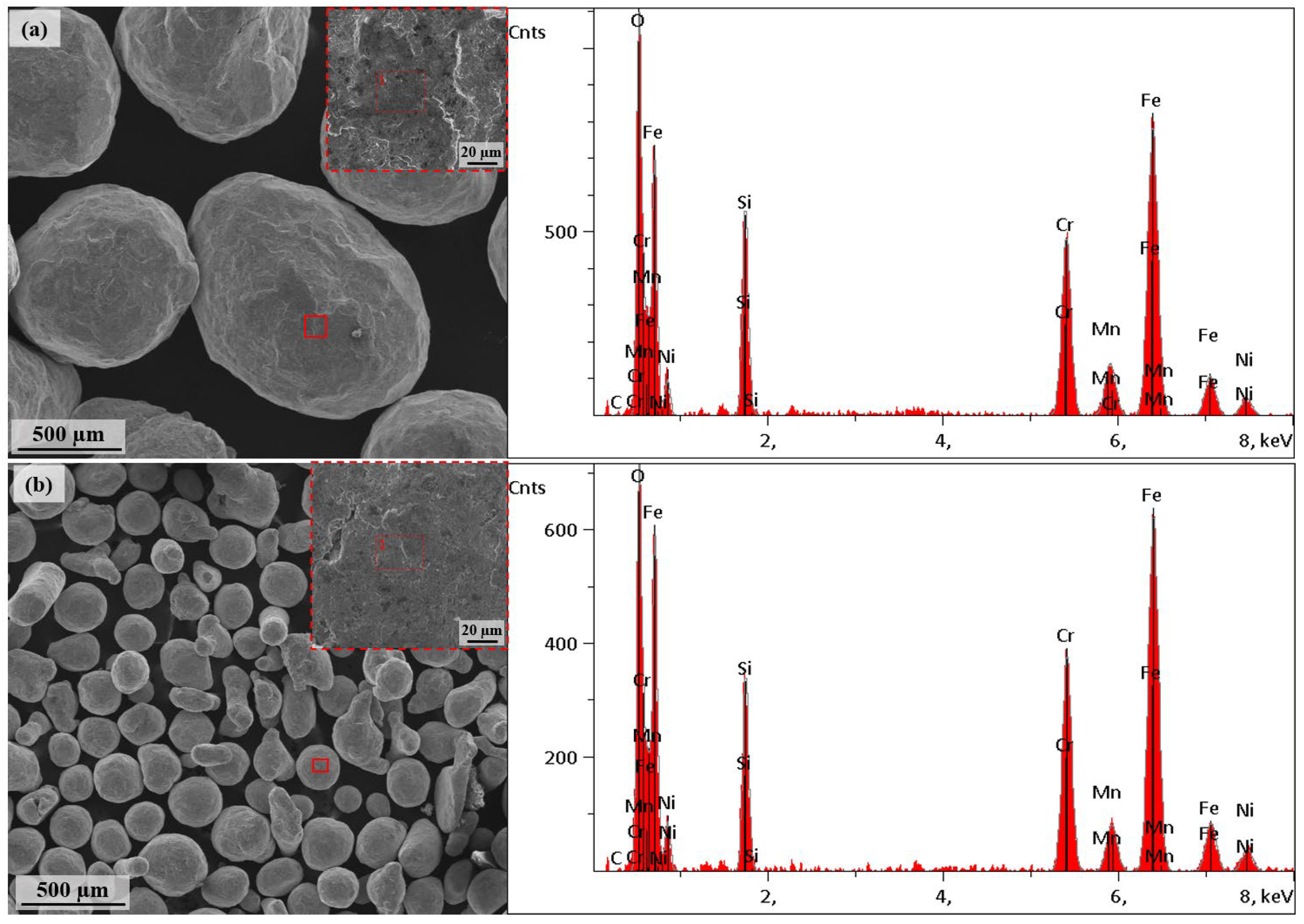
Figure 2.
Representation of surface topography analysis of shot-peened Ti6Al4V alloy.
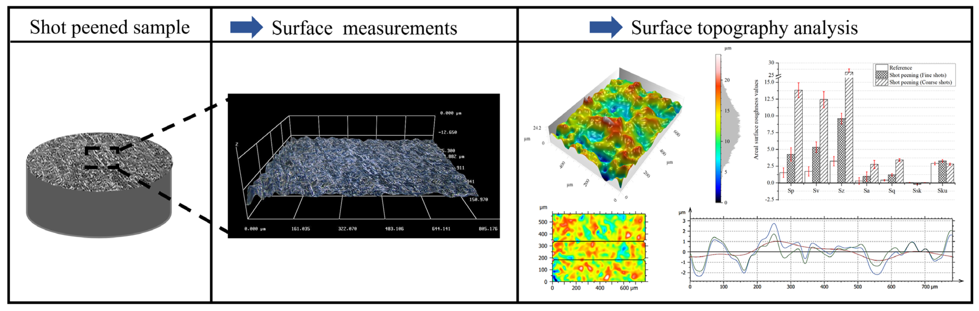
Figure 3.
(a) Low magnification and (b) high magnification SEM images of the surface of conventionally shot-peened Ti6Al4V.
Figure 3.
(a) Low magnification and (b) high magnification SEM images of the surface of conventionally shot-peened Ti6Al4V.
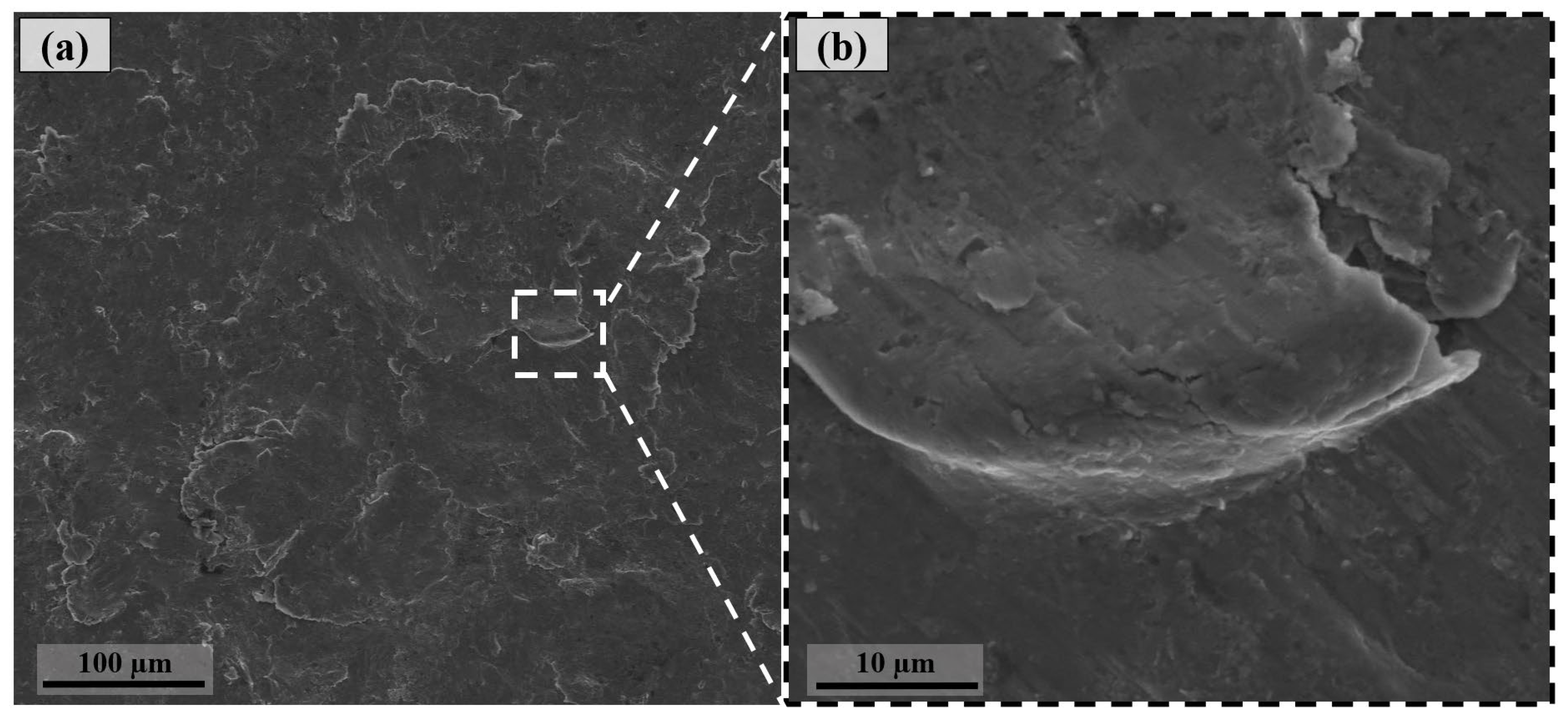
Figure 4.
(a) Low magnification and (b) high magnification SEM images of the surface of fine shot-peened Ti6Al4V.
Figure 4.
(a) Low magnification and (b) high magnification SEM images of the surface of fine shot-peened Ti6Al4V.
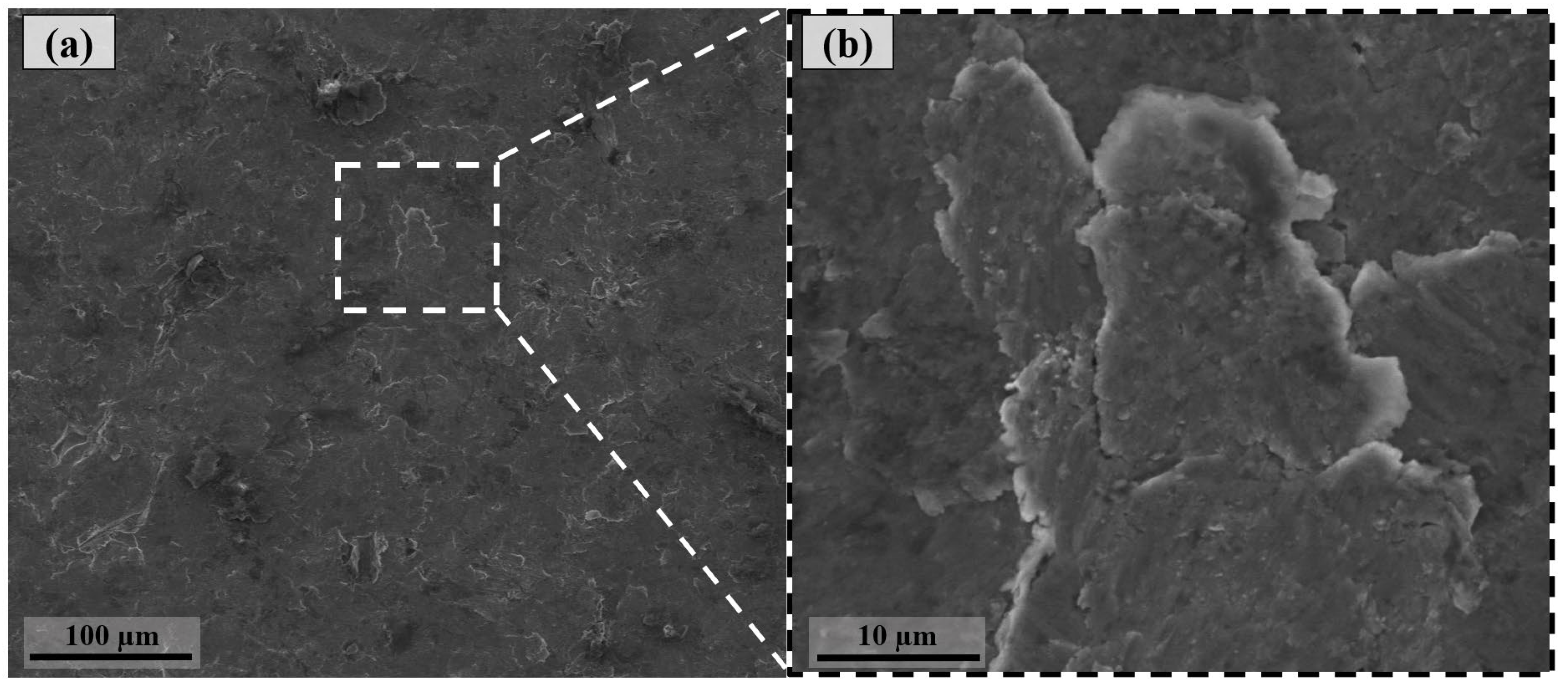
Figure 5.
(a) Three-dimensional surface topography, (b) 2D roughness map, and (c) areal surface roughness values of conventionally shot-peened Ti6Al4V.
Figure 5.
(a) Three-dimensional surface topography, (b) 2D roughness map, and (c) areal surface roughness values of conventionally shot-peened Ti6Al4V.
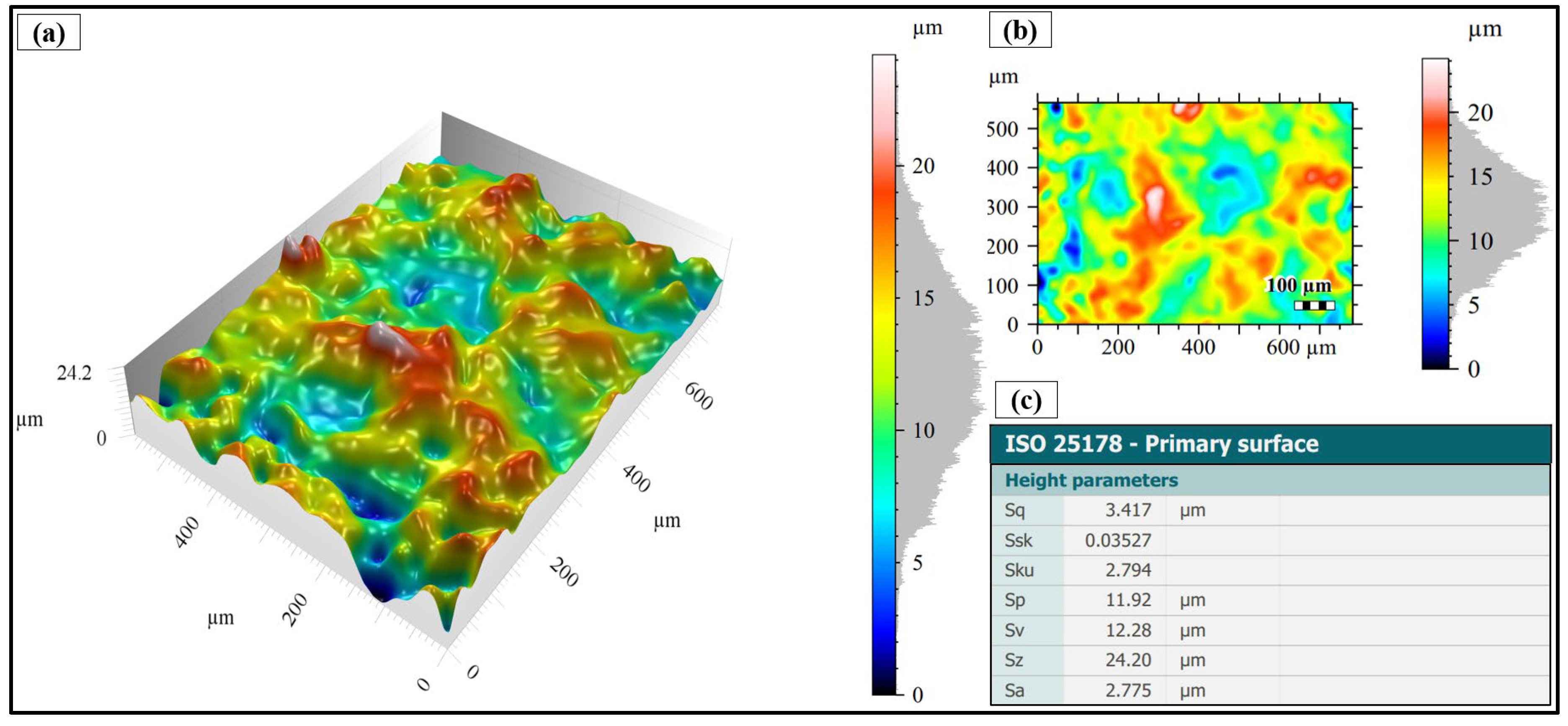
Figure 6.
(a) Three-dimensional surface topography, (b) 2D roughness map, and (c) areal surface roughness values of fine shot-peened Ti6Al4V.
Figure 6.
(a) Three-dimensional surface topography, (b) 2D roughness map, and (c) areal surface roughness values of fine shot-peened Ti6Al4V.
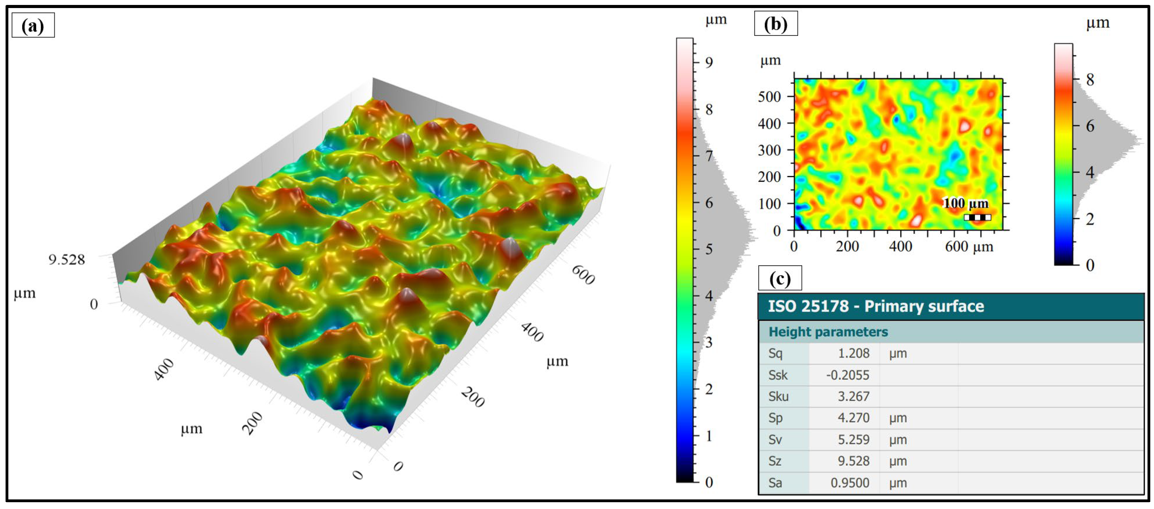
Figure 7.
(a) Areal surface roughness results and (b) roughness profiles of conventionally and fine shot-peened Ti6Al4V.
Figure 7.
(a) Areal surface roughness results and (b) roughness profiles of conventionally and fine shot-peened Ti6Al4V.
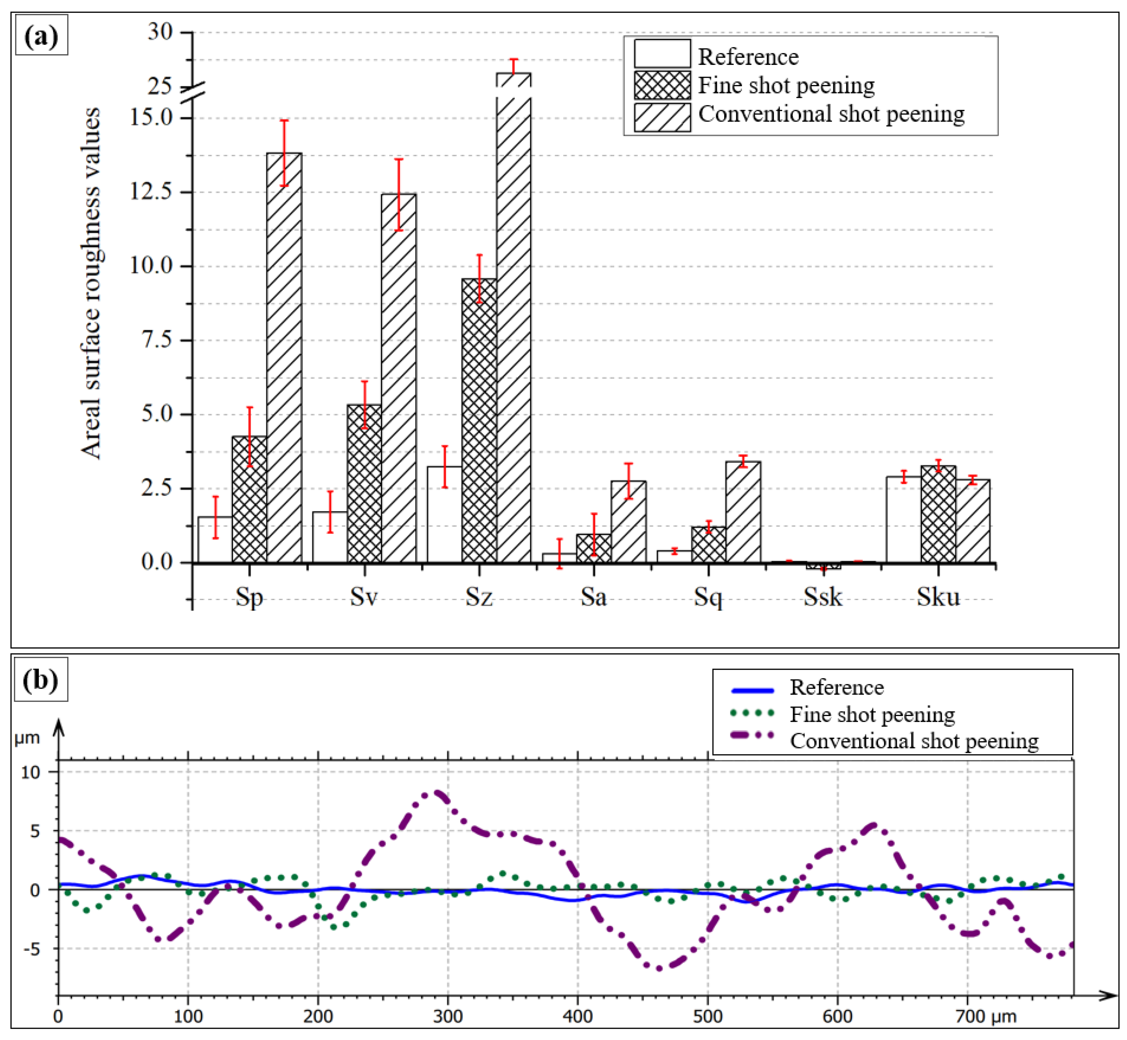
Figure 8.
Contact angle measurements.
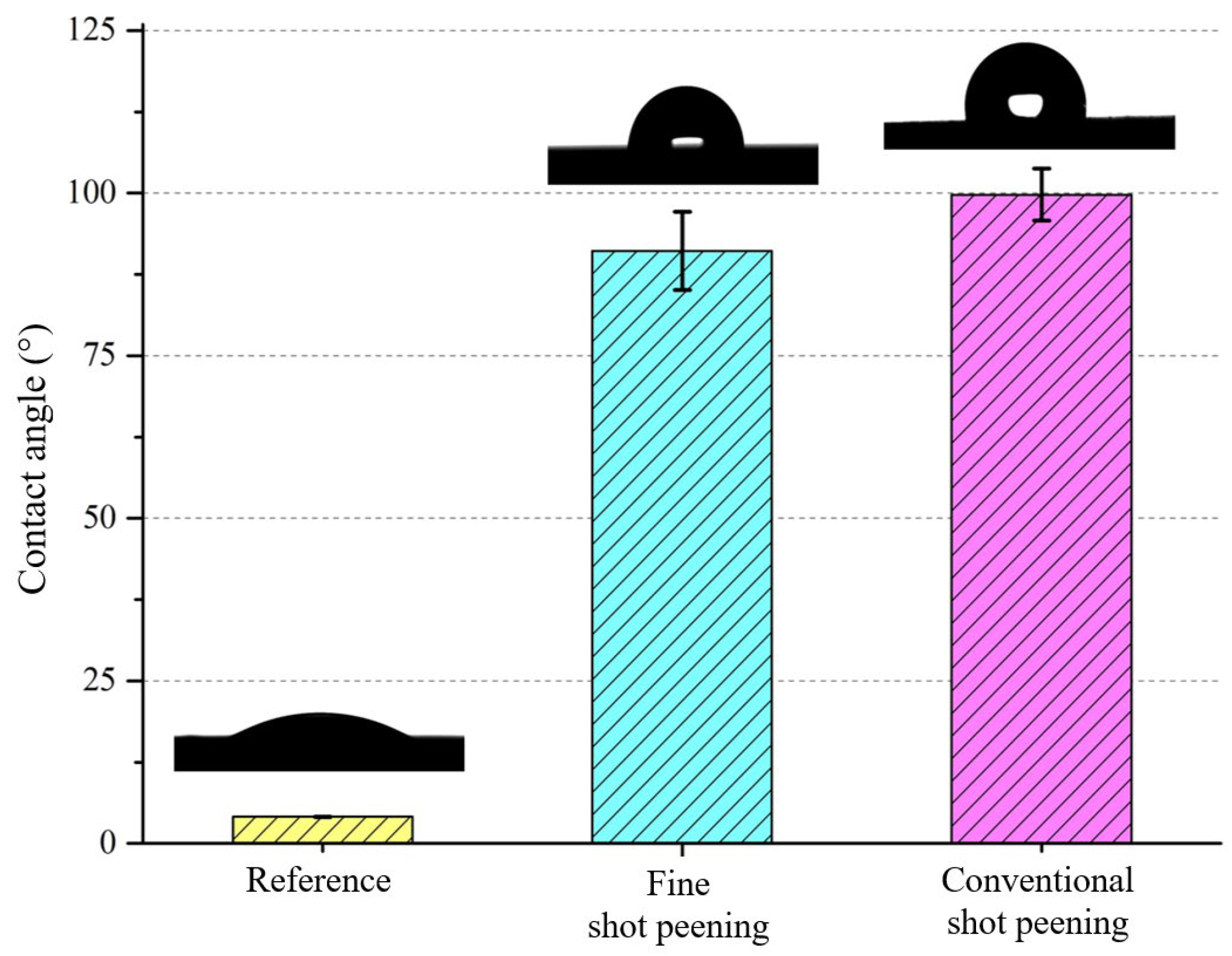
Figure 9.
Antibacterial performance of samples against S. aureus after 6 hours of incubation. Representative images of bacterial colony formation on (a) control agar plate (no material), (b) untreated Ti6Al4V reference sample, (c) fine shot-peened Ti6Al4V surface, and (d) conventionally shot-peened Ti6Al4V surface.
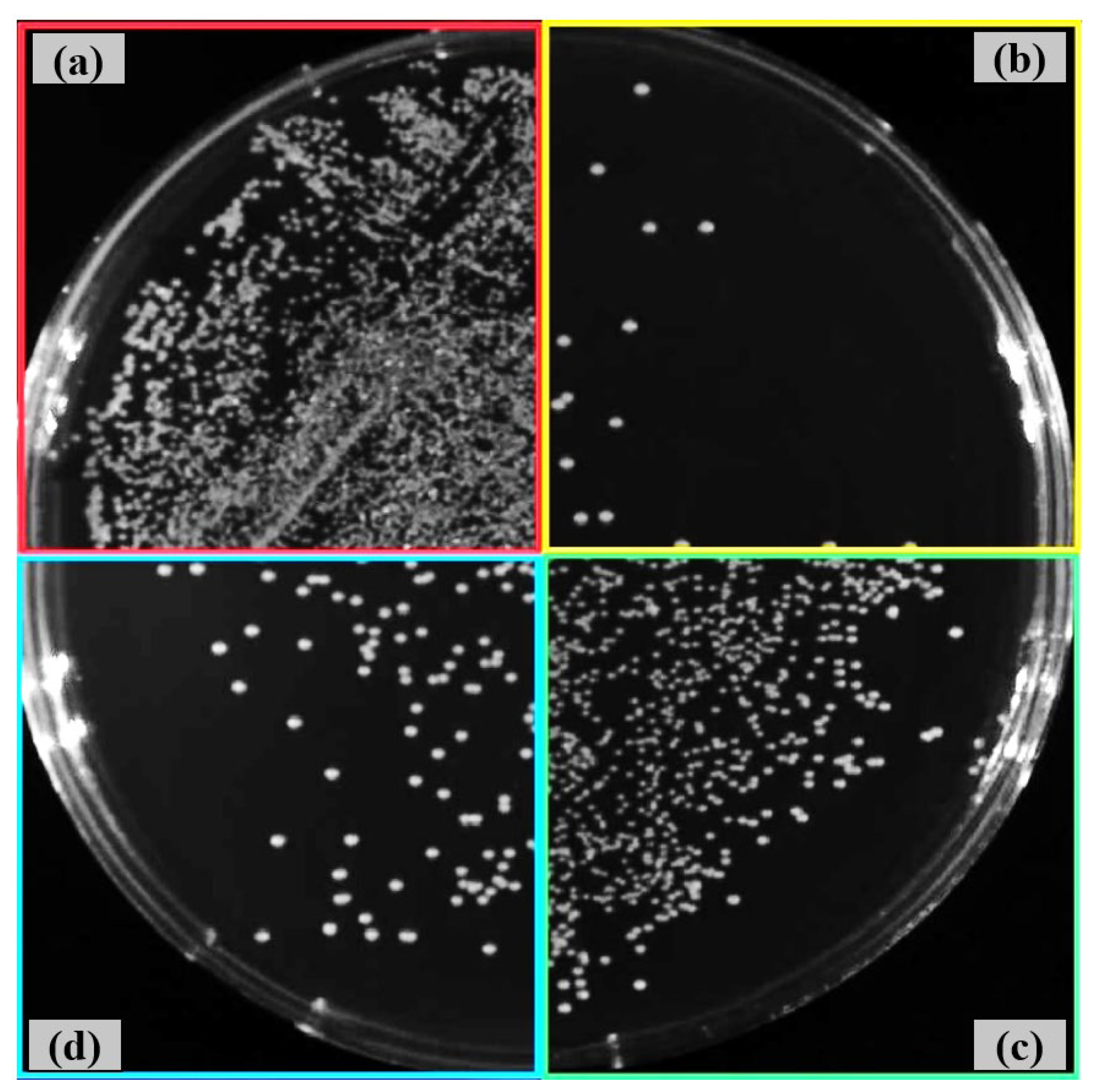
Figure 10.
Antibacterial performance of samples against E. Coli after 6 hours of incubation. Representative images of bacterial colony formation on (a) control agar plate (no material), (b) untreated Ti6Al4V reference sample, (c) fine shot-peened Ti6Al4V surface, and (d) conventional shot-peened Ti6Al4V surface.
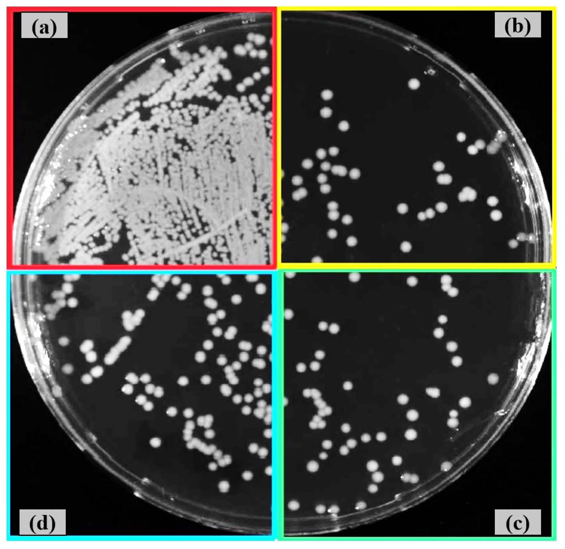
Figure 11.
Reduction rates (%) of E. coli and S. aureus after 6 h incubation on Ti6Al4V surfaces: untreated, fine shot-peened, and conventionally shot-peened.
Figure 11.
Reduction rates (%) of E. coli and S. aureus after 6 h incubation on Ti6Al4V surfaces: untreated, fine shot-peened, and conventionally shot-peened.


Table 1.
Chemical composition of Ti6Al4V alloy.
| Chemical Composition (wt. %) | |||||
|---|---|---|---|---|---|
| V | Al | Fe | Sn | Si | Ti |
| 2.769 | 5.629 | 0.089 | 0.006 | 0.052 | 91.455 |
Table 2.
Shot peening parameters used for peening of the Ti6Al4V alloy.
| Parameter | Value |
|---|---|
| Shot type | Stainless steel shots |
| Shot hardness | 460 HV |
| Shot size | 100-200 µm (Fine shot peening) |
| 700-1000 µm (Conventional shot peening) | |
| Acceleration pressure | 5 bar |
| Duration | 2.5 min |
| Standoff distance | 40 mm |
| Nozzle diameter | 4.76 mm |
Disclaimer/Publisher’s Note: The statements, opinions and data contained in all publications are solely those of the individual author(s) and contributor(s) and not of MDPI and/or the editor(s). MDPI and/or the editor(s) disclaim responsibility for any injury to people or property resulting from any ideas, methods, instructions or products referred to in the content. |
© 2025 by the authors. Licensee MDPI, Basel, Switzerland. This article is an open access article distributed under the terms and conditions of the Creative Commons Attribution (CC BY) license (http://creativecommons.org/licenses/by/4.0/).
Copyright: This open access article is published under a Creative Commons CC BY 4.0 license, which permit the free download, distribution, and reuse, provided that the author and preprint are cited in any reuse.


