Submitted:
14 August 2025
Posted:
14 August 2025
You are already at the latest version
Abstract
In recent days graphene oxide has attracted great attention in wide areas for its structural, mechanical and thermal properties which enables it to be tailored in different applications. Ozonation of the graphene oxide has been noted to increase the degree of oxidation. Investigation of X-ray diffraction by several groups observed data showing a reduced interlayer spacing in the OGO films relative to GO, while other groups have observed an increase in the interlayer distance. This research therefore aimed to investigate and clarify the discrepancy. The results from this experiment agree that Ozonation results leads to reduced interlayer spacing in OGO films relative to GO.
Keywords:
graphene oxide
; ozonated graphene oxide
; X-ray diffraction
; interlayer distance
Introduction
Graphene oxide (GO) contains several chemical functional groups that are attached to the graphite basal plane and can be manipulated to tailor GO for specific applications through chemical reactions. Ozonation of the graphite oxide has been noted to increase the degree of oxidation, hence increase in hydroxyl,laxtol, ester and ketone content in GO(Gao, Wu et al. 2014). Various researchers have looked at the X-ray diffraction (XRD) for free standing OGO films, Gao, Wu etal(Gao, Wu et al. 2014)observed data showing a reduced interlayer spacing in the OGO films relative to GO, in his observation a (002) diffraction peak at 2 theta of 11.8 degree was observed for GO films, which corresponded to an interlayer distance of 7.5A at ca.20% relative humidity. In OGO a sharper (002) peak was observed in his work at 12.2 degree that corresponded to an interlayer distance of 7.3A (at the same humidity). Other researchers have observed an increase in the interlayer distance in the oxidation of the graphite oxide(Jeong, Jin et al. 2009) This research therefore aimed to investigate and clarify the discrepancy.
GO Preparation
GO was prepared according to the improved method of (Marcano, Kosynkin et al. 2010) whereby a 0.1 mixture of concentrated H2SO4/ H3PO4 (360:40ml) was added to a mixture of graphite flakes (3.0g, 1wt equiv) and KMnO4 (18.0g,6wt equiv), this reaction produced a light
exotherm to 35-40 °C. The reaction was then heated to 50°C and stirred for 12h. After this process, the reaction was cooled to room temperature and poured into ice (400mL) then 3ml H2O2 was added and the solution was washed with water for several times and later resuspended in DI water for use
UV-Vis
The UV-Vis was done using Berkman coulter (DUR spectrophotometer) 800, from 200.0nm wavelength to 800.0nm, the wavelength interval was 1.0nm while the scan speed was 1200nm/min. The temperature was 25°C.
Raman Spectra
Raman spectra were done by DXR Raman microscope (thermo scientific) with 532nm laser at room temperature.
Fourier Transformation infrared (FT-IR)
Fourier transformation infrared (FT-IR) spectra of the samples were obtained between 400 and 4000cm-1 on KBr powder with FTIR spectrometer (AVATAR 360, Nicolet, Madison,USA). A minimum of 24 scans were performed.
X-Ray Diffraction (XRD)
X-ray diffraction (XRD) patterns of the samples were examined using a reflection scan with nickel filtered Cu Ka radiation (D8 advance Brunker-AXS, Germany) (°=1.5418Å).The measurements were performed at 2 Theta (degree) between 20°C and 80°C
Ozonation
The OGO was prepared as described by (Gao, Wu et al. 2014) (Gao, Wu et al. 2014). This involved the Ozonation where O3 gas was passed through a GO dispersion with a mild water bath sonication for one hour. A noticeable colour change of the solution to light brown was observed after Ozonation.
Results and Discussion
The UV-Vis of GO yields a distinct peak at ca.230nm, which has been assigned to various oxygen functionalities on the graphine-oxide surface (Gao, Wu et al. 2014).
Figure 1.
(a &b): graphine oxide suspended in DI water. a) Shows the GO solution before H2O2 is added, b) shows the GO solution after the H2O2.
Figure 1.
(a &b): graphine oxide suspended in DI water. a) Shows the GO solution before H2O2 is added, b) shows the GO solution after the H2O2.
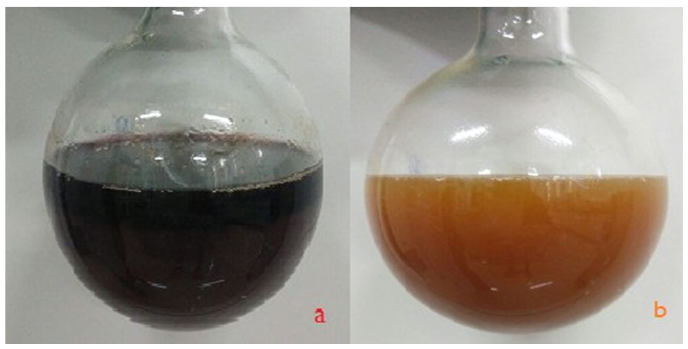
Figure 2.
UV-Vis for GO and OGO.
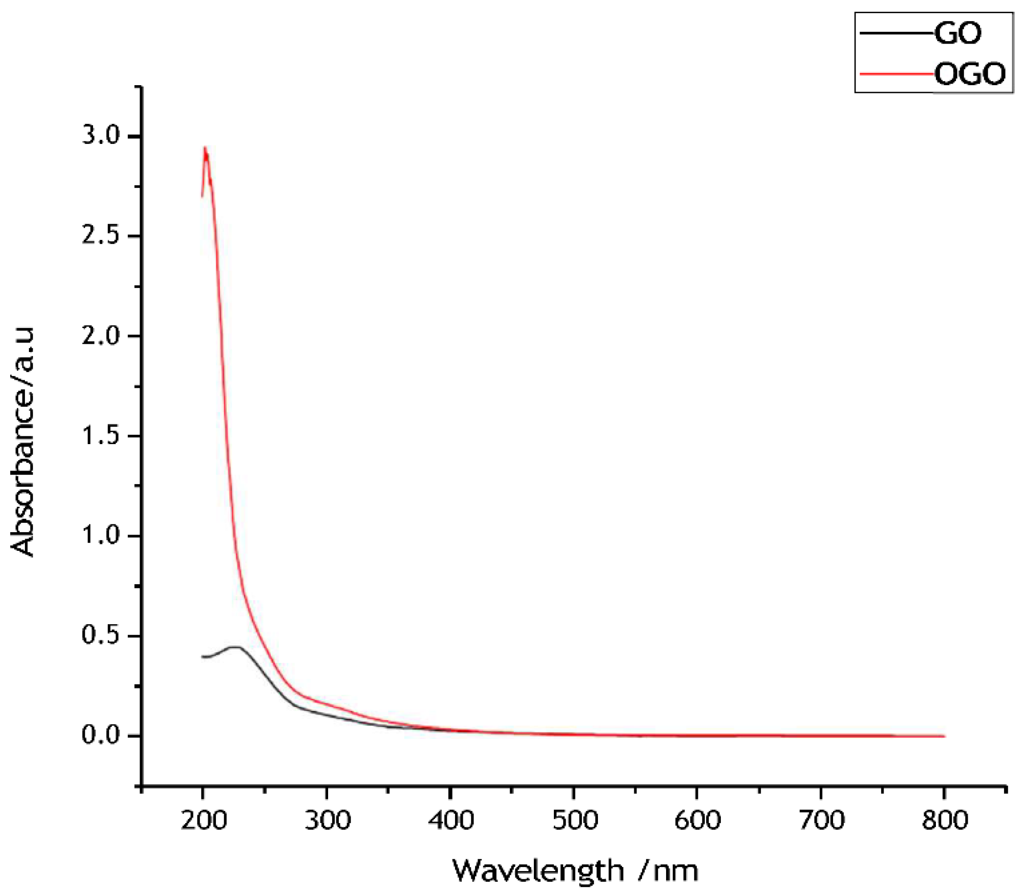
Figure 3.
Raman spectra for GO and OGO.
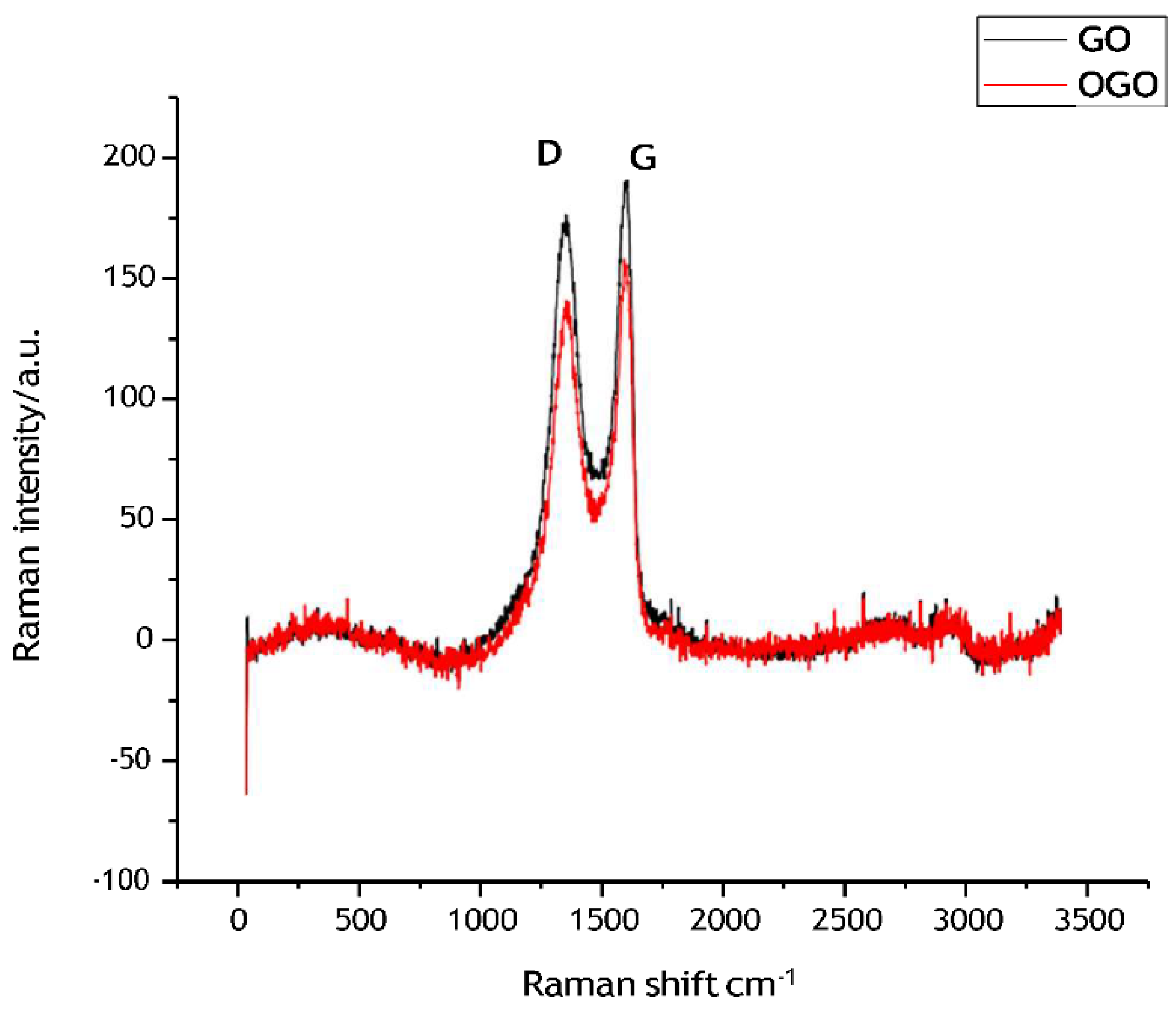
The ratio of ID/IG of GO is 173.57/139.50=1.24 while that of OGO is 189.85/158.66= 1.02 which shows a decrease.
Figure 4.
a&b Fourier transformation infrared (FT-IR) spectra for GO and OGO.
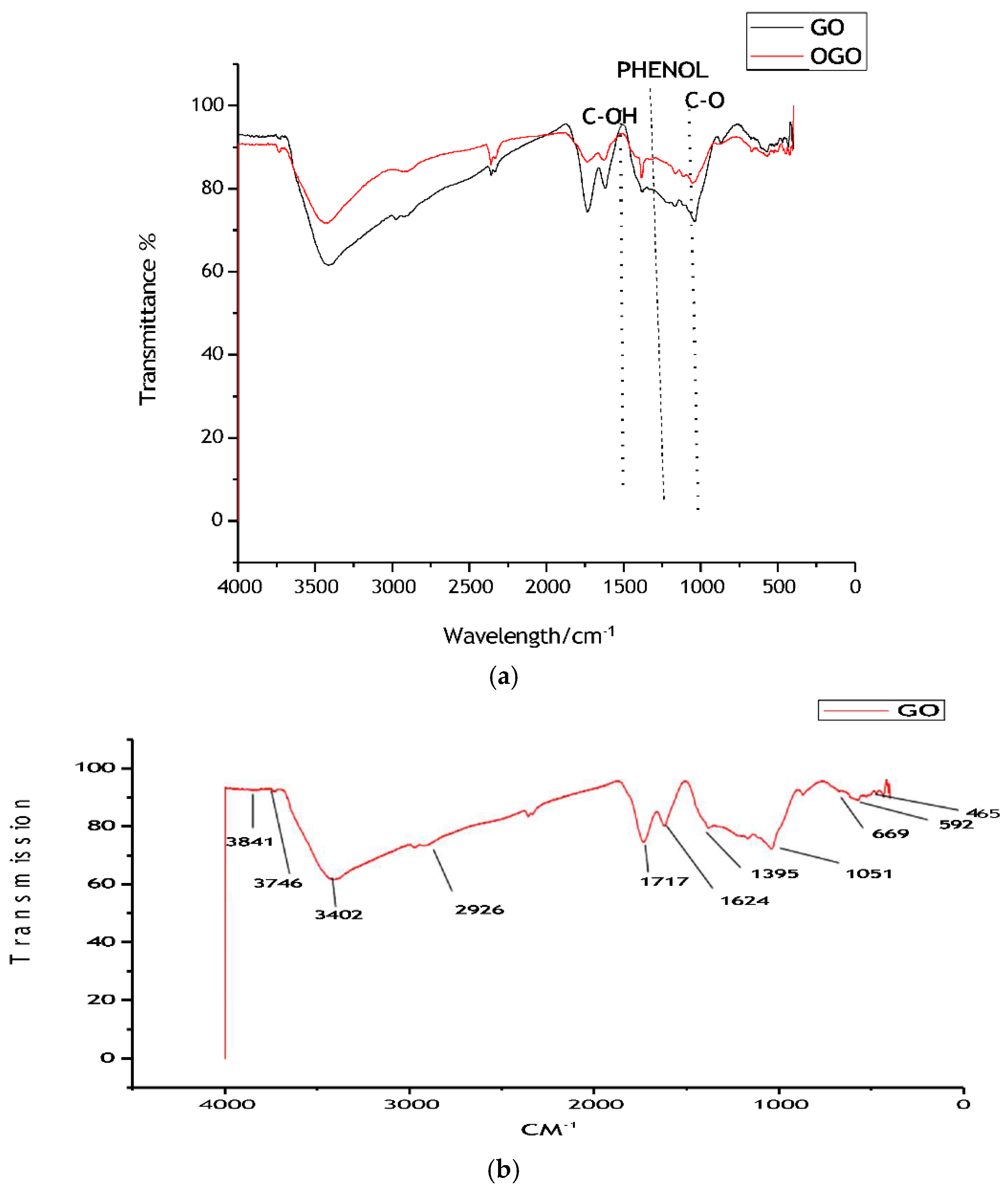
Figure 5.
XRD for GO and OGO. Software used for XRD data interpretation: (Software Used: Jade5lv).
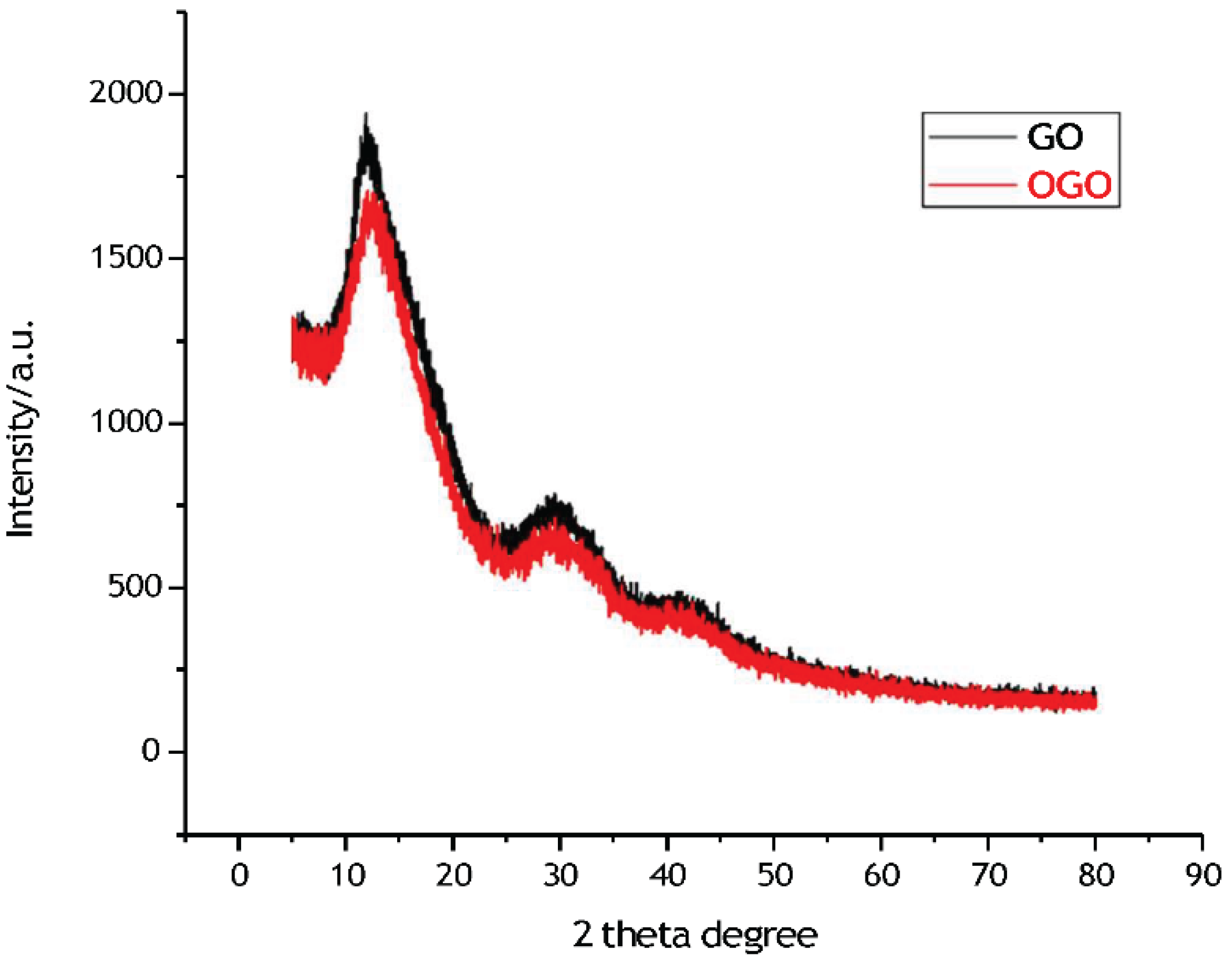
Graphene oxide had the diffraction angle at 2 theta degree and interplanar spacing values at 11.9 degree and 7.4 Å respectively whereas OGO peak was observed at 12.548(12.6) degree and 7.04861 (7.1) Å. The data from UV-VIS, Fourier transformation infrared (FT-IR) and Raman spectra confirmed the conversion of graphite oxide to ozonated graphine oxide. It agrees with other work done on graphene oxide.
Conclusions
My finding agrees with the work of (Gao, Wu et al. 2014) that the Ozonation results in the decrease of the interplanar distance OGO 12.6 degree 7.1Å interlayer distance relative to GO at
11.9 degree and 7.4 Å interlayer distance. Further research is needed graphene oxide oxidation by O2 and O3.
Acknowledgment
I would like to thank the Chinese government scholarship council for CSC scholarship.
References
- Gao, W., G. Wu, M. T. Janicke, D. A. Cullen, R. Mukundan, J. K. Baldwin, E. L. Brosha, C. Galande, P. M. Ajayan, K. L. More, A. M. Dattelbaum and P. Zelenay (2014). “Ozonated Graphene Oxide Film as a Proton-Exchange Membrane.” Angewandte Chemie International Edition 53(14): 3588-3593. [CrossRef]
- Jeong, H. K., M. H. Jin, K. P. So, S. C. Lim and Y. H. Lee (2009). “Tailoring the characteristics of graphite oxides by different oxidation times.” Journal of Physics D: Applied Physics 42(6): 065418. [CrossRef]
- Marcano, D. C., D. V. Kosynkin, J. M. Berlin, A. Sinitskii, Z. Sun, A. Slesarev, L. B. Alemany.
- W. Lu and J. M. Tour (2010). “Improved Synthesis of Graphene Oxide.” ACS Nano 4(8): 4806- 4814.
Disclaimer/Publisher’s Note: The statements, opinions and data contained in all publications are solely those of the individual author(s) and contributor(s) and not of MDPI and/or the editor(s). MDPI and/or the editor(s) disclaim responsibility for any injury to people or property resulting from any ideas, methods, instructions or products referred to in the content. |
© 2025 by the authors. Licensee MDPI, Basel, Switzerland. This article is an open access article distributed under the terms and conditions of the Creative Commons Attribution (CC BY) license (http://creativecommons.org/licenses/by/4.0/).
Copyright: This open access article is published under a Creative Commons CC BY 4.0 license, which permit the free download, distribution, and reuse, provided that the author and preprint are cited in any reuse.


