
Submitted:
07 August 2025
Posted:
08 August 2025
You are already at the latest version
Abstract
IFN-γ, a key player in the pathogenesis of deep vein thrombosis (DVT), acts as a bridge between immune responses and coagulation activation. It promotes endothelial activation, leukocyte recruitment, cytokine release, and coagulation imbalance. This critical pro-inflammatory and prothrombotic role makes IFN-γ a promising molecular target in DVT management beyond traditional anticoagulation methods. Exploring pharmacological inhibition of IFN-γ signaling or its downstream effectors could open new therapeutic pathways for DVT, enhancing resolution and averting post-thrombotic complications. This review summarizes pathophysiology, diagnostics, and management of DVT, and adds further information on the role of IFN-γ as a key cytokine to target to advance DVT therapeutics.
Keywords:
deep vein thrombosis (DVT)
; IFN-γ
; Neutrophil Extracellular Traps (NETs)
; Natural Killer (NK) cells
; pathophysiology
; management
1. Introduction
Deep vein thrombosis (DVT) can occur in various parts of the body, with the legs being the most common location [1,2,3]. It affects approximately 0.1 % of the population annually [4]. Primary causes include decreased blood flow, which can happen due to stasis, where blood stays at one location for an extended period of time. Other risk factors for DVT include immobility, malignancy, hip fracture, pregnancy, recent surgery, and a family history of the condition [1,5,6]. Several studies have demonstrated the occurrence of DVT in cancer patients undergoing immunotherapies, including immune checkpoint inhibitors (ICIs), immunotherapy agents (ipilimumab, nivolumab, pembrolizumab, atezolizumab, avelumab, or durvalumab), chimeric antigen receptor (CAR) T-cells, and bispecific T-cell engagers [7,8].
To diagnose DVT, healthcare professionals utilize various tests to accurately identify the condition. These diagnostic methods include the D-dimer blood test, this test detects elevated levels of D-dimer in the blood, signaling the existence of blood clots. Other tests are duplex ultrasound, which aids in visualizing the veins to detect any signs of DVT, and venography is another method to visualize the veins and identify potential blood clots. Additionally, magnetic resonance imaging (MRI) scans are utilized to detect DVT in different regions of the body [9,10]. These diagnostic tools play a crucial role in identifying and confirming the presence of DVT, allowing for timely and appropriate medical intervention
The pathophysiology involves immune-related endothelial activation, cytokine production, neutrophil extracellular traps (NETs), and C-Reactive protein (CRP)-mediated immunothrombosis [6,11,12]. Currently available therapeutic and management approaches of DVT include: anticoagulants and thrombolytics, and compression stockings help prevent clot formation and further complications [10,13]. In critical situations, clot removal surgery may be necessary. Inferior vena cava (IVC) filters can be inserted to prevent clots from traveling to the lungs [14]. Anticoagulant treatments traditionally focus on coagulation factors, overlooking the crucial aspect of inflammation. Inflammatory conditions in DVT can lead to increased production of IFN-γ, highlighting the significance of this cytokine in the inflammatory response to DVT [3,15]. Inflammatory response can be acute as a protective mechanism or can be chronic, leading to tissue changes and thrombus formation. Symptoms of DVT may include pain, swelling, redness, and visible veins in the affected area [10]. It is crucial to address DVT promptly, as it can lead to life-threatening complications such as pulmonary embolism when a blood clot travels to the lungs [1,5,16].
Exploring the role of immune cells introduces new possibilities for supplementary anti-inflammatory approaches [12,17]. These may involve targeting cytokines, NETosis, or adjusting macrophage phenotypes. The aim is to enhance thrombus resolution, minimize complications, and mitigate bleeding risks [17]. Research has shown that inhibiting IFN-γ signaling can lead to thrombosis resolution and reduced fibrosis in DVT animal models [18]. According to the updated American Society of Hematology guidelines, monitoring the IFN-γ levels of DVT patients is essential [14]. These diverse approaches aim to effectively manage DVT and reduce associated risks, highlighting the importance of tailored treatment strategies based on individual patient needs
In this insightful review, the critical role of immune cells in the pathophysiology of DVT is explored, alongside the diagnostic approach and management strategies for DVT. The report emphasizes the importance of IFN-γ in triggering DVT and proposes the inhibition of IFN-γ as a potential treatment for this condition.
2. Role of Immune Cells in the Pathophysiology of DVT
Immune cells are key players in inflammatory processes that contribute to clot formation, playing a vital role in the development and progression of DVT. The interaction between the innate immune system and the coagulation cascade leads to the initiation and propagation of thrombosis [17,19]. DVT, a condition resulting from a complex interplay among coagulation, vascular endothelium, and immune system components, involves a significant role played by the immune system [17]. Innate immune cells like neutrophils, monocytes/macrophages, and lymphocytes actively engage in various stages of venous thrombi formation. This interaction has given rise to the concept of immunothrombosis, where thrombosis is intricately connected to immune activation and inflammatory responses.
Neutrophils, the predominant leukocytes in thrombi, contribute to DVT through the generation of neutrophil extracellular traps (NETs). These web-like structures made of chromatin not only ensnare pathogens but also serve as a scaffold for fibrin and platelets, facilitating clot development and stability [12,17]. NETosis plays a central role in the initial stages of thrombus formation, fueling coagulation and inflammation processes [12]. Monocytes infiltrate thrombi and transform into macrophages, expressing tissue factor (TF) to trigger coagulation cascades and recruit platelets. Additionally, macrophages aid in thrombus resolution by releasing fibrinolytic enzymes crucial for breaking down fibrin and collagen, leading to remodeling. During the resolution phase, pro-inflammatory macrophages ("M1") transition into pro-resolving ("M2") phenotypes. NK cells, B cells, and T cells also impact thrombus progression [17].
These cells trigger activated endothelial cells, leading to the release of Weibel-Palade bodies, reactive oxygen species, inflammasomes, and cytokines such as IL-6 and MCP-1 contribute to a pro-inflammatory microenvironment [12]. This environment promotes immune cell recruitment and activation, leading to coagulation and thrombus maturation [17]. Tissue factor expression on immune cells and platelets initiates thrombin generation and fibrin production, facilitating the formation of the thrombus scaffold [17].
Immune cells play a crucial role in not just initiating and spreading responses, but also in regulating the breakdown of blood clots [17]. Macrophages are key players in this process as they secrete fibrinolytic enzymes and facilitate the restructuring of the extracellular matrix through collagenolysis, ultimately restoring blood flow. Neutrophils also contribute to clot dissolution, although their dysregulation can potentially prolong inflammation. Additionally, endothelial cells participate by re-endothelializing the thrombus and can undergo changes that influence the resolution of the clot. When these processes fail, it can result in issues like chronic venous obstruction, valvular damage, and post-thrombotic syndrome (PTS) [17].
Venous stasis, endothelial injury/dysfunction, and hypercoagulability (known as Virchow’s triad) initiate immune cell adhesion to activated endothelium, involving neutrophils and monocytes [20]. These cells release proinflammatory cytokines, express tissue factor (TF), and trigger platelet activation, promoting localized thrombin production and fibrin mesh formation. Neutrophils undergo NETosis, releasing DNA and proteins that entrap erythrocytes and platelets, enhancing clot stability. Following thrombus formation, macrophages infiltrate to engulf debris, release plasminogen activators and matrix metalloproteinases (MMPs), facilitating the shift from inflammation to repair phases crucial for clot dissolution and vein wall healing [17]. Diverse lymphocyte subsets regulate inflammation levels, influencing the resolution or aggravation of thrombotic processes alongside other immune factors. Molecules like IL-6, MCP-1, MIP-1, and CINC are elevated in thrombotic areas, orchestrating immune recruitment and activation. Effective thrombus resolution hinges on well-coordinated immune signaling to promote fibrinolysis, collagen restructuring, and endothelial repair. Imbalance in this process can lead to adverse outcomes, such as post-thrombotic syndrome [21,22].
Recent research has shed light on the impaired function and levels of natural killer (NK) cells in the blood of individuals with DVT and pulmonary embolism [23,24,25]. NK cells may have a role in the development and progression of DVT, potentially impacting its prognosis. Particularly, through the production of interferon-gamma (IFN-γ), NK cells have been shown to trigger the formation of neutrophil extracellular traps (NETs) [26,27]. These NETs, released by neutrophils, contribute to thrombus formation, thereby increasing the risk of DVT [26]. Studies have indicated reduced thrombotic formation in NK cell-depleted or IFN-γ knock-down mice, suggesting a correlation between NK cells, IFN-γ, and venous thrombosis [26]. Further observations have revealed an imbalance of NK cell subsets in DVT patients' peripheral blood, with significantly low levels of CD56dimCD16+ NK cells and notably higher levels of CD56-CD16+ NK cells. Additionally, markers like citrullinated histone (H3) were identified in the peripheral blood of DVT patients [23]. While additional research is essential, these findings suggest that NK cells, through their role in NET formation and the alterations in their subsets, could potentially influence the severity and prognosis of DVT. Moreover, targeting IFN-γ might serve as a therapeutic intervention aimed at inhibiting thrombosis.
3. IFN-γ and Its Role in the Pathophysiology of DVT
Interferon gamma (IFN-γ) plays a crucial role as a key type II interferon cytokine in regulating pro-inflammatory and immunoregulatory processes during inflammation [28]. This pivotal cytokine is predominantly generated by immune cells such as NK cells, activated Th1 CD4+ T cells, and CD8+ cytotoxic T cells [29,30,31,32]. These immune cells secrete IFN-γ in response of pathogens or when stimulated with cytokines like IL-2, IL-12, IL-18, etc [31,33]. This cytokine plays a crucial role in coordinating both innate and adaptive immune responses [32,34]. By triggering cellular mechanisms that combat pathogens, tumors, and regulate inflammation, IFN-γ serves as a linchpin in the immune system's functionality [32]. Its functions include activating macrophages, improving antigen presentation, stimulating the expression of inflammatory mediators, and influencing hematopoiesis. Notably, IFN-γ serves a dual purpose by promoting inflammation while also regulating feedback mechanisms to prevent excessive immune responses, thereby contributing to immune homeostasis [32]. The signaling of IFN-γ primarily occurs through its heterodimeric receptor complex (IFNGR1/IFNGR2), setting off the JAK-STAT1 pathway [30,34,35,36,37]. This activation leads to the transcription of interferon-stimulated genes (ISGs) that are closely associated with various aspects of immunity, inflammation, cell cycle regulation, apoptosis, and antigen presentation [30]. IFN-γ stimulates the production of pro-inflammatory cytokines, facilitates the generation of reactive oxygen/nitrogen species (ROS/RNS), and bolsters leukocyte recruitment and activation [38]. The intricate interplay orchestrated by IFN-γ demonstrates its profound impact on bolstering the body's defense mechanisms against a spectrum of threats, but it can also contribute to inflammatory conditions [29,38]. The dysregulation of IFN-γ signaling is associated with autoimmune diseases, infectious disease pathology, cancer immunology, and chronic inflammatory conditions [28,34,35].
IFN-γ plays a pivotal role in the pathophysiology of DVT [12,39]. It impacts endothelial cells by modifying their barrier function, increasing permeability, and inducing the expression of adhesion molecules like ICAM-1 and VCAM-1, thereby promoting inflammation. Additionally, IFN-γ triggers the production of pro-inflammatory cytokines (TNF-α, IL-1β), chemokines, and enhances the activation of the coagulation cascade by boosting tissue factor on monocytes/macrophages. By activating macrophages, recruiting Th1 cells, and supporting their differentiation, IFN-γ sustains local inflammation and thrombogenic signals. Furthermore, it influences the expression of anticoagulants and fibrinolytic factors, leading to a pro-thrombotic environment by reducing thrombomodulin levels. IFN-γ also stimulates reactive oxygen species (ROS) and reactive nitrogen species (RNS) in vascular cells, potentially causing oxidative endothelial damage and aiding in clot formation. In essence, IFN-γ emerges as a critical factor driving the inflammatory milieu that encourages the development, enlargement, and dissemination of blood clots in DVT [38].
4. Targeting IFN-γ and Its Signaling to Advance Therapeutics of DVT
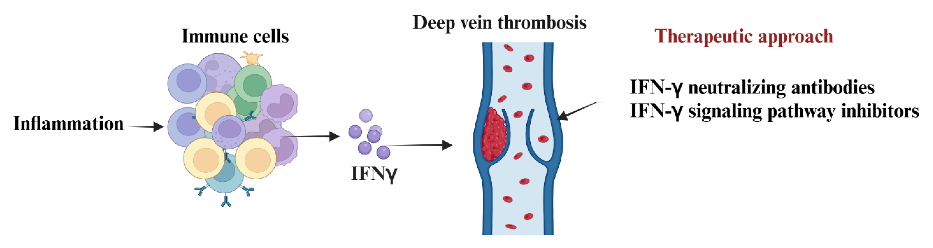
Targeting IFN-γ and its signaling could be a promising approach to combat the inflammation-driven progression of DVT [38,39,40]. Potential interventions include using IFN-γ neutralizing antibodies or inhibitors like monoclonal antibodies (e.g., emapalumab, FDA-approved for other cytokine storm syndromes) to alleviate IFN-γ-induced inflammation [41,42]. Additionally, JAK-STAT pathway inhibitors such as ruxolitinib could be utilized to impede the downstream signaling of IFN-γ, thereby reducing prothrombotic inflammation [43,44,45]. Moreover, by modulating endothelial function through targeting IFN-γ-induced pathways like STAT1, p38 MAPK, and small GTPases, it may be possible to maintain vascular barrier integrity effectively [46]. While the current standard management for DVT focuses on anticoagulation to prevent thrombus extension and embolization, addressing the inflammatory aspect, particularly pivotal cytokines like IFN-γ, holds the potential to enhance outcomes, minimize recurrence, and promote thrombus resolution (Figure 1).
Recent research findings reveal that the use of anti-IFN-γ monoclonal antibodies post-DVT significantly hastened the resolution of venous thrombus and reduced fibrosis in murine models [47]. Remarkably, this treatment demonstrated these benefits without affecting the overall coagulation function systemically [47]. Administration of antibodies against IFN-γ accelerated thrombotic resolution in DVT wild-type mice [39,48], and also reduced the number of NETs formed [49]. Selective dampening of IFN-γ-mediated inflammation presents a novel approach to enhancing thrombus breakdown and promoting vein wall healing. This targeted strategy offers a distinct advantage over conventional anticoagulation methods. While standard anticoagulants focus on preventing clot progression, they fall short in expediting natural clot resolution and mitigating inflammation [50]. By neutralizing IFN-γ, a more tailored and effective treatment path emerges, fostering optimal recovery outcomes. Further experimental and clinical investigations are crucial to assess the impact of IFN-γ modulation in the treatment of venous thrombosis.
5. Conclusion
Investing further in the role of immune cells in the onset and progression of DVT could help to develop advanced therapeutics for DVT. Exciting insights into immune responses shed light on the mechanisms behind certain conditions, offering potential paths for future therapies. Evidence points to the crucial involvement of NK cells, especially in NET formation, in the progression of DVT. The emerging importance of NK cell subsets and associated markers for diagnosing DVT and tailoring treatments underscores their relevance in comprehending and addressing this condition. This analysis underscores the intricate impact of IFN-γ on both the development and treatment of DVT. Interferon-gamma (IFN-γ) plays a crucial role in the development of DVT. It promotes endothelial dysfunction, inflammatory cell recruitment, procoagulant activity, and vascular inflammation, all contributing to thrombus formation and progression. Targeting IFN-γ signaling presents a promising approach to complement traditional anticoagulation methods in addressing inflammation-driven thrombotic mechanisms in DVT (Figure 1).
Author Contributions
AJ was the principal investigator, obtained the funding, designed the study, and wrote the manuscript along with KK. KK performed the literature search and wrote the manuscript. All figures “created with BioRender.com.”.
Conflicts of Interest Statement
The authors declare that the work reviewed in the article was conducted in the absence of any commercial or financial relationships that could be construed as a potential conflict of interest.
References
- Duffy S. Understanding patient assessment and treatment in deep vein thrombosis. Nurs Stand. 2022;37(11):71-5. [CrossRef]
- Waheed SM, Kudaravalli P, Hotwagner DT. Deep Vein Thrombosis. StatPearls. Treasure Island (FL) ineligible companies. Disclosure: Pujitha Kudaravalli declares no relevant financial relationships with ineligible companies. Disclosure: David Hotwagner declares no relevant financial relationships with ineligible companies.: StatPearls Publishing Copyright © 2025, StatPearls Publishing LLC.; 2025.
- Navarrete S, Solar C, Tapia R, Pereira J, Fuentes E, Palomo I. Pathophysiology of deep vein thrombosis. Clinical and Experimental Medicine. 2023;23(3):645-54. [CrossRef]
- Kesieme E, Kesieme C, Jebbin N, Irekpita E, Dongo A. Deep vein thrombosis: a clinical review. Journal of Blood Medicine. 2011;2(null):59-69. [CrossRef]
- Bonner L, Johnson J. Deep vein thrombosis: diagnosis and treatment. Nurs Stand. 2014;28(21):51-8; quiz 60. [CrossRef]
- Navarrete S, Solar C, Tapia R, Pereira J, Fuentes E, Palomo I. Pathophysiology of deep vein thrombosis. Clin Exp Med. 2023;23(3):645-54. [CrossRef]
- Fowler C, Pastores SM. Venous Thromboembolic Events in Cancer Immunotherapy: A Narrative Review. Journal of Clinical Medicine. 2025;14(14):4926. [CrossRef]
- Roopkumar J, Kim AS, Bicky T, Hobbs BP, Khorana AA. Venous Thromboembolism in Cancer Patients Receiving Immunotherapy. Blood. 2018;132(Supplement 1):2510-. [CrossRef]
- Linkins LA, Takach Lapner S. Review of D-dimer testing: Good, Bad, and Ugly. Int J Lab Hematol. 2017;39 Suppl 1:98-103. [CrossRef]
- Kruger PC, Eikelboom JW, Douketis JD, Hankey GJ. Deep vein thrombosis: update on diagnosis and management. Med J Aust. 2019;210(11):516-24. [CrossRef]
- Schulman S, Makatsariya A, Khizroeva J, Bitsadze V, Kapanadze D. The Basic Principles of Pathophysiology of Venous Thrombosis. Int J Mol Sci. 2024;25(21). [CrossRef]
- Budnik I, Brill A. Immune Factors in Deep Vein Thrombosis Initiation. Trends in Immunology. 2018;39(8):610-23. [CrossRef]
- Stone J, Hangge P, Albadawi H, Wallace A, Shamoun F, Knuttien MG, et al. Deep vein thrombosis: pathogenesis, diagnosis, and medical management. Cardiovasc Diagn Ther. 2017;7(Suppl 3):S276-s84. [CrossRef]
- Arnold MJ. Venous Thromboembolism: Management Guidelines from the American Society of Hematology. Am Fam Physician. 2021;104(4):429-31.
- Luther N, Shahneh F, Brähler M, Krebs F, Jäckel S, Subramaniam S, et al. Innate Effector-Memory T-Cell Activation Regulates Post-Thrombotic Vein Wall Inflammation and Thrombus Resolution. Circulation Research. 2016;119(12):1286-95. [CrossRef]
- Nitta D, Mitani H, Ishimura R, Moriya M, Fujimoto Y, Ishiwata S, et al. Deep vein thrombosis risk stratification. Int Heart J. 2013;54(3):166-70. [CrossRef]
- Rayes J, Brill A. Hot under the clot: venous thrombogenesis is an inflammatory process. Blood. 2024;144(5):477-89. [CrossRef]
- Mukhopadhyay S, Johnson TA, Duru N, Buzza MS, Pawar NR, Sarkar R, et al. Fibrinolysis and Inflammation in Venous Thrombus Resolution. Front Immunol. 2019;10:1348. [CrossRef]
- Qi N, Lyu Z, Huang L, Zhao Y, Zhang W, Zhou X, et al. Investigating the dual causative pathways linking immune cells and venous thromboembolism via Mendelian randomization analysis. Thrombosis Journal. 2025;23(1):8. [CrossRef]
- Poredos P, Jezovnik MK. Endothelial Dysfunction and Venous Thrombosis. Angiology. 2018;69(7):564-7. [CrossRef]
- Cronkite DA, Strutt TM. The Regulation of Inflammation by Innate and Adaptive Lymphocytes. J Immunol Res. 2018;2018:1467538. [CrossRef]
- Kim Y, Greenleaf WJ, Bendall SC. Systems biology approaches to unravel lymphocyte subsets and function. Current Opinion in Immunology. 2023;82:102323. [CrossRef]
- Alhabibi AM, Wahab MA, Sakr AK, Abd El-Hamid SM, Zakaria MY, Althoqapy AA, et al. The Diagnostic Utility of Natural Killer Cell Subsets in Deep Vein Thrombosis. Vasc Health Risk Manag. 2023;19:779-87. [CrossRef]
- Duan Q, Gong Z, Song H, Wang L, Yang F, Lv W, et al. Symptomatic venous thromboembolism is a disease related to infection and immune dysfunction. Int J Med Sci. 2012;9(6):453-61. [CrossRef]
- Zhang X, Wang Q, Shen Y, Song H, Gong Z, Wang L. Compromised natural killer cells in pulmonary embolism. Int J Clin Exp Pathol. 2015;8(7):8244-51.
- Bertin FR, Rys RN, Mathieu C, Laurance S, Lemarié CA, Blostein MD. Natural killer cells induce neutrophil extracellular trap formation in venous thrombosis. Journal of Thrombosis and Haemostasis. 2019;17(2):403-14. [CrossRef]
- Becker C, Reinhardt C. Unexpected role of natural killer cell‐derived interferon‐γ as a driver of NETosis and DVT. Journal of Thrombosis and Haemostasis. 2019;17(2):400-2. [CrossRef]
- Kopitar-Jerala N. The Role of Interferons in Inflammation and Inflammasome Activation. Front Immunol. 2017;8:873. [CrossRef]
- Alspach E, Lussier DM, Schreiber RD. Interferon γ and Its Important Roles in Promoting and Inhibiting Spontaneous and Therapeutic Cancer Immunity. Cold Spring Harb Perspect Biol. 2019;11(3). [CrossRef]
- Sica A, Dorman L, Viggiano V, Cippitelli M, Ghosh P, Rice N, et al. Interaction of NF-kappaB and NFAT with the interferon-gamma promoter. J Biol Chem. 1997;272(48):30412-20. [CrossRef]
- Szabo SJ, Kim ST, Costa GL, Zhang X, Fathman CG, Glimcher LH. A novel transcription factor, T-bet, directs Th1 lineage commitment. Cell. 2000;100(6):655-69. [CrossRef]
- Schoenborn JR, Wilson CB. Regulation of interferon-gamma during innate and adaptive immune responses. Adv Immunol. 2007;96:41-101. [CrossRef]
- Kannan Y, Yu J, Raices RM, Seshadri S, Wei M, Caligiuri MA, et al. IκBζ augments IL-12– and IL-18–mediated IFN-γ production in human NK cells. Blood. 2011;117(10):2855-63. [CrossRef]
- Mendoza JL, Escalante NK, Jude KM, Sotolongo Bellon J, Su L, Horton TM, et al. Structure of the IFNγ receptor complex guides design of biased agonists. Nature. 2019;567(7746):56-60. [CrossRef]
- Zaidi MR, Merlino G. The Two Faces of Interferon-γ in Cancer. Clinical Cancer Research. 2011;17(19):6118-24. [CrossRef]
- Pearce EL, Mullen AC, Martins GA, Krawczyk CM, Hutchins AS, Zediak VP, et al. Control of Effector CD8<sup>+</sup> T Cell Function by the Transcription Factor <i>Eomesodermin</i>. Science. 2003;302(5647):1041-3. [CrossRef]
- Schoenborn JR, Wilson CB. Regulation of Interferon-γ During Innate and Adaptive Immune Responses. Advances in Immunology. 96: Academic Press; 2007. p. 41-101.
- Ng CT, Fong LY, Abdullah MNH. Interferon-gamma (IFN-γ): Reviewing its mechanisms and signaling pathways on the regulation of endothelial barrier function. Cytokine. 2023;166:156208. [CrossRef]
- Nosaka M, Ishida Y, Kimura A, Kuninaka Y, Inui M, Mukaida N, et al. Absence of IFN-γ accelerates thrombus resolution through enhanced MMP-9 and VEGF expression in mice. J Clin Invest. 2011;121(7):2911-20. [CrossRef]
- Ivashkiv LB. IFNγ: signalling, epigenetics and roles in immunity, metabolism, disease and cancer immunotherapy. Nature Reviews Immunology. 2018;18(9):545-58. [CrossRef]
- Manni S, Del Bufalo F, Merli P, Silvestris DA, Guercio M, Caruso S, et al. Neutralizing IFNγ improves safety without compromising efficacy of CAR-T cell therapy in B-cell malignancies. Nat Commun. 2023;14(1):3423. [CrossRef]
- Vallurupalli M, Berliner N. Emapalumab for the treatment of relapsed/refractory hemophagocytic lymphohistiocytosis. Blood. 2019;134(21):1783-6. [CrossRef]
- Goker Bagca B, Biray Avci C. The potential of JAK/STAT pathway inhibition by ruxolitinib in the treatment of COVID-19. Cytokine Growth Factor Rev. 2020;54:51-62. [CrossRef]
- Ma Y, Chen X, Wang M, Meng J, Zhu D, Chen L, et al. Ruxolitinib targets JAK-STAT signaling to modulate neutrophil activation in refractory macrophage activation syndrome. Blood. 2025;146(5):612-27. [CrossRef]
- Shah RR. Challenges, opportunities, and therapeutic potential of JAK inhibitors and their derived PROTACs (2022 – 2023). Expert Opinion on Therapeutic Patents. 2025;35(6):571-82. [CrossRef]
- Ng CT, Fong LY, Tan JJ, Abdullah MNH. Endothelial barrier disruptive effect of IFN-Ƴ and TNF-α: Synergism of pro-inflammatory cytokines. Cytokine. 2025;190:156922. [CrossRef]
- Mukhopadhyay S, Johnson TA, Duru N, Buzza MS, Pawar NR, Sarkar R, et al. Fibrinolysis and Inflammation in Venous Thrombus Resolution. Frontiers in Immunology. 2019;Volume 10 - 2019. [CrossRef]
- Nosaka M, Ishida Y, Kimura A, Kuninaka Y, Inui M, Mukaida N, et al. Absence of IFN-γ accelerates thrombus resolution through enhanced MMP-9 and VEGF expression in mice. The Journal of Clinical Investigation. 2011;121(7):2911-20. [CrossRef]
- Bertin FR, Rys RN, Mathieu C, Laurance S, Lemarié CA, Blostein MD. Natural killer cells induce neutrophil extracellular trap formation in venous thrombosis. J Thromb Haemost. 2019;17(2):403-14. [CrossRef]
- Luther N, Shahneh F, Brähler M, Krebs F, Jäckel S, Subramaniam S, et al. Innate Effector-Memory T-Cell Activation Regulates Post-Thrombotic Vein Wall Inflammation and Thrombus Resolution. Circ Res. 2016;119(12):1286-95. [CrossRef]
Figure 1.
Illustration showing the role of IFN-γ in the pathophysiology and management of DVT.
Disclaimer/Publisher’s Note: The statements, opinions and data contained in all publications are solely those of the individual author(s) and contributor(s) and not of MDPI and/or the editor(s). MDPI and/or the editor(s) disclaim responsibility for any injury to people or property resulting from any ideas, methods, instructions or products referred to in the content. |
© 2025 by the authors. Licensee MDPI, Basel, Switzerland. This article is an open access article distributed under the terms and conditions of the Creative Commons Attribution (CC BY) license (http://creativecommons.org/licenses/by/4.0/).
Copyright: This open access article is published under a Creative Commons CC BY 4.0 license, which permit the free download, distribution, and reuse, provided that the author and preprint are cited in any reuse.