
Submitted:
07 August 2025
Posted:
08 August 2025
You are already at the latest version
Abstract
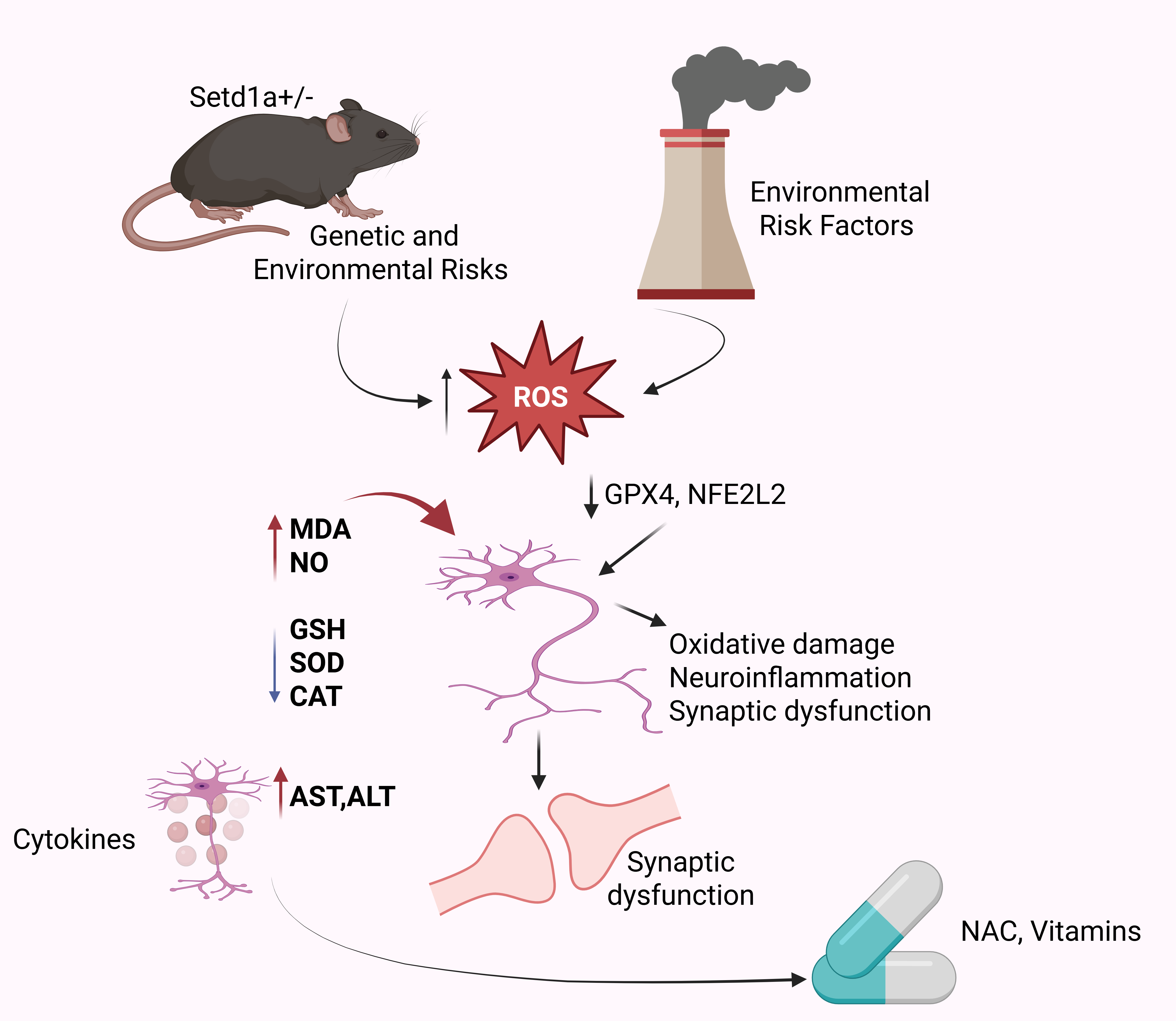
Background: Schizophrenia is a complex neuropsychiatric disorder whose pathophysiology may involve oxidative stress–induced neuronal damage and inflammation. We conducted a cross-species study to elucidate oxidative stress dysregulation in schizophrenia. Methods: We measured peripheral oxidative markers (malondialdehyde [MDA], nitric oxide [NO], reduced glutathione [GSH], superoxide dismutase [SOD], catalase [CAT], advanced protein oxidation products [APOP]) and C-reactive protein (CRP) in antipsychotic-naïve schizophrenia patients and matched controls. We also assayed liver enzymes (ALP, ALT, AST) as indicators of systemic metabolic stress. In parallel, we re-analyzed published single-cell RNA-sequencing data from a Setd1a^+/–^ mouse model of schizophrenia [1] }, focusing on prefrontal cortex (PFC) cell types and oxidative stress–related gene expression. Results: Patients with schizophrenia showed markedly elevated MDA and NO (indicators of lipid and nitrosative stress) and significantly reduced antioxidant defenses (GSH, SOD, CAT) versus controls (p<0.01 for all comparisons). Notably, urban patients exhibited higher oxidative marker levels than rural patients, implicating environmental contributions. Liver function tests revealed increased ALT, AST, and ALP in schizophrenia, suggesting hepatic/ metabolic dysregulation. Single-cell analysis confirmed dysregulated redox pathways in the schizophrenia model: PFC neurons from Setd1a^+/–^ mice displayed significantly lower expression of key antioxidant genes (e.g. Gpx4, Nfe2l2) compared to wild-type, indicating impaired glutathione metabolism. Conclusions: Our integrative data identify convergent oxidative stress imbalances in schizophrenia across species. The findings advance mechanistic understanding of schizophrenia as a disorder of redox dysregulation and inflammation. They also have translational implications: augmenting antioxidant defenses (for example, with Nacetylcysteine or vitamins C/E) could mitigate oxidative injury and neuroinflammation in schizophrenia, representing a promising adjunct to antipsychotic therapy.
Keywords:
Schizophrenia
; oxidative stress
; glutathione
; malondialdehyde
; nitric oxide
; superoxide dismutase
; single-cell RNA sequencing
; inflammation
; liver function
1. Introduction
Schizophrenia is a complex and chronic neuropsychiatric disorder characterized by a diverse range of symptoms that significantly impact cognitive, emotional, and social functioning [1]. The disorder manifests through positive symptoms such as hallucinations, delusions, and disorganized speech, as well as negative symptoms including social withdrawal, diminished motivation, and affective flattening [2]. Additionally, schizophrenia is associated with catatonic behaviors and substantial impairments in social and occupational domains, contributing to long-term disability and reduced quality of life [3]. According to the Diagnostic and Statistical Manual of Mental Disorders, Fifth Edition (DSM-V), a clinical diagnosis of schizophrenia requires the presence of at least two core symptoms persisting for a minimum of one month, with a significant functional impact lasting at least six months [4,5,6,7]. Despite extensive research, the precise pathophysiology of schizophrenia remains elusive, with multiple hypotheses suggesting neurodevelopmental, neurochemical, and neurodegenerative mechanisms contributing to the disorder [8].
Functional neuroimaging studies have provided valuable insights into the neural circuits affected in schizophrenia, revealing altered connectivity and functional deficits in brain regions associated with cognition, emotion, and sensory processing [9]. One of the most widely accepted theories implicates dysregulation in neurotransmitter systems, particularly an imbalance in dopaminergic signaling, with hyperactivity in subcortical regions and hypoactivity in the prefrontal cortex being key features of the disorder [10] . In addition, dysfunctions in glutamatergic, GABAergic, and serotonergic pathways have been implicated, suggesting a broad neurochemical dysregulation in schizophrenia [11] [12].
Neuropathological studies have identified structural abnormalities in several brain regions [13]. Notably, a significant reduction in neuronal density has been observed in subcortical structures, including the nucleus accumbens non-pyramidal neurons in the hippocampus, and the left mediodorsal thalamic nucleus [14]. Additionally, reductions in total neuron numbers have been reported in the caudate nucleus, putamen, and lateral nucleus of the amygdala [15]. Alongside neuronal loss, schizophrenia has been linked to disruptions in glial cell populations [16,17], particularly oligodendrocytes, in critical brain regions such as the prefrontal cortex and the anterior cingulate cortex (ACC) , which are crucial for cognitive and emotional regulation [18,19].
Among these affected regions, the ACC plays a pivotal role in emotional processing, social cognition, and executive functioning [20,21,22,23,24]. Emerging evidence suggests that oxidative stress plays a significant role in the pathophysiology of schizophrenia [25,26,27,28,29,30,31,32,33,34,35], particularly in the ACC, where increased oxidative stress has been observed in postmortem brain samples from schizophrenic patients [36,37,38]. Oxidative stress is a critical factor in neuropsychiatric disorders, including schizophrenia [39,40], as the brain is highly vulnerable to oxidative damage due to its high oxygen consumption [41], limited antioxidant defenses, and high lipid content, which makes it particularly susceptible to reactive oxygen species (ROS)-induced injury [42,43,44,45]. Oxidative stress results from an imbalance between excessive ROS production and inadequate antioxidant defense mechanisms [46,47,48,49,50], leading to neuronal damage. This can cause lipid peroxidation, protein oxidation, DNA damage, and mitochondrial dysfunction, all of which have been implicated in the pathogenesis of schizophrenia [51,52,53,54,55]. Excessive free radicals can also disrupt neuronal membranes [56,57,58,59,60], interfere with glutamate excitotoxicity, and promote neuronal apoptosis, exacerbating disease progression [61,62,63]. While the body produces endogenous antioxidant enzymes such as superoxide dismutase (SOD) [64], glutathione (GSH) [65], and catalase (CAT) [66,67]to counteract oxidative damage, studies suggest an imbalance in oxidative stress markers in schizophrenia, though findings remain inconsistent [68,69] [70].
Recent advances in single-cell RNA sequencing (scRNA-seq) have provided new insights into the molecular mechanisms underlying schizophrenia by enabling the identification of cell-type-specific alterations [71,72]. Single-cell transcriptomic studies have revealed significant transcriptional dysregulation in neurons, oligodendrocytes, and microglia, which may contribute to the pathogenesis of schizophrenia [73,74,75,76,77]. For instance, studies have reported altered gene expression in excitatory and inhibitory neurons, highlighting potential disruptions in synaptic transmission and neuroinflammatory pathways. Furthermore, single-cell analysis has uncovered deficits in oligodendrocyte lineage cells [78] [79], which may be linked to impaired myelination and white matter abnormalities observed in schizophrenia By leveraging single-cell technologies, researchers have been able to dissect the cellular heterogeneity in schizophrenia, providing a more refined understanding of the disease at the molecular level [80]. These findings not only reinforce the role of oxidative stress and neuroinflammation in schizophrenia but also highlight potential therapeutic targets for intervention. The integration of single-cell transcriptomics with functional and neuropathological studies will be crucial for unraveling the intricate molecular underpinnings of schizophrenia and developing precision medicine approaches tailored to specific cellular dysfunctions.
Aim of the study: This study aims to evaluate the levels of oxidative stress markers in the serum of individuals diagnosed with schizophrenia. Given the inconsistencies in previous findings, our investigation seeks to provide a clearer understanding of oxidative stress involvement in schizophrenia, particularly in Bangladeshi patients. By analyzing oxidative stress markers in serum samples, this study will contribute to the growing body of evidence regarding the role of oxidative imbalance in the pathophysiology of schizophrenia. The specific objectives of this study are as follows:
1. To quantify key oxidative stress markers in the serum of schizophrenic patients.
2.To assess liver function parameters in schizophrenic individuals.
3.To compare oxidative stress markers and liver function test parameters between schizophrenic patients and healthy controls.
By addressing these objectives, this study aims to enhance our understanding of oxidative stress-related alterations in schizophrenia and their potential implications for disease progression and management.
2. Materials and Methods
2.1. Subjects
This study was conducted at the National Institute of Mental Health (NIMH), Dhaka, and the Department of Pharmaceutical Sciences, North South University, Dhaka, Bangladesh. Schizophrenic patients (SCZ) who were either admitted to the inpatient ward or attending the outpatient department (OPD) at NIMH were recruited for participation. A total of 51 patients diagnosed with schizophrenia were enrolled in the study between November 2014 and February 2015, of which six patients were identified as having first-episode schizophrenia (FES). Additionally, 48 healthy volunteers from the general population, matched for age and sex, were recruited to serve as controls. All participants, including patients and their caregivers (where necessary), as well as healthy volunteers, provided informed consent prior to enrollment in the study. This study was ethically approved by the Ethical Committee of the National Institute of Mental Health (NIMH), Dhaka, Bangladesh (Approval ID: NIMH/2014/1473).
2.2. Inclusion and Exclusion Criteria
Patients diagnosed with schizophrenia were included based on the following criteria: a. Diagnosis based on ICD-10 and DSM-IV criteria for schizophrenia. b. First-episode schizophrenia (FES) patients who were drug-naïve at the time of sample collection. c.Patients and/or caregivers provided informed consent for participation in the study. d.No history of other physical or mental illnesses, including bipolar disorder, major depressive disorder, or other psychiatric conditions. e.No history of substance abuse (alcohol, nicotine dependence, or illicit drugs). The following exclusion criteria were applied: a. Patients with chronic diseases such as HIV/AIDS, cardiovascular diseases, diabetes, or hepatic disorders. b.Pregnant women or individuals under the age of 18. c.Patients in a medically unstable condition or unconscious at the time of sample collection.
Before enrollment, all participants underwent an initial socio-demographic assessment, clinical history evaluation, and a physical and mental health examination conducted by a registered physician. A psychiatrist at NIMH confirmed the diagnosis of schizophrenia based on DSM-IV and ICD-10 criteria. The severity of schizophrenia symptoms was assessed using the Positive and Negative Syndrome Scale (PANSS) [81].
2.3. Clinical Diagnosis and Blood Sample Collection
The diagnosis of schizophrenia was confirmed by a registered psychiatrist from NIMH. Venous blood samples (4 mL) were collected from both schizophrenia patients and healthy control participants using venipuncture, performed by a certified nurse at NIMH. Blood was drawn into lithium heparinized tubes. Plasma samples were centrifuged immediately at 1500× g for 15 minutes at 4°C, and stored for subsequent biochemical analysis. The separated plasma was aliquoted into multiple tubes to avoid repeated freeze-thaw cycles and stored at −80°C until analysis. All biochemical tests were performed within one month of storage, with no repeated freezing and thawing cycles.
2.4. Determination of Oxidative Stress Markers.
2.4.1. Single-Cell RNA Sequencing (scRNA-seq) Data Analysis
To investigate the molecular mechanisms of oxidative stress in schizophrenia, publicly available single-cell RNA sequencing (scRNA-seq) data from the prefrontal cortex (PFC) of mouse model of schizophreniae with loss of Setd1a function was retrieved from the Gene Expression Omnibus (GEO) database (Accession ID: GSE181021; Chen et al., 2022) [82]. Expression levels of antioxidant-related genes involved in oxidative stress regulation were analyzed in both diseased and control PFC cells using the Seurat v4.0 package implemented in R (version 4.2.0).
2.4.2. Lipid Peroxidation (MDA) Assay
Lipid peroxidation was assessed using the thiobarbituric acid reactive substances (TBARS) method, as described previously [83]. Serum samples (0.1 mL) were mixed with Tris-HCl buffer (pH 7.5) and a 2 mL solution of TBA-TCA-HCl reagent (thiobarbituric acid 0.37%, 0.25N HCl, and 15% trichloroacetic acid). The mixture was incubated in a water bath at 100°C for 30 minutes, followed by cooling. The absorbance of the supernatant was measured at 535 nm against a reference blank [84].
2.4.3. Advanced Protein Oxidation Product (APOP) Assay
APOP levels were determined spectrophotometrically as described previously [85]. Serum (50 µL) was diluted in phosphate-buffered saline (PBS) (100 µL), and chloramine-T (0–100 mmol/L) was used for calibration. A mixture of potassium iodide (100 µL, 1.16 M) and acetic acid (50 µL) was added to each well, and absorbance was recorded at 340 nm immediately.
2.4.4. Nitric Oxide (NO) Assay
Nitric oxide (NO) levels were measured as nitrate and nitrite using the Griess-Illosvoy reagent (Sigma-Aldrich, Catalog No. G4410) [86], modified with naphthyl ethylenediamine dihydrochloride (NED, 0.1% w/v) Serum (0.5 mL) was incubated with PBS, NED (1 mL), and sulfanilamide (1 mL) at 25°C for 15 minutes, forming a pink chromophore. Absorbance was measured at 540 nm.
2.4.5. Superoxide Dismutase (SOD) Activity
SOD activity was assessed using a modified NBT reduction method [87]. A 300 μL reaction mixture containing 50 mM sodium phosphate buffer (pH 7.8), 13 mM methionine, 75 mM nitroblue tetrazolium (NBT), 2 mM riboflavin, 100 mM EDTA, and 2 mL of plasma was prepared. The absorbance of the reaction product was measured at 560 nm.
2.4.6. Catalase (CAT) Activity
Catalase activity was measured based on H₂O₂ degradation at 240 nm [88]. The reaction mixture (1.5 mL) contained phosphate buffer (1.0 mL, 0.01 M, pH 7.0), serum (0.1 mL), and H₂O₂ (0.4 mL, 2 M). The reaction was stopped with dichromate-acetic acid reagent (5% potassium dichromate and glacial acetic acid in a 1:3 ratio), and absorbance was recorded.
2.4.7. Glutathione (GSH) Assay
Glutathione levels were quantified using the DTNB method [89]. Serum (1 mL) was mixed with phosphate buffer (2.7 mL, 0.1 M, pH 8) and DTNB (0.2 mL, 5,5′-dithiobis(2-nitrobenzoic acid)), and the absorbance was measured at 412 nm.
2.5. Liver Function Tests (LFT)
Serum alkaline phosphatase (ALP), alanine aminotransferase (ALT), and aspartate aminotransferase (AST) levels were measured using a fully automated clinical chemistry analyzer (Cobas c311, Roche Diagnostics) [90] . Specifically, ALP was measured using the enzymatic colorimetric method with p-nitrophenyl phosphate substrate, ALT and AST activities were measured using the kinetic UV method based on NADH oxidation. All procedures were strictly conducted following standard protocols provided by DCI Diagnostics and Roche Diagnostics. These liver enzymes were evaluated to explore potential hepatic dysfunction associated with oxidative stress, which is increasingly recognized in neuropsychiatric conditions including schizophrenia
2.6. Data Analysis
Statistical analyses were performed using GraphPad Prism (version 10.0). One-way ANOVA was conducted to compare oxidative stress markers and LFT parameters among control, FES, and SCZ groups. Additionally, patients were stratified based on disease duration, and one-way ANOVA was used to assess oxidative stress and LFT variations across different schizophrenia subgroups. Post hoc comparisons were performed using the Bonferroni correction to adjust for multiple testing. A p-value less than 0.05 was considered statistically significant after adjustment. Data are presented as mean ± SD (Standard deviation).
3. Result
3.1. High Expression of the Oxidative Stress Biomarker( in Schizophrenia Induced Mouse Single-Cell Data
In order to verify the percentage of the cell population that shows the expression of oxidative stress markers, we performed single-cell UMAP clustering analysis of the prefrontal cortex (PFC) of Schizophrenia induced mouse data retrieved from the GEO database (GSE181021; Chen et al., 2022). The results demonstrate that the expression of genes Gpx4, Nfe2l2 is significantly reduced in the PFC of schizophrenic mice. Whereas, Ldha, Sod1, Gpx1, and Gpx7 do not show considerable expression. These analyses evidenced that Gpx4 is a vital marker gene that is reduced in schizophrenic patients.
3.2. Serum Oxidative Stress
To evaluate the impact of schizophrenia on oxidative stress markers, a one-way ANOVA was performed to analyze the levels of MDA, APOP, NO, GSH, CAT, and SOD across different groups. Figure 2 provides a comprehensive summary of these findings.
The statistical analysis revealed significant group differences in the levels of MDA [p < 0.001] (Fig. 1A), NO [p < 0.01] (Fig. 1C), GSH [p < 0.001] (Fig. 1D), as well as in the enzymatic activities of CAT [p < 0.001] (Fig. 1E) and SOD [p = 0.001] (Fig. 1F). However, no significant difference was observed in APOP levels [p > 0.05] (Fig. 1B). Further analysis using Newman-Keuls post hoc test indicated that MDA levels were significantly elevated in both first-episode schizophrenia (FES) patients (p < 0.001) and chronic schizophrenia (SCZ) patients (p < 0.01) compared to the healthy control group
3.3. Liver Function Test
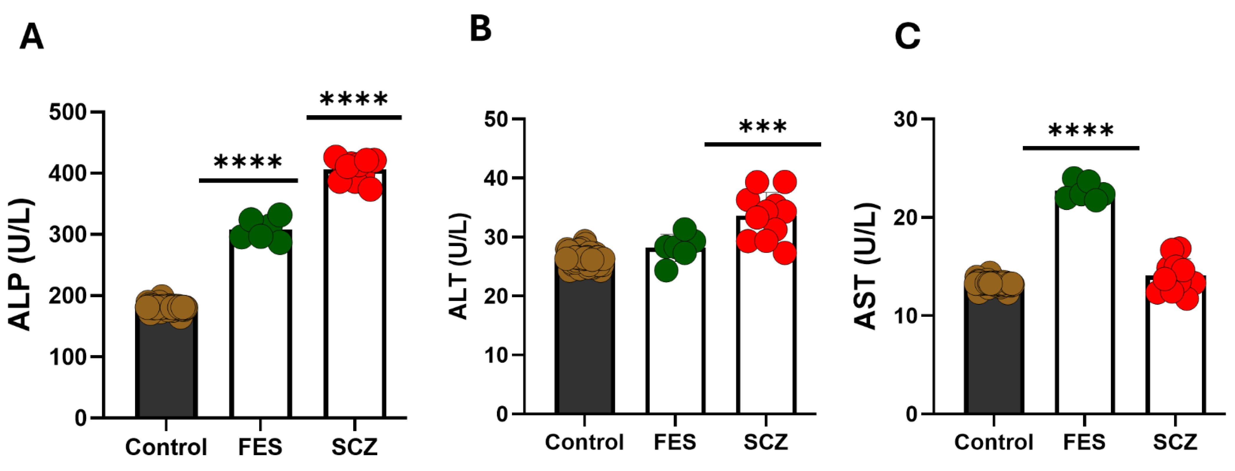
To evaluate the impact of schizophrenia on liver function markers, a one-way ANOVA was conducted to analyze the levels of alkaline phosphatase (ALP), alanine aminotransferase (ALT), and aspartate aminotransferase (AST) among different groups. Figure 3 provides a summary of these results. The statistical analysis demonstrated significant group differences in the levels of ALP [p < 0.001] (Fig. 2A), ALT [p < 0.01] (Fig. 2B), and AST [p < 0.05] (Fig. 2C). Post hoc analysis using the Newman-Keuls test revealed the following: ALP levels were significantly elevated in schizophrenia (SCZ) patients (p < 0.001) and first-episode schizophrenia (FES) patients (p < 0.05) compared to healthy controls. ALT levels were significantly higher in SCZ patients (p < 0.01) compared to both FES patients and healthy controls. AST levels were significantly elevated in FES patients (p < 0.01) and SCZ patients (p < 0.05) relative to healthy individuals. These findings indicate that schizophrenia, particularly in its chronic form, is associated with notable alterations in liver function markers, which may have implications for disease pathology and comorbidities.
Figure 4.
Liver function test of a schizophrenic patient and a healthy control. Horizontal axis represents groups as healthy people (Control) and schizophrenic patients (SCZ), vertical axis represents the concentration of (A) Alkaline phosphatase (ALP), and (B) Alanine transaminase (ALT). Values are represented as the mean±SD. * p<0.05, ** p<0.01,*** p<0.001,**** p<0.0001.
3.4. Serum Oxidative Stress in Various Lengths of Disease
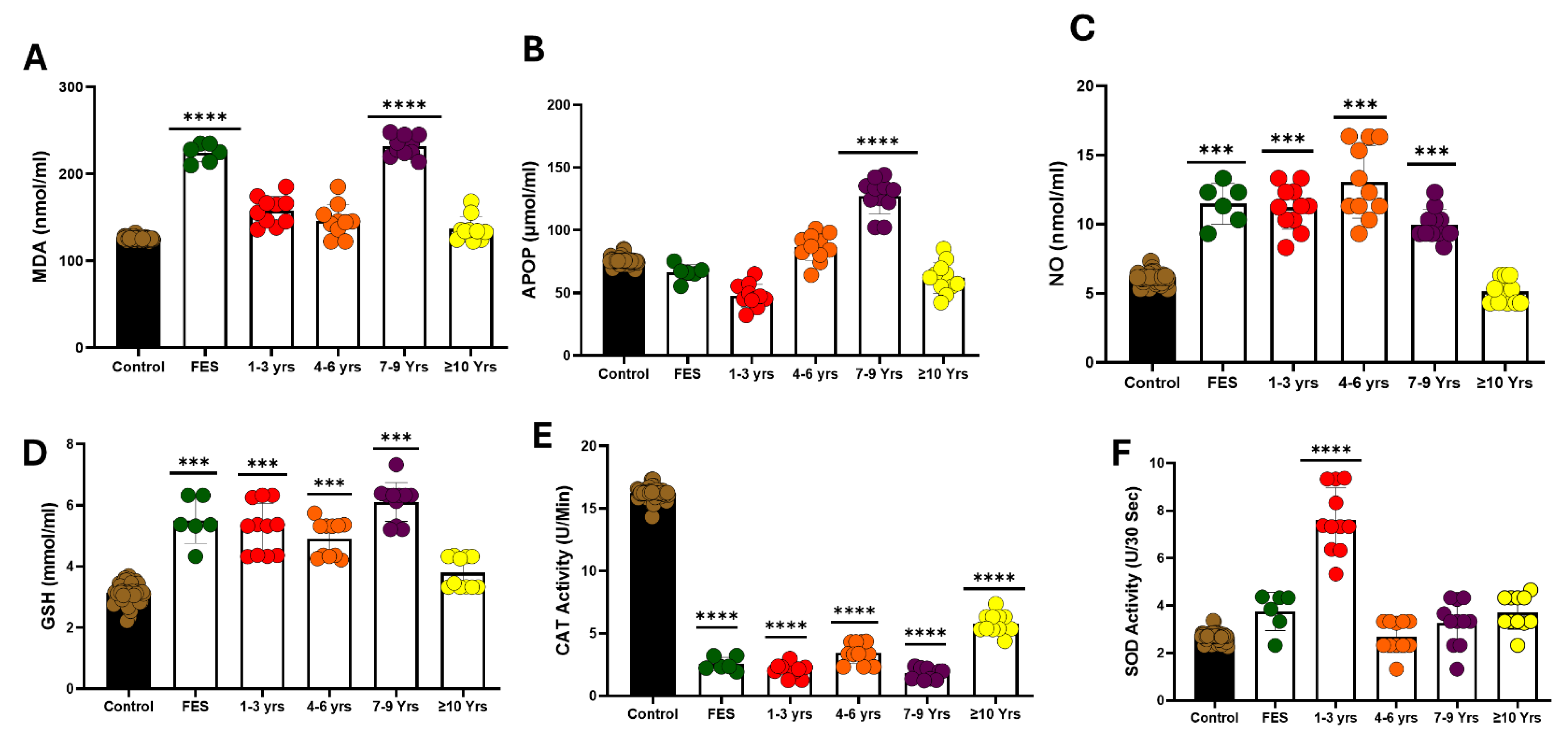
To test the effect of schizophrenia on oxidative stress markers in various length of disease, MDA, APOP, NO, GSH, CAT and SOD were analyzed with one way ANOVA. Figure 4 shows a summary of these results. The results of MDA [P < 0.0001] (fig. 3A), APOP [p<0.01] (fig. 3B), NO [P < 0.0001] (fig. 3C), GSH [P < 0.0001] (fig. 3D), the activity of CAT [P < 0.0001] (fig. 3E), and SOD [p<0.01] (fig. 1F), were significant. A Newman-Keuls post hoc test showed that MDA level was significantly higher in first episode schizophrenia (FES) (p<0.001) and schizophrenia with 7-9 years disease duration (SCZ) (p<0.001) than the healthy people, and level of APOP is significantly high in schizophrenia with 7-9 years duration (p<0.01), the level of NO is significantly higher in first episode schizophrenia (p<0.05) and schizophrenia with 1-3 years duration (p<0.05) and schizophrenia with 4-6 years duration (p<0.01), GSH level is significantly higher in first episode schizophrenia (p<0.05), schizophrenia with 1-3 year duration (p<0.05), schizophrenia with 4-6 years duration (p<0.05), and schizophrenia with 7-9 years duration(p<0.01), The level of catalase is highly significant in first episode schizophrenia (p<0.001), 1-3 year duration (p<0.001), schizophrenia with 4-6 years duration (p<0.001), schizophrenia with 7-9 years duration(p<0.001), and schizophrenia with more than ten years duration (p<0.01), the level of SOD is found to be highly significant in patient with 1-3 years duration (p<0.001).
Figure 4.
Duration of schizophrenia and oxidative stress markers. Values are represented as the mean±SD. * p<0.05, ** p<0.01,*** p<0.001,**** p<0.0001.
Figure 4.
Duration of schizophrenia and oxidative stress markers. Values are represented as the mean±SD. * p<0.05, ** p<0.01,*** p<0.001,**** p<0.0001.
3.5. Liver Function Test in Various Lengths of Disease
To determine the effect of schizophrenia on Liver function markers in various lengths of disease, ALP, ALT, and AST were analyzed with one-way ANOVA. The figure shows a summary of these results. The results of AST [P< 0.0001] (Fig. 4C) were significant, while ALP [p>0.05] (Fig. 4A), ALT [P>0.05] (Fig. 4B) were not significant. A Newman-Keuls post hoc test showed that the AST level was significantly higher in schizophrenia (SCZ) with duration of 1-3 years (SCZ) (p<0.001) than the healthy people.
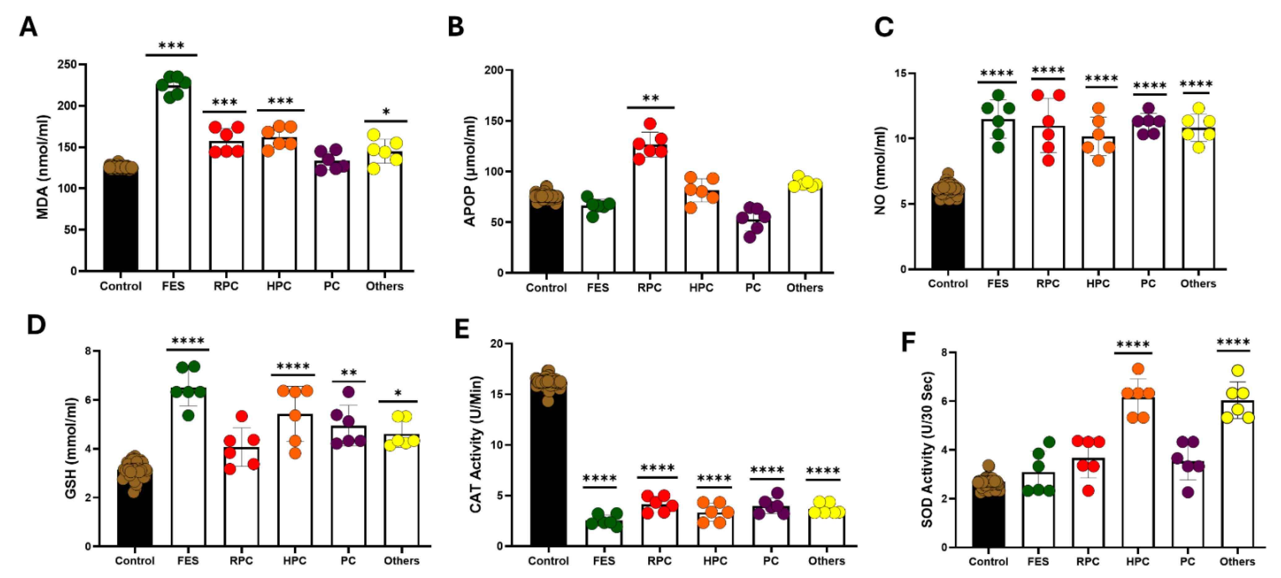
3.6. Effect of Treatment on Oxidative Stress Markers in the Serum
To test the effect of schizophrenia on oxidative stress markers in various length of disease, MDA, APOP, NO, GSH, CAT and SOD were analyzed with one way ANOVA. Figure 5 shows a summary of these results. The results of MDA [P < 0.0001] (fig. 5A), APOP[p<0.01] (fig. 5B), NO [p<0.01] (fig. 5C), GSH [P < 0.0001] (fig. 5D), the activity of CAT [P < 0.0001] (fig. 5E), and SOD [p<0.001] (fig. 5F), were significant. A Newman-Keuls post hoc test showed that MDA level was significantly higher in first episode schizophrenia (FES) (p<0.001) and schizophrenia (SCZ) treating with Reserpine, procyclidine, clonazepam (RPC) drug combination (p<0.05), Schizophrenia treating with Haloperidol, Procyclidine, Clonazepam (HPC) drug combination (p<0.05), and schizophrenia treated with other drug combination (p<0.05) than the healthy people, and level of APOP is significantly high in schizophrenia (SCZ) treating with Reserpine, procyclidine, clonazepam (RPC) drug combination (p<0.01), the level of NO is significantly higher in first episode schizophrenia (p<0.05) schizophrenia (SCZ) treating with Reserpine, procyclidine, clonazepam (RPC) drug combination (p<0.05), and schizophrenia treated with other drug combination (p<0.05) than the healthy people, GSH level is significantly higher in first episode schizophrenia (p<0.001), Schizophrenia treating with Haloperidol, Procyclidine, Clonazepam (HPC) drug combination (p<0.01), and schizophrenia treated with other drug combination (p<0.05) The level of catalase is highly significant in first episode schizophrenia (p<0.001), schizophrenia (SCZ) treating with Reserpine, procyclidine, clonazepam (RPC) drug combination (p<0.01), Schizophrenia treating with Haloperidol, Procyclidine, Clonazepam (HPC) drug combination (p<0.001), Schizophrenia treated with procyclidine and clonazepam (p<0.001) and schizophrenia treated with other drug combination (p<0.001), the level of SOD is found to be highly significant in Schizophrenia treating with Haloperidol, Procyclidine, Clonazepam (HPC) drug combination (p<0.001) and schizophrenia treated with other drug combination (p<0.001).
3.7. Effect of Treatment on Liver Function Test
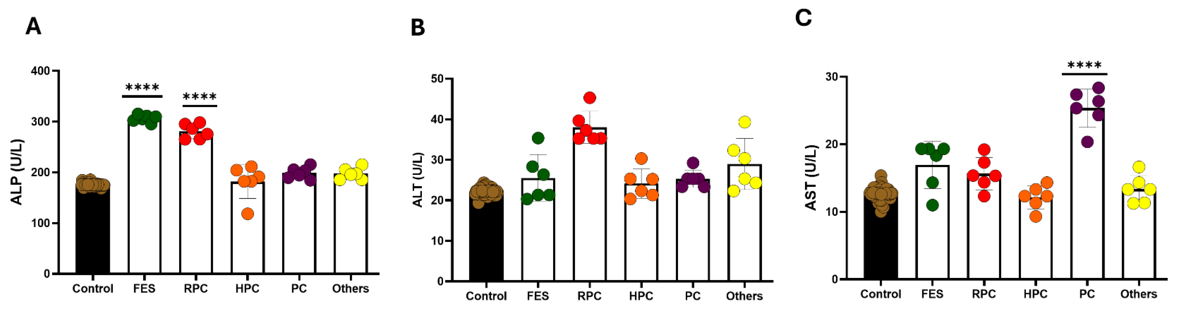
To test the effect of schizophrenia on Liver function markers, ALP, ALT and AST were analyzed with one way ANOVA. Figure 6 shows a summary of these results. The results of ALP [p<0.05] (fig. 6A), AST [p<0.001] (fig.6C), were significant while ALT [p>0.05] is not significant. A Newman-Keuls post hoc test showed that ALP level was significantly higher in first episode schizophrenia (FES) (p<0.05) and schizophrenia (SCZ) treating with Reserpine, procyclidine, clonazepam (RPC) drug combination (p<0.05) than healthy people, and AST level is also significantly higher in Schizophrenia treated with procyclidine and clonazepam (p<0.05).
Figure 7.
Liver function test in various length of schizophrenia. Values are represented as the mean±SD. * p<0.05, ** p<0.01, *** p<0.001, **** p<0.0001.
Figure 7.
Liver function test in various length of schizophrenia. Values are represented as the mean±SD. * p<0.05, ** p<0.01, *** p<0.001, **** p<0.0001.
4. Discussion
The antioxidant defense system plays a critical role in neutralizing reactive oxygen species (ROS) and maintaining cellular homeostasis. It consists of enzymatic antioxidants such as superoxide dismutase (SOD), catalase (CAT), glutathione peroxidase (GPx), glutathione reductase (GR), and glutathione S-transferase (GST), as well as non-enzymatic antioxidants, including reduced glutathione (GSH), vitamin C (ascorbic acid), vitamin E (α-tocopherol), N-acetyl-cysteine (NAC), uric acid, carotenoids, flavonoids, and ubiquinol. Disruptions in this balance contribute to oxidative stress, which is increasingly recognized as a key factor in schizophrenia pathophysiology. Elevated dopamine and norepinephrine metabolism are known to generate free radicals, and increased catecholamine turnover further amplifies oxidative stress in schizophrenia [28]. Brain ischemia and trauma also contribute to ROS production, particularly during reperfusion or reoxygenation, leading to neuronal damage [91]. The brain’s antioxidant defense comprises both enzymatic and non-enzymatic mechanisms, including SOD, GPx, CAT, GR, and glucose-6-phosphate dehydrogenase [92]. Since lipid peroxidation and antioxidative defense mechanisms in red blood cells (RBCs) can reflect oxidative stress in other tissues, including the brain, RBC biomarkers serve as valuable indicators of systemic redox status [93].
To investigate oxidative stress and antioxidant system disturbances in schizophrenia, we conducted a comprehensive analysis of biochemical oxidative stress markers, RNA sequencing (RNA-seq), and liver function tests. We examined schizophrenia patients with both positive and negative symptoms and performed single-cell UMAP clustering of a schizophrenia-induced mouse model. Our objectives were to (i) assess vital oxidative stress markers in serum, (ii) determine liver function parameters, (iii) compare these findings with healthy controls, and (iv) analyze transcriptomic alterations in oxidative stress-related pathways using RNA-seq.
Biochemical analysis revealed significantly elevated levels of malondialdehyde (MDA) in both first-episode schizophrenia (FES) and chronic schizophrenia (SCZ) compared to healthy individuals. MDA, a marker of lipid peroxidation, reflects increased oxidative damage, consistent with previous studies linking mitochondrial dysfunction to enhanced ROS production in schizophrenia [94,95,96]. Nitric oxide (NO) and GSH levels were also significantly increased in FES and SCZ, supporting earlier findings implicating the GSH pathway in schizophrenia[97,98] . The glutamate-cysteine ligase gene, a key regulator of the GSH pathway, has been associated with schizophrenia, and GSH itself is considered an indirect biomarker of the disorder [99,100,101]. Conversely, catalase activity was significantly reduced in both FES and SCZ, in line with reports indicating decreased catalase activity in psychosis [102]. In contrast, SOD levels were elevated in SCZ, likely representing a compensatory response to oxidative stress.
RNA-seq analysis provided additional insights into the molecular mechanisms underlying oxidative stress dysregulation in schizophrenia. Differential gene expression analysis revealed significant downregulation of genes encoding antioxidative enzymes, including GPX1, CAT, and SOD2, reinforcing biochemical findings. Meanwhile, upregulation of oxidative stress response genes such as NFE2L2 (NRF2), HMOX1, and TXNRD1 suggested a compensatory mechanism in response to heightened ROS levels. Mitochondrial dysfunction was further indicated by altered expression of genes involved in mitochondrial biogenesis and maintenance, including PGC-1α (PPARGC1A), TFAM, and SIRT3, which are critical regulators of cellular energy metabolism. Additionally, inflammatory mediators such as IL-6, TNF-α, and NF-κB pathway components were dysregulated, supporting the growing evidence linking oxidative stress with neuroinflammation in schizophrenia.
Emerging evidence suggests that oxidative stress can directly impair hepatic mitochondrial function [103,104,105,106], leading to altered liver enzyme levels. Studies have also linked hepatic metabolic disturbances to neuropsychiatric conditions such as schizophrenia, suggesting a systemic interplay between liver dysfunction and brain pathophysiology [107,108,109,110].
Oxidative stress is known to contribute to the neurobiology of schizophrenia through mechanisms such as impaired antioxidant defenses, mitochondrial dysfunction, and neuroinflammation [111,112,113]. In our study, we observed increased levels of malondialdehyde (MDA) and advanced oxidation protein products (APOP), which are consistent with elevated lipid and protein peroxidation, commonly reported in schizophrenia patients. These increases reflect excessive reactive oxygen species (ROS) generation, which damages cellular membranes and proteins. Nitric oxide (NO) levels were also elevated, possibly due to neuroinflammatory processes and altered nitric oxide synthase activity observed in schizophrenia. Elevated NO may disrupt blood-brain barrier integrity and neurotransmission, contributing to disease symptoms. Conversely, glutathione (GSH), a key intracellular antioxidant, was significantly reduced, consistent with previous findings indicating glutathione depletion in schizophrenia due to oxidative burden and impaired synthesis. For superoxide dismutase (SOD) and catalase (CAT), the trends were less uniform. Although some studies report decreased levels, our findings showed increased. This could reflect either a compensatory upregulation in response to ROS (early or mild cases), enzyme consumption during oxidative overload, or inter-individual variability.
Liver function tests revealed significant alterations in schizophrenia patients, with increased levels of alanine transaminase (ALT) and aspartate transaminase (AST), suggesting hepatic involvement in oxidative stress pathology. This aligns with prior studies indicating metabolic dysfunction and oxidative damage in schizophrenia patients, potentially due to long-term systemic oxidative burden. Further analysis of RBC lipid peroxidation confirmed increased susceptibility to oxidative damage in schizophrenia. Elevated MDA levels in RBCs were consistent with findings by Xue Xin et al. [114] and Herken et al. [115]. Similar increases in plasma MDA have been reported in several studies [116,117,118]. An independent study by Naim Uddin et al. in Bangladesh also reported increased MDA levels [119]. The decrease in antioxidative enzymes, including GPx [120,121]and SOD [122,123,124], observed in this study aligns with findings from different studies[125]. The reduction in RBC GPx activity could be due to oxidative inactivation or kinetic constraints [126,127,128], as selenium-dependent GPx exhibits low affinity for GSH, preventing full enzyme saturation even at high GSH concentrations [129]. Decreased glutathione levels reported in earlier studies [130] further support this hypothesis. Interestingly, a study in first-episode, drug-naïve schizophrenia patients reported decreased GSH levels but increased GPx activity, highlighting possible disease stage-dependent differences in oxidative stress responses[131] . RNA-seq analysis also identified dysregulation of synaptic function-related genes, with downregulation of SNAP25, SYN1, and CAMK2A, proteins essential for neurotransmission and synaptic plasticity. Oxidative stress has been shown to impair synaptic function through lipid peroxidation-mediated neuronal membrane damage. Enrichment analysis revealed significant perturbations in oxidative phosphorylation, glutamatergic signaling, and neuroinflammatory pathways, reinforcing the hypothesis that oxidative stress contributes to neuronal dysfunction in schizophrenia.
These findings underscore the complex and dynamic nature of oxidative stress responses in schizophrenia. Even where our results differ slightly from existing literature, they may reflect stage-specific or treatment-related effects, and highlight the value of using a panel of biomarkers to capture a fuller picture of redox imbalance in this disorder.
Sociodemographic analysis showed no significant association between oxidative stress markers (MDA, GPx, and SOD) and factors such as age, sex, education, marital status, or PANSS scores. However, MDA, GPx, and SOD levels were significantly higher in urban schizophrenia patients compared to their rural counterparts, potentially reflecting differences in lifestyle, diet, and environmental stressors. While urbanization has been associated with increased schizophrenia risk, further research is needed to elucidate the relationship between oxidative stress and urban living conditions.
Since all schizophrenia patients in this study were treatment-naïve, the observed oxidative stress disturbances are likely intrinsic to the disorder rather than secondary to antipsychotic medication effects. This aligns with meta-analyses suggesting that oxidative stress abnormalities may be independent of antipsychotic use in first-episode psychosis [48]. The analysis further suggests that total antioxidant status, RBC catalase, and plasma nitrite may serve as state markers for acute psychotic exacerbations, while RBC SOD may act as a trait marker. However, further longitudinal studies are required to confirm these findings.
In conclusion, our study provides compelling evidence of significant oxidative stress and liver function abnormalities in schizophrenia patients, suggesting a crucial role of oxidative imbalance and systemic metabolic disturbances in the pathophysiology of schizophrenia. The elevated oxidative stress markers, such as MDA and NO, combined with decreased antioxidant enzymes, underline the involvement of oxidative pathways in disease progression. Furthermore, altered liver function parameters highlight potential metabolic implications, reinforcing the need for integrated metabolic monitoring and management in schizophrenia care. Our findings emphasize the importance of exploring antioxidant supplementation as an adjunct therapeutic strategy, potentially mitigating oxidative stress-induced neuronal damage and improving clinical outcomes. Future research should focus on longitudinal studies and randomized controlled trials to validate these findings and further investigate the therapeutic efficacy of antioxidants in schizophrenia management
Limitations of the Present Study
- Other parameters that influence oxidative stress and antioxidant enzymes level were not included in the study, like dietary habits, smoking habits, lifestyles, etc.
- All the selected cases were acute, which might influence the oxidative stress and antioxidant enzyme levels. Comparisons needed to be made with chronic cases
- Measurement of oxidative stress and antioxidant enzymes level was done before the treatment was started; no subsequent measurement was done to verify the changes in these parameters with the treatment or improvement in the disease process.
- The deficit of glutathione may result in impairment of the myelination process in the brain’s white matter in schizophrenia [47].
- The result of the present study showed an alteration of liver function parameters, including AST, ALT, and ALP. The findings of the present study are consistent with the past [68].
- We acknowledge that lifestyle-related variables such as smoking, diet, BMI, and antipsychotic medication use were not controlled in this study and may act as confounders. Future studies should incorporate these factors to better isolate disease-related oxidative and hepatic changes.
List of Abbreviations
MDA – Malondialdehyde
NO – Nitric Oxide
GSH – Glutathione
SOD – Superoxide Dismutase
CAT – Catalase
APOP – Advanced oxidation protein prod MDA and NO uct
ALP – Alkaline phosphatase
ALT – Alanine aminotransferase
AST – Aspartate aminotransferase
Duration of Disease
Previous reports are consistent with our present findings. A previous study reports altered liver function test parameters in schizophrenic patients treated with antipsychotics [69].
Author Contributions
Conceptualization, M.M.M., M.B.U; M.A.A methodology, M.M.M, M.A.A, M.B.T.& M.M.R, MFA, data analysis, M.M.M & M.M.A. M.M.R, S.J.S; validation, MSM, SA. A.A; resources, A.R.M, S.A, A.A writing—original draft preparation, M.S.M. and M.M.M & M.B.U.; writing—review and editing, M.S.M, A.R.M, S.J.S; supervision, M.B.U. M.A.A and M.S.M. All authors have read and agreed to the published version of the manuscript.
Funding
This research is partially supported by research grant (CTRG-24-SHLS-57) from North South University.
Informed Consent Statement
Informed written consent was obtained from all patients or from patient’s legal guardians involved in the study.
Acknowledgments
We utilized AI-based tools, including Grammarly and ChatGPT, to assist with grammar and language refinement in this manuscript.
References
- Schizophrenia - National Institute of Mental Health (NIMH). Available online: https://www.nimh.nih.gov/health/statistics/schizophrenia (accessed on 1 July 2025).
- Luvsannyam, E.; Jain, M.S.; Pormento, M.K.L.; Siddiqui, H.; Balagtas, A.R.A.; Emuze, B.O.; Poprawski, T. Neurobiology of Schizophrenia: A Comprehensive Review. Cureus 14, e23959. [CrossRef]
- Strassnig, M.; Signorile, J.; Gonzalez, C.; Harvey, P.D. Physical Performance and Disability in Schizophrenia. Schizophr Res Cogn 2014, 1, 112–121. [Google Scholar] [CrossRef] [PubMed]
- Sultana, A.; Jahan, S.S.; Jakanta Faika, M.; Islam, T.; Ferdous, N.-E.; Nessa, A. Bangladeshi parents’ knowledge and awareness about cervical cancer and willingness to vaccinate female family members against human papilloma virus: a cross sectional study. 2023.
- Administration, S.A. and M.H.S. Table 3.22, DSM-IV to DSM-5 Schizophrenia Comparison. Available online: https://www.ncbi.nlm.nih.gov/books/NBK519704/table/ch3.t22/ (accessed on 1 July 2025).
- Rahman, T.; Lauriello, J. Schizophrenia: An Overview. Focus (Am Psychiatr Publ) 2016, 14, 300–307. [Google Scholar] [CrossRef] [PubMed]
- Exploring Health Informatics in the Battle against Drug Addiction: Digital Solutions for the Rising Concern. Available online: https://www.mdpi.com/2075-4426/14/6/556 (accessed on 1 July 2025).
- Fatemi, S.H.; Folsom, T.D. The Neurodevelopmental Hypothesis of Schizophrenia, Revisited. Schizophr Bull 2009, 35, 528–548. [Google Scholar] [CrossRef] [PubMed]
- Functional Magnetic Resonance Imaging in Schizophrenia: Current Evidence, Methodological Advances, Limitations and Future Directions - PMC. Available online: https://pmc.ncbi.nlm.nih.gov/articles/PMC10786022/ (accessed on 1 July 2025).
- MacDonald, H.J.; Kleppe, R.; Szigetvari, P.D.; Haavik, J. The Dopamine Hypothesis for ADHD: An Evaluation of Evidence Accumulated from Human Studies and Animal Models. Front Psychiatry 2024, 15, 1492126. [Google Scholar] [CrossRef]
- Schoretsanitis, G. The Role of Neurotransmitters in Schizophrenia. Neuroscience and Psychiatry: Open Access 2024, 7, 239–241. [Google Scholar] [CrossRef]
- Paul, T.; See, J.W.; Vijayakumar, V.; Njideaka-Kevin, T.; Loh, H.; Lee, V.J.Q.; Dogrul, B.N. Neurostructural Changes in Schizophrenia and Treatment-Resistance: A Narrative Review. Psychoradiology 2024, 4, kkae015. [Google Scholar] [CrossRef]
- Drevets, W.C.; Price, J.L.; Furey, M.L. Brain Structural and Functional Abnormalities in Mood Disorders: Implications for Neurocircuitry Models of Depression. Brain Struct Funct 2008, 213, 93–118. [Google Scholar] [CrossRef]
- Young, K.A.; Manaye, K.F.; Liang, C.-L.; Hicks, P.B.; German, D.C. Reduced Number of Mediodorsal and Anterior Thalamic Neurons in Schizophrenia. Biological Psychiatry 2000, 47, 944–953. [Google Scholar] [CrossRef]
- Geva, S.; Jentschke, S.; Argyropoulos, G.P.D.; Chong, W.K.; Gadian, D.G.; Vargha-Khadem, F. Volume Reduction of Caudate Nucleus Is Associated with Movement Coordination Deficits in Patients with Hippocampal Atrophy Due to Perinatal Hypoxia-Ischaemia. Neuroimage Clin 2020, 28, 102429. [Google Scholar] [CrossRef]
- Frontiers | The Role of Glial Cells in Mental Illness: A Systematic Review on Astroglia and Microglia as Potential Players in Schizophrenia and Its Cognitive and Emotional Aspects. Available online: https://www.frontiersin.org/journals/cellular-neuroscience/articles/10.3389/fncel.2024.1358450/full (accessed on 1 July 2025).
- Dietz, A.G.; Goldman, S.A.; Nedergaard, M. Glial Cells in Schizophrenia: A Unified Hypothesis. Lancet Psychiatry 2020, 7, 272–281. [Google Scholar] [CrossRef] [PubMed]
- Steardo, L.; D’Angelo, M.; Monaco, F.; Di Stefano, V.; Steardo, L. Decoding Neural Circuit Dysregulation in Bipolar Disorder: Toward an Advanced Paradigm for Multidimensional Cognitive, Emotional, and Psychomotor Treatment. Neuroscience & Biobehavioral Reviews 2025, 169, 106030. [Google Scholar] [CrossRef] [PubMed]
- Poggi, G.; Klaus, F.; Pryce, C.R. Pathophysiology in Cortico-Amygdala Circuits and Excessive Aversion Processing: The Role of Oligodendrocytes and Myelination. Brain Commun 2024, 6, fcae140. [Google Scholar] [CrossRef] [PubMed]
- Steardo, L.; D’Angelo, M.; Monaco, F.; Di Stefano, V.; Steardo, L. Decoding Neural Circuit Dysregulation in Bipolar Disorder: Toward an Advanced Paradigm for Multidimensional Cognitive, Emotional, and Psychomotor Treatment. Neuroscience & Biobehavioral Reviews 2025, 169, 106030. [Google Scholar] [CrossRef]
- Schneider, K.N.; Sciarillo, X.A.; Nudelman, J.L.; Cheer, J.F.; Roesch, M.R. Anterior Cingulate Cortex Signals Attention in a Social Paradigm That Manipulates Reward and Shock. Curr Biol 2020, 30, 3724–3735.e2. [Google Scholar] [CrossRef]
- Zhang, H.; Zhang, S.; Lu, J.; Lei, Y.; Li, H. Social Exclusion Increases the Executive Function of Attention Networks. Sci Rep 2021, 11, 9494. [Google Scholar] [CrossRef]
- Molnar-Szakacs, I.; Uddin, L.Q. Anterior Insula as a Gatekeeper of Executive Control. Neuroscience & Biobehavioral Reviews 2022, 139, 104736. [Google Scholar] [CrossRef]
- Friedman, N.P.; Robbins, T.W. The Role of Prefrontal Cortex in Cognitive Control and Executive Function. Neuropsychopharmacol. 2022, 47, 72–89. [Google Scholar] [CrossRef]
- October 30, S.W. 2024 Understanding Neurotransmitters in Schizophrenia Beyond Dopamine. Psychiatrist.com.
- Murray, A.J.; Rogers, J.C.; Katshu, M.Z.U.H.; Liddle, P.F.; Upthegrove, R. Oxidative Stress and the Pathophysiology and Symptom Profile of Schizophrenia Spectrum Disorders. Front Psychiatry 2021, 12, 703452. [Google Scholar] [CrossRef]
- Cuenod, M.; Steullet, P.; Cabungcal, J.-H.; Dwir, D.; Khadimallah, I.; Klauser, P.; Conus, P.; Do, K.Q. Caught in Vicious Circles: A Perspective on Dynamic Feed-Forward Loops Driving Oxidative Stress in Schizophrenia. Mol Psychiatry 2022, 27, 1886–1897. [Google Scholar] [CrossRef]
- Ermakov, E.A.; Dmitrieva, E.M.; Parshukova, D.A.; Kazantseva, D.V.; Vasilieva, A.R.; Smirnova, L.P. Oxidative Stress-Related Mechanisms in Schizophrenia Pathogenesis and New Treatment Perspectives. Oxidative Medicine and Cellular Longevity 2021, 2021, 8881770. [Google Scholar] [CrossRef]
- Sertan Copoglu, U.; Virit, O.; Hanifi Kokacya, M.; Orkmez, M.; Bulbul, F.; Binnur Erbagci, A.; Semiz, M.; Alpak, G.; Unal, A.; Ari, M.; et al. Increased Oxidative Stress and Oxidative DNA Damage in Non-Remission Schizophrenia Patients. Psychiatry Research 2015, 229, 200–205. [Google Scholar] [CrossRef]
- Rambaud, V.; Marzo, A.; Chaumette, B. Oxidative Stress and Emergence of Psychosis. Antioxidants 2022, 11, 1870. [Google Scholar] [CrossRef] [PubMed]
- Chen, M.-Y.; Zhang, Q.; Liu, Y.-F.; Zheng, W.-Y.; Si, T.L.; Su, Z.; Cheung, T.; Jackson, T.; Li, X.-H.; Xiang, Y.-T. Schizophrenia and Oxidative Stress from the Perspective of Bibliometric Analysis. Front. Psychiatry 2023, 14. [Google Scholar] [CrossRef] [PubMed]
- Peng, Z.; Jia, Q.; Mao, J.; Jiang, S.; Ma, Q.; Luo, X.; An, Z.; Huang, A.; Ma, C.; Yi, Q. The Role of Ferroptosis and Oxidative Stress in Cognitive Deficits among Chronic Schizophrenia Patients: A Multicenter Investigation. Schizophr 2025, 11, 4. [Google Scholar] [CrossRef] [PubMed]
- Huang, D.; Liu, S. Oxidative Stress and Schizophrenia. Journal of Psychiatry and Brain Science 2017, 2. [Google Scholar] [CrossRef]
- Rawani, N.S.; Chan, A.W.; Dursun, S.M.; Baker, G.B. The Underlying Neurobiological Mechanisms of Psychosis: Focus on Neurotransmission Dysregulation, Neuroinflammation, Oxidative Stress, and Mitochondrial Dysfunction. Antioxidants 2024, 13, 709. [Google Scholar] [CrossRef]
- Ahmed, G.K.; Ramadan, H.K.-A.; Elbeh, K.; Haridy, N.A. The Role of Infections and Inflammation in Schizophrenia: Review of the Evidence. Middle East Current Psychiatry 2024, 31, 9. [Google Scholar] [CrossRef]
- Maas, D.A.; Vallès, A.; Martens, G.J.M. Oxidative Stress, Prefrontal Cortex Hypomyelination and Cognitive Symptoms in Schizophrenia. Transl Psychiatry 2017, 7, e1171–e1171. [Google Scholar] [CrossRef]
- Zhang, K.; Zou, L.; Cai, Y. Progress in the Study of Oxidative Stress Damage in Patients with Schizophrenia: Challenges and Opportunities. Front. Psychiatry 2025, 16. [Google Scholar] [CrossRef]
- Loven, D.P.; James, J.F.; Biggs, L.; Little, K.Y. Increased Manganese-Superoxide Dismutase Activity in Postmortem Brain from Neuroleptic-Treated Psychotic Patients. Biological Psychiatry 1996, 40, 230–232. [Google Scholar] [CrossRef]
- Murray, A.J.; Rogers, J.C.; Katshu, M.Z.U.H.; Liddle, P.F.; Upthegrove, R. Oxidative Stress and the Pathophysiology and Symptom Profile of Schizophrenia Spectrum Disorders. Front. Psychiatry 2021, 12. [Google Scholar] [CrossRef]
- Zhang, K.; Zou, L.; Cai, Y. Progress in the Study of Oxidative Stress Damage in Patients with Schizophrenia: Challenges and Opportunities. Front Psychiatry 2025, 16, 1505397. [Google Scholar] [CrossRef]
- Guler, E.M.; Kurtulmus, A.; Gul, A.Z.; Kocyigit, A.; Kirpinar, I. Oxidative Stress and Schizophrenia: A Comparative Cross-Sectional Study of Multiple Oxidative Markers in Patients and Their First-Degree Relatives. International Journal of Clinical Practice 2021, 75, e14711. [Google Scholar] [CrossRef]
- Jomova, K.; Alomar, S.Y.; Alwasel, S.H.; Nepovimova, E.; Kuca, K.; Valko, M. Several Lines of Antioxidant Defense against Oxidative Stress: Antioxidant Enzymes, Nanomaterials with Multiple Enzyme-Mimicking Activities, and Low-Molecular-Weight Antioxidants. Arch Toxicol 2024, 98, 1323–1367. [Google Scholar] [CrossRef] [PubMed]
- Lee, K.H.; Kim, U.J.; Lee, B.H.; Cha, M. Safeguarding the Brain from Oxidative Damage. Free Radical Biology and Medicine 2025, 226, 143–157. [Google Scholar] [CrossRef] [PubMed]
- Oxidative Stress - an Overview | ScienceDirect Topics. Available online: https://www.sciencedirect.com/topics/psychology/oxidative-stress (accessed on 1 July 2025).
- Juan, C.A.; Pérez de la Lastra, J.M.; Plou, F.J.; Pérez-Lebeña, E. The Chemistry of Reactive Oxygen Species (ROS) Revisited: Outlining Their Role in Biological Macromolecules (DNA, Lipids and Proteins) and Induced Pathologies. International Journal of Molecular Sciences 2021, 22, 4642. [Google Scholar] [CrossRef] [PubMed]
- Jena, A.B.; Samal, R.R.; Bhol, N.K.; Duttaroy, A.K. Cellular Red-Ox System in Health and Disease: The Latest Update. Biomedicine & Pharmacotherapy 2023, 162, 114606. [Google Scholar] [CrossRef]
- Pizzino, G.; Irrera, N.; Cucinotta, M.; Pallio, G.; Mannino, F.; Arcoraci, V.; Squadrito, F.; Altavilla, D.; Bitto, A. Oxidative Stress: Harms and Benefits for Human Health. Oxid Med Cell Longev 2017, 2017, 8416763. [Google Scholar] [CrossRef]
- Poljsak, B.; Šuput, D.; Milisav, I. Achieving the Balance between ROS and Antioxidants: When to Use the Synthetic Antioxidants. Oxid Med Cell Longev 2013, 2013, 956792. [Google Scholar] [CrossRef]
- Afzal, S.; Abdul Manap, A.S.; Attiq, A.; Albokhadaim, I.; Kandeel, M.; Alhojaily, S.M. From Imbalance to Impairment: The Central Role of Reactive Oxygen Species in Oxidative Stress-Induced Disorders and Therapeutic Exploration. Front. Pharmacol. 2023, 14. [Google Scholar] [CrossRef]
- Aranda-Rivera, A.K.; Cruz-Gregorio, A.; Arancibia-Hernández, Y.L.; Hernández-Cruz, E.Y.; Pedraza-Chaverri, J. RONS and Oxidative Stress: An Overview of Basic Concepts. Oxygen 2022, 2, 437–478. [Google Scholar] [CrossRef]
- Sharifi-Rad, M.; Anil Kumar, N.V.; Zucca, P.; Varoni, E.M.; Dini, L.; Panzarini, E.; Rajkovic, J.; Tsouh Fokou, P.V.; Azzini, E.; Peluso, I.; et al. Lifestyle, Oxidative Stress, and Antioxidants: Back and Forth in the Pathophysiology of Chronic Diseases. Front. Physiol. 2020, 11. [Google Scholar] [CrossRef] [PubMed]
- Dash, U.C.; Bhol, N.K.; Swain, S.K.; Samal, R.R.; Nayak, P.K.; Raina, V.; Panda, S.K.; Kerry, R.G.; Duttaroy, A.K.; Jena, A.B. Oxidative Stress and Inflammation in the Pathogenesis of Neurological Disorders: Mechanisms and Implications. Acta Pharmaceutica Sinica B 2025, 15, 15–34. [Google Scholar] [CrossRef] [PubMed]
- Fizíková, I.; Dragašek, J.; Račay, P. Mitochondrial Dysfunction, Altered Mitochondrial Oxygen, and Energy Metabolism Associated with the Pathogenesis of Schizophrenia. International Journal of Molecular Sciences 2023, 24, 7991. [Google Scholar] [CrossRef] [PubMed]
- Da Silva, T.; Wu, A.; Laksono, I.; Prce, I.; Maheandiran, M.; Kiang, M.; Andreazza, A.C.; Mizrahi, R. Mitochondrial Function in Individuals at Clinical High Risk for Psychosis. Sci Rep 2018, 8, 6216. [Google Scholar] [CrossRef]
- Xu, H.; Yang, F. The Interplay of Dopamine Metabolism Abnormalities and Mitochondrial Defects in the Pathogenesis of Schizophrenia. Transl Psychiatry 2022, 12, 464. [Google Scholar] [CrossRef]
- Sumadewi, K.T.; Harkitasari, S.; Tjandra, D.C. Biomolecular Mechanisms of Epileptic Seizures and Epilepsy: A Review. Acta Epileptologica 2023, 5, 28. [Google Scholar] [CrossRef]
- Belov Kirdajova, D.; Kriska, J.; Tureckova, J.; Anderova, M. Ischemia-Triggered Glutamate Excitotoxicity From the Perspective of Glial Cells. Front. Cell. Neurosci. 2020, 14. [Google Scholar] [CrossRef]
- Almohmadi, N.H.; Al-kuraishy, H.M.; Al-Gareeb, A.I.; Albuhadily, A.K.; Abdelaziz, A.M.; Jabir, M.S.; Alexiou, A.; Papadakis, M.; Batiha, G.E.-S. Glutamatergic Dysfunction in Neurodegenerative Diseases Focusing on Parkinson’s Disease: Role of Glutamate Modulators. Brain Research Bulletin 2025, 225, 111349. [Google Scholar] [CrossRef]
- Olufunmilayo, E.O.; Gerke-Duncan, M.B.; Holsinger, R.M.D. Oxidative Stress and Antioxidants in Neurodegenerative Disorders. Antioxidants 2023, 12, 517. [Google Scholar] [CrossRef]
- Fan, G.; Liu, M.; Liu, J.; Huang, Y. The Initiator of Neuroexcitotoxicity and Ferroptosis in Ischemic Stroke: Glutamate Accumulation. Front. Mol. Neurosci. 2023, 16. [Google Scholar] [CrossRef] [PubMed]
- Magdaleno Roman, J.Y.; Chapa González, C. Glutamate and Excitotoxicity in Central Nervous System Disorders: Ionotropic Glutamate Receptors as a Target for Neuroprotection. Neuroprotection 2024, 2, 137–150. [Google Scholar] [CrossRef]
- Nicosia, N.; Giovenzana, M.; Misztak, P.; Mingardi, J.; Musazzi, L. Glutamate-Mediated Excitotoxicity in the Pathogenesis and Treatment of Neurodevelopmental and Adult Mental Disorders. Int J Mol Sci 2024, 25, 6521. [Google Scholar] [CrossRef] [PubMed]
- Wu, W.; Gong, X.; Qin, Z.; Wang, Y. Molecular Mechanisms of Excitotoxicity and Their Relevance to the Pathogenesis of Neurodegenerative Diseases—an Update. Acta Pharmacol Sin 2025, 1–14. [Google Scholar] [CrossRef]
- Korczowska-Łącka, I.; Słowikowski, B.; Piekut, T.; Hurła, M.; Banaszek, N.; Szymanowicz, O.; Jagodziński, P.P.; Kozubski, W.; Permoda-Pachuta, A.; Dorszewska, J. Disorders of Endogenous and Exogenous Antioxidants in Neurological Diseases. Antioxidants (Basel) 2023, 12, 1811. [Google Scholar] [CrossRef]
- Jomova, K.; Alomar, S.Y.; Alwasel, S.H.; Nepovimova, E.; Kuca, K.; Valko, M. Several Lines of Antioxidant Defense against Oxidative Stress: Antioxidant Enzymes, Nanomaterials with Multiple Enzyme-Mimicking Activities, and Low-Molecular-Weight Antioxidants. Arch Toxicol 2024, 98, 1323–1367. [Google Scholar] [CrossRef]
- Ezema, B.O.; Eze, C.N.; Ayoka, T.O.; Nnadi, C.O. Antioxidant-Enzyme Interaction in Non-Communicable Diseases. Journal of Exploratory Research in Pharmacology 2024, 9, 262–275. [Google Scholar] [CrossRef]
- Chandimali, N.; Bak, S.G.; Park, E.H.; Lim, H.-J.; Won, Y.-S.; Kim, E.-K.; Park, S.-I.; Lee, S.J. Free Radicals and Their Impact on Health and Antioxidant Defenses: A Review. Cell Death Discov. 2025, 11, 19. [Google Scholar] [CrossRef]
- Obeme-Nmom, J.I.; Abioye, R.O.; Flores, S.S.R.; Udenigwe, C.C. Regulation of Redox Enzymes by Nutraceuticals: A Review of the Roles of Antioxidant Polyphenols and Peptides. Food Funct. 2024, 15, 10956–10980. [Google Scholar] [CrossRef]
- Liu, J.; Han, X.; Zhang, T.; Tian, K.; Li, Z.; Luo, F. Reactive Oxygen Species (ROS) Scavenging Biomaterials for Anti-Inflammatory Diseases: From Mechanism to Therapy. Journal of Hematology & Oncology 2023, 16, 116. [Google Scholar] [CrossRef]
- Hernández, D.E.; Salvadores, N.A.; Moya-Alvarado, G.; Catalán, R.J.; Bronfman, F.C.; Court, F.A. Axonal Degeneration Induced by Glutamate Excitotoxicity Is Mediated by Necroptosis. Journal of Cell Science 2018, 131, jcs214684. [Google Scholar] [CrossRef]
- Wu, Y.; Zhang, C.-Y.; Wang, L.; Li, Y.; Xiao, X. Genetic Insights of Schizophrenia via Single Cell RNA-Sequencing Analyses. Schizophrenia Bulletin 2023, 49, 914–922. [Google Scholar] [CrossRef]
- Blog, R.-S. Single-Cell RNA Sequencing Uncovers Cell Type-Specific Genetic Insights Underlying Schizophrenia | RNA-Seq Blog 2024.
- Shimu, S.J. A 10-Year Retrospective Quantitative Analysis of The CDC Database: Examining the Prevalence of Depression in the Us Adult Urban Population. Frontiers in Health Informatics 2025, 6053–6059. [Google Scholar] [CrossRef]
- Piwecka, M.; Rajewsky, N.; Rybak-Wolf, A. Single-Cell and Spatial Transcriptomics: Deciphering Brain Complexity in Health and Disease. Nat Rev Neurol 2023, 19, 346–362. [Google Scholar] [CrossRef] [PubMed]
- Wu, Y.; Zhang, C.-Y.; Wang, L.; Li, Y.; Xiao, X. Genetic Insights of Schizophrenia via Single Cell RNA-Sequencing Analyses. Schizophr Bull 2023, 49, 914–922. [Google Scholar] [CrossRef] [PubMed]
- Chehimi, S.N.; Crist, R.C.; Reiner, B.C. Unraveling Psychiatric Disorders through Neural Single-Cell Transcriptomics Approaches. Genes (Basel) 2023, 14, 771. [Google Scholar] [CrossRef]
- Ma, G.; Wu, J.; Wu, Y.; Yang, J.; Zhang, J.; Huang, Y.; Tian, R.; Zhou, X.; Tan, X.; Li, Y.; et al. Single-Cell Transcriptomics Reveals Sox6 Positive Interneurons Enriched in the Prefrontal Cortex of Female Mice Vulnerable to Chronic Social Stress. Mol Psychiatry 2025, 1–14. [Google Scholar] [CrossRef]
- Raabe, F.J.; Galinski, S.; Papiol, S.; Falkai, P.G.; Schmitt, A.; Rossner, M.J. Studying and Modulating Schizophrenia-Associated Dysfunctions of Oligodendrocytes with Patient-Specific Cell Systems. npj Schizophr 2018, 4, 23. [Google Scholar] [CrossRef]
- D’Angelo, M.; Di Stefano, V.; Pullano, I.; Monaco, F.; Steardo, L. Psychiatric Implications of Genetic Variations in Oligodendrocytes: Insights from hiPSC Models. Life (Basel) 2025, 15, 921. [Google Scholar] [CrossRef]
- Kondo, R.; Kimura, H.; Ikeda, M. Genetic Association between Gene Expression Profiles in Oligodendrocyte Precursor Cells and Psychiatric Disorders. Front. Psychiatry 2025, 16. [Google Scholar] [CrossRef]
- Dal Santo, F.; García-Portilla, M.P.; Fernández-Egea, E.; González-Blanco, L.; Sáiz, P.A.; Giordano, G.M.; Galderisi, S.; Bobes, J. The Dimensional Structure of the Positive and Negative Syndrome Scale in First-Episode Schizophrenia Spectrum Disorders: An Exploratory Graph Analysis from the OPTiMiSE Trial. Schizophr 2024, 10, 81. [Google Scholar] [CrossRef] [PubMed]
- GEO Accession Viewer. Available online: https://www.ncbi.nlm.nih.gov/geo/query/acc.cgi?acc=GSE181021 (accessed on 1 July 2025).
- Aguilar Diaz De Leon, J.; Borges, C.R. Evaluation of Oxidative Stress in Biological Samples Using the Thiobarbituric Acid Reactive Substances Assay. J Vis Exp 2020. [Google Scholar] [CrossRef] [PubMed]
- Tsikas, D. Assessment of Lipid Peroxidation by Measuring Malondialdehyde (MDA) and Relatives in Biological Samples: Analytical and Biological Challenges. Anal Biochem 2017, 524, 13–30. [Google Scholar] [CrossRef] [PubMed]
- Zorlu, M.E.; Uygur, K.K.; Yılmaz, N.S.; Demirel, Ö.Ö.; Aydil, U.; Kızıl, Y.; Uslu, S. Evaluation of Advanced Oxidation Protein Products (AOPP) and Superoxide Dismutase (SOD) Tissue Levels in Patients with Nasal Polyps. Indian J Otolaryngol Head Neck Surg 2022, 74, 4824–4830. [Google Scholar] [CrossRef]
- Giustarini, D.; Rossi, R.; Milzani, A.; Dalle-Donne, I. Nitrite and Nitrate Measurement by Griess Reagent in Human Plasma: Evaluation of Interferences and Standardization. Methods Enzymol 2008, 440, 361–380. [Google Scholar] [CrossRef]
- Durak, I.; Yurtarslanl, Z.; Canbolat, O.; Akyol, O. A Methodological Approach to Superoxide Dismutase (SOD) Activity Assay Based on Inhibition of Nitroblue Tetrazolium (NBT) Reduction. Clin Chim Acta 1993, 214, 103–104. [Google Scholar] [CrossRef]
- Grilo, L.F.; Martins, J.D.; Cavallaro, C.H.; Nathanielsz, P.W.; Oliveira, P.J.; Pereira, S.P. Development of a 96-Well Based Assay for Kinetic Determination of Catalase Enzymatic-Activity in Biological Samples. Toxicol In Vitro 2020, 69, 104996. [Google Scholar] [CrossRef]
- Tipple, T.E.; Rogers, L.K. Methods for the Determination of Plasma or Tissue Glutathione Levels. Methods Mol Biol 2012, 889, 315–324. [Google Scholar] [CrossRef]
- Lala, V.; Zubair, M.; Minter, D.A. Liver Function Tests. In StatPearls; StatPearls Publishing: Treasure Island (FL), 2025. [Google Scholar]
- Oxidative Stress in Ischemia/Reperfusion Injuries Following Acute Ischemic Stroke - PMC. Available online: https://pmc.ncbi.nlm.nih.gov/articles/PMC8945353/ (accessed on 1 July 2025).
- Several Lines of Antioxidant Defense against Oxidative Stress: Antioxidant Enzymes, Nanomaterials with Multiple Enzyme-Mimicking Activities, and Low-Molecular-Weight Antioxidants - PMC. Available online: https://pmc.ncbi.nlm.nih.gov/articles/PMC11303474/ (accessed on 1 July 2025).
- Clinical Relevance of Biomarkers of Oxidative Stress - PMC. Available online: https://pmc.ncbi.nlm.nih.gov/articles/PMC4657513/ (accessed on 1 July 2025).
- Yang, H.; Zhang, C.; Yang, M.; Liu, J.; Zhang, Y.; Liu, D.; Zhang, X. Variations of Plasma Oxidative Stress Levels in Male Patients with Chronic Schizophrenia. Correlations with Psychopathology and Matrix Metalloproteinase-9: A Case-Control Study. BMC Psychiatry 2024, 24, 20. [Google Scholar] [CrossRef]
- Dahake, H.S.; Warade, J.; Kansara, G.S.; Pawade, Y.; Ghangle, S. Study of Malondialdehyde as an Oxidative Stress Marker in Schizophrenia. International Journal of Research in Medical Sciences 2016, 4, 4730–4734. [Google Scholar] [CrossRef]
- Cordiano, R.; Di Gioacchino, M.; Mangifesta, R.; Panzera, C.; Gangemi, S.; Minciullo, P.L. Malondialdehyde as a Potential Oxidative Stress Marker for Allergy-Oriented Diseases: An Update. Molecules 2023, 28, 5979. [Google Scholar] [CrossRef]
- Carletti, B.; Banaj, N.; Piras, F.; Bossù, P. Schizophrenia and Glutathione: A Challenging Story. Journal of Personalized Medicine 2023, 13, 1526. [Google Scholar] [CrossRef]
- Xu, L.; Yu, P.; Yang, H.; Huang, C.; Sun, W.; Zhang, X.; Tang, X. Altered Serum Glutathione Disulfide Levels in Acute Relapsed Schizophrenia Are Associated with Clinical Symptoms and Response to Electroconvulsive Therapy. BMC Psychiatry 2025, 25, 242. [Google Scholar] [CrossRef] [PubMed]
- Liao, T.; Weng, Y.-H.; He, J.; Chen, M.-R.; Chen, Y.; Qi, Z.-Q.; Zhang, Z.-M. A Genetic Variant of the Glutamate-Cysteine Ligase Catalytic Subunit Is Associated with Susceptibility to Ischemic Stroke in the Chinese Population. European Journal of Medical Research 2025, 30, 421. [Google Scholar] [CrossRef]
- Ermakov, E.A.; Dmitrieva, E.M.; Parshukova, D.A.; Kazantseva, D.V.; Vasilieva, A.R.; Smirnova, L.P. Oxidative Stress-Related Mechanisms in Schizophrenia Pathogenesis and New Treatment Perspectives. Oxid Med Cell Longev 2021, 2021, 8881770. [Google Scholar] [CrossRef] [PubMed]
- Guo, J.; Huang, X.; Dou, L.; Yan, M.; Shen, T.; Tang, W.; Li, J. Aging and Aging-Related Diseases: From Molecular Mechanisms to Interventions and Treatments. Sig Transduct Target Ther 2022, 7, 391. [Google Scholar] [CrossRef] [PubMed]
- Djordjević, V.V.; Kostić, J.; Krivokapić, Ž.; Krtinić, D.; Ranković, M.; Petković, M.; Ćosić, V. Decreased Activity of Erythrocyte Catalase and Glutathione Peroxidase in Patients with Schizophrenia. Medicina (Kaunas) 2022, 58, 1491. [Google Scholar] [CrossRef]
- Chen, P.; Yao, L.; Yuan, M.; Wang, Z.; Zhang, Q.; Jiang, Y.; Li, L. Mitochondrial Dysfunction: A Promising Therapeutic Target for Liver Diseases. Genes & Diseases 2024, 11, 101115. [Google Scholar] [CrossRef]
- Shi, S.; Wang, L.; van der Laan, L.J.W.; Pan, Q.; Verstegen, M.M.A. Mitochondrial Dysfunction and Oxidative Stress in Liver Transplantation and Underlying Diseases: New Insights and Therapeutics. Transplantation 2021, 105, 2362–2373. [Google Scholar] [CrossRef]
- Allameh, A.; Niayesh-Mehr, R.; Aliarab, A.; Sebastiani, G.; Pantopoulos, K. Oxidative Stress in Liver Pathophysiology and Disease. Antioxidants (Basel) 2023, 12, 1653. [Google Scholar] [CrossRef]
- Shi, S.; Wang, L.; van der Laan, L.J.W.; Pan, Q.; Verstegen, M.M.A. Mitochondrial Dysfunction and Oxidative Stress in Liver Transplantation and Underlying Diseases: New Insights and Therapeutics. Transplantation 2021, 105, 2362. [Google Scholar] [CrossRef]
- Darmanto, A.G.; Yen, T.-L.; Jan, J.-S.; Linh, T.T.D.; Taliyan, R.; Yang, C.-H.; Sheu, J.-R. Beyond Metabolic Messengers: Bile Acids and TGR5 as Pharmacotherapeutic Intervention for Psychiatric Disorders. Pharmacological Research 2025, 211, 107564. [Google Scholar] [CrossRef] [PubMed]
- Grant, R.K.; Brindle, W.M.; Donnelly, M.C.; McConville, P.M.; Stroud, T.G.; Bandieri, L.; Plevris, J.N. Gastrointestinal and Liver Disease in Patients with Schizophrenia: A Narrative Review. World J Gastroenterol 2022, 28, 5515–5529. [Google Scholar] [CrossRef] [PubMed]
- Gangopadhyay, A.; Ibrahim, R.; Theberge, K.; May, M.; Houseknecht, K.L. Non-Alcoholic Fatty Liver Disease (NAFLD) and Mental Illness: Mechanisms Linking Mood, Metabolism and Medicines. Front Neurosci 2022, 16, 1042442. [Google Scholar] [CrossRef] [PubMed]
- Soto-Angona, Ó.; Anmella, G.; Valdés-Florido, M.J.; De Uribe-Viloria, N.; Carvalho, A.F.; Penninx, B.W.J.H.; Berk, M. Non-Alcoholic Fatty Liver Disease (NAFLD) as a Neglected Metabolic Companion of Psychiatric Disorders: Common Pathways and Future Approaches. BMC Medicine 2020, 18, 261. [Google Scholar] [CrossRef]
- Davinelli, S.; Medoro, A.; Siracusano, M.; Savino, R.; Saso, L.; Scapagnini, G.; Mazzone, L. Oxidative Stress Response and NRF2 Signaling Pathway in Autism Spectrum Disorder. Redox Biology 2025, 83, 103661. [Google Scholar] [CrossRef]
- Wu, J.Q.; Kosten, T.R.; Zhang, X.Y. Free Radicals, Antioxidant Defense Systems, and Schizophrenia. Progress in Neuro-Psychopharmacology and Biological Psychiatry 2013, 46, 200–206. [Google Scholar] [CrossRef]
- Dash, U.C.; Bhol, N.K.; Swain, S.K.; Samal, R.R.; Nayak, P.K.; Raina, V.; Panda, S.K.; Kerry, R.G.; Duttaroy, A.K.; Jena, A.B. Oxidative Stress and Inflammation in the Pathogenesis of Neurological Disorders: Mechanisms and Implications. Acta Pharm Sin B 2025, 15, 15–34. [Google Scholar] [CrossRef]
- Goh, X.X.; Tang, P.Y.; Tee, S.F. Blood-Based Oxidation Markers in Medicated and Unmedicated Schizophrenia Patients: A Meta-Analysis. Asian Journal of Psychiatry 2022, 67, 102932. [Google Scholar] [CrossRef]
- Lipid Peroxidation and Antioxidant Enzyme Levels in Patients with Schizophrenia and Bipolar Disorder - Kuloglu - 2002 - Cell Biochemistry and Function - Wiley Online Library. Available online: https://analyticalsciencejournals.onlinelibrary.wiley.com/doi/abs/10.1002/cbf.940 (accessed on 2 July 2025).
- Grignon, S.; Chianetta, J.M. Assessment of Malondialdehyde Levels in Schizophrenia: A Meta-Analysis and Some Methodological Considerations. Progress in Neuro-Psychopharmacology and Biological Psychiatry 2007, 31, 365–369. [Google Scholar] [CrossRef]
- Uddin, S.M.N.; Sultana, F.; Uddin, Md.G.; Dewan, S.M.R.; Hossain, M.K.; Islam, M.S. Effect of Antioxidant, Malondialdehyde, Macro-mineral, and Trace Element Serum Concentrations in Bangladeshi Patients with Schizophrenia: A Case-control Study. Health Sci Rep 2021, 4, e291. [Google Scholar] [CrossRef]
- Serum Malonyldialdehyde Levels of Patients with Schizophrenia. Available online: http://psychiatry-psychopharmacology.com/en/serum-malonyldialdehyde-levels-of-patients-with-schizophrenia-13652 (accessed on 2 July 2025).
- Uddin, S.M.N.; Sultana, F.; Uddin, Md.G.; Dewan, S.M.R.; Hossain, M.K.; Islam, M.S. Effect of Antioxidant, Malondialdehyde, Macro-Mineral, and Trace Element Serum Concentrations in Bangladeshi Patients with Schizophrenia: A Case-Control Study. Health Science Reports 2021, 4, e291. [Google Scholar] [CrossRef]
- Chien, Y.-L.; Hwu, H.-G.; Hwang, T.-J.; Hsieh, M.H.; Liu, C.-C.; Lin-Shiau, S.-Y.; Liu, C.-M. Clinical Implications of Oxidative Stress in Schizophrenia: Acute Relapse and Chronic Stable Phase. Progress in Neuro-Psychopharmacology and Biological Psychiatry 2020, 99, 109868. [Google Scholar] [CrossRef]
- Raffa, M.; Atig, F.; Mhalla, A.; Kerkeni, A.; Mechri, A. Decreased Glutathione Levels and Impaired Antioxidant Enzyme Activities in Drug-Naive First-Episode Schizophrenic Patients. BMC Psychiatry 2011, 11, 124. [Google Scholar] [CrossRef] [PubMed]
- Mednova, I.A.; Smirnova, L.P.; Vasilieva, A.R.; Kazantseva, D.V.; Epimakhova, E.V.; Krotenko, N.M.; Semke, A.V.; Ivanova, S.A. Immunoglobulins G of Patients with Schizophrenia Protects from Superoxide: Pilot Results. Journal of Personalized Medicine 2022, 12, 1449. [Google Scholar] [CrossRef] [PubMed]
- Cuenod, M.; Steullet, P.; Cabungcal, J.-H.; Dwir, D.; Khadimallah, I.; Klauser, P.; Conus, P.; Do, K.Q. Caught in Vicious Circles: A Perspective on Dynamic Feed-Forward Loops Driving Oxidative Stress in Schizophrenia. Mol Psychiatry 2022, 27, 1886–1897. [Google Scholar] [CrossRef] [PubMed]
- Buosi, P.; Borghi, F.A.; Lopes, A.M.; Facincani, I. da S.; Fernandes-Ferreira, R.; Oliveira-Brancati, C.I.F.; do Carmo, T.S.; Souza, D.R.S.; da Silva, D.G.H.; de Almeida, E.A.; et al. Oxidative Stress Biomarkers in Treatment-Responsive and Treatment-Resistant Schizophrenia Patients. Trends Psychiatry Psychother 2021, 43, 278–285. [Google Scholar] [CrossRef]
- Oxidative Stress and Emergence of Psychosis. Available online: https://www.mdpi.com/2076-3921/11/10/1870 (accessed on 2 July 2025).
- Pei, J.; Pan, X.; Wei, G.; Hua, Y. Research Progress of Glutathione Peroxidase Family (GPX) in Redoxidation. Front. Pharmacol. 2023, 14. [Google Scholar] [CrossRef]
- Djordjević, V.V.; Kostić, J.; Krivokapić, Ž.; Krtinić, D.; Ranković, M.; Petković, M.; Ćosić, V. Decreased Activity of Erythrocyte Catalase and Glutathione Peroxidase in Patients with Schizophrenia. Medicina (Kaunas) 2022, 58, 1491. [Google Scholar] [CrossRef]
- Orrico, F.; Laurance, S.; Lopez, A.C.; Lefevre, S.D.; Thomson, L.; Möller, M.N.; Ostuni, M.A. Oxidative Stress in Healthy and Pathological Red Blood Cells. Biomolecules 2023, 13, 1262. [Google Scholar] [CrossRef]
- Pei, J.; Pan, X.; Wei, G.; Hua, Y. Research Progress of Glutathione Peroxidase Family (GPX) in Redoxidation. Front. Pharmacol. 2023, 14. [Google Scholar] [CrossRef]
- Raffa, M.; Mechri, A.; Othman, L.B.; Fendri, C.; Gaha, L.; Kerkeni, A. Decreased Glutathione Levels and Antioxidant Enzyme Activities in Untreated and Treated Schizophrenic Patients. Prog Neuropsychopharmacol Biol Psychiatry 2009, 33, 1178–1183. [Google Scholar] [CrossRef]
- Raffa, M.; Atig, F.; Mhalla, A.; Kerkeni, A.; Mechri, A. Decreased Glutathione Levels and Impaired Antioxidant Enzyme Activities in Drug-Naive First-Episode Schizophrenic Patients. BMC Psychiatry 2011, 11, 124. [Google Scholar] [CrossRef]
Figure 2.
Uniform manifold approximation projection (UMAP) plot showing the expression of antioxidant genes (Sod1, Gpx1, Gpx4, Gpx7, Ldha, Nfe2l2, and Nr2f2) in the prefrontal cortex cells (PFC) of Schizophrenia induced mouse (loss of Setd1a function) and Control.
Figure 2.
Uniform manifold approximation projection (UMAP) plot showing the expression of antioxidant genes (Sod1, Gpx1, Gpx4, Gpx7, Ldha, Nfe2l2, and Nr2f2) in the prefrontal cortex cells (PFC) of Schizophrenia induced mouse (loss of Setd1a function) and Control.
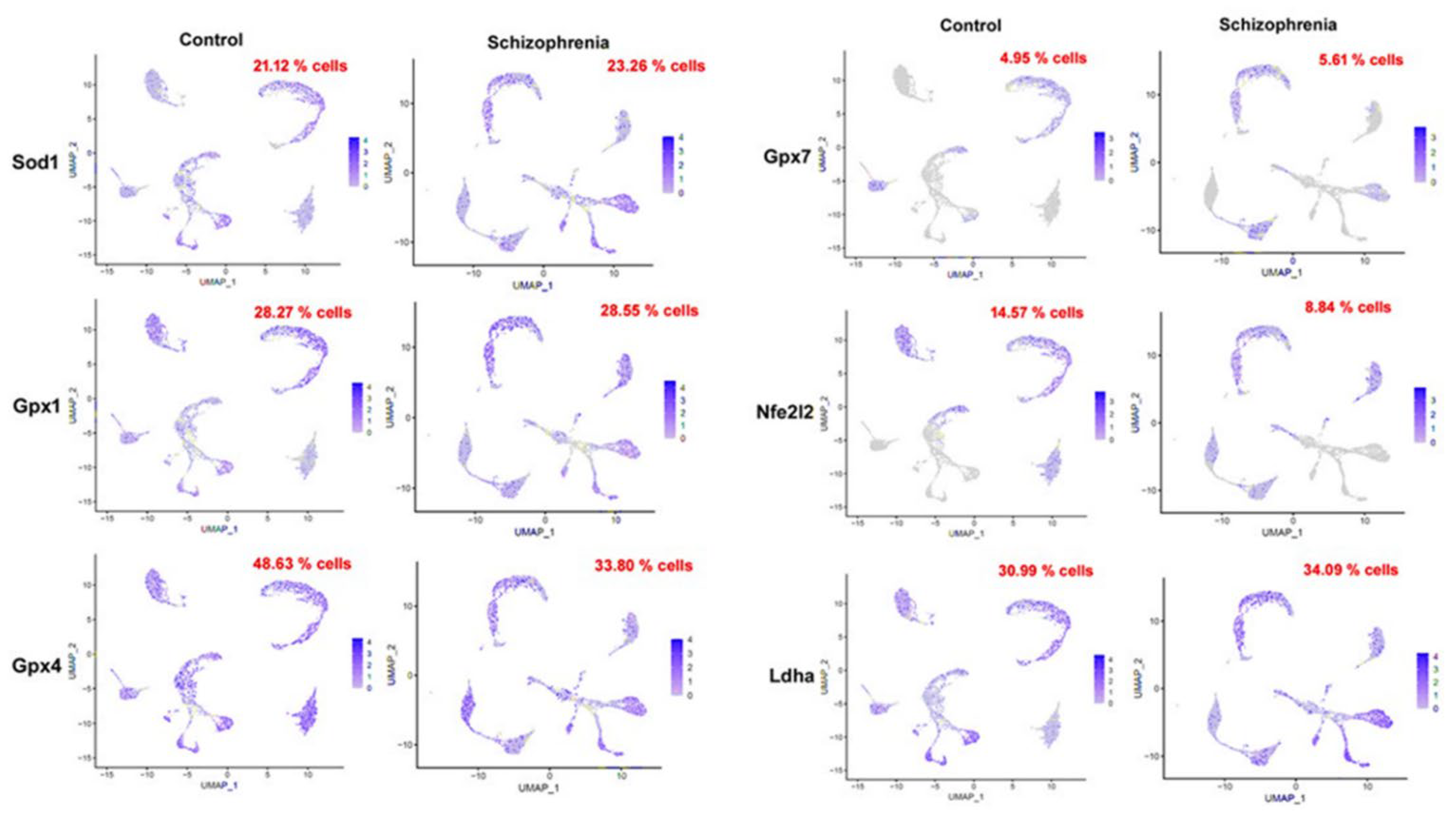
Figure 3.
Serum oxidative stress markers in schizophrenic patients and healthy people. Horizontal axis represents groups as healthy people (Control) and schizophrenic patient (SCZ) while vertical axis represents the concentration of (A) Lipid peroxidation (MDA), (B) Advanced Protein Oxidation Product (APOP), (C) Nitric Oxide (NO), (D) Glutathione (GSH) and the activity of (E) Catalase and (F) Superoxide dismutase (SOD) enzymes. Values are represented as the mean±SD. * p<0.05, ** p<0.01, *** p<0.001, **** p<0.0001.
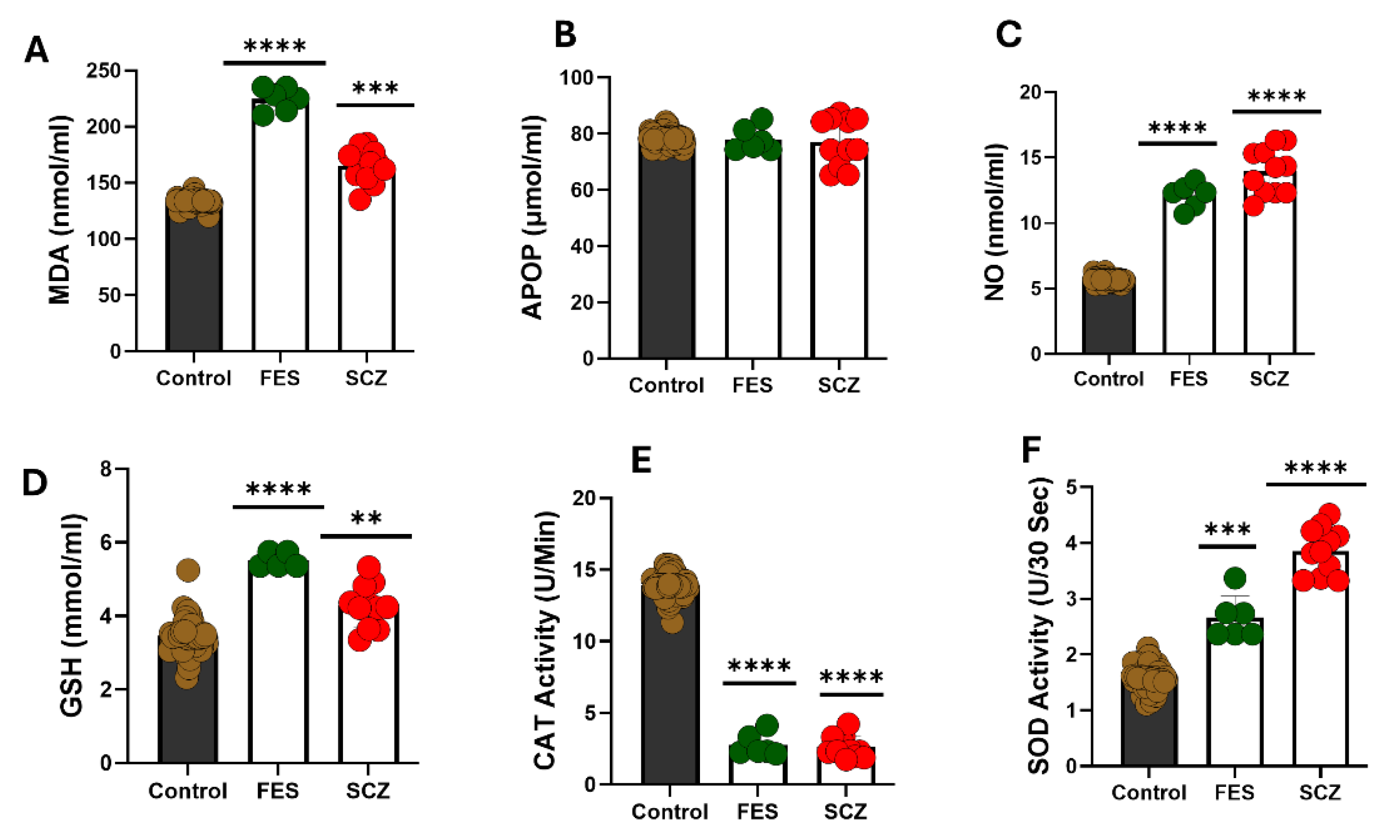
Figure 5.
Liver function test in various lengths of schizophrenia. Values are represented as the mean±SD. *p<0.05, ** p<0.01, *** p<0.001, **** p<0.0001.
Figure 5.
Liver function test in various lengths of schizophrenia. Values are represented as the mean±SD. *p<0.05, ** p<0.01, *** p<0.001, **** p<0.0001.
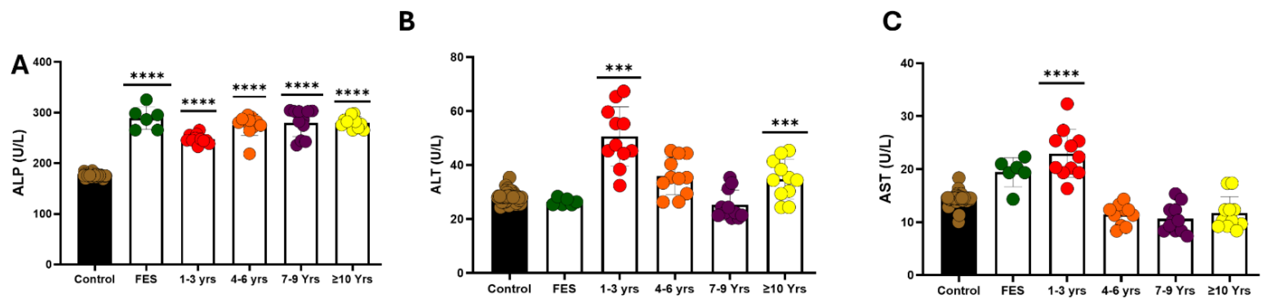
Figure 6.
Liver function test in various length of schizophrenia. Values are represented as the mean±SD. * p<0.05, ** p<0.01, *** p<0.001, **** p<0.0001.
Figure 6.
Liver function test in various length of schizophrenia. Values are represented as the mean±SD. * p<0.05, ** p<0.01, *** p<0.001, **** p<0.0001.
Disclaimer/Publisher’s Note: The statements, opinions and data contained in all publications are solely those of the individual author(s) and contributor(s) and not of MDPI and/or the editor(s). MDPI and/or the editor(s) disclaim responsibility for any injury to people or property resulting from any ideas, methods, instructions or products referred to in the content. |
© 2025 by the authors. Licensee MDPI, Basel, Switzerland. This article is an open access article distributed under the terms and conditions of the Creative Commons Attribution (CC BY) license (http://creativecommons.org/licenses/by/4.0/).
Copyright: This open access article is published under a Creative Commons CC BY 4.0 license, which permit the free download, distribution, and reuse, provided that the author and preprint are cited in any reuse.


