Submitted:
06 August 2025
Posted:
07 August 2025
You are already at the latest version
Abstract
The possibility of modifying the surface chemistry of materials, synthetizing inorganic particles in natural aqueous extracts of plants (at low temperature, avoiding calcination), opens the doors to undoubtedly very interesting scenarios for innovative functionalization strategies that are increasingly eco-sustainable and rich in interesting chemical-physical and biochemical properties. Among the aerial plant, Satureja montana exhibits interesting antibacterial, antifungal, antimicrobial and antioxidant activities due to the rich volatile and non-volatile compounds (as activators/phase controllers characterized by Gas Chromatography-Mass Spectrometry, GC-MS), contained in the aqueous extracts. For the first time, the aqueous extract of Satureja montana plant was applied for the green synthesis of TiO2 and CaCO3 particles, characterized by X-Ray diffraction (XRD), Raman, Infrared/IR spectroscopies, and Scanning Electron Microscopy-SEM/coupled by microanalysis EDX. Screening through antimicrobial assays under indoor passive sedimentation conditions showed opposite trends for both kind of inorganic particles. TiO₂ Anatase spherical particles (400<<600 nm) increase microbial growth, proportionally to increasing particles concentration (dose-dependent response). Probably, the low surface area (1.20 m2/g), low porosity (pore volume 0.0023 cm3/g; pore size of 560 nm) and smooth spherical morphology, don’t provoke chemical or physical damages to microorganisms, and therefore microbial growth is not inhibited. This occurs in the presence of medium/low concentrations of ROS (oxygenated radical species), which are chemically active in generating oxidative stress and cell death. In fact, the chemically reactive defects found in these TiO2/Anatase particles are predominantly carbonyl in nature, less reactive than carboxylic ones in producing ROS. Instead, S. montana-functionalized CaCO₃ prismatic microparticles (1µm x 1µm x 1µm) exhibit strong and dose-dependent antimicrobial activities, achieving near-complete inhibition at 50 mg mL⁻¹. This could be related to the molecular activators/phase controllers that induced defects, edges, dislocations and chemical active surface functionalities on the prismatic calcite particles, making them capable of inhibiting microbial activity. In particular, the presence of carboxyl groups C=(O)-OH, recorded by FTIR on the prismatic surfaces of CaCO3 MPs/SM particles, could be responsible for the production of ROS, the latter known to be dangerous sentinels of oxidative stress and cell death.
Keywords:
Satureja montana
; plant extract-functionalized microparticles
; GC-MS extract analysis
; oxygenated terpenoids as activator/phase controllers
; Terpinene
; Thymoquinone
; Carvacrol
; Thymol
; L-Linalool
; preliminary antimicrobial screening
1. Introduction
The possibility of synthesizing and especially functionalizing Micro and Nanomaterials in eco-sustainable work environments certainly opens the doors to new and interesting possibilities in obtaining smart nanomaterials with extraordinary chemical-physical [1], biochemical [2] and medical properties [3] of great relevance. For a long time, surface chemistry has been exclusively concerned with predominantly chemical functionalization reactions. The great variety of natural substances, such as essential oils [4]and natural extracts [5] from medicinal plants [6], allows to obtain (on a large scale mass production) increasingly eco-friendly micro; and nanomaterials characterized by excellent properties/features of clinical-medical [7], environmental [8], Cultural Heritage restoration/conservation [9]and food packaging interest [10]. Recently, several interesting review papers provide innovative advancements in green synthesis strategies to produce eco-friendly materials, focusing on raw materials, their reaction mechanism and surface Chemistry, challenges involved in different application fields [11,12,13].
The strategies based on the green synthetic approach offer several advantages over traditional mechanism production of new materials (as reported in Scheme 1). Traditional chemical synthesis routes are the subject of debate (in the international scientific community) because they involve the use of chemical reagents with an environmental impact and also contribute to the production of others (such as secondary waste products present in large volumes) that are difficult to dispose of in the environment [14]. Furthermore, conventional chemical synthesis involves much higher costs than strategies based on natural substances, easily available in the environment (at low cost and/or zero cost) and with low impact on humans, surfaces, materials and the ecosystem. The great advantage of using natural substances, present in the ecosystem, also allows the adoption of synthetic procedures that do not require high temperatures, high working pressures and non-aqueous solvents (which often produce reaction waste with a high impact on operators/end-users and the work environment). The ease of finding natural substances and their abundance allows for scaling up production to an industrial level, thus designing and creating new materials ready to be launched on the market [15]. In particular, several scientific publications dated 2025 (current year) focus on the scalable production of micro; and nanomaterials from extracts, essential oils and derivatives of natural plants [16,17,18,19,20,21,22,23,24,25]. The main reason is related to plants’ widespread diffusion, to their medicinal and officinal properties, widely used in the past to heal, cure, nourish, purify (for example water) and to produce the colors used in classical Artworks (i.e., ancient Polychromy) [26].
The possibility of replicating processes that occur in Nature also represents a great scientific challenge because it would allow us to understand, simulate and reproduce completely spontaneous processes present in the World [27]. Not only that but simulating and reproducing many natural synthetic pathways would also guarantee the preservation of Nature and its vital processes, as well as the safeguarding of History (Our Past) through the care of Cultural Heritage (using the same raw materials, with which they were assembled in past eras of Human History) [28]. Among these new synthetic routes [16,17,18,19,20,21,22,23,24,25], those in which micro, and nanoparticles nucleate and subsequently grow, simultaneously with the extraction of natural substances from plants, are very interesting. In fact, natural substances that also play the role of “templates in the green synthesis” of particles have enormous advantages over chemical and/or physical templates, because they do not have to be removed at the end of the synthesis and become part of the surface functionalization [29,30,31,32,33,34,35,36,37,38]. Indeed, natural substances (contained in plant extracts, essential oils and exudates) usually known as biochemical additives, play the main role of phase controllers during green synthesis approach.
This new synthetic strategy is also fast on a time scale (no time consuming) because in a single reactive process the particles are produced from natural substances that shape them (in terms of shape, size and defects), also functionalizing them (in only “one pot green synthetic approach”).
In this work, a simultaneous synthesis and functionalization process of TiO2 and CaCO3 microparticles (MPs), starting from natural extracts of Satureja montana (SM), was optimized. Especially, Satureja montana plant species (which is native from the Mediterranean region and cultivated all over Europe, Russia, and Turkey [39,40]) has been selected because of interesting antimicrobial properties, the great potential synergism with antibiotics, anti-inflammatory, antibacterial, antifungal, and antioxidant features [41,42,43,44,45]. Recently, Rai et al. (2025) [46] demonstrate the high efficiency of the synergistic combinations with two well-known antibiotics (gentamicin and amphotericin B) and Essential Oils (EOs), this latter extracted from SM, against a panel of pathogenic bacteria and candida (responsible for the extensive formation of biofilms on polymaterial surfaces). Natural Plant Extracts (NPEs), EOs and phytochemicals represent a smart alternative strategy to control Salmonella biofilm formation [47]. Thymol, carvacrol, and eugenol, notably from different SM species, exhibit excellent antibiofilm activity, inducing stress-related proteins in Salmonella. Plant-derived extracts and phytochemicals (including plant extracts, essential oils, and purified phytochemicals, individually or combined ones) act against both planktonic and biofilm forms by disrupting biofilm formation, motility and surfaces adhesion. Phyto-synthesized microparticles offer a very promising approach, combating microbial biofilms while being cost-effective, environmentally friendly and highly compatible with the polymaterial surfaces to be treated and with the end-users [47]. Integrating plant extracts and synthetic microparticles demonstrates potential in addressing biofilm-related issues and antibiotic resistance (fundamental features for applications in the medical, pharmacological, nutritional fields and also for the conservation of Cultural Heritage). For this purpose, during the past time, Rinaldi et al. (2021) [48] optimized Nano-Emulsion (NEs) formulations to ensure food safety quality and counteract the antibiotic resistance of poultry associated E. coli, if applied in poultry farms. To improve the essential oils (EO) properties, oil in water Nano-Emulsions (NEs) composed of SMEOs (Satureja montana Essential Oils) and Tween-80 were synthetized, fully characterized, and their antimicrobial and antibiofilm properties assayed against Escherichia coli strains isolated from healthy chicken. Since surfactant and oil composition can strongly influence NE features and their application fields, a ternary phase diagram was evaluated to select the best surfactant/oil/water ratio composition.
During 2021, Oliveira-Pinto et al. [49] combined SMEOS with the Zein microparticles (ZNPs) in an optimized formulation suitable to reduce the X. euvesicatoria (Xeu) amount, which is responsible for the Tomato Bacterial spot (Bs), and simultaneously to improve plants’ health, mainly for their antimicrobial activity on Xeu pathogens. In the following year 2022, the same authors Oliveira-Pinto et al. [50] report about the Nano clays production and characterization, such as Montmorillonite (NMT), demonstrating their excellent antimicrobial activities, or their role as delivery tools/stabilizers for organic compounds, such as natural extracts from plants and/or essential oils (EOs), which also exhibit antimicrobial activity against plant pathogens [50]. Results highlight that the NMT was able to reduce Xeu bacterial amount, while reducing ROS (Radical Oxygenated Species) production and keeping the transcript levels of the defense-related genes close to those of the control samples (used for comparison). Generally, the results show that NMT has a great potential to integrate Bs management approaches, considering its antimicrobial activity, and that EO and/or Nano clays could be successfully employed as new disease preventive strategies, since they enhance the healthy shoots’ defense, thus potentially reducing the pathogen establishment.
Currently, SM has never been used to synthesize/functionalize with its extracts, TiO2 and CaCO3 microparticles (MPs) as done in this work, which for this reason is innovative (in terms of products synthesized and characterized). According to this, TiO2 nanoparticles (i.e., TiO2 NPs) have been previously synthetized and functionalized by applying Jasminum and Magnolia champaca flower extracts, as reported by Sangeetha et al. (2024) [51]. These authors demonstrate that NPs synthesis applying natural extracts/oils led to rutile phase of TiO2 NPs while chemical synthesis produced anatase phase. Green synthesis yielded larger crystallite size TiO2 compared to chemical synthesis, also enhancing the photoluminescence emission efficiency. In this current year 2025, Pham et al. [52], for the first time, fabricate innovative aqueous Nano Emulsions (NEs) based on anthraquinone-rich extract of P. cuspitaum and TiO2 nanoparticles, together with surfactants, evaluating the corresponding antifungal efficiencies, against the melon fungal diseases. At a concentration of 0.5%, the NE formulation completely inhibited the mycelial growth of Botrytis cinerea, Phytophthora spp., Fusarium oxysporum, Colletotrichum spp., Sclerotium spp., Rhizoctonia spp., and the germination of Eryshipe cichorasearum spores, respectively [52]. Other authors, as Wang et al. (2025) [53], develop a new chitosan-based nanocomposite films incorporating nano-TiO2 and Daisy Essential Oil for the preservation of Actinidia arguta (which represents a small fruit rich in many vitamins and minerals especially vitamin C is much higher than other fruits [54]. The films exhibited enhanced mechanical strength, antimicrobial activity, and antioxidant capacity. Application of these nanocomposite films on Actinidia arguta reveals significant improvements in weight loss, hardness, decay rate, total microbial count, respiration rate, soluble solids content, vitamin C, titratable acidity, and enzyme activities during food storage. Experimental data show that chitosan/nano-TiO2/DEO nanocomposite films effectively extend the shelf life and preserve the quality of Actinidia arguta, providing a promising strategy for the natural and eco-sustainable food preservation chain.
According to these literatures, TiO2 particles described above have been synthetized by calcination treatments, carried out at high temperatures [55], instead, we propose a sustainable low-temperature method (avoiding calcination), as already reported by Prof. Eckhart Wollenweber [56]. This latter, which allows to produce particles in the aqueous natural extracts of SM at lower temperatures, result useful to study exudate/extract flavonoids but without removing Volatile Organic Compounds (VOCs), which are responsible for the main antibacterial, antimicrobial, antifungal, antioxidant and photocatalytic properties, opening up a range of application opportunities in many industrial sectors.
Even more innovative is the approach performed in this work, regarding the synthesis and functionalization of calcite microparticles (CaCO3 MPs) with SM extract (for the first time synthetized here). Indeed, starting from 2023 some authors such as Meydan et al. (2023) [57], describe the green synthesis of calcium carbonate nanoparticles (CaCO3 NPs), by applying the Gum Arabic, as a natural polymer for green synthesis. The synthesized CaCO3 NPs showed good photocatalytic activity to methylene blue (MB) dye degradation, which percentage degradation was 93% after 120 min. Also, the cytotoxicity of CaCO3 NPs has been examined on the normal L929 and cancer CT26 cell lines and the IC50 value was about 250 μg/mL for cancer cells [57]. During the same year, other authors as Nelwamondo et al. (2023) [58], demonstrate the MgO and CaCO3/Moringa oleifera nanoparticles production and assessed the green nanoparticles effects on groundnut genotypes. The effect of biosynthesized MgO and CaCO3 nanoparticles (NPs) using Moringa oleifera natural extract on the growth and yield of groundnut genotypes (exposed to different concentrations of green NPs) has been widely investigated [58]. The findings demonstrated that foliar application of MgO/CaCO3/ Moringa oleifera natural extract NPs positively affected groundnut biomass production. There is a great deal of evidence signifying that foliar applications of 50 mg/L of MgO 100 mg/L CaCO3 contributed greatly to plant growth and crop production, and for this purpose the green NPS are very recommended as Nano-fertilizers application rate for groundnut production [58].
In our very recent publications cited as Valentini et al. (2024) [59], authors report about the use of Fe-Alg-CaCO3 microparticles (MPs) as foliar fertilizer on lettuce plants in an aquaponic system. MPs were produced starting from CaCO3 nanoparticles (used here as precursors of MPs), these latter synthetized by applying a biomimetic approach, optimized by Valentini et al. (2022) [60]. The research investigated Lactuca sativa L. cv. Foglia di Quercia Verde plants as the experimental cultivar. Three iron concentrations (10, 50, and 250 ppm) were tested, with 15 plants per treatment group, plus a control group receiving only sterile double-distilled water. This study investigated rosette diameter, root length, fresh weight, soluble solids concentration, levels of photosynthetic pigments, and phenolic and flavonoid content. The optimized concentration of 250 ppm for treatment provided the most significant enhancements in both biomass yield and quality, highlighting the potential of precision fertilizers to boost eco-sustainability and efficiency in aquaponic systems. During 2025, Valentini et al. [61], add a further important scientific result concerning the application of green Zn-Alg-CaCO3 MPs as an eco-sustainable solution for precision fertilization of Tomato plants (Solanum lycopersicum L. cv. Principe Borghese) in aquaponic agriculture approach. Several parameters have been investigated as antioxidant activity, phenolic, flavonoid, and lycopene contents, which particularly enhanced at the highest concentrations of green Zn-Alg-CaCO3 MPs. Importantly, no increase in zinc element was detected in recirculating water, highlighting the environmental safety of foliar application. These experimental results highlight the potential of Zn-Alg-CaCO3 MPs as a sustainable solution for precision fertilization in aquaponic agriculture. According to this, the authors wanted to combine for the first time the exceptional properties of natural SM extracts (especially the anti-biofilm features) with the production and functionalization of microstructured calcite (CaCO3 MPs), to be used in fields other than those widely reported in this introduction, such as the conservation and restoration of Cultural Heritage and Historical-Artistic Heritage/surfaces. During the past time, in our previous work [62], we demonstrated that homemade CaCO3 nanoparticles/nanofiller were successfully employed for reinforcing the plaster of Palazzetto Alessandro Lancia in Rome (Italy). During 2017, Valentini et al. [63] described the role of nanostructured CaCO3 for the deacidification of Library and Archival Materials (i.e., indoor Cultural Heritage). Furthermore, Valentini et al. (2022) [60] applied CaCO3 nanoparticles for the consolidation of Pietraforte (a particular type of Florentine sandstone) which makes up the Bell Tower of San Lorenzo (belonging to the corresponding monumental complex of San Lorenzo), in Florence (Italy).
Considering all this, the aim of this work is to synthesize inorganic particles (i.e., TiO2 and CaCO3) having a green and sustainable chemistry approach (based on SM’s aqueous extract, carried out at low temperatures), which encompasses many interesting antimicrobial features for future applications in many fields, such as clinical-medical, pharmaceutical, nutraceutical, food chemistry, agricultural and even in the conservation of Cultural Heritage.
Finally, for clarity of discussion in the text, Scheme 2 reports the nomenclature of the materials cited and applied in the work, including the acronyms and abbreviations used.
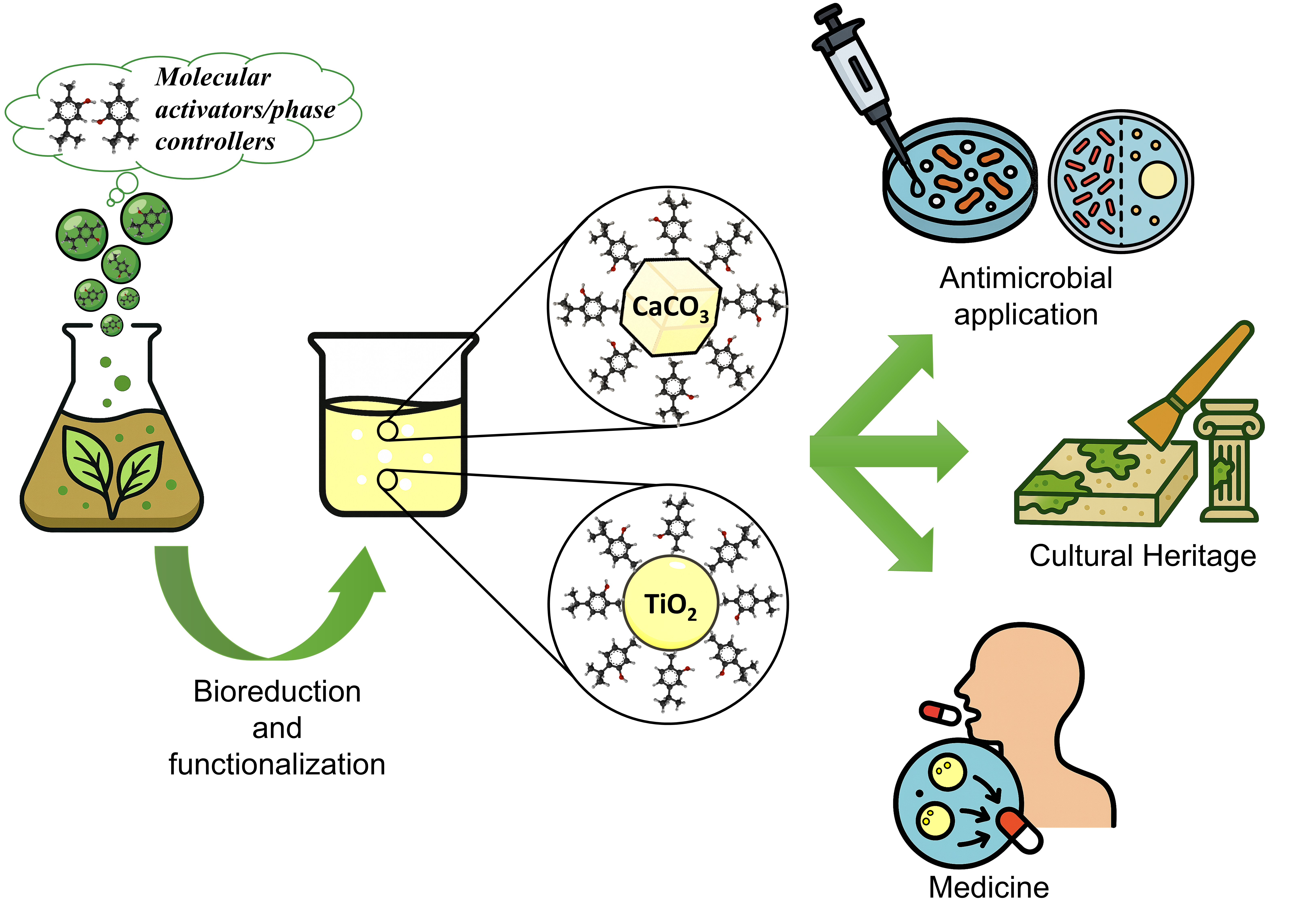
Scheme 1.
Green synthesis routes for the functionalization of micro- and nanoparticles using natural extracts, essential oils and exudates from natural plants.
Scheme 1.
Green synthesis routes for the functionalization of micro- and nanoparticles using natural extracts, essential oils and exudates from natural plants.

Scheme 2.
Nomenclature, acronyms/abbreviations, description of all materials cited in the text.
| Nomenclature | Acronyms abbreviations |
Description | Notes |
|---|---|---|---|
| Titanium Oxide | TiO2 MPs/SM | Functionalized particles produced in natural extracts of plants | Microparticles (in size) as confirmed in this work |
| Calcium Carbonate | CaCO3 MPs/SM | Functionalized particles produced in natural extracts of plants | Microparticles (in size) as confirmed in this work |
| Microparticles | MPs | Microparticles are tiny spherical particles, typically ranging from 1 to 1000 micrometers in diameter. | Microparticles used for biological assay in this work |
| Nanoparticles | NPs | Nanoparticles are incredibly small particles, typically between 1 and 100 nanometers in size | [1] |
| Satureja montana | SM | Plant species, which is native from the Mediterranean region and cultivated all over Europe, Russia, and Turkey |
Satureja montana L., Lamiaceae, commonly known as winter savory This work |
| Natural Plant Extracts | NPEs | Extracts may be derived from whole plants or from specific parts of plants such as leaves, stems, barks, roots, flowers and/or fruits | Natural Extracts (by drying the leaves of the plants) were performed at low temperature and in bi-distilled water. This work |
| Essential Oils | EOs | Essential Oils are precious mixtures of liquid and volatile aromatic substances, extracted mainly by steam distillation from plant material coming from various types of aromatic plants; they can also be obtained by cold pressing, as in the case of oils derived from citrus peel or by solvent extraction. | [47,48,49,50] |
| Nano-Emulsions | (NEs) | Nanoemulsions are nano-sized emulsions (20–200 nm), thermodynamically stable isotropic system in which two immiscible liquids are mixed to form a single phase | [48,49,50] |
| Satureja montana Essential Oils and Tween-80 | SMEOs | Oil in water Nano-Emulsions (NEs) composed of SMEOs (Satureja montana Essential Oils) and Tween-80 | [48,49] |
| Montmorillonite | NMT | Montmorillonite is a naturally occurring clay mineral belonging to the smectite group, primarily of aluminosilicate | [50] |
| Chitosan-nano-TiO2 Daisy Essential Oil | chitosan/nano TiO2/DEO | Chitosan film, incorporating nano-TiO2, functionalized with Daisy Essential Oil | [53,54] |
| Magnesium Oxide and Calcium Carbonate Moringa oleifera natural extract | MgO/CaCO3/ Moringa oleifera NPs | Biosynthesized MgO and CaCO3 nanoparticles (NPs) using Moringa oleifera natural extract | [58] |
| Iron-Calcium Alginate-Calcium Carbonate microparticles/microspheres | Fe-Alg-CaCO3 MPs | Foliar fertilizer on lettuce plants in an aquaponic system | [59] |
| Zinc- Alginate-Calcium Carbonate microparticles | Zn-Alg-CaCO3 MPs | eco-sustainable solution for precision fertilization of Tomato plants in aquaponic agriculture approach | [61] |
| Volatile Organic Compounds | VOCs | Volatile organic compounds are a group of chemicals that easily evaporate at room temperature and are commonly found in various products used indoors and outdoors (in this study terpenes family) | [56] |
| Methylene Blue | MB | Methylene blue is used as a bacteriologic stain or dye and indicator | [57] |
| Radical Oxygenated Species | ROS | Reactive Oxygen Species (ROS) are unstable, highly reactive molecules containing oxygen that can cause damage to cells by reacting with other molecules. | [49,50,64,65] |
2. Results and Discussion
This chapter is organized as follows: first, the results related to the morphological and structural characterization of the new functionalized microparticles are reported and described. Then, the chemical composition of the natural SM extracts is discussed, by means of GC-MS analysis; and finally, the results related to the study of the antibacterial, antimicrobial, antioxidant and antibiofilm properties, found in appropriate bacterial strains selected and treated at different concentrations of new functionalized particles, are widely reported and discussed.
2.1. Functionalized Microparticles and Their Characterization Study
2.1.1. TiO2 MPs/SM
The TiO2 particles contained in the alcoholic suspension, shows instead a spherical shape, with diameters ranging from 160 nm to 1.3 µm, with a prevalence of elements with diameters around 400-600 nm (Figure 1A). The surface of the micro/nanospheres appears smooth and the observations at high magnification (Figure 1B) suggest that the spherical elements have variable sizes because they arise from the nucleation of TIO2 around a core of amorphous organic substance and then they gradually growth in size by TIO2 apposition. The amorphous aggregates of organic matter are indicated by the arrows in Figure 1B.
The XRD analysis revealed a slightly noisy diffractogram (Figure 2A) characterized by broad peaks centered at 11.74°, 17.45°, 21.77°, 24.60°, 27.91°, 30.54°, 32.90°, and 35.63° (2θ, Mo Kα). All detected reflections were attributed to the anatase phase of TiO2, with the most intense peak at 11.74°, corresponding to the (101) plane [66,67,68]. The remaining signals were assigned to the (112), (200), (211), (204), (220), (301), and (312) planes, respectively. Nevertheless, the presence of brookite (~10%) cannot be entirely ruled out, as the peaks at 11.74° and 21.77° may also match the (111) and (231) planes of this TiO2 polymorph [68,69]. However, the absence of the typical high-intensity doublet that characterizes the brookite phase suggests that the sample is predominantly composed of the anatase form (~90%).
Raman spectroscopy provided further confirmation of the TiO2 crystalline phases (Figure 2B and Table 1). The spectrum revealed the presence of characteristic bands of the anatase polymorph, confirming the results obtained by XRD analysis. A strong band was observed at 146 cm−1, corresponding to the Eg vibrational mode of anatase, along with the B1g mode at 411 cm−1. Additional bands were detected at 517 cm−1 and 623 cm−1, attributable to the A1g + B1g and Eg modes of the anatase phase, respectively [70,71,72]. Two minor shoulders were also observed between 219 cm−1 and 279 cm−1, which can be attributed to the B1g and B2g vibrational modes of brookite, suggesting the possible presence of this secondary polymorph in low concentration [72,73]. Nevertheless, a partial contribution from the low-frequency Eg mode of anatase, typically appearing as a broad shoulder around 200 cm−1, cannot be excluded [70,71,72]. In addition, the Raman spectrum exhibited broad fluorescence related features at ̴ 874, ̴ 1627, and ̴ 3352 cm−1, which are likely associated with the vibrational modes of the organic functional groups/phase controllers used to functionalize/draw the particles (see Figure S3 in Supplementary Information). Specifically, the band at 874 cm−1 may correspond to ν(C–C), ν(C–O) stretching and δ(C–H) bending vibrations, while the band at 1627 cm−1 may arise from ν(C=C) or ν(C=O) stretching, as well as δ(O–H) bending [74,75,76,77,78,79,80,81,82,83]. The broad signal at 3352 cm−1 is attributable to ν(O–H) stretching vibrations, indicative of hydroxyl groups of phytocompounds based activators [84].
FTIR-ATR analysis confirmed the successful synthesis and functionalization of TiO2 nanoparticles with Satureja montana extract (Figure 2C). A broad and intense absorption band observed in the range 800–600 cm−1 was attributed to Ti–O–Ti stretching and bending vibrations, typical of titanium dioxide frameworks [85,86]. Signals at approximately 3400 cm−1 and 1630 cm−1 were assigned to O–H stretching and bending vibrations, respectively, indicating the presence of hydroxyl groups, either due to surface hydroxylation or originating from phenolic and terpenoid constituents of the plant extract [87,88,89,90,91]. Additionally, the spectrum displayed several distinctive bands associated with phytocompounds from the extract. Notably, peaks at 2974 cm−1 and 2893 cm−1 correspond to the asymmetric stretching of –CH3 groups and the symmetric stretching of –CH2 groups, respectively. Absorption bands between 1454 and 1395 cm−1 were assigned to C–H bending vibrations, specifically the asymmetric and symmetric bending modes of methyl and methylene groups. Furthermore, the band at 1268 cm−1 was attributed to C–O stretching vibrations, while the signal at 1051 cm−1 indicated C–O or C–C stretching modes [90,91]. All the FTIR band assignments are also reported in Table 1.
Table 1.
Raman and FTIR band assignments of TiO2 MPs /SM.
| Band (cm−1) | Assignment | Ref. | ||
| RAMAN SPECTROSCOPY | ||||
| 146 | Eg mode (anatase) | [70,71,72] | ||
| 219-279 | Eg (anatase) B1g + B2g mode (brookite) |
[70,71,72,73] | ||
| 411 | B1g mode (anatase) | [70,71,72] | ||
| 517 | A1g + B1g mode (anatase) | [70,71,72] | ||
| 623 | Eg mode (anatase) | [70,71,72] | ||
| ̴ 874 | νC–OH νC–C δCH/CH2 |
[74,75,76,77,78,79,80,81,82,83] | ||
| ̴ 1627 | νC=O νC=C δO-H |
[74,75,76,77,78,79,80,81,82] | ||
| ̴ 3352 | νO-H | [84] | ||
| FTIR SPECTROSCOPY | ||||
| 3400 | νO–H | [87,88,89,90,91] | ||
| 1630 | δO–H νC=O νC=C |
[87,88,89,90,91] | ||
| 2974 | νas –CH3 | [90,91] | ||
| 2893 | νs –CH2 | [90,91] | ||
| 1454, 1395 | δas/s –CH3 δas/s –CH2 |
[90,91] | ||
| 1268 | νC–O νC–C |
[90,91] | ||
| 1051 | νC–O νC–C ring vibration |
[90,91] | ||
| 800-600 | ν+δ Ti–O–Ti | [85,86] | ||
Vibrational bands assignments follow conventional spectroscopic notation: ν = stretching; δ = bending; s = symmetric; as = asymmetric.
2.1.2. CaCO3 MPs/SM
The CaCO3 particles in the examined alcohol suspension, appear as crystals with a prismatic shape, sharp edges and variable size (Figure 3A), with an average of 1 µm x 1 µm x 1 µm. Amorphous aggregates of organic matter (indicated by the arrows in Figure 3B) are adherent onto the smooth surface of the crystals. The image B (high magnification) shows that the prismatic crystals’ structure is the result of a stratification of thin and smooth plates (like cheddar cheese layered tiles).
The diffractogram obtained for CaCO3 MPs/SM reveals calcite as the predominant crystalline phase (Figure 4A). The most intense diffraction peak is observed at 2θ = 29.43°, corresponding to the (104) plane, a characteristic reflection of calcite [92,93,94,95]. Additional peaks, including those at 31.46°, 56.54°, and beyond 58.20°, exhibit lower intensities. Nevertheless, the overall diffraction pattern unambiguously confirms the presence of calcite, with no significant traces of other CaCO3 polymorphs [92,93,94,95]. These findings suggest that Satureja montana extract favors the formation of the calcitic phase over other crystalline structures (i.e., predominant phase).
Complementary insights were provided by Raman spectroscopy, which revealed vibrational features attributable to both the calcitic phase and organic phytocompounds introduced by SM extract during synthesis (Figure 4B, and Table 2). The presence of calcite is confirmed by the bands at 248 cm−1, 718 cm−1, and 1092 cm−1. Specifically, the 1092 cm−1 band corresponds to the symmetric stretching vibration of the CO32− group (A1g mode), while the 718 cm−1 band is attributed to the in-plane bending vibration of the carbonate ion (Eg mode) [96,97]. The signal at 248 cm−1 relates to lattice translational vibrations (Eg mode) but may also partially overlap with skeletal torsional and deformation modes of aromatic phytocompounds, suggesting nanoparticle surface functionalization [74,75,76,77,78,82]. Moreover, other spectral bands support the presence of flavonoids and phenolic terpenoids on the carbonate surface. In the low-frequency region, several additional Raman bands (320, 412, 507, and 626 cm−1) are assigned to skeletal deformations, twisting, and out-of-plane vibrations of aromatic ring systems [74,75,76,77,78,82].
The band at 746 cm−1 arises from a combination of C–C stretching, C–O bending, and aromatic ring deformation, while the signal at 890 cm−1 is linked to C–OH and C–C stretching, as well as CH and CH2 bending vibrations [74,75,76,77,78,82]. Other bands such as 1009 cm−1 and 1180 cm−1 are attributed to in-plane CH bending, C–O stretching, and aromatic skeletal vibrations, whereas the band at 1339 cm−1 is associated with CH3 bending (notably of isopropyl groups) and CH2 scissoring modes [74,75,76,77,78,79,80,81,82].
The FTIR spectrum further confirms the dual nature of the material, revealing contributions from both the inorganic carbonate phase and the organic constituents (Figure 4C, and Table 2). The bands at 1404 cm−1 and 870 cm−1 correspond to the asymmetric stretching and out-of-plane bending vibrations of CO32−, respectively. The additional signal at 705 cm−1, attributed to in-plane bending of CO32−, confirms the identification of the calcite phase [98,99]. A broad band in the 3600–3000 cm−1 range is assigned to O–H stretching vibrations, originating from adsorbed water, surface hydroxyl groups, and phenolic or alcoholic phytocompounds. The presence of aliphatic chains, typical of plant-derived molecules, is confirmed by the bands at 2951, 2922, and 2856 cm−1, which are associated with asymmetric and symmetric C–H stretching vibrations of methyl (CH3) and methylene (CH2) groups [87,88,89,90,91]. A sharp absorption band at 1710 cm−1 corresponds to carbonyl (C=O) stretching, while the band at 1637 cm−1 is mainly due to molecular water bending (δO–H), with potential contributions from C=O and C=C stretching vibrations in aromatic systems [87,88,89,90,91]. Finally, the bands at 1231 cm−1 and 1070 cm−1 are assigned to C–O and C–C stretching modes, as well as to skeletal vibrations of aromatic rings, further confirming the presence of oxygenated phytocompounds on the nanoparticle surface.
2.2. Chemical Composition of NPEs from SM by GC-MS
GC-MS analyses were performed using both targeted and untargeted approaches to comprehensively characterize the volatile fraction of the Satureja montana aqueous extract (SM). The integration of both methods allowed not only the identification of a defined set of known volatile constituents but also the detection of additional, previously unconsidered compounds, potentially involved in the green synthesis and surface functionalization of microparticles.
2.2.1. Targeted Analysis
A targeted GC-MS analysis was performed on the aqueous extract of SM immediately after preparation, with the aim of characterizing its volatile composition and identifying compounds potentially involved in the surface functionalization of microparticles. The analysis was carried out using a calibrated terpene standard mixture, allowing the semi-quantitative determination of 24 selected volatile organic compounds (see Table 3).
The extract was dominated by oxygenated monoterpenes, with thymol being by far the most abundant constituent (63,906 ng/mL), followed by terpinen-4-ol (10,305 ng/mL) and thymoquinone (8,271 ng/mL). Other noteworthy compounds included γ-terpinene (882 ng/mL), carvacrol (5,669 ng/mL), α-terpinene (3,814 ng/mL), and sabinene (1,054 ng/mL). The presence of α-thujone (282 ng/mL), α-phellandrene (264 ng/mL), and α-terpinolene (284 ng/mL) further confirmed the richness of the terpene fraction. Trace amounts of carvacrol methyl ether (23 ng/mL), caryophyllene oxide (135 ng/mL), L-linalool (1,425 ng/mL), and para-cymene (157 ng/mL) were also identified.
The volatile profile of the extract included monoterpene hydrocarbons (e.g., sabinene, α-thujone, α-terpinene), oxygenated derivatives (e.g., thymol, terpinen-4-ol, caryophyllene oxide), and aromatic compounds (e.g., p-cymene, thymoquinone), reflecting a chemically diverse composition.
Notably, thymol and terpinen-4-ol showed the highest peak areas, followed by thymoquinone, carvacrol, and α-terpinene, while compounds like caryophyllene oxide and l-linalool were present at lower levels. This distribution confirms the predominance of oxygenated monoterpenes among the most abundant constituents.
The analysis was conducted on a single, well-characterized extract, providing a representative chemical fingerprint under the tested conditions. Although the lack of replicates prevents statistical evaluation, the dataset offers a consistent reference for subsequent applications using the same preparation.
2.2.2. Untargeted Analysis
In addition to the targeted screening, a non-targeted GC-HRMS approach was employed to comprehensively explore the chemical complexity of the Satureja montana aqueous extract and to identify additional constituents potentially involved in the green synthesis and surface functionalization of microparticles. This strategy aimed to complement the targeted dataset by detecting molecules not included in the calibration mixture but potentially relevant in mechanistic or functional terms.
Following peak deconvolution, a total of 643 individual features were detected. After procedural blank subtraction and spectral quality filtering (Total Score > 90), 169 compounds were identified with high confidence. These were subsequently categorized into major chemical families based on their molecular structures and functional groups. As shown in Figure 5, alcohols (22.6%), aromatic hydrocarbons (17.3%), and ketones (8.9%) were among the most represented classes. Esters, polycyclic aromatic hydrocarbons, monoterpenes, and furan derivatives were also identified, while a substantial fraction (34.5%) fell under the category “Others,” encompassing structurally ambiguous, multifunctional, or less-characterized phytochemicals. These results provide a broad overview of the chemical landscape of the extract and highlight its richness in oxygenated and aromatic species, which are likely to influence its reactivity and interaction with inorganic surfaces.
The full dataset, including retention times, average peak areas, molecular formulas, exact and theoretical masses, and NIST match scores, is reported in Supplementary Table S1. In many cases, compounds were detected at multiple retention times but shared the same molecular formula and theoretical mass. These signals, retained in the final table, likely represent structural or conformational isomers and are discussed collectively based on their elemental identity.
To focus the discussion on the most relevant compounds, the 22 molecules with the highest peak areas—each with a Total Score > 95 and a Peak Rating > 7.5—were selected and visualized in Figure 6. For clarity, thymol and endo-borneol, although confirmed as major constituents, were excluded from the graph due to their disproportionately high abundance, which would distort the scale. Their identity is confirmed and discussed elsewhere in the text.
The selected compounds span a broad retention time range and include molecules with well-defined structure as well as entries reported solely by molecular formula (e.g., C10H16, C10H14, C13H22O). These formula-based entries indicate the detection of multiple peaks with identical mass and elemental composition but distinct chromatographic behaviors, suggesting the presence of unresolved isomeric species. Although further structural elucidation is needed, such components are retained for their potential contribution to the extract’s reactivity and its role in downstream applications.
In the following paragraphs, the results regarding the application of TiO2 MPs/SM and CaCO3 MPs/SM in bacterial cultures are described, and the order of presentation will respect that followed for all other results previously shown.
2.3. Antimicrobial Screening
In the present study, the antimicrobial activity of TiO2 and CaCO3 MPs was evaluated within an indoor environmental setting. Passive sedimentation method was employed, as this approach is widely regarded as one of the most cost-effective, standardized, and reproducible methods for such investigation [100] The antimicrobial activity of TiO2 microparticles (MPs) and TiO2 MPs functionalized with S. montana leaf extract (TiO2 MPs/SM) was evaluated in comparison with the untreated control (CT) (Figure 7A). In the control condition, passive air sampling yielded 66.33 ± 5.51 CFU/plate, representing the baseline microbial load in the indoor environment. When TiO2 MPs/SM were applied, low to moderate concentrations (0.2–4 mg/mL) produced a statistically non-significant increase in CFU compared to the control, indicating a limited impact on microbial deposition. However, at 10 mg/mL a statistically significant increase was observed 1132.00 ± 46.13 (p < 0.0001), and at the highest concentrations (20–50 mg/mL) the number of CFU was found to be too numerous to count (TNTC), suggesting an unexpected promotion of microbial accumulation rather than inhibition. A similar but more pronounced trend was observed for TiO2 MPs, where even the lowest dose (0.2 mg/mL) caused a dramatic increase in microbial growth 1776.00 ± 166.40 CFU/plate (p < 0.0001), further escalating at 2 mg/mL (2785.00 ± 190.31 CFU/plate), with higher concentrations resulting in TNTC. Compared to the CT, both formulations failed to reduce microbial contamination. Contrary to what was observed with TiO2 MPs data regarding the efficiency of antibacterial activity, promising results were observed with CaCO3 MPs data shown in Figure 7B. Compared to the CT (106.00 ± 13.52 CFU/plate), the application of CaCO3 MPs and CaCO3 MPs/SM induced a dose-dependent modulation of microbial growth.
For CaCO3 MPs/SM, low concentrations (0.2–2 mg/mL) did not significantly differ from the control, maintaining CFU counts close to CT, respectively of 102.00 ± 2.00 and 97 ± 1.73 CFU/plate. However, starting from 4 mg/mL, a statistically significant reduction was observed with a value of 75.32 ± 16.04 CFU/plate (p < 0.01), which became even more pronounced at 10 mg/mL with a total number of CFU per plate equal 55.67 ± 9.01 CFU/plate (p < 0.0001). At 20 mg/mL and 50 mg/mL, microbial growth was almost completely suppressed showing values equal to respectively 6.33 ± 4.04 and 1 ± 1 CFU/plate (p < 0.0001), indicating a strong antimicrobial effect at high doses. In contrast, the application of CaCO3 MPs did not produce statistically significant reductions compared to CT at any tested concentration, with CFU counts ranging from 111.70 ± 7.23 at 0.2 mg/mL to 96.00 ± 5.29 at 50 mg/mL, thus remaining within the same range registered for CT.
Results are also summarized on Table 4.
The data presented in the table clearly demonstrate contrasting antimicrobial behaviours between TiO2 and CaCO3-based microparticles (MPs), particularly when functionalized with Satureja montana (SM) extract. TiO2 MPs, both in their pristine (as prepared) and SM-functionalized forms, not only failed to reduce microbial contamination but exhibited a dose-dependent increase in colony-forming units (CFUs), with values exceeding the countable limit (>2500 CFU/plate) at concentrations ≥4 mg mL-1. This suggests a possible aggregation or nutrient-like effect that supports microbial proliferation and cell growth. Instead, CaCO3 MPs/SM showed a clear dose-dependent reduction in microbial load, with statistically significant decreases in CFUs starting from 4 mg mL-1 and nearly complete inhibition at higher doses (20–50 mg mL-1). All this finds a reasonable and valid explanation in many factors (undoubtedly regulated by the activators/phase controllers) such as the shape/morphology of the particles, their size, the presence of chemically reactive defects, potentially capable of developing ROS (main inhibitors of cell growth).
Especially, the photocatalytic activity of TiO2 microparticles is mainly determined by their crystalline phases (anatase has the highest photocatalytic activity), grain size, specific surface area, pore structure, crystallinity, defects, and edges, [100]. The spherical microparticles characterized in this study have dimensions greater than 100 nm (between 400 and 600 nm), are completely smooth and without edges (see the SEM micrographs on Figure 1). They do not exhibit significant roughness, as shown by the BET data (see Table S3 in Supplementary Information), both in terms of porosity (pore volume 0.0023 cm3/g; pore size of 560 nm as macropores, according to the International Union of Pure and Applied Chemistry, IUPAC [101]), and surface area (1.20 m2/g). Furthermore, TiO2 MPs/SM particles possess carbonyl groups, such as aldehydes and ketones (see the well-defined FTIR signal cantered around 1630 cm-1) that do not have the same rate of oxygen radical-ROS production, when compared to carboxyl groups; C(=O)O-H, which upon oxidative decarboxylation (and other radical-mediated processes) tend to release ROS radicals [64]. Low to moderate ROS levels can act as signaling molecules, promoting cell survival, proliferation, and even resistance to certain therapies [65]. Low reactive oxygen species (ROS) levels can indeed stimulate cell proliferation and inhibit apoptosis, as highlighted on Figure 7A.
These functional groups characterized by FTIR are molecular activators/phase controllers, whose role in the synthesis of inorganic particles depends on many parameters, such as: the nature of the inorganic precursor, the type of synthesis performed, the nature of the selected plants, the different concentrations of the activators/phase controllers in the different plants used, the derivatives of the latter employed, i.e., aqueous and/or hydroalcoholic extracts, essential oils and finally exudates. In accordance with what was previously stated, we modified the synthesis temperature (as reported in Supplementary Information, working at lower temperature see Scheme S1) to obtain a brookite/anatase heterophase which, as known from the literature [102], could exhibit superior photocatalytic and antimicrobial activities (for several cases of study, as reported in [102]. Unfortunately, under our working conditions (see Supplementary Information), even the mixed brookite/anatase phase did not show significant antimicrobial activity and therefore future studies will be necessary to improve and optimize the green synthesis with regard to TiO2 particles in all its crystalline phases (homogeneous and/or mixed phases/heterophases), considering the crucial role exhibited by the natural plants phase controllers/molecular activators.
The results obtained in this study have been compared with those reported in very recent literature (current year 2025 and beyond), all summarized in Table 5, where the following considerations could be highlighted:
- anatase and rutile films with the same dimensions (600 nm and grain size of 100 nm; surface area of 1.35 m2/g) and different shape/morphology as a film, exhibit photodynamic antimicrobial chemotherapy activity against methicillin-resistant Staphylococcus aureus [101]. Only one case of amorphous TiO2 spheres, with larger dimensions (φ=2000 nm and surface area of 0.85 m2/g) still shows antimicrobial activity (as reported on Table 5).
- in all other case studies, involving mesoporous TiO2 (3≤φ<50 nm) with higher surface area (ranging from 60 to 36 m2/g), statistically significant antibacterial, antimicrobial and antioxidant activities are still present (see Table 5 and all the references reported therein).
A different trend (in terms of antimicrobial features, see Figure 7B) was obtained in presence of porous micrometric CaCO3 MPs/SM prismatic particles (see SEM micrographs on Figure 3 and BET data on Table S3. in Supplementary Section). The phase controllers/activators induce (during the nucleation and growth of calcite particles) lattice defects, such as vacancies or dislocations, that represent irregularities in the material structure. These defects can act as active sites for chemical reactions (i.e., oxidation, reduction, hydrolysis, etc.;) or alter the electronic structure of the materials [108]. The edges of material structures offer a higher density of reactive sites compared to the basal plane [109] by favouring/promoting the formation of C(=O)OH carboxylic groups, the main sources of ROS, that induce growth inhibition of microorganisms (as clearly highlighted on Figure 7B, in the text). These results are also compared with the most recent literature reported on Table 6, where it is possible to note how calcite nanoparticles, synthesized with different eco-sustainable approaches and equipped with different shapes, size and roughness can induce antimicrobial, antibacterial, antioxidant effects on selected bacterial cultures and strains, thanks to the presence of chemically reactive sites such as carboxylic groups (mainly C(=O)-OH), (see references cited into Table 6). The higher density of oxygenated functional groups on the structural defects and edges, also provides to CaCO3 MPs/SM with prismatic shape and micrometric size, a Z potential value equal to -25.14 mV due to the acidic dissociation of carboxylic groups C(=O)OH/into carboxylate (C=(O)O-)) anions and phenols/into phenoxide or phenate anion, the latter more acidic than aliphatic alcohols at the working pH (pH=6.6 in bidistilled water, as working medium). These reactive functional groups were also quantified through the volumetric titration of the acidic sites, as reported in literature [110]. The carboxyl groups quantified on the CaCO3 MPs/SM particles were found to be equal to 41.31 nmol/mg (while those on the TiO2 MPs/SM spherical particles were found to be equal to 14.66 nmol/mg, in comparison), see also Table S4 on Supplementary text. Furthermore, particles with a negative Zeta-potential value (see Table S4 in Supporting information), on the other hand, could be attracted to the positively charged bacterial cell surface (especially Staphylococcus aureus as a valuable Gram-positive bacterial model) through electrostatic interactions, facilitating their attachment and potentially disrupting the cell membrane.
Finally, we have not included in this manuscript the VP-SEM observations of the bacterial cultures used for the analyses presented in this work, this because such ultrastructural morphological analysis requires the development of a specific protocol. We are therefore developing an experimental model that will allow us to observe, using VP-SEM, the effect of TiO2 and CaCO3 administration on bacterial cultures. Once the optimal experimental conditions have been established, we will conduct the morphological analysis to confirm the effect of crystal shape on bacterial growth capacity (for detail of the experimental model we are developing, Supplementary Material Section).
3. Materials and Methods
In accordance with what reported above for the results and their discussion, also the experimental part will follow the same order of presentation, that is: materials/measurement procedures for the synthesis and characterization of microparticles and natural extracts from SM; in the second part the materials/measurement procedures for the characterization of the antibacterial, antimicrobial, anti-oxidant, anti-biofilm properties of microparticles functionalized with natural extracts of SM, will be described, respectively.
3.1. Plant Materials and Chemicals/Reagents
SM plants (Satureja montana L., Lamiaceae, commonly known as winter savory) were collected from Orto Botanico di Roma Sapienza University (Italy), during the flowering phase (on July 2024), where the term of harvest had a significant effect on the chemical content of Essential Oils and Natural Extracts in Satureja montana [115]. Each plant during the sampling step was hand-harvested into Ziploc storage containers and transported to the laboratory. Leaves were manually removed and let it dry at room temperature and under pressure for two weeks until extraction.
All Chemical reagents are of analytical grade and purchased from Merck (Germany), including Titanium Tetra Isopropoxide (TTIP, C12H28O4Ti, 97%), ethanol (C2H5OH, 96%), n-hexane (C6H14, 98.5%), and the following GC-MS standards: α-humulene (C15H24, 90%), α-phellandrene (C10H16, 85%), α-pinene (C10H16, 98%), α-terpinolene (C10H16, 95%), aromadendrene oxide 2 (C15H24O, 95%), β-caryophyllene (C15H24, 80%), β-myrcene (C10H16, 90%), β-pinene (C10H16, 99%), carvacrol (C10H14O, 98%), carvacrol methyl ether (C11H16O, 90%), caryophyllene oxide (C15H24O, 98%), cis-α-bisabolene (C15H24, 93%), cis-sabinene hydrate (C10H18O, 97%), D-carvone (C10H14O, 98%), geraniol (C10H18O, 98%), (R)-(+)-limonene (C10H16, 97%), γ-terpinene (C10H16, 95%), L-linalool (C10H18O, 95%), para-cymene (C10H14, 99%), sabinene (C10H16, 75%), (+)-terpinen-4-ol (C10H18O, 98%), and thymol (C10H14O, 99%). Other used chemicals and solvents were of the highest analytical grade, as perdeuterated fluoranthene (Chemical Research 2000 S.r.l., Milan, Italy) and NaCl (Merck, Germany). Bidistilled water is obtained with the Millipore system (Milli-Q® EQ 7000) and all aqueous and/or hydroalcoholic solutions are freshly prepared every day, to carry out all the measurements.
3.2. Preparation of TiO2 Microparticles in SM’ Natural Extracts (TiO2 MPs/SM)
SM’ natural extracts were obtained by drying the leaves of the plants (3 plants). These latter were harvested, dried, and subsequently ground using a mortar. To prepare the extract, 2 g of the pulverized leaves were immersed in 60 mL of bidistilled water and heated to 40°C under continuous stirring for 1 hour. The resulting extract was filtered, cooled to room temperature and stored in a dark glass bottle at 4°C. This protocol agrees with the literature [116], but we have modified it for the aqueous (and not organic) solvent and for the thermal treatment at lower temperature.
TiO2 particles were synthesized starting from Titanium Isopropoxide (TTIP, used here as titanium oxides precursor) in ethanol as working medium, according to the literature [117]. Briefly, 5 mL of TTIP is dissolved in 5 ml of ethanol solution and 2 mL of aqueous SM’s extract (added drop by drop into the reaction environment), under continuous stirring for 3 hours at Room Temperature (RT). The solid was vacuum filtered, washed thoroughly with 250 mL of double-distilled water, and finally dried in an oven at 60°C (without calcination step, which is known to occur at temperatures well above that used in this study). The resulting TiO2 MPs/SM were collected and processed with further characterization measurements.
3.3. Preparation of CaCO3 Microparticles in SM’ Natural Extracts (CaCO3 MPs/SM)
SM’ natural extracts were obtained according to the previous paragraph. CaCO3 particles were synthesized starting from calcium chloride (CaCl2) and sodium carbonate (Na2CO3) used here as precursor, according to the literature [118,119]. Briefly, 200 ml of 50mM CaCl2 and 50 ml of aqueous SM’s extract were mixed together under magnetic stirring at 400 rpm for 5 min (at R.T.). Then, 250 ml of 50 mM Na2CO3 was added to the previous solution, which is left under magnetic stirring at 400 rpm for 15 min (at R.T.). The solid was vacuum filtered, washed thoroughly with 250 mL of double-distilled water, and finally dried in an oven at 60°C. The resulting CaCO3 MPs/SM were collected and processed with further characterization measurements.
3.4. (TiO2 MPs/SM) and (CaCO3 MPs/SM) Characterization Study
In this paragraph, the order of presentation of the measuring apparatus and experimental procedures performed will follow the one adopted previously in the text, for the presentation of the experimental results.
3.4.1. SEM/EDX
The method is the same for both CaCO3 microparticles and TiO2 microparticles. An ethanol-based (100%) suspension containing a small amount of microparticles was prepared in a glass tube and exposed to one hour of sonication. Upon completion, 10 µL of the suspension was rapidly drop-cast onto 200 mesh copper grids coated with Formvar film (Ted Pella, USA). These grids were then set atop absorbent paper inside a Petri dish, covered, and left to dry under ambient conditions. Once the grids were fully dried, they were mounted onto aluminum stubs using carbon tape and introduced into a sputter coater (Emitec K550, Emitech Corato, Italy), where they were coated with platinum at a current of 15 mA for 1.5 minutes. This sputtering process was conducted twice to ensure uniform coating. For the Scanning Electron Microscopy and image analysis, samples were examined using a Variable Pressure Scanning Electron Microscope (VP-SEM), specifically the Hitachi SU3500 model (Hitachi, Tokyo, Japan), operated under high vacuum conditions at an accelerating voltage of 15–20 kV [120,121,122]. This SEM system is equipped with dual energy-dispersive X-ray spectroscopy (dEDS) detectors (Bruker XFlash® 6|60), enabling simultaneous multimodal imaging and spatially resolved chemical mapping. This advanced analytical capability is particularly effective for investigating biological surfaces in their native state. The XFlash® 6|60 is well-suited for nanoanalytical applications involving materials with relatively low X-ray emission [117,118]. For the image analysis, the SEM micrographs were collected using Hitachi Map 3D software, version 8.2 (Digital Surf, Besançon, France) [123,124].
3.4.2. XRD
Powder X-ray diffraction (PXRD) analyses were carried out using a Rigaku SmartLab SE powder diffractometer (Rigaku, Tokyo, Japan), operating with a Cu Kα radiation anode at 40 kV and 30 mA, and equipped with a Rigaku D/teX Ultra 250 silicon strip detector. As the instrument is designed for powder analysis, samples were analyzed directly, without any additional preparation. To improve peak resolution in the high-angle region and to detect weaker diffraction signals, a second set of PXRD measurements was acquired using a D8 Advance diffractometer (Bruker, Billerica, MA, USA) equipped with a Mo Kα radiation source. The use of a shorter X-ray wavelength enabled the detection of additional reflections that were not clearly resolved under Cu Kα irradiation.
3.4.3. Raman Spectroscopy
Samples were analysed using a micro-Raman system equipped with a Cobolt 08-DPL 532 nm solid-state laser (Cobolt AB, Solna, Sweden), coupled with an HRS500 spectrometer (Teledyne Princeton Instruments, Trenton, NJ, USA) with a 50 cm focal length, and a 100× objective (NA = 0.9, model MPLFLN100XBD, Olympus Corporation, Tokyo, Japan), ensuring high spatial resolution. All spectra were collected directly on the microparticle powders without specific sample preparation.
3.4.4. FTIR Spectroscopy
The Fourier Transform Infrared spectroscopy (FTIR) measurements were performed in attenuated total reflection (ATR) mode. A Nicolet Summit FTIR spectrometer (Thermo Fisher Scientific Inc., Waltham, MA, USA), equipped with an Everest™ Diamond ATR accessory (Thermo Fisher Scientific Inc., Waltham, MA, USA), was used. Spectra were acquired with the following parameters: 32 scans per sample, a resolution of 8 cm−1, and a spectral range of 4000–600 cm−1. As the analysis was performed using FTIR in ATR mode, the powdered samples were analyzed by placing them directly in contact with the diamond crystal, without any prior preparation.
3.4.5. GC-MS Analysis of SM Extract: Molecular Composition
The chemical composition of the aqueous extracts obtained from Satureja montana leaves was investigated using an untargeted approach based on gas chromatography coupled with high-resolution Orbitrap mass spectrometry (GC-HRMS). The analyses were performed on a Thermo Scientific™ system operated in full scan mode, with a resolving power of up to 50,000 FWHM at m/z 272 and mass accuracy below 1 ppm.
Headspace solid-phase microextraction (HS-SPME) was performed using a Tri Plus RSH autosampler (Thermo Fisher Scientific Inc., Waltham, MA, USA) coupled with an 80 µm thick, 10 mm long multi-adsorbent DVB/CarbonWR/PDMS fiber (Supelco, Merck KGaA, Darmstadt, Germany). The fiber was initially conditioned at 280°C for 60 minutes. For all samples, the fiber was pre-conditioned for 5 minutes at 270°C and post-conditioned for 3 minutes at 270°C after injection. Headspace extraction of samples was carried out in 20 mL vials containing 2 g of NaCl, previously dried at 300°C for 4 hours. To these vials, 10 mL of sample were added, spiked with 20 ng of perdeuterated fluoranthene (Chemical Research 2000 S.r.l., Milan, Italy) as an internal standard. Each vial was conditioned at 70°C for 25 minutes prior to fiber sampling. The SPME fiber was exposed to the headspace vapors for 10 minutes under agitation at 5 revolutions/minute. The SPME needle penetrated the vial by 40 mm, ensuring the full 10 mm length of the fiber was exposed. Subsequently, the fiber was desorbed for 5 minutes at 270°C in a split/splitless injector in split mode (1:50).
Chromatographic separation was performed using an Rxi-5Sil MS column (30 m x 0.25 mm I.D. x 0.25 µm film thickness; Restek Corporation, Bellefonte, PA, USA), operated under a programmed temperature gradient. The carrier gas was helium 5.5 (Nippon Gases Italia S.r.l., Milan, Italy) at a constant flow rate of 1 mL/min. The GC oven temperature program started at 40°C, held for 5 minutes, then increased at 6°C/min to 150°C, and subsequently at 15°C/min to 280°C, with a final isothermal hold of 3 minutes.
A dedicated analytical batch was designed to ensure reproducibility and background control. This batch included procedural blanks—Milli-Q water and filtered water—processed and analyzed under identical conditions as the SM extract samples. These blanks were used for background subtraction during data processing. Additionally, the batch included a five-point calibration curve of a terpene standard mixture at concentrations of 0.08, 0.20, 1.00, 2.00, and 2.01 ng/µL, to each point of which 20 ng of perdeuterated fluoranthene internal standard was added, enabling semi-quantitative analysis of the identified compounds.
Raw data were processed using Compound Discoverer™ 3.3.3.200 software following a two-step deconvolution workflow. In the first step, all features were extracted and aligned across the dataset, with blank subtraction applied to remove procedural background. In the second step, the same dataset was reprocessed to quantify the analytes by comparing the response intensities against the terpene standard mixture, enabling a semi-quantitative estimation of compound abundance.
Peak detection parameters were configured to ensure robust compound annotation. The mass tolerance was set at 5 ppm, the minimum spectral signal-to-noise ratio at 10, and the minimum chromatographic peak S/N threshold at 3. A total ion current (TIC) threshold of 1,000,000 was applied based on signal intensities observed in real samples. The ion overlap for spectral matching was fixed at 98%, and high-resolution filtering (HRF) scoring was enabled. For compound identification via spectral libraries, both the similarity index (SI) and reverse similarity index (RSI) thresholds were set at 600.
All detected features were filtered for reproducibility across replicates, retention time consistency, and spectral match quality. Confidently identified compounds were grouped into major chemical families, including monoterpenes, sesquiterpenes, diterpenes, phenolic compounds, and long-chain fatty acid derivatives. This comprehensive VOCs fingerprint of Satureja montana provided the chemical basis for interpreting the role of the molecular activators/phase controllers during the inorganic particles synthesis.
3.5. Experimental Biological Testing and Sampling
To evaluate the potential antimicrobial properties of TiO2 and CaCO3 MPs, 500 µL of each solution at the previously specified concentrations were aseptically dispensed onto Petri dishes containing 25 mL of culture medium (Plate Count Agar, Liofilchem). The solution was evenly distributed across the agar surface under sterile conditions to ensure homogeneous coverage, and plates were subsequently used for the sampling phase. The sampling was performed using the passive sedimentation method, which measures the amount of microorganisms settle on surfaces by exposing settle plates to air for a defined period of time [125]. In detail, rather standard 9 cm Petri dishes were used to collect the biological particles as sediments, randomly positioned in a public indoor area of 25 m2, according to the 1/1/1 scheme (for 1 hour, 1 meter above the floor and about 1 meter away from walls and major obstacles) [126]. For each treatment were used six opened MPs treated Petri dishes and six untreated plates were used as controls (CT). At the end of the exposure period the plates were incubated at a temperature of 25°C for 48 h. Bacterial colonies were manually counted by placing each Petri dish on a gridded plate and enumerating colonies within each cell, and the results are expressed as CFU per plate (CFU/plate).
3.5.1. Statistical Analyses
Statistical analyses were performed using GraphPad Prism 10.1.2 (GraphPad Software Inc., San Diego, USA). Data normality was verified with the Shapiro–Wilk test (p<0.05), allowing parametric analysis by ANOVA followed by Tukey’s post-hoc test. Significance was set at p<0.05.
4. Conclusions
TiO2/A and CaCO3/C microparticles, modified with the aqueous extract of SM plant in the main form of Anatase (A) and Calcite (C), were synthesized, characterized and tested for their antimicrobial activity. The TiO2 MPs/SM particles (mainly anatase but also the tested heterophase brookite/anatase) did not show significant antimicrobial activity and exhibited a dose-dependent increase in colony-forming units (CFUs), with values exceeding the countable limit (>2500 CFU/plate) at concentrations ≥4 mg mL-1. This suggests a possible aggregation or nutrient-like effect that supports microbial proliferation and cell growth. A low to moderate level of ROS concentration on the non-porous TiO2/A microparticles, could be responsible for bacterial proliferation and cell growth. What has been observed experimentally is confirmed by the identification of carbonyl-type chemical defects (aldehyde-ketones, by FTIR), which are less reactive in producing ROS than carboxylic-type functional groups.
Instead, CaCO3 MPs/SM exhibit strong and dose-dependent antimicrobial activities, achieving near-complete inhibition at 50 mg mL−1. Most likely, the same molecular activators/phase controllers, contained in SM plant extracts, in presence of calcite precursors (under the same low temperature conditions), are able to induce the particles growth with morphology (prismatic particles), size, porosity and edges/defects/chemical functionalities responsible for a very evident synergistic inhibitory effect, against the strains of microorganisms used in this study. Especially, chemical reactive defects are identifiable as carboxylic groups (by FTIR spectroscopic analysis, C(=O)-OH), which represent the main source of ROS, able to inhibit the bacterial growth.
In conclusion, the SM aqueous extract used for the first time in the synthesis of calcite and anatase particles (avoiding calcination) is an excellent source of molecular activators/phase controllers, especially in the presence of the CaCO3 MPs/SM precursors.
However, for the TiO2 MPs/SM anatase particles, the activators/phase controllers did not appear to induce antimicrobial effects. For this case study, it would be advisable to use other plants with higher concentrations of activators/phase controllers; or to experiment with SM essential oils and/or plant exudates, which are much more concentrated than the aqueous extract (used in this work), in terms of oxygenated terpenoids.
Supplementary Materials
The following supporting information can be downloaded at the website of this paper posted on Preprints.org, File S1: The full dataset, including retention times, average peak areas, molecular formulas, exact and theoretical masses, and NIST match scores, is reported in Supplementary Table S1. File S2: The XRD and Raman spectra of mixed sample of brookite (major phase)/anatase (minor phase) are reported on Figure S1 and S2, respectively. On Table S2, the Raman assignment bands are also summarized. Raman spectrum of the organic activators/phase controllers into TiO2 MPs/SM sample, has been also reported on Figure S3. File S3: The BET porosity data (surface area, pore volume and pore size) are reported on Table S3. File S4: while the Z-potential /DLS values are reported on Table S4. File S5: the VP-SEM protocol that will be optimized is reported on Text S4. Also, Supplementary References were included.
Author Contributions
Conceptualization, F.V., R.B., F.S., L.R., M.R., and M.R.; methodology, F.V., E.G., R.B., M.R., R. De M., M.R., and I.C.; formal analysis, I.C., L.R, R.R., R. De M., and M.C.; resources, F.V. and A.C.; data curation, I.C., C.Z., M.C; E.G. and M.R.; writing—original draft preparation, F.V., M.R., A.M. and R.B.; writing—review and editing, M.R., M.R., A.M., I.C., and C.Z.; supervision, F.V., A.C. and I.A.; project administration, F.V., I.A.; funding acquisition, F.V and A.C. All authors have read and agreed to the published version of the manuscript.
Funding
This research was supported by a grant for “Economic valorisation of patents in favour of micro, small and medium-sized enterprises”, called “Brevetti +” CUP: C85F20000440008), co-financed by the Ministry of Economic Development. Tor Vergata Brevetti POC-TV.B.POC according to the proof of concepts of the Italian Minister of Economic Development, “Tor Vergata Brevetti Poc—TV.B. Poc”, with CUP code C86I20000130004, admitted to financing with communication from the National Agency for Investment Attraction and Business Development S.p.A.—Invitalia prot. no. 0139954 of 09/18/2020 and subsequent financing granting act prot. no. 0175114 of 11/10/2020.
Institutional Review Board Statement
Not applicable.
Informed Consent Statement
Not applicable.
Data Availability Statement
Data are contained within the article.
Acknowledgments
We are grateful to: Roberto Braglia and Antonella Canini for preparing sterile media and plates for the growth of the bacteria and microorganism, and to Massimo Reverberi and Roberto Braglia at the Botanical Garden of Sapienza University and the Botanical Garden of Rome Tor Vergata University, respectively, for helping us identify and harvest the plant materials, and to Ettore Guerriero and Marina Cerasa for the GC-MS analysis and terpenoids identification from extract of Satureja montana. The authors would also like to thank Prof. Olga Russina and the CNIS (Center of Nanotechnology for Engineering of Roma Sapienza) of Sapienza University (Rome) for the X-ray diffraction (XRD) measurements. All the authors thank the Department of Physics of the Sapienza University of Rome for the in-depth analysis of Raman signals regarding TiO2 polymorphs.
Conflicts of Interest
The authors declare no conflicts of interest.
References
- Patra, A.R.; Pattnaik, A.; Ghosh, P. The Latest Breakthroughs in Green and Hybrid Nanoparticle Synthesis for Multifaceted Environmental Applications. J Taiwan Inst Chem Eng 2025, 106157. [CrossRef]
- Pattnaik, A.; Sahu, J.N.; Poonia, A.K.; Ghosh, P. Current Perspective of Nano-Engineered Metal Oxide Based Photocatalysts in Advanced Oxidation Processes for Degradation of Organic Pollutants in Wastewater. Chemical Engineering Research and Design 2023, 190, 667–686. [CrossRef]
- Suman, T.Y.; Radhika Rajasree, S.R.; Ramkumar, R.; Rajthilak, C.; Perumal, P. The Green Synthesis of Gold Nanoparticles Using an Aqueous Root Extract of Morinda Citrifolia L. Spectrochim Acta A Mol Biomol Spectrosc 2014, 118, 11–16. [CrossRef]
- Velmurugan, P.; Anbalagan, K.; Manosathyadevan, M.; Lee, K.J.; Cho, M.; Lee, S.M.; Park, J.H.; Oh, S.G.; Bang, K.S.; Oh, B.T. Green Synthesis of Silver and Gold Nanoparticles Using Zingiber Officinale Root Extract and Antibacterial Activity of Silver Nanoparticles against Food Pathogens. Bioprocess Biosyst Eng 2014, 37, 1935–1943. [CrossRef]
- Behravan, M.; Hossein Panahi, A.; Naghizadeh, A.; Ziaee, M.; Mahdavi, R.; Mirzapour, A. Facile Green Synthesis of Silver Nanoparticles Using Berberis Vulgaris Leaf and Root Aqueous Extract and Its Antibacterial Activity. Int J Biol Macromol 2019, 124, 148–154. [CrossRef]
- Saeed, S.Y.; Mazhar, K.; Raees, L.; Mukhtiar, A.; Khan, F.; Khan, M. Green Synthesis of Cobalt Oxide Nanoparticles Using Roots Extract of Ziziphus Oxyphylla Edgew Its Characterization and Antibacterial Activity. Mater Res Express 2022, 9, 105001. [CrossRef]
- Ganesh Kumar, V.; Dinesh Gokavarapu, S.; Rajeswari, A.; Stalin Dhas, T.; Karthick, V.; Kapadia, Z.; Shrestha, T.; Barathy, I.A.; Roy, A.; Sinha, S. Facile Green Synthesis of Gold Nanoparticles Using Leaf Extract of Antidiabetic Potent Cassia Auriculata. Colloids Surf B Biointerfaces 2011, 87, 159–163. [CrossRef]
- Flexa-Ribeiro, B.; Garcia, M.D.N.; Silva, A.C. de J.; Carvalho, J.C.T.; Rocha, L.; Faustino, S.M.M.; Fernandes, C.P.; da Silva, H.F.; Machado, F.P.; Hage-Melim, L.I. da S.; et al. Essential Oil from Curcuma Longa Leaves: Using Nanotechnology to Make a Promising Eco-Friendly Bio-Based Pesticide from Medicinal Plant Waste. Molecules 2025, 30, 1023. [CrossRef]
- Savković, Ž.; Džamić, A.; Veselinović, J.; Grbić, M.L.; Stupar, M. Exploring the Potential of Essential Oils against Airborne Fungi from Cultural Heritage Conservation Premises. Science of Nature 2025, 112, 32. [CrossRef]
- Bautista-Hernández, I.; Gómez-García, R.; Martínez-Ávila, G.C.G.; Medina-Herrera, N.; González-Hernández, M.D. Unlocking Essential Oils’ Potential as Sustainable Food Additives: Current State and Future Perspectives for Industrial Applications. Sustainability (Switzerland) 2025, 17, 2053. [CrossRef]
- Omidian, H.; Cubeddu, L.X.; Gill, E.J. Harnessing Nanotechnology to Enhance Essential Oil Applications. Molecules 2025, 30, 520. [CrossRef]
- Naeem, M.; Sana; Aftab, T.; Khan, M.M.A. Conclusions and Future Prospects of Plant-Based Essential Oils. In Essential Oil-Bearing Plants: Agro-techniques, Phytochemicals, and Healthcare Applications; Elsevier, 2025; pp. 381–384 ISBN 9780443248603.
- Silva, E.F. da; Santos, F.A.L. dos; Pires, H.M.; Bastos, L.M.; Ribeiro, L.N. de M. Lipid Nanoparticles Carrying Essential Oils for Multiple Applications as Antimicrobials. Pharmaceutics 2025, 17, 178. [CrossRef]
- Olaleru, S.A.; Molokwu, M.I.; Mathew, S.; Ejidike, I.P.; Oyebamiji, O.O. Enhanced Photocatalytic Degradation of Methylene Blue Dye Using TiO 2 Nanoparticles Obtained via Chemical and Green Synthesis: A Comparative Analysis. Pure and Applied Chemistry 2025, 97, 541–553. [CrossRef]
- Ivanova, S.; Gvozdeva, Y.; Staynova, R.; Grekova-Kafalova, D.; Nalbantova, V.; Benbassat, N.; Koleva, N.; Ivanov, K. Essential Oils – a Review of the Natural Evolution of Applications and Some Future Perspectives. Pharmacia 2025, 72, 1–12. [CrossRef]
- Mishra, A.K.; Verma, S. Importance of Green Chemistry and Nano Techniques in Chemical Synthesis. In Contemporary Trends in Chemical, Pharmaceutical and Life Sciences Volume V; Bhumi Publishing: Pune, India, 2025; Vol. V, pp. 72–83 ISBN 978-93-48620-08-8.
- Patadiya, A.; Mehta, D.; Karuppiah, N. Bridging Nature and Nanotechnology: A Review on the Potential of Herbal Nanoparticles in Medicine. E3S Web of Conferences 2025, 619, 05005. [CrossRef]
- Sun, G.; Wang, L.; Dong, Z.; Zhang, Y.; Yang, Y.; Hu, M.; Fang, H. The Current Status, Hotspots, and Development Trends of Nanoemulsions: A Comprehensive Bibliometric Review. Int J Nanomedicine 2025, 20, 2937–2968. [CrossRef]
- Kirubakaran, D.; Wahid, J.B.A.; Karmegam, N.; Jeevika, R.; Sellapillai, L.; Rajkumar, M.; SenthilKumar, K.J. A Comprehensive Review on the Green Synthesis of Nanoparticles: Advancements in Biomedical and Environmental Applications. Biomedical Materials and Devices 2025. [CrossRef]
- Kirubakaran, D.; Bupesh, G.; Wahid, J.B.A.; Murugeswaran, R.; Ramalingam, J.; Arokiyaraj, S.; Sivasakthi, V.; Panigrahi, J. Green Synthesis of Zinc Oxide Nanoparticles Using Acmella Caulirhiza Leaf Extract: Characterization and Assessment of Antibacterial, Antioxidant, Anti-Inflammatory and Hemolytic Properties. Biomedical Materials and Devices 2025. [CrossRef]
- Sati, A.; Ranade, T.N.; Mali, S.N.; Ahmad Yasin, H.K.; Pratap, A. Silver Nanoparticles (AgNPs): Comprehensive Insights into Bio/Synthesis, Key Influencing Factors, Multifaceted Applications, and Toxicity─A 2024 Update. ACS Omega 2025, 10, 7549–7582. [CrossRef]
- Nagime, P. V; Shaikh, N.M.; Singh, S.; Chandak, V.S.; Chidrawar, V.R.; Nweye, E.P. Metallic Nanostructures: An Updated Review on Synthesis, Stability, Safety, and Applications with Tremendous Multifunctional Opportunities. Pharm Nanotechnol 2025, 13. [CrossRef]
- Soomro, R.; Abdelmonem, M.; Meli, A.D.; Panhwar, M.; Che Abdullah, C.A. A Novel Plant-Based Approach for Synthesis of Iron Oxide Nanoparticles and Cancer Therapy. Discover Chemistry 2025, 2, 25. [CrossRef]
- Perveen, F.; Farmn Ullah; Assad Rehman; Haidar Zaman; Waseem Abbas; Muhammad Khan; Muhammad Bilal; Abbas Khan A Review on Nanotechnology-Driven Green Synthesis of Silver Nanoparticles Using Nigella Sativa. Insights-Journal of Health and Rehabilitation 2025, 3, 121–128. [CrossRef]
- Shah, S.T.; Sari, I.P.; Yanto, D.H.Y.; Chowdhury, Z.Z.; Bashir, M.N.; Badruddin, I.A.; Hussien, M.; Lee, J.S. Nature’s Nanofactories: Biogenic Synthesis of Metal Nanoparticles for Sustainable Technologies. Green Chem Lett Rev 2025, 18. [CrossRef]
- Narduzzi, M.; Pelosi, C.; Vinciguerra, V.; Antonelli, C.; Vettraino, A.M. Natural Protection for Historic Mural Paintings: Thymus Serpyllum Essential Oil vs. Paracoccus IBR3 2025.
- Abady, M.M.; Mohammed, D.M.; Soliman, T.N.; Shalaby, R.A.; Sakr, F.A. Sustainable Synthesis of Nanomaterials Using Different Renewable Sources. Bull Natl Res Cent 2025, 49, 24. [CrossRef]
- Min, W. A Scientometric Review of Cultural Heritage Management and Sustainable Development through Evolutionary Perspectives. npj Heritage Science 2025, 13, 215. [CrossRef]
- Srujana, T.L.; Rao, K.J.; Korumilli, T. Natural Biogenic Templates for Nanomaterial Synthesis: Advances, Applications, and Environmental Perspectives. ACS Biomater Sci Eng 2025, 11, 1291–1316. [CrossRef]
- Sachdeva, B.; Nisha, N.; Baby, N.; Aggarwal, K.; Singh, A.; Kumari, K.; Chandra, R.; Singh, S. Advancements in Silver-Based Nanocatalysts for Organic Transformations and Other Applications: A Comprehensive Review (2019-2024). RSC Adv 2025, 15, 17591–17634. [CrossRef]
- Kouadri, I.; Seghir, B. Ben; Cherif, N.F.; Rebiai, A. Nanomaterials Synthesis and Medicinal Plant Extracts. In Interdisciplinary Biotechnological Advances; 2025; Vol. 103, pp. 103–131.
- Hamad, I.; Aleidi, S.M.; Alshaer, W.; Twal, S.; Al Olabi, M.; Bustanji, Y. Advancements and Global Perspectives in the Green Synthesis of Silver Nanoparticles: A Two-Decade Analysis. Pharmacia 2025, 72, 1–13. [CrossRef]
- Mangundu, P.; Makaudi, R.; Paumo, H.K.; Ramalapa, B.; Tshweu, L.; Raleie, N.; Katata-Seru, L. Plant-Derived Natural Products and Their Nano Transformation: A Sustainable Option Towards Desert Locust Infestations. ChemistryOpen 2025, 14. [CrossRef]
- Sarmah, K.; Anbalagan, T.; Marimuthu, M.; Mariappan, P.; Angappan, S.; Vaithiyanathan, S. Innovative Formulation Strategies for Botanical- and Essential Oil-Based Insecticides. J Pest Sci (2004) 2025, 98, 1–30. [CrossRef]
- Yu, L.; Peng, J.; Han, Q.; Huang, W.; Jiang, Y.; Ruan, Y.; Liu, X.; Milcovich, G.; Weng, X. Encapsulation of Thyme Essential Oil in Dendritic Mesoporous Silica Nanoparticles: Enhanced Antimycotic Properties and ROS-Mediated Inhibition Mechanism. Int J Pharm 2025, 669, 125057. [CrossRef]
- Siam, A.M.J.; Abu-Zurayk, R.; Siam, N.; Abdelkheir, R.M.; Shibli, R. Forest Tree and Woody Plant-Based Biosynthesis of Nanoparticles and Their Applications. Nanomaterials 2025, 15, 845. [CrossRef]
- Xia, L.; Zhou, C.; Liu, X.; Yu, Y.; Xie, Q.; Lin, H.; Xiong, X.; Zhang, S.; Liang, W.; Shao, H. Transforming Bone Cancer Treatment: A Comprehensive Review of Green-Synthesized Metal Nanoparticles. Cancer Cell International 2025, 25, 193. [CrossRef]
- Yao, Y.; Xu, Z.; Ding, H.; Yang, S.; Chen, B.; Zhou, M.; Zhu, Y.; Yang, A.; Yan, X.; Liang, C.; et al. Carrier-Free Nanoparticles—New Strategy of Improving Druggability of Natural Products. Journal of Nanobiotechnology 2025, 23, 108. [CrossRef]
- Saghabashi, A.; Sadeghi, A.; Ghodratie, M.; hatami, H.; Alavi Rostami, S.F.; Yazdehi, M.; Mahmoudi, E.; Najafi, S.; Mottaghiyan, Z. Antimicrobial Potential of Satureja Species: A Review of Bioactive Compounds and Molecular Mechanisms. Plant Biotechnology Persa 2025, 7, 0–0. [CrossRef]
- Capdevila, S.; Grau, D.; Cristóbal, R.; Moré, E.; De las Heras, X. Chemical Composition of Wild Populations of Thymus Vulgaris and Satureja Montana in Central Catalonia, Spain. JSFA reports 2025, 5, 234–246. [CrossRef]
- Tancheva, L.; Dragomanova, S.; Abarova, S.; Grigorova, V.B.; Gavazova, V.; Stanciu, D.; Tzonev, S.; Kalfin, R. The Potential of Polyphenols Derived from Satureja Montana (Lamiaceae) in Prevention and Treatment of Various Mental Disorders, Including Dementia 2025.
- Štrbac, F.; Krnjajić, S.; Ratajac, R.; Rinaldi, L.; Musella, V.; Castagna, F.; Stojanović, D.; Simin, N.; Orčić, D.; Bosco, A. Anthelmintic Activity of Winter Savory (Satureja Montana L.) Essential Oil against Gastrointestinal Nematodes of Sheep. BMC Vet Res 2025, 21, 405. [CrossRef]
- Zahraoui, E.M. Essential Oils: Antifungal Activity and Study Methods. Moroccan Journal of Agricultural Sciences 2025, 6, 99–108. [CrossRef]
- Shanaida, M.; Korablova, O.; Bakalets, D.; Potikha, N.; Rakhmetov, D. Chemotaxonomic Characteristics of Satureja Coerulea (Lamiaceae Family) Based on Analysis of Its Bioactive Compounds. Biomedical and Pharmacology Journal 2025, 18, 559–568. [CrossRef]
- Demyashkin, G.; Parshenkov, M.; Tokov, A.; Sataieva, T.; Shevkoplyas, L.; Said, B.; Shamil, G. Therapeutic Efficacy of Plant-Based Hydrogels in Burn Wound Healing: Focus on Satureja Montana L. and Origanum Vulgare L. Scr Med (Brno) 2025, 56, 27–35. [CrossRef]
- Rai, M.K.; Kon, K.V. Fighting Multidrug Resistance with Herbal Extracts, Essential Oils and Their Components; Elsevier: Cambridge, MA, USA, 2013; ISBN 9780123985392.
- Sahu, N.; Upadhyay, P. Phytochemical-Based Drugs for Salmonella Biofilm Disruption. In Salmonella Biofilms: Formation, Resistance, and Therapeutics; Publishing, B., Ed.; Royal Society of Chemistry: Maharashtra, India, 2025; pp. 123–143 ISBN 9781837677047.
- Rinaldi, F.; Maurizi, L.; Conte, A.L.; Marazzato, M.; Maccelli, A.; Crestoni, M.E.; Hanieh, P.N.; Forte, J.; Conte, M.P.; Zagaglia, C.; et al. Nanoemulsions of Satureja Montana Essential Oil: Antimicrobial and Antibiofilm Activity against Avian Escherichia Coli Strains. Pharmaceutics 2021, 13, 134. [CrossRef]
- Oliveira-Pinto, P.R.; Mariz-Ponte, N.; Sousa, R.M.O.F.; Torres, A.; Tavares, F.; Ribeiro, A.; Cavaco-Paulo, A.; Fernandes-Ferreira, M.; Santos, C. Satureja Montana Essential Oil, Zein Nanoparticles and Their Combination as a Biocontrol Strategy to Reduce Bacterial Spot Disease on Tomato Plants. Horticulturae 2021, 7, 584. [CrossRef]
- Oliveira-Pinto, P.R.; Mariz-Ponte, N.; Gil, R.L.; Cunha, E.; Amorim, C.G.; Montenegro, M.C.B.S.M.; Fernandes-Ferreira, M.; Sousa, R.M.O.F.; Santos, C. Montmorillonite Nanoclay and Formulation with Satureja Montana Essential Oil as a Tool to Alleviate Xanthomonas Euvesicatoria Load on Solanum Lycopersicum. Applied Nano 2022, 3, 126–142. [CrossRef]
- Sangeetha, A.; Ambli, A.; Nagabhushana, B.M. Green and Chemical Synthesis of TiO2 Nanoparticles: An In-Depth Comparative Analysis and Photoluminescence Study. Nano-Structures & Nano-Objects 2024, 40, 101408. [CrossRef]
- Pham, D.Q.; Chu, T. Van; Phan, H.T.; Nguyen, S.V.; Le, T.T.; Tran, D.Q.; Nguyen, H.K.; Vo, A.T.K.; Tran, L.D.; Tran, K.N.; et al. Antifungal Nanoformulation of Botanical Anthraquinone and TiO2 against Melon Phytopathogenic Fungi: Preparation, in Vitro Bioassays and Field Test. Not Bot Horti Agrobot Cluj Napoca 2025, 53, 14108. [CrossRef]
- Wang, Y.; Zhang, Y.; Ma, Y.; Liu, J.; Zhang, R.; Zhao, J. Preparation and Application of Chitosan/Nano-TiO2/Daisy Essential Oil Composite Films in the Preservation of Actinidia Arguta. Food Chem X 2025, 26, 102303. [CrossRef]
- Macedo, C.; Costa, P.C.; Rodrigues, F. Bioactive Compounds from Actinidia Arguta Fruit as a New Strategy to Fight Glioblastoma. Food Research International 2024, 175, 113770. [CrossRef]
- Shakeel, N.; Piwoński, I.; Iqbal, P.; Kisielewska, A. Green Synthesis of Titanium Dioxide Nanoparticles: Physicochemical Characterization and Applications: A Review. Int J Mol Sci 2025, 26, 5454. [CrossRef]
- Wollenweber, E.; Dörr, M.; Rustaiyan, A.; Roitman, J.N.; Graven, E.H. Notes: Exudate Flavonoids of Some Salvia and a Trichostema Species. Zeitschrift für Naturforschung C 1992, 47, 782–784. [CrossRef]
- Ghadiri Soltan Meydan, T.; Samareh Moosavi, S.; Sabouri, Z.; Darroudi, M. Green Synthesis of CaCO3 Nanoparticles for Photocatalysis and Cytotoxicity. Bioprocess Biosyst Eng 2023, 46, 727–734. [CrossRef]
- Nelwamondo, A.M.; Kaningini, A.G.; Ngmenzuma, T.Y.; Maseko, S.T.; Maaza, M.; Mohale, K.C. Biosynthesis of Magnesium Oxide and Calcium Carbonate Nanoparticles Using Moringa Oleifera Extract and Their Effectiveness on the Growth, Yield and Photosynthetic Performance of Groundnut (Arachis Hypogaea L.) Genotypes. Heliyon 2023, 9, e19419. [CrossRef]
- Frassine, D.; Braglia, R.; Scuderi, F.; Redi, E.L.; Valentini, F.; Relucenti, M.; Colasanti, I.A.; Macchia, A.; Allegrini, I.; Gismondi, A.; et al. Enhancing Lettuce (Lactuca Sativa) Productivity: Foliar Sprayed Fe-Alg-CaCO3 MPs as Fertilizers for Aquaponics Cultivation. Plants 2024, 13, 3416. [CrossRef]
- Valentini, F.; Pallecchi, P.; Relucenti, M.; Donfrancesco, O.; Sottili, G.; Pettiti, I.; Mussi, V. Characterization of Calcium Carbonate Nanoparticles with Architectural Application for the Consolidation of Pietraforte. Anal Lett 2022, 55, 93–108. [CrossRef]
- Frassine, D.; Braglia, R.; Scuderi, F.; Redi, E.L.; Valentini, F.; Relucenti, M.; Colasanti, I.A.; Zaratti, C.; Macchia, A.; Allegrini, I.; et al. Smart Foliar Fertilizer Based on Zn-Alg-CaCO3 Microparticles Improves Aquaponic Tomato Cultivation. Sci Rep 2025, 15, 18092. [CrossRef]
- Valentini, F.; Bertini, F.; Carbone, M.; Antiochia, R.; Favero, G.; Boaretto, A.; Gazzoli, D. Calcite Nanoparticles as Possible Nano-Fillers for Reinforcing the Plaster of Palazzetto Alessandro Lancia in Rome. European Journal of Science and Theology 2016, 12, 245–259.
- Bicchieri, M.; Valentini, F.; Calcaterra, A.; Talamo, M. Newly Developed Nano-Calcium Carbonate and Nano-Calcium Propanoate for the Deacidification of Library and Archival Materials. J Anal Methods Chem 2017, 2017, 2372789. [CrossRef]
- Li, L.; Yao, Y.; Fu, N. Free Carboxylic Acids: The Trend of Radical Decarboxylative Functionalization. European J Org Chem 2023, 26, e202300166. [CrossRef]
- Ju, S.; Singh, M.K.; Han, S.; Ranbhise, J.; Ha, J.; Choe, W.; Yoon, K.S.; Yeo, S.G.; Kim, S.S.; Kang, I. Oxidative Stress and Cancer Therapy: Controlling Cancer Cells Using Reactive Oxygen Species. Int J Mol Sci 2024, 25. [CrossRef]
- Kheamrutai Thamaphat; Limsuwan, P.; Boonlaer Ngotawornchai Phase Characterization of TiO2 Powder by XRD and TEM. Kasetsart Journal (Natural Science) 2008, 42, 357–361.
- You, Y.F.; Xu, C.H.; Xu, S.S.; Cao, S.; Wang, J.P.; Huang, Y.B.; Shi, S.Q. Structural Characterization and Optical Property of TiO2 Powders Prepared by the Sol-Gel Method. Ceram Int 2014, 40, 8659–8666. [CrossRef]
- El-Sheikh, S.M.; Khedr, T.M.; Zhang, G.; Vogiazi, V.; Ismail, A.A.; O’Shea, K.; Dionysiou, D.D. Tailored Synthesis of Anatase–Brookite Heterojunction Photocatalysts for Degradation of Cylindrospermopsin under UV–Vis Light. Chemical Engineering Journal 2017, 310, 428–436. [CrossRef]
- Bellardita, M.; Di Paola, A.; Megna, B.; Palmisano, L. Absolute Crystallinity and Photocatalytic Activity of Brookite TiO2 Samples. Appl Catal B 2017, 201, 150–158. [CrossRef]
- Rezaee, M.; Mousavi Khoie, S.M.; Liu, K.H. The Role of Brookite in Mechanical Activation of Anatase-to-Rutile Transformation of Nanocrystalline TiO2: An XRD and Raman Spectroscopy Investigation. CrystEngComm 2011, 13, 5055–5061. [CrossRef]
- Yan, J.; Wu, G.; Guan, N.; Li, L.; Li, Z.; Cao, X. Understanding the Effect of Surface/Bulk Defects on the Photocatalytic Activity of TiO2: Anatase versus Rutile. Physical Chemistry Chemical Physics 2013, 15, 10978–10988. [CrossRef]
- Kandiel, T.A.; Robben, L.; Alkaima, A.; Bahnemann, D. Brookite versus Anatase TiO2 Photocatalysts: Phase Transformations and Photocatalytic Activities. Photochemical & Photobiological Sciences 2013, 12, 602–609. [CrossRef]
- Hu, W.; Li, L.; Li, G.; Tang, C.; Sun, L. High-Quality Brookite TiO2 Flowers: Synthesis, Characterization, and Dielectric Performance. Cryst Growth Des 2009, 9, 3676–3682. [CrossRef]
- Anjos, O.; Santos, A.J.A.; Paixão, V.; Estevinho, L.M. Physicochemical Characterization of Lavandula Spp. Honey with FT-Raman Spectroscopy. Talanta 2018, 178, 43–48. [CrossRef]
- Espina, A.; Sanchez-Cortes, S.; Jurašeková, Z. Vibrational Study (Raman, SERS, and IR) of Plant Gallnut Polyphenols Related to the Fabrication of Iron Gall Inks. Molecules 2022, 27, 279. [CrossRef]
- Minteguiaga, M.; Dellacassa, E.; Iramain, M.A.; Catalán, C.A.N.; Brandán, S.A. FT-IR, FT-Raman, UV–Vis, NMR and Structural Studies of Carquejyl Acetate, a Distinctive Component of the Essential Oil from Baccharis Trimera (Less.) DC. (Asteraceae). J Mol Struct 2019, 1177, 499–510. [CrossRef]
- Vargas Jentzsch, P.; Sandoval Pauker, C.; Zárate Pozo, P.; Sinche Serra, M.; Jácome Camacho, G.; Rueda-Ayala, V.; Garrido, P.; Ramos Guerrero, L.; Ciobotă, V. Raman Spectroscopy in the Detection of Adulterated Essential Oils: The Case of Nonvolatile Adulterants. Journal of Raman Spectroscopy 2021, 52, 1055–1063. [CrossRef]
- Calheiros, R.; Machado, N.F.L.; Fiuza, S.M.; Gaspar, A.; Garrido, J.; Milhazes, N.; Borges, F.; Marques, M.P.M. Antioxidant Phenolic Esters with Potential Anticancer Activity: A Raman Spectroscopy Study. Journal of Raman Spectroscopy 2008, 39, 95–107. [CrossRef]
- Schulz, H.; Özkan, G.; Baranska, M.; Krüger, H.; Özcan, M. Characterisation of Essential Oil Plants from Turkey by IR and Raman Spectroscopy. Vib Spectrosc 2005, 39, 249–256. [CrossRef]
- Lafhal, S.; Vanloot, P.; Bombarda, I.; Valls, R.; Kister, J.; Dupuy, N. Raman Spectroscopy for Identification and Quantification Analysis of Essential Oil Varieties: A Multivariate Approach Applied to Lavender and Lavandin Essential Oils. Journal of Raman Spectroscopy 2015, 46, 577–585. [CrossRef]
- Rodríguez-Solana, R.; Daferera, D.J.; Mitsi, C.; Trigas, P.; Polissiou, M.; Tarantilis, P.A. Comparative Chemotype Determination of Lamiaceae Plants by Means of GC-MS, FT-IR, and Dispersive-Raman Spectroscopic Techniques and GC-FID Quantification. Ind Crops Prod 2014, 62, 22–33. [CrossRef]
- Ram Kumar, A.; Selvaraj, S.; Azam, M.; Sheeja Mol, G.; Kanagathara, N.; Alam, M.; Jayaprakash, P. Spectroscopic, Biological, and Topological Insights on Lemonol as a Potential Anticancer Agent. ACS Omega 2023, 8, 31548–31566. [CrossRef]
- Ertani, A.; Pizzeghello, D.; Francioso, O.; Tinti, A.; Nardi, S. Biological Activity of Vegetal Extracts Containing Phenols on Plant Metabolism. Molecules 2016, 21, 205. [CrossRef]
- Yang, M.; Han, L.; Xu, Y.; Ke, H.; Zhou, N.; Dong, H.; Liu, S.; Qiao, G. Near Infrared Spectroscopic Study of Trioctahedral Chlorites and Its Remote Sensing Application. Open Geosciences 2019, 11, 815–828. [CrossRef]
- Blessymol, B.; Yasotha, P.; Kalaiselvi, V.; Gopi, S. An Antioxidant Study of Titanium Dioxide (TiO2) Nanoparticles against Mace of Nutmeg in Myristica Fragrans Houtt, Rhizomes of Curcuma Longa Linn and Kaempferia Galanga Extracts. Results Chem 2024, 7, 101291. [CrossRef]
- Pushpamalini, T.; Keerthana, M.; Sangavi, R.; Nagaraj, A.; Kamaraj, P. Comparative Analysis of Green Synthesis of TiO2 Nanoparticles Using Four Different Leaf Extract. Mater Today Proc 2021, 40, S180–S184. [CrossRef]
- Ahmadi, O.; Jafarizadeh-Malmiri, H. Intensification Process in Thyme Essential Oil Nanoemulsion Preparation Based on Subcritical Water as Green Solvent and Six Different Emulsifiers. Green Processing and Synthesis 2021, 10, 430–439. [CrossRef]
- Agatonovic-Kustrin, S.; Ristivojevic, P.; Gegechkori, V.; Litvinova, T.M.; Morton, D.W. Essential Oil Quality and Purity Evaluation via Ft-Ir Spectroscopy and Pattern Recognition Techniques. Applied Sciences (Switzerland) 2020, 10, 1–12. [CrossRef]
- Abdallah, R.A.; El-Borady, O.M.; El-Sayed, A.F.; Fawzy, M. A Comparative Study of Chemo- and Phytosynthesized Silver Nanoparticles Using Ceratophyllum Demersum Extract: Characterization and Biological Activities. Biomass Convers Biorefin 2025. [CrossRef]
- Falcioni, R.; Moriwaki, T.; Gibin, M.S.; Vollmann, A.; Pattaro, M.C.; Giacomelli, M.E.; Sato, F.; Nanni, M.R.; Antunes, W.C. Classification and Prediction by Pigment Content in Lettuce (Lactuca Sativa L.) Varieties Using Machine Learning and ATR-FTIR Spectroscopy. Plants 2022, 11, 3413. [CrossRef]
- Silverstein, R.M.; Webster, F.X.; Kiemle, D. Spectrometric Identification of Organic Compounds; 7th Editio.; Wiley: Hoboken, New Jersey, United States, 2005; ISBN 1118311655.
- Juhasz-Bortuzzo, J.A.; Myszka, B.; Silva, R.; Boccaccini, A.R. Sonosynthesis of Vaterite-Type Calcium Carbonate. Cryst Growth Des 2017, 17, 2351–2356. [CrossRef]
- Zhou, G.T.; Yu, J.C.; Wang, X.C.; Zhang, L.Z. Sonochemical Synthesis of Aragonite-Type Calcium Carbonate with Different Morphologies. New Journal of Chemistry 2004, 28, 1027–1031. [CrossRef]
- Ramasamy, V.; Anand, P.; Suresh, G. Synthesis and Characterization of Polymer-Mediated CaCO3 Nanoparticles Using Limestone: A Novel Approach. Advanced Powder Technology 2018, 29, 818–834. [CrossRef]
- Swain, S.K.; Pradhan, G.C.; Dash, S.; Mohanty, F.; Behera, L. Preparation and Characterization of Bionanocomposites Based on Soluble Starch/Nano CaCO3. Polym Compos 2018, 39, E82–E89. [CrossRef]
- Kim, Y.; Caumon, M.C.; Barres, O.; Sall, A.; Cauzid, J. Identification and Composition of Carbonate Minerals of the Calcite Structure by Raman and Infrared Spectroscopies Using Portable Devices. Spectrochim Acta A Mol Biomol Spectrosc 2021, 261, 119980. [CrossRef]
- Alves, J.F.; Edwards, H.G.M.; Korsakov, A.; de Oliveira, L.F.C. Revisiting the Raman Spectra of Carbonate Minerals. Minerals 2023, 13, 1358. [CrossRef]
- Legodi, M.A.; De Waal, D.; Potgieter, J.H. Quantitative Determination of CaCO3 in Cement Blends by FT-IR. Appl Spectrosc 2001, 55, 361–365. [CrossRef]
- Vagenas, N. V.; Gatsouli, A.; Kontoyannis, C.G. Quantitative Analysis of Synthetic Calcium Carbonate Polymorphs Using FT-IR Spectroscopy. Talanta 2003, 59, 831–836. [CrossRef]
- Gismondi, A.; Di Marco, G.; Redi, E.L.; Ferrucci, L.; Cantonetti, M.; Canini, A. The Antimicrobial Activity of Lavandula Angustifolia Mill. Essential Oil against Staphylococcus Species in a Hospital Environment. J Herb Med 2021, 26. [CrossRef]
- Sundaram, T.; Rajendran, S.; Natarajan, S.; Vinayagam, S.; Rajamohan, R.; Lackner, M. Environmental Fate and Transformation of TiO2 Nanoparticles: A Comprehensive Assessment. Alexandria Engineering Journal 2025, 115, 264–276.
- Khalaf Kenawe, L.; Abbas, R.S.; Kadhim, A.S. Comparative Study of Alcohol Clove Extract, TiO2 Nanoparticles, and Clove Extract-TiO2 Complex in Controlling Bacterial Growth in Burn Infections. Microbes and Infectious Diseases 2025, 6, 680–688. [CrossRef]
- Fletes-Vargas, G.; Rodríguez-Rodríguez, R.; Pinto, N.V.; Kato, K.C.; Carneiro, G.; Rodrigues, A.P.; Rodrigues-Martins, H.; Espinosa-Andrews, H. TiO2 Nanoparticles Loaded with Polygonum Cuspidatum Extract for Wound Healing Applications: Exploring Their Hemolytic, Antioxidant, Cytotoxic, and Antimicrobial Properties. Nanomaterials 2025, 15, 926. [CrossRef]
- Al-darwesh, M.Y.; Babakr, K.A.; Qader, I.N.; Mohammed, M.A. Antimicrobial, Anti-Inflammatory, and Anticancer Potential of Green Synthesis TiO2 Nanoparticles Using Sophora Flavescens Root Extract. Chemical Papers 2025, 79, 1207–1221. [CrossRef]
- Raj, L.F.A.A.; Annushrie, A.; Namasivayam, S.K.R. Anti Bacterial Efficacy of Photo Catalytic Active Titanium Di Oxide (TiO2) Nanoparticles Synthesized via Green Science Principles against Food Spoilage Pathogenic Bacteria. Microbe (Netherlands) 2025, 7. [CrossRef]
- Shah, R.R.; Kaewgun, S.; Lee, B.I.; Tzeng, T.R.J. The Antibacterial Effects of Biphasic Brookite-Anatase Titanium Dioxide Nanoparticles on Multiple-Drug-Resistant Staphylococcus Aureus. In Proceedings of the Journal of Biomedical Nanotechnology; September 2008; Vol. 4, pp. 339–348.
- Delgado, L.P.; Figueroa-Torres, M.Z.; Ceballos-Chuc, M.C.; García-Rodríguez, R.; Alvarado-Gil, J.J.; Oskam, G.; Rodriguez-Gattorno, G. “Tailoring the TiO2 Phases through Microwave-Assisted Hydrothermal Synthesis: Comparative Assessment of Bactericidal Activity.” Materials Science and Engineering C 2020, 117. [CrossRef]
- Zhang, C.; Dabbs, D.M.; Liu, L.M.; Aksay, I.A.; Car, R.; Selloni, A. Combined Effects of Functional Groups, Lattice Defects, and Edges in the Infrared Spectra of Graphene Oxide. Journal of Physical Chemistry C 2015, 119, 18167–18176. [CrossRef]
- Nguyet Nga, D.T.; Le Nhat Trang, N.; Hoang, V.T.; Ngo, X.D.; Nhung, P.T.; Tri, D.Q.; Cuong, N.D.; Tuan, P.A.; Huy, T.Q.; Le, A.T. Elucidating the Roles of Oxygen Functional Groups and Defect Density of Electrochemically Exfoliated GO on the Kinetic Parameters towards Furazolidone Detection. RSC Adv 2022, 12, 27855–27867. [CrossRef]
- Mardente, S.; Aventaggiato, M.; Splendiani, E.; Mari, E.; Zicari, A.; Catanzaro, G.; Po, A.; Coppola, L.; Tafani, M. Extra-Cellular Vesicles Derived from Thyroid Cancer Cells Promote the Epithelial to Mesenchymal Transition (EMT) and the Transfer of Malignant Phenotypes through Immune Mediated Mechanisms. Int J Mol Sci 2023, 24. [CrossRef]
- Chen, X.; Liao, D.; Zhou, Q.; Li, X.; Sun, J.; Tong, Z.; Lai, L.; Sun, L.; Zhou, G. Hollow Calcium Carbonate Microspheres Prepared from Waste Eggshells as PH-Responsive Controlled-Release Carvacrol for Efficient and Safe Antimicrobial Agents in Pork Preservation. Food Control 2025, 176. [CrossRef]
- Koley, R.; Mandal, M.; Mondal, A.; Debnath, P.; Mondal, A.; Mondal, N.K. Synthesis of Calcium Carbonate Nanoparticles from Mollusc Shell Waste and Its Efficacy towards Plant Growth and Development. Sustainable Chemistry One World 2025, 6. [CrossRef]
- Motlhalamme, T.; Mohamed, H.; Kaningini, A.G.; More, G.K.; Thema, F.T.; Mohale, K.C.; Maaza, M. Bio-Synthesized Calcium Carbonate (CaCO3) Nanoparticles: Their Anti-Fungal Properties and Application as Nanofertilizer on Lycopersicon Esculentum Growth and Gas Exchange Measurements. Plant Nano Biology 2023, 6, 100050. [CrossRef]
- Motlhalamme, T.; Kaningini, A.G.; Thema, F.T.; Mohale, K.C.; Maaza, M. Nanotechnology in Agriculture: Exploring the Influence of Calcium Carbonate Nanoparticles on Tomato Leaf and Fruit Metabolomic Profiles. South African Journal of Botany 2025, 180, 615–624. [CrossRef]
- Wesolowska, A.; Grzeszczuk, M.; Jadczak, D. Influence of Harvest Term on the Content of Carvacrol, p-Cymene, Î3-Terpinene and Î2-Caryophyllene in the Essential Oil of Satureja Montana. Not Bot Horti Agrobot Cluj Napoca 2014, 42, 392–397. [CrossRef]
- Serrano, C.; Matos, O.; Teixeira, B.; Ramos, C.; Neng, N.; Nogueira, J.; Nunes, M.L.; Marques, A. Antioxidant and Antimicrobial Activity of Satureja Montana L. Extracts. J Sci Food Agric 2011, 91, 1554–1560. [CrossRef]
- Aravind, M.; Amalanathan, M.; Mary, M. Synthesis of TiO2 Nanoparticles by Chemical and Green Synthesis Methods and Their Multifaceted Properties. SN Appl Sci 2021, 3, 1–10. [CrossRef]
- Bahrom, H.; Goncharenko, A.A.; Fatkhutdinova, L.I.; Peltek, O.O.; Muslimov, A.R.; Koval, O.Y.; Eliseev, I.E.; Manchev, A.; Gorin, D.; Shishkin, I.I.; et al. Controllable Synthesis of Calcium Carbonate with Different Geometry: Comprehensive Analysis of Particle Formation, Cellular Uptake, and Biocompatibility. ACS Sustain Chem Eng 2019, 7, 19142–19156. [CrossRef]
- Garg, R.; Kumari, M.; Kumar, M.; Dhiman, S.; Garg, R. Green Synthesis of Calcium Carbonate Nanoparticles Using Waste Fruit Peel Extract. Mater Today Proc 2021, 46, 6665–6668. [CrossRef]
- Correr, S.; Makabe, S.; Heyn, R.; Relucenti, M.; Naguro, T.; Familiari, G. Microplicae-like Structures of the Fallopian Tube in Postmenopausal Women as Shown by Electron Microscopy. Histol Histopathol 2006, 21, 219–226. [CrossRef]
- Relucenti, M.; Heyn, R.; Correr, S.; Familiari, G. Cumulus Oophorus Extracellular Matrix in the Human Oocyte: A Role for Adhesive Proteins. In Proceedings of the Italian Journal of Anatomy and Embryology; 2005; Vol. 110, pp. 219–224.
- Relucenti, M.; Heyn, R.; Petruzziello, L.; Pugliese, G.; Taurino, M.; Familiari, G. Detecting Microcalcifications in Atherosclerotic Plaques by a Simple Trichromic Staining Method for Epoxy Embedded Carotid Endarterectomies. European Journal of Histochemistry 2010, 54, 143–147. [CrossRef]
- Di Giorgio, G.; Relucenti, M.; Iaculli, F.; Salucci, A.; Donfrancesco, O.; Polimeni, A.; Bossù, M. The Application of a Fluoride-and-Vitamin D Solution to Deciduous Teeth Promotes Formation of Persistent Mineral Crystals: A Morphological Ex-Vivo Study. Materials 2023, 16. [CrossRef]
- Bossù, M.; Matassa, R.; Relucenti, M.; Iaculli, F.; Salucci, A.; Di Giorgio, G.; Familiari, G.; Polimeni, A.; Di Carlo, S. Morpho-Chemical Observations of Human Deciduous Teeth Enamel in Response to Biomimetic Toothpastes Treatment. Materials 2020, 13. [CrossRef]
- Viani, I.; Colucci, M.E.; Pergreffi, M.; Rossi, D.; Veronesi, L.; Bizzarro, A.; Capobianco, E.; Affanni, P.; Zoni, R.; Saccani, E.; et al. Passive Air Sampling: The Use of the Index of Microbial Air Contamination. Acta Biomedica 2020, 91, 92–105. [CrossRef]
- Pasquarella, C.; Pitzurra, O.; Savino, A. The Index of Microbial Air Contamination. Journal of Hospital Infection 2000, 46, 241–256.
Figure 1.
SEM of TiO2 microspheres. A) a microspheres cluster is presented, spherical structures are visible, with variable sizes ranging from 160nm up to 1.3 µm, magnification 10K. B) A higher magnification of a microspheres cluster shows the amorphous organic material on the surface of microparticles (arrows), magnification 25 K. C) and D) highlight the microanalysis, where: C) is the SEM image of the examined sample, the red rectangle shows the ROI from which the spectrum was extracted; D) is the spectrum clearly shows the titanium/Ti peak; the copper peaks are due to the grid on which the sample was deposited. The other elements are C and O.
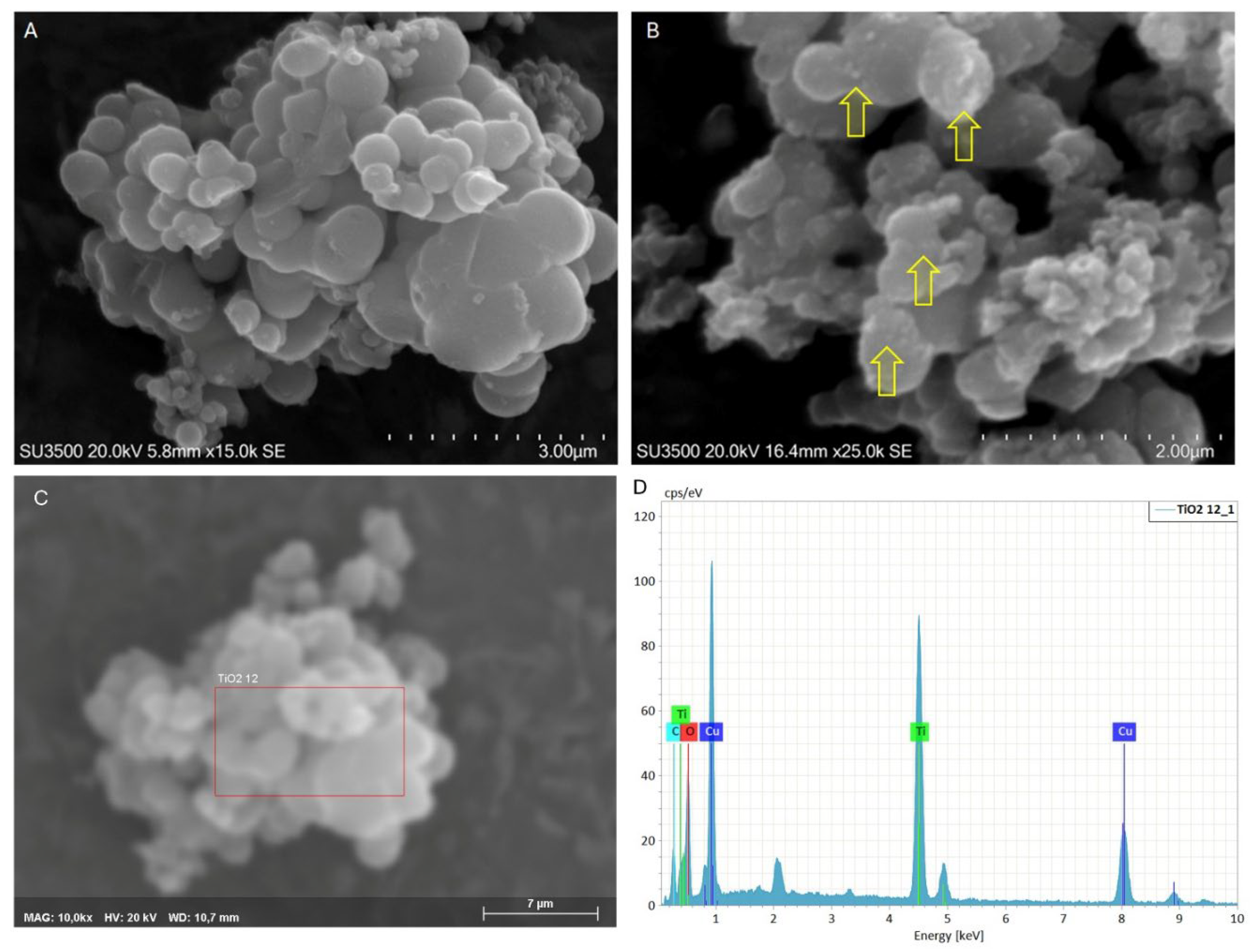
Figure 2.
XRD (A); Raman (B) and FTIR (C) for TiO2 MPs/SM. (D): scheme of spherical TiO2 synthesis.
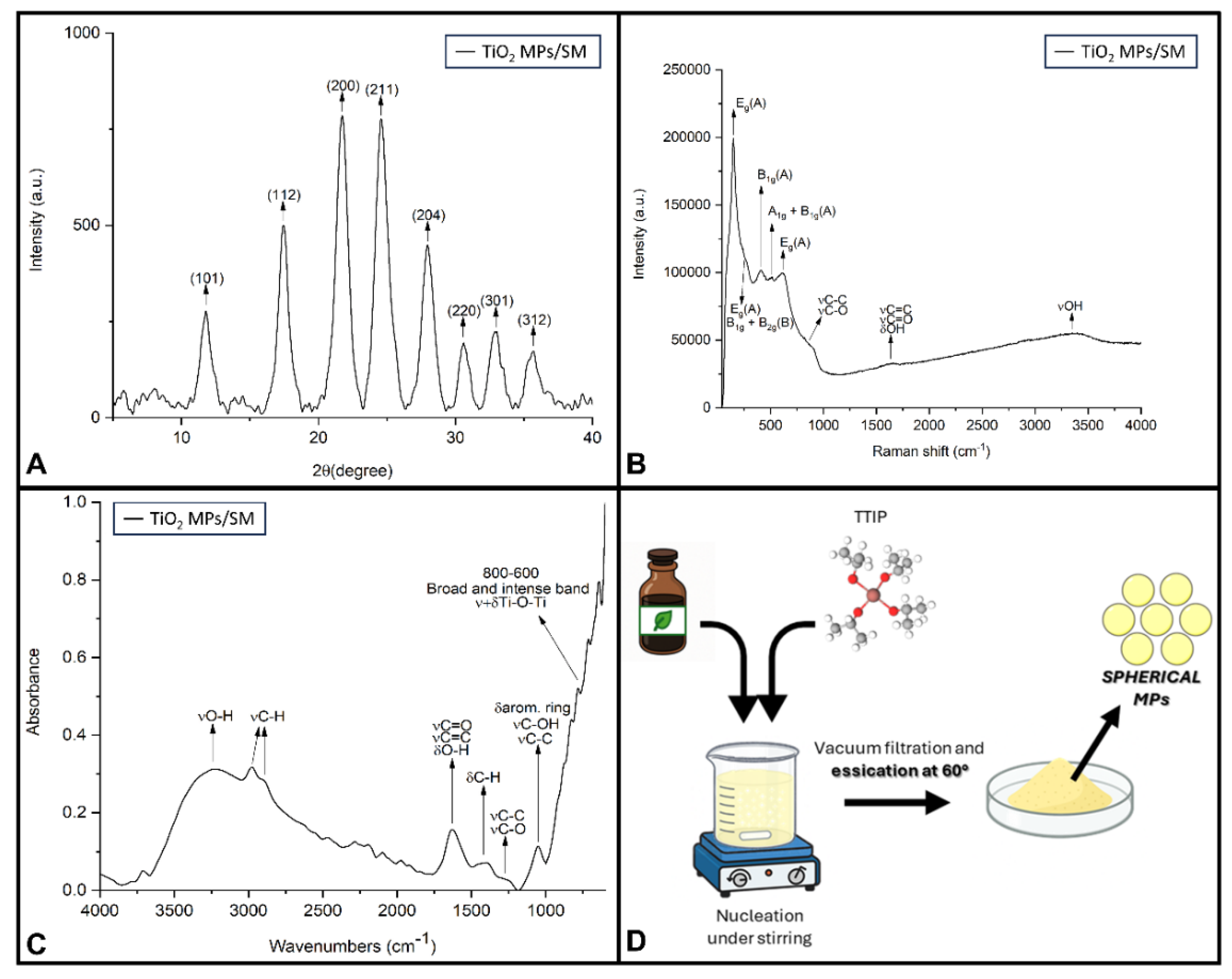
Figure 3.
SEM of CaCO3 Crystal. A) a crystal cluster is presented, prismatic structures with sharp edges are visible, one on the top of each other, magnification 10K. B) A higher magnification of a Crystal cluster shows the amorphous organic material on the surface of prismatic crystals (arrows), and evidence that they are formed by a layered structure of thin plates, magnification 25 K. C) and D) highlight the microanalysis, where: C) is SEM image of the examined sample, the red rectangle shows the ROI from which the spectrum was extracted; D) is the spectrum clearly shows the calcium/Ca peak; the copper peaks are due to the grid on which the sample was deposited. The other elements are C and O.
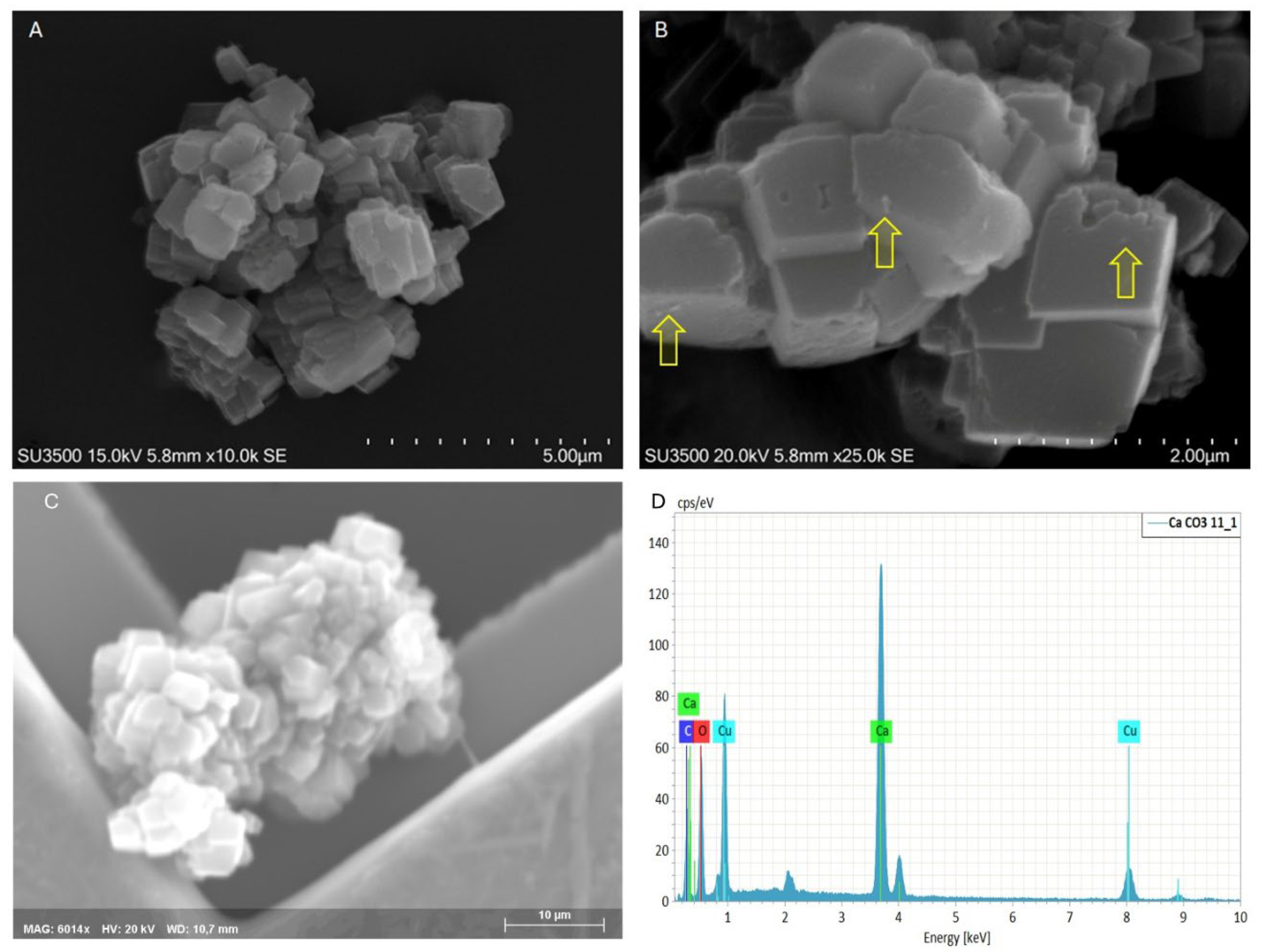
Figure 4.
XRD (A); Raman (B) and FTIR (C) of CaCO3 MPs/SM. (D): scheme of polygonal CaCO3 synthesis.
Figure 4.
XRD (A); Raman (B) and FTIR (C) of CaCO3 MPs/SM. (D): scheme of polygonal CaCO3 synthesis.
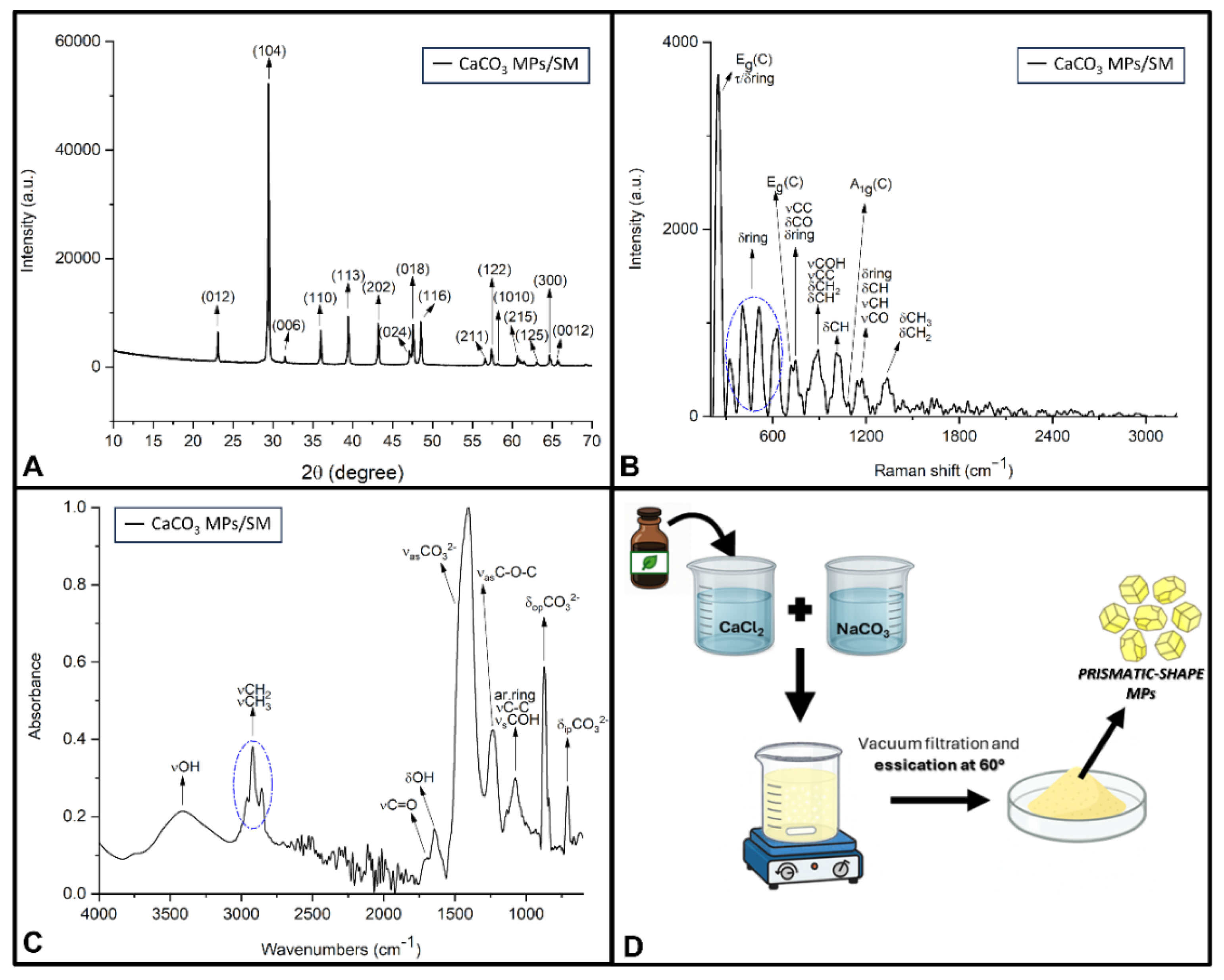
Figure 5.
Chemical classification of high-confidence compounds identified in the untargeted GC-HRMS analysis of Satureja montana aqueous extract. The chart is based on the compound list reported in Supplementary Table S1, where molecules were grouped into chemical families according to their molecular structures and functional groups.
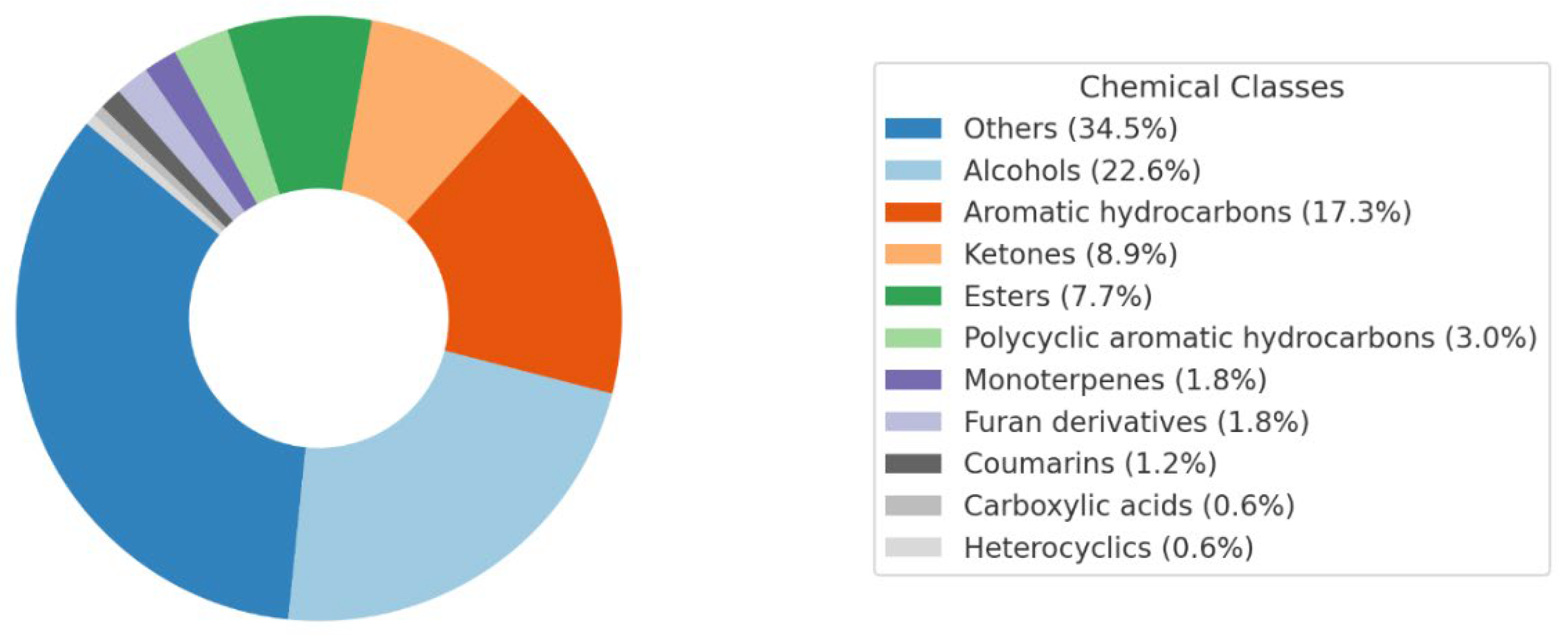
Figure 6.
Histogram plot of the 22 most abundant compounds identified in the SM extract based on peak area, ordered by retention time. Thymol and endo-borneol were excluded from the graph due to their disproportionately high peak areas, which would distort the scale. Since it’s a single sample, there’s no standard deviation. The bars indicate the relative abundance of the compounds in the sample because they are chromatographic peak areas, not quantities.
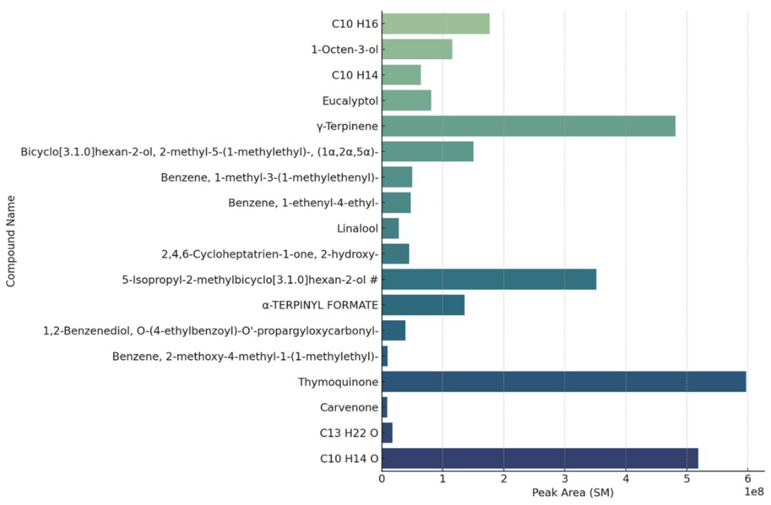
Figure 7.
Mean ± SD (n=6) of CFU/plate following passive sedimentation method under different TiO2 (A) and CaCO3 MPs (B) treatments. TNTC: Too Numerous To Count (CFU>2500); CT: untreated control. Statistical significance relative to the untreated control is indicated by asterisks: * p<0.05; ** p<0.005; *** p<0.001; **** p<0.0001.
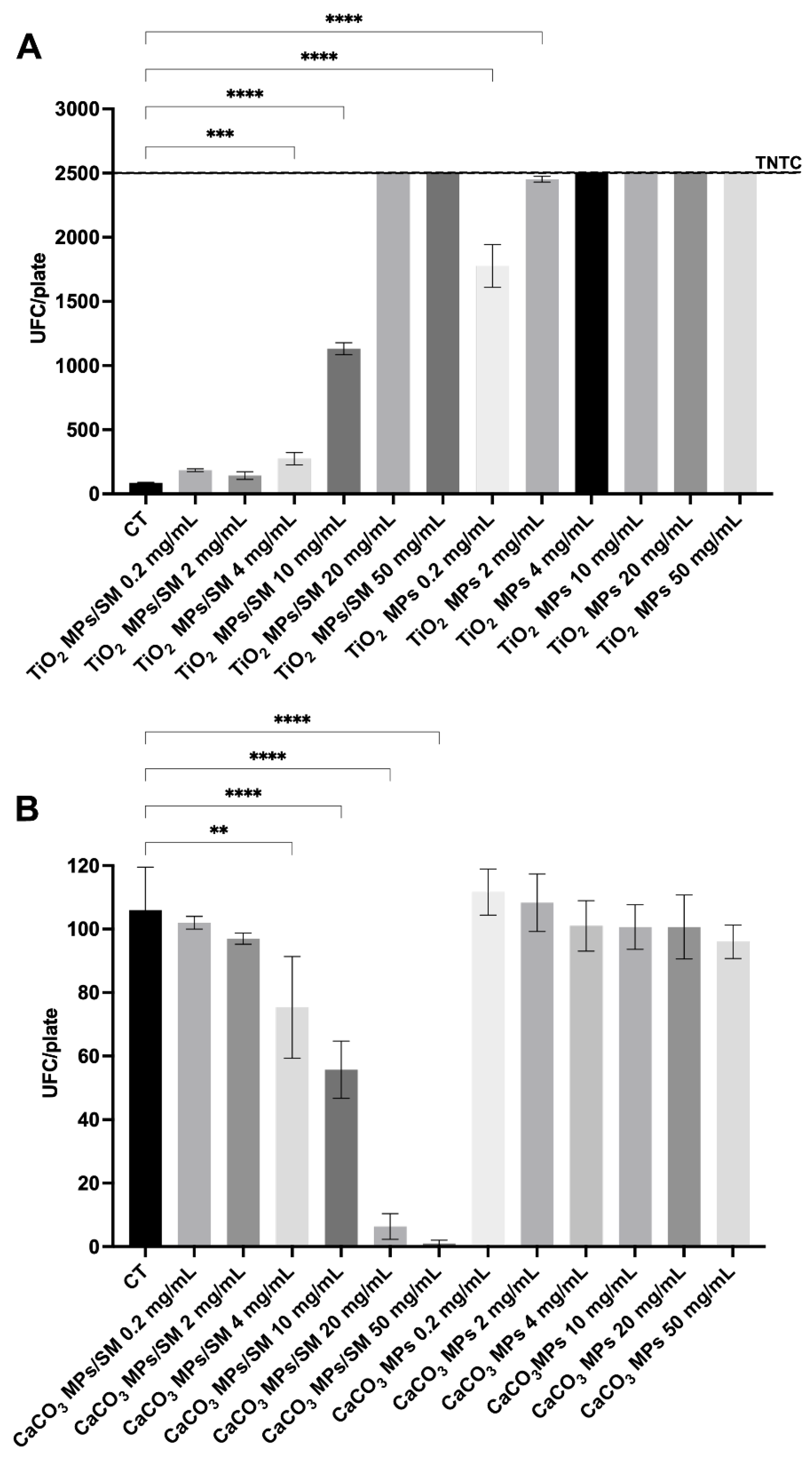
Table 2.
Raman and FTIR band assignments of CaCO3 MPs /SM.
| Band (cm−1) | Assignment | Reference |
| RAMAN SPECTROSCOPY | ||
| 248 | Eg lattice mode (calcite) τ+δ aromatic ring |
[96,97] |
| 320 | τ aromatic ring | [74,75,76,77,78,82] |
| 412 | δop+τ aromatic ring | [74,75,76,77,78,82] |
| 507 | δop +τ aromatic ring | [74,75,76,77,78,82] |
| 626 | δop +τ aromatic ring | [74,75,76,77,78,82] |
| 718 | Eg mode (δipCO32−) (calcite) | [96,97] |
| 746 | νC–C δC–O δ aromatic ring |
[74,75,76,77,78,82] |
| 890 | νC–OH νC–C δCH/CH2 |
[74,75,76,77,78,82] |
| 1009 | δipCH νC–O δ aromatic skeletal |
[75,76,77,78,79,80,81,82] |
| 1092 | A1g mode (νsCO32−) (calcite) | [96,97] |
| 1180 | δCH νC–O δ aromatic skeletal |
[74,75,76,77,78,79,80,81,82,83] |
| 1339 | δCH3 (isopropyl) δCH2 |
[74,75,76,77,78,79,80,81,82,83] |
| FTIR-ATR SPECTROSCOPY | ||
| 3600-3000 | νO–H | [88,89,90,91] |
| 2951 | νas –CH3 | [87,88,89,90,91] |
| 2922 | νas –CH2 | [87,88,89,90,91] |
| 2856 | νs –CH2 | [87,88,89,90,91] |
| 1710 | νC=O | [87,88,89,90,91] |
| 1637 | δO–H νC=O νC=C |
[87,88,89,90,91] |
| 1404 | νasCO32− (calcite) | [98,99] |
| 1231 | νC–O νC–C δ aromatic skeletal |
[87,88,89,90,91] |
| 1070 | νC–O νC–C δ aromatic skeletal |
[87,88,89,90,91] |
| 870 | δopCO32− (calcite) | [98,99] |
| 705 | δipCO32− (calcite) | [98,99] |
Vibrational bands assignments follow conventional spectroscopic notation: ν = stretching; δ = bending; τ = twisting; s = symmetric; as = asymmetric; ip = in-plane; op = out-of-plane.
Table 3.
Semi-quantitative concentrations of VOCs identified by targeted GC-MS (n.d. = not detected).
Table 3.
Semi-quantitative concentrations of VOCs identified by targeted GC-MS (n.d. = not detected).
| Compound |
Concentration [ng/ml] |
| α-Thujone | 282 |
| α-pinene | n.d. |
| Camphene | n.d. |
| Sabinene | 1054 |
| β-Pinene | n.d. |
| α-Phellandrene | 264 |
| α-Terpinene | 3814 |
| para-cymene | 157 |
| (R)-(+)-Limonene | 535 |
| α-Terpinolene | 284 |
| γ-Terpinene | 882 |
| Terpinen 4-ol | 10305 |
| Carvacrol methyl ether | 23.4 |
| L-Carvone | n.d. |
| Thymoquinone | 8271. |
| Geraniol | n.d. |
| Thymol | 63906 |
| Carvacrol | 5669 |
| β-Caryophyllene | n.d. |
| α-Humulene | 4.0 |
| caryophyllene oxide | 135 |
| cis-α-bisabolene (Levomenol) | 4.7 |
| L-Linalool | 1425 |
| aromadendrene oxide 2 | n.d. |
Table 4.
Mean ± SD (n=6) of CFU/plate following passive sedimentation method under different TiO2 and CaCO3 MPs treatments. CT: untreated control. Statistical significance relative to the untreated control is indicated by asterisks: * p<0.05; ** p<0.005; *** p<0.001; **** p<0.0001.
Table 4.
Mean ± SD (n=6) of CFU/plate following passive sedimentation method under different TiO2 and CaCO3 MPs treatments. CT: untreated control. Statistical significance relative to the untreated control is indicated by asterisks: * p<0.05; ** p<0.005; *** p<0.001; **** p<0.0001.
| Treatment | Concentration (mg/mL) | CFU/plate |
MPs Size _SEM (nm/µm) |
| Titanium Oxide | |||
| CT | - | 86.00 ± 3.60 | Diameters ranging from 160 nm to 1.3 µm, with a prevalence of elements with diameters around 400-600 nm |
| TiO2 MPs/SM | 0.2 | 184.00 ± 10.58 | |
| 2 | 142.40 ± 29.70 | ||
| 4 | 274.00 ± 47.62 *** | ||
| 10 | 1132.00 ± 46.13 **** | ||
| 20 | >2500 | ||
| 50 | >2500 | ||
| TiO2 MPs | 0.2 | 1776.00 ± 166.40 **** | |
| 2 | 2452.00 ± 22.27 **** | ||
| 4 | >2500 | ||
| 10 | >2500 | ||
| 20 | >2500 | ||
| 50 | >2500 | ||
| Calcium Carbonate | |||
| CT | - | 106.00 ± 13.53 |
CaCO3 particles have prismatic shape, sharp edges and variable size, with an average of 1 µm x 1 µm x 1 µm |
| CaCO3 MPs/SM | 0.2 | 102.00 ± 2.00 | |
| 2 | 97.00 ± 1.73 | ||
| 4 | 75.33 ± 16.04 ** | ||
| 10 | 55.67 ± 9.02 **** | ||
| 20 | 6.33 ± 4.04 **** | ||
| 50 | 1.00 ± 1.00 **** | ||
| CaCO3 MPs | 0.2 | 111.7 ± 7.23 | |
| 2 | 108.3 ± 9.07 | ||
| 4 | 101.00 ± 7.93 | ||
| 10 | 100.70 ± 7.02 | ||
| 20 | 100.70 ± 10.07 | ||
| 50 | 96.00 ± 5.29 | ||
Table 5.
All the characterization parameters for TiO2 MPs/SM. B: Brookite; A: Anatase; R: Rutile in the text.
Table 5.
All the characterization parameters for TiO2 MPs/SM. B: Brookite; A: Anatase; R: Rutile in the text.
| Samples |
SEM (φ: nm) Shape |
XRD 2θ (plane) |
Raman cm-1 (mode) |
SM extract composition (GC-MS) |
References | Notes |
| TiO2 MPs/SM | 400 – 600 nm µ-Spheres |
11.74° (101)/A 17.45° (112)/A 21.77° (200)/A 24.60° (211)/A 27.91° (204)/A 30.54° (220)/A 32.90° (301)/A 35.63° (312)/A 11.74° (111) /B 21.77° (231)/B |
(146, 219, 623) Eg/A (411, 517) B1g/A (517) A1g/A (219) B1g/B (279) B2g/B |
α-Thujone α-pinene Camphene Sabinene β-Pinene α-Phellandrene alfaTerpinene para-cymene (R)-(+)-Limonene α-Terpinolene γ-Terpinene Terpinen 4-ol Carvacrol methyl ether L-Carvone Thymoquinone Geraniol Thymol Carvacrol β-Caryophyllene α-Humulene caryophyllene oxide cis-α-bisabolene (Levomenol) L-Linalool |
This work (2025) | No antimicrobial performances have been observed |
| Anatase and rutile film |
600 nm size, grain size: 100 nm film |
The anatase films appear as flat terraces with mono-atomic steps. The ‘grains’ have a slightly rectangular shape and two kinds of rectangular grains oriented 90 to each other are observed. These are due to the before mentioned twinning of the films |
Not reported in the text | Not reported in the text | [101] | Photodynamic antimicrobial chemotherapy activity against Methicillin resistance Staphylococcus aureus |
| TiO2 NPs/ clove extract | 19-33 nm Spheres |
Not reported | Not reported | Not reported for the clove extract | [102] | Alcohol-based clove extract combined with TiO2 nanoparticles is a potent antimicrobial agent, capable of inhibiting VMRSA growth even at low concentrations |
| TiO2 NPs from Sigma Aldrich (Saint Louis, MO, USA) Loaded with Polygonum cuspidatum Extract | 21 nm (as reported by Sigma Aldrich) Spheres |
Not reported in the article, where commercially available nanoparticles have been applied | Not reported in the article, where commercially available nanoparticles have been applied | Not reported for the Polygonum cuspidatum Extract | [103] | Incorporating P. cuspidatum root extract into TiO2 nanoparticles significantly enhances their antioxidant and hemocompatibility properties. TiO2-loaded extract NPs exhibited excellent antibacterial properties against the tested strains |
| Green synthesis TiO2 nanoparticles using Sophora flavescens root extract |
8-24 nm Spheres |
(100) A (004) A (204) A (105) A (211) A (204) A |
Not reported only FTIR | Not reported for the Sophora flavescens root extract | [104] | Their stability and ability to penetrate bacteria cells enhance their antibacterial efficacy, particularly against gram-positive bacteria |
| TiO2 nanoparticles were synthesized using Talinum fruticosum leaf extract |
3-12 nm Spheres |
24.92° (101) A 68.99° (200) R |
Not reported only FTIR | Not reported for the Talinum fruticosum leaf extract | [105] | TiO2 showed photocatalytic activity and anti-inflammatory property as the protein degradation inhibition capacity |
| TiO2 P25 TiO2 Br200 |
mean diameter of 40 nm 10-15 nm |
79 % anatase/A 21% rutile/ R 45% anatase/A 2% rutile/R 53% brookite/B |
Not reported | No plant extract was applied for the synthesis of these nanoparticles | [106] | Biphasic brookite-anatase nanoparticles, due to their smaller particle size, may increase the efficiency of TiO2 nanoparticles to inhibit bacterial growth by promoting a greater surface area contact ratio |
|
TiO2, anatase, brookite and rutile, have been synthetized through a microwave-assisted hydrothermal method using amorphous TiO2 as a common precursor |
2000 nm amorphous (spheres) 15 nm A (spheres) 20 nm Degussa P25 (spheres) 20 nm B (spheres) 20 nm R rod-shaped |
Degussa P25 (80% anatase and 20% rutile), A (100% purity) B (94% purity) R (100% purity) |
(144, 196, 639 cm−1) Eg/A (399, 519 cm−1) B1g/A (513 cm−1) /A (127, 154, 194, 247, 412, 640 cm−1), A1g/B (133, 159, 215, 320, 415, 502 cm−1) B1g/B (235, 450 cm-1) /R |
TiO2 without functionalization, as reported in the full text | [107] | The bactericidal activity and photocatalytic antibacterial effectiveness of each material were evaluated through the determination of the minimum inhibitory and bactericidal concentrations, and via the mortality kinetic method under ultraviolet (UV) illumination under similar conditions with two bacterial groups of unique cellular structures: Gram-negative Escherichia coli (E. coli) and Gram-positive Staphylococcus aureus (S. aureus). |
Degussa P25: selected samples of the amorphous TiO2, a reference sample of Degussa P25 (80% anatase and 20% rutile), commercially available, as reported in the ref. [108].
Table 6.
All the characterization parameters for CaCO3 MPs/SM. C: calcite/CaCO3.
| Samples |
SEM (φ: nm) Shape |
XRD 2θ (plane) |
Raman cm-1 (mode) |
SM extract composition (GC-MS) |
References | Notes |
| CaCO3 MPs/SM | 1 µm x 1 µm x 1 µm Prismatic shape |
29.43° (104) /C 31.46° /C 56.54° /C 58.20° /C |
248 cm−1 (Eg) C lattice 718 cm−1 (Eg) C 1092 cm−1 (A1g) C |
α-Thujone α-pinene Camphene Sabinene β-Pinene α-Phellandrene alfaTerpinene para-cymene (R)-(+)-Limonene α-Terpinolene γ-Terpinene Terpinen 4-ol Carvacrol methyl ether L-Carvone Thymoquinone Geraniol Thymol Carvacrol β-Caryophyllene α-Humulene caryophyllene oxide cis-α-bisabolene (Levomenol) L-Linalool |
This work (2025) |
CaCO3 MPs/SM exhibit antimicrobial properties, depending on the particle concentrations, shape, defects, edges, dislocations and density of oxygenated functionalities on edges |
| Hollow calcium carbonate microspheres loaded by carvacrol | 1.97 ± 0.61 μm hollow microspheres |
XRD analysis of the CaCO3 hollow microspheres revealed that they are primarily aragonite (standard card: JCPDS#33–0268) | Not reported in the text | Not reported in the text for the carvacrol loading | [111] | The antimicrobial results indicated that microSCaCA effectively inhibited and killed E. coli and S. aureus, markedly enhancing the efficacy of CA, and exhibiting cellular safety. In practical preservation applications, microSCaCA significantly improved the preservation of pork during 4 ◦C storage, inhibiting key indicators such as TVB-N, pH, TBARS, and TVC, effectively delaying the oxidation of fats and proteins and suppressing the growth of microorganisms during the storage period |
| CaCO3 nanoparticles have been synthetized by using dried powder of mollusc shells |
60-70 nm spheres |
23.1◦ (012) 29.3◦ (104) 31.4° (006) 35.9◦ (110) 39.4◦ (113) 42.7◦ (202) 47.5◦ (018) 48.5◦ (116) 57.4◦ (122) 60.7◦ (214) 64.4◦ (300) 65.4◦ (0012) (JCPDS No. 85–1108) |
Not reported in the text but only FTIR | The synthesis of calcium carbonate nanoparticles from mollusk shell waste occurred without any natural plant extracts. Therefore, this characterization is not specifically contemplated in the text. | [112] | A low level of oxidative stress was recorded under all treatments of CaCO3NPs, and the highest decrease in MDA and O2•- contents of about 12 % and 23 % were recorded under 30 mg/L of CaCO3NPs compared to the control, respectively. The antioxidant system was improved under CaCO3NPs treatments and the highest increase of about 22 % and 37 % in CAT and SOD activity were recorded at 30 mg/L of CaCO3NPs compared to the control, respectively. |
| The extract of Hyphaene thebaica L. Mart (Egyptian doum palm) fruits was used as the reducing/capping agent for the synthesis of CaCO3 nanoparticles |
60 nm - 180 nm shapes from quasi-spherical, to cubical with equiaxed morphology |
012 104 110 113 202, 018, 116 211 122 214 300 correspond to the rhombohedral calcite phase of CaCO3, which is following with the X-pert standard card #47–1743 |
Not reported in the text but only FTIR | Not reported in the text for the extract of Hyphaene thebaica L. Mart (Egyptian doum palm) fruits |
[113] | CaCO3 NPs exhibit antifungal activity and therefore, the findings of this study suggest that the inclusion of a green synthesis of CaCO3 NPs as a nonfertilizer has the potential to promote tomato growth and yield. |
| The extract of Hyphaene thebaica L. Mart (Egyptian doum palm) fruits was used as the reducing/capping agent for the synthesis of CaCO3 nanoparticles |
60 nm - 180 nm shapes from quasi-spherical, to cubical with equiaxed morphology |
012 104 110 113 202, 018, 116 211 122 214 300 correspond to the rhombohedral calcite phase of CaCO3, which is following with the X-pert standard card #47–1743 |
Not reported | Not reported for the extract of Hyphaene thebaica L. Mart (Egyptian doum palm) fruits. This investigation has been reported only for the metabolomes of both tomato cultivars (Heinz-1370andMoneymaker), before and after CaCO3 NPs inoculation/treatments. |
[114] | The application of CaCO3NPs increased the presence of terpenoids and flavonoids in both fruits and leaves compared with the untreated plants. Metabolites such as 13-hydroxy abscisic acid, Dantaxusin A, and Sinuatol were identified in the leaves of the Moneymaker cultivar, with 30-O-linolenoylglyceryl6-O-galactopyranosyl-galactopyranoside, Olean-12-en-28-oic acid and scutianthraquinone B present in the fruits. |
Disclaimer/Publisher’s Note: The statements, opinions and data contained in all publications are solely those of the individual author(s) and contributor(s) and not of MDPI and/or the editor(s). MDPI and/or the editor(s) disclaim responsibility for any injury to people or property resulting from any ideas, methods, instructions or products referred to in the content. |
© 2025 by the authors. Licensee MDPI, Basel, Switzerland. This article is an open access article distributed under the terms and conditions of the Creative Commons Attribution (CC BY) license (http://creativecommons.org/licenses/by/4.0/).
Copyright: This open access article is published under a Creative Commons CC BY 4.0 license, which permit the free download, distribution, and reuse, provided that the author and preprint are cited in any reuse.


