
Preprint
Review

This version is not peer-reviewed.

Propposal of an Energy Dissipating Ptahway Regulated by the Extracellular Glucose Concentration That Precedes the Sustained Stimulation of Insulin Secretion

A peer-reviewed version of this preprint was published in:
Cells 2025, 14(21), 1644. https://doi.org/10.3390/cells14211644

Submitted:

01 August 2025

Posted:

04 August 2025

You are already at the latest version

Abstract

It is debated whether β-cells bioenergetic regulation is controlled by ATP supply or ATP demand. We proposed the hypothesis that an energy dissipating process precedes the stimulation of a continuous stimulation of insulin secretion by glucose. This process is mediated by connexin 36 hemichannels (Cx36H) or Cx36 connexons. Cx36H oligomers are expressed at the plasma membrane and their gating activity is activated by plasma membrane depolarization after the closure of K+ATP channels by glucose (<5 mM) metabolism. This initial depolarization might be responsible for the 1st phase of insulin secretion. Subsequent opening of CX36H increases β-cell plasma membrane permeability, allowing the efflux of metabolites (less than 1KD) (GABA, adenine nucleotides) and K+. This provokes a breakdown of oxidative glucose metabolism and repolarization of the plasma membrane. As extracellular glucose concentration increases (<< 5 mM), it exerts a progressive inhibition of Cx36H gating, allowing a continuous stimulation of insulin secretion (2nd phase). Glucose feature to regulate Cx36H closing with sigmoidal kinetics (8 mM IC50, and maximum around 20 mM) has been confirmed in mouse Cx36 connexin expression in Xenopus oocytes and in mouse islets stimulated by a range of glucose concentrations in the presence of 70 mM KCl. Its gating activity was also inhibited by some non-metabolized glucose analogues. Cx36H gating offers also the opportunity to use metabolic stimuli in vivo to potentiate the glucose secretory effect by, permeant or not, metabolic stimuli. It is concluded that β-cell bioenergetic regulation proceeds via ATP supply. This new proposal of a physiological function for Cx36Hs in β-cells place them as new susceptibility locus for type 1 and type 2 diabetes.

Keywords: 
;  ;  ;  ;  

Introduction

The background supporting this hypothetical mechanism of the bioenergetic β-cell regulation was the finding that connexin 36, specifically expressed in the β-cells, is sensitive to plasma membrane depolarization, and other extracellular ionic changes, after its oligomerization to Cx36 hemichannels (Cx36H). The resultant changes in Cx36H are regulated by the extracellular glucose concentration (1).
Going a little backwards, the original finding in the experimental pathway leading to the proposed hypothesis was that KCl β-cell depolarization, overlapped to a metabolic stimulus, deranges partially the insulin secretory response. 70 mM KCl decreased significantly the second phase response to 20 mM glucose either in the absence and presence of 10 mM glutamine (-35%, p<0.03, -47%, p<0.05) (2). However, the islet pre-exposure to 5 mM glucose plus 70 mM KCl for 45 minutes induced a strong suppression of the first (-69%, p<0.01) and second (-49%, p< 0.4) phase of a subsequent stimulation of insulin secretion by 20 mM glucose for 30 minutes (2).
The simultaneous combination of 70 mM KCl together with some branched-chain oxoacids induced a very strong suppression of islet insulin secretion, as compared with control islets in the absence of KCl. As an example, 10 mM KIC (oxo-4-methylpentanoate, OMP) second phase secretion was suppressed within 61% (p<0.0001). Similar results were obtained with KC (oxo-3-methylbutyrate) (3).
It was also demonstrated that branched-chain oxoacids promote the metabolic flux in the GABA-shunt, strongly diminishing the islet content of the γ-amino acid (3). It was then considered that KCl depolarization might induce a release of intracellular GABA cutting off the supply of substrate to the GABA-shunt. It could be demonstrated that 70 mM KCL increased the islet release of GABA and taurine by more than 100% in islets pre-loaded with 10 mM glutamine and decreased their islet contents (-32 and –40 %, respectively). In support of this “non-metabolic” inhibition of branched-chain oxoacids stimulated secretion, metabolic suppression of GABA metabolism by gabaculine (GABA transaminase inhibitor) suppressed 20 mM glucose, 10 mM semialdehyde succinic acid (SSA), a membrane permeable precursor of succinic acid) (2), and KIC (oxo-4-methylpentanoate; OMP) stimulation of insulin secretion (4).

Experimental Data

Structural Characteristics of Connexins

The mammalian family of connexins comprises 20 different members codified by different genes (5). They are represented as nCxZ (n,species; Z, molecular weight). Cx36 is expressed in neurons and glia, adrenal medullary chromaffin cells, and pancreatic β-cells (6); the latest express specifically only Cx36, due to their absence of the transcriptional repressor RE-1 Silencing Transcription Factor (REST) (7). In the transition from endoplasmic reticulum to the Golgi, 6 identical or different members, oligomerize to constitute a quaternary protein structure named homomeric or heteromeric connexon, depending on the presence of a single member or a mixture of them; Cx36 connexons are homomeric. Connexons inserted in the plasma membrane constitute transmembrane channels allowing diffusion of ions, second messengers, and metabolites between the cytoplasm and the extracellular space, with an exclusion limit around 1 kD. They are also designed as hemichannels because they are the precursors of gap-junction channels resulting from the tight apposition of two connexons of adjacent cells allowing the molecular exchange between the two coupled cells (ionic and metabolic coupling). They cluster in lipid rafts of the plasma membrane where calcium-dependent cell adhesion molecules promote a close contact between adjacent cells (“gap junctions”).

Functional Features of Connexins

The physiological function of gap-junction channels has been better consolidated than the role of the uncoupled connexons or hemichannels. Whereas Cx36 gap-junction channels mediate the ionic and molecular exchange between β-cells, Cx36H communicates β-cell cytosol with the extracellular space. In islets, the role of Cx36 gap-junction channels was initiated by Meda P. (5) and their role brilliantly explained in the coordinated regulation of insulin secretion to glucose in the islet core of β-cells (8). However, the physiological role of hemichannels has not been yet firmly established, in general, and even in the β-cell in particular. In fact, an earlier study failed to detect nuclear staining with ethidium bromide of MIN6 cells or freshly isolated mouse islets incubated at 20 mM glucose (9). At 20 mM glucose, MIN6 cells released some ATP that was increased by tetraethyl ammonium and even further in the absence of extracellular Ca2+. However, as ATP release was prevented by 200 µM diazoxide, the authors concluded that it was possibly mediated by the exocytotic pathway. With the actual information, probably the failure to detect Cx36 hemichannels was due to the presence of a high glucose concentration (20 mM) in the experiments that, as commented below, exerts an inhibitory effect on Cx36 hemichannels gating. Moreover, MIN6 cells seem to have a low expression of the mCx36 gene (10).

Properties of CX36H Overexpressed in Xenopus oocytes

Connexins exogenously expressed in Xenopus oocytes exhibit gating properties: the molecular conductance is approximately twice that of gap-junction channels. According to the authors, “the most compelling data on the properties of unpaired connexons or hemichannels was initially derived from oocytes expressing Cx46 and more recently Cx50”. In Cx43 hemichannels, their conductance activates around 0 mV and increases dramatically at + 60 mV and then it decays to a residual rate and, in the absence of divalent cations (Ca2+ and Mg2+), hemichannels opening becomes more active at negative potentials (11). Years ago, it was found that omission of divalent cations (Ca2+ and Mg2+) depleted ATP content of ob/ob islets, and its mechanism was surprising and unknown (12); today, it may be ascribed to an increased plasma membrane permeability due to activation of Cx36 opening (Scemes,2009) (9). As gating regulation differs among the different connexins, it was crucial to characterize the regulatory properties of Cx36, the unique connexin expressed in β-cells.
Overexpression of mCx36 gene (GJD2) in Xenopus oocytes increased plasma membrane permeability, as revealed by the uptake of extracellular propidium iodide at normal conditions (-42 ± 5 mV, 1.5 mM Ca2+, and1 mM Mg2+) (1). Electrophysiological studies showed that Cx36H gating activity was increased in the range of plasma membrane potential between – 40 and + 100 mV and membrane permeability was further enhanced by omitting divalent cations from the incubation medium (1). Cx36 hemichannel gating activity was deactivated by returning to hyperpolarized potential. Voltage activation of Cx36H gating was almost completely suppressed by 10 µM mefloquine (a known Cx36 inhibitor without off-target effects in the range 10 to 50 µM (1,13). More relevant, glucose blocked hemichannel currents activated by depolarization in a dose-dependent (sigmoidal) manner with an IC50 close to 8 mM and around 85 % of inhibition at 20 mM glucose (1).

ATP movements through plasma Cx36H hemichannels activated by plasma membrane depolarization in murine islets at different glucose concentrations

1. ATP release

Gaiting regulation of Cx36H may vary when expressed in native β-cells. Therefore, Cx36H opening was indirectly evaluated in isolated islets measuring the efflux of molecules, sensitive to inhibition by mefloquine. Mouse islets subject to 70 mM KCl depolarization at 5 mM glucose showed an increased release of ATP, measured with luciferin/luciferase, that was almost completely suppressed by 20 mM glucose. Islets from germinal knockout of Cx36 gene (Cx36-/- mouse) did not release ATP after depolarization at any glucose concentration (1).

2. Changes in β-cell ATP content and ATP/ADP ratio in rat and murine islets

Rat islets depolarized at 3 mM glucose in the presence of 250 µM diazoxide showed a gradual and statistically significant decrease of their ATP content as larger depolarizations were induced (15, 30, and 70 mM KCL) without any modification of their ATP/ADP ratio (1). 15 mM KCl depolarization (-21 mV) drove the membrane potential to the level of the plateau phase reached by high glucose; 40 mM KCl depolarized the membrane by 40 mV and the potential stabilized at the peak level of the Ca2+ spikes. These KCl driven depolarizations are of similar magnitude to those induced by glucose. The accompanying Islet ATP decline was completely prevented by the presence of 20 mM glucose; omission of medium Ca2+did not affect ATP depletion, excluding that ATP release was mediated by exocytosis. Mefloquine (Cx36 hemichannel inhibitor) did also prevent the depolarization-induced (70 mM KCL) loss of islet ATP.
Other metabolic secretagogues, like KIC and semialdehyde SSA, increasing islet ATP content and ATP/ADP ratio to the same levels as 20 mM glucose, failed to maintain ATP content under the same depolarizing conditions (1).
The restoring effect of 20 mM glucose did not only prevent the islet ATP fall but increased further both ATP content and ATP/ADP ratio to stimulatory values for secretion in the presence of 70 mM KCl in a sigmoidal dose-dependent manner: a half maximal effect was observed at 8 mM and a maximum around 20 mM, very similar to the sigmoidal relationship between glucose concentration and stimulated insulin secretion, suggesting its possible implication in the physiological mechanism of stimulation of insulin secretion (1).
Similar results were obtained in native mouse islets depolarized with 70 mM KCl at 5 mM glucose (1); islet ATP content was reduced approximately three-fold without changing the ATP/ADP ratio. The ATP depletion was prevented by 50 µM mefloquine and 20 mM glucose. To rule out that 20 mM glucose might counteract the ATP loss through an increased metabolism, the effects of non-metabolized (L-glucose, 3-0-mehyl-glucose) or poorly metabolized (D-galactose) structural glucose analogs were checked. Each of the analogs at 15 mM exhibited a significant recovery of the islet ATP content without modification of the ATP/ADP ratio (1). Glucose inhibition of KCl-induced islet ATP depletion showed a dose response with an IC50 value at 8 mM and a maximum effect at 20 mM; moreover, it did also increase both islet ATP and ATP/ADP ratio to stimulatory values for secretion in the presence of 70 mM KCl. Glucose lack of effect on the gating of gap-junction channels might be due to the occlusion of the glucose site on CX36H after the tight association of two hemichannels to form a gap junction channel between neighboring β-cells. By contrast, mefloquine inhibits the conductance of both Cx36H and gap-junction channels.

3. Changes in β-cell ATP content and ATP/ADP ratio in transgenic islets with homo and heterozygous knockout of connexin 36 (Cx36-/- and Cx6+/- murine islets) (1)

Cx36-/- islets showed abnormally elevated ATP levels at 5 mM glucose, very close (no significant difference) to those reached at 20 mM glucose but an ATP/ADP ratio characteristic of non-stimulated islets. Does it mean that native islets, opposite Cx36-/- islets, are losing adenine nucleotides regularly to maintain a basal cytosolic ATP concentration? In our modest opinion, there is no information concerning whether β-cells pay a high energy cost (oxidative phosphorylation) to synthesize adenine nucleotides, as compared with other biochemical or physiological loads?
Basal and stimulated ATP content and ATP/ADP ratio in heterozygous Cx36+/-islets were intermediate of the corresponding values in control and Cx36-/-islets. At 20 mM glucose, islets from the three different mouse phenotypes showed similar increases in their ATP content and ATP/ADP ratio above the basal values.

4. Content and release of GABA and taurine mediated by plasma membrane depolarization activation of Cx36 hemichannels in rat islets (14)

Mouse islets depolarized at 5 mM glucose by 70 mM KCl in the presence of 250 µM diazoxide showed a decrease of its GABA and taurine contents and an increase of their release; these effects were potentiated by omission of the extracellular calcium concentration. The combination of KCl and Ca2+ omission was more reluctant to inhibition by mefloquine that caused a partial recovery of GABA content and release at 50 µM.

5.Β-. cell insulin secretion by glucose in the absence of Cx36 (1).

In Vivo Effects

Cx6-/- mice showed significantly greater glucose excursions in plasma glucose at 15 and 30 minutes in an intraperitoneal glucose tolerance test; the area under the curve was significantly higher than in wild type mice. Basal plasma insulin levels were significantly lower in Cx6-/- mice.

Insulin Secretion by Glucose

A gradual decrease of the insulin response to 20 mM glucose was observed in the three islet phenotypes, from controls (Cx36+/+) to transgenic islets (1): Cx36+/- response was intermediate between controls and Cx36-/- which exhibited a single and diminished first phase. Both phases (1st and 2nd) of insulin secretion were significantly suppressed as compared with native islets; however, the 1stphase was always present, besides the absence of Cx36H. Mefloquine diminished partially 20 mM glucose-induced secretion in Cx36+/+ and Cx6+/- perifused islets.

Secretory effect of extracellular ATP on insulin secretion in native, transgenic mice islets with knockout of the Cx36 gene, and native rat islets.

These experiments were designed to confirm that open (by 70 mM KCl) Cx36H allows adenine nucleotide diffusion in both senses, dependent on the concentration gradient between extracellular space and cytoplasm. This might also help to check whether extracellular ATP, at 5 mM glucose, could stimulate a sufficient and sustained secretion of insulin. In control (Cx36+/+) islets, only the first phase of insulin secretion due to KCl was not modified by 5 mM ATP but it induced a significant second phase (p<0.05). Cx36+/- islets responded to 5 mM ATP with a weak stimulation of the first and second phases of secretion; no secretory response was recorded in Cx36-/- islets.
In rat islets, 5 mM ATP in the presence of 5 mM glucose and 70 mM KCl stimulated a significant increase of the 2nd but not the 1stphase of secretion, in comparison with control islets (in the absence of ATP). 10 mM ATP triggered a significant increase of both phases of insulin secretion; the second phase of insulin secretion was significantly higher at 10mM than at 5 mM ATP (14).

Bioenergetic Regulation of β-Cells

The review on the regulation of β-cell bioenergetics written by Prof. Nicholls D.G. brilliantly describes the general concepts necessary to understand it (15). Therefore, a very clarifying paragraph of its review is translated:
“In the β-cell, the downstream dissipative pathways that balance the upstream delivery of glucose-derived pyruvate to the mitochondrion are of critical importance but are commonly neglected. These include the abnormally high endogenous mitochondrial proton leak and the largely unexplored quantitation of ATP utilizing pathways. In the absence of these dissipative pathways, even the slowest trickle through the glycolytic pathway would eventually result in a maximal ATP/ADP ratio approaching thermodynamic equilibrium. It is therefore important to understand how the β-cell, which has evolved to vary the Gibbs free energy of the cytosolic ATP hydrolysis reaction (“ΔGp = ΔGATP”) as a function of upstream substrate availability, differs from, for example, heart muscle, which possesses feedback mechanisms to ensure that ΔGp (“mitochondrial electrochemical membrane potential or proton motif force”) remains as constant as possible in response to huge variations in ATP demand”. In our opinion, the nature of the dissipating pathway implicated in a balanced regulation of mitochondrial oxidative phosphorylation in the β-cells has not yet been defined.

Does Cx36 Hemichannels-Dependent Repolarization Oppose K+ATP Channel Dependent Depolarization?

As demonstrated earlier, at basal glucose (0 to 3-5 mM), β-cell glucose metabolism is essentially anaerobic and ATP production, and the cytosolic ATP /ADP ratio must be low (16). Above 5 mM, glycolytic-derived pyruvate starts to be oxidized in mitochondria (17) and the ATP generated in oxidative phosphorylation closures K+ATP channels causing plasma membrane depolarization (1st step). This initial step activates voltage-dependent Ca2+channels and the resulting increase in cytosolic Ca2+activates mitochondrion metabolism that activates insulin secretion. It is tempting to speculate whether in the 1st step of β-cell activation, a burst of membrane potential depolarization would be responsible for the first phase of insulin secretion as observed in the pioneering recordings of a prompt stimulation of insulin secretion by glucose that is followed by a period of electrical silencing (17,18). Perhaps, in the more physiological ramp stimulation by glucose there would be better intermingling of both processes.
According to our hypothesis, membrane depolarization activates the gating of Cx36 hemichannels (unknown timing relationship) and facilitates the communication between cytosol and extracellular milieu (2nd step). This means that β-cell membrane permeability is increased, facilitating not only the exchange of molecular components but perhaps also contributing to antagonizing membrane depolarization due to a certain dissipation of the K+ gradient (11). It is not known whether the repolarization current is of similar magnitude, or smaller enough, to not compete with the K+ATP dependent depolarization current. It must depend on the number of Cx36 hemichannels, compared to the K+ATP channels.

Cx36 hemichannels mediate the release of important molecular entities necessary for an intact mitochondrial metabolism (ATP and GABA).
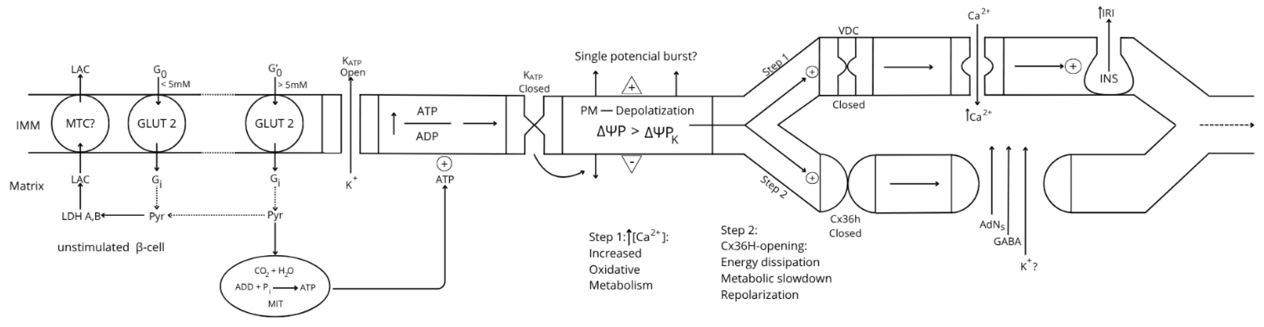
Cx36 hemichannels opening allows the extracellular efflux of important molecular components needed to mediate the stimulus-secretion coupling mechanism of glucose-induced insulin release. On one side, loss of islet intracellular GABA, highly implicated in the coupling between citric acid cycle and the GABA-shunt, as demonstrated with several metabolic stimulus (glucose, branch-chain ketoacids, semialdehyde succinic acid), restricts their oxidative metabolism (23). Due to the limiting activity of β- cell lactate dehydrogenase, no further increase in anerobic glycolysis is possible (absence of Pasteur effect) to improve substrate-link ATP synthesis. Moreover, the loss of cytosolic adenine nucleotides, ADP, might maintain mitochondrial respiration at state IV, interrupting oxidative phosphorylation. Perhaps, a suppression of the cytosolic adenine nucleotide pool might also reduce the Gibbs energy released in the hydrolysis of ATP (↓ ΔGATP) also contributing to a metabolic slowdown. The accompanying fall of the electrochemical mitochondrial membrane potential (↓ΔGp), commented above, would also, perhaps primordially, condition the value of cytoplasmic ΔGATP that probably also contributes to a slowdown of β-cell glucose metabolism by deficient substrate phosphorylation (15).
Figure 1. Relationship between variations of β-cell plasma membrane voltage and bioenergetic mitochondrial function.
Preprints 170791 g001
A careful lecture of a review by Henquin J.C. (18) is highly recommended to appreciate the parallelism observed between the two processes. “The timing of the main ionic events accompanying membrane depolarization by glucose are as follows (see Figure 2): 1. A square-wave increase of glucose concentration (from 3 to 10 mM) triggers initially an increase of Rb+ (tracer for K+) efflux coinciding with a long first burst of membrane voltage depolarization with continuous spike activity. 2. It follows a decrease of Rb+ efflux, and a repolarization (silent interval) first demonstrated by Sehlin J. and Freinkel N.” (20). According to Henquin JC (18), “it was tempting to suggest that a biphasic change in K+ membrane permeability contributes to the biphasic pattern of electrical activity and eventually of insulin release, but such a proposal awaits experimental support”. In other words, the first long voltage burst would be responsible for the first phase of glucose-induced insulin secretion. 3. “After the silent interval, β-cell plasma membrane depolarizes again to the threshold potential when glucose rises to 6-7 mM and repetitive slow waves are generated. This second and steady round of depolarization is probably responsible for the second phase of glucose-induced insulin secretion. A further increase of the glucose concentration does not affect the absolute value of the membrane potential, but the slow waves lengthen whereas the intervals among them shorten. At 18 mM glucose, the membrane is permanently depolarized at the plateau potential, with continuous spike activity”.
The new proposal for the participation of Cx36Hs in the bioenergetic regulation of the stimulus- secretion coupling provides experimental support for their implication in the biphasic changes observed in Rb+ efflux and in the plasma membrane depolarization / repolarization in the second step after the first one of glucose metabolism-dependent depolarization by closure of K+ channels.
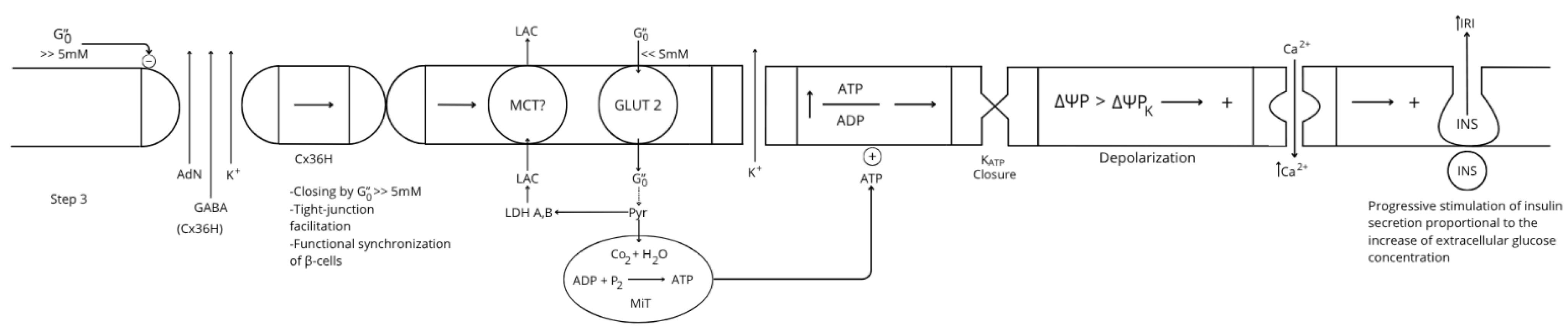
The step of Cx36 hemichannels activation (2ndstep) mediates an energy dissipative pathway just before definitive activation of glucose-induced insulin secretion. A continuous rise of the extracellular glucose concentration after a meal will reach a value where it enters its inhibitory concentration range on Cx36 hemichannels provoking its closure (3d step). It was striking to find that the inhibition kinetic of glucose is sigmoidal with a half concentration of 8 mM glucose, very similar to the kinetics of glucose stimulation of insulin secretion (1). Closing Cx36 hemichannels will favor β-cell intercommunication through tight junction channels promoting the synchrony of the insulin secretory response of more β-cells in the islet core.
Figure 3. Two different ways of regulation a cellular steady bioenergetics transition.
Preprints 170791 g003
According to different authors, many cells accelerate its oxidative phosphorylation when they experience a physiological workload with an important ATP cost; these cells are categorized as “fuel utilizers” and they are activated by “ATP demand”. Their cytosolic ATP levels are constant, independently of their stimuli (21, 22).
Others, like the β-cells, react to a stimulus increasing their cytosolic ATP content dependent on the stimulus concentration (glucose in the β-cells) and not by an increased physiological workload. These cells are considered “fuel sensors” and their oxidative phosphorylation is supposed to be regulated by “ATP supply” but not by “ATP demand” (21). However, this concept is now debated.
At variance with fuel utilizer cells, stimulated β-cells do not increase their oxidative phosphorylation by an increased ATP demand (increased insulin secretion) but, contrary to them, they dissipate biochemical energy before sustained stimulation of insulin secretion that is promoted proportionally to the ATP supply dependent on the magnitude of the flux generated through glucose oxidative metabolism.

Experimental Conditions Considered to Be an ATP Demand to β-Cells

The energetic consumption and dependence of the main ATP-demanding cellular processes on OxPhos have been measured in some mammalian cells (categorized as “fuel utilizers”). The results uncover a bioenergetic hierarchy in which the higher O2 (ATP) consumers were protein and RNA/DNA synthesis, followed by sodium cycling through the Na+/K+-ATPase, Ca+ ATPase, unidentified ATP consumers, and proton leak (23).
There is almost general agreement that sulfonylureas in general suppress islet ATP levels and that it is due to an excess of ATP consumption over its synthesis provoked by an excessive workload, supposedly due to a high stimulation of glucose-induced insulin secretion (?). However, glucose-induced secretion is also generally reduced by the combination of glucose and sulfonylureas. These data have been commented on before in a previous review (23) and are referred here to different sulfonylureas: Tolbutamide (25,26); Glibenclamide (27); Gliburide (28). DetimaryP et al. (26) also showed that 30 mM KCL suppressed significantly the ATP/ADP ratio of mouse islets incubated at 10 mM glucose.
Stimulation of the plasma membrane Na+-K+-ATPase with glibenclamide and meglitinide, supposed to depolarized β-cells, suppressed significantly islet concentrations of all three adenine nucleotides at 10 mM glucose without affecting glucose oxidation. The effect was also attributed to an elevated ATP consumption by the activated Na+-K+-pump (29).
All these experimental maneuvers intended to increase the ATP demand of the β-cells resulted unexpectedly in their exhaustion, probably caused by loss of clue metabolites (GABA, adenine nucleotides) by an excessive depolarization-induced opening of β-cell hemichannels.

Cx36 gating opens an opportunity window to potentiate glucose-induced secretion by glucose.

This aspect of the stimulus-secretion coupling of insulin secretion will be explained with reference to a particular set of experiments performed in rat perifused islets stimulated with glucose and succinic acid dimethyl ester (SAD) whose secretory response was strongly potentiated by pyruvate (30) (Figure4). The latter does not permeate the plasma membrane of rat β-cells because of their deficiency of the monocarboxylate transporter (MCT1 and 2) at their plasma membrane (31).
As expected, 10 mM pyruvate alone failed to trigger any insulin secretory response at all. However, it potentiated by +64% each of the individual responses to 20 mM glucose and 10 mM SAD that were of similar magnitude (Fig 4A). Moreover, the combination of 20 mM glucose together with 10 mM SAD (Fig.4B) reproduced the potentiation by 10 mM pyruvate of the individual secretory responses to 20 mM glucose and 10 mM SAD, increasing insulin release around 60-70%. This potentiation of glucose-induced insulin secretion by pyruvate and SAD are an example of regulation by ATP supply because the resultant increased secretion load does not cause any feedback suppression.
Interpreting these experimental results according to our new proposal of regulation of the bioenergetics of β-cells, the facilitated plasma membrane permeability of pyruvate might be attributed to the opening of Cx36 connexon hemichannels occurring after plasma membrane depolarization closes K+ATP channels. This possibility would enable the potentiation of glucose-induced insulin secretion in vivo administering metabolic stimuli, permeable or not, that would be taken up by open Cx36 connexons during step 2 of the β-cell bioenergetic pathway. Perhaps this feature of β-cell plasma membrane would challenge any pharmacological treatment of type-1 and/or 2 diabetes in the future.

Are Cx36 proteins as risk factors for the development of type-1 and type-2 diabetes?

An early report by Allagnant et al (32) showed that 20 mM glucose repressed Cx36 gene expression in insulin secreting cells diminishing both its protein and mRNA levels. Glucose-induced transcription repression was ascribed to a putative cAMP response element (CRE) located between bases –566 and –556 of the Cx36 gene promoter. The D-glucose effect was not reproduced by the non-metabolized glucose analogues L-glucose and 3-O-methyl-D-glucose at 20 mM; however, 2-deoxy-D-glucose, phosphorylated by glucokinase but poorly further metabolized in glycolysis or pentose phosphate pathway, induced a 50% suppression of Cx36 protein levels. The authors did not find any glucose-responsive element (GlRE) or carbohydrate-responsive element (ChoRE) in their bioinformatic search of the human 3945 bp fragment of the human Cx36 promoter used in the experiments. It would be interesting to investigate the hypothetical Cx36 amino acid sequence recognizing the glucose molecule and its corresponding nucleotide codifying region to search its polymorphisms in the human sequence. As our proposal is the first attributing a physiological role to Cx36 hemichannels in β-cells, it is yet unknown whether their genetic polymorphisms might affect their modulation by glucose. One has to keep in mind that the hypothetical Cx36 H site for glucose is not expected to affect gap junction channel gating that is supposed to be occluded by the formation of connexons.
Islet Cx36 has been implicated in both Type-1 and Type-2 diabetes, but their mechanisms are yet unknown. Farnsworth N.L., and Benninger R. K.P. (33) have reviewed the putative roles of Cx36 in the development of both type 1 and type 2 diabetes. Recent studies suggest that, in type 1 diabetes development, Cx36 gap junction channels modulate ER and oxidative stress, and apoptosis induced by proinflammatory cytokines. According to the authors, the gene coding for Cx36 is a susceptibility locus for type 2 diabetes although no direct implication of gap junction coupling has been found in humans with type 2 diabetes. However, prediabetic mice subject to a high fat diet experience a decrease in their Cx36 gap junction coupling, under the prevailing hyperglycemic and hyperlipidemic conditions, that will lead to a loss of synchronization of electrical activity and [Ca2+] i oscillations (33).
Figure 1. Evolution of the stimulus-secretion coupling mechanism of glucose induced insulin secretion implicating a hypothetical role of the β-plasma membrane Cx36 hemichannels.
First to the left, the plasma membrane of a β-cell at rest (glucose concentration < 5 mM), characterized by a zero net flux of K+ at its K+-equilibrium potential (ΔΨPK = -60 mV). This resting β-cell depends predominantly on anaerobic glycolysis, with a restricted ATP production rate due to the absence of mitochondrial oxidative metabolism. As extracellular glucose concentration rises (> 5mM), glycolysis exceeds the capacity of lactate dehydrogenase (LDH) to generate lactate (LAC) and the extra pyruvate (Pyr) produced is derived to mitochondria for its oxidative metabolism, increasing cytosolic ATP concentration (Step 1). Subsequently, K+ATP channels are closed, and the plasma membrane depolarizes (ΔΨP > ΔΨPK, less negative) exhibiting an oscillatory burst of action potentials caused by the increased influx of Ca2+, due to the opening of the voltage-dependent channels (VDC). The final consequence would be the stimulation of insulin secretion (first phase?). In Step 2, the previous membrane depolarization induces depolarization and opening of Cx36 hemichannels, resulting in a repolarization of the plasma membrane, due to a glucose metabolism slowdown and an increased efflux of K+.
MCT (plasma membrane monocarboxylate transporter); GLUT 2 (glucose transporter 2 or HK IV).
Figure 2. Effect of a change of glucose concentration from 3 to 10 mM on the membrane potential of a single mouse β-cell (Graphic kindly provided by Henquin J.C.).
Figure 3. Step 3 of the stimulus-secretion coupling mechanism of glucose induced insulin secretion implicating a hypothetical role of the β-plasma membrane Cx36 hemichannels.
As the extracellular glucose concentration continues to increase (>> 5 mM), it enters its range of hyperbolic inhibition of Cx36 hemichannels’ inhibition that leads to their progressive closing dependent on the final glucose concentration reached. That makes possible a partial or maximum recovery of step 1 and all their components: K+ATP-closure by a recovered glucose metabolism, membrane depolarization, and an increase of cytosolic Ca2+ concentration that allows a continuous stimulation of insulin secretion (2nd phase?). The insulin response will return spontaneously to basal values when the extracellular glucose declines back to its basal concentration (5 mM).

References

  1. Pizarro-Delgado,J, Fasciani I, Temperan A., Romero N., González-Nieto D., Alonso-Magdalena A., Nualart-Martí A., et al, Inhibition of connexin hemichannels by glucose contributes to the stimulation of insulin secretion, Am. J. Physiol. Endo. Metab., Am. J. Physiol. Endocrinol. Metab. 306: E1354-E1366, 2014. [CrossRef]
  2. Pizarro-DelgadoJ, Braun M., Hernández-Fisac I., Martín-del-Rio R., Tamarit-Rodriguez J., Glucose promotion of GABA metabolism contributes to the stimulation of insulin secretion in β-cells, Biochem. J. 431: 381-389, 2010. [CrossRef]
  3. Pizarro-Delgado, Hernández-Fisac I., Martín-del-Rio R., Tamarit-Rodriguez J., Branched-chain 2-oxoacids transamination incresase GABA-shunt metabolism and insulin secretion in isolated islets, Biochem. J., 410: 359-368, 2009. [CrossRef]
  4. Hernández-Fisac I., Pizarro-Delgado J., Fernández-Pascual S., Ortsäter H., Pizarro-Delgado J., Martín-del-Rio R., Bergsten P., Tamarit-Rodriguez J., Oxo-4-methypentanoic acid directs the metabolism of GABA into the Krebs cycle in rat pancreatic islets, Biochem. J. 400:81-89, 2006. [CrossRef]
  5. Cigliola V., Allagnant F., Berchtold L. A., Lamprianou S., Haefliger J.-A., Meda P., Role of connexins and pannexins in the pancreas, Pancreas 44: 1234-1244, 2015. [CrossRef]
  6. NakashimaT, Ke-Yong Wang K.-Y, Matsuoka H., Ogawa K., Harada K., Inoue M., Developmental changes in gap Junction expression in rat adrenal medullary chromaffin cells Acta Histochem. Cytochem. 57 (6): 189-197, 2024.7. [CrossRef]
  7. Martin D., Allagnant F., Gesina E., Calle D., Gjinovichi A., Waeber G., Meda P., hHaefliger J.-A., Specific silencing of the REST target genes in insulin-secreting cells uncovers their participation in beta cell survival, PlosOne 7(9), e45844, 2012.
  8. Ravier M. A., Güldenagel M., Charollais A., Gjinovci A., Caille D., Söhl G., Wollheim K- B. et al, Loss of connexin36 channels alters β-cell coupling, islet synchronization of glucose-induced Ca2+ and insulin oscillations, and basal insulin release, Diabetes 54:1798-1807, 2005. [CrossRef]
  9. Scemes E., Bavamian S., Charollais A., Spray D. C., Meda P., Lack of “hemichannel” activity in insulin producing cells, Cell Commun. Adhes. 15 (1): 143-154, 2008.
  10. Puginier, E.; Leal-Fischer, K.; Gaitan, J.; Lallouet, M.; Scotti, P.-A.; Raoux, M.; Lang, J. Extracellular electrophysiology on clonal human β-cell spheroids. Front. Endocrinol. 2024, 15, 1402880. [CrossRef]
  11. Spray B. RB. R., Functional connexin “hemichannels”: a critical appraisal, Glia 54: 758-773, 2006.
  12. Tamarit-Rodriguez J., Hellman B., Sehlin J., Metabolic characteristic s of pancreatic β-cells exposed to calcium-transporting ionophores, Biochem. Biophys. Acta 496: 167-174 , 1977. [CrossRef]
  13. Iglesias R., Spray D. C., Scemes E., Mefloquine blockade of pannexin 1 currents: resolution of a conflict, Cell Commun. Adhes., 16: 131-137, 2010. [CrossRef]
  14. Pizarro-Delgado J., KCl-permeabilized pancreatic islets: An experimental model to explore the messenger role of ATP in the mechanism of insulin secretion, PlosOne 10 (10): e014096, 2015. [CrossRef]
  15. Nicholls D.G., The pancreatic β-cell: A bioenergetic perspective, Physiol. Rev. 96: 1385-1447, 2016. [CrossRef]
  16. Tamarit-Rodriguez J., Idahl L. Å., Lactate production in pancreatic islets, Diabetes 47: 1219-1223, 1998.
  17. Ashcroft F.M., Rorsman P., Electrophysiology of the pancreatic β-cell, Prog. Biophys. molec. Biol. 54: 87-143, 1989.
  18. Henquin J. C., Meissner H. P., Significance of ionic fluxes and changes in membrane potential for stimulus-secretion coupling in pancreatic B-cells, Experientia 40: 1043-1052, 1984. [CrossRef]
  19. Tamarit-Rodriguez J., Metabolic Role of GABA in the Secretory Function of Pancreatic β-Cells: Its Hypothetical Implication in β-Cell Degradation in Type 2 Diabetes, Metabolites 2023, 13, 697. [CrossRef]
  20. Sehlin J., Freinkel N., Biphasic modulation of K+ permeability in pancreatic islets during stimulation with glucose, Diabetes 32: 820-824, 1983.
  21. Affourtit C., Alberts B., Barlow J., Carré J.E., Wynne A., Control of pancreatic β-cell bioenergetics, Biochemical Society Transactions, DOI/ 10.1042/BST20170505, 2018. [CrossRef]
  22. Wikstrom, J.D.; Sereda, S.B.; Stiles, L.; Elorza, A.; Allister, E.M.; Neilson, A.; Ferrick, D.A.; Wheeler, M.B.; Shirihai, O.S., A novel high-throughput assay for islet respiration reveals uncoupling of rodent and human islets. PLoS ONE 2012, 7, e33023. [CrossRef]
  23. Buttgereit, F.; Brandt, M.D. A hierarchy of ATP-consuming processes in mammalian cells. Biochem. J. 1995, 312, 163–167.
  24. Tamarit-Rodriguez J., Stimulus–Secretion Coupling Mechanisms of Glucose-Induced Insulin Secretion: Biochemical Discrepancies Among the Canonical, ADP Privation, and GABA-Shunt, Int J. Mol. Sci. 26, 2947, 2025 (https://doi.org/10.3390/ijms260729). [CrossRef]
  25. Panten, B. J. ZiJnkler, S. Scheit, K. Kirchhoff and S. Lenzen, Regulation of energy metabolism in pancreatic islets by glucose and tolbutamide, Diabetologia(1986)29:648-654. [CrossRef]
  26. Detimary P., Gilon P., Henquin J.C., Interplay between cytoplasmic Ca2+ and the ATP/ADP ratio: a feedback control mechanism in mouse pancreatic islets, Biochem. J. (1998) 333, 269–274. [CrossRef]
  27. Sweet I. R., Cook D., DeJulio E., Wallen A. R., Khalil G., Callis J., Reems J., Regulation of ATP/ADP in Pancreatic Islets, Diabetes 53:401–409, 2004. [CrossRef]
  28. Doliba N. M., Vatamaniuk M. Z, Buettger C. W., Qin W., Collins H. W., Wehrli S. L., Carr R. D., Matschinsky F., Differential Effects of Glucose and Glyburide on Energetics and Na1 Levels of bHC9 Cells. Nuclear Magnetic Resonance Spectroscopy and Respirometry Studies, Diabetes 52: 394–402, 2003. [CrossRef]
  29. Elmi A., Idahl L.Å., Sehlin J., Relationships between the Na+/K+ pump and ATP and ADP content in mouse pancreatic islets: effects of meglitinide and glibenclamide, Brit. J. Pharmacol. 131: 1700 – 1706, 2000.
  30. Mukala -Nsengu A., Fernández-Pascual S., Martín F., Martín-del-Rio R., Tamarit-Rodriguez J., Similar effects of succinic acid dimethyl ester and glucose islet calcium oscillations and -cell insulin release, Biochem. Pharmacol. 67: 981-988, 2004.
  31. Granja S., Tavares-Valente D., Queirós O., Baltazar F., Value of pH regulators in the diagnosis, prognosis and treatment of cancer, Seminars in Cancer Biology 43:17-34, 2017. [CrossRef]
  32. Allagnat F, Martin D., Condorelli D. F., Waeber G. Haefliger J.-A., Glucose represses connexin36 in insulin-secreting cells, J. Cell Sci. 118: 5335-5344, 2005. [CrossRef]
  33. Nikki L. Farnsworth N.L. and Benninger R. K. P., New insights into the role of connexins in pancreatic islet function and diabetes, FEBS Lett. 588(8): 1278-1287, 2014.
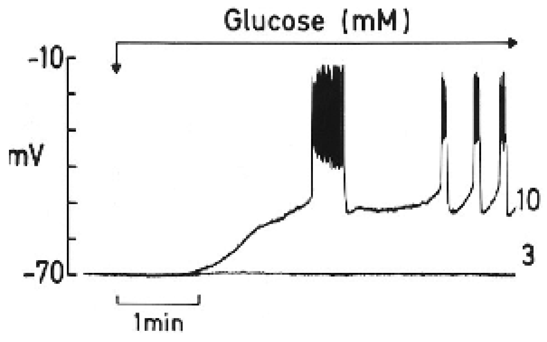
Figure 2. Extracellular (blood) glucose control of Cx36 hemichannels gating.
Preprints 170791 g002
Disclaimer/Publisher’s Note: The statements, opinions and data contained in all publications are solely those of the individual author(s) and contributor(s) and not of MDPI and/or the editor(s). MDPI and/or the editor(s) disclaim responsibility for any injury to people or property resulting from any ideas, methods, instructions or products referred to in the content.
Copyright: This open access article is published under a Creative Commons CC BY 4.0 license, which permit the free download, distribution, and reuse, provided that the author and preprint are cited in any reuse.