Submitted:
17 July 2025
Posted:
18 July 2025
You are already at the latest version
Abstract
Background/Objectives: Traumatic injuries to the brain and spinal cord remain highly challenging to treat due to the complex biological processes involved and the limited regenerative capacity of neural tissues. Recent advances in nanotechnology offer new opportunities to address these limitations. This review aims to summarize current and emerging applications of nanotechnology for improving diagnosis, targeted therapy, and tissue regeneration in brain and spinal cord injuries. Methods: We conducted a comprehensive review of recent studies investigating the interaction of nanomaterials with neural cells such as microglia, astrocytes, and neurons. Key areas of focus include nanoscale diagnostic markers, nanoparticle-mediated drug delivery systems, nanostructured scaffolds, and combined strategies integrating gene therapy, neuro-protection, rehabilitation, and artificial intelligence. Results: Emerging evidence indi-cates that nanotechnology can enable real-time, minimally invasive detection of in-flammation, oxidative stress, and cellular damage. Targeted drug delivery using na-noparticles shows potential for enhanced therapeutic effects with fewer side effects, especially when combined with electrical stimulation to promote nerve regeneration. Additionally, nanostructured scaffolds can mimic the brain’s microenvironment, sup-porting the regrowth and reconnection of damaged neural tissue. Integrating AI tools may further help design personalized nanoparticle treatments. Conclusions: Nano-technology offers promising new avenues for the diagnosis and treatment of traumatic brain and spinal cord injuries. By improving targeting, enhancing regeneration, and enabling personalized approaches, these innovations could accelerate recovery and provide hope for patients with acute or chronic neurological damage.
Keywords:
nanotechnology
; traumatic brain injury
; spinal cord injury
; Neural Regeneration e Targeted Drug Delivery
1. Introduction
Traumatic injuries to the brain and spinal cord represent a major cause of disability worldwide, with limited therapeutic options and often irreversible consequences [1]. Traumatic brain injury (TBI) and spinal cord injury (SCI) trigger complex cascades of molecular and cellular events, including inflammation, oxidative stress, axonal degeneration, and synaptic disruption, which ultimately compromise functional recovery [1]. Traditional therapeutic strategies, encompassing surgical interventions, neuroprotective drugs, and physical rehabilitation, offer only partial efficacy and fail to fully restore the intricate architecture of the central nervous system (CNS) [1]. In recent years, nanotechnology has emerged as a transformative platform capable of addressing the multifaceted challenges associated with CNS injuries [2,3]. Nanostructured materials offer unprecedented opportunities for ultra-precise diagnostics, targeted therapeutic delivery, and the engineering of bioactive scaffolds that can promote tissue regeneration [4,5]. Unlike conventional approaches, nanotechnologies can operate at the molecular scale, interact specifically with damaged tissues, and dynamically respond to the evolving pathological microenvironment [2,3].
This review aims to present an integrated vision for nanotechnology-driven neuroregeneration, structured around three fundamental pillars: ultra-precise diagnosis using biosensitive nanomarkers for real-time monitoring of injury progression [6,7,8]; targeted multi-modal therapy through smart nanoparticle systems and electro-conductive nanomaterials [5,6]; (3) neurobiological regeneration using nanostructured scaffolds designed to mimic the extracellular matrix and support axonal growth and synaptic plasticity [4,5].
2. Materials and Methods
This narrative review was conducted to examine the current and emerging applications of nanotechnology in the field of neuroregeneration, with particular attention to spinal cord and brain injuries. A comprehensive literature search was carried out using PubMed, Scopus, Web of Science, and Google Scholar. The search focused on publications from the past decade written in English, including preclinical and clinical studies, original research articles, systematic reviews, and meta-analyses. Studies were selected based on their relevance to the diagnostic, therapeutic, and rehabilitative use of nanomaterials in central nervous system injuries. Articles were manually screened through titles, abstracts, and full texts, with particular attention to methodological rigor and clinical or translational significance. Non-peer-reviewed sources, duplicates, and studies outside the scope of nanotechnology-based neuroregeneration were excluded. The final selection reflects a critical synthesis of the most significant and innovative contributions in the field, highlighting both established mechanisms and emerging directions for clinical application.
Generative artificial intelligence (ChatGPT by OpenAI) was used to assist with language refinement and the organization of content in this manuscript. All scientific interpretations, critical analyses, and final text were reviewed and approved by the authors.
3. Molecular and Cellular Mechanisms of Injury: Inflammation and Oxidative Stress
The primary mechanical insult to the CNS rapidly activates resident immune cells, such as microglia and astrocytes, leading to the release of pro-inflammatory cytokines (e.g., TNF-α, IL-1β, IL-6) and chemokines[11,12,13]. This inflammatory response, although initially protective, often evolves into a chronic, self-perpetuating state that exacerbates secondary neuronal damage [15,16,17,18]. Concomitantly, the injury triggers a burst of oxidative stress through the excessive production of reactive oxygen species (ROS) and reactive nitrogen species (RNS). These molecules induce lipid peroxidation, protein dysfunction, and DNA damage, severely compromising cellular integrity and promoting apoptotic and necrotic pathways [15]. Following injury, axonal tracts undergo Wallerian degeneration, characterized by fragmentation of axons distal to the lesion and disruption of the myelin sheath. The loss of myelin further impairs axonal conductivity and exposes neurons to additional cytotoxic insults [12,15]. Oligodendrocyte apoptosis, a hallmark of secondary injury, limits spontaneous remyelination and contributes to chronic deficits [17]. Beyond structural damage, TBI and SCI severely affect synaptic architecture and function. Disruption of synaptic homeostasis impairs neurotransmitter release, receptor signaling, and synaptic plasticity mechanisms such as long-term potentiation and depression [15]. Altered synaptic dynamics hinder the re-establishment of functional circuits necessary for recovery. Scar formation, primarily driven by reactive astrocytes (astrogliosis) and the deposition of extracellular matrix inhibitors (e.g., chondroitin sulfate proteoglycans), further impedes axonal regrowth [15]. The blood-brain barrier (BBB) and blood-spinal cord barrier (BSCB) become compromised, leading to edema and infiltration of peripheral immune cells, compounding tissue damage [1,12,19]. Importantly, recent evidence highlights that chronic inflammation itself acts as an active barrier to axon regeneration, maintaining a hostile microenvironment that prevents effective neuronal repair [18].
4. Nanotechnology for Ultra-Precise Diagnosis
In recent years, the use of nanomarkers, functionalized nanoparticles, has revolutionized the field of medical imaging. These nanoscale structures can be engineered at the molecular level to target specific biological features, such as cancer cells, receptors, or enzymes, enabling non-invasive, real-time visualization of disease processes with unprecedented precision. Nanomarkers are nanoscale construct, as quantum dots, gold nanoparticles, and magnetic nanoparticles, obtained to detect specific molecular signatures associated with injury, including inflammation, oxidative stress, and neuronal apoptosis [1,2,20]. A particularly innovative aspect of nanomarkers is their ability to emit physical signals, optical, magnetic, or acoustic, making them compatible with various advanced imaging modalities. By functionalizing nanoparticle surfaces with ligands (e.g., antibodies, peptides) targeting injury-specific biomarkers, these probes can selectively accumulate at damaged sites. Depending on their composition and functionalization, nanomarkers can interact with magnetic fields, emit near-infrared (NIR) fluorescence, absorb light and generate acoustic signals, or incorporate radioactive isotopes. This versatility makes them suitable for techniques such as magnetic resonance imaging (MRI), positron emission tomography (PET), photoacoustic imaging, and near-infrared fluorescence (NIRF) imaging [2,4,20]. In MRI, for example, superparamagnetic iron oxide nanoparticles (SPIONs) are commonly used to enhance image contrast by altering the relaxation times of surrounding hydrogen nuclei. These nanoparticles are biocompatible and can be coated with targeting ligands to home in on specific tissues or tumors [21]. For PET imaging, nanomarkers can be labeled with radioactive isotopes such as fluorine-18 or gallium-68. These isotopes enable the real-time tracking of metabolic processes and disease markers, providing functional information that complements structural imaging [22]. Photoacoustic imaging is an emerging hybrid technique that combines the high contrast of optical imaging with the depth resolution of ultrasound. Nanoparticles that strongly absorb light—such as gold nanorods or carbon nanotubes—can convert pulsed laser energy into acoustic waves, enabling detailed imaging of vascular and tumor structures [23]. In NIR fluorescence imaging, nanoparticles embedded with fluorescent dyes or quantum dots emit light in the near-infrared range (700–900 nm), which penetrates deeper into biological tissues and reduces background noise due to lower autofluorescence. This technique is particularly useful for intraoperative imaging and surgical guidance [24]. Moreover, a growing trend is the development of multimodal nanomarkers, which can be detected by more than one imaging technique. For instance, a single nanoparticle may combine magnetic and fluorescent properties, enabling pre-operative MRI scans and real-time NIRF visualization during surgery. This approach offers a more comprehensive diagnostic profile, bridging anatomical and molecular information for improved decision-making. Furthermore, theranostic nanoparticles, platforms combining therapeutic and diagnostic function, offer dynamic monitoring of disease progression while simultaneously delivering therapeutic agents [20]. Compared to conventional biomarkers measured in cerebrospinal fluid (CSF) or blood, nanosensors offer the advantage of in situ monitoring without the need for invasive procedures. Moreover, their ability to provide dynamic, real-time information can significantly improve clinical decision-making, enabling the early adjustment of therapeutic strategies based on evolving injury profiles [1,2,4,20].
5. Targeted Multi-Modal Therapeutic Strategies
The complexity of CNS injuries demands multifaceted therapeutic interventions that can address inflammation, oxidative stress, neuronal loss, and impaired regeneration. Nanotechnology provides a unique platform for the development of multi-modal therapeutic strategies that combine targeted drug delivery, electrical stimulation, and tissue engineering approaches to promote functional recovery.
5.1. Nanoparticle-Based Targeted Delivery for the Treatment of Spinal Cord Injury
Nanoparticles have emerged as a powerful therapeutic platform in the treatment of SCI, offering a level of spatial and molecular precision that conventional pharmacological approaches lack. By exploiting their nanoscale size and the ability to be surface-modified with bioactive molecules, nanoparticles can be engineered to deliver therapeutic agents directly to the site of injury. This capability is particularly important in the context of SCI, where the blood–spinal cord barrier (BSCB), inflammation, and tissue degradation severely limit the effectiveness of systemic treatments. One of the fundamental advantages of nanoparticles is their capacity for targeted delivery, achieved through surface functionalization with ligands, peptides, or monoclonal antibodies. These moieties enable the recognition of molecular markers specifically upregulated in inflamed or damaged tissues, such as adhesion molecules (e.g., ICAM-1, VCAM-1) or integrins. Once administered, these functionalized nanoparticles exhibit enhanced accumulation at the injury site, thereby reducing off-target distribution and minimizing systemic side effects [25,26,27]. In addition to anti-inflammatory and antioxidant compounds, nanoparticles have been successfully employed to deliver neurotrophic factors, which play an essential role in neural survival, regeneration, and synaptic repair. Molecules such as brain-derived neurotrophic factor (BDNF) and insulin-like growth factor-1 (IGF-1) are known to promote axonal growth and functional recovery post-injury. However, their systemic delivery is limited by rapid degradation and poor penetration across the BSCB. Encapsulation within nanoparticles protects these proteins from enzymatic degradation and enables sustained, localized release at the lesion site. Experimental studies have demonstrated that nanoparticle-mediated delivery of BDNF and IGF-1 promotes neuronal survival and axonal regrowth, contributing to improved histological and functional outcomes in animal models of SCI [28]. Another promising application of nanotechnology in SCI therapy is gene delivery. Nanoparticles can act as non-viral vectors to transport nucleic acids, including plasmid DNA, siRNA, mRNA, or CRISPR-Cas9 components, into specific cell populations within the injured spinal cord. This strategy enables modulation of gene expression pathways involved in inflammation, apoptosis, and regeneration. For instance, the delivery of anti-apoptotic genes or the silencing of pro-inflammatory cytokines using siRNA-loaded nanoparticles has been shown to reduce secondary damage and enhance the intrinsic capacity for repair. These non-viral systems present significant advantages over viral vectors, including lower immunogenicity, greater biosafety, and the potential for transient and controllable gene expression [26,27]. The integration of nanocarriers into SCI therapy represents a multidisciplinary advancement that bridges materials science, molecular biology, and neurology. Their multifunctionality, tunable properties, and ability to respond to the local microenvironment make them ideal candidates for combination therapies that simultaneously address multiple aspects of SCI pathology. As research in this field continues to evolve, future efforts will need to focus on improving the translation potential of these systems by enhancing their biocompatibility, optimizing dosing strategies, and validating their long-term efficacy and safety in clinical settings.
5.2. Electro-Nanohybrid Stimulation
The transition from conventional oral or systemic therapies to technologically advanced regenerative strategies mark a significant shift in the treatment of SCI. Among these, electro-nanohybrid stimulation, the integration of electrically conductive nanomaterials into neural repair platforms, has emerged as a compelling approach to facilitate neuronal regeneration by directly modulating the injured microenvironment. Electrically active nanomaterials, including carbon nanotubes (CNTs), graphene derivatives, and conductive polymers such as polypyrrole, possess the unique ability to transduce electrical signals across disrupted axonal pathways [27]. These materials can be incorporated into scaffolds, hydrogels, or injectable delivery systems, where they serve a dual purpose: providing physical support for cell adhesion and axonal guidance and delivering electrical cues that stimulate neuronal growth and connectivity. Studies have shown that localized electrical stimulation via conductive nanoscaffolds promotes neurite extension, synaptic plasticity, and remyelination, all critical processes in the restoration of function after SCI (Figure 1). The electrical signals are thought to modulate ion channels, influence intracellular signaling pathways (e.g., ERK/MAPK, PI3K/Akt), and enhance the secretion of neurotrophic factors by both neurons and glial cells. In parallel, recent advances in multifunctional nanomaterials have led to the development of bioactive nanofibers with intrinsic reactive oxygen species (ROS) scavenging capabilities. Following SCI, the accumulation of ROS contributes to secondary damage by promoting inflammation, lipid peroxidation, and cell death. Incorporating antioxidant properties into electroconductive materials enables the reduction of oxidative stress, thereby protecting neurons and glial cells in the perilesional region [29].
Moreover, these hybrid systems have demonstrated immunomodulatory effects, notably the promotion of M2 macrophage polarization. The M2 phenotype is associated with anti-inflammatory and tissue-repair functions, in contrast to the pro-inflammatory M1 phenotype. By modulating macrophage activity, electro-nanohybrids help create a regeneration-permissive environment that supports axonal regrowth and limits scar formation. Importantly, the tunability of nanomaterial properties, such as surface charge, conductivity, mechanical stiffness, and topography, allows precise control over cellular responses. For example, aligned conductive nanofibers can guide directional axonal growth, while dynamically responsive materials can be activated externally to synchronize electrical stimulation with cellular events. In summary, electro-nanohybrid stimulation represents a convergence of nanotechnology, materials science, and neuroengineering, offering a multi-pronged approach to spinal cord repair. By combining structural, electrical, biochemical, and immunological functionalities into a single platform, these systems address the complex and multifactorial nature of SCI pathology more effectively than single-modality therapies. As preclinical models continue to demonstrate the potential of this technology, ongoing research is now focused on improving scalability, long-term biocompatibility, and integration with implantable bioelectronic devices for future clinical applications.
Legend: Spinal Cord Injury: Damaged axons and disrupted blood–spinal cord barrier with inflammation. Nanoparticle: Engineered particle with surface ligands for targeted drug or factor delivery. Conductive Nanoscaffold: 3D structure made of conductive nanomaterials enabling local electrical stimulation. Localized Electrical Stimulation: Stimuli that promote neuron activation and tissue regeneration. Neurite Outgrowth: Extension of new axons from neurons. Synaptic Plasticity: Strengthening and reformation of neural connections. Remyelination: Restoration of the myelin sheath around regenerated axons.
6. Nanotechnological Strategies for CNS Drug Delivery
One of the most significant challenges in treating CNS disorders, including spinal cord injury (SCI), is the restricted permeability of the BBB and BSCB. These tightly regulated interfaces serve as protective barriers, maintaining CNS homeostasis by limiting the entry of potentially harmful substances. However, this same protective function severely limits the penetration of therapeutic agents, including neurotrophic factors, anti-inflammatory drugs, and genetic materials (Figure 2) [30].
To address this issue, nanoparticles have been increasingly explored as delivery vectors due to their physiochemical properties, which can be optimized to facilitate barrier crossing. Key design parameters include particle size (typically <100 nm), surface charge (often neutral or slightly positive to favor uptake), and surface functionalization with ligands such as transferrin, lactoferrin, apolipoprotein E, or peptides like TAT. These ligands bind to specific receptors on the endothelial cells of the BBB/BSCB, enabling receptor-mediated transcytosis [19,27]. Moreover, following SCI, the integrity of the BSCB is temporarily compromised due to inflammatory responses, oxidative stress, and vascular disruption. This creates transient permeability windows that nanoparticles can exploit to penetrate more efficiently into the injured spinal cord. Timed delivery during this window allows for maximized therapeutic impact with minimized systemic exposure. Beyond delivery, advanced nanocarriers have been designed for theragnostic applications, integrating both therapeutic and diagnostic functions within a single platform. For example, iron oxide nanoparticles or quantum dots can be co-loaded with drugs and visualized via MRI or near-infrared fluorescence imaging. These systems allow real-time tracking of biodistribution, accumulation at the lesion site, and correlation with therapeutic outcomes [20]. This dual functionality improves clinical decision-making and supports personalized treatment regimens. Critically, these strategies must also ensure biocompatibility, non-immunogenicity, and controlled drug release to maintain safety and therapeutic efficacy.
While preclinical studies have demonstrated promising results, further optimization and rigorous validation in large animal models and early-phase clinical trials are needed to ensure translational success.
The reconstruction of neural circuits after TBI or SCI requires not only cellular survival but also the physical guidance of axonal growth and synapse formation. Nanostructured scaffolds represent a promising strategy to recreate the extracellular matrix (ECM), providing biochemical, mechanical, and electrical cues necessary for effective neuroregeneration. Advanced nanofabrication techniques have enabled the creation of scaffolds that mimic the architecture and composition of the native ECM [27]. These scaffolds can be composed of biocompatible materials such as collagen, hyaluronic acid, chitosan, or synthetic polymers (e.g., PLGA, PCL) functionalized with neurotrophic factors or adhesion molecules (e.g., laminin, fibronectin). The nanoscale topography, including fiber orientation and pore size, plays a crucial role in guiding axonal alignment, enhancing neurite extension, and supporting cell migration [27]. Electrospun nanofibers, in particular, can be aligned to direct axonal regrowth along specific paths, mimicking the organized structure of white matter tracts. To further enhance regenerative outcomes, scaffolds can be functionalized with bioactive agents (e.g., BDNF, NGF, VEGF) that promote neuronal survival, angiogenesis, and synaptic plasticity [26,27]. Additionally, incorporating conductive nanomaterials such as graphene or carbon nanotubes allows scaffolds to transmit electrical signals, synergistically stimulating neuronal activity and enhancing axon regeneration [29]. Recent developments have introduced multifunctional scaffolds capable of simultaneously clearing reactive oxygen species (ROS), modulating inflammation, and stimulating electrical activity, thereby creating a pro-regenerative microenvironment [29] [Table 2]. Combining nanoscaffolds with stem cells or induced oligodendrocyte precursor cells (iOPCs) offers an even more powerful regenerative strategy. For instance, human umbilical cord mesenchymal stem cells differentiated into OPCs using TET3 overexpression have demonstrated the capacity to promote remyelination and functional recovery after SCI [17]. By embedding these cells into nanostructured matrices, it is possible to provide physical support, biochemical cues, and protection from hostile inflammatory environments, thereby enhancing cell survival, differentiation, and integration into host tissue [17]. The therapeutic efficacy of nanotechnologies for CNS injuries critically depends on their interactions with the various cellular players involved in injury and repair processes (see Table 1). Understanding how nanomaterials influence microglia, astrocytes, neurons, and oligodendrocytes is essential for optimizing regenerative strategies and minimizing adverse effects. Following injury, microglia rapidly become activated and can adopt either a pro-inflammatory (M1) or pro-regenerative (M2) phenotype. Similarly, astrocytes may undergo reactive astrogliosis, leading to scar formation and inhibition of axonal regeneration [6]. Nanomaterials can modulate these glial responses in a beneficial way. Certain nanoparticles, such as Prussian blue nanoparticles and antioxidant nanomaterials, have been shown to reduce pro-inflammatory cytokine production (e.g., TNF-α, IL-1β) and promote M2 polarization of microglia, supporting a regenerative environment [3,29]. Surface-modified nanocarriers can interact with astrocytes to limit glial scar formation and enhance the secretion of neurotrophic factors [27]. By reprogramming the inflammatory response, nanotechnologies can shift the microenvironment from a neurotoxic to a neuroprotective state. Axonal damage and demyelination are major contributors to functional loss after CNS injury. Nanostructured materials contribute to axonal repair through promoting axonal regrowth: Aligned electrospun nanofibers provide topographical guidance that stimulates axon elongation along desired trajectories [27]; enhancing remyelination: Conductive scaffolds combined with stem cell therapies (e.g., iOPCs) can support oligodendrocyte maturation and efficient remyelination. Nanocarriers delivering growth factors such as IGF-1, PDGF-AA, or gene editing tools (e.g., CRISPR-Cas9 systems) may further potentiate axonal repair [17,27]. Recovery of CNS function also requires the restoration of synaptic networks. Nanoparticles can aid in synaptic plasticity by delivering BDNF, NT-3, or other synaptogenic molecules directly to the injury site [26,27]; modulating intracellular signaling cascades (e.g., Ca²⁺ influx, cAMP/PKA pathways) that govern synaptic remodeling; acting as nanoscaffolds that facilitate cell-cell contacts and synaptogenesis by mimicking the 3D nanoarchitecture of synaptic clefts. This targeted support of synaptic repair is crucial for regaining motor, sensory, and cognitive functions after TBI and SCI.
7. AI-Guided Personalized Nanomedicine
Personalized medicine aims to design interventions that match the individual characteristics of each patient’s injury, genetics, and molecular profile, by predicting optimal nanoparticle formulations (e.g., size, surface chemistry, cargo) based on injury type and patient-specific biomarkers [20]; optimizing dosing schedules and delivery routes using machine learning algorithms trained on preclinical and clinical datasets; monitoring treatment responses in real-time through AI-enhanced imaging and biomarker analysis. By combining nanotechnology with AI-driven personalization, it becomes possible to maximize therapeutic efficacy while minimizing risks — moving toward a precision neuroregenerative therapy model.
8. Integration with Conventional Therapies and Personalized Nanomedicine
Although nanotechnology-based approaches have shown remarkable promise in experimental models of CNS injury, their integration with established therapeutic modalities is crucial for translating these innovations into clinical practice. Nanotechnology-based approaches can be integrated synergistically with conventional treatments such as physical rehabilitation, exercise protocols, and gene therapies to maximize outcomes. Exercise-induced exerkines, such as neurotrophic factors, may act synergistically with nanotherapy to enhance regeneration [28]. Recent evidence also suggests that cognitive multisensory rehabilitation, combined with advanced nanoparticle therapies, may reshape neural networks involved in body awareness and pain modulation, providing a holistic approach to functional recovery [31].
8.1. Physical Rehabilitation
Physical rehabilitation remains a basis in the management of TBI and SCI. Nanotherapeutics can synergize with rehabilitative strategies to enhance functional recovery as exercise-induced secretion of exerkines (e.g., IGF-1, VEGF, BDNF) can be potentiated by nanocarriers delivering complementary neuroprotective agents; structured exercise regimens can facilitate nanoparticle distribution and enhance cellular uptake within injured neural tissues, improving therapeutic outcomes [28] (Table 2). Recent studies also suggest that combining neuromuscular stimulation, task-specific training, and nanoscaffold implantation can promote superior motor recovery compared to isolated interventions. Even integrating cognitive rehabilitation with nanotechnology-based therapies may be particularly beneficial for addressing sensory and cognitive deficits post-injury. Multisensory rehabilitation programs have been shown to induce neural plasticity and reorganization of brain networks related to body awareness and pain processing. Smart nanomaterials capable of modulating synaptic activity could be paired with cognitive exercises to amplify neuroplastic changes at both the structural and functional levels [31]. This integrated approach holds promise for more holistic recovery of both motor and cognitive domains (Table 3).
9. Discussion
Traumatic injuries to the brain and spinal cord remain among the most formidable challenges in clinical neuroscience, largely due to the intrinsic complexity of central nervous system (CNS) repair mechanisms. While conventional therapies provide some benefit, they often fall short of achieving full functional restoration.
Nanotechnology has emerged as a transformative frontier, offering ultra-precise diagnostic tools, targeted delivery systems for therapeutic agents, and innovative scaffolding platforms capable of overcoming many of the biological barriers to regeneration. Nanomarkers, in particular, are leading the way in precision medicine by providing highly sensitive and specific tools for molecular and functional imaging. Their integration with biomedical imaging enhances diagnostic accuracy and paves the way for image-guided interventions, early detection, and personalized therapeutic strategies.
Moreover, nanomaterials can interact dynamically with key cellular players—such as microglia, astrocytes, neurons, and axons—thereby modulating the injury microenvironment and promoting neural repair. This convergence of nanotechnology and neuroscience opens new therapeutic horizons, with the potential to significantly improve outcomes, reduce systemic side effects, and bring renewed hope to patients affected by both acute and chronic neurotrauma.
Looking ahead, the integration of nanotechnology with conventional rehabilitation strategies and AI-driven personalized approaches is poised to usher in a new era of precision neuroregeneration. Future research should prioritize the optimization of biocompatibility, a deeper understanding of long-term biodistribution, and the advancement of translational studies to ensure the successful transition of these promising technologies from bench to bedside.
10. Conclusions
Traumatic injuries to the central nervous system, particularly the brain and spinal cord, remain among the most formidable challenges in regenerative medicine. Nanotechnology has emerged as a transformative paradigm, offering ultra-precise diagnostics, targeted therapeutic delivery, and bioengineered scaffolds that can overcome many of the biological barriers to neural repair.
By enabling dynamic interaction with immune and neural cells, integrating with advanced imaging systems, and supporting personalized treatment through artificial intelligence, nanomedicine provides a multi-dimensional approach to neuroregeneration. When combined with physical and cognitive rehabilitation, these strategies have the potential to significantly enhance functional recovery and improve quality of life for patients.
While preclinical results are highly promising, clinical translation requires further work to optimize biocompatibility, ensure long-term safety, and develop standardized therapeutic protocols. Nevertheless, the convergence of nanotechnology, neuroscience, and personalized medicine marks a decisive step toward a new era of precision neuroregeneration—bringing renewed hope to individuals affected by both acute and chronic neurotrauma.
Author Contributions
For research articles with several authors, a short paragraph specifying their individual contributions must be provided. The following statements should be used “Conceptualization, L.R. and G.R..; methodology, L.R., G.R., RSC; data curation, L.R., G.R., RSC; writing—original draft preparation, L.R. and G.R.; writing—review and editing, L.R., G.R.; visualization, RSC.; supervision, RSC; project administration, L.R. All authors have read and agreed to the published version of the manuscript.
Funding
This research received no external funding.
Institutional Review Board Statement
Not applicable.
Informed Consent Statement
Not applicable.
Data Availability Statement
none
Acknowledgments
none
Conflicts of Interest
The authors declare no conflicts of interest.
References
- Tran AP, Warren PM, Silver J. The Biology of Regeneration Failure and Success After Spinal Cord Injury. Physiol Rev. 2018;98(2):881–917. [CrossRef]
- Silva GA. Nanotechnology approaches to crossing the blood-brain barrier and drug delivery to the CNS. BMC Neurosci. 2008;9 Suppl 3(Suppl 3):S4. [CrossRef]
- Patel T, Zhou J, Piepmeier JM, Saltzman WM. Polymeric nanoparticles for drug delivery to the central nervous system. Adv Drug Deliv Rev. 2012;64(7):701–705. [CrossRef]
- Chen S, John JV, McCarthy A, Xie J. New forms of electrospun nanofiber materials for biomedical applications. J Mater Chem B. 2020;8(17):3733–3746. [CrossRef]
- Wang W, Hou Y, Martinez D, Kurniawan D, Chiang WH, Bartolo P. Carbon Nanomaterials for Electro-Active Structures: A Review. Polymers (Basel). 2020;12(12):2946. [CrossRef]
- Gupta AK, Gupta M. Synthesis and surface engineering of iron oxide nanoparticles for biomedical applications. Biomaterials. 2005;26(18):3995–4021. [CrossRef]
- Jokerst JV, Gambhir SS. Molecular imaging with theranostic nanoparticles. Acc Chem Res. 2011;44(10):1050–1060. [CrossRef]
- Blanco-Andujar C, Walter A, Cotin G, et al. Design of iron oxide-based nanoparticles for MRI and magnetic hyperthermia. Nanomedicine (Lond). 2016;11(14):1889–1910. [CrossRef]
- Burnouf T, Agrahari V, Agrahari V. Extracellular Vesicles As Nanomedicine: Hopes And Hurdles In Clinical Translation. Int J Nanomedicine. 2019;14:8847–8859. [CrossRef]
- Bafekry A, Faraji M, Fadlallah MM, et al. Effect of adsorption and substitutional B doping at different concentrations on the electronic and magnetic properties of a BeO monolayer: a first-principles study. Phys Chem Chem Phys. 2021;23(43):24922–24931. [CrossRef]
- Gong W, Zhang T, Che M, et al. Recent advances in nanomaterials for the treatment of spinal cord injury. Mater Today Bio. 2022;18:100524. [CrossRef]
- Yang Q, Lu D, Wu J, et al. Nanoparticles for the treatment of spinal cord injury. Neural Regen Res. 2025;20(6):1665–1680. [CrossRef]
- Gu X, Zhang S, Ma W. Prussian blue nanotechnology in the treatment of spinal cord injury: application and challenges. Front Bioeng Biotechnol. 2024;12:1474711. [CrossRef]
- Wang W, Yong J, Marciano P, O'Hare Doig R, Mao G, Clark J. The Translation of Nanomedicines in the Contexts of Spinal Cord Injury and Repair. Cells. 2024;13(7):569. [CrossRef]
- Hu X, Xu W, Ren Y, et al. Spinal cord injury: molecular mechanisms and therapeutic interventions. Signal Transduct Target Ther. 2023;8(1):245. [CrossRef]
- Gu X, Zhang S, Ma W. Bibliometric analysis of nanotechnology in spinal cord injury: current status and emerging frontiers. Front Pharmacol. 2024;15:1473599. [CrossRef]
- Zhang Y, Peng Z, Guo M, et al. TET3-facilitated differentiation of human umbilical cord mesenchymal stem cells into oligodendrocyte precursor cells for spinal cord injury recovery. J Transl Med. 2024;22(1):1118. [CrossRef]
- Stewart AN, Bosse-Joseph CC, Kumari R, et al. Nonresolving Neuroinflammation Regulates Axon Regeneration in Chronic Spinal Cord Injury. J Neurosci. 2025;45(1):e1017242024. [CrossRef]
- Yu S, Chen X, Yang T, et al. Revealing the mechanisms of blood-brain barrier in chronic neurodegenerative disease: an opportunity for therapeutic intervention. Rev Neurosci. 2024;35(8):895–916. [CrossRef]
- Toader C, Dumitru AV, Eva L, Serban M, Covache-Busuioc RA, Ciurea AV. Nanoparticle Strategies for Treating CNS Disorders: A Comprehensive Review of Drug Delivery and Theranostic Applications International Journal of Molecular Sciences (2024).
- Gupta, A. K., & Gupta, M. (2005). Synthesis and surface engineering of iron oxide nanoparticles for biomedical applications. Biomaterials, 26(18), 3995–4021. [CrossRef]
- Lee, D. E., Koo, H., Sun, I. C., Ryu, J. H., Kim, K., & Kwon, I. C. (2010). Multifunctional nanoparticles for multimodal imaging and theragnosis. Chemical Society Reviews, 41(7), 2656–2672. [CrossRef]
- Jokerst, J. V., & Gambhir, S. S. (2011). Molecular imaging with theranostic nanoparticles. Accounts of Chemical Research, 44(10), 1050–1060. [CrossRef]
- Hong, G., Antaris, A. L., & Dai, H. (2017). Near-infrared fluorophores for biomedical imaging. Nature Biomedical Engineering, 1(1), 0010. [CrossRef]
- Papa S, Ferrari R, De Paola M, et al. Polymeric nanoparticle system to target activated microglia/macrophages in spinal cord injury. J Control Release. 2014;174:15-26. [CrossRef]
- Fortun J, Puzis R, Pearse DD, Gage FH, Bunge MB. Muscle injection of AAV-NT3 promotes anatomical reorganization of CST axons and improves behavioral outcome following SCI. J Neurotrauma. 2009;26(7):941-953. [CrossRef]
- Liu Z, Ran Y, Huang Q, Wang Z, Zhang W, Wu F, et al. Targeted delivery of gene therapy for spinal cord injury using non-viral polymeric nanoparticles. Curr Pharm Des. 2020;26(11):1295–309.
- Li Q, Li C, Zhang X. Research Progress on the Effects of Different Exercise Modes on the Secretion of Exerkines After Spinal Cord Injury. Cell Mol Neurobiol. 2024;44(1):62. Published 2024 Oct 1. [CrossRef]
- Zhang Q, Zheng J, Li L, et al. Bioinspired conductive oriented nanofiber felt with efficient ROS clearance and anti-inflammation for inducing M2 macrophage polarization and accelerating spinal cord injury repair. Bioact Mater. 2024;46:173-194. Published 2024 Dec 13. [CrossRef]
- Wu D, Chen Q, Chen X, Han F, Chen Z, Wang Y. The blood-brain barrier: structure, regulation, and drug delivery. Signal Transduct Target Ther. 2023;8(1):217. Published 2023 May 25. [CrossRef]
- Van de Winckel A, Carpentier ST, Deng W, et al. Identifying Body Awareness-Related Brain Network Changes after Cognitive Multisensory Rehabilitation for Neuropathic Pain Relief in Adults with Spinal Cord Injury: Delayed Treatment arm Phase I Randomized Controlled Trial. Preprint. medRxiv. 2023;2023.02.09.23285713. Published 2023 Feb 10. [CrossRef]
Figure 1.
Targeted Neuroregeneration Using Conductive Nanoscaffolds.
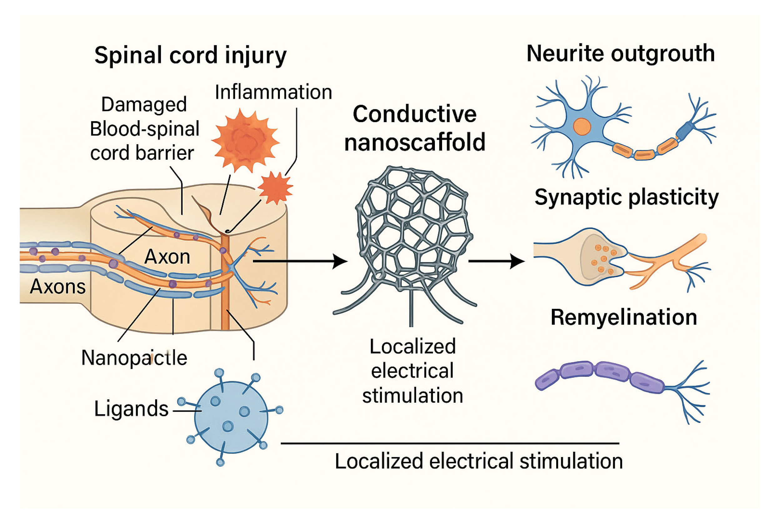
Figure 2.
Mechanisms of nanoparticle transport across the blood-brain barrier (BBB) and blood-spinal cord barrier (BSCB). The left panel illustrates receptor-mediated transcytosis of ligand-functionalized nanoparticles (NPs) across the intact BBB. The right panel depicts nanoparticle infiltration through a compromised BSCB following spinal cord injury (SCI), where inflammation-induced barrier disruption allows enhanced passive diffusion. These strategies enable targeted delivery of therapeutic agents into the central nervous system (CNS), improving treatment efficacy and reducing systemic toxicity.
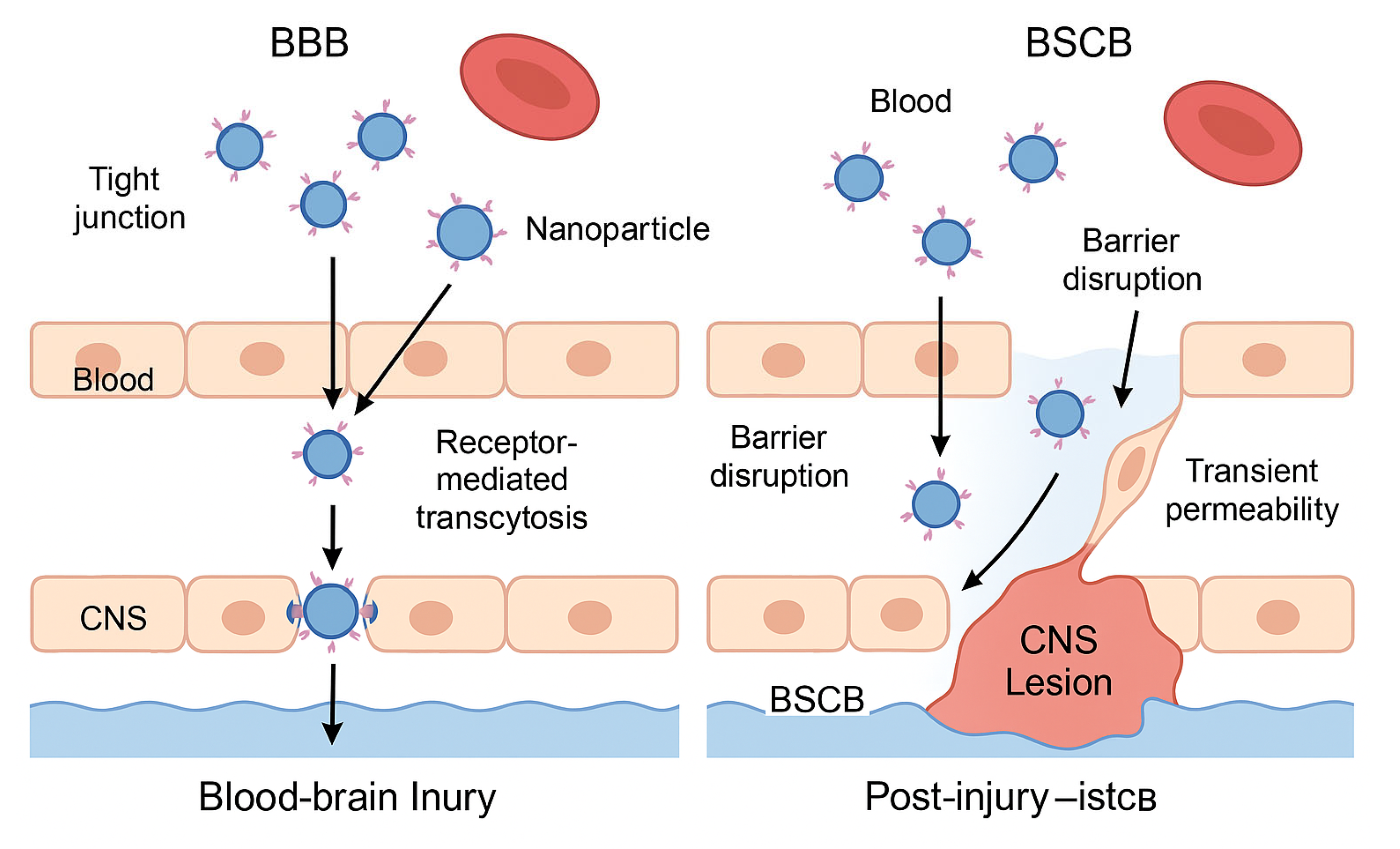

Table 1.
Comparative Table: Strategies to Cross the BBB and BSCB with Nanoparticles.
| Parameter | BBB (Blood-Brain Barrier) | BSCB (Blood-Spinal Cord Barrier) |
| Structure | Continuous endothelium with tight junctions; highly selective | Similar to BBB, slightly more permeable under physiological conditions |
| Therapeutic challenge | Blocks over 98% of systemically administered drugs | Less restrictive but still limits large or hydrophilic molecules |
| Nanoparticle strategy | Functionalization with ligands for receptor-mediated transcytosis (e.g., transferrin) | Exploitation of increased permeability after injury |
| Entry mechanism | Receptor-mediated transcytosis across endothelial cells | Passive diffusion through transiently disrupted barrier |
| Optimal timing | Constant, but difficult without targeting ligands | Subacute phase: hours to days post-injury, during inflammation |
| Clinical applications | Alzheimer's, brain tumors, encephalitis | Spinal cord injury, multiple sclerosis, spinal inflammation |
| Nanotherapy advantages | Targeted access with potential for theranostic monitoring | High efficiency when administered during post-injury permeability window |
Table 2.
This table summarizes the key types of nanoparticles being investigated for the treatment of SCI, their therapeutic cargos, main advantages and limitations, and current clinical development status.
Table 2.
This table summarizes the key types of nanoparticles being investigated for the treatment of SCI, their therapeutic cargos, main advantages and limitations, and current clinical development status.
| Nanoparticle Type | Therapeutic Cargo | Main Advantages | Limitations | Clinical Status |
| Polymeric | Anti-inflammatory drugs, growth factors, antioxidants | Customizable, sustained release, biocompatible | Scalability, burst release, clearance rate | Preclinical, some early-phase trials |
| Liposomal | Hydrophilic/lipophilic drugs, peptides | Good biocompatibility, approved in other indications | Stability, drug leakage, short half-life | Clinically approved for other diseases |
| Exosome-derived | Endogenous miRNAs, proteins, neurotrophic factors | Natural origin, low immunogenicity, high targeting potential | Isolation/purification challenges, batch variability | Preclinical studies |
| Magnetic (e.g., iron oxide) | Drugs + external magnetic control | Magnetic guidance, imaging compatibility | Possible long-term toxicity, low biodegradability | Used in oncology trials, not yet in SCI |
| Prussian Blue | Antioxidants, anti-inflammatory agents | Strong ROS scavenging, neuroprotection | Limited clinical validation, synthesis complexity | Preclinical |
| Carbon Nanotube (CNT) | Electrical stimulation interfaces | Restores neuronal conductivity, bioelectrical signaling | Biocompatibility issues, inflammatory potential | Preclinical (neural interface models) |
Table 3.
This table summarizes emerging applications of nanotechnology designed to enhance rehabilitation outcomes in spinal cord injury (SCI). Each row highlights a distinct area of application, describing the mechanism, functional impact, and development stage of the technology.
Table 3.
This table summarizes emerging applications of nanotechnology designed to enhance rehabilitation outcomes in spinal cord injury (SCI). Each row highlights a distinct area of application, describing the mechanism, functional impact, and development stage of the technology.
| Application Area | Description | Rehabilitative Benefit | Development Status |
| Neurotrophic Factor Delivery | Use of nanoparticles to deliver agents like BDNF, NGF, or IGF-1 to enhance plasticity during motor rehabilitation. | Amplifies the effect of activity-based therapies by promoting synaptic and axonal plasticity. | Preclinical |
| Bioelectronic Interfaces | Integration of conductive nanomaterials into scaffolds or implants to restore electrical signaling and support neuronal reactivation. | Enables functional reactivation of spinal circuits and synergy with FES or robotic training. | Preclinical to early prototyping |
| Nanosensors for Monitoring | Implantable or wearable nanosensors to monitor inflammation, neural activity, or metabolic markers during therapy. | Personalizes rehabilitation intensity and timing based on real-time physiological data. | Emerging technology |
| Gene Modulation via Nanocarriers | Nanoparticles carrying siRNA or miRNA to modulate genes involved in inhibitory signaling or regeneration during rehabilitation phases. | Maximizes the molecular environment's responsiveness to training by modulating key signaling pathways. | Preclinical studies in animal models |
Disclaimer/Publisher’s Note: The statements, opinions and data contained in all publications are solely those of the individual author(s) and contributor(s) and not of MDPI and/or the editor(s). MDPI and/or the editor(s) disclaim responsibility for any injury to people or property resulting from any ideas, methods, instructions or products referred to in the content. |
© 2025 by the authors. Licensee MDPI, Basel, Switzerland. This article is an open access article distributed under the terms and conditions of the Creative Commons Attribution (CC BY) license (http://creativecommons.org/licenses/by/4.0/).
Copyright: This open access article is published under a Creative Commons CC BY 4.0 license, which permit the free download, distribution, and reuse, provided that the author and preprint are cited in any reuse.


