Submitted:
07 July 2025
Posted:
07 July 2025
Read the latest preprint version here
Abstract
Ganoderma lucidum, a medicinal mushroom with a rich ethnobotanical history, was investigated using wild specimens collected from high-altitude regions of Nepal. This study aimed to identify key bioactive compounds and assess the influence of solvent type—water, ethanol, methanol, and acetone—on extraction efficiency and biological activity. Extracts were evaluated for antioxidant potential, cytotoxicity against HeLa cells, and phytochemical composition via gas chromatography–mass spectrometry (GC-MS). Solvent type significantly affected both yield and bioactivity. Acetone yielded the highest crude extract (3.43 mg/g), while ethanol extract exhibited the highest total phenolic (376.50 ± 9.32 mg PGE/g) and flavonoid content (30.33 ± 0.50 mg QEs/g). Methanol extract was richest in lycopene (0.0670 ± 0.001 mg/g) and β-carotene (0.4536 ± 0.000 mg/g). Ethanol extract demonstrated consistently strong DPPH, superoxide, hydroxyl, and nitric oxide radical scavenging activity, along with high reducing power. All extracts showed dose-dependent cytotoxicity against HeLa cells, with ethanol and water extracts showing the greatest inhibition (>65% at 1000 µg/mL). GC-MS profiling identified solvent-specific bioactive compounds including sterols, terpenoids, polyphenols, and fatty acids. Notably, pharmacologically relevant compounds such as hinokione, ferruginol, ergosterol, and geranylgeraniol were detected. These findings demonstrate the therapeutic potential of G. lucidum, underscore the importance of solvent selection, and suggest that high-altitude ecological conditions may influence its bioactive metabolite profile.
Keywords:
Ganoderma lucidum
; solvent extraction
; antioxidant activity
; cytotoxicity
; high-altitude fungi
; DPPH
; MTT assay
; taxonomy
; GC-MS
1. Introduction
Ganoderma lucidum, commonly known as Lingzhi in China, Reishi in Japan, and Dadu chyau in Nepal, is a polypore mushroom with a long and storied history of use in East Asian medicine for promoting health and longevity [1]. It is often referred to as the “King of Herbs” or the “Mushroom of Immortality.” Its medicinal use dates back over 2,000 years, with its effects documented in ancient scripts like the Shen Nong Ben Cao Jing from China’s Eastern Han dynasty (25-220 AD) [2]. Traditionally, it has been used to treat a variety of ailments and is believed to enhance stamina, increase brain power, improve circulation, and strengthen the immune system [3].
Modern research has begun to validate these traditional claims, attributing the mushroom’s medicinal properties to its rich and varied chemical composition. Bioactive compounds such as polysaccharides, triterpenes, adenosine, organic germanium, phenolic compounds, flavonoids, and ergosterol contribute to its therapeutic effects, which include antioxidant, anti-cancer, anti-inflammatory, and antimicrobial activities [4,5,6,7,8]. For instance, triterpenic acids have demonstrated significant anti-cancer effects, while polysaccharides have shown anti-diabetic and antibiotic properties [9,10,11,12]. The antioxidant properties of G. lucidum are particularly noteworthy, as they neutralize reactive oxygen species (ROS) and reactive nitrogen species (RNS), which are implicated in the development of chronic diseases such as cancer, aging, diabetes, cardiovascular disorders, and neurodegeneration [4,13]. Studies have shown that polysaccharides in G. lucidum exhibit potent ROS-scavenging activity and can inhibit tumor growth through immunomodulation and the induction of apoptosis [6,9,10]. Furthermore, its anti-inflammatory effects have been observed in animal models, suggesting significant therapeutic potential [14].
Nepal, with its diverse geography ranging from the Terai plains to the high Himalayas, provides a unique environment for a wide variety of mushrooms [15,16,17,18,19]. While approximately 1,300 mushroom species have been reported in Nepal, with 73 identified as having medicinal value, the therapeutic potential of most, including native G. lucidum, remains largely unexplored [20,21]. Research on Nepalese mushrooms, initiated by the Nepal Agricultural Research Council (NARC) in 1974, has historically focused more on cultivation and taxonomy rather than on the detailed analysis of their bioactive properties.
The unique environmental conditions of Nepal’s high-altitude regions—characterized by lower oxygen levels, intense UV radiation, and distinct soil compositions—may lead to the production of G. lucidum strains with enhanced or novel bioactive profiles [22,23]. Despite Nepal’s rich biodiversity, research on high-altitude G. lucidum is scarce, leaving a gap in our understanding of its full potential. This study, therefore, aims to investigate the bioactive constituents, antioxidant activities, and cytotoxic effects of wild G. lucidum collected from the high altitudes of Nepal, utilizing various solvent extracts to comprehensively characterize its mycochemical composition and therapeutic promise.
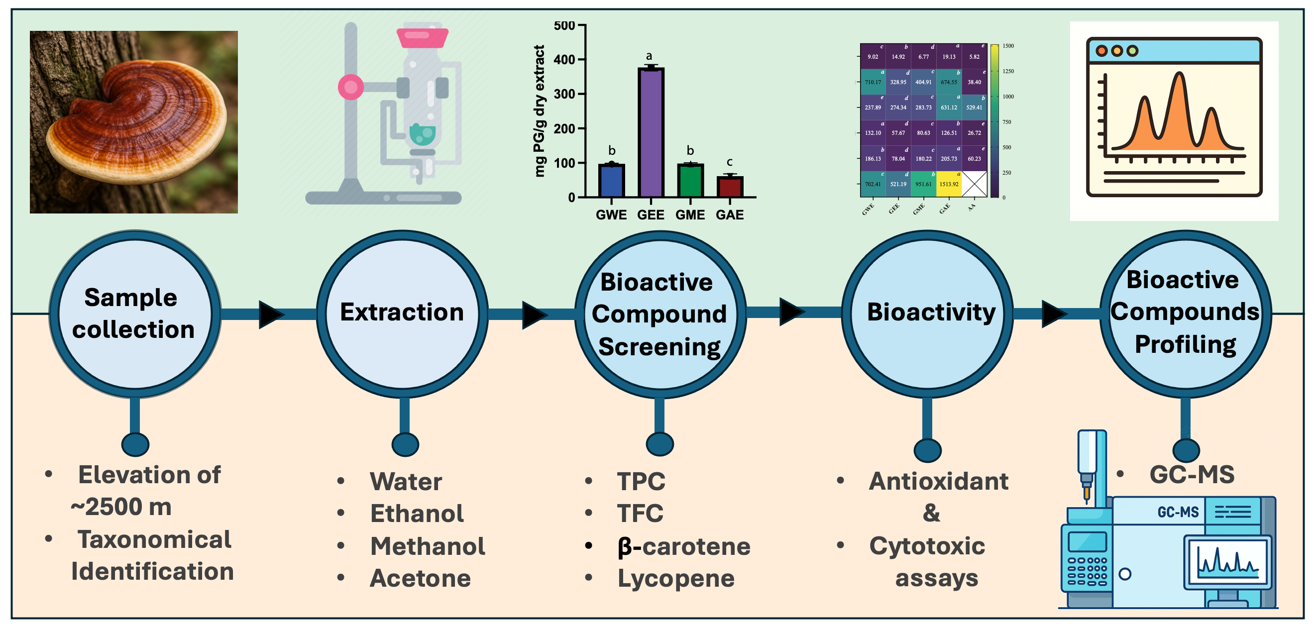
Figure 1.
Overview of the study workflow, from G. lucidum collection and solvent extraction to bioactive compound screening, biological activity assays, and GC-MS profiling.
Figure 1.
Overview of the study workflow, from G. lucidum collection and solvent extraction to bioactive compound screening, biological activity assays, and GC-MS profiling.
2. Materials and Methods
2.1. Collection and Identification
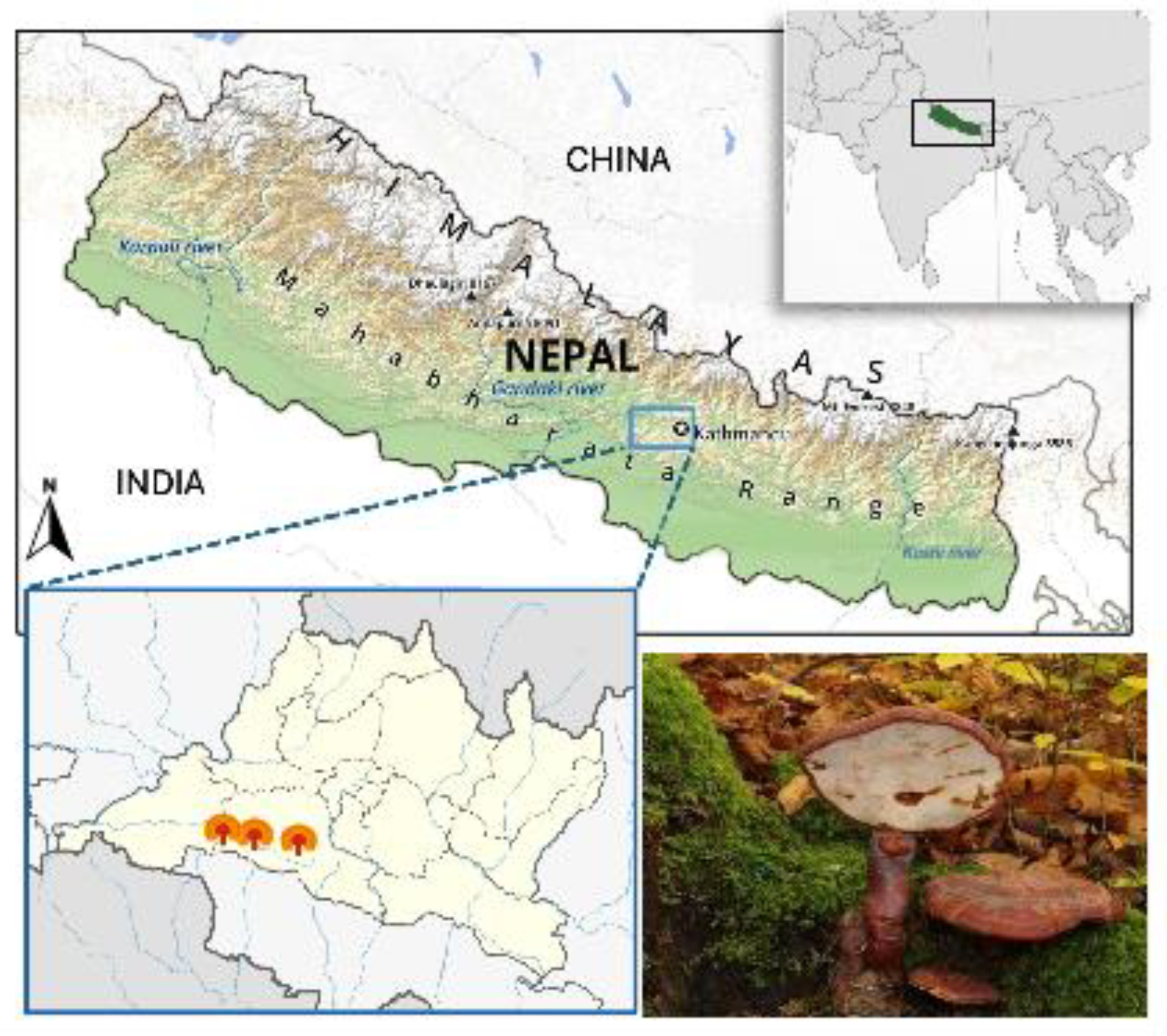
Fresh fruiting bodies of wild G. lucidum were collected from a dead trunk of Quercus lanata on Chandragiri Hill, Nepal (elevation 7482 ft; latitude 27.67402; longitude 85.19874) (Figure. 2). The specimens were identified based on detailed macro- and micro-morphological characteristics (Table 1).
Figure 2.
Map of Nepal in a global context (A), G. lucidum collection site (B) and its fruiting body growing on Quercus lanata (C).
Figure 2.
Map of Nepal in a global context (A), G. lucidum collection site (B) and its fruiting body growing on Quercus lanata (C).
2.2. Sample Preparation and Extraction
The samples were cleaned and oven-dried gradually from 45 °C to 60 °C for 3 days until a constant weight was attained. The dried mushroom was milled into a fine powder and stored in airtight containers for future use. 10 gm of G. lucidum were subjected for extraction. Solvent extraction was performed for 10 hrs in a Soxhlet apparatus with 250 mL each of water (GWE, 100 °C), 70% ethanol (GEE, 60 °C), 80% methanol (GME, 70 °C), and 50% acetone (GAE, 50 °C). Extracts were concentrated under vacuum in a rotary evaporator (50 °C) and stored in dark vials at 4 °C. The calculation of the % yield was done for each solvent using the formula,
2.3. Estimation of Total Phenolic, Flavonoid, β-carotene and Lycopene
2.3.1. Total Phenolic Content
Total phenolic content was determined using the Folin-Ciocalteu method with pyrogallol as the standard [24]. A calibration curve was established by measuring the absorbance of pyrogallol standards (10-100 µg/mL) at 760 nm. Sample solutions (1 mL) were reacted with 5 mL of 10% Folin-Ciocalteu reagent for 5 minutes at room temperature. Subsequently, 4 mL of Na2CO3 solution was added, and samples were vigorously mixed and incubated in the dark for 2 hours at room temperature. Absorbance was read at 760 nm, and results were expressed as pyrogallol equivalents (mg/g dry extract).
2.3.2. Total Flavonoid Content
Total flavonoid content was determined using the aluminum chloride colorimetric method, adapted from Shraim et al., 2021[25]. Quercetin standard curve was prepared by diluting a 10 mg quercetin stock in 50% methanol to concentrations ranging from 10-100 µg/mL, with absorbance measured at 415 nm. Sample aliquots (1.0 mL) were mixed with 0.5 mL of 1.2% aluminum chloride, 0.5 mL of 120 mM potassium acetate, and 1 mL of distilled water. After 30 minutes of incubation at room temperature, absorbance was read at 415 nm. Results were expressed as mg quercetin equivalents per gram of dry extract (mg QE/g dry weight).
2.3.3. Estimation of β-Carotene and Lycopene Content
β-carotene and lycopene were determined as per Prakash et al., 2016 [26]. Dried extracts (100 mg) were extracted with 10 mL of acetone-hexane (4:6) for 1 min, then filtered. Filtrate absorbance was measured at 453, 505, 645, and 663 nm. Carotenoid concentrations (mg/100 mL) were calculated using the following equations:
Lycopene = (−0.0458 × A663) + (0.372 × A505) + (0.0806 × A453)
β-carotene = (0.216 × A663) − (0.304 × A505) + (0.452 × A453)
Results are presented as mg carotenoid per gram of dry extract (mg carotenoid/g dry extract)
2.4. Determination of In Vitro Antioxidant Activities
2.4.1. DPPH (2, 2-diphenyl-1-picryl-hydrazyl) Assay
Different concentrations of extracts (20-100 µg/mL) were prepared. To 1 mL of each extract concentration, 2 mL of ice-cold 0.1 mM DPPH solution was added. The mixtures were incubated in the dark at room temperature for 30 min. Absorbance was then measured at 517 nm against a methanol blank [27]. A 3 mL DPPH solution was used as the control. Percentage inhibition was calculated using the formula:
where A0 is the absorbance of the control and A1 is the absorbance of the sample.
% inhibition = [(A₀-A₁)/(A₀)] x 100
2.4.2. Nitric Oxide Radical Scavenging Assay
Nitric oxide radical scavenging activity was determined with slight modification from Alam et al., 2013 [28]. Samples or ascorbic acid (20-100 µg/mL, 1 mL) were mixed with 2 mL of 10 mM sodium nitroprusside in phosphate buffer and incubated at 25°C for 2.5 hours. To 3 mL of the incubated solution, 3 mL of Griess reagent (1% sulfanilamide, 0.1% naphthylethylenediamine dihydrochloride in 2% H3PO3) was added. Absorbance of the pink color was measured at 540 nm against a blank. Ascorbic acid served as a positive control. Percentage inhibition was calculated using:
where A0 is the absorbance of the control and A1 is the absorbance of the sample.
% inhibition = [(A₀-A₁)/(A₀)] x 100
2.4.3. Hydroxyl Radical Scavenging Assay
Hydroxyl radical scavenging activity was assessed by measuring the inhibition of salicylic acid hydroxylation [29]. The 7 mL reaction mixture contained 1 mL of sample/standard (100-500 µg/mL), 2 mL of 6 mM FeSO4, 2 mL of 6 mM H2O2, and 2 mL of 6 mM salicylic acid. After incubation at 37 °C for 1 hour, absorbance was measured at 510 nm due to the color change of salicylic acid. Scavenging activity was calculated as follows:
% inhibition = [(A₀-A₁)/(A₀)] x 100
Where A0 is the absorbance of the control and A1 is the absorbance of the sample.
2.4.4. Superoxide Radical Scavenging Assay
Superoxide radical scavenging activity was assessed by a modified pyrogallol auto-oxidation method [29]. The reaction mixture contained 4.5 mL of 50 mM Tris-HCl buffer (pH 8.2), 0.4 mL of 25 mM pyrogallol, and 1 mL of sample (0.1-0.5 mg/mL). After 5 min incubation at 25°C, the reaction was terminated by adding 1 mL of 8 mM HCl. Absorbance was measured at 420 nm. Ascorbic acid served as the positive control. Superoxide radical scavenging activity was calculated using the formula:
% inhibition = [(A₀-A₁)/(A₀)] x 100
Where A0 is the absorbance of the control and A1 is the absorbance of the sample.
2.4.5. Reducing Power Assay
The reducing power of samples was assessed via the ferric reducing antioxidant power (FRAP) assay [30]. One mL of sample or standard (20-100 µg/mL) was combined with 2.5 mL of 0.2 M phosphate buffer (pH 6.6) and 2.5 mL of 1% (w/v) K3Fe(CN)6. After 20 min incubation at 50 °C in a water bath, 2.5 mL of 10% (w/v) trichloroacetic acid was added to terminate the reaction. The mixture was centrifuged at 3000 rpm for 10 min. A 2.5 mL aliquot of the supernatant was then mixed with 2.5 mL distilled water and 0.5 mL of 0.1% (w/v) FeCl3. Absorbance was measured at 700 nm against a blank.
2.5. Cytotoxicity Assay
HeLa cells were seeded in a 96-well plate. After 24 hours, cells were treated with dimethylsulfoxide or various extract concentrations for 48 hrs. Post-treatment, media were removed and replaced with 100 µL fresh medium containing MTT (3-(4,5-dimethylthiazol-2-yl)-2,5-diphenyltetrazolium bromide) reagent (final concentration 0.4 mg/mL). Plates were incubated at 37 °C for 3 hrs, during which intracellular purple formazan was observed. Next, 100 µL of solubilization solution (4 mM HCl, 0.1% NP40 in isopropanol) was added, and plates were kept in the dark for 15 minutes at room temperature. Absorbance was measured at 570 nm using a microplate reader [31].
2.6. Gas Chromatography–Mass Spectrometry (GC-MS) Analysis
GC-MS analysis of G. lucidum extracts was conducted using a GCMS-QP2010 Ultra (Shimadzu, Kyoto, Japan) equipped with an Rtx-5 M5 capillary column (30 m × 0.02 mm i.d., 0.25 µm film thickness; Restek, Bellefonte, PA, USA). The operating conditions, including solvent cut-off, temperature program, and MS scan parameters, were identical to those described by Tiwari et al. 2023 [32]. Compounds were identified using NIST libraries (NIST 14, Gaithersburg, MD, USA).
2.7. Statistical Analysis
Statistical analysis was performed using GraphPad Prism 10 (GraphPad Software, San Diego, CA, USA). Experiments were conducted in triplicate, expressed as mean ± SD. One-way ANOVA with Tukey’s post-hoc test compared extracts (p < 0.05).
3. Results
3.1. Extraction Yield
Extraction efficiency is affected by the chemical nature of bioactive compounds, the extraction method used, sample particle size, the solvent used, as well as the presence of interfering substances [33,34]. The yield of extraction depends on the solvent with varying polarity, temperature, pH, extraction time, and composition of the sample [33,34]. Extraction efficiency (% yield) varied significantly (p < 0.05) among solvents, with acetone yielding the highest crude extract (GAE; 5.01%), followed by ethanol (GEE; 3.43%), methanol (GME; 2.98%), and water (GWE; 2.29%) (Table 2).
3.1. Estimation of Total Phenolic and Flavonoid Content
Phenolic compounds are recognized as potent chain-breaking antioxidants due to the radical-scavenging capabilities of their hydroxyl groups [35]. The total phenolic content (TPC) exhibited significant variation among the tested solvents (Figure 3A). Ethanol extract (GEE) demonstrated the highest TPC (376.5 ± 9.3 mg PGE/g), which was significantly greater (p < 0.05) than that of methanol extract (GME; 97.3 ± 2.8 mg PGE/g), water extract (GWE; 96.6 ± 2.6 mg PGE/g), and acetone extract (GAE; 60.5 ± 7.4 mg PGE/g). Notably, the TPC of GEE exceeded values previously reported for 62 wild mushrooms from Nepal, including other Ganoderma species, and 29 other diverse mushroom species [16,17,36].
Flavonoids were quantified using the aluminum chloride method. GEE and GWE exhibited significantly higher TFCs, with values of 30.33 ± 0.5 mg QEs/g extract and 26.73 ± 0.6 mg QEs/g extract, respectively (Figure 3B). The low flavonoid content was observed in GME; 6.34 ± 0.4 mg QEs/g dry extract and GAE; 7.95 ± 0.23 mg QEs/g extract. Notably, GEE contained approximately 4.7-fold higher total flavonoids compared to GME.
3.2. Estimation of β-Carotene and Lycopene
The concentrations of lycopene and β-carotene in G. lucidum extracts were estimated spectrophotometrically. Carotenoid analysis demonstrated limited solvent efficacy. β-carotene content was highest in GME (0.4536 ± 0.000 mg/g), followed by GEE (0.1982 ± 0.006 mg/g), GWE (0.1595 ± 0.001 mg/g), and GAE (0.0944 ± 0.001 mg/g) (Figure 4A).
Similarly, lycopene content varied considerably among the extracts, ranging from 0.0163 ± 0.000 to 0.0670 ± 0.001 mg/g of dry extract (Figure 4B). The highest lycopene content was found in the methanolic extract (GME; 0.0670 ± 0.001 mg/g), followed by the ethanolic extract (GEE; 0.0308 ± 0.004 mg/g), water extract (GWE; 0.0175 ± 0.002 mg/g), and acetone extract (GAE; 0.0163 ± 0.000 mg/g). However, overall β-carotene and lycopene contents were comparatively low when contrasted with the phenolic and flavonoid contents of the same extracts.
3.3. Comparative In-Vitro Antioxidant Activities
Antioxidant activity cannot be definitively concluded from a single assay due to the diverse mechanisms involved and variations between in vitro test models. These diverse mechanims include free radical scavenging, metal ion chelation, reducing power, single electron transfer, and others [37,38]. Therefore, this study employed multiple in vitro antioxidant assays (DPPH radical scavenging, superoxide radical scavenging, hydroxyl radical scavenging, nitric oxide radical scavenging, and reducing power) to comprehensively evaluate and compare the antioxidant potential of G. lucidum solvent extracts.
3.3.1. DPPH Radical Scavenging Activity
The DPPH radical scavenging activity of the extracts (at 100 µg/mL) ranged from 86.58 ± 4.65% to 95.59 ± 0.17% (Figure 5). Methanolic extract (GME) exhibited the highest activity (95.59 ± 0.17%), followed by acetone extract (GAE; 92.13 ± 0.34%), ethanolic extract (GEE; 90.91 ± 0.06%), and water extract (GWE; 86.58 ± 4.65%). Ascorbic acid, as a standard, showed 97.26 ± 0.69% inhibition. All extracts demonstrated high antioxidant activity, scavenging over 80% of the DPPH radical even at 80 µg/mL. These findings indicate better scavenging activity compared to some previously reported wild mushrooms from Nepal, including G. lucidum [16,17,36].
3.3.2. Superoxide Radical Scavenging Activity
Superoxide radical scavenging activity was positively correlated with increasing extract concentrations (p < 0.05) (Figure 6). At 500 µg/mL, GEE exhibited the highest scavenging activity (72.24 ± 0.61%), followed by GME (54.84 ± 1.72%), GAE (39.37 ± 1.70%), and GWE (38.70 ± 0.65%). Ascorbic acid showed 98.80 ± 0.15% scavenging. The observed order of activity was: GEE > GME > GAE > GWE. These results suggest the extracts’ ability to scavenge superoxide anion radicals, potentially preventing oxidative damage, likely attributed to the electron-donating capacity of their phenolic hydroxyl groups [39].
3.3.3. Hydroxyl Radical Scavenging Activity
All samples exhibited significant dose-dependent hydroxyl radical scavenging activity. At tested concentrations, GME showed the highest activity (77.88 ± 0.15%), followed by GEE (76.62 ± 0.28%), and GWE (65.72 ± 0.13%), with GAE showing comparatively lower activity (42.12 ± 1.59%) (Figure 7). Ascorbic acid (47.73 ± 0.13%) served as a standard. Notably, the IC50 values for GWE, GEE, and GME were lower than that of ascorbic acid, indicating their superior hydroxyl radical scavenging potential. The potent hydroxyl radical scavenging capacity of G. lucidum extracts suggests a preventive role against lipid peroxidation initiation and protection against DNA damage, mutagenesis, and cytotoxicity [40,41,42,43].
3.3.4. Nitric Oxide Radical Scavenging Activity
The extracts demonstrated good inhibition of nitric oxide radicals. At 100 µg/mL, GEE exhibited the highest scavenging potential (81.51%), followed by GME (63.81%), GAE (41.88%), and GWE (41.62%) (Figire 8). Ascorbic acid showed 97.02% inhibition. The order of activity was GEE > GME > GAE ≈ GWE. The ability of the extracts to scavenge nitric oxide suggests a potential to prevent peroxynitrite formation and a protective role against nitrosamine-mediated carcinogenesis in the digestive tract [44].
3.3.5. Reducing Power Assay
The reducing power assay (electron-donating capacity) further confirmed ethanol’s dominance (0.353 ± 0.003 at 100 µg/mL), aligning with radical scavenging trends (Figure 9). The reducing power of the extracts at 100 µg/mL followed the order: GEE (0.353 ± 0.002) > GME (0.176 ± 0.002) > GWE (0.164 ± 0.005) > GAE (0.158 ± 0.001). Standard ascorbic acid showed a reducing power of 0.493 ± 0.001 at the same concentration. The reducing powers of the ethanolic extracts were notably higher than those reported for other G. lucidum, Boletus edulis, and Pleurotus ostreatus [45,46,47,48]. This reducing capacity is likely due to their hydrogen-donating ability of the compounds present in the extracts, which can halt peroxide formation and terminate radical chain reactions [49,50].
3.4. MTT-Based Cytotoxicity Assay in HeLa Cells
Following the characterization of bioactive compounds and antioxidant activities, the cytotoxic potential of G. lucidum extracts was evaluated against human cervical cancer (HeLa) cells via MTT assay. Extracts were tested at concentrations of 100, 500, and 1000 µg/mL, and results are presented in Figure 10. All extracts exhibited a dose-dependent inhibition of HeLa cell viability. At the highest concentration tested (1000 µg/mL), GEE and GWE extracts demonstrated significantly higher (p < 0.05) cytotoxicity, suppressing cell proliferation by >65%. Specifically, GEE achieved 82.53 ± 1.46% inhibition and GWE 67.28 ± 1.39% inhibition. In comparison, GME and GAE showed more moderate inhibition at 1000 µg/mL, with 51.87 ± 2.1% and 34.78 ± 4.69 % inhibition, respectively.
3.5. IC50 Comparison of Extraction Solvents for Antioxidant and Cytotoxicity Activities
The extraction efficiency and bioactive potential of G. lucidum were solvent-dependent, with GEE having the highest total phenolic (376.50 ± 9.32 PGE/g) and flavonoid (30.33 ± 0.50 QE/g) content, while GME showed higher carotenoids (lycopene: 0.067 ± 0.001 mg/g; β-carotene: 0.454 ± 0.000 mg/g) (Figure 11). Similarly, antioxidant assays revealed solvent-specific efficacy: GWE, GEE, GME, GAE exhibited an exceptional DPPH radical scavenging comparable to ascorbic acid, IC50: 5.82 µg/mL), whereas GEE dominated in superoxide (IC50: 328.95 µg/mL), nitric oxide (IC50: 57.67 µg/mL), and reducing power (IC50: 78.04 µg/mL) assays (Figure 11). Hydroxyl radical inhibition was strongest in GWE (IC50: 237.89 µg/mL), closely followed by GEE (274.34 µg/mL). Cytotoxicity via MTT assay demonstrated dose-dependent inhibition, with GEE (IC50: 520.19 µg/mL) and GWE (702.41 µg/mL) exhibiting moderate activity (Figure 11). Collectively, ethanol was found to be superior in extracting phenolic and flavonoid-rich fractions with high antioxidant capacity, despite GWE’s exceptional DPPH activity, likely due to ethanol extracting polar bioactive compounds with higher antioxidant capacity [51,52,53]. The correlation between ethanol’s high phenolic/flavonoid content and multi-target bioactivity positions it as the optimal solvent for extracting compounds with therapeutic potential against oxidative stress and cancer.
3.6. Solvent-Dependent Variation in Bioactive Compounds via GC-MS Profiling
GC-MS analysis of G. lucidum extracts showed solvent-dependent profiles of bioactive compounds, including sterols, triterpenoids, terpenoids, and polyphenols (Table 3). Fatty acids dominated ethanol (53.18%) and methanol (48.57%) extracts, with 9,12-octadecadienoic acid (linoleic derivative: 20.60% in ethanol, 14.05% in methanol) and pentadecanoic acid (14.52% in ethanol) as major constituents. Acetone exhibited the lowest fatty acid content (5.64%) but uniquely contained ergosterol (Vitamin D2 precursor) and retinoic acid. Polar protic solvents (ethanol, methanol) efficiently extracted free fatty acids and esters, including (E)-9-octadecenoic acid ethyl ester (oleic derivative: 3.86% in ethanol), while acetone’s mid-polarity favored sterols (7,22-ergostadienone, 9(11)-dehydroergosteryl benzoate) and triterpenoids (ergosta-4,6,8(14),22-tetraen-3-one). Similarly, Hinokione, an abietene diterpene, was detected across solvent extracts (0.9% in GAE, 2.9% in GEE, and 5.5% in GME) (Table 3).
Pharmacologically significant compounds included ferruginol (exclusive to ethanol), nerolidol acetate (methanol-specific), geranylgeraniol (anti-inflammatory terpenoid), and Hinokione (anti-inflammatory and anticancer) (Table 3). Ethanol and methanol extracts had the highest polyphenol, diterpenoid, and fatty acid content, whereas acetone had higher sterols and triterpenoids, demonstrating solvent polarity as a critical determinant of bioactive compound selectivity. These findings show G. lucidum’s diverse phytochemical composition and the impact of solvent choice in optimizing targeted metabolite extraction.
4. Discussion
This study demonstrated the solvent-dependent extraction of bioactive compounds from G. lucidum collected from high-altitude regions of Nepal, supporting our initial hypothesis that unique environmental factors at these altitudes influence the mushroom’s secondary metabolite profile. The observed variability in extraction yield across different solvents, with acetone yielding the highest, followed by ethanol, methanol, and water, demonstrates the role of solvent polarity in determining extraction efficiency. This observation aligns with established principles of phytochemistry, where solvent polarity dictates the solubilization and subsequent extraction of specific compound classes [33,94,95,96,97,98,99,100,101].
GEE displayed the highest TPC and TFC, correlating strongly with its superior antioxidant and cytotoxic performance (Figure 4-10). GEE’s TPC exceeded that of 62 wild mushrooms previously reported in Nepal and outperformed other Ganoderma species and various commercial mushrooms [16,17,36]. GEE also contained approximately 4.7 times more flavonoids than GME, with flavonoid levels surpassing those reported in G. applanatum and G. resinaceum [36,102]. This suggests that flavonoids likely constitute a significant portion of the total phenolic content in the tested extracts. Collectively, these findings support the efficacy of ethanol in extracting phenolic and flavonoid-rich fractions with potential nutraceutical value.
Carotenoid extraction with methanol proved to be most effective among the tested solvents due to its polar nature, disrupting the cellular matrix to release hydrophobic carotenoids, consistent with previous reports of carotenoid content in G. lucidum [94]. Nevertheless, overall carotenoid yields were relatively low compared to the phenolic and flavonoid content (Figure 4, 5). The values obtained for GME were higher than those previously reported in Turkish mushrooms [103] and some Indian strains of G. lucidum, but still lower than those observed in wild Portuguese mushrooms [47]. This reinforces the role of solvent polarity in selective compound recovery.
Multiple in vitro antioxidant assays, including DPPH, superoxide, hydroxyl, nitric oxide radical scavenging, and reducing power assays, confirmed the strong antioxidant potential of the extracts. All extracts scavenged over 80% of DPPH radicals at 80–100 µg/mL, with GEE and GME showing the strongest activity, likely due to their higher phenolic content. Superoxide scavenging was highest in GEE (72.24 ± 0.61%), suggesting strong potential to neutralize ROS via electron donation [39]. Hydroxyl radical IC₅₀ values for GWE, GEE, and GME were lower than that of ascorbic acid, indicating a potent ability to counter lipid peroxidation and DNA damage [40,41,42]. Nitric oxide scavenging by GEE (81.51%) indicates its role in mitigating nitrosative stress and possible prevention of nitrosamine-mediated carcinogenesis. Reducing power, another key antioxidant indicator, was highest in GEE and significantly exceeded values for Boletus edulis and Pleurotus ostreatus [45,46,47,48]. This capacity is linked to the hydrogen-donating ability of flavonoids and phenolics [49,50].
The cytotoxicity of the extracts was assessed against HeLa cells using the MTT assay. All extracts demonstrated dose-dependent inhibition of cell viability. At 1000 µg/mL, GEE exhibited strongest cytotoxic effect (82.53 ± 1.46%), followed by GWE (Figure 10). These results are consistent with earlier reports on the anticancer effects of G. lucidum, suggesting that ethanol and water extracts contain compounds that may induce apoptosis, modulate immune responses, and arresting cell cycle progression [48,94,104,105]. The anti-proliferative effects of G. lucidum extracts are well documented in the literature and have been reported in a variety of cancer cell lines, including HeLa (cervical cancer), A549 (lung cancer), LS174 (colon cancer), and MCF-7 (breast cancer) cells [105,106]. As described in recent studies, including the work by Prabhu et al. (2023), these cytotoxic effects are largely attributed to the presence of bioactive compounds such as pentadecanoic acid, 14-methyl ester; hexanoic acid; (Z,Z)-9,12-octadecadienoic acid methyl ester; ergosta-4,6,8(14),22-tetraen-3-one (ergosta-tetraenone); 7,22-ergostadienone; and various Ganoderma-derived polysaccharides [105]. Notably, our GC-MS profiling confirmed the presence of these compounds in the solvent extracts of G. lucidum, providing mechanistic support for the observed cytotoxicity in HeLa cells and reinforcing their potential therapeutic relevance in cancer treatment.
When IC₅₀ values were compared across assays, ethanol emerged as the most effective solvent for extracting multifunctional bioactives (Figure 11). GEE had the lowest IC₅₀ values in superoxide (328.95 µg/mL), nitric oxide (57.67 µg/mL), and reducing power (78.04 µg/mL) assays. Although GWE had stronger hydroxyl radical inhibition, GEE consistently performed across multiple assays and demonstrated superior cytotoxicity (IC₅₀: 520.19 µg/mL). These findings highlight ethanol’s extraction of polar antioxidant and anticancer agents with broad-spectrum activity.
GC-MS analysis confirmed solvent-specific extraction efficiency, identifying steroids, terpenoids, diterpenoids, triterpenoids, polyphenols, and fatty acids. Polyunsaturated fatty acids were most abundant in ethanol and methanol. One of the major bioactive constituents gaining a lot of attention recently is found in all three extracts was Hinokione, an abietane-type diterpene known for its significant anticancer and anti-inflammatory activities [85]. Hinokione, an abietane-type diterpene with established anticancer and anti-inflammatory properties, was identified in all extracts. Hinokione has been shown to exhibit cytotoxicity against MV-3 and MIAPaCa-2 human cancer cell lines with IC50 values of 34.1 and 17.9 µM, respectively, and has demonstrated β-cell regeneration and hypoglycemic effects in zebrafish [85,107,108]. Ferruginol, another abietane diterpenoid with neuroprotective and anticancer activity, was exclusively present in GEE. It has shown antiproliferative activity in melanoma (Sk-MEL28) and various cancer cell lines, including prostate, lung, gastric, and breast cancers, as well as efficacy in CL1-5 xenograft mouse models [73,75,77,78]. Methanol extract contained nerolidol acetate, a sesquiterpene with antioxidant, antibacterial, anti-biofilm, antifungal, and anticancer properties [88,89,90]. Geranylgeraniol, an anti-inflammatory isoprenoid, was also detected in methanol and ethanol extracts, likely contributing to their antioxidant activity [81,82,83,84]. GAE was rich in ergosterol and retinoic acid, with ergosterol comprising more than two-thirds of the total extracted compounds. As a vitamin D precursor, ergosterol has potential for addressing vitamin D deficiency-associated diseases, including cancers, rheumatoid arthritis, and multiple sclerosis [109]. Estrogenic derivatives such as 7,22-ergostadienone and 9(11)-dehydroergosteryl benzoate, known for their therapeutic applications, were found across all extracts (Table 3).
Collectively, the GC-MS dataset underscores the profound impact of solvent choice on the chemical profile of mushroom extracts and the types of bioactive molecules recovered. These solvent-dependent metabolic signatures not only explain the variation in antioxidant and cytotoxic activities observed across assays but also provide mechanistic insight into the functional contributions of specific compound classes. The selective enrichment of fatty acids, sterols, and terpenoids by distinct solvents offers a strategic basis for tailoring extraction protocols to maximize therapeutic yield. Building upon these findings, future investigations should focused on isolating and functionally characterizing the specific bioactive compounds responsible for the observed activities through selective extraction and purification of bioactive candidate compounds. Testing these isolated compounds will provide a clearer understanding of their therapeutic potential.
5. Conclusions
Our study aimed to investigate the therapeutic potential of G. lucidum from Nepal’s high-altitude regions, and our findings strongly confirms it as a key source of bioactive compounds. Through this work, we have shown that the choice of extraction solvent is critical, significantly impacting not only the yield but also the specific bioactive compounds obtained, and consequently, their biological activities. While acetone yielded the most crude extract, ethanol and methanol extract showed higher phenolic and flavonoid content, correlating with high antioxidant activity across a spectrum of in vitro assays. The ethanol and water extracts also demonstrated a powerful ability to inhibit HeLa cell growth. GC-MS analysis identified diverse array of beneficial compounds, including fatty acids, sterols like ergosterol, and various terpenoids (diterpenoids, triterpenoids). The specific distribution of these compounds varied depending on the extraction solvents, and they collectively contribute to the observed health benefits. Future research should focus on optimizing extraction methods and characterizing these individual compounds to maximize specific bioactivities, which will be critical for bridging the gap between traditional use and modern applications.
Author Contributions
Conceptualization, I.T., A.P., S.T., and S.C.A.; methodology, I.T., A.P., and S.C.A.; software, I.T., and S.T.; validation, I.T., A.P., S.T., and S.C.A.; formal analysis, I.T.; investigation, I.T., A.P.; resources, S.C.A.; data curation, I.T., and A.P.; writing—original draft preparation, I.T., S.T.; writing—review and editing, I.T., A.P., S.T., and S.C.A.; visualization, I.T.; supervision, S.C.A.; project administration, I.T., S.T.; funding acquisition, I.T., and S.C.A. All authors have read and agreed to the published version of the manuscript.
Funding
The involved research received no external funding.
Institutional Review Board Statement
Not applicable
Informed Consent Statement
Not applicable
Data Availability Statement
All relevant data are available upon request. Please email the corresponding author.
Acknowledgments
Authors of this paper are highly grateful to Nabina Maharjan and Dilip Bhattrai. We are also thankful to Nepal Academy of Science and Technology and SANN International College for providing the funding and laboratory facilities.
Conflicts of Interest
Authors shares no conflicts of interest.
References
- Zhou, L.W.; Cao, Y.; Wu, S.H.; Vlasák, J.; Li, D.W.; Li, M.J.; Dai, Y.C. Global Diversity of the Ganoderma Lucidum Complex (Ganodermataceae, Polyporales) Inferred from Morphology and Multilocus Phylogeny. Phytochemistry 2015, 114, 7–15. [Google Scholar] [CrossRef] [PubMed]
- Bishop, K.S.; Kao, C.H.J.; Xu, Y.; Glucina, M.P.; Paterson, R.R.M.; Ferguson, L.R. From 2000 Years of Ganoderma Lucidum to Recent Developments in Nutraceuticals. Phytochemistry 2015, 114, 56–65. [Google Scholar] [CrossRef] [PubMed]
- Ekiz, E.; Oz, E.; Abd El-Aty, A.M.; Proestos, C.; Brennan, C.; Zeng, M.; Tomasevic, I.; Elobeid, T.; Çadırcı, K.; Bayrak, M.; et al. Exploring the Potential Medicinal Benefits of Ganoderma Lucidum: From Metabolic Disorders to Coronavirus Infections. Foods 2023, 12. [Google Scholar] [CrossRef]
- Du, Y.; Tian, L.; Wang, Y.; Li, Z.; Xu, Z. Chemodiversity, Pharmacological Activity, and Biosynthesis of Specialized Metabolites from Medicinal Model Fungi Ganoderma Lucidum. Chinese Medicine 2024, 19. [Google Scholar] [CrossRef] [PubMed]
- Baby, S.; Johnson, A.J.; Govindan, B. Secondary Metabolites from Ganoderma. Phytochemistry 2015, 114, 66–101. [Google Scholar] [CrossRef]
- Zhu, M.; Chang, Q.; Wong, L.K.; Chong, F.S.; Li, R.C. Triterpene Antioxidants from Ganoderma Lucidum. Phytother Res. 1999, 529–31. [Google Scholar] [CrossRef]
- Yuen, J.W.M.; Gohel, M.D.I.; Yuen, J.W.M.; Gohel, M.D.I. Anticancer Effects of Ganoderma Lucidum: A Review of Scientific Evidence. Nutr Cancer. 2005, 37–41. [Google Scholar] [CrossRef]
- Lettre, D.P. Pharmacognostic Standardization of Ganoderma Lucidum: A Commercially Explored Medicinal Mushroom. 2015.
- Ferreira, I.C.F.R.; Heleno, S.A.; Reis, F.S.; Stojkovic, D.; Queiroz, M.J.R.P.; Vasconcelos, M.H.; Sokovic, M. Chemical Features of Ganoderma Polysaccharides with Antioxidant, Antitumor and Antimicrobial Activities. Phytochemistry 2015, 114, 38–55. [Google Scholar] [CrossRef]
- Mohan, K.; Padmanaban, M.; Uthayakumar, V. Isolation, Structural Characterization and Antioxidant Activities of Polysaccharide from Ganoderma Lucidum (Higher Basidiomycetes). American Journal of Bio and Life Sci. 2015, 3, 168–175. [Google Scholar]
- Heleno, S.A.; Barros, L.; Martins, A.; João, M.; Queiroz, R.P. Fruiting Body, Spores and in Vitro Produced Mycelium of Ganoderma Lucidum from Northeast Portugal: A Comparative Study of the Antioxidant Potential of Phenolic and Polysaccharidic Extracts. Food Research Int. 2012, 135-140,.
- Niedermeyer, T.H.J.; Lindequist, U.; Mentel, R.; Go, D.; Schmidt, E.; Thurow, K.; Lalk, M. Antiviral Terpenoid Constituents of Ganoderma Pfeifferi. J Nat Prod. 2005, 1728–1731. [Google Scholar] [CrossRef]
- Martínez-Montemayor, M.M.; Ling, T.; Suárez-Arroyo, I.J.; Ortiz-Soto, G.; Santiago-Negrón, C.L.; Lacourt-Ventura, M.Y.; Valentín-Acevedo, A.; Lang, W.H.; Rivas, F. Identification of Biologically Active Ganoderma Lucidum Compounds and Synthesis of Improved Derivatives That Confer Anti-Cancer Activities in Vitro. Front Pharmacol 2019, 10. [Google Scholar] [CrossRef] [PubMed]
- Hasnat, M.A.; Pervin, M.; Cha, K.M.; Kim, S.K.; Lim, B.O. Anti-Inflammatory Activity on Mice of Extract of Ganoderma Lucidum Grown on Rice via Modulation of MAPK and NF-ΚB Pathways. Phytochemistry 2015, 114, 125–136. [Google Scholar] [CrossRef] [PubMed]
- Uddin, K.; Shrestha, H.L.; Murthy, M.S.R.; Bajracharya, B.; Shrestha, B.; Gilani, H.; Pradhan, S.; Dangol, B. Development of 2010 National Land Cover Database for the Nepal. J Environ Manage 2015, 148, 82–90. [Google Scholar] [CrossRef] [PubMed]
- Hai Bang, T.; Suhara, H.; Doi, K.; Ishikawa, H.; Fukami, K.; Parajuli, G.P.; Katakura, Y.; Yamashita, S.; Watanabe, K.; Adhikari, M.K.; et al. Wild Mushrooms in Nepal: Some Potential Candidates as Antioxidant and ACE-Inhibition Sources. Evidence-based Complementary and Alternative Medicine 2014, 2014. [Google Scholar] [CrossRef]
- Tamrakar, S.; Tran, H.B.; Nishida, M.; Kaifuchi, S.; Suhara, H.; Doi, K.; Fukami, K.; Parajuli, G.P.; Shimizu, K. Antioxidative Activities of 62 Wild Mushrooms from Nepal and the Phenolic Profile of Some Selected Species. J Nat Med 2016, 70, 769–779. [Google Scholar] [CrossRef]
- Christensen, M.; Bhattarai, S.; Devkota, S.; Larsen, H.O. Collection and Use of Wild Edible Fungi in Nepal. Econ Bot. 2008.
- Devkota, S.; Fang, W.; Arunachalam, K.; Phyo, K.M.M.; Shakya, B. Systematic Review of Fungi, Their Diversity and Role in Ecosystem Services from the Far Eastern Himalayan Landscape (FHL). Heliyon 2023. [Google Scholar] [CrossRef]
- Adhikari, M.K. Mushrooms of Nepal; Durrieu, G. , Cotter, H.V.T., Eds.; Self-Published: Kathmandu, Nepal, 2014. [Google Scholar]
- Paudel, P.K.; Bhattarai, B.P.; Kindlmann, P. An Overview of the Biodiversity in Nepal. In Himalayan Biodiversity in the Changing World; Springer Netherlands, 2012; pp. 1–40 ISBN 9789400718029.
- Pan, L.; Yang, N.; Sui, Y.; Li, Y.; Zhao, W.; Zhang, L.; Mu, L.; Tang, Z. Altitudinal Variation on Metabolites, Elements, and Antioxidant Activities of Medicinal Plant Asarum. Metabolites 2023, 13. [Google Scholar] [CrossRef]
- Rodríguez-Hernández, D. Secondary Metabolites as a Survival Strategy in Plants of High Mountain Habitats. Bol Latinoam Caribe Plantas Med Aromat 2019, 18, 444–458. [Google Scholar] [CrossRef]
- Jadhav, A.P.; Kareparamban, J. a; Nikam, P.H.; Kadam, V.J. Spectrophotometric Estimation of Ferulic Acid from Ferula Asafoetida by Folin—Ciocalteu ’ s Reagent. Der Pharmacia Sinica 2012, 3, 680–684. [Google Scholar]
- Shraim, A.M.; Ahmed, T.A.; Rahman, M.M.; Hijji, Y.M. Determination of Total Flavonoid Content by Aluminum Chloride Assay: A Critical Evaluation. LWT 2021, 150. [Google Scholar] [CrossRef]
- Prakash, V.; Rana, S.; Sagar, A. In Vitro Antioxidant Activity of Methanolic Extract of Ganoderma Lucidum ( Curt.) P. Karst. 2016, 51–54.
- Chang, S.T.; Wu, J.H.; Wang, S.Y.; Kang, P.L.; Yang, N.S.; Shyur, L.F. Antioxidant Activity of Extracts from Acacia Confusa Bark and Heartwood. J Agric Food Chem 2001, 49, 3420–3424. [Google Scholar] [CrossRef] [PubMed]
- Alam, N.; Bristi, N.J. Review on in Vivo and in Vitro Methods Evaluation of Antioxidant Activity. Saudi Pharmaceutical Journal 2013, 21, 143–152. [Google Scholar] [CrossRef] [PubMed]
- Liu, J.; Jia, L.; Kan, J.; Jin, C.H. In Vitro and in Vivo Antioxidant Activity of Ethanolic Extract of White Button Mushroom (Agaricus Bisporus). Food Chem Toxicol 2013, 51, 310–316. [Google Scholar] [CrossRef]
- El Jemli, M.; Kamal, R.; Marmouzi, I.; Zerrouki, A.; Cherrah, Y.; Alaoui, K. Radical-Scavenging Activity and Ferric Reducing Ability of Juniperus Thurifera (L.), J. Oxycedrus (L.), J. Phoenicea (L.) and Tetraclinis Articulata (L.). Adv Pharmacol Sci 2016, 2016. [Google Scholar] [CrossRef]
- Li, X.M.; Luo, X.G.; He, J.F.; Wang, N.; Zhou, H.; Yang, P.L.; Zhang, T.C. Induction of Apoptosis in Human Cervical Carcinoma Hela Cells by Active Compounds from Hypericum Ascyron L. Oncol Lett 2018, 15, 3944–3950. Oncol Lett 2018, 15, 3944–3950. [Google Scholar] [CrossRef]
- Tiwari, S.; Dhakal, N. Analysis of Variations in Biomolecules during Various Growth Phases of Freshwater Microalgae Chlorella Sp. Applied Food Biotechnology 2023, 10, 73–84. [Google Scholar] [CrossRef]
- Do, Q.D.; Angkawijaya, A.E.; Tran-Nguyen, P.L.; Huynh, L.H.; Soetaredjo, F.E.; Ismadji, S.; Ju, Y.H. Effect of Extraction Solvent on Total Phenol Content, Total Flavonoid Content, and Antioxidant Activity of Limnophila Aromatica. J Food Drug Anal 2014, 22, 296–302. [Google Scholar] [CrossRef] [PubMed]
- Gil-Martín, E.; Forbes-Hernández, T.; Romero, A.; Cianciosi, D.; Giampieri, F.; Battino, M. Influence of the Extraction Method on the Recovery of Bioactive Phenolic Compounds from Food Industry By-Products. Food Chem, 1319. [Google Scholar] [CrossRef]
- Platzer, M.; Kiese, S.; Tybussek, T.; Herfellner, T.; Schneider, F.; Schweiggert-Weisz, U.; Eisner, P. Radical Scavenging Mechanisms of Phenolic Compounds: A Quantitative Structure-Property Relationship (QSPR) Study. Front Nutr 2022, 9. [Google Scholar] [CrossRef]
- Zengin, G.; Sarikurkcu, C.; Gunes, E.; Uysal, A.; Ceylan, R.; Uysal, S.; Gungor, H.; Aktumsek, A. Two Ganoderma Species: Profiling of Phenolic Compounds by HPLC-DAD, Antioxidant, Antimicrobial and Inhibitory Activities on Key Enzymes Linked to Diabetes Mellitus, Alzheimer’s Disease and Skin Disorders. Food Funct 2015, 6, 2794–2802. [Google Scholar] [CrossRef]
- Apak, R.; Özyürek, M.; Güçlü, K.; Çapanoʇlu, E. Antioxidant Activity/Capacity Measurement. 1. Classification, Physicochemical Principles, Mechanisms, and Electron Transfer (ET)-Based Assays. J Agric Food Chem 2016, 64, 997–1027. [Google Scholar] [CrossRef]
- Alam, M.N.; Bristi, N.J.; Rafiquzzaman, M. Review on in Vivo and in Vitro Methods Evaluation of Antioxidant Activity. Saudi Pharmaceutical Journal 2013, 21, 143–152. [Google Scholar] [CrossRef] [PubMed]
- Bravo, L.; Sources, D.; Significance, N. Polyphenols: Chemistry, Dietary Sources, Metabolism, and Nutritional Significance. Nutr Rev 1998, 56, 317–333. [Google Scholar] [CrossRef] [PubMed]
- Esmaeili, M.A.; Sonboli, A. Antioxidant, Free Radical Scavenging Activities of Salvia Brachyantha and Its Protective Effect against Oxidative Cardiac Cell Injury. Food and Chemical Toxicology 2010, 48, 846–853. [Google Scholar] [CrossRef]
- Kim, K.C.; Kim, I.G. Ganoderma Lucidum Extract Protects DNA from Strand Breakage Caused by Hydroxyl Radical and UV Irradiation. Int J Mol Med 1999, 4, 273–277. [Google Scholar] [CrossRef] [PubMed]
- Lakshmi, B.; Ajith, T. a; Sheena, N.; Gunapalan, N.; Janardhanan, K.K. Antiperoxidative, Anti-Inflammatory, and Antimutagenic Activities of Ethanol Extract of the Mycelium of Ganoderma Lucidum Occurring in South India. Teratog Carcinog Mutagen 2003, Suppl 1, 85–97. [Google Scholar] [CrossRef]
- Lee, J.M.; Kwon, H.; Jeong, H.; Lee, J.W.; Lee, S.Y.; Baek, S.J.; Surh, Y.J. Inhibition of Lipid Peroxidation and Oxidative DNA Damage by Ganoderma Lucidum. Phytotherapy Research 2001, 15, 245–249. [Google Scholar] [CrossRef]
- Pacher, P.; Beckman, J.S.; Liaudet, L. Nitric Oxide and Peroxynitrite in Health and Disease; Physiol Rev. 2007, 315-424. [CrossRef]
- Vamanu, E.; Nita, S. Antioxidant Capacity and the Correlation with Major Phenolic Compounds, Anthocyanin, and Tocopherol Content in Various Extracts from the Wild Edible Boletus Edulis Mushroom. Biomed Res Int 2013, 2013. [Google Scholar] [CrossRef] [PubMed]
- Jayakumar, T.; Thomas, P.A.; Geraldine, P. In-Vitro Antioxidant Activities of an Ethanolic Extract of the Oyster Mushroom, Pleurotus Ostreatus. Innovative Food Science and Emerging Technologies 2009, 10, 228–234. [Google Scholar] [CrossRef]
- Barros, L.; Ferreira, M.J.; Queirós, B.; Ferreira, I.C.F.R.; Baptista, P. Total Phenols, Ascorbic Acid, B-Carotene and Lycopene in Portuguese Wild Edible Mushrooms and Their Antioxidant Activities. Food Chem 2007, 103, 413–419. [Google Scholar] [CrossRef]
- Rajasekaran, M.; Rajasekaran, M.; Kalaimagal, C. In Vitro Antioxidant Activity of Ethanolic Extract of a Medicinal Mushroom, Ganoderma Lucidum; 2011.
- Abdullah, N.; Ismail, S.M.; Aminudin, N.; Shuib, A.S.; Lau, B.F. Evaluation of Selected Culinary-Medicinal Mushrooms for Antioxidant and ACE Inhibitory Activities. Evidence-based Complementary and Alternative Medicine 2012, 2012. [Google Scholar] [CrossRef]
- Lim, C.S.H.; Lim, S.L. Ferric Reducing Capacity Versus Ferric Reducing Antioxidant Power for Measuring Total Antioxidant Capacity. Lab Med 2013, 44, 51–55. [Google Scholar] [CrossRef]
- Kozhantayeva, A.; Tursynova, N.; Kolpek, A.; Aibuldinov, Y.; Tursynova, A.; Mashan, T.; Mukazhanova, Z.; Ibrayeva, M.; Zeinuldina, A.; Nurlybayeva, A.; et al. Phytochemical Profiling, Antioxidant and Antimicrobial Potentials of Ethanol and Ethyl Acetate Extracts of Chamaenerion Latifolium L. Pharmaceuticals 2024, 17. [Google Scholar] [CrossRef] [PubMed]
- Dai, J.; Mumper, R.J. Plant Phenolics: Extraction, Analysis and Their Antioxidant and Anticancer Properties. Molecules 2010, 15, 7313–7352. [Google Scholar] [CrossRef]
- Lefebvre, T.; Destandau, E.; Lesellier, E. Selective Extraction of Bioactive Compounds from Plants Using Recent Extraction Techniques: J Chromatogr A 2021, 1635:461770. [CrossRef]
- Thang, T.D.; Kuo, P.C.; Hwang, T.L.; Yang, M.L.; Ngoc, N.T.B.; Han, T.T.N.; Lin, C.W.; Wu, T.S. Triterpenoids and Steroids from Ganoderma Mastoporum and Their Inhibitory Effects on Superoxide Anion Generation and Elastase Release. Molecules 2013, 18, 14285–14292. [Google Scholar] [CrossRef] [PubMed]
- Gao, J.; Wang, L.W.; Zheng, H.C.; Damirin, A.; Ma, C.M. Cytotoxic Constituents of Lasiosphaera Fenzlii on Different Cell Lines and the Synergistic Effects with Paclitaxel. Nat Prod Res 2016, 30, 1862–1865. [Google Scholar] [CrossRef]
- Ramos-Ligonio, A.; López-Monteon, A.; Lagunes-Castro, M. de la S.; Suárez-Medellín, J.; Espinoza, C.; Mendoza, G.; Trigos, Á. In Vitro Expression of Toll-like Receptors and Proinflammatory Molecules Induced by Ergosta-7,22-Dien-3-One Isolated from a Wild Mexican Strain of Ganoderma Oerstedii (Agaricomycetes). Int J Med Mushrooms 2017, 19, 203–211. [Google Scholar] [CrossRef]
- Krivošija, S.; Nastić, N.; Karadžić Banjac, M.; Kovačević, S.; Podunavac-Kuzmanović, S.; Vidović, S. Supercritical Extraction and Compound Profiling of Diverse Edible Mushroom Species. Foods 2025, 14. [Google Scholar] [CrossRef]
- Parmar, R.; Kumar, D. Study of Chemical Composition in Wild Edible Mushroom Pleurotus Cornucopiae (Paulet) from Himachal Pradesh, India by Using Fourier Transforms Infrared Spectrometry (FTIR), Gas Chromatography-Mass Spectrometry (GCMS) and X-Ray Fluorescence (XRF). Biological Forum—An International Journal 2015.
- Akwu, N.A.; Naidoo, Y.; Singh, M.; Lin, J.; Aribisala, J.O.; Sabiu, S.; Lekhooa, M.; Aremu, A.O. Phytochemistry, Antibacterial and Antioxidant Activities of Grewia Lasiocarpa E. Mey. Ex Harv. Fungal Endophytes: A Computational and Experimental Validation Study. Chem Biodivers 2025. [CrossRef]
- Das Gupta, S.; Suh, N. Tocopherols in Cancer: An Update. Mol Nutr Food Res 2016, 60, 1354–63. [Google Scholar] [CrossRef]
- Huang, H.; He, Y.; Cui, X.X.; Goodin, S.; Wang, H.; Du, Z.Y.; Li, D.; Zhang, K.; Tony Kong, A.N.; Dipaola, R.S.; et al. Potent Inhibitory Effect of β-Tocopherol on Prostate Cancer Cells Cultured in Vitro and Grown as Xenograft Tumors in Vivo. J Agric Food Chem 2014, 62, 10752–10758. [Google Scholar] [CrossRef]
- Biernacki, K.; Daśko, M.; Ciupak, O.; Kubiński, K.; Rachon, J.; Demkowicz, S. Novel 1,2,4-Oxadiazole Derivatives in Drug Discovery. Pharmaceuticals 2020, 13. [Google Scholar] [CrossRef]
- Glomb, T.; Świątek, P. Antimicrobial Activity of 1,3,4-Oxadiazole Derivatives. Int J Mol Sci 2021, 22, 6979. [Google Scholar] [CrossRef]
- Siwach, A.; Verma, P.K. Therapeutic Potential of Oxadiazole or Furadiazole Containing Compounds. BMC Chem 2020, 14. [Google Scholar] [CrossRef] [PubMed]
- Ehrsam, D.; Porta, F.; Mori, M.; Meyer Zu Schwabedissen, H.E.; Via, L.D.; Garcia-Argaez, A.N.; Basile, L.; Meneghetti, F.; Villa, S.; Gelain, A. Unravelling the Antiproliferative Activity of 1,2,5-Oxadiazole Derivatives. Anticancer Res 2019, 39, 3453–3461. [Google Scholar] [CrossRef]
- Luczynski, M.; Kudelko, A. Synthesis and Biological Activity of 1,3,4-Oxadiazoles Used in Medicine and Agriculture. Applied Sciences (Switzerland) 2022, 12, 3756. [Google Scholar] [CrossRef]
- Su, J.C.; Pan, Q.; Xu, X.; Wei, X.; Lei, X.; Zhang, P. Structurally Diverse Steroids from an Endophyte of Aspergillus Tennesseensis 1022LEF Attenuates LPS-Induced Inflammatory Response through the Cholinergic Anti-Inflammatory Pathway. Chem Biol Interact 2022, 362. [Google Scholar] [CrossRef]
- Zhao, Y.Y.; Cheng, X.L.; Cui, J.H.; Yan, X.R.; Wei, F.; Bai, X.; Lin, R.C. Effect of Ergosta-4,6,8(14),22-Tetraen-3-One (Ergone) on Adenine-Induced Chronic Renal Failure Rat: A Serum Metabonomic Study Based on Ultra Performance Liquid Chromatography/High-Sensitivity Mass Spectrometry Coupled with MassLynx i-FIT Algorithm. Clinica Chimica Acta 2012, 413, 1438–1445. [Google Scholar] [CrossRef]
- Zhao, Y.Y.; Shen, X.; Chao, X.; Ho, C.C.; Cheng, X.L.; Zhang, Y.; Lin, R.C.; Du, K.J.; Luo, W.J.; Chen, J.Y.; et al. Ergosta-4,6,8(14),22-Tetraen-3-One Induces G2/M Cell Cycle Arrest and Apoptosis in Human Hepatocellular Carcinoma HepG2 Cells. Biochim Biophys Acta Gen Subj 2011, 1810, 384–390. [Google Scholar] [CrossRef] [PubMed]
- Nilkhet, S.; Vongthip, W.; Lertpatipanpong, P.; Prasansuklab, A.; Tencomnao, T.; Chuchawankul, S.; Baek, S.J. Ergosterol Inhibits the Proliferation of Breast Cancer Cells by Suppressing AKT/GSK-3beta/Beta-Catenin Pathway. Sci Rep 2024, 14. [Google Scholar] [CrossRef]
- Dupont, S.; Fleurat-Lessard, P.; Cruz, R.G.; Lafarge, C.; Grangeteau, C.; Yahou, F.; Gerbeau-Pissot, P.; Abrahão Júnior, O.; Gervais, P.; Simon-Plas, F.; et al. Antioxidant Properties of Ergosterol and Its Role in Yeast Resistance to Oxidation. Antioxidants 2021, 10. [Google Scholar] [CrossRef]
- Rangsinth, P.; Sharika, R.; Pattarachotanant, N.; Duangjan, C.; Wongwan, C.; Sillapachaiyaporn, C.; Nilkhet, S.; Wongsirojkul, N.; Prasansuklab, A.; Tencomnao, T.; et al. Potential Beneficial Effects and Pharmacological Properties of Ergosterol, a Common Bioactive Compound in Edible Mushrooms. Foods 2023, 12. [Google Scholar] [CrossRef]
- Li, W.; Cao, J.; Wang, X.; Zhang, Y.; Sun, Q.; Jiang, Y.; Yao, J.; Li, C.; Wang, Y.; Wang, W. Ferruginol Restores SIRT1-PGC-1α-Mediated Mitochondrial Biogenesis and Fatty Acid Oxidation for the Treatment of DOX-Induced Cardiotoxicity. Front Pharmacol 2021, 12. [Google Scholar] [CrossRef] [PubMed]
- Varbanov, M.; Philippot, S.; González-Cardenete, M.A. Anticoronavirus Evaluation of Antimicrobial Diterpenoids: Application of New Ferruginol Analogues. Viruses 2023, 15. [Google Scholar] [CrossRef] [PubMed]
- Bispo de Jesus, M.; Zambuzzi, W.F.; Ruela de Sousa, R.R.; Areche, C.; Santos de Souza, A.C.; Aoyama, H.; Schmeda-Hirschmann, G.; Rodríguez, J.A.; Monteiro de Souza Brito, A.R.; Peppelenbosch, M.P.; et al. Ferruginol Suppresses Survival Signaling Pathways in Androgen-Independent Human Prostate Cancer Cells. Biochimie 2008, 90, 843–854. [Google Scholar] [CrossRef] [PubMed]
- Salih, A.M.; Al-Qurainy, F.; Tarroum, M.; Khan, S.; Nadeem, M.; Shaikhaldein, H.O.; Alansi, S. Phytochemical Compound Profile and the Estimation of the Ferruginol Compound in Different Parts (Roots, Leaves, and Seeds) of Juniperus Procera. Separations 2022, 9. [Google Scholar] [CrossRef]
- Shao, L.; González-Cardenete, M.A.; Prieto-Garcia, J.M. In Vitro Cytotoxic Effects of Ferruginol Analogues in Sk-MEL28 Human Melanoma Cells. Int J Mol Sci 2023, 24. [Google Scholar] [CrossRef] [PubMed]
- Bispo de Jesus, M.; Zambuzzi, W.F.; Ruela de Sousa, R.R.; Areche, C.; Santos de Souza, A.C.; Aoyama, H.; Schmeda-Hirschmann, G.; Rodríguez, J.A.; Monteiro de Souza Brito, A.R.; Peppelenbosch, M.P.; et al. Ferruginol Suppresses Survival Signaling Pathways in Androgen-Independent Human Prostate Cancer Cells. Biochimie 2008, 90, 843–854. [Google Scholar] [CrossRef]
- Ho, S.T.; Tung, Y.T.; Kuo, Y.H.; Lin, C.C.; Wu, J.H. Ferruginol Inhibits Non-Small Cell Lung Cancer Growth by Inducing Caspase-Associated Apoptosis. Integr Cancer Ther 2015, 14, 86–97. [Google Scholar] [CrossRef]
- Wang, X.; Cao, G.; Ding, D.; Li, F.; Zhao, X.; Wang, J.; Yang, Y. Ferruginol Prevents Degeneration of Dopaminergic Neurons by Enhancing Clearance of α-Synuclein in Neuronal Cells. Fitoterapia 2022, 156. [Google Scholar] [CrossRef]
- Sediva, A.; Orlicky, M.; Vrabcova, P.; Klocperk, A.; Kalina, T.; Fujiwara, H.; Hsu, F.-F.; Bambouskova, M. Geranylgeraniol Supplementation Leads to an Improvement in Inflammatory Parameters and Reversal of the Disease Specific Protein Signature in Patients with Hyper-IgD Syndrome 2024, doi.org/10.1101/2024.07.17.24309492.
- Gheith, R.; Sharp, M.; Stefan, M.; Ottinger, C.; Lowery, R.; Wilson, J. The Effects of Geranylgeraniol on Blood Safety and Sex Hormone Profiles in Healthy Adults: A Dose-Escalation, Randomized, Placebo-Controlled Trial. Nutraceuticals 2023, 3, 605–618. [Google Scholar] [CrossRef]
- Chung, E.; Elmassry, M.M.; Cao, J.J.; Kaur, G.; Dufour, J.M.; Hamood, A.N.; Shen, C.L. Beneficial Effect of Dietary Geranylgeraniol on Glucose Homeostasis and Bone Microstructure in Obese Mice Is Associated with Suppression of Proinflammation and Modification of Gut Microbiome. Nutrition Research 2021, 93, 27–37. [Google Scholar] [CrossRef]
- Chin, K.Y.; Ekeuku, S.O.; Trias, A. The Role of Geranylgeraniol in Managing Bisphosphonate-Related Osteonecrosis of the Jaw. Front Pharmacol 2022, 13. [Google Scholar] [CrossRef]
- Wang, Z.; Yu, Z.W.; Zhang, Y.; Wang, W.H.; Wu, X.Y.; Liu, S.Z.; Bin, Y.L.; Cai, B.P.; Huang, S.Y.; Fang, M.J.; et al. Hinokione: An Abietene Diterpene with Pancreatic β Cells Regeneration and Hypoglycemic Activity, and Other Derivatives with Novel Structures from the Woods of Agathis Dammara. J Nat Med 2024, 78, 849–862. [Google Scholar] [CrossRef] [PubMed]
- Gáborová, M.; Šmejkal, K.; Kubínová, R. Abietane Diterpenes of the Genus Plectranthus Sensu Lato. Molecules 2022, 27. [Google Scholar] [CrossRef]
- Ulubelen, A.; Topcu, G.; Johansson, C.B. Norditerpenoids and Diterpenoids from Salvia Multicaulis with Antituberculous Activity. J Nat Prod 1997, 60, 1275–1280. [Google Scholar] [CrossRef] [PubMed]
- Dong, J.R.; Chang, W.W.; Chen, S.M. Nerolidol Inhibits Proliferation of Leiomyoma Cells via Reactive Oxygen Species-Induced DNA Damage and Downregulation of the ATM/Akt Pathway. Phytochemistry 2021, 191. [Google Scholar] [CrossRef]
- Judzentiene, A.; Butkiene, R.; Budiene, J.; Tomi, F.; Casanova, J. Composition of Seed Essential Oils of Rhododendron Tomentosu. Nat Prod Commun. 2012, 227–30. [Google Scholar] [PubMed]
- Chan, W.K.; Tan, L.T.H.; Chan, K.G.; Lee, L.H.; Goh, B.H. Nerolidol: A Sesquiterpene Alcohol with Multi-Faceted Pharmacological and Biological Activities. Molecules 2016, 21. [Google Scholar] [CrossRef]
- Pouso, M.R.; Cairrao, E. Effect of Retinoic Acid on the Neurovascular Unit: A Review. Brain Res Bull 2022, 184, 34–45. [Google Scholar] [CrossRef]
- Lee, H.P.; Casadesus, G.; Zhu, X.; Lee, H.G.; Perry, G.; Smith, M.A.; Gustaw-Rothenberg, K.; Lerner, A. All-Trans Retinoic Acid as a Novel Therapeutic Strategy for Alzheimer’s Disease. Expert Rev Neurother 2009, 9, 1615–1621. [Google Scholar] [CrossRef]
- Pino-Lagos, K.; Benson, M.J.; Noelle, R.J. Retinoic Acid in the Immune System. Ann N Y Acad Sci 2008, 1143, 170–187. [Google Scholar] [CrossRef]
- Erbiai, E.H.; Amina, B.; Kaoutar, A.; Saidi, R.; Lamrani, Z.; Pinto, E.; Esteves da Silva, J.C.G.; Maouni, A.; Pinto da Silva, L. Chemical Characterization and Evaluation of Antimicrobial Properties of the Wild Medicinal Mushroom Ganoderma Lucidum Growing in Northern Moroccan Forests. Life 2023, 13. [Google Scholar] [CrossRef] [PubMed]
- Zhou, K.; Yu, L. Effects of Extraction Solvent on Wheat Bran Antioxidant Activity Estimation. LWT—Food Science and Technology 2004, 37, 717–721. [Google Scholar] [CrossRef]
- Yousuf, B.; Panesar, P.S.; Chopra, H.K.; Gul, K. Characterization of Secondary Metabolites from Various Solvent Extracts of Saffron Floral Waste. Proceedings of the National Academy of Sciences, India Section B: Biological Sciences 2015. [CrossRef]
- Packialakshmi, N.; Naziya, S. Fourier Transform Infrared Spectroscopy Analysis of Various Solvent Extracts of Caralluma Fimbriyata. 2014, 04, 20–25. 04. [CrossRef]
- Tomsone, L.; Kruma, Z.; Galoburda, R. Comparison of Different Solvents and Extraction Methods for Isolation of Phenolic Compounds from Horseradish Roots. International Scholarly and Scientific Research & Innovation 2012, 6, 1164–1169. [Google Scholar]
- Pham, H.; Nguyen, V.; Vuong, Q.; Bowyer, M.; Scarlett, C. Effect of Extraction Solvents and Drying Methods on the Physicochemical and Antioxidant Properties of Helicteres Hirsuta Lour Leaves. Technologies (Basel) 2015, 3, 285–301. [Google Scholar] [CrossRef]
- Do, Q.D.; Angkawijaya, A.E.; Tran-Nguyen, P.L.; Huynh, L.H.; Soetaredjo, F.E.; Ismadji, S.; Ju, Y.H. Effect of Extraction Solvent on Total Phenol Content, Total Flavonoid Content, and Antioxidant Activity of Limnophila Aromatica. J Food Drug Anal 2014, 22, 296–302. [Google Scholar] [CrossRef] [PubMed]
- Avcı, E.; Avcı, G.A.; Kose, D.A. Determination of Antioxidant and Antimicrobial Activities of Medically Important Mushrooms Using Different Solvents and Chemical Composition via GC / MS Analyses. Journal of Food and Nutrition Research 2014, 2, 429–434. [Google Scholar] [CrossRef]
- Nagaraj, K.; Mallikarjun, N.; Naika, R.; Venugopal, T.M. Antioxdative Activities of Wild Macro Fungi Ganoderma Applanatum (PERS.) PAT. Asian Journal of Pharmaceutical and Clinical Research 2014, 7, 166–171. [Google Scholar]
- Tel, G.; Ozturk, M.; Duru, M.E.; Turkoglu, A. Antioxidant and Anticholinesterase Activities of Five Wild Mushroom Species with Total Bioactive Contents. Pharm Biol 2015, 53, 824–830. [Google Scholar] [CrossRef]
- Gao, X.; Homayoonfal, M. Exploring the Anti-Cancer Potential of Ganoderma Lucidum Polysaccharides (GLPs) and Their Versatile Role in Enhancing Drug Delivery Systems: A Multifaceted Approach to Combat Cancer. Cancer Cell Int 2023, 23. [Google Scholar] [CrossRef]
- Mousavi, S.M.; Hashemi, S.A.; Gholami, A.; Omidifar, N.; Chiang, W.H.; Neralla, V.R.; Yousefi, K.; Shokripour, M. Ganoderma Lucidum Methanolic Extract as a Potent Phytoconstituent: Characterization, In-Vitro Antimicrobial and Cytotoxic Activity. Sci Rep 2023, 13. [Google Scholar] [CrossRef]
- Veljović, S.; Veljović, M.; Nikićević, N.; Despotović, S.; Radulović, S.; Nikšić, M.; Filipović, L. Chemical Composition, Antiproliferative and Antioxidant Activity of Differently Processed Ganoderma Lucidum Ethanol Extracts. J Food Sci Technol 2017, 54, 1312–1320. [Google Scholar] [CrossRef] [PubMed]
- Fronza, M.; Murillo, R.; Ślusarczyk, S.; Adams, M.; Hamburger, M.; Heinzmann, B.; Laufer, S.; Merfort, I. In Vitro Cytotoxic Activity of Abietane Diterpenes from Peltodon Longipes as well as Salvia Miltiorrhiza and Salvia Sahendica. Bioorg Med Chem 2011, 19, 4876–4881. [Google Scholar] [CrossRef] [PubMed]
- Li, S.; Wang, P.; Deng, G.; Yuan, W.; Su, Z. Cytotoxic Compounds from Invasive Giant Salvinia (Salvinia Molesta) against Human Tumor Cells. Bioorg Med Chem Lett 2013, 23, 6682–6687. [Google Scholar] [CrossRef] [PubMed]
- Cardwell, G.; Bornman, J.F.; James, A.P.; Black, L.J. A Review of Mushrooms as a Potential Source of Dietary Vitamin D. Nutrients 2018, 10. [Google Scholar] [CrossRef]
Figure 3.
Total phenolic content (A) and Total Flavonoid Content (B) expressed as pyrogallol and quercetin equivalents (mg/g dry extract), respectively. Values with the same letter (a-d) are not significantly different; different letters indicate significant differences between solvents (p < 0.05).
Figure 3.
Total phenolic content (A) and Total Flavonoid Content (B) expressed as pyrogallol and quercetin equivalents (mg/g dry extract), respectively. Values with the same letter (a-d) are not significantly different; different letters indicate significant differences between solvents (p < 0.05).
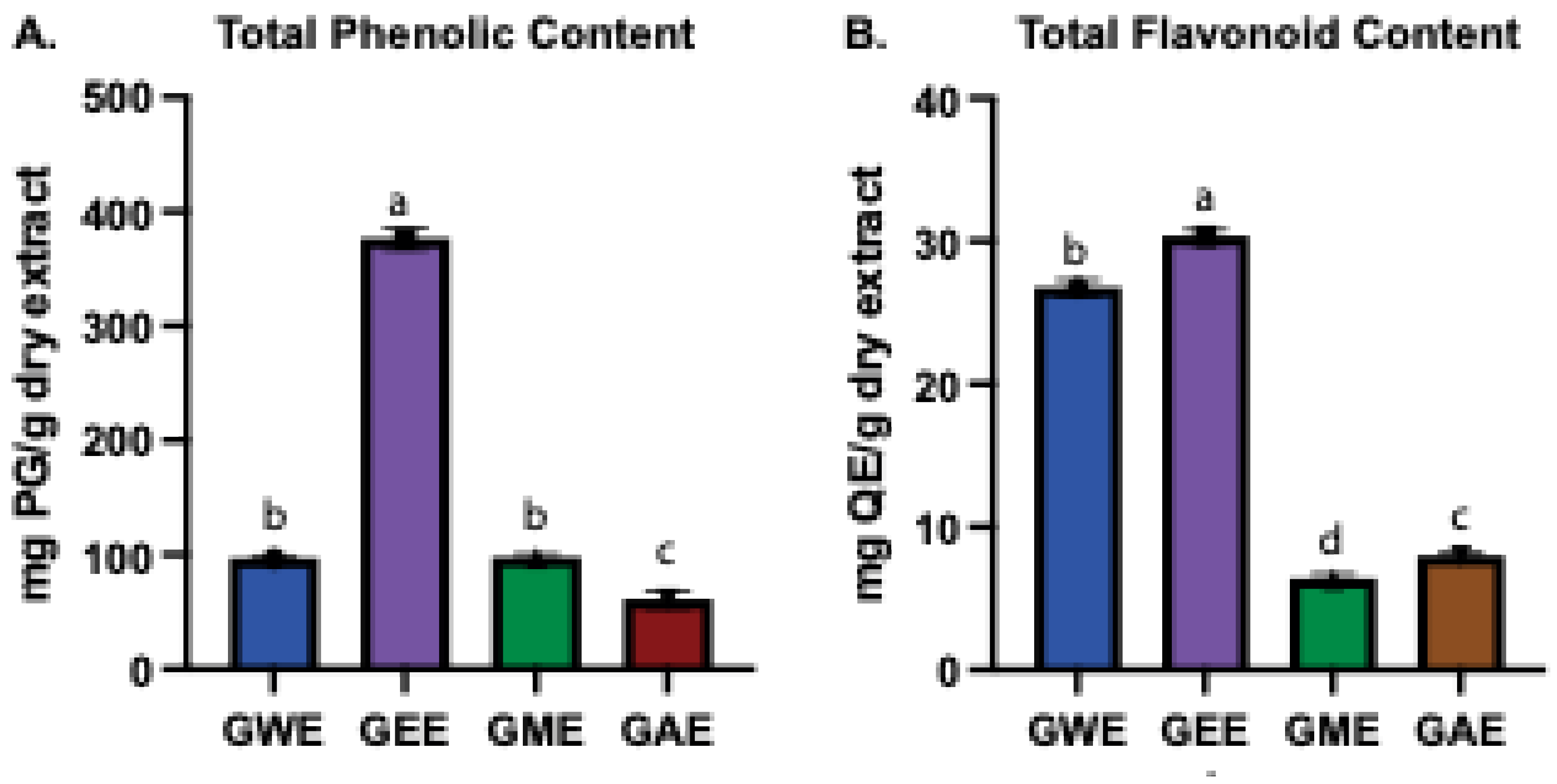
Figure 4.
β-carotene (A) and lycopene (B) content as mg/100 mL extract in various solvent extract. Values with the same letter (a-d) are not significantly different; different letters indicate significant differences between solvents (p < 0.05).
Figure 4.
β-carotene (A) and lycopene (B) content as mg/100 mL extract in various solvent extract. Values with the same letter (a-d) are not significantly different; different letters indicate significant differences between solvents (p < 0.05).
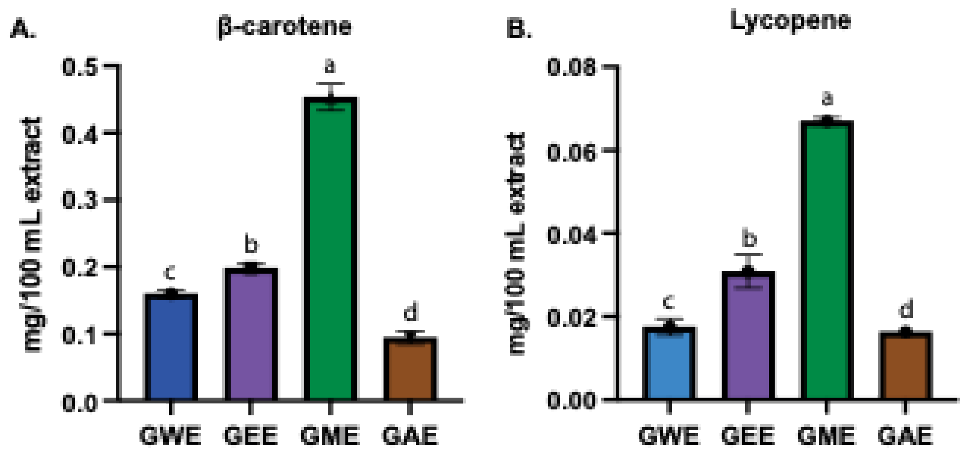
Figure 5.
DPPH radical scavenging activity (%) of G. lucidum extracts prepared with four different solvents, compared to the standard antioxidant ascorbic acid.
Figure 5.
DPPH radical scavenging activity (%) of G. lucidum extracts prepared with four different solvents, compared to the standard antioxidant ascorbic acid.
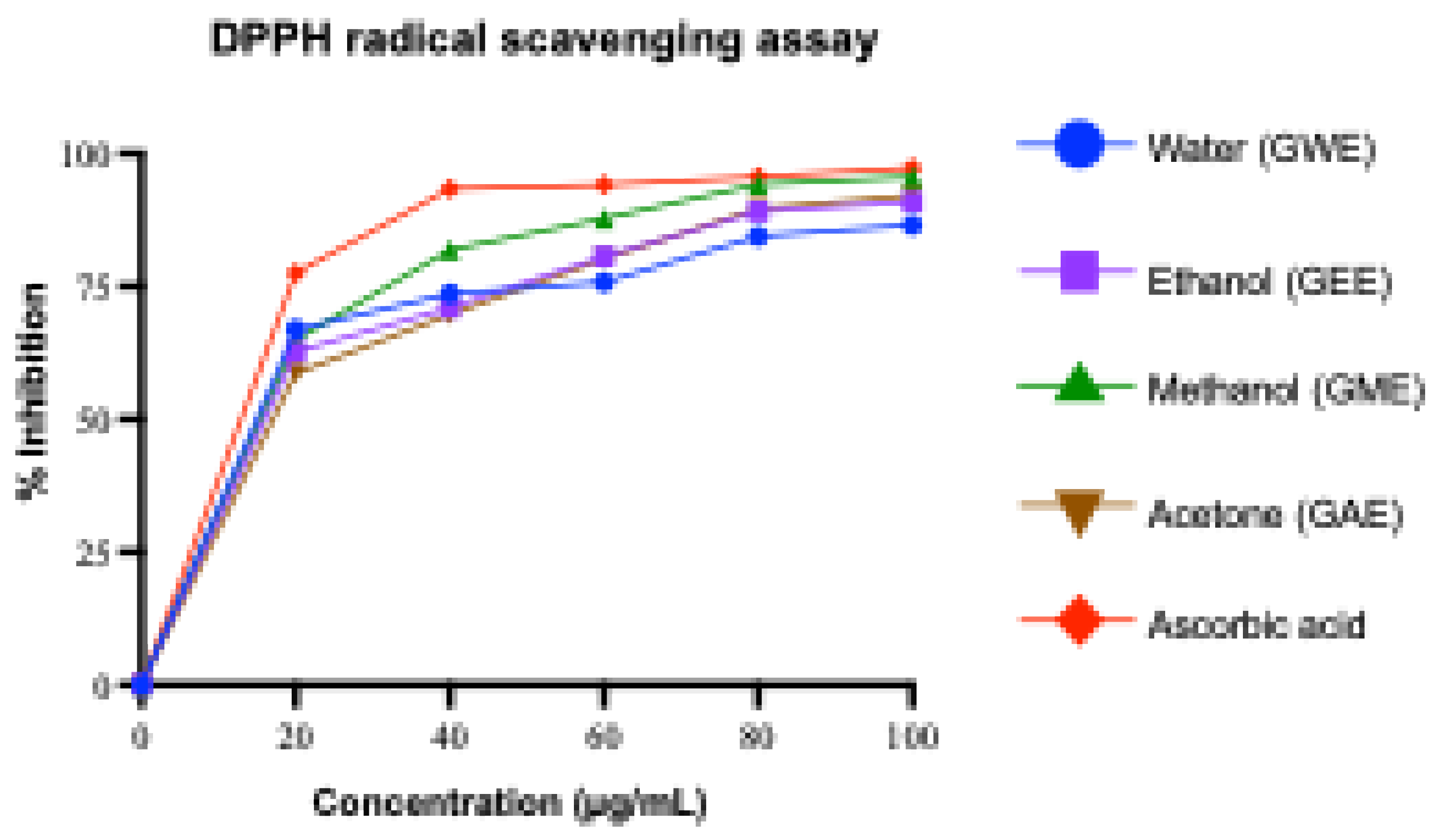
Figure 6.
Superoxide radical scavenging activity (%) of G. lucidum extracts prepared with four different solvents, compared to the standard antioxidant ascorbic acid. .
Figure 6.
Superoxide radical scavenging activity (%) of G. lucidum extracts prepared with four different solvents, compared to the standard antioxidant ascorbic acid. .
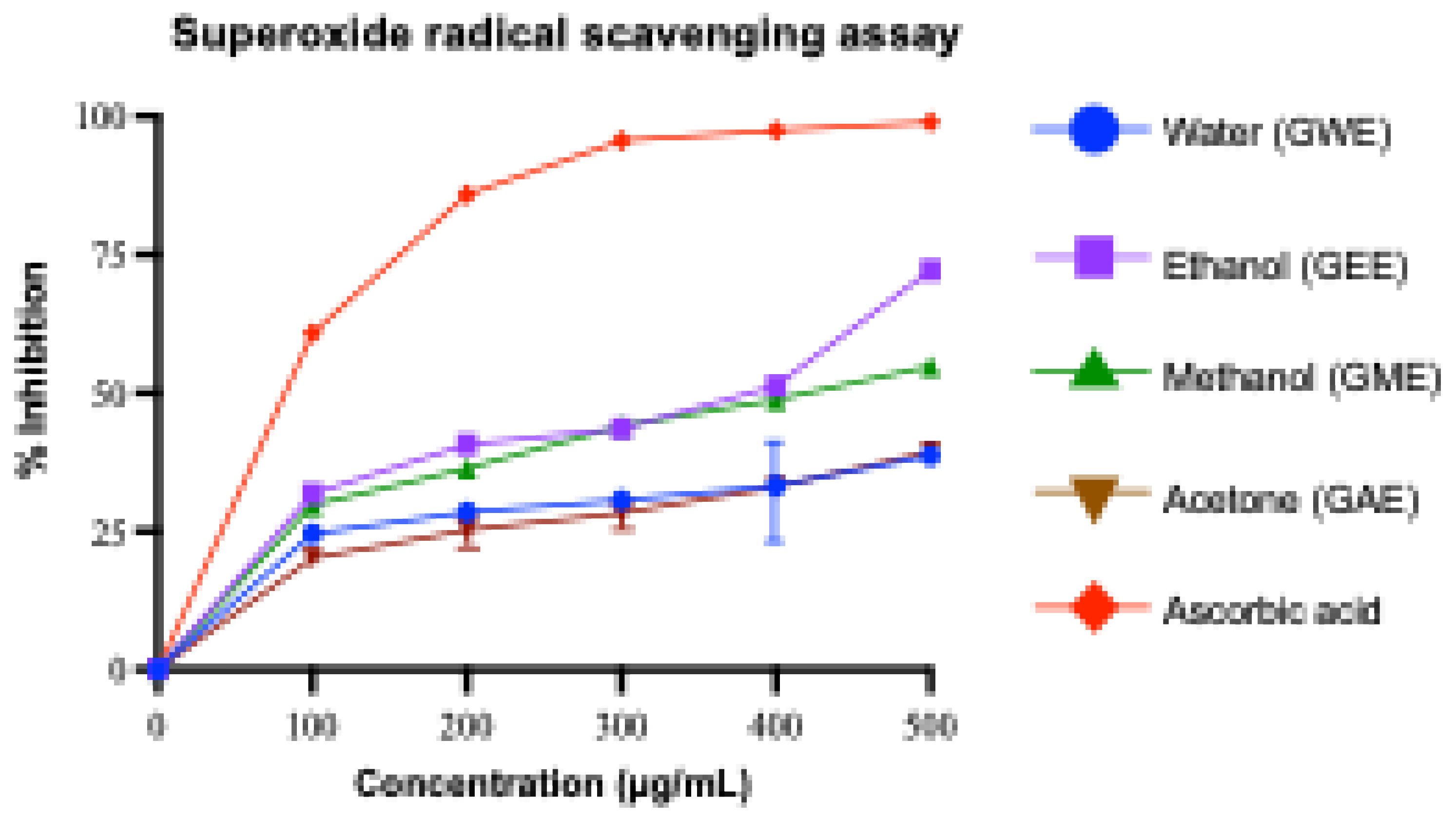
Figure 7.
Hydroxyl radical scavenging activity (%) of G. lucidum extracts prepared with four different solvents, compared to the standard antioxidant ascorbic acid.
Figure 7.
Hydroxyl radical scavenging activity (%) of G. lucidum extracts prepared with four different solvents, compared to the standard antioxidant ascorbic acid.

Figure 8.
Nitric oxide radical scavenging activity (%) of G. lucidum extracts prepared with four different solvents, compared to the standard antioxidant ascorbic acid. .
Figure 8.
Nitric oxide radical scavenging activity (%) of G. lucidum extracts prepared with four different solvents, compared to the standard antioxidant ascorbic acid. .
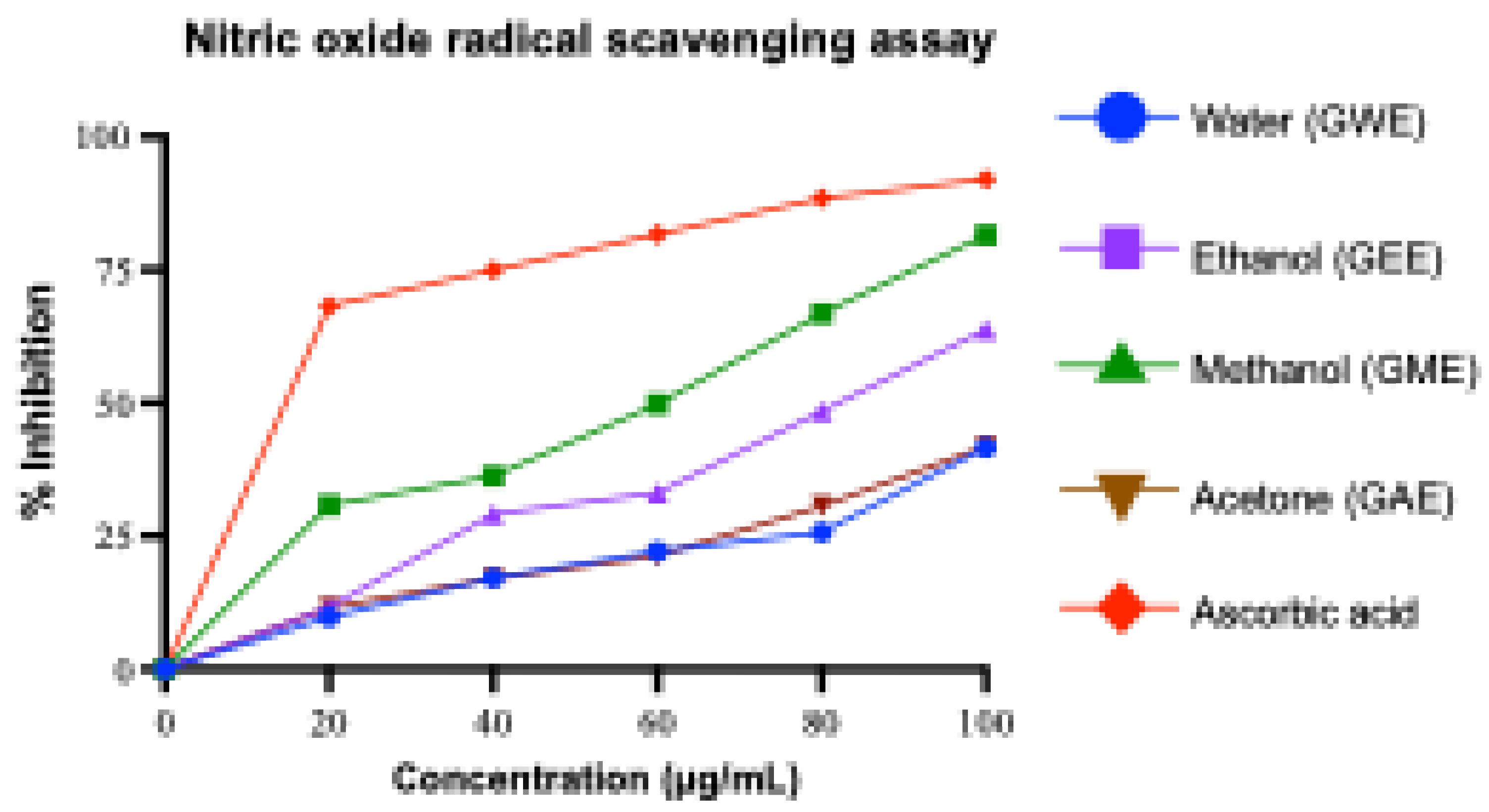
Figure 9.
Reducing power assay of G. lucidum extracts prepared with four different solvents, compared to the standard antioxidant ascorbic acid.
Figure 9.
Reducing power assay of G. lucidum extracts prepared with four different solvents, compared to the standard antioxidant ascorbic acid.
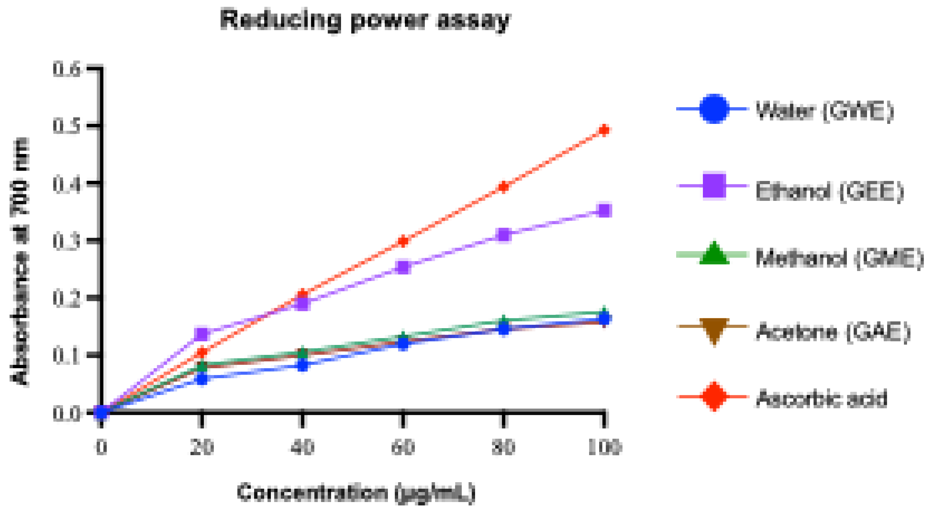
Figure 10.
Cytotoxic activity of G. lucidum extracts against HeLa cervical cancer cell lines at varying concentration. For each concentration, values with the same letter (a-e) are not significantly different; different letters indicate significant differences between solvent extracts (p < 0.05).
Figure 10.
Cytotoxic activity of G. lucidum extracts against HeLa cervical cancer cell lines at varying concentration. For each concentration, values with the same letter (a-e) are not significantly different; different letters indicate significant differences between solvent extracts (p < 0.05).
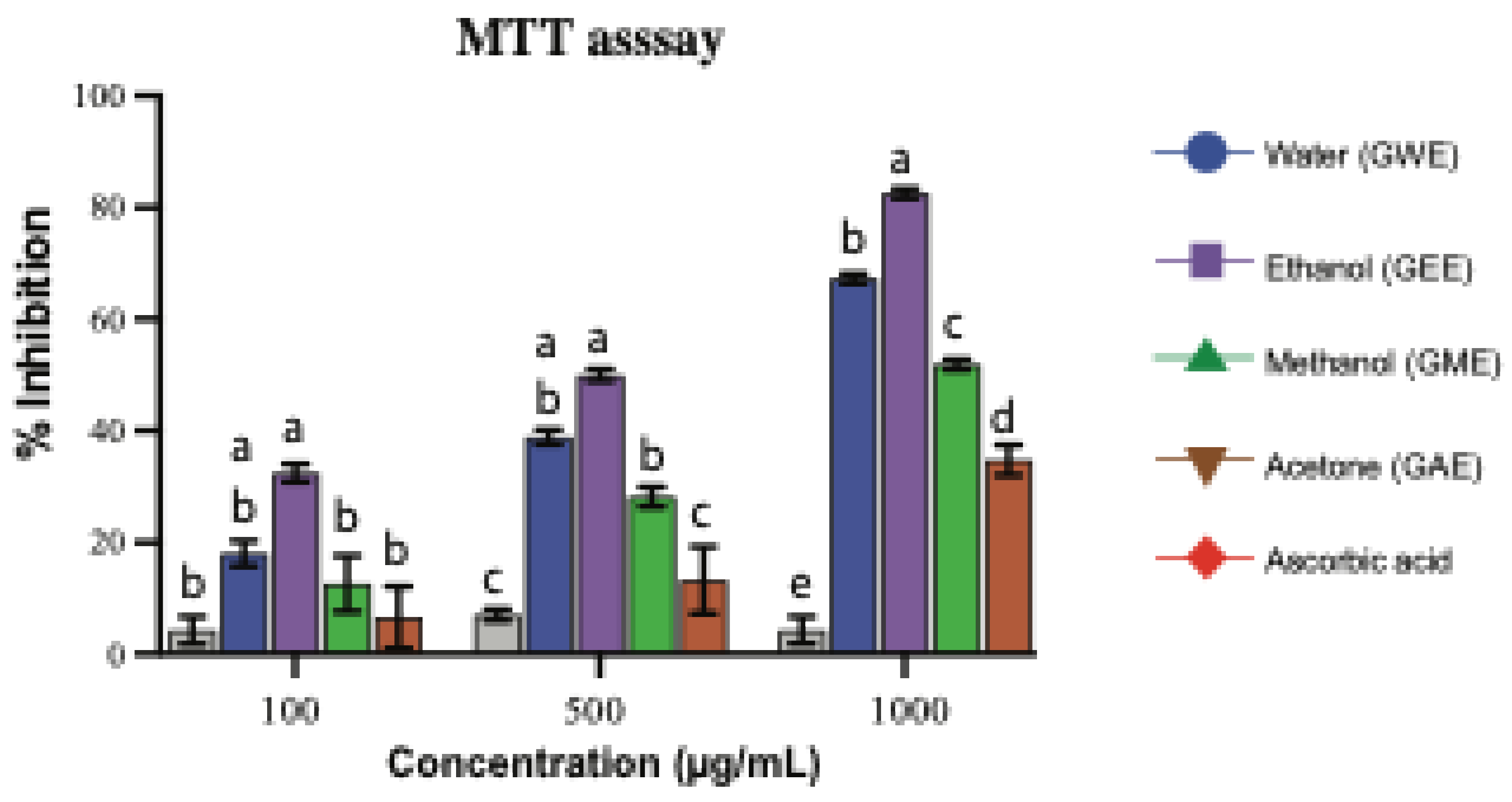
Figure 11.
Heatmap of IC50 values obtained from various antioxidant and cytotoxic assays. Values are expressed in µg/mL. Within each assay, values with the same letter (a-e) are not significantly different; however, different letters indicate significant differences between solvents (p < 0.05). Ascorbic acid: AA.
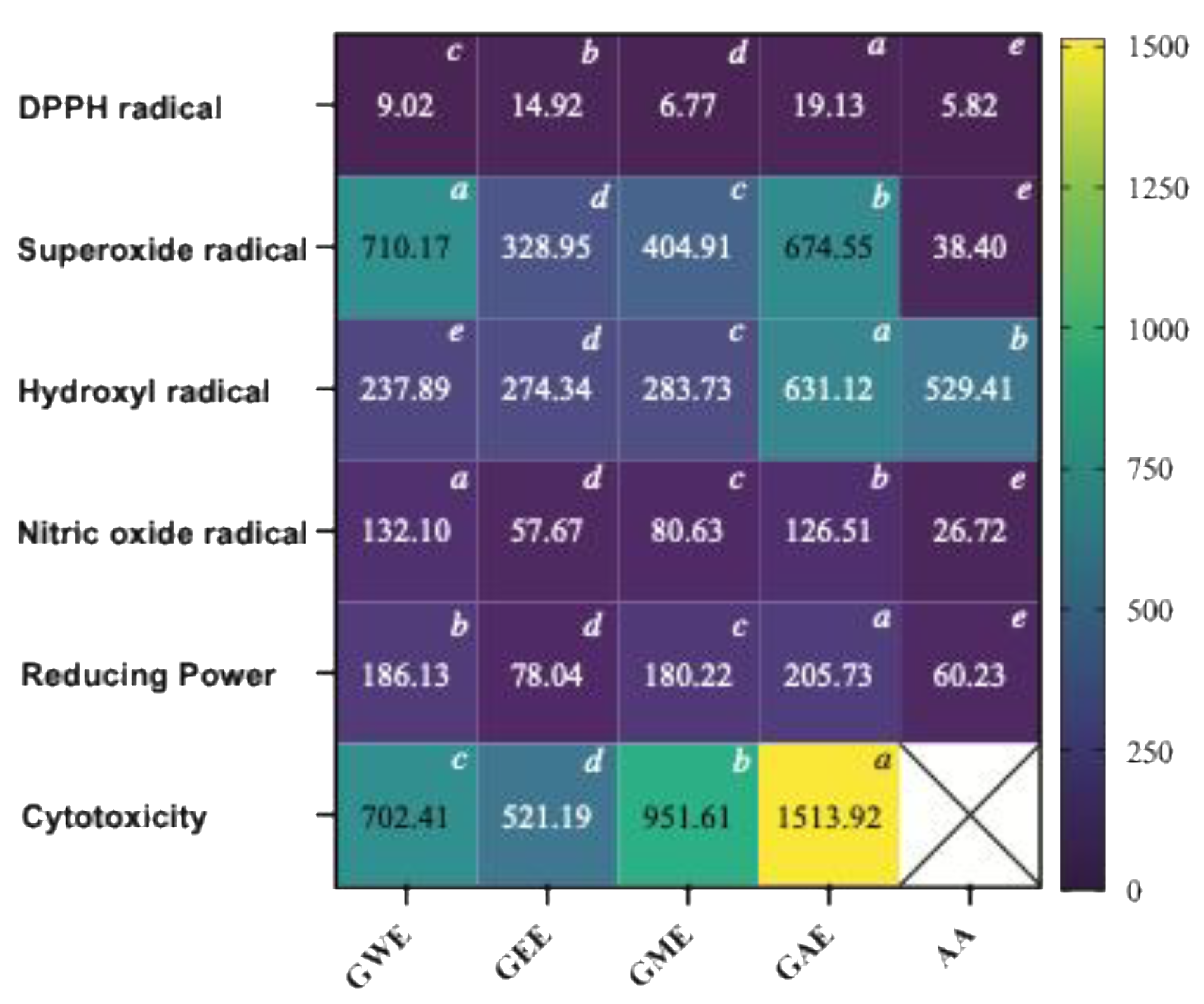

Table 1.
Summary of the ecological, morphological, anatomical, and taxonomic characteristics of G. lucidum.
Table 1.
Summary of the ecological, morphological, anatomical, and taxonomic characteristics of G. lucidum.
| Parameter | Description |
|---|---|
| Collection month | September–October |
| Location | Chandragiri Hill, Kathmandu, Nepal |
| Elevation | 7482 ft (2280 m) above sea level |
| Coordinates | Latitude: 27.67402° N; Longitude: 85.19874° E |
| Ecosystem type | Solitary |
| Substrate | Wood, stump, log, stick, base of tree, bark |
| Host tree | Quercus lanata |
| Rot type | White-rot |
| Surrounding trees (20 ft radius) | Predominantly hardwoods |
| Basidiocarp size Texture |
7–12 × 11–19 × 1.5 cm Woody to corky |
| Stipe | Sub-sessile to laterally stipitate, 2–3 cm |
| Pileus shape | Reniform |
| Upper surface | Laccate, dark reddish to purplish, yellowish at margins; brittle, soft |
| Margin | Blunt, rounded, brown-white |
| Pore surface | Creamy to milky coffee; ~5 pores/mm |
| Tube layer | 2–9 mm long, white turning brown when brushed or aged |
| Context | 9 mm thick, brown, without horny deposition |
| Cutis type | Thick-walled claviform with diverticula; 35–42 × 6–8.5 µm |
| Hyphal system | Trimitic: Generative (3.3 µm, hyaline, thin-walled, with clamp); Skeletal (5.8–7.5 µm, brown, thick); Binding (5–7.5 µm, brown) |
| Basidiospores | 8.3–10 × 6.6 µm; yellowish-brown |
| Identification authority |
Prof. Mahesh Kumar Adhikari, Dept. of Plant Resources, Kathmandu |
Table 2.
Percentage yield of various solvent extracts.
| Extract | Weight of sample before extraction (gm) | Weight obtained after extraction (gm) | % Yield value |
| Water | 10 | 0.229 | 2.29 d |
| Ethanol | 10 | 0.343 | 3.43 b |
| Methanol | 10 | 0.298 | 2.98 c |
| Acetone | 10 | 0.501 | 5.01 a |
Values with the same letter (a-d) are not significantly different; different letters indicate significant differences between solvents (p < 0.05).
Table 3.
Summary of key compounds detected by the GC-MS analysis in various solvents and their reported pharmacological relevance.
Table 3.
Summary of key compounds detected by the GC-MS analysis in various solvents and their reported pharmacological relevance.
| Compound Name | Solvent Extracts (% area) | Compound class | Key pharmacological relevance | Reference | ||
| GEE | GME | GAE | ||||
| 7,22-Ergostadienone | 3.54 | 2.90 | 2.55 | Sterol | Antithrombotic activity with cardiovascular benefit; antidiabetic, anticancer, and neuroprotective effects; Pro-inflammatory properties (activating Toll-like receptors, cytokines, and chemokines) | [51,54,55,56,57] |
| 9(11)-Dehydroergosteryl 3,5-dinitrobenzoate | 2.90 | 3.13 | 2.70 | Sterol conjugate | Anti-inflammatory; antibacterial (MRSA and S. aureus); and cytotoxic properties | [58,59] |
| δ-Tocopherol | 2.13 | 3.91 | 0.75 | Tocopherol | Antioxidant; anti-inflammatory (primarily via inhibiting protein kinase C and reducing eicosanoid production); anticancer (both in vitro and in vivo prostate xenograft models); cvardiovascular and neuroprotective | [60,61] |
| 4-[5-(2-bromophenyl)-1,2,4-oxadiazol-3-yl]-1,2,5-oxadiazol-3-amine | - | - | 0.35 | Synthetic heterocycle | Anticancer (potentially via targeting Topoisomerase II relaxation activity); antibacterial; anti-inflammatory; analgesic properties; antioxidant | [62,63,64,65,66] |
| Ergosta-tetraenone | 3.86 | - | 1.67 | Sterol derivative | Anticancer (via G2/M arrest and apoptosis induction); nephroprotection (mitigation of renal damage in mouse model); anti-inflammatory | [67,68,69] |
| Ergosterol | - | - | 73.99 | Sterol | Vitamin D2 precursor; lipid soluble antioxidant; anticancer effects (cell cycle arrest and modulates Wnt/β-catenin signaling pathway); antimicrobial; antidiabetic; immunomodulatory effects | [70,71,72] |
| Ferruginol | 3.18 | - | - | Abietane diterpene | Anticancer (apoptosis induction in melanoma, prostate, lung, and ovarian cancer cells); neuroprotective (reduces α-synuclein toxicity and restores LTP in Alzheimer’s models); cardioprotective (both invitro and in vivo models); antimicrobial and antiviral | [73,74,75,76,77,78,79,80] |
| Geranylgeraniol | 5.26 | - | 0.89 | Diterpenoid alcohol | Anti-inflammatory (NF-κB inhibition; ↓ IL-1β, TNF-α, IL-6, COX-2); pain relief; bone and muscle support (muscle regeneration and prevents bisphosphonate-related bone damage); antimicrobial activity; hormonal balance; glucose homeostasis | [81,82,83,84] |
| Hinokione | 2.9 | 5.5 | 0.9 | Abietane diterpene | Anticancer; anti-inflammatory; hypoglycemic & β-Cell regenerative properties (promotes β-cell differentiation and improved glycemia in zebrafish); antibacterial; antioxidant | [85,86,87] |
| Nerolidol acetate | - | 1.70 | - | Sesquiterpene ester | Anticancer; anti-inflammatory; neuroprotective; antimicrobial; antifungal; antioxidant | [88,89,90] |
| Retinoic acid | - | - | 0.50 | Retinoid | Acne and photoaging (promotes cell differentiation and skin repair); anti-cancer (induces differentiation of malignant promyelocytes in acute promyeloid leukemia); neuroprotective | [91,92,93] |
Disclaimer/Publisher’s Note: The statements, opinions and data contained in all publications are solely those of the individual author(s) and contributor(s) and not of MDPI and/or the editor(s). MDPI and/or the editor(s) disclaim responsibility for any injury to people or property resulting from any ideas, methods, instructions or products referred to in the content. |
© 2025 by the authors. Licensee MDPI, Basel, Switzerland. This article is an open access article distributed under the terms and conditions of the Creative Commons Attribution (CC BY) license (http://creativecommons.org/licenses/by/4.0/).
Copyright: This open access article is published under a Creative Commons CC BY 4.0 license, which permit the free download, distribution, and reuse, provided that the author and preprint are cited in any reuse.


