Submitted:
23 June 2025
Posted:
26 June 2025
Read the latest preprint version here
Abstract
What is known today as Koch’s triangle (the triangular locus of the atrioventricular node) is delimited by the fibrous attachment of septal tricuspid leaflet, aperture of the coronary sinus and the tendon linking the inferior vena cava valve (Eustachian) and central fibrous body. It was a construct of several exceptional anatomists: Francesco Todaro (Italy), Sunao Tawara (Japan), Arthur keith (United Kingdom), Walter Koch (Germany) and Julius Tandler (Austria). The contributions of Arthur Keith in this topic have been particularly overlooked.It is shown here the development of the concept, and why the eponym Koch it is not fair due to the significant contributions of several anatomists. The triangular locus of atrioventricular node or triangle of atrioventricular node are more instructive and impartial names (Ogden, 2007; Woywodt and Matteson, 2007).
Keywords:
Atrioventricular node
; Koch’s triangle
; Atrioventricular conduction system
; Triangular locus of atrioventricular node
; Triangle of atrioventricular node
; Heart anatomy
Introduction
What is known today as Koch’s triangle (the triangle of the atrioventricular node) is delimited by the fibrous attachment of septal tricuspid leaflet, aperture of the coronary sinus and the tendon linking the inferior vena cava valve ( Eustachian) and central fibrous body (Keith, 1906; Koch, 1909; Tandler, 1913; James, 1999; Drenckham, 2004; Stranding, 2016). It was a construct developed by exceptional anatomists. They were Francesco Todaro ( February 14 th, 1839 in Tripi ,Messina ; October 22 nd, 1918 Rome ) , Sunao Tawara (July 5th, 1873 Nakajima, in Aki -Ōita; January 19th,, 1952 Nakatsu) , Ludwig Aschoff (January 10 th , 1866 in Berlin; 24. June 1942 in Freiburg ,Breisgau), Arthur Keith (February 5th, 1866 in Old Machar, Aberdeenshire ;January 7th, 1955 Downe, Kent) and Carl Eduard Walter Koch ( 3rd. Mai 1880 in Dortmund; 21th November 1962 in Gretesch bei Osnabrück) . Julius Tandler ( February 16 1869 in Iglau/Mähren; August 25 1936 in Moscow) gave the name” Dreieck”, (triangle), focusing on Koch’s contributions (Tandler, 1913). Selected refences on the biographies of these scientists are: Francesco Todaro (Ottaviani, 2019), Ludwig Aschoff (Fye, 1999), Sunao Tawara (Suma, 2001; De Almeida et al., 2020; De Almeida, 2023), Arthur Keith (Keith, 1942; Clark, 1955), Walter Koch (James, 1999; Conti, 2011), J Tandler (Kyle and Shampo, 1990).
It is shown how the contributions of all these authors progressed to this important macro and microscopic concept, the locus of atrioventricular node, and why Koch’s triangle is not a fair eponym and should be replaced by a more informative and unbiased term: atrioventricular node triangle or triangular locus of the atrioventricular node. Previously, the late Thomas James (Ottaviani, 2011) have criticized the soundness of this eponym and ,controversially, the utility of the concept (James, 1999). The utility of the concept, the locus of atrioventricular node, since then has been largely confirmed (Mori et al., 2015). However, the criticisms of Thomas James about the soundness of the eponym although right, lacks a clear temporal description of the events culminating in the concept and two important gaps: the contributions of Tawara, not only based in his thesis (Tawara, 1906a, 2000), and Arthur Keith. In particular, the contributions of Arthur Keith on this , so far as I know have not been considered except by a brief mention given in a recent paper (De Almeida, 2023).
The announcement of the Knoten (node) and its place.
In a congress of the German physiological society held in Marburg, 14-16 June of 1905 the Knoten (node) was announced for the first time as a structure confined in the atrial portion of the atrio ventricular bundle (Aschoff, 1905) . Ludwig Aschoff , Tawara’s mentor and then head of the anatomic pathological institute at Marburg, Germany, gave a succinct but clear summary of Tarawa’s, in course, investigation and presented microscopic preparations produced by Tawara (Aschoff, 1905). A significant excerpt from Aschoff’ seminar is selected: “Atrial fibers first appear as a closed bundle at the coronary sinus. They then run anteriorly along the atrial septum to the area of the insertion of the medial tricuspid leaflet, where they form a tangled network and then, together with the plexus from which the fibers of the ventricular segment arise, form the so-called node. This lies as a flat, ridged structure in the fibrous septum between the tricuspid and mitral insertions, and from it the ventricular fibers pass anteriorly through the fibrous septum in an easily traceable bundle to the ventricular septum”(Aschoff, 1905). This communication was published in September 26th in the Muenchener Medizinische wochenscrift ( Munich Medical Weekly) (Aschoff 1905). Tawara himself had produced an account of his almost finished doctoral thesis : ”Die topographie und histologie der brückenfasern “ (The topography and histology of the bridge fibers ) received by the Zentrallblatt fur Physiologie ( key papers in Physiology) in April 17th 1905 and published at the end of January 1906 (Tawara, 1906b) . That was not a brief communication but a full seven pages paper without illustrations. Tawara anticipated much of the material that would be further developed in his 1906 monography (Tawara, 1906a), which has now an English translation (Tawara, 2000). Tawara stressed in that paper: “So far, I have only been able to identify one center that has been precisely characterized histologically, and which, in my opinion, has its center in a mesh-like node in the atrial septum, directly above the atrioventricular fibrous septum. From this node, a fiber bundle runs backward, often to the anterior border of the coronary sinus, and a second fiber bundle runs anteriorly downward through the atrioventricular fibrous septum, where it continues with the system of Purkinje fibers …”. Tawara mentioned as well that the system is present in birds besides mammals. He particularly stressed that the atrial node is fully irrigated and defined its microscopic aspects: “These fibers are characterized by various features: ...they are narrower than the remaining fibers of the atrial muscle and are very rich in nuclei and exhibit slight fibrillar differentiation, limited only to the peripheral layer. The protoplasm of these fibers always stains a different color than the other muscle fibers. In their imperfect development, they resemble embryonic muscle fibers, but can already be clearly distinguished from the rest of the muscles in the newborn “. Tawara (Tawara, 1906c) , on second half of February , also published the histologic analysis of the Hering’s (Henrich Ewald Hering (May 3rd, 1866 Vienna; December 16th, 1948 Papenhusen)) material on the bundle transection (Hering, 1905; Tawara, 1906c) (Figure 1). In this publication he depicted the position of the node and the bundle. These would be further improved in his monography. Previously, Hering (Hering, 1905) had provided macroscopic photographs illustrating in the real hearts where the bundle should be sectioned to produce atrio- ventricular dissociation ( Figure 2)
The Development of the Concept of a Triangular Locus
James Mackenzie (April 12th, 1853 Pictonhill Scone; January 25th, 1925 London) at Burnley read the Aschoff’s report (Aschoff, 1905) and communicated it to Arthur Keith who was struggling without success to reproduce His junior’s (His, 1949) (Wilhelm His Jr (December 29th,1863 Basel; November 10th, 1934 Brombach- Lörrach)) and Hering’s (Hering, 1905) results on the atrioventricular bundle. Arthur Keith afterwards sent in February 3rd 1906 a letter to The Lancet that was published in March 3rd 1906 (Keith, 1906) . Keith exposed that he was indeed preparing a previous letter to The Lancet denying the existence of the bundle: “There may be an error in my observation or in my technique. I simply draw attention to the fact that I have failed to find a muscular bundle in the position described by His and Hering and that before it is accepted as the anatomical basis of Stokes-Adams disease its constancy must be proved. At least it is well to have the existence of the bundle of His verified at the very commencement, so that it may not have a circulation of two centuries before it is called in as false coin. Some days after writing the part of this letter given above Dr. James Mackenzie sent me a cutting from the Muenchner Medizinische Wochenschrift of Sept. 26th, 1905 p. 1904, in which Dr. L. Aschoff gave an account " Ueber die Untersuchungen des Herrn Dr. Tawara, die Bruckenfasern betreffend und Demonstration der zugehorigen mikroskopischen Praparate” (On the investigations of Dr. Tawara, concerning the bridge fibers and demonstration of the associated microscopic preparations) ….With this account to guide me I renewed my search with success” (Keith, 1906) .
It is worthy to recognize that Keith, although reading the description of the node by Aschoff (Aschoff, 1905) overlooked the description of the “Knoten” in his letter , apparently he was more focused in identify the atrioventricular bridging fibers of the atrioventricular bundle (His bundle). Confirming that he wrote ” since it is probable that others may find the initial difficulty I experienced in dealing with this intricate region of the heart I propose to give briefly by the aid of a diagram some of the points which have proved most useful to me”(Figure 3 A). This diagram, as mentioned (De Almeida, 2023), is a first description of the landmarks delimiting in the atrial side a triangular area ( red dotted lines in Figure.3) whose components are known today as Koch’s triangle. Unfortunately, Keith not portrayed the Knoten (node) described by Aschoff’s communication, but only the atrioventricular bundle as described by Aschoff. However, he described the fibrous tendon linking inferior vena cava valve (Eustachian) to central fibrous body as a novelty:” In the musculature beneath the endocardium will be found two fibrous bands (see g, h, in Figureure) which are of great interest but, so far as I know, have never been described. These bands begins in the membranous space and run through the musculature of the septum to the Eustachian (g) and Thebesian (h)…”(Keith, 1906) . Keith was unaware that Francesco Todaro (Todaro, 1895) had described the fibrous structure arising from vena cava inferior valve (Eustachian valve) in 1895 (Figure 4). Todaro’s description is more complete than that of Keith: “I immediately noticed that from the right posterior portal/part of the tendinous center, a fibrous ribbon started; and, having followed it, with great care, among the muscular fibers below, I accompanied it up to the posterior pillar of the oval fossa, from where I made sure that it entered through the posterior horn, which originates here, in the Eustachian valve, following it along its entire free margin. I have confirmed this observation later, in many other cases; and I have always found the tendon constant, even when the Eustachian valve is almost deficient ”(Todaro, 1895). Keith described too the nodal artery: “A large artery (with a caliber of 1 to 1. 5 millimeters) invariably perforates the central fibrous body just behind the bundle ; the artery is always bigger than the muscular bundle at the point of perforation”(Keith, 1906). Nevertheless, Keith described, inside the triangular area, a component which is not today mentioned in the triangular locus of the atrioventricular node: a separate tendon arising from coronary sinus valve. Keith, however could be describing a case with a double Tendon as reported previously (James, 1999).
It is almost certain that Keith read also the Tawara’s paper (Tawara, 1906b) because one description is not present in Aschoff’s paper (Aschoff, 1905). He mentioned in the letter to The Lancet” … as Tawara described, fine, multinucleated cells with indistinct striation; I observed also, as he did, that the fibers were accompanied by numerous nerve fibers, and there are cells, too, which have the appearance of ganglionic nerve cells”. The Keith intention was to give a more didactic presentation of Tawara discoveries. He achieved that but neglected the Knoten(node), on the other side he was the first to add the tendon of inferior vena cava in this area as a useful delimitation (Keith, 1906).
Tawara’s monography “Das Reizleitungssystem des Säugetierherzens. Eine anatomisch-histologische Studie über das Atrioventrikular bündel und die Purkinjeschen Fäden“ (Tawara, 1906a) ( The conduction system of the mammalian heart. An anatomical-histological study of the atrioventricular bundle and Purkinje's fibers.(Tawara, 2000) ) was finished December 24th 1905 at Marburg and published at the beginning of 1906. The Aschoff’s preface was written in March 19th. In that monography he further developed what was announced before by his Mentor and by himself. Tawara first described the node (knoten) in a human heart (No. 136. The two-year-old child heart). The description is given in the section “Topography of the Atrioventricular Connecting System”. Tawara had previously examined ten dog hearts in which an atrial segment of the connecting system is described, a description compatible with the node. The word knoten (node), however, was not mentioned in these dogs’ hearts descriptions but he had drawn it ( dog heart no 166, Figure3 ) Figure 7A. In consequence, Tawara crystalized the concept of the node (Knoten) just when he studied the human heart No 136 (Figure 5); “By examining the staged microscopic sections, a peculiar muscle group is observed in section No. 105, which is entirely different from the atrial musculature in terms of abundancy and form of nuclei as well as in the arrangement of the muscle fibers. The muscle group is located about 1.5 mm below the lowest attachment of the noncoronary aortic leaflet, in other words, at the infero posterior margin of the membranous septum. It adheres to the atrioventricular fibrous septum (s), i.e. to the origin of the aorta. When compared with the adjacent atrial muscle fibers, the fibers are much smaller and less differentiated. The arrangement is extremely irregular, giving the appearance of a complicated glomerulation - I call this site “the node*” (k). In the following sections, this bundle becomes gradually thicker and extends anteriorly. Consequently, the tip of the bundle protrudes into the fibrous septum (section No. 110). When projected to the left endocardial surface, the extension of the bundle occupies the posterior three quarters of the noncoronary aortic leaflet. The muscle fibers connect with the adjacent ordinary atrial muscle fibers (v) posteriorly and at the right hand margin of the node (Figure. 1 = section No. 110.) (Tawara, 2000). Tawara monography provided drawings helping the macroscopic and microscopic localization of the Knoten (Atrioventricular node). (Figure 1A, B; Figure.6A, B) Tawara macroscopic Figureures show the evident relation of the node with the septal tricuspid leaflet and mitral aortic leaflet, coronary sinus ostium, commissure of septal and antero superior tricuspid leaflet, right and non-coronary leaflets, central fibrous body and membranous septum (Figure 1; Figure 6A, B). The inferior vena cava tendon (Todaro’s tendon), however, is not described or mentioned. In some of his histological Figureures he sketched the tendon but he did not make reference to it . Tawara clearly represented the tendon in Figure 120 plate 1. in a dog heart (Figure 7A). and possibly in Figure 139 in a human heart. (Figure 7B). In general, the upper border of the other Figureures are too close to the fibrous skeleton to see the tendon in different cut levels. However, his descriptions, and Figureures of the node and its relation with surroundings structures are completely original and precise. Shortly, Tawara was the first to describe all components of the triangular locus in its relation with the node excluding the inferior vena cava valve tendon. Koch (Koch, 1909)) as Keith described the tendon of vena cava and assembled it with the other structures previously described by Tawara, constructing the triangle . Koch emphasized the presence of the node and atrioventricular bundle in a Figureure whose main objective was to show the advances he obtained in morphology of sinoatrial node (Koch, 1909; Keith and Mackenzie, 1910)( Figure .8) .Indeed, the main objective of the whole paper was to detail his findings in sinoatrial node and discuss its probable function (Koch, 1909) . He wrote “On the other hand, at another location in the entry of the atrial cavity, more anteriorly near the upper border of the right atrial appendage, there is a peculiar muscular system discovered by Keith and described in more detail by me in a previously published work, which in many respects can be considered parallel to the atrial part of the conduction system between the atrium and the ventricle. I was able to demonstrate it at the time, at the border between the atrium and the cava in the sulcus already discussed, beginning approximately at the upper edge of the atrial appendage. As recent observations have shown me, it appears to extend along the sulcus, its fibers extending outward and downward almost 1 cm further than I previously stated (2 cm) (Figure. 3), where it splits and disappears upward into the musculature of the cava and downward into the atrial musculature…. I consider the demonstration of this muscular system discovered by Keith to be not unimportant, since with a better understanding of its function, disturbances in it may perhaps be able to be used to explain physiological and pathological cardiac processes.”
Dreieck ( triangle ) is not mentioned nor in this paper neither in his magnum opus (Koch, 1922). Koch did not gave credit to Keith, on this particular aspect, who just emphasized the fibers of the atrial portion of the bundle inside of the triangular locus. Both did not gave credit to Todaro (Todaro, 1895) .Both did not stress or write that they were delimiting a triangular area . Tandler (Tandler, 1913) named for the first time the locus as a Koch’s Dreieck ( triangle) (James, 1999)”:Topographically, the nodule was described almost entirely consistently by all authors, although the relationship to the aortic valves was particularly emphasized. This relationship, at least for locating the nodule, does not seem particularly well chosen, since the nodule is most easily located from the right side of the atrial septum. Koch relocated the nodule into the triangle he described. Koch outlined this triangle as follows: The caudal border is formed by the insertion of the tricuspid artery, the calcaneal border by a fold that forms when the junction of the Eustachian and Thebesian valves is tensed to the right with forceps. The fold then runs obliquely forward and downward toward the membranous septum. In my opinion, it is the expression of the tension of Todaro's tendon, which has already been described elsewhere. The posterior border of this triangular field is represented by the orifice of the coronary sinus. Near the apex of this triangle, as Koch quite rightly points out, lies the node. We already emphasized elsewhere that Koch's deductions, as if this field were originally a sensory area, are not valid for evolutionary reasons. However, Koch's topographical definition is certainly acceptable.”. Tandler forgot also that Tawara had described the main relations of the node in right atrial side.
Limitations of the Study
The author did not intended to be exhaustive or dogmatic in matters of the priority of the discoveries. As exposed here Keith and Koch have rediscovered the findings of Todaro and we cannot affirm with absolute certainty that Todaro was the first to describe the tendon of the inferior vena cava valve. These does not invalidate the thesis: to know, that the triangular locus of the atrioventricular node was a collective construct. To prove it we just need one clear and logical sequence of temporally sound events as shown here.
Conclusion
In summary the triangular locus of the atrioventricular node was a collective construct as the majority of human scientific endeavors(Ogden, 2007; Woywodt and Matteson, 2007).
Conflict of interest: There is no conflict of interest.
Acknowledgments: Many thanks to the librarians of Free University of Berlin who gently sent me the paper of Koch W (1909) Weiter Mitteilungen über den Sinusknoten des Herzens. . Verh Deutscth Ges Pathol 13:85–92
Declaration: the author have used as an auxiliary tool for the translation of German original texts , in print and online German-English dictionaries as well as Google translator.
Copyrights: The Figureures shown in this article are free of copyrights.
Ethical issues: The ethical issues are not involved in this article whose main novelty is a revision of an historical error, an eponym widely used in cardiac anatomy.
References
- Almeida MC, De. Heinrich Ewald Hering’s discovery of the heart pacemaker: Hering, Tawara and Aschoff’s search for its morphological basis, the sinoatrial node, and why they failed. Anat Sci Int 2023, 98, 482–492. [Google Scholar] [CrossRef] [PubMed]
- Almeida MC De, Sánchez-Quintana D, Davis N, Charles FR, Chikweto A, Sylvester W, Loukas M, Anderson RH. The ox atrioventricular conduction axis compared to human in relation to the original investigation of sunao tawara. Clin Anat 2020, 33. [Google Scholar]
- Aschoff, L. Ueber die Untersuchungen des Herrn Dr. Tawara, die Bruckenfasern betreffend und Demonstration der zugehorigen mikroskopischen Praparate. Muenchener Medizinische wochenscrift 1905, 52, 674. [Google Scholar]
- Clark, W. Arthur Keith 1866-1955. Biogr Mem Fellows R Soc 1955, 1, 144–151. [Google Scholar]
- Conti, AA. Calling the heart by name: distinguished eponyms in the history of cardiac anatomy. Heart Surg Forum 2011, 14, E183–E187. [Google Scholar] [CrossRef] [PubMed]
- Drenckham, D. 2004. Benninghoff Anatomie. 16th Ed. München: Urban and Fischer. Elsevier.
- Fye, WB. Ludwig Aschoff. Clin Cardiol 1999, 22, 545–546. [Google Scholar] [PubMed]
- Hering, HE. Nachweis, dass das His’sche Uebergangsbündel Vorhof und Kammer des Säugethierherzens functionell verbindet - Zweite Mittheilung. Pflüger, Arch für die Gesammte Physiol des Menschen und der Thiere 1905, 108, 267–280. [Google Scholar] [CrossRef]
- His, W. The activity of the embryonic human heart and its significance for the understanding of the heart movement in the adult (english translation). J Hist Med Allied Sci 1949, 4, 289–318. [Google Scholar] [CrossRef]
- James, TN. The tendons of Todaro and the “Triangle of Koch”: Lessons from eponymous hagiolatry. J Cardiovasc Electrophysiol 1999, 10, 1478–1496. [Google Scholar] [CrossRef]
- Keith, A. The auriculo ventricular bundle of His. Lancet 1906, 167, 623–625. [Google Scholar] [CrossRef]
- Keith, A. The Sino-auricular node: A Historical note. Heart 1942, 4, 77–79. [Google Scholar] [CrossRef]
- Keith A, Mackenzie I. Recent Researches on the anatomy of the Heart. Lancet 1910, 175, 101–103. [Google Scholar] [CrossRef]
- Koch, W. Weiter Mitteilungen über den Sinusknoten des Herzens. . Verh Deutscth Ges Pathol 1909, 13, 85–92. [Google Scholar]
- Koch, W. 1922. Der Funktionelle Bau Des Menschlichen Herzens. 1st Ed. Berlin: Urban & Schwazenberger. 1–146 p.
- Kyle RA, Shampo MA. Professor Julius Tandler, Anatomist and Public Health Advocate. Mayo Clin Proc 1990, 65, 530. [Google Scholar] [CrossRef]
- Mori S, Nishii T, Takaya T, Kashio K, Kasamatsu A, Takamine S, Ito T, Fujiwara S, Kono AK, Hirata KI. Clinical structural anatomy of the inferior pyramidal space reconstructed from the living heart: Three-dimensional visualization using multidetector-row computed tomography. Clin Anat 2015, 28, 878–887. [Google Scholar] [CrossRef]
- Ogden, DJ. Time to drop eponyms: Curbing Medicalese is the issue. BMJ 2007, 335, 528. [Google Scholar] [CrossRef]
- Ottaviani, A. 2019. Francesco Todaro. In: Dizionario Biografico Degli Italiani, Istituto della Enciclopedia Italiana fondata da Giovanni Treccani S.p.A,.
- Ottaviani G., Dr. Thomas N. James, MD, MACP (1925-2010): in memoriam. Am J Cardiol 2011, 108, 330. [Google Scholar] [CrossRef]
- Stranding, S. 2016. Gray`s Anatomy. 41st Ed. Elsevier. 2251 p.
- Suma, K. Sunao Tawara: a father of modern cardiology. Pacing Clin Electrophysiol 2001, 24, 88–96. [Google Scholar] [CrossRef]
- Tandler, J. 1913. Anatomie Des Herzens. 1st Ed. Jena: Gustav Fischer. 279 p.
- Tawara, S. 1906a. Das Reizleitungssystem Des Säugetierherzens. Eine Anatomisch-Histologische Studie Über Das Atrioventrikularbündel Und Die Purkinjeschen Fäden. Jena: Gustav Fischer. 203 p.
- Tawara, S. Die Topographie und Histologie der Brückenfasern. Ein Beitrag zur Lehre von der Bedeutung der Purkinjeschen Fäden. Zentrallblatt fur Physiol 1906, 19, 70–76. [Google Scholar]
- Tawara, S. 1906c. Anatomisch-histologische Nachpriifung der Schnittfuhrung an den von Prof. H.E. Hering ubersandten Hundeherzen. Zentrallblatt fur Physiol 111:300-302.
- Tawara, S. 2000. The Conduction System of the Mammalian Heart: An Anatomico-Histological Study of the Atrioventricular Bundle and the Purkinje Fibers. 1st Englis Ed. London: Imperial College Press. 1–229 p.
- Todaro, F. 1895. Novelle Ricerche Sopra La Struttura Delle Orecchiette Del Cuore Umano e Sopra Ta Valvola Di Eustachio.. Firenze.
- Woywodt A, Matteson E. Should eponyms be abandoned? Yes. BMJ 2007, 335, 424. [Google Scholar] [CrossRef]
Figure 1.
A Representation of the level of experimental transection done by H E Hering in the His bundle of four dog hearts. Published in Tawara, S. 1906a.“ Anatomisch-Histologische Nachprüfung der Schnittfuhrung an den von Prof. H.E. Hering Ubersandten Hundeherzen. Zentrallblatt für Physiologie 111::300–302 “ (Anatomical–histological verification of the incision made by Prof. H.E. Herring on dog hearts”. K = atrioventricular node; S = atrioventricular fibrous septum; W = experimental transection; t = bifurcating bundle; right branch (r) and left branch (ll); B Didactic representation of human His bundle and important reference points for planning the experimental transection. S Tawara 1906. “ Das Reizleitungssystem Des Säugetierherzens. Eine Anatomisch-Histologische Studie Über Das Atrioventrikularbündel Und Die Purkinjeschen Fäden“ (The conduction system of the mammalian heart. An anatomical–histological study of the atrioventricular bundle and Purkinje's network) Jena: Gustav Fischer. Right side view = superior drawing. Left side view = inferior drawing; P = membranous septum; t = bifurcating bundle;right branch (r) and left branch l (ll); X = frontier between atrium and ventricle: m = aortic mitral leaflet; ba –ad = septal and superior tricuspid leaflet position; ef = suggested transection line.
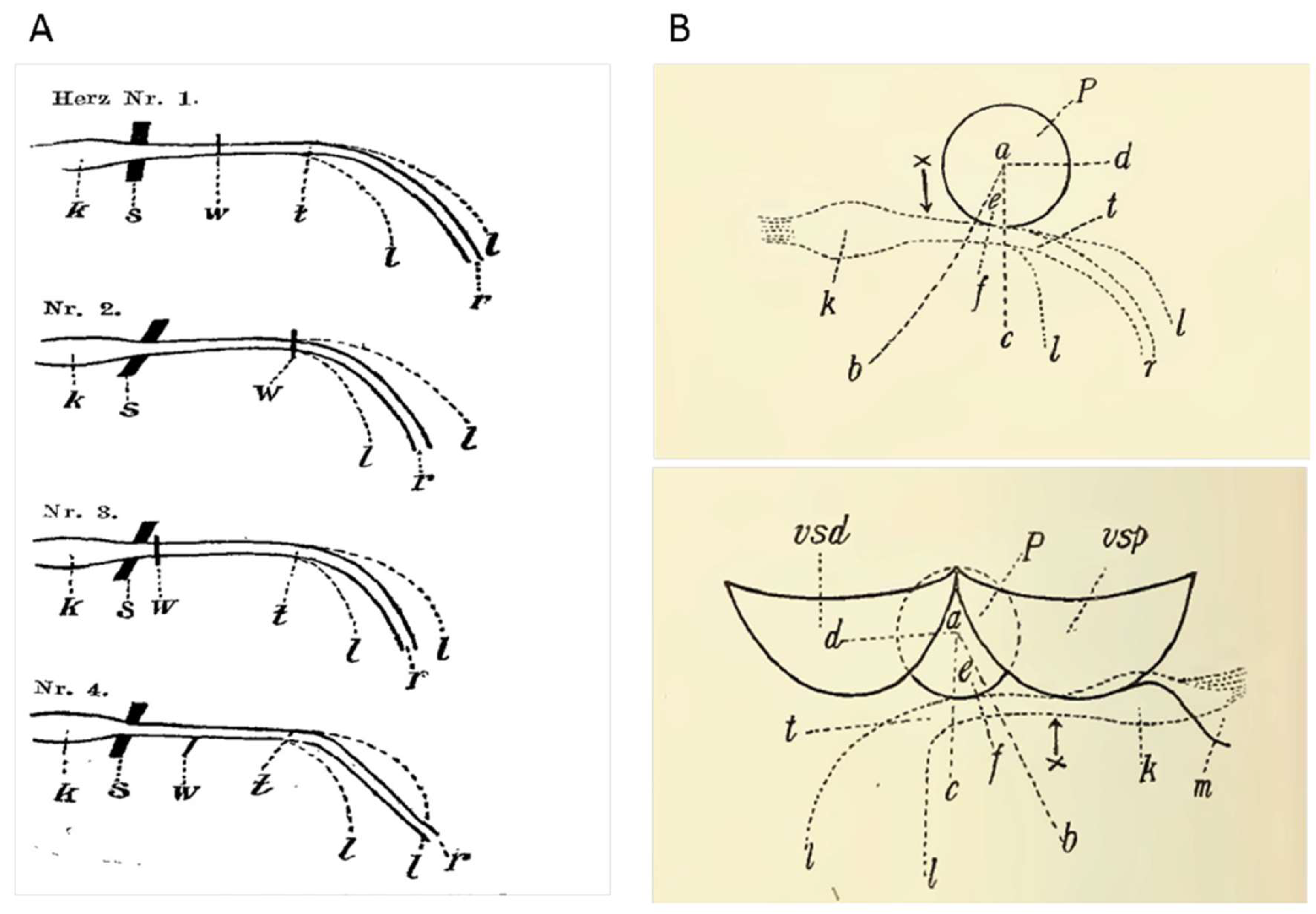
Figure 2.
Cutting through the transition/crossing bundle in the dog's heart. (First experiment on February 21, 1905. ) from H. E. Hering, Hering HE (1905) “ Nachweis, dass das His’sche Uebergangsbündel Vorhof und Kammer des Säugethierherzens functionell verbinde”t (Evidence that the His junctional bundle functionally connects the atrium and ventricle of the mammalian heart ) - Zweite Mittheilung. Pflüger, Arch für die Gesammte Physiol des Menschen und der Thiere 108:267–280.A Right atrium and ventricle opened. AO= aorta; RA= right Atrium; R V= right ventricle; RVAW= right ventricle anterior wall; SP= section point.B Left atrium and ventricle opened. AO= aorta; LV= left ventricle; RAV= right aortic valve; PAV= posterior aortic valve; SP= Section point.
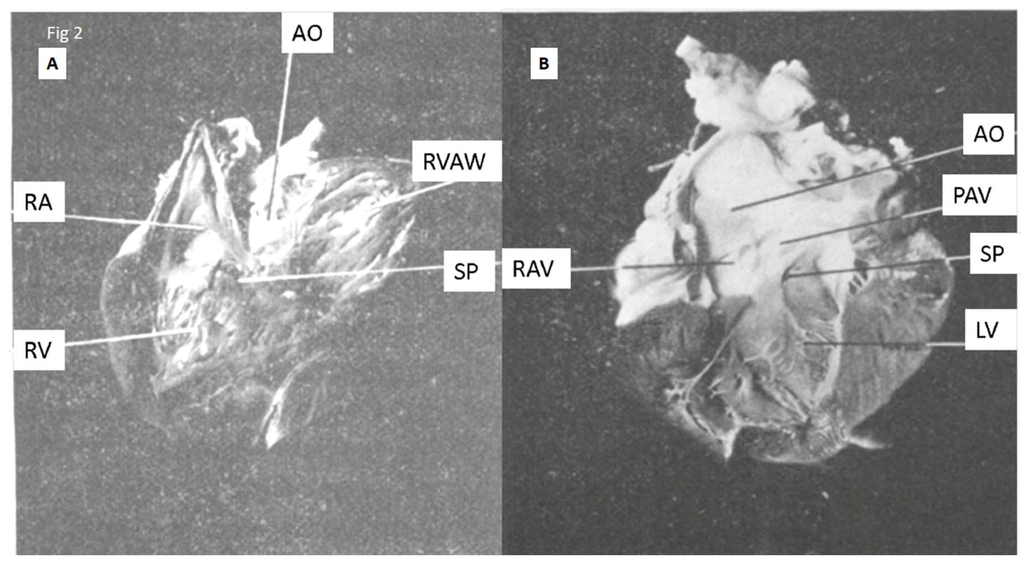
Figure 3.
A. Original drawing of Arthur Keith published in Keith A. 1906 modified by us.. “The auriculo-ventricular bundle of His. Lancet 167:623–625.” He drew it to show the important landmarks to find the His bundle and the atrioventricular node . As far as we know it is the first representation of what is today know as Koch’s triangle. The doted red line triangle was provided by us to emphasize the triangular geometry of the area. The triangle sides are a= attachment of septal cusp of tricuspid leaflet; g= fibrous tendon of Eustachian valve; n= order of coronary sinus ; “ a, Septal cusp of tricuspid. b, Infundibular cusp. c, Ventricular part of membranous space. d, Auricular part of membranous space. e, Position of bundle of His in auricle. f, Position of bundle of His in ventricle. g, Fibrous band of Eustachian valve. h, Fibrous band of Thebesian valve. k, Annulus ovalis. l, Septum ovale. m, Eustachian valve. n, Coronary sinus. o, Thebesian valve. p, Base of right ventricle. q, Base of right ventricle (infundibular part). r, Wall of right auricle. s, Aorta. t, Interventricular septum”. B Figureure published by W. Koch, Koch W (1909) and modified by us “Weiter Mitteilungen über den Sinusknoten des Herzens (Further aspects about the sinus node of the heart) . Verh Deutscth Ges Pathol 13:85–92”. The identification labels were provided by us as well as the dotted red line triangle to emphasize the triangular geometry of the area. The original legend read as : “Schematic drawing of the location of the sinus node (Keith),( black) line at the border between the cava and the auricle , and the atrioventricular node (Tawara),( black) spot above the insertion of the tricuspid septal leaflet. The white line in the atrium demarcates the coronary sinus from the rest of the atrium. (S. Ziegler's Beitr., Vol. XLII, p. 219.)”AO= aorta; APM= anterior papillary muscle: CS= aperture of the coronary sinus; LV= left ventricle; FO= fossa ovalis; SAN=sinoatrial node; ST= septal tricuspid leaflet; VCI= inferior caval vein; VCS= superior caval vein. The whitish tread was posteriorly identified as the Todaro’s tendon. The identification labels were provided by us as well as the dotted red line triangle to emphasize the triangular geometry of the area .
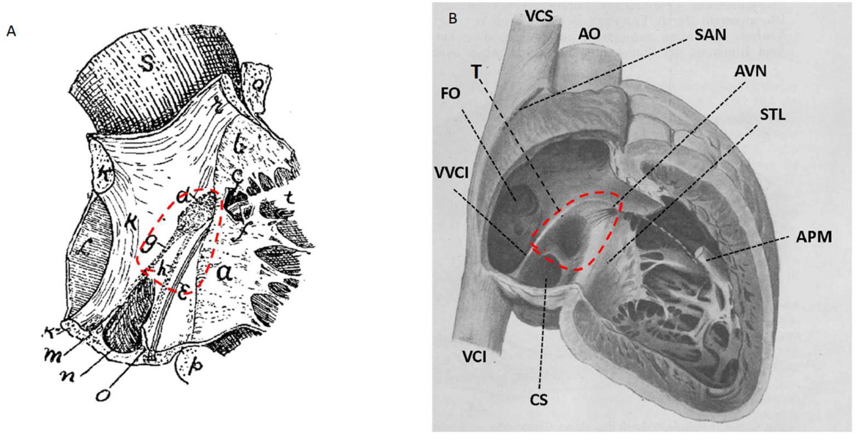
Figure 4.
Anterior/lateral wall of right atrium removed showing the fibrous connexon between inferior vena cava valve and central fibrous body (Todaro’s tendon). From Tandler Tandler J (1913) “Anatomie Des Herzens” (Anatomia do coração), 1st edn. Gustav Fischer, Jena. Aa= aorta; Ct= terminal crest; LV= limbo of fossa oval (ring of Viussens) Raymond Vieussens (born 1641; Vigan; (16Th ; 1715Montpellier ) valve of inferior vena cava; (Eustachian) Bartolomeo Eustachi (San Severino March , 1500-1510; Fossombrone, August 27th 1574) ; VTh= valva do seio coronário (Thebesian) Adam Christian Thebesius (January 1686, in Tschistey, Guhrau, Duchy of Glogau; November 10th, 1732 in Hirschberg, Duchy of Schweidnitz); TT= Tendon of Todaro.
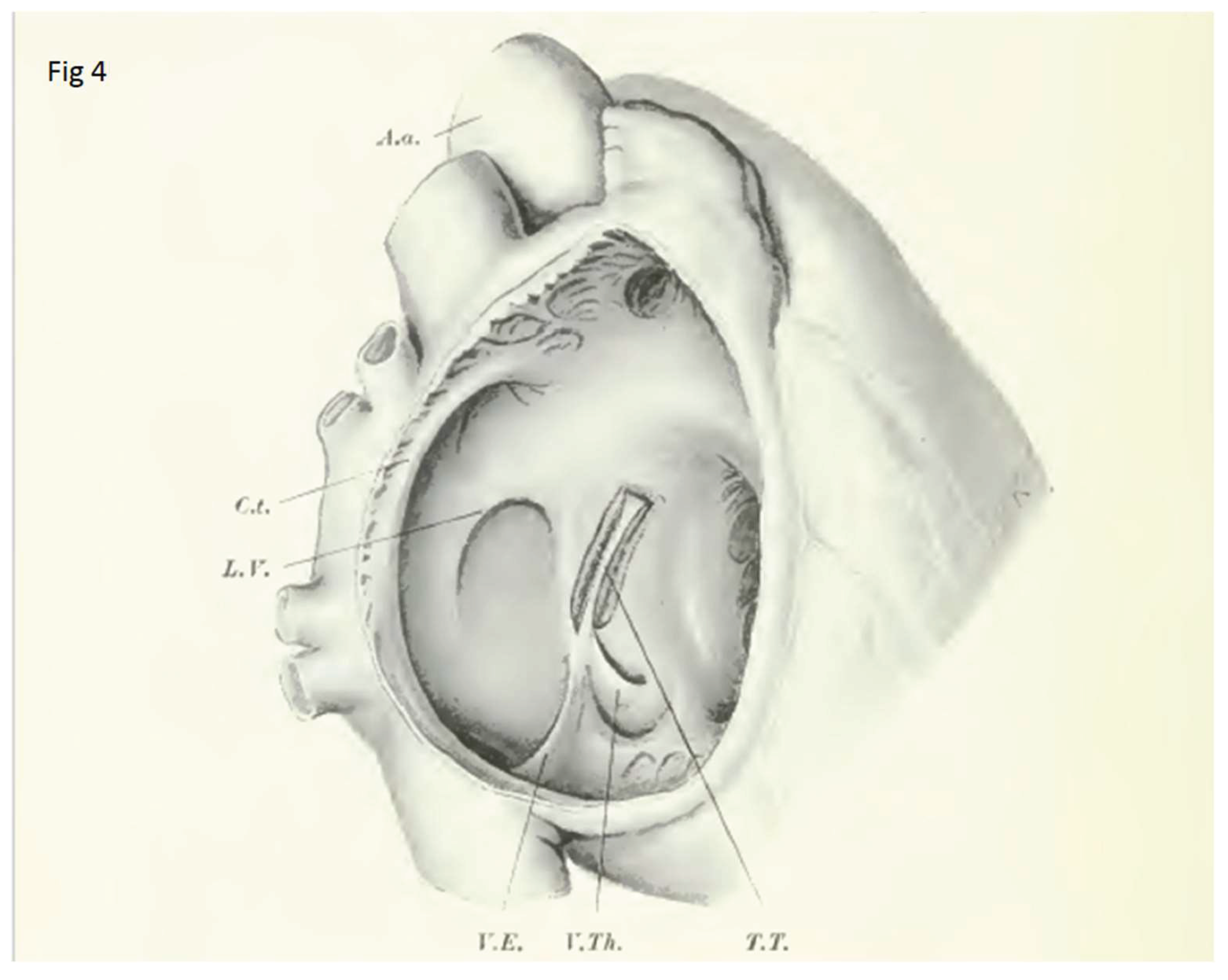
Figure 5.
Sections of Human heart ( No. 136. The two-year-old child heart) from S Tawara 1906 , “Das Reizleitungssystem Des Säugetierherzens. Eine Anatomisch-Histologische Studie Über Das Atrioventrikularbündel Und Die Purkinjeschen Fäden ‘The conduction system of the mammalian heart“(. An anatomical–histological study of the atrioventricular bundle and Purkinje's network.) Jena: Gustav Fischer“ . The upper portion of the septum(from the free edge of the noncoronary aortic leaflet) and the lower portion (from a line about 8 mm below the lowest attachment of the noncoronary aortic leaflet) were cut away parallel to the upper horizontal section line” translation by Kozo Suma& Munehiro Shimada (Tawara 2000 English translation ). The cuts are so represented in vertical position and not in horizontal anatomic position.. “ v = musculature of the atrial septum; s = the atrioventricular fibrous septum; m = anterior mitral leaflet; t = septal leaflet of the tricuspid valve; km = the ventricular septum; h = initial portion of the ventricular bundle of the connecting system;sf = a tendinous fiber for the septal leaflet of the tricuspid valve; 1 & r = the left and the right bundle branch of the connecting system; It = a part of muscle fibers of the left bundle branch entering a tendinous fibe;h = deepest attachment of the noncoronary aortic leaflet;; k = node, i.e. atrial segment of the connecting system” translation by Kozo Suma& Munehiro Shimada ( Tawara 2000 English translation).
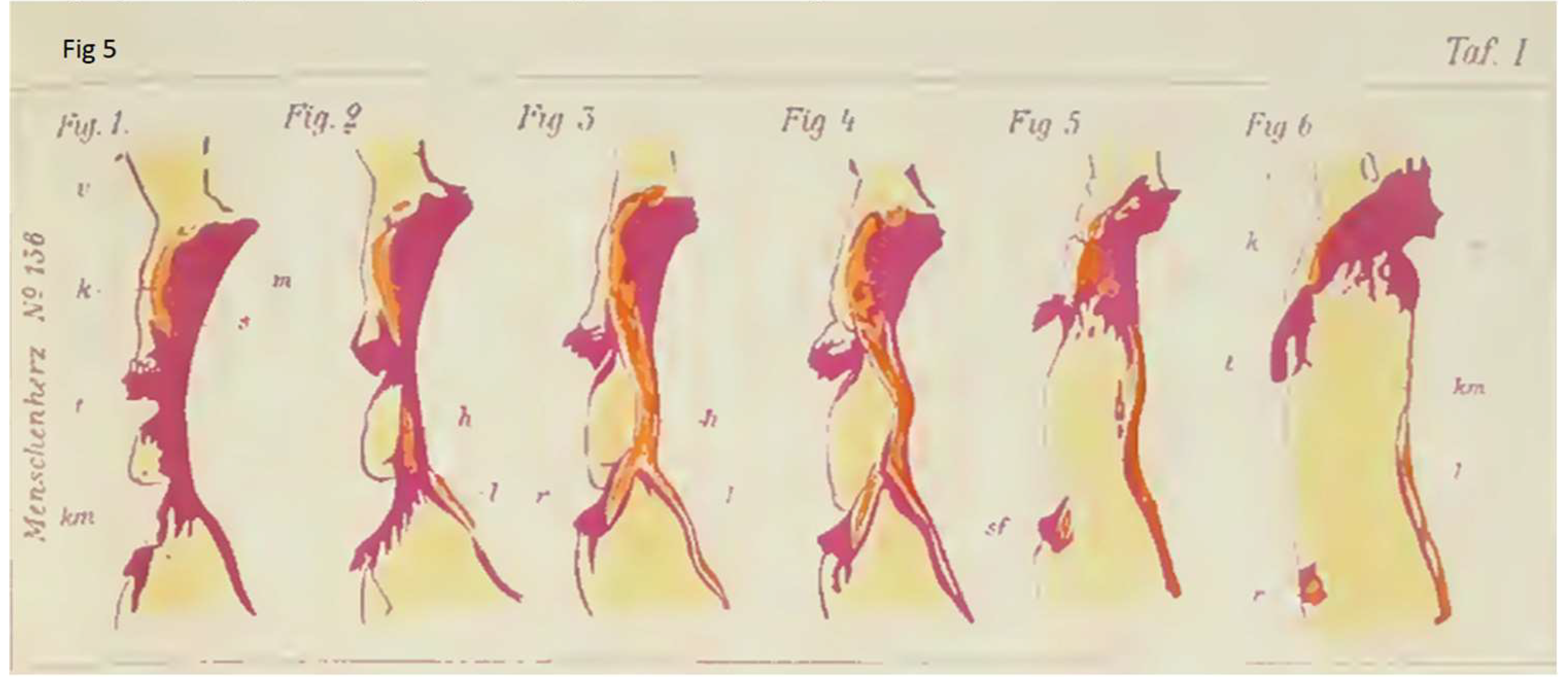
Figure 6.
Human heart opened on right A and left sides B showing the atrioventricular conduction system. ). Modified from S Tawara 1906 “,Das Reizleitungssystem Des Säugetierherzens. Eine Anatomisch-Histologische Studie Über Das Atrioventrikularbündel Und Die Purkinjeschen Fäden”(The conduction system of the mammalian heart. An anatomical–histological study of the atrioventricular bundle and Purkinje's network”’ Jena: Gustav Fischer. “. The heart is positioned in anatomical position and the nomenclature was changed accordingly. O= The white circle was inserted by us to show the localization of membranous septum.A APM= anterior papillary muscle; AVN= atrioventricular node; IVS= interventricular septum; IPM= inferior papillary muscle; ITL= inferior tricuspid leaflet; IW= inferior free wall; PNT= Purkinje network threads; RA= right atrium; RBB= right bundle branch; SATL= supero anterior tricuspid leaflet; SPM= septal papillary muscle; SVC= superior vena cava. O= Localization of membranous septum.B Ao= aorta; AVN= atrioventricular node; ISPM= infero septal papillary muscle; LA= left Atrium; LAA= left Atrium appendage; LBB/H= left bundle branch/bifurcation of his bundle; NCL= noncoronary aortic sinus leaflet; PNT= = Purkinje network threads; RCSL= right coronary aortic sinus leaflet ; SPLM= supero lateral papillary muscle; SPLM*=sectioned part of supero lateral papillary muscle; O= Localization of membranous septum.
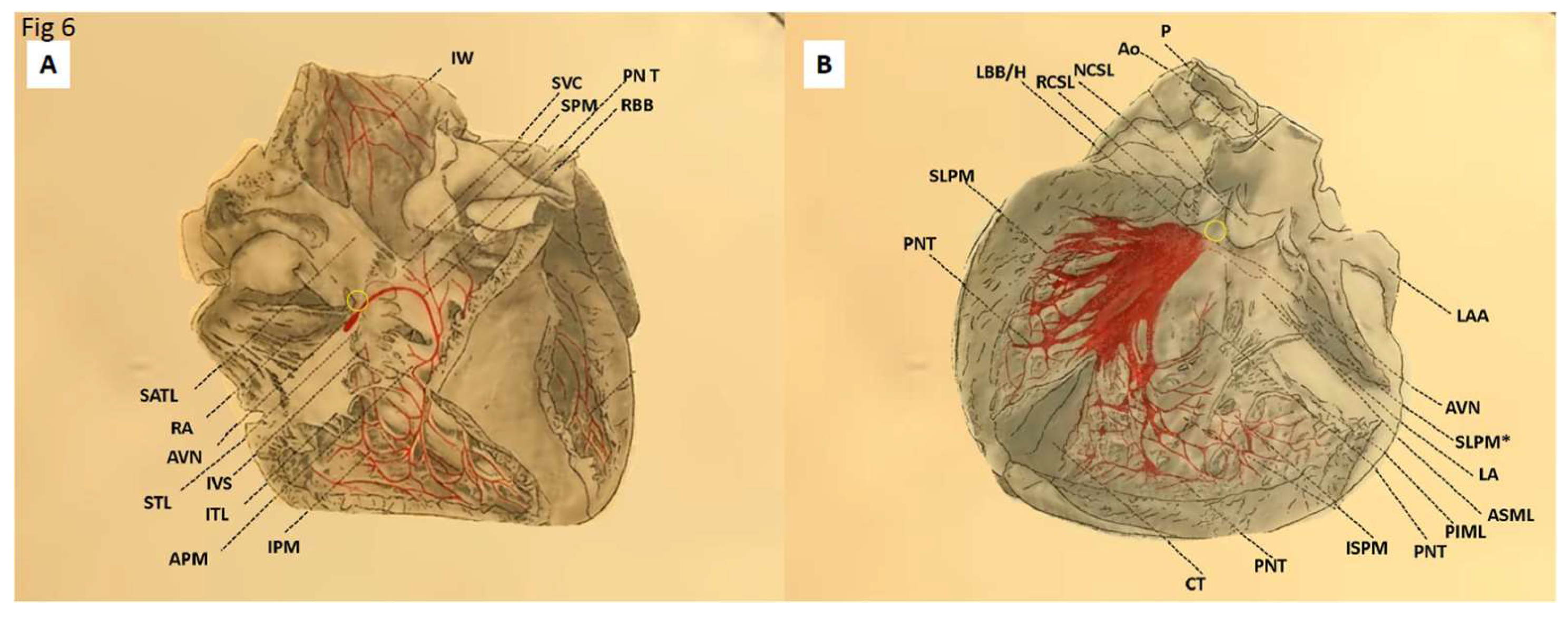
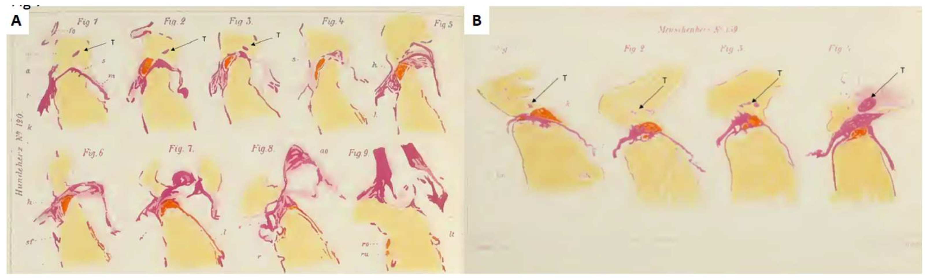
Figure 7.
Sections of dog heart No 120. The three-day-old dog heart, f rom S Tawara 1906 modified by us. We added T= Todaro tendon in pictures or tendon of inferior vena cava valve. “,Das Reizleitungssystem Des Säugetierherzens. Eine Anatomisch-Histologische Studie Über Das Atrioventrikularbündel Und Die Purkinjeschen Fäden (The conduction system of the mammalian heart. An anatomical–histological study of the atrioventricular bundle and Purkinje's network). Jena: Gustav Fischer”. “The axis of the heart was positioned as perpendicularly as possible, the ventricular septum being in the sagittal plane. Every sixth section was stained with Haematoxylin and van Gieson”. The heart “was cut in the frontal plane serially with a thickness of nine microns in the posteroanterior direction. The axis of the heart was positioned as perpendicularly as possible, the ventricular septum being in the sagittal plane.” , translation by Kozo Suma& Munehiro Shimada ( Tawara 2000 English translation). ). “ fo = the oval fossa; v = musculature of the atrial septum; a = tongue like process of the atrial musculature ;s = the atrioventricular fibrous septum; m = anterior mitral leaflet t = septal leaflet of the tricuspid valve; k = the ventricular septum; h = initial portion of the ventricular bundle of the connecting system; sf = a tendinous fiber for the septal leaflet of the tricuspid valve;1 & r = the left and the right bundle branch of the connecting system; ao = the aorta like cord; It = a part of muscle fibers of the left bundle branch entering a tendinous fiber; ro& ru = the right bundle branch separates into two groups, translation by Kozo Suma& Munehiro Shimada ( Tawara 2000 engl translation). This is one ten initial dogs in which Tawara did not mention Knoten ( node) but just atrial segment of the connecting system. See text.). T= Todaro tendon in pictures or tendon of inferior vena cava valve. B Human heart No. 139 . The heart of a female fetus, 31.5 cm long, f rom S Tawara 1906 “Das Reizleitungssystem Des Säugetierherzens. Eine Anatomisch-Histologische Studie Über Das Atrioventrikularbündel Und Die Purkinjeschen Fäden“ (The conduction system of the mammalian heart. An anatomical–histological study of the atrioventricular bundle and Purkinje's network)’ Jena: Gustav Fischer. The sections were done postero anteriorly, parallel to the long axis of the heart and perpendicular to the septum. We added T= Todaro tendon in pictures or tendon of inferior vena cava valve. “v = the atrial septum; k = node, i.e. atrial segment of the connecting system; rn = the anterior mitral leaflet; t = septal leaflet of the tricuspid valve; s =the atrioventricular fibrous septum;km = ventricular septum; 1 & r = the left and the right bundle branch of the connecting system; sf = a tendinous fiber for septal leaflet of the tricuspid valve; va = leaflets of the aortic valve” . Translation by Kozo Suma& Munehiro Shimada ( Tawara 2000 English translation). T= Todaro tendon or tendon of inferior vena cava valve.
Disclaimer/Publisher’s Note: The statements, opinions and data contained in all publications are solely those of the individual author(s) and contributor(s) and not of MDPI and/or the editor(s). MDPI and/or the editor(s) disclaim responsibility for any injury to people or property resulting from any ideas, methods, instructions or products referred to in the content. |
© 2025 by the authors. Licensee MDPI, Basel, Switzerland. This article is an open access article distributed under the terms and conditions of the Creative Commons Attribution (CC BY) license (http://creativecommons.org/licenses/by/4.0/).
Copyright: This open access article is published under a Creative Commons CC BY 4.0 license, which permit the free download, distribution, and reuse, provided that the author and preprint are cited in any reuse.


