
Submitted:
25 July 2025
Posted:
28 July 2025
You are already at the latest version
Abstract
The efficacy of topical drug delivery via eye drops is often achieved at the expense of tolerability and consequently, efforts are being made to design strategies that minimize the adverse effects associated with the passage of active pharmaceutical ingredients (APIs) across the ocular surface. Many of these approaches are too complex, costly and challenging to implement on an industrial scale, yet there is increasing evidence that hylan A, a very high molecular weight hyaluronic acid (≥3.0 MDa), may be a promising vehicle for topical drug delivery of ocular therapies. In this review we explore how the mucoadhesive and viscoelastic properties of eye drop formulations based on hylan A help extend the residence time of APIs at the ocular surface, while maintaining patient comfort. Moreover, we examine how hylan A facilitates the dissolution and stabilisation of APIs, as well as their transport across the ocular epithelial barrier without the need to use toxic penetration enhancers, thereby preserving ocular surface health. Finally, we present evidence indicating that the intrinsic biological properties of hylan A, including its anti-inflammatory effects, help mitigate side effects commonly associated with certain APIs. To illustrate these advantages, we examine the pioneering use of a hylan A-based aqueous eye drop formulation as a vehicle to deliver latanoprost, a prostaglandin analogue widely used in the treatment of glaucoma. This case study demonstrates the potential of hylan A-based eye drops to offer safer and more effective topical drug delivery, especially for long-term ocular therapies where tolerability and biocompatibility are critical.
Keywords:
hylan A
; hyaluronic acid
; very high molecular weight
; eye drops
; ocular drug delivery
; ocular surface health
; mucoadhesion
; viscoelasticity
; latanoprost
1. Introduction
The anterior segment of the eye is comprised of the conjunctiva, cornea and anterior chamber, and it is the primary site of several common ocular disorders. The accessibility of this anterior segment makes it well-suited for topical drug delivery, most commonly in the form of eye drops that offer therapeutic opportunities for a range of eye disorders. These approaches offer many advantages, including ease of use, non-invasiveness and lower cost compared to other technologies, establishing eye drops containing active pharmaceutical ingredients (APIs) as first-line therapies for many anterior segment disorders.
However, topical drug delivery via eye drops also faces some limitations due to the anatomy of the eye and tear dynamics. One challenge is the rapid turnover of the tear film, which causes a large proportion of the eye drop to quickly drain into the nasolacrimal duct, reducing the residence time of the API. The API that resists this initial clearance must still then penetrate the most superficial epithelial barrier and consequently, only a small fraction of the dose applied will ultimately reach deeper structures like the anterior chamber (Allyn et al., 2021, Hansen et al., 2024, Ahmed et al., 2023). A traditional approach to improving topical drug delivery via eye drops has been to include penetration enhancers in the formulations. These facilitate API penetration in different ways, by enhancing penetration of the glycocalyx of the corneal and conjunctival surface epithelium, by loosening tight junctions to compromise epithelial integrity, and by altering cell membrane properties. For example, metal chelators like ethylene diamine tetraacetic acid (EDTA) may be used as penetration enhancers as they disrupt the tight junctions between epithelial cells by sequestering calcium ions (Moiseev et al., 2019, Ye et al., 2012).
Eye drop formulations often contain additives to enhance the solubility and stability of the API (e.g. surfactants to dissolve lipophilic APIs), prolonging its retention at the ocular surface (e.g. polymers enhancing viscosity), or to maintain sterility during use and storage (i.e. preservatives). Some additives have adverse effects at the ocular surface, producing irritation, dryness and allergic reactions (Kahook et al., 2024, Fineide et al., 2024). This is particularly concerning when treating chronic conditions where long-term daily administration is required, and these adverse effects may reduce patient adherence and persistence (Baudouin et al., 2025). By contrast, adverse effects that are associated with the intrinsic molecular properties of the APIs may be less easy to palliate, although they may be alleviated or exacerbated by additives in the formulation (Hedengran and Kolko, 2023).
Intense efforts are being made to enhance eye drop formulations for topical drug delivery (Wang and Wang, 2022), testing the incorporation of various additives to enhance parameters like API solubility, retention time and penetration into the eye. However, identifying optimal combinations is still largely considered a challenge, as improvements in one aspect are often either insufficient or may come at the expense of another. For example, preservative-free eye drops produce fewer ocular surface symptoms but they do not appear to be associated with consistently improved patient adherence, for example in the treatment of glaucoma (Jayaram et al., 2023). This may be due to adverse effects associated with other additives or to the intrinsic properties of the APIs themselves. Hence, there is still a need to develop formulations that enhance API bioavailability while avoiding side effects, and that ideally provide additional benefits to the ocular surface.
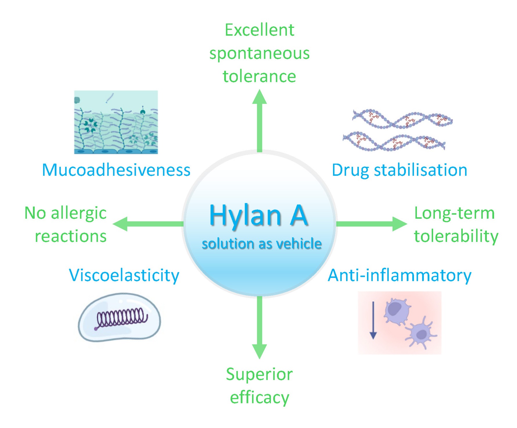
Here we focus on a novel vehicle for topical drug delivery via eye drops, a buffered saline solution containing 0.15% of a linear, very high molecular weight (MW) hyaluronic acid (HA; hylan A, ≥3.0 MDa) (Müller-Lierheim, 2020, Bron et al., 2022). We will provide evidence of the promise this aqueous hylan A formulation offers to address several key challenges, enhancing ocular surface retention, API solubilisation and stabilisation, and API transport, while maintaining a healthy ocular surface. To illustrate these benefits, we will examine the pioneering use of this aqueous hylan A formulation as a vehicle for latanoprost, a prostaglandin analogue and a frontline approach to treat glaucoma.
2. An Aqueous Hylan A Formulation with Optimal Viscoelastic Properties Enhances Ocular Surface Retention of APIs
Achieving increased ocular surface retention by adjusting the formulation of eye drops is desirable as it means the API will stay at the ocular surface longer, thereby enhancing its therapeutic efficacy and drug delivery to the eye. One approach often used to extend ocular surface retention is to increase the viscosity of the eye drop, which hinders drainage from the ocular surface. This can be achieved with synthetic polymers like polyvinyl alcohols (PVAs), natural polymers like HA, or with derivatives of natural polymers such as hypromellose (HPMC) (Grassiri et al., 2021). However, excessively high viscosity can cause problems, like increased reflex tearing, which can accelerate API clearance, exacerbate discomfort and cause temporary visual blurring (Grassiri et al., 2021, Giri et al., 2024). To avoid these issues, eye drops can be formulated with a polymeric solution that exhibits viscoelastic properties closely mimicking the flow of healthy human tears. Ideally, such formulations maintain the high viscosity of natural tears when the eye is open, promoting prolonged residence times, while they respect the low viscosity of natural tears during blinking, thereby limiting the friction between eyelid and epithelial surface to ensure comfort and avoid altered vision (Arshinoff et al., 2021b, Tiffany, 1994, Müller-Lierheim, 2020).
The flow of dissolved polymers is determined by their ability to entangle (Graessley, 2005). HA is a linear, naturally occurring polysaccharide and an essential component of the vertebrate extracellular matrix (Almond, 2007). HA can be produced through fermentation, adopting a wide range of MWs (i.e. chain lengths) up to several million Daltons (MDa) (Fallacara et al., 2018). Longer HA chains in solution become more easily entangled, which enhances their viscoelasticity. Accordingly, an aqueous solution with 0.15% hylan A (≥3.0 MDa) exhibits flow behaviour mimicking that of natural tears, whereas an aqueous solution containing 0.15% lower MW HA does not produce this effect (Arshinoff et al., 2021a, Müller-Lierheim, 2020). This is primarily attributed to the very high MW of hylan A without the need for additional viscosity-enhancing agents. A comprehensive examination of this topic has been presented in a previous article (Müller-Lierheim, 2020).
Mucoadhesion is an even more critical factor than viscosity in determining the retention of APIs at the ocular surface. This feature refers to the interaction with mucus, which is mainly composed of water (>95%) and mucins, a family of large, highly glycosylated hydrophilic proteins (Andrews et al., 2009, Grassiri et al., 2021). Mucins are secreted into the mucoaqueous layer of the tear film at the ocular surface, and they also exist as cell membrane-bound proteins within the glycocalyx formed by the superficial corneal and conjunctival epithelial cells (Baudouin et al., 2019). The flexibility of polymer chains drives their entanglement with mucin chains and the formation of hydrogen bonds (Andrews et al., 2009, Smart, 2005). In aqueous solution, HA is a very flexible polyanion able to entangle intimately with and adhere to the mucin molecules at the ocular surface. The mucoadhesive properties of aqueous HA are influenced strongly by its MW, with high MW but not low MW HA binding readily to membrane-bound mucins, enhancing the cellular barrier against pathogens and prolonging local drug retention (Hansen et al., 2017). Indeed, the mucoadhesive performance of linear HA increases in direct proportion with its MW, with crosslinked HA and other polymers commonly used in eye drops exhibiting lower mucoadhesion (except for xanthan gum) (Guarise et al., 2023). Accordingly, an aqueous hylan A formulation would be expected to exhibit superior mucoadhesiveness at the ocular surface given its very high MW. Together with its viscoelastic properties comparable to natural tears, an aqueous 0.15% hylan A formulation is therefore likely to be a suitable vehicle for ocular drug delivery, producing adequate ocular surface retention and patient comfort.
3. An Aqueous Hylan A Formulation Improves API Solubilisation, Stability and Ocular Transport
A major challenge when using eye drops for topical drug delivery is the poor aqueous solubility and stability of many APIs, which restricts their bioavailability. While advanced solutions have been developed to address this issue (Ahmed et al., 2023, Giri et al., 2024), they are often complex, costly, and may face lengthy evaluation and regulatory approval processes before reaching the market and patients (Giri et al., 2024, Tenpattinam et al., 2025). A simpler alternative, supported by extensive biocompatibility and clinical data, is the use of linear, natural HA in aqueous solution. This approach differs from the use of HA/drug chemical conjugates or HA modified nanoparticles or micelles, although all these strategies exploit similar properties of HA (Jiang and Xu, 2023, Zhang et al., 2018, Fallacara et al., 2018, Guter and Breunig, 2017, Buckley et al., 2022).
At physiological pH in aqueous solution, HA is a negatively charged polyanion and it forms salts generally referred to as hyaluronan or hyaluronate (e.g. sodium hyaluronate), which are very hydrophilic and consequently, surrounded by water molecules. More precisely, water molecules link the HA hydrophilic functional groups (e.g. carboxyl, COOH) to hydrogen bonds that stabilize the secondary structure of the biopolymer, i.e. a two-fold helix. In this extended conformation, HA chains also form hydrophobic domains within their secondary structure that can interact non-covalently with other hydrophobic molecules in an aqueous environment (Rouse et al., 2007, Ghosh et al., 1994). This feature can improve the solubility of APIs in aqueous solutions, enhancing their chemical stability by decreasing water accessibility and inhibiting hydrolysis, or by reducing the access of enzymes to the API. When the secondary structure of HA molecules provokes entanglement, an extended three-dimensional network can form (i.e. a tertiary structure) in which the strong intermolecular interactions between HA chains reduce the availability of these hydrophobic domains and the ability of HA to interact with hydrophobic molecules (Rouse et al., 2007). Both the concentration and MW of HA influence the transition from secondary to tertiary structures in aqueous solutions (Fallacara et al., 2018). Indeed, based on the properties of HA studied previously (Rouse et al., 2007, Ghosh et al., 1994), it is plausible that a 0.15% hylan A aqueous solution contains enough HA molecules in conformations that can interact with hydrophobic molecules to improve the solubility and stability of the latter.
There is some evidence as to how hylan A might facilitate the transport of other molecules into the eye. In vertebrates, several cell surface receptors for HA exist, the best studied being cluster-determined 44 (CD44) (Aruffo et al., 1990, Wang et al., 2025). The binding of HA molecules to CD44 involves its engagement with multiple CD44 receptor sites (Ruppert et al., 2014), such that the avidity of HA increases with its MW and high MW HA binds much more strongly to this receptor than low MW forms (Lee-Sayer et al., 2015). Among the known functions of the CD44 receptor is its role in facilitating HA internalization and its subsequent degradation (Knudson et al., 2002). When high MW HA binds to CD44 it can be cleaved into intermediate-sized fragments by cell surface enzymes (membrane-bound hyaluronidases), which can then be internalized by and directed to intracellular compartments where they are further degraded (Wang et al., 2025, Garantziotis and Savani, 2019). CD44 receptors have been identified in both corneal and conjunctival epithelial cells (Lardner and van Setten, 2020, Zhu et al., 1997, Lerner et al., 1998), such that this pathway is a potential route for API delivery to deeper ocular regions without compromising cell membrane integrity when these molecules interact with HA. Receptors involved in alternative uptake pathways include the HA receptor for endocytosis (HARE) (Zhou et al., 2000, Harris and Baker, 2020), which is also found on corneal epithelial cells (Falkowski et al., 2003) and might facilitate the delivery of molecules that interact with HA (Müller-Lierheim, 2020).
4. The Benefits of Using an Aqueous Hylan A Formulation for Ocular Surface Health
When the inherent properties of the API in a therapeutic eye drop produce adverse effects, it is important to include substances in these formulations that counteract such effects and that favour ocular surface health. Beyond the benefits outlined above, hylan A has consistently been demonstrated to promote better ocular surface health than lower MW HA, primarily due to its unique very high MW (Müller-Lierheim, 2020).
The MW of HA affects its biological activity in both healthy and diseased states (Bohaumilitzky et al., 2017, Cyphert et al., 2015). In homeostatic conditions, HA predominantly exists as a high MW polymer with biophysical properties consistent with its activity as a lubricant, space-filler and shock absorber in joints and connective tissues. Moreover, it has anti-inflammatory, anti-proliferative and anti-angiogenic effects, and multiple studies have highlighted its role as a tissue protectant and promoter of homeostasis after injury and inflammation. By contrast, in pathological circumstances HA fragmentation is enhanced and produces more low MW HA that is linked to inflammation (Bohaumilitzky et al., 2017, Cyphert et al., 2015, Monslow et al., 2015, Garantziotis and Savani, 2019). Furthermore, unlike low MW HA, high MW HA reduces peripheral nociceptor activity (Gomis et al., 2004, Caires et al., 2015) as well as inflammatory and neuropathic pain (Bonet et al., 2020, Ferrari et al., 2018), including pain induced by chemotherapy (Bonet et al., 2022) or surgery (Zhang et al., 2024) in preclinical models. Elsewhere, tear film stability was improved when eye drops with 0.15% aqueous hylan A (Comfort Shield, i.com medical, Munich, Germany) were evaluated in a preclinical model of environmental dry eye stress (Table 1), reducing ocular surface damage and inflammation when compared to the use of low MW HA or secretagogues (secretion stimulating drugs) (Kojima et al., 2020).
Clinical data suggest that the physiological activity of very high MW 0.15% hylan A eye drops is effective to treat severe ocular disease and that they may even be superior to autologous serum eye drops (Table 1) (Beck et al., 2019). These benefits were backed up in the HYLAN M clinical study, where switching from optimized artificial tear treatments to 0.15% hylan A eye drops significantly improved symptoms in severe dry eye patients (within four weeks), including visual stability, discomfort and pain (Table 1) (Medic et al., 2024, van Setten et al., 2020a, Alsheikh et al., 2021). Moreover, 0.15% hylan A eye drops appeared to increase the length of corneal nerves (van Setten et al., 2020b), which are typically compromised in this population (Benitez-Del-Castillo et al., 2007, Shetty et al., 2023, Galor et al., 2025). The benefits of hylan A for corneal nerves were further demonstrated in patients who underwent corneal surgery, procedures known to cause unavoidable corneal nerve damage. In this context, daily 0.15% hylan A eye drop application after surgery accelerated the recovery of corneal nerve structure and sensitivity relative to eye drops containing low MW HA, while also improving ocular surface symptoms (Özkan et al., 2025). In addition, hylan A treatment helped prevent the increase in inflammation-related immune cells observed three months after surgery when low MW HA was used (Table 1) (Özkan et al., 2025).
5. An Aqueous Hylan A Formulation as New Vehicle for Latanoprost to Manage Elevated Intraocular Pressure
Alterations to the anterior segment may influence the development of diseases that affect structures in the posterior eye segment, such as the retina and optic nerve. One prominent example is glaucoma, one of the most common worldwide causes of irreversible blindness. Glaucoma often develops when aqueous humour drainage from the anterior chamber is impaired or unbalanced, leading to an elevation in intraocular pressure (IOP) that can provoke structural changes in the posterior segment of the eye and optic nerve injury. Currently, IOP is the only risk factor that can be modulated to prevent glaucoma progression (Weinreb et al., 2014, Jayaram et al., 2023). Thus, reducing IOP via eye drops containing APIs is the primary strategy to treat glaucoma (Jayaram et al., 2023).
Prostaglandin analogues increase aqueous humour outflow via the unconventional pathway, such that latanoprost has become a frequently used treatment (Jayaram et al., 2023) and one of the most effective APIs used in eye drops to lower IOP and slow glaucoma progression (Li et al., 2016). Prostaglandin analogues produce well documented adverse effects at the ocular surface (Kolko et al., 2023) and at therapeutic concentrations, topical latanoprost can cause ocular surface damage in a mouse model that resembles dry eye disease, primarily through inflammatory mechanisms (Yang et al., 2018). Moreover, many commercial IOP lowering eye drops contain the quaternary ammonium cationic detergent benzalkonium chloride (BAK) as a preservative and penetration enhancer (Kahook et al., 2024), although long-term BAK use has negative effects at the ocular surface (Baudouin et al., 2010) and an emergence of preservative-free IOP lowering eye drops has occurred in recent years (Konstas et al., 2021, Hollo et al., 2018, Kim et al., 2021, Kahook et al., 2024). These formulations may include other penetration enhancers like EDTA, which while less problematic can potentially cause long-term ocular surface damage (Villani et al., 2016, Halder and Khopade, 2020, Ye et al., 2012). Other additives like polyethylene glycol (PEG) and propylene glycol (PG) act as lubricants, surfactants and co-solubilisers, yet they may also contribute to dry eye disease (Gomes et al., 2017).
The prevalence of ocular surface disease is very high in individuals with glaucoma, particularly in those with uncontrolled glaucoma and those who use multiple topical medications (Fechtner et al., 2010, Skalicky et al., 2012, Baudouin et al., 2012b). A large-scale study found a higher IOP in glaucoma patients with severe ocular surface disease than in those with no or mild disease (Baudouin et al., 2012b). Moreover, there is evidence that chronic ocular surface inflammation increases outflow resistance, further favouring IOP elevation (Baudouin et al., 2012a, Batra et al., 2014, Baudouin et al., 2021). Therefore, adopting a management strategy for ocular surface problems, particularly inflammation, is key to improving the effectiveness of IOP control. This can be achieved by reducing the toxicity of glaucoma medications and incorporating supportive therapies (e.g. ocular surface lubrication and anti-inflammatory treatment) (Dubrulle et al., 2018, Messmer et al., 2024, Kemer et al., 2024).
To address these challenges, a novel latanoprost ophthalmic formulation has been developed using an aqueous hylan A solution as the vehicle, leveraging the advantages outlined in the previous sections (Figure 1). The formulation is a preservative-free 0.15% hylan A solution prepared in isotonic phosphate buffered saline at pH 7.4, a stable vehicle to which 20 μg/ml of latanoprost is added (Müller-Lierheim, 2021). Notably, this concentration exceeds the normal solubility of latanoprost in water by approximately 8 μg/ml (Table 2), probably facilitated by an interaction between latanoprost and hylan A. In one subject with ocular hypertension, this new formulation lowered the IOP more than a commercial latanoprost eye drop (Table 2), despite the higher concentration in the latter (50 μg/ml latanoprost) (Müller-Lierheim, 2021). This effect further suggests that hylan A may facilitate the transport of latanoprost into the eye (Müller-Lierheim, 2020, Bron et al., 2022), as seen in a rat model where the therapeutic concentration of latanoprost in the animals’ aqueous humour was similar after the administration of a hylan A-based eye drop containing 14 μg/ml latanoprost to that achieved with a commercial formulation containing 50 μg/ml latanoprost (Table 2). Indeed, this commercial formulation not only contained around 3.5 times more latanoprost but it also included the penetration enhancer EDTA (Higa et al., 2024). The same formulations were also tested in a mouse model in which the novel hylan A-based formulation induced less inflammation, caused fewer ocular surface alterations and retained better corneal epithelial barrier integrity than the commercial formulation, while controlling IOP better. Significantly, all the ocular surface parameters analysed in animals treated with the hylan A-based latanoprost formulation were indistinguishable from those of the untreated wild-type controls (Table 2) (Dogru et al., 2023).
Taken together, these findings support the potential of this novel latanoprost formulation, which is free of toxic additives, that uses a lower amount of the API, and that takes advantage of the lubricating and anti-inflammatory properties of hylan A. By promoting ocular surface health, this formulation may enhance the reduction of IOP by chronic treatments, as preclinical results indicate. Further clinical studies are being prepared to confirm these potential benefits.
6. Hylan A-Based Eye Drops as Next Generation Vehicles for Therapeutic API Delivery
A variety of new delivery systems for topical drugs are being developed that offer promise to advance ocular therapy, such as hydrogels, nanoparticles carriers, contact lenses and ocular inserts (Zeppieri et al., 2025). However, they pose significant challenges related to implantation, removal, long-term biocompatibility and patient acceptance. Many of these new technologies are also costly and difficult to scale-up for industrial production. In the case of glaucoma, which affects around 100 million people worldwide across a wide range of socioeconomic and healthcare settings (Kolko et al., 2023), it is essential to establish a range of treatment options. Thus, eye drops continue to represent a practical, accessible and widely accepted option.
There are many ongoing efforts to improve eye drop formulations for topical drug delivery, with different additives being tested to enhance critical parameters like mucoadhesiveness, viscoelasticity and API solubility, stability and ocular transport, as well as to favour ocular surface health. While optimizing one parameter in many new formulations compromises another, the hylan A-based eye drops presented here seem to represent a promising solution to simultaneously address all these critical aspects (Figure 1). Indeed, the evidence supports the efficacy of hylan A-based eye drops to treat various ocular surface conditions (Table 1), also representing a useful vehicle for latanoprost (Table 2). Although the benefits observed can mostly be attributed to the unique properties of hylan A, other physicochemical properties of the formulation also contribute positively, such as pH, osmolarity and the buffer employed (Hedengran and Kolko, 2023, Higa et al., 2024).
We envisage that hylan A-based eye drops will represent a platform that could support a wide range of ocular therapies. A further promising application is the topical delivery of cyclosporine A (CsA), an immunosuppressive drug commonly used to manage the inflammatory component of chronic dry eye disease (Patil et al., 2025, Craig et al., 2017). This is a condition where effectively overcoming ocular drug delivery barriers while avoiding adverse effects remains a challenge, mainly due to the large MW and hydrophobic nature of CsA (Nagai and Otake, 2022, Periman et al., 2020). Hylan A-based eye drops could address these limitations, as already seen with latanoprost (Müller-Lierheim, 2021, Higa et al., 2024, Dogru et al., 2023). Beyond its use as a vehicle, hylan A itself produces anti-inflammatory effects (Kojima et al., 2020, Bron et al., 2022) that may act synergistically with CsA to more effectively treat chronic dry eye disease. Moreover, given the involvement of neurosensory abnormalities in dry eye disease (Craig et al., 2017), the neurotrophic effects of hylan A (Özkan et al., 2025, van Setten et al., 2020b) may offer further therapeutic benefits. Interestingly, recent findings in vitro showed that CsA can alter HA metabolism in orbital fibroblasts (Galgoczi et al., 2024). If similar effects occur in vivo, this could have negative effects on ocular surface health. In such a case, co-formulation of CsA with hylan A may help maintain physiological HA homeostasis and mitigate potential adverse effects.
In summary, the use of hylan A as a vehicle represents a promising step towards a new generation of eye drops for topical ocular drug delivery, designed to improve API bioavailability, reduce adverse effects and support long-term ocular surface health in a range of therapeutic indications.
Author Contributions
Writing—original draft preparation: J.P.-M. and W.G.K.M.-L; writing—review and editing: J.P.-M. and W.G.K.M.-L; visualization: J.P.-M. and W.G.K.M.-L; All authors have read and agreed to the published version of the manuscript.
Funding
This research received no external funding.
Conflicts of Interest
The author Wolfgang G. K. Müller-Lierheim is the Chief Executive Officer of i.com medical GmbH, a company developing and manufacturing hyaluronan containing eye drops. The author Jesús Pujol-Martí is the Chief Scientific Officer at i.com medical GmbH.
References
- AHMED, S.; AMIN, M. M.; SAYED, S. Ocular Drug Delivery: a Comprehensive Review. AAPS PharmSciTech 2023, 24, 66. [Google Scholar] [CrossRef]
- ALLYN, M. M.; LUO, R. H.; HELLWARTH, E. B.; SWINDLE-REILLY, K. E. Considerations for Polymers Used in Ocular Drug Delivery. Front Med (Lausanne) 2021, 8, 787644. [Google Scholar] [CrossRef] [PubMed]
- ALMOND, A. Hyaluronan. Cell Mol Life Sci 2007, 64, 1591–6. [Google Scholar] [CrossRef] [PubMed]
- ALSHEIKH, O.; ALZAAIDI, S.; VARGAS, J. M.; AL-SHARIF, E.; ALRAJEH, M.; ALSEMARI, M. A.; ALHOMMADI, A.; ALSAATI, A.; ALJWAISER, N.; ALSHAHWAN, E.; ABDULHAFIZ, M.; ELSAYED, R.; MULLER-LIERHEIM, W. G. K. Effectiveness of 0.15% hylan A eye drops in ameliorating symptoms of severe dry eye patients in Saudi Arabia. Saudi J Ophthalmol 2021, 35, 293–298. [Google Scholar] [CrossRef] [PubMed]
- ANDREWS, G. P.; LAVERTY, T. P.; JONES, D. S. Mucoadhesive polymeric platforms for controlled drug delivery. Eur J Pharm Biopharm 2009, 71, 505–18. [Google Scholar] [CrossRef]
- ARSHINOFF, S.; HOFMANN, I.; NAE, H. Rheological behavior of commercial artificial tear solutions. J Cataract Refract Surg 2021a, 47, 649–654. [Google Scholar] [CrossRef]
- ARSHINOFF, S.; HOFMANN, I.; NAE, H. Role of rheology in tears and artificial tears. J Cataract Refract Surg 2021b, 47, 655–661. [Google Scholar] [CrossRef]
- ARUFFO, A.; STAMENKOVIC, I.; MELNICK, M.; UNDERHILL, C. B.; SEED, B. CD44 is the principal cell surface receptor for hyaluronate. Cell 1990, 61, 1303–13. [Google Scholar] [CrossRef]
- BATRA, R.; TAILOR, R.; MOHAMED, S. Ocular surface disease exacerbated glaucoma: optimizing the ocular surface improves intraocular pressure control. J Glaucoma 2014, 23, 56–60. [Google Scholar] [CrossRef]
- BAUDOUIN, C.; DENOYER, A.; DESBENOIT, N.; HAMM, G.; GRISE, A. In vitro and in vivo experimental studies on trabecular meshwork degeneration induced by benzalkonium chloride (an American Ophthalmological Society thesis). Trans Am Ophthalmol Soc, 2012a; pp. 40–63. [Google Scholar]
- BAUDOUIN, C.; KOLKO, M.; MELIK-PARSADANIANTZ, S.; MESSMER, E. M. Inflammation in Glaucoma: From the back to the front of the eye, and beyond. Prog Retin Eye Res 2021, 83, 100916. [Google Scholar] [CrossRef]
- BAUDOUIN, C.; LABBE, A.; LIANG, H.; PAULY, A.; BRIGNOLE-BAUDOUIN, F. Preservatives in eyedrops: the good, the bad and the ugly. Prog Retin Eye Res 2010, 29, 312–34. [Google Scholar] [CrossRef] [PubMed]
- BAUDOUIN, C.; MYERS, J. S.; VAN TASSEL, S. H.; GOYAL, N. A.; MARTINEZ-DE-LA-CASA, J.; NG, A.; EVANS, J. S. Adherence and Persistence on Prostaglandin Analogues for Glaucoma: A Systematic Review and Meta-Analysis. Am J Ophthalmol 2025, 275, 99–113. [Google Scholar] [CrossRef] [PubMed]
- BAUDOUIN, C.; RENARD, J. P.; NORDMANN, J. P.; DENIS, P.; LACHKAR, Y.; SELLEM, E.; ROULAND, J. F.; JEANBAT, V.; BOUEE, S. Prevalence and risk factors for ocular surface disease among patients treated over the long term for glaucoma or ocular hypertension. Eur J Ophthalmol 2012b. [Google Scholar] [CrossRef]
- BAUDOUIN, C.; ROLANDO, M.; BENITEZ DEL CASTILLO, J. M.; MESSMER, E. M.; FIGUEIREDO, F. C.; IRKEC, M.; VAN SETTEN, G.; LABETOULLE, M. Reconsidering the central role of mucins in dry eye and ocular surface diseases. Prog Retin Eye Res 2019, 71, 68–87. [Google Scholar] [CrossRef]
- BECK, R.; STACHS, O.; KOSCHMIEDER, A.; MUELLER-LIERHEIM, W. G. K.; PESCHEL, S.; VAN SETTEN, G. B. Hyaluronic Acid as an Alternative to Autologous Human Serum Eye Drops: Initial Clinical Results with High-Molecular-Weight Hyaluronic Acid Eye Drops. Case Rep Ophthalmol 2019, 10, 244–255. [Google Scholar] [CrossRef]
- BENITEZ-DEL-CASTILLO, J. M.; ACOSTA, M. C.; WASSFI, M. A.; DIAZ-VALLE, D.; GEGUNDEZ, J. A.; FERNANDEZ, C.; GARCIA-SANCHEZ, J. Relation between corneal innervation with confocal microscopy and corneal sensitivity with noncontact esthesiometry in patients with dry eye. Invest Ophthalmol Vis Sci 2007, 48, 173–81. [Google Scholar] [CrossRef]
- BOHAUMILITZKY, L.; HUBER, A. K.; STORK, E. M.; WENGERT, S.; WOELFL, F.; BOEHM, H. A Trickster in Disguise: Hyaluronan's Ambivalent Roles in the Matrix. Front Oncol 2017, 7, 242. [Google Scholar] [CrossRef] [PubMed]
- BONET, I. J. M.; ARALDI, D.; KHOMULA, E. V.; BOGEN, O.; GREEN, P. G.; LEVINE, J. D. Mechanisms Mediating High-Molecular-Weight Hyaluronan-Induced Antihyperalgesia. J Neurosci 2020, 40, 6477–6488. [Google Scholar] [CrossRef]
- BONET, I. J. M.; STAURENGO-FERRARI, L.; ARALDI, D.; GREEN, P. G.; LEVINE, J. D. Second messengers mediating high-molecular-weight hyaluronan-induced antihyperalgesia in rats with chemotherapy-induced peripheral neuropathy. Pain 2022, 163, 1728–1739. [Google Scholar] [CrossRef]
- BRON, A. J.; DOGRU, M.; HORWATH-WINTER, J.; KOJIMA, T.; KOVACS, I.; MULLER-LIERHEIM, W. G. K.; VAN SETTEN, G. B.; BELMONTE, C. Reflections on the Ocular Surface: Summary of the Presentations at the 4th Coronis Foundation Ophthalmic Symposium Debate: "A Multifactorial Approach to Ocular Surface Disorders "(August 31 2021). Front Biosci (Landmark Ed); 2022; 27, p. 142. [Google Scholar]
- BUCKLEY, C.; MURPHY, E. J.; MONTGOMERY, T. R.; MAJOR, I. Hyaluronic Acid: A Review of the Drug Delivery Capabilities of This Naturally Occurring Polysaccharide. Polymers (Basel) 2022, 14. [Google Scholar] [CrossRef]
- CAIRES, R.; LUIS, E.; TABERNER, F. J.; FERNANDEZ-BALLESTER, G.; FERRER-MONTIEL, A.; BALAZS, E. A.; GOMIS, A.; BELMONTE, C.; DE LA PENA, E. Hyaluronan modulates TRPV1 channel opening, reducing peripheral nociceptor activity and pain. Nat Commun 2015, 6, 8095. [Google Scholar] [CrossRef]
- CRAIG, J. P.; NICHOLS, K. K.; AKPEK, E. K.; CAFFERY, B.; DUA, H. S.; JOO, C. K.; LIU, Z.; NELSON, J. D.; NICHOLS, J. J.; TSUBOTA, K.; STAPLETON, F. TFOS DEWS II Definition and Classification Report. Ocul Surf 2017, 15, 276–283. [Google Scholar] [CrossRef]
- CYPHERT, J. M.; TREMPUS, C. S.; GARANTZIOTIS, S. Size Matters: Molecular Weight Specificity of Hyaluronan Effects in Cell Biology. Int J Cell Biol 2015, 563818. [Google Scholar] [CrossRef] [PubMed]
- DOGRU, M.; KOJIMA, T.; HIGA, K.; IGARASHI, A.; KUDO, H.; MULLER-LIERHEIM, W. G. K.; TSUBOTA, K.; NEGISHI, K. The Effect of High Molecular Weight Hyaluronic Acid and Latanoprost Eyedrops on Tear Functions and Ocular Surface Status in C57/BL6 Mice. J Clin Med 2023, 12. [Google Scholar] [CrossRef] [PubMed]
- DUBRULLE, P.; LABBE, A.; BRASNU, E.; LIANG, H.; HAMARD, P.; MEZIANI, L.; BAUDOUIN, C. Influence of Treating Ocular Surface Disease on Intraocular Pressure in Glaucoma Patients Intolerant to Their Topical Treatments: A Report of 10 Cases. J Glaucoma 2018, 27, 1105–1111. [Google Scholar] [CrossRef]
- FALKOWSKI, M.; SCHLEDZEWSKI, K.; HANSEN, B.; GOERDT, S. Expression of stabilin-2, a novel fasciclin-like hyaluronan receptor protein, in murine sinusoidal endothelia, avascular tissues, and at solid/liquid interfaces. Histochem Cell Biol 2003, 120, 361–9. [Google Scholar] [CrossRef]
- FALLACARA, A.; BALDINI, E.; MANFREDINI, S.; VERTUANI, S. Hyaluronic Acid in the Third Millennium. Polymers (Basel) 2018, 10. [Google Scholar] [CrossRef]
- FECHTNER, R. D.; GODFREY, D. G.; BUDENZ, D.; STEWART, J. A.; STEWART, W. C.; JASEK, M. C. Prevalence of ocular surface complaints in patients with glaucoma using topical intraocular pressure-lowering medications. Cornea 2010, 29, 618–21. [Google Scholar] [CrossRef] [PubMed]
- FERRARI, L. F.; KHOMULA, E. V.; ARALDI, D.; LEVINE, J. D. CD44 Signaling Mediates High Molecular Weight Hyaluronan-Induced Antihyperalgesia. J Neurosci 2018, 38, 308–321. [Google Scholar] [CrossRef]
- FINEIDE, F.; MAGNO, M.; DAHLO, K.; KOLKO, M.; HEEGAARD, S.; VEHOF, J.; UTHEIM, T. Topical glaucoma medications-Possible implications on the meibomian glands. Acta Ophthalmol 2024, 102, 735–748. [Google Scholar] [CrossRef]
- GALGOCZI, E.; MOLNAR, Z.; KATKO, M.; UJHELYI, B.; STEIBER, Z.; NAGY, E. Cyclosporin A inhibits PDGF-BB induced hyaluronan synthesis in orbital fibroblasts. Chem Biol Interact 2024, 396, 111045. [Google Scholar] [CrossRef]
- GALOR, A.; GALLAR, J.; ACOSTA, M. C.; MESEGUER, V.; BENITEZ-DEL-CASTILLO, J. M.; STACHS, O.; SZENTMARY, N.; VERSURA, P.; MULLER-LIERHEIM, W. G. K.; BELMONTE, C.; PUJOL-MARTI, J. CORONIS symposium 2023: Scientific and clinical frontiers in ocular surface innervation. Acta Ophthalmol; 2025; 103, pp. e240–e255. [Google Scholar]
- GARANTZIOTIS, S.; SAVANI, R. C. Hyaluronan biology: A complex balancing act of structure, function, location and context. Matrix Biol 2019, 78-79, 1–10. [Google Scholar] [CrossRef]
- GHOSH, P.; HUTADILOK, N.; ADAM, N.; LENTINI, A. Interactions of hyaluronan (hyaluronic acid) with phospholipids as determined by gel permeation chromatography, multi-angle laser-light-scattering photometry and 1H-NMR spectroscopy. Int J Biol Macromol 1994, 16, 237–44. [Google Scholar] [CrossRef]
- GIRI, B. R.; JAKKA, D.; SANDOVAL, M. A.; KULKARNI, V. R.; BAO, Q. Advancements in Ocular Therapy: A Review of Emerging Drug Delivery Approaches and Pharmaceutical Technologies. Pharmaceutics 2024, 16. [Google Scholar] [CrossRef]
- GOMES, J. A. P.; AZAR, D. T.; BAUDOUIN, C.; EFRON, N.; HIRAYAMA, M.; HORWATH-WINTER, J.; KIM, T.; MEHTA, J. S.; MESSMER, E. M.; PEPOSE, J. S.; SANGWAN, V. S.; WEINER, A. L.; WILSON, S. E.; WOLFFSOHN, J. S. TFOS DEWS II iatrogenic report. Ocul Surf 2017, 15, 511–538. [Google Scholar] [CrossRef]
- GOMIS, A.; PAWLAK, M.; BALAZS, E. A.; SCHMIDT, R. F.; BELMONTE, C. Effects of different molecular weight elastoviscous hyaluronan solutions on articular nociceptive afferents. Arthritis Rheum 2004, 50, 314–26. [Google Scholar] [CrossRef]
- GRAESSLEY, W. W. The entanglement concept in polymer rheology. The entanglement concept in polymer rheology 2005, 1–179. [Google Scholar]
- GRASSIRI, B.; ZAMBITO, Y.; BERNKOP-SCHNURCH, A. Strategies to prolong the residence time of drug delivery systems on ocular surface. Adv Colloid Interface Sci 2021, 288, 102342. [Google Scholar] [CrossRef] [PubMed]
- GUARISE, C.; ACQUASALIENTE, L.; PASUT, G.; PAVAN, M.; SOATO, M.; GAROFOLIN, G.; BENINATTO, R.; GIACOMEL, E.; SARTORI, E.; GALESSO, D. The role of high molecular weight hyaluronic acid in mucoadhesion on an ocular surface model. J Mech Behav Biomed Mater 2023, 143, 105908. [Google Scholar] [CrossRef] [PubMed]
- GUTER, M.; BREUNIG, M. Hyaluronan as a promising excipient for ocular drug delivery. Eur J Pharm Biopharm 2017, 113, 34–49. [Google Scholar] [CrossRef] [PubMed]
- HALDER, A.; KHOPADE, A. J. Physiochemical Properties and Cytotoxicity of a Benzalkonium Chloride-Free, Micellar Emulsion Ophthalmic Formulation of Latanoprost. Clin Ophthalmol 2020, 14, 3057–3064. [Google Scholar] [CrossRef]
- HANSEN, I. M.; EBBESEN, M. F.; KASPERSEN, L.; THOMSEN, T.; BIENK, K.; CAI, Y.; MALLE, B. M.; HOWARD, K. A. Hyaluronic Acid Molecular Weight-Dependent Modulation of Mucin Nanostructure for Potential Mucosal Therapeutic Applications. Mol Pharm 2017, 14, 2359–2367. [Google Scholar] [CrossRef] [PubMed]
- HANSEN, M. E.; IBRAHIM, Y.; DESAI, T. A.; KOVAL, M. Nanostructure-Mediated Transport of Therapeutics through Epithelial Barriers. Int J Mol Sci 2024, 25. [Google Scholar] [CrossRef]
- HARRIS, E. N.; BAKER, E. Role of the Hyaluronan Receptor, Stabilin-2/HARE, in Health and Disease. Int J Mol Sci 2020, 21. [Google Scholar] [CrossRef] [PubMed]
- HEDENGRAN, A.; KOLKO, M. The molecular aspect of anti-glaucomatous eye drops-are we harming our patients? Mol Aspects Med 2023, 93, 101195. [Google Scholar] [CrossRef] [PubMed]
- HIGA, K.; KIMOTO, R.; KOJIMA, T.; DOGRU, M.; MULLER-LIERHEIM, W. G. K.; SHIMAZAKI, J. Therapeutic Aqueous Humor Concentrations of Latanoprost Attained in Rats by Administration in a Very-High-Molecular-Weight Hyaluronic Acid Eye Drop. Pharmaceutics 2024, 16. [Google Scholar] [CrossRef] [PubMed]
- HOLLO, G.; KATSANOS, A.; BOBORIDIS, K. G.; IRKEC, M.; KONSTAS, A. G. P. Preservative-Free Prostaglandin Analogs and Prostaglandin/Timolol Fixed Combinations in the Treatment of Glaucoma: Efficacy, Safety and Potential Advantages. Drugs 2018, 78, 39–64. [Google Scholar] [CrossRef]
- JAYARAM, H.; KOLKO, M.; FRIEDMAN, D. S.; GAZZARD, G. Glaucoma: now and beyond. Lancet 2023, 402, 1788–1801. [Google Scholar] [CrossRef]
- JIANG, H.; XU, Z. Hyaluronic acid-based nanoparticles to deliver drugs to the ocular posterior segment. Drug Deliv 2023, 30, 2204206. [Google Scholar] [CrossRef]
- KAHOOK, M. Y.; RAPUANO, C. J.; MESSMER, E. M.; RADCLIFFE, N. M.; GALOR, A.; BAUDOUIN, C. Preservatives and ocular surface disease: A review. Ocul Surf 2024, 34, 213–224. [Google Scholar] [CrossRef]
- KEMER, O. E.; MEKALA, P.; DAVE, B.; KOONER, K. Managing Ocular Surface Disease in Glaucoma Treatment: A Systematic Review. Bioengineering (Basel) 2024, 11. [Google Scholar] [CrossRef] [PubMed]
- KIM, J. M.; PARK, S. W.; SEONG, M.; HA, S. J.; LEE, J. W.; RHO, S.; LEE, C. E.; KIM, K. N.; KIM, T. W.; SUNG, K. R.; KIM, C. Y. Comparison of the Safety and Efficacy between Preserved and Preservative-Free Latanoprost and Preservative-Free Tafluprost. Pharmaceuticals (Basel) 2021, 14. [Google Scholar] [CrossRef]
- KNUDSON, W.; CHOW, G.; KNUDSON, C. B. CD44-mediated uptake and degradation of hyaluronan. Matrix Biol 2002, 21, 15–23. [Google Scholar] [CrossRef]
- KOJIMA, T.; NAGATA, T.; KUDO, H.; MULLER-LIERHEIM, W. G. K.; VAN SETTEN, G. B.; DOGRU, M.; TSUBOTA, K. The Effects of High Molecular Weight Hyaluronic Acid Eye Drop Application in Environmental Dry Eye Stress Model Mice. Int J Mol Sci 2020, 21. [Google Scholar] [CrossRef]
- KOLKO, M.; GAZZARD, G.; BAUDOUIN, C.; BEIER, S.; BRIGNOLE-BAUDOUIN, F.; CVENKEL, B.; FINEIDE, F.; HEDENGRAN, A.; HOMMER, A.; JESPERSEN, E.; MESSMER, E. M.; MURTHY, R.; SULLIVAN, A. G.; TATHAM, A. J.; UTHEIM, T. P.; VITTRUP, M.; SULLIVAN, D. A. Impact of glaucoma medications on the ocular surface and how ocular surface disease can influence glaucoma treatment. Ocul Surf 2023, 29, 456–468. [Google Scholar] [CrossRef] [PubMed]
- KONSTAS, A. G.; LABBE, A.; KATSANOS, A.; MEIER-GIBBONS, F.; IRKEC, M.; BOBORIDIS, K. G.; HOLLO, G.; GARCIA-FEIJOO, J.; DUTTON, G. N.; BAUDOUIN, C. The treatment of glaucoma using topical preservative-free agents: an evaluation of safety and tolerability. Expert Opin Drug Saf 2021, 20, 453–466. [Google Scholar] [CrossRef] [PubMed]
- LARDNER, E.; VAN SETTEN, G. B. Detection of TSG-6-like protein in human corneal epithelium. Simultaneous presence with CD44 and hyaluronic acid. J Fr Ophtalmol 2020, 43, 879–883. [Google Scholar] [CrossRef]
- LEE-SAYER, S. S.; DONG, Y.; ARIF, A. A.; OLSSON, M.; BROWN, K. L.; JOHNSON, P. The where, when, how, and why of hyaluronan binding by immune cells. Front Immunol 2015, 6, 150. [Google Scholar] [CrossRef]
- LERNER, L. E.; SCHWARTZ, D. M.; HWANG, D. G.; HOWES, E. L.; STERN, R. Hyaluronan and CD44 in the human cornea and limbal conjunctiva. Exp Eye Res 1998, 67, 481–4. [Google Scholar] [CrossRef]
- LI, T.; LINDSLEY, K.; ROUSE, B.; HONG, H.; SHI, Q.; FRIEDMAN, D. S.; WORMALD, R.; DICKERSIN, K. Comparative Effectiveness of First-Line Medications for Primary Open-Angle Glaucoma: A Systematic Review and Network Meta-analysis. Ophthalmology 2016, 123, 129–40. [Google Scholar] [CrossRef]
- MEDIC, N.; BOLDIN, I.; BERISHA, B.; MATIJAK-KRONSCHACHNER, B.; AMINFAR, H.; SCHWANTZER, G.; MULLER-LIERHEIM, W. G. K.; VAN SETTEN, G. B.; HORWATH-WINTER, J. Application frequency-key indicator for the efficiency of severe dry eye disease treatment-evidence for the importance of molecular weight of hyaluronan in lubricating agents. Acta Ophthalmol 2024, 102, e663–e671. [Google Scholar] [CrossRef]
- MESSMER, E. M.; BAUDOUIN, C.; BENITEZ-DEL-CASTILLO, J. M.; IESTER, M.; ANTON, A.; THYGESEN, J.; TOPOUZIS, F. Expert Consensus Recommendations for the Management of Ocular Surface Inflammation in Patients With Glaucoma. J Glaucoma 2024, 33, 715–727. [Google Scholar] [CrossRef] [PubMed]
- MOISEEV, R. V.; MORRISON, P. W. J.; STEELE, F.; KHUTORYANSKIY, V. V. Penetration Enhancers in Ocular Drug Delivery. Pharmaceutics 2019, 11. [Google Scholar] [CrossRef]
- MONSLOW, J.; GOVINDARAJU, P.; PURE, E. Hyaluronan-a functional and structural sweet spot in the tissue microenvironment. Front Immunol 2015, 6, 231. [Google Scholar] [CrossRef] [PubMed]
- MÜLLER-LIERHEIM, W. G. Hylan a: a novel transporter for Latanoprost in the treatment of ocular hypertension. Biomedical Journal of Scientific & Technical Research 2021, 37. [Google Scholar] [CrossRef]
- MÜLLER-LIERHEIM, W. G. K. Why Chain Length of Hyaluronan in Eye Drops Matters. Diagnostics (Basel) 2020, 10. [Google Scholar] [CrossRef]
- NAGAI, N.; OTAKE, H. Novel drug delivery systems for the management of dry eye. Adv Drug Deliv Rev 2022, 191, 114582. [Google Scholar] [CrossRef]
- ÖZKAN, G.; TURHAN, S. A.; TOKER, E. Effect of high and low molecular weight sodium hyaluronic acid eye drops on corneal recovery after crosslinking in keratoconus patients. BMJ Open Ophthalmol 2025, 10. [Google Scholar] [CrossRef] [PubMed]
- PATIL, S.; SAWALE, G.; GHUGE, S.; SATHAYE, S. Quintessence of currently approved and upcoming treatments for dry eye disease. Graefes Arch Clin Exp Ophthalmol 2025, 263, 269–278. [Google Scholar] [CrossRef]
- PERIMAN, L. M.; MAH, F. S.; KARPECKI, P. M. A Review of the Mechanism of Action of Cyclosporine A: The Role of Cyclosporine A in Dry Eye Disease and Recent Formulation Developments. Clin Ophthalmol 2020, 14, 4187–4200. [Google Scholar] [CrossRef]
- ROUSE, J. J.; WHATELEY, T. L.; THOMAS, M.; ECCLESTON, G. M. Controlled drug delivery to the lung: Influence of hyaluronic acid solution conformation on its adsorption to hydrophobic drug particles. Int J Pharm 2007, 330, 175–82. [Google Scholar] [CrossRef]
- RUPPERT, S. M.; HAWN, T. R.; ARRIGONI, A.; WIGHT, T. N.; BOLLYKY, P. L. Tissue integrity signals communicated by high-molecular weight hyaluronan and the resolution of inflammation. Immunol Res 2014, 58, 186–92. [Google Scholar] [CrossRef] [PubMed]
- SHETTY, R.; DUA, H. S.; TONG, L.; KUNDU, G.; KHAMAR, P.; GORIMANIPALLI, B.; D'SOUZA, S. Role of in vivo confocal microscopy in dry eye disease and eye pain. Indian J Ophthalmol 2023, 71, 1099–1104. [Google Scholar] [CrossRef]
- SKALICKY, S. E.; GOLDBERG, I.; MCCLUSKEY, P. Ocular surface disease and quality of life in patients with glaucoma. Am J Ophthalmol 2012, 153, 1–9 e2. [Google Scholar] [CrossRef]
- SMART, J. D. The basics and underlying mechanisms of mucoadhesion. Adv Drug Deliv Rev 2005, 57, 1556–68. [Google Scholar] [CrossRef]
- TENPATTINAM, S. S.; BUKKE, S. P. N.; KUSUMA, P. K.; ONOHUEAN, H.; MOTHILAL, M.; KRISHNAMARAJU, U.; GORUNTLA, N.; YADESA, T. M. Self-assembled nanoparticles in ocular delivery: a comprehensive review. Discover Applied Sciences 2025, 7, 52. [Google Scholar] [CrossRef]
- TIFFANY, J. M. Viscoelastic properties of human tears and polymer solutions. Adv Exp Med Biol 1994, 350, 267–70. [Google Scholar] [PubMed]
- VAN SETTEN, G. B.; BAUDOUIN, C.; HORWATH-WINTER, J.; BOHRINGER, D.; STACHS, O.; TOKER, E.; AL-ZAAIDI, S.; BENITEZ-DEL-CASTILLO, J. M.; BECK, R.; AL-SHEIKH, O.; SEITZ, B.; BARABINO, S.; REITSAMER, H. A.; MULLER-LIERHEIM, W. G. K. The HYLAN M Study: Efficacy of 0.15% High Molecular Weight Hyaluronan Fluid in the Treatment of Severe Dry Eye Disease in a Multicenter Randomized Trial. J Clin Med 2020a, 9. [Google Scholar] [CrossRef]
- VAN SETTEN, G. B.; STACHS, O.; DUPAS, B.; TURHAN, S. A.; SEITZ, B.; REITSAMER, H.; WINTER, K.; HORWATH-WINTER, J.; GUTHOFF, R. F.; MULLER-LIERHEIM, W. G. K. High Molecular Weight Hyaluronan Promotes Corneal Nerve Growth in Severe Dry Eyes. J Clin Med 2020b, 9. [Google Scholar] [CrossRef] [PubMed]
- VILLANI, E.; SACCHI, M.; MAGNANI, F.; NICODEMO, A.; WILLIAMS, S. E.; ROSSI, A.; RATIGLIA, R.; DE CILLA, S.; NUCCI, P. The Ocular Surface in Medically Controlled Glaucoma: An In Vivo Confocal Study. Invest Ophthalmol Vis Sci 2016, 57, 1003–10. [Google Scholar] [CrossRef]
- WANG, X.; LIU, X.; LI, C.; LI, J.; QIU, M.; WANG, Y.; HAN, W. Effects of molecular weights on the bioactivity of hyaluronic acid: A review. Carbohydr Res 2025, 552, 109472. [Google Scholar] [CrossRef] [PubMed]
- WANG, Y.; WANG, C. Novel Eye Drop Delivery Systems: Advance on Formulation Design Strategies Targeting Anterior and Posterior Segments of the Eye. Pharmaceutics 2022, 14. [Google Scholar] [CrossRef]
- WEINREB, R. N.; AUNG, T.; MEDEIROS, F. A. The pathophysiology and treatment of glaucoma: a review. JAMA 2014, 311, 1901–11. [Google Scholar] [CrossRef]
- YANG, Y.; HUANG, C.; LIN, X.; WU, Y.; OUYANG, W.; TANG, L.; YE, S.; WANG, Y.; LI, W.; ZHANG, X.; LIU, Z. 0.005% Preservative-Free Latanoprost Induces Dry Eye-Like Ocular Surface Damage via Promotion of Inflammation in Mice. Invest Ophthalmol Vis Sci 2018, 59, 3375–3384. [Google Scholar] [CrossRef]
- YE, J.; WU, H.; WU, Y.; WANG, C.; ZHANG, H.; SHI, X.; YANG, J. High molecular weight hyaluronan decreases oxidative DNA damage induced by EDTA in human corneal epithelial cells. Eye (Lond) 2012, 26, 1012–20. [Google Scholar] [CrossRef]
- ZEPPIERI, M.; GAGLIANO, C.; TOGNETTO, D.; MUSA, M.; ROSSI, F. B.; GREGGIO, A.; GUALANDI, G.; GALAN, A.; BABIGHIAN, S. Unraveling the Mechanisms, Clinical Impact, Comparisons, and Safety Profiles of Slow-Release Therapies in Glaucoma. Pharmaceutics 2025, 17. [Google Scholar] [CrossRef] [PubMed]
- ZHANG, C.; HUANG, Q.; FORD, N. C.; LIMJUNYAWONG, N.; LIN, Q.; YANG, F.; CUI, X.; UNIYAL, A.; LIU, J.; MAHABOLE, M.; HE, H.; WANG, X.; DUFF, I.; WANG, Y.; WAN, J.; ZHU, G.; RAJA, S. N.; JIA, H.; YANG, D.; DONG, X.; CAO, X.; TSENG, S. C.; HE, S.; GUAN, Y. Human birth tissue products as a non-opioid medicine to inhibit post-surgical pain. Elife 2024, 13. [Google Scholar] [CrossRef]
- ZHANG, Y.; SUN, T.; JIANG, C. Biomacromolecules as carriers in drug delivery and tissue engineering. Acta Pharm Sin B 2018, 8, 34–50. [Google Scholar] [CrossRef]
- ZHOU, B.; WEIGEL, J. A.; FAUSS, L.; WEIGEL, P. H. Identification of the hyaluronan receptor for endocytosis (HARE). J Biol Chem 2000, 275, 37733–41. [Google Scholar] [CrossRef]
- ZHU, S. N.; NOLLE, B.; DUNCKER, G. Expression of adhesion molecule CD44 on human corneas. Br J Ophthalmol 1997, 81, 80–4. [Google Scholar] [CrossRef] [PubMed]
Figure 1.
Qualitative comparison of physicochemical and physiological properties of two aqueous eye drop formulations containing 0.15% HA: one with low MW HA and the other with hylan A, a very high MW HA.
Figure 1.
Qualitative comparison of physicochemical and physiological properties of two aqueous eye drop formulations containing 0.15% HA: one with low MW HA and the other with hylan A, a very high MW HA.
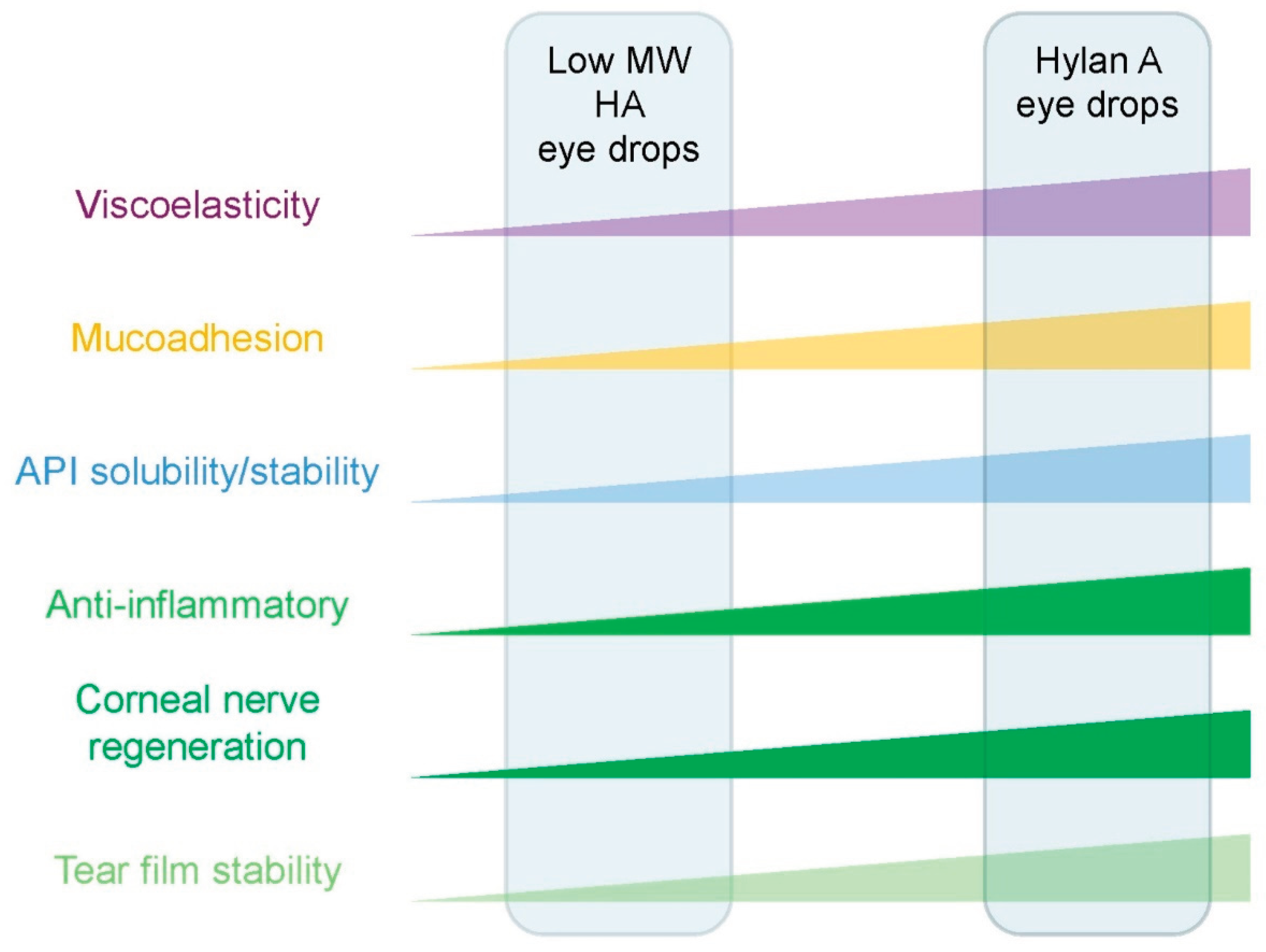

Table 1.
Summary of the studies demonstrating the benefits of the 0.15% hylan A eye drop formulations in ocular surface health.
Table 1.
Summary of the studies demonstrating the benefits of the 0.15% hylan A eye drop formulations in ocular surface health.
| Study | Study type |
Model/ Patients |
Comparisons | Conclusions |
| Kojima et al., 2020 | Preclinical | Mouse model of environmental dry eye disease | Low MW HA eye drops, secretagogue eye drops | Improved tear film stability. Reduced ocular surface damage. Less inflammation with 0.15% hylan A eye drops. |
| Beck et al., 2019 | Clinical | 11 patients treated with autologous serum eye drops | Autologous serum eye drops | Eye drops containing 0.15% hylan A effectively treat severe ocular disease. These 0.15% hylan A eye drops may replace eye drops of autologous serum. |
| van Setten et al., 2020a | Clinical HYLAN M study |
84 patients with severe dry eye disease | Optimized artificial tear treatments | Symptoms rapidly improved by switching to 0.15% hylan A eye drops include: visual stability, discomfort and pain. |
| van Setten et al., 2020b | Clinical. Subgroup analysis of the HYLAN M study | 16 patients | Optimized artificial tear treatments | Switching to 0.15% hylan A eye drops promotes corneal nerve growth. |
| Medic et al., 2024 | Clinical Subgroup analysis of the HYLAN M study |
47 patients | HA containing artificial tears (15 commercial brands with HA of diverse MWs) | Fewer 0.15% hylan A eye drops required than those with lower MW HA. Eye drops containing 0.15% hylan A have better clinical effects. |
|
Özkan et al., 2025 |
Clinical | 63 eyes from 55 patients with keratoconus following corneal crosslinking (CXL) | Low MW HA eye drops | After CXL, 0.15% hylan A eye drops produce: faster corneal nerve regeneration; faster recovery of sensitivity; improved ocular symptoms; and fewer inflammation related immune cells than low MW HA eye drops. |
Table 2.
Summary of the studies demonstrating the benefits of a novel 0.15% hylan A eye drop formulation as a vehicle for latanoprost.
Table 2.
Summary of the studies demonstrating the benefits of a novel 0.15% hylan A eye drop formulation as a vehicle for latanoprost.
| Study | Study type |
Model/ Patients |
Comparators | Conclusions |
| Müller-Lierheim, 2021 | Formulation solubility and stability | N/A | N/A | A preservative-free 0.15% hylan A solution in isotonic phosphate buffered saline (pH 7.4) enhances latanoprost solubility by 75% |
| Dogru et al., 2023 | Preclinical | Standard mouse strain | Commercial eye drops with 50 μg/mL latanoprost |
A hylan A-based eye drop with 14 μg/mL latanoprost preserves ocular surface parameters while effectively lowering IOP |
| Higa et al., 2024 | Preclinical | Standard strain rat | Commercial eye drops with 50 μg/mL latanoprost |
A hylan A-based eye drop with 14 μg/mL latanoprost achieves comparable therapeutic latanoprost levels in aqueous humour as a 50 μg/mL commercial formulation |
| Müller-Lierheim, 2021 | Proof-of-concept | One subject with ocular hypertension | Commercial eye drops with 50 μg/mL latanoprost |
A hylan A-based eye drop with 20 μg/mL latanoprost lowers IOP better than a commercial latanoprost eye drop with a higher API concentration |
Disclaimer/Publisher’s Note: The statements, opinions and data contained in all publications are solely those of the individual author(s) and contributor(s) and not of MDPI and/or the editor(s). MDPI and/or the editor(s) disclaim responsibility for any injury to people or property resulting from any ideas, methods, instructions or products referred to in the content. |
© 2025 by the authors. Licensee MDPI, Basel, Switzerland. This article is an open access article distributed under the terms and conditions of the Creative Commons Attribution (CC BY) license (http://creativecommons.org/licenses/by/4.0/).
Copyright: This open access article is published under a Creative Commons CC BY 4.0 license, which permit the free download, distribution, and reuse, provided that the author and preprint are cited in any reuse.


