Submitted:
02 June 2025
Posted:
03 June 2025
You are already at the latest version
Abstract
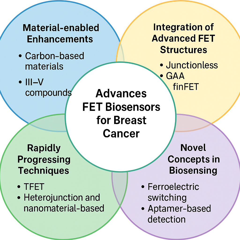
As a consequence of high sensitivity, rapid response, and potential miniaturization, FET-based biosensors have become the leading class of label-free, real-time diagnostic devices due to their favorable characteristics. In the past few years, their use for the purpose of breast cancer detection has come into focus due to the increasing demand for an early, accurate, and non invasive device for diagnostics. This paper offers a thorough examination of advanced FET biosensor structures on Junctionless, Gate All Around (GAA), Dual Material Gate (DMG), and finFET structures, and discusses the integration of these for biosensing. This paper presents the role of nanomaterial-enabled enhancements of FET sensitivity and selectivity for breast cancer biomarkers, including carbon-based materials, metal oxide semiconductors, and III–V compounds such as GaN and InGaAs. It reviews dielectric modulated TFETs, heterojunction and nanomaterial-based FETs, GaN/CNT-based devices, and unconventional hybrid biosensors to specific breast cancer markers including ERα, BRCA1, c-erbB-2, MUC1, and CA125. Through the review, we will discusses the rapidly progressing technological advances in FET bio sensor manufacture and function. We also present a couple of concepts in biosensing that are novel, including polarity-controlled tunneling, ferroelectric switching for logical inference, microfluidic integration, and aptamer-based detection techniques. Lastly, we discuss the main challenges of device reproducibility, biocompatibility, and large-scale integration with future directions for making next-generation FET biosensors for multi-marker breast cancer.
Keywords:
FET biosensors
; TFET
; breast cancer detection
; nanomaterials
; GaN FinFET
; InGaAs heterojunction
; label-free diagnostics
; dielectric modulation
; microfluidics
; graphene
; carbon nanotubes
; III-V semiconductors
; real-time biosensing
; charge plasma
; sensor sensitivity enhancement
Introduction
In recent years, the demand for highly sensitive, real-time, and label-free biosensing platforms has surged in response to the growing need for early-stage disease detection, particularly in cancer diagnostics. Among the most promising technologies, Field Effect Transistor (FET)-based biosensors have emerged as a powerful tool due to their miniaturization potential, compatibility with CMOS processes, and high electrical sensitivity. These biosensors have shown exceptional promise in the detection of breast cancer biomarkers, offering the potential for point-of-care testing using minimally invasive samples such as saliva, serum, and other biofluids [1].
FET biosensors find wide-ranging applications in areas like early cancer diagnostics, personalized medicine, wearable healthcare devices, and real-time biological monitoring. Their ability to detect critical breast cancer-related biomarkers—such as Estrogen Receptor α (ERα), BRCA1, CA125, MUC1, and c-erbB-2—makes them extremely valuable in both research and clinical settings [2]. The integration of nanoscale materials and advanced fabrication techniques has further enabled their deployment in compact, high-performance diagnostic platforms.
Historically, biosensing relied on traditional methods such as fluorescence-based detection, electrochemical sensors, and surface plasmon resonance (SPR) [3]. While effective, these methods often involved complex procedures, bulky equipment, and limited sensitivity at ultra-low analyte concentrations. The development of MOSFET-based biosensors, including Ion-Sensitive Field Effect Transistors (ISFETs) and Extended Gate FETs (EGFETs), marked a significant shift toward label-free, electrically readable biosensing devices [4]. These sensors rely on modulation of the surface potential near the gate to detect biological interactions, offering faster response times and simpler integration into electronic systems.
In response to limitations in traditional FET designs, several advanced architectures have been demonstrated. These include Junctionless FETs, Gate-All-Around (GAA) nanowire FETs, Dual-Material Gate (DMG) FETs, and FinFET-based sensors—all of which offer improved electrostatic control, scalability, and enhanced sensitivity [5]. Parallel to this, the adoption of nanomaterials such as graphene, MoS₂, carbon nanotubes (CNTs), indium selenide (InSe), and III-V semiconductors like gallium nitride (GaN) has led to dramatic improvements in biosensor performance [6]. These developments are especially valuable for detecting breast cancer biomarkers at extremely low concentrations, with high selectivity and rapid response.
The core principle behind FET biosensors lies in the modulation of channel conductivity due to the electrostatic interaction between the gate and biomolecules. Device innovations such as tunnel FETs (TFETs), ferroelectric FETs (FeFETs), and charge-plasma-based FETs have enabled significant advancements in terms of subthreshold performance, charge sensitivity, and energy efficiency. While traditional MOSFET biosensors benefit from simpler fabrication, advanced structures offer superior analytical capabilities at the cost of increased complexity and process variation [7]. The selection of appropriate device architecture and material thus plays a crucial role in the success of biosensor applications.The review concludes by identifying current limitations and projecting future directions, including challenges in device stability, scalability, and integration into real-world diagnostic systems.
Advanced FET Biosensor Architectures and Fabrication
Junctionless and Gate-All-Around (GAA) Nanowire Based Fabrication
The Junctionless Cylindrical Gate-All-Around (CGAA) Si Nanowire (SiNW) MOSFET-based biosensor is designed to detect neutral biomolecules label free. Uricase, Streptavidin, Proteins, Biotin, ChOx, and APTES can be found by observing the changing of dielectric constant. The sensor has high sensitivity, as evidenced by its performance: DIBL(Drain-Induced Barrier Lowering) is 50.47mV/V; subthreshold slope (SS) close to 60 mV/dec; currents ratio of about 9.01×10¹².This sensitivity is mainly attributable to advanced techniques like Triple Material Gate Engineering, using materials such as Molybdenum, Tantalum and Tungsten. These materials make it easier to control the channel, so that even small changes caused by biomolecules binding with it can be accurately detected. A junctionless design removes p-n junctions, thereby reducing short-channel effects (SCEs) and ensuring consistent device performance [8]The Junctionless Gate-All-Around (JLGAA-NTFET) biosensor takes this further with its nanocavity designed to trap biomolecules within it, which decreases leakage current and increases the accuracy of detection. In this way, it is able to detect molecules like ATS with one of the highest dielectric constants. The result is much improved sensitivity and specificity of the sensor [9].To get rid of the variegated and inconsistent results, such as the sense of detecting proteins, such as Streptavidin, the dielectric-modulated, junctionless double gate (JL-DG) MOSFET biosensor is designed . This design avoids abrupt doping between static, channel and drain, reduces the variability of results and improves consistency. The double gate structure enhances control, reduces short-channel effects, and improves the ability of the sensor to detect even small changes. A specially created misaligned cavity increases sensitivity further for biomolecules that have more space, while benefiting those that interact with the surface sensors of this cunningly designed device. By adjusting the size of this cavity, the gate capacitance is increased, which in turn improves sensitivity and specificity while also ensuring reliable detection of biomolecules [10].Figure 1
These sensors are highly sensitive and specific due to their junctionless design and advanced gate engineering that brings better control while reducing short-channel effects. Modeled on the Spacer-Engineered Reconfigurable Silicon Nanowire Schottky Barrier Transistor (SE R-Si NW SBT) biosensor, but aimed at protein detection, it has even higher sensitivity and specificity than the conventional C-RFET(Capacitively Regulated Field-Effect Transistor) biosensor [11]. Within this category of biosensors, the LODs and dynamic ranges provided are not specific; however, its sensitivity and selectivity via spacer-engineered structures plus dual gate structure, make it a very strong sensor for label-free biosensing devices [12].The label-free gate-all-around tunnel field-effect transistor (GAA TFET) biosensor is an improved design over previous ones and boasts the ability to detect within a defined range clinically relevant protein biomarkers. Although the LOD is not specified exactly, the GAA TFET biosensor’s design featuring III-V materials and particular etching at the source-to-channel junction, makes its capacity to detect biomarkers at low concentrations much better, especially in subthreshold region. This design offers a more precise and flexible mechanism for label-free biosensing, with higher sensitivity and selectivity[13].
Dual-Material Gate and FinFET-Based Fabrication
The biosensor of Dual-Materialgate Stack Double-Gate Fin Field-Effect Transistor (DM-GS-DG FinFET) is designed with a nanogap cavity in the gate dielectric region, which is critical for detecting biomolecules by sensing changes in dielectric properties. By making the design more sensitive in this way it is particularly good for detecting biomolecules with different dielectric constants and charge densities. This can be infer from its increases in ON-current, subthreshold swing (SS), transconductance (gm) and output conductance (gd) [14].Advanced fabrication techniques, such as the creation of the nanogap cavity, further improve this sensitivity by increasing the surface area for biomolecules. Biomolecules can interact while at the same time making their response to changes in dielectric properties more marked. This is vital for detecting low-concentration biomolecules. In the dual-material gate stack, the sensor’s electrical properties are also being microscopically controlled. This means improvements in how it handles the electric field and the distribution of charge. Compared to the conventional planar transistor, it raises sensitivity and avoids potential problem states. Furthermore by resorting to materials like nickel-silicide (NiSi) contacts and high-dielectric constants (high-k) dielectrics, the ability of the sensor to inject charge carriers and control the gate are much increased. This is because these are substances that allow the sensor to detect very small changes in the dielectric properties of biomolecules. The sensor can also detect changes in the dielectric properties of molecules that have lower dielectric constants or charge densities [15]The Dual-Metal Double-Gate Vertical Tunnel Field-Effect Transistor (DMDG-VTFET) provides a further improvement, making the sensor even more sensitive and robust by optimizing the distribution of electric field and carrier transport in its dual-gate design. This design greatly enhances the switching ratio, subthreshold swing, and detection without interference from short-channel effects. It is essential for really sensing biomolecular changes, making the sensor a highly effective tool in medical diagnostics and pathogen detection. The top-down approach to manufacturing DMDG-VTFET, permits precise patterning and etching, making it possible to create nanoscale features which are essential for high performance biosensors. This method further increases the surface area available for detection, enhancing sensitivity. As a result, the sensor is able to detect biomolecules at very low concentrations, possibly in the femtomolar (fM) to picomolar (pM) range [16].The synergistic action of nanogap cavities, dual-gate designs, high-k dielectrics, nickel-silicide not only optimizes electric field control, but also improves sensitivity and specificity through improved surface interactions.
A hetero-dielectric Junctionless Tunnel Field
Effect Transistor (HD-JL-TFET) based biomolecule-detecting sensor (HD-JL-TFET)
has the principle of band-to-band tunnelling (BTBT). This sensor concerns
itself with proteins and their dielectric properties. The sensor shows high
sensitivity and specificity as it measures changes in dielectric constant (K)
of biomolecules, which affect drain current (IDS).In addition, such advanced
fabrication techniques as using a nanogap near the source end between the gate
and channel serve to enhance sensitivity. If the sensor detects small changes
in dielectric properties, biomolecules can be detected more easily. Precise
doping of the source, channel and drain areas reduces variability and increases
sensitivity. Moreover, nanocavities created within either the gate-insulator or
gate-metal portion of the gate allow for efficient immobilization of
biomolecules; not only can small amounts be detected but they can also be
immobilized in an even manner [17]. Fabrication
by advanced techniques like the silicon-germanium dielectrically modulated
stacked source/drain (SiGe-DMTG SON) MOSFET biosensor also produces high
sensitivity and specificity. These complex techniques include the introduction
of nanocavities near the gate for specific biomolecule immobilization. A
dual-metal gate structure was used to improve the electric field control and
gate capacitance in response to biomolecule binding. The stacked source/drain
configuration and trenched gate design boost sensitivity further by increasing
the efficiency of carrier injection and cutting parasitic resistances. At the
same time, technical leakages are minimized and other unwanted interactions, which
can negatively affect performance are avoided by employing the
silicon-on-nothing (SON) method [18].Improved
sensitivity and specificity result from these advanced fabrication techniques
that optimize biomolecules interaction with the sensor.
Ion-Sensitive and Extended Gate Field Effect Transistor (ISFET and EGFET) Biosensors
The sensor is an Ion-Sensitive Field-Effect Transistor (ISFET) biosensor, with the enzyme tyrosinase as its protein sensor. The protein sensor is able to detect chlorogenic acid at a low level of 2 muM and ranges between 2 muM to 40 muM. Once the concentration becomes higher than this point, it becomes inadequate for measurement purposes again. The specificity of this sensor is determined from the enzyme tyrosinase action on different phenolic compounds. The strongest signals were obtained for diphenols and trihydroxyphenols. Detection sensitivity of the biosensor was increased by Back-End-of-the-Line (BEOL) post-treatment, which removed the passivation layer. Now, hafnium oxide’s surface comes into direct contact with analyte and makes it more responsive[19]. An Extended-Gate Field-Effect Transistor (EG-FET) sensor, using papain immobilized on gold nanoparticles (Au NPs), is equipped to deal with some areas where ISFETs are not ideal. This system has a lower limit of detection (LOD) for Cystatin C (Cys C) of only 0.05 ag/μL, and its dynamic range extends from 5 ag/μL up to 50 ng/μL. The high specificity and sensitivity of the EG-FET sensor are achieved through the use of advanced manufacturing technologies. This ensures its consistent performance is not disturbed by other substances[20].Additional progress has been made on the Extended Gate Field-Effect Transistor (EGFET) aptasensor for histone proteins. This system has a reduced limit of detection at 5 pM for histone H4. Specifically, thiol-modified RNA aptamers and poly(ethylene glycol) methyl ether thiol (PEG) have been linked up to immobilize on the surface. This decreases nonspecific adsorption, enhancing sensor sensitivity and selectivity[21].These advances are the result of new materials such as hafnium oxide and gold nanoparticles; advanced methods of production like Atomic Layer Deposition (ALD) and laser-induced graphene (LIG) optimize their performance.
The ISFET sensor detected the α-galactosidase A
enzyme, with a LOD of 9.56 × 10⁻¹¹ M and a dynamic range from 1 × 10-11
M to 3.2 × 10⁻⁸ M. With melibiose as the substrate, the sensor shows
a high specificity for α-galactosidase A. It can distinguish normal and
inhibited enzyme activities. Compared with conventional fluorescent assays, the
sensor can detect enzyme concentrations as low as 1 × 10⁻¹⁰ M within
15 minutes, giving faster signal accumulation times. By utilizing fabrication
techniques compatible with standard complementary metal-oxide-semiconductor
(CMOS) processes, the ISFET sensor achieves a high level of sensitivity, which
reduces noise and improves signal accuracy. Post-BEOL processes can reduce
non-idealities such as capacitve attenuation, trapped charges, and drift,
resulting in more reliable measurements. In addition, the sensor is
microfluidically packaged, which is achieved by three-dimensional (3D)
printing, thereby allowing precise control of the reaction environment and
reducing sample contamination while improving measurement reproducibility [22].In contrast, the Extended Gate Field-Effect
Transistor (EGFET) biosensor uses glucose oxidase (GOx) immobilized on vanadium
pentoxide xerogel (V2O5) as its glucose sensor. The sensor’s fabrication
techniques, include covalent immobilization of GOx on vanadium pentoxide
xerogel and dip-coating for uniform thin-film application. The inclusion of
Nafion helps to reduce mass loss and enhances film adhesion without
interferring with charge diffusion; this engenders the sensor’s long-term
stability and reusability [23]. Therefore, by
using advanced fabrication techniques that highly control both the reaction
environment and enzyme attachment to the support, both ISFET and EGFET sensors
can be made better, more accurate and reliable. Figure 2
GaN, AlGaN/GaN-Based, and Other III-V Material Fabrication
The sensor is designed to detect sensitive materials completely without tagging. That carried a potential advantage over conventional techniques such as PCR. It does this with dual gate technology and an InGaN notch, both of which serve to confine electrical carrier movement and reduce leakage. Because of that, it managed a 61% increase in drain current sensibility when detecting Uricase. Drain current reached 4. 602 A/mm with transconductance (gm) added good for 18 milismen/mm (mS/mm). However, it does not give the sensor’s performance indicators equivalent to limit of detection (LOD) and dynamic range. Consequently, some doubts remain about the effectiveness of the product. Consequently, advanced techniques which create nano gaps underneath the gate are utilized. These gaps make it easy for biomolecules to be placed in exactly the right location when the dielectric constant changes and such a change should necessarily affect performance [24]. Despite this rise in sensibility however, the sensor still offers no details about specificity, which is essential for accurate biosensing. The sensor also utilizes double-gate (DG) technology to further control the electric field and reduce the interference of undesired channel effects. It achieves a drain current sensitivity (SION) of 0.328 and a trans-conductance sensitivity (Sgm) of 0.197 [25]. However, even with these developments, the sensor does not effectively address the issue of specificity. The Graphene Field-Effect Transistor (GFET) biosensor gets around some of these problems by delivering far superior sensitivity improvements when targeting SARS-CoV-2 in blood plasma and saliva. Although once again there’s no individual LOD data on its range of charge currents, the bands qualified show differences of up to 80 microamperes(μA) and 89.4 μA, respectively. Use of precise lithography and oxygen(O2) plasma etching in the GFET biosensor gives the sample uniform and accurately shaped channels, thereby both improving sensitivity and contributing toward solving the specificity problem. This makes it an excellent platform for detecting viral proteins with less noise and clearer signals [26].
According to another biosensor called the GaAs 1-xSb x cylindrical dielectric-modulated field-effect transistor (DM-FET), proteins like gelatin, biotin, and bacteriophage can be easily detected, which need no labels. It showed high sensitivity, and specificity, as demonstrated by various metrics, including an OFF to ON current ratio sensitivity (SCR) of 41.20%, saturation current sensitivity (SION) 16.68%, threshold voltage sensitivity (SVth) 52.69%, transconductance sensitivity (Sgm) 50.66%, and OFF-state current sensitivity (SIOFF) 99.92%. These impressive results come from advanced fabrication techniques, such as the device’s cylindrical shape offering better gate control than flat devices and an embedded nanogap cavity that increases the area where the sensor interacts with biomolecules. The sensor also includes a thin radial chromium layer in between oxide and gate electrode to isolate biomolecules inside the cavity, thereby reducing noise and improving specificity. However, the design may still struggle with reducing leakage and ambipolar currents, which could affect sensitivity at very low biomolecule concentrations [27].These improvements are taken further in the Dielectric-Modulated Split-Drain Z-shaped Gate Tunnel Field-Effect Transistor (DM-SDZ-TFET) biosensor. This design uses split-drain architectures and varied doping profiles to reduce ambipolar and leakage currents, thus boosting sensitivity significantly. This sensor is particularly effective at detecting proteins, can be concluded from the fact that it can reach an on-current (Ion) in the order of 10-4 A/μm with a dielectric constant (K) of 12 (gelatin) and has a switching current ratio sensitivity (SIon/Ioff) around 1.5 * 104. Precise location of the gate oxide etching to form a nanocavity improves biomolecule immobilization, directly changing electrical characteristics of the sensor. The use of ultra-high vacuum electron-beam deposition and in situ post-deposition annealing ensures direct, high-quality contacts between oxides (HfO2 and SiO2) and silicon, leading to excellent interface quality. Also, the inclusion of an n+ pocket in the source region increases tunneling rate by providing more paths, further enhancing sensitivity. Although the DM-SDZ-TFET shows great improvements, there is still a need for fine-tuning the sensor surface in terms of detecting even smaller changes in dielectric properties [28].The enzyme-free biosensor goes further. It concentrates on detecting proteins like uricase, streptavidin, biotin, apetide, keratin by observing changes in dielectric properties. This sensor increases sensitivity through precisely formed nanocavities underneath two metal gates, which serve as the controlled location to place biomolecules and allow interaction with target molecules. The dual-material gate (DMG) design used in the sensor improves control over electric field and surface potential, leading to even greater sensitivity. Additionally, the overlapped gate-on-drain structure enhances tunneling effect, which is vital for detecting small changes in dielectric properties. Advanced lithography techniques ensure high precision in the sensor’s components, reducing defects and thus increasing performance. During fabrication, the dielectric modulation technique is used again to enhance sensor’s ability to detect minor changes in the dielectric constant of target molecules. This achieves superior sensitivity and accuracy compared to previous designs [29]. The remarkable performance of these biosensors is due to advanced fabrication methods and precision designs that maximize its interaction with biomolecules and improve electric response.
Advanced Fabrication Techniques and Specialized Applications
The dielectric-modified thin-film transistor (DMTFT) biosensor All the distinctive characteristics of a nano-cavity to the gate insulator is used to improve the sensitivity and specificity. This cavity can gather biomolecules, which changes the dielectric properties and gate capacitance. However, it may result that the semiconducting thin-film is unprotected during the earlier stages of production. To overcome this last obstacle, making certain to maintain its high mobility and reliability, the thin film is added [30].The dielectric-modulated silicon-on-insulator junctionless Fin field-effect transistor (DM-SOI-JL-FinFET) biosensor. It conserves and expands on it by utilizing reactive ion etching (RIE) to make extremely uniform nanocavities. These cavities can only let in certain biomolecules, thus enhancing specificity. Electron beam lithography is also used to create fins with a high aspect ratio. This increases the surface area available for biomolecule interaction and thus makes the biosensor more sensitive [31].Keeping sensor performance consistent is a challenge. Take the aluminum nitride/beta gallium oxide high-electron-mobility transistor (AlN/β-Ga2O3 HEMT) based biosensor as an example. To counter this weakness, it uses high-resolution lithography to position the gate, source, and drain electrodes precisely. As a result, leakage currents decrease and the space available to biomolecule interaction increases. A Schottky contact with a specific gate work function offers further sensitivity and specificity that ensure efficient charge transfer [32].Finally, charge plasma doping and formation of a heterojunction with a low bandgap material (Si0.5Ge0.5) at its source region are all part of the newly proposed biosensor. This lowers the bandgap and hence raises tunneling efficiency. When it comes to ON-state current and overall sensitivity and specificity, this application gives an improvement over earlier architecture [33].This is due to precise nanofabrication that improves biomolecule targeting, charge transfer efficiency, and sensor reliability.
Nanomaterials for High-Performance FET Biosensors
Carbon-Based Materials
In the Graphene Field Effect Transistor (GFET), the materials used for detecting Myoglobin are essential for high sensitivity and specificity. However, graphene’s zero bandgap creates difficulty to realize clear on/off switching, which is required for some sensing applications [34]. For this purpose, Field Effect Transistor reduced graphene oxide (rGO) is used. Conductive, like graphene, rGO incorporates oxygen functional groups that can control its electronic properties. This provides finer switches for the sensor ‘s on/off states, making it more effective in detecting the smaller electrical signal changes which occur during antigen-antibody interaction [35].Contrary to the above problem, rGO promotes specificity but also bring some troubles. To address this, the carbon nanotube(CNT) Field Effect Transistor (FET) uses carbon nanotubes which are an effective way of addressing these challenges. Nanotubes have the triple benefits of high conductivity, large surface area, and relative freedom from non-specific interactions or cross-reaction foreign particles, properties that make them ideally suited to biosensor operation [36].However Owing to their metallic content, CNTs can introduce noise. This problem is avoided altogether in the single-walled carbon nanotube (SWCNT) based biosensor for Human Serum Albumin (HSA) detection. In this case the sensor uses CoMoCAT SG 65 SWCNTs, 90% of which are semiconducting, so as to improve its on/off current ratio and reliability [37].Although with SWCNTs a higher sensitivity and lower noise were achieved, the graphene oxide (GO) field effect transistor (FET) for COVID-19 spike protein detection takes sensitivity plus specificity even further. It uses gold nanoparticles on platinum and palladium metal foils in the SGL It therefore overcomes earlier materials ‘ weaknesses, ensuring good performance in real-time diagnostics [38].Each materials by fine-crafting conductivity, surface area and binding specificity well-tuned bio-sensors for improved diagnostic accuracy can be manufactured. Figure 3
Metal-Oxide Semiconductors
The sensitivity and specificity of the MoS2-based dual-gate MOSFET biosensor can be optimized by the material used. MoS2 has high carrier mobility, excellent thermal stability and a moderate bandgap; together these conduce to great sensitivity. Its very thin structure also provides large specific surface areas for interaction with target biomolecules. At the same time, MoS2 has an admirable ON/OFF current ratio that clarifies whether or not target molecules are present. However, the complexity of making and maintaining specific chemical groups or biological molecules in the nanogap may affect MoS2 sensor’s performance [39].To solve this problem the sensor uses a simple but practical method, gold-plated electrodes. These electrodes are both stable and able to conduct, thus providing an ideal surface for immobilizing biomolecules and ensuring efficient electron transfer. Deionized water and ethanol are used to prepare high purity solvents which reduce impurity levels; this makes nanomaterials effective for medulloblastoma detection [40]. The stability and conductivity gained by gold-plated electrodes is a good achievement, but getting biomolecules to insert and orient themselves correctly is still a problem. This difficulty can be overcome by preparing gold electrodes in combination with thioglycolic acid (TGA) and reagents like N, N’-Dicyclohexylcarbodiimide (DCC) and N-Hydroxysuccinimide (NHS). These substances form strong bonds that hold the antibody securely on the electrode surface, ensuring the right mode of orientation and the best antigen binding. This will improve the sensitivity and specificity to detect biomarkers for breast cancer detection [41].
For the IGZO Field-Effect Transistor (FET) biosensor array, it is crucial that the indium gallium zinc oxide has the right material. Its features with high electron mobility, excellent electrical properties, and low off-current advance the signal-to-noise ratio to improve the sensitivity of test values. With its ample surface area, on the other hand, IGZO also creates a big arena for immobilizing antibodies. This makes it more likely that we will be able to trap target proteins [42].While IGZO ensures consistency in sensor signals, MoS2 further offers a big advantage to stability with its perfect interface that does not have dangling bonds. So it is quite stable here for reliable chip biosensing, addressing potential problems of stability at design time. MoS2’s semiconductor properties, which include a decent bandgap and high sensitivity to charged substances, enhance the sensitivity of electrical signal measurements and make it readily responsive to target protein binding [43].The advantages of MoS2 are further enhanced by the indium tin oxide (ITO) material used in double-gate oxide-semiconductor thin-film transistor (TFT) technology for SARS-CoV-2 detection. On the surface of ITO, ultraviolet (UV)-ozone treatment increases the number of hydroxyl groups, conducive to linkers binding and subsequent antibody immobilization. This increased hydrophilicity guarantees that it will interact strongly and specifically with antibodies, giving higher specificity and sensitivity in detecting target proteins. Compared as well to the limitations of MoS2, this approach improves the efficiency of linkage and functionalizing ITO for bioassays and especially the total binding efficiency [44].These materials offer excellent performance because of their ability to accurately control electrical properties and surface chemistry, thus maximizing both the capture of biomolecules and the accuracy of signal detection. Figure 4
Gallium Nitride (GaN) and Related Materials
The choice of materials is the key to sensor performance. Gallium Nitride (GaAs) is preferred material as it has higher electron mobility and lower noise as compared to Silicon; it can therefore detect smaller current changes caused by diffusion processes of biomolecular molecule interactions. The Aluminium Gallium Nitride/Galium Nitride (AlGaN/GaN) heterostructure further increases this sensitivity. It forms a two-dimensional electron gas (2DEG) channel that is greatly sensitive to changes in concentration when analytes bind on it [45].Gold surfaces in the GaN High Electron Mobility Transistor (HEMT) sensor assure strong stable binding of antibodies. Another method involves using EDC and NHS chemicals to prepare more reactive binding sites. It enhances the precision of antibody attachment and improves antigen binding better [46].The Double-Gate Aluminum Gallium Nitride/Gallium Nitride Metal Oxide Semiconductor High Electron Mobility Transistor (DG-AlGaN/GaN MOSHEMT) sensor uses Nickel/Gold (Ni/Au) and Aluminium/Gold (Au/Al) contacts. These contacts facilitate better charge flow, which in turn eases biomolecule interactions with the sensor [47].The Electrical Double Layer (EDL)-Gated Field-Effect Transistor (FET)-based biosensor (BioFET) uses gold electrodes and SU-8 photoresist. These materials will guarantee that the sensor is biocompatible, accurately patterned as well as working consistently. This combination resolves many issues in achieving high sensitivity and specificity [48].These materials and methods taken together result in more accurate and reliable sensors.
Silicon-Based Nanowires and Related Structures
Silicon nanowires are the basis of the silicon nanowire field-effect transistor (SiNW-FET) biosensor. At just a few nano meter in diameter, these nanowires have excellent semiconductor properties and a large surface area, making them especially effective for biosensing. When target molecules bind to these nanowires, the electrical signals that result represent changes in the natural language of all life. This leads to very reliable performance, based as it is on tried and tested silicon technology[49]. However, there are limits to the functionalization of silicon nanowires. The silicon nanowire bio field-effect transistor (SiNW Bio-FET) deals with this issue by treating the surface with APTES (amino propyltriethoxysilane) and glutaraldehyde. Treatment in this way ensures that antibodies bind evenly, minimizing non-specific interactions [50].To enhance long-term stability, the silicon nanowire field-effect transistor (SiNWFET) for cardiac troponin I (cTnI) detection integrates into silicon’s own chemical properties. This means that antibody binding is stronger and more durable over time, significantly increasing the sensor’s sensitivity and specificity [51]. However, silicon’s reactivity could cause problems in different environments. The extended gate field-effect transistor (EGFET) biosensor solves this by using hafnium oxide (HfO2), a material that offers better stability and is more responsive. This guarantees consistent performance [52].Finally, high sensitivity at very low concentrations is necessary. The meta nano-channel bio-field-effect transistor (MNC BioFET), which uses silicon-on-insulator (SOI) wafers and a high-quality dielectric material, such as thin thermal oxide. These materials create an effective interface and take away unwanted electrical effects. The result is ultra low detection limits along with extremely good sensor performance [53]. These improvements involve careful selection of materials that increase binding efficiency, stability and long lifetime.
FET-Based Biosensors for Breast Cancer Detection
Dielectric-Modulated TFETs for Breast Cancer Detection
It is very important to diagnose breast cancer on an early stage so they could be treated effectively to achieve the highest chances of survival. As a smart screening tool, biosensors have evolved into identifying the biomarkers of the breast cancer initiation, progression and treatment. For the purpose of breast cancer cell lines detection, this work proposes dielectric modulated dopingless dual metal gate heterojunction Tunnel Field Effect Transistor (DM–DL–DMG–HJTFET) which has a nanogap underneath the gate electrode towards the source side where the breast cells are immobilized. The different regions inside a breast tissue also have different dielectric property such as cancerous and normally healthy breast have different cell conductivity such that it can be helpful to early breast cancer detection. The biosensor is simulated using the SILVACO ATLAS TCAD tool and the calibrated results are utilized for the in depth analysis of the energy band diagram, electric field and surface potential. Various partially and non-uniformly filled cavities with mixed cell proportions are analyzed with respect to practical functionality. The evaluated sensitivity is in terms of drain current, threshold voltage, transconductance and current ratio and reflects considerable improvement in sensitivity, suitable for point of care (POC) diagnostics [54]. On this basis, a biosensor is proposed for the first time using a dielectric modulated polarity control tunnel field effect transistor (DM—PC—TFET) specifically for BCCs detection using the BCCs dielectric contrast with the dielectric of the healthy cells at microwave frequencies. Early detection is assisted by device electrical characteristics changing when BCCs fill the sensing region. A device sensitivity analysis is carried out, and the device possesses high sensitivity in terms of drain current and transconductance. In addition, selectivity, noise, and linearity characteristics as well as sensitivity to cell charge density, temperature, geometry and non-uniform cell arrangement were evaluated [55]. A novel dielectric modulated step channel Junctionless tunnel field effect transistor (DM-SC-JLTFET) designed for label free detection using dielectric constant values of breast cancer cells captured at 200 MHz using open ended coaxial probe technique further enhances the feature for detection capability. The efficacious biosensor incorporating only a dash of charge plasma is simplified via a concept of the charge plasma to suppress random dopant fluctuation (RDF), thereby improving the efficacy as well as simplifying the fabrication. The device shows the potential of being a strong alternative to the traditional breast cancer detection methods (e.g., mammography) with high sensitivity and low subthreshold swing (SS) in the reduced substrate thickness and with a step channel structure[8]. Silvaco Atlas TCAD simulation further advances the design of a dielectrically modulated drain pocket dual gate Tunnel Field Effect Transistor (DM DPDG TFET) biosensor with an integrated nanocavity towards the detection of, breast cancer cells. The high sensitivity and superior electrical performance of this device can be inferred from its response to changes in the dielectric constant of the nanocavity caused by immobilized biomolecules, namely MDA-MB-231 cells (k=22). Device characteristics are optimized by improving the thickness of the nanocavity and the length of the oxide layer, and sensitivity is evaluated based on on-state current (Ion), threshold voltage (Vth), transconductance (gm), and subthreshold swing (SS). Better performance than existing biosensors in terms of cost and simplicity; larger ion-to-off ratio; consequently, to find applications in identifying breast cancer as well as other diseases such as SARS COV 2 [56], but further improvement in device architecture, nanocavity design, and dielectric tuning enhances sensitivity in biosensors.
Heterojunction FETs for Cancer Biomarkers
The recent years have seen breast cancer become one of the most common carcinomas and the most common cause of cancer-related deaths. Early-stage detection is vital for reducing deaths through early, effective treatments. For the same, the DMG-HJ-SOI-TFET device is proposed for the label-free detection of breast cancer cells (BCCs) using their unique dielectric constant values [57]. By using InSb/Si heterojunction and nanocavity architecture, ON current and sensitivity for sensing T47D cells are enhanced while power consumption and leakage are minimized, making it for point-of-care diagnostics. To address the limitations of sensing selectivity and speed in the fluid samples such as blood, a dual cavity HJ-DL-TFET biosensor is proposed using a heterojunction of In₁₋ₓGaₓAs/Si [1]. This provides fast, real-time detection without the complexity of traditional methods, modifies the interface charges of biomarkers such as C-erbB-2 in saliva and serum in an efficient way to improve tunneling probability and sensitivity. The III–V GAA TFET biosensor is developed to improve performance under low power [58]. To achieve high sensitivity and fast response towards both breast and prostate cancer biomarkers, it operates in the subthreshold region of the device and uses the transconductance generation factor (gm/IDS) as the metric. Based on charge deduction and cavity length tuning, this work demonstrates precise sensitivity under different health conditions and across males and females, which is also effective in handling limitations associated with reducing response times and variability in earlier models. In final, to solve the design flexibility and enhance the detection capabilities, CP-GS-ArcTFET biosensor is proposed by employing charge plasma and gate stack engineering with various central angles (θ) [59]. In addition to improving analog performance indicators such as gain bandwidth product and intrinsic delay, this design has the advantage of increasing sensitivity with current ratio metrics, particularly at high levels of C erbB 2. It establishes an advanced approach that surpasses the prior models by combining a high ON/OFF ratio, a low subthreshold swing, and improved control over electrical parameters, thereby enabling more robust, flexible, and accurate biosensing across diverse diagnostic settings. Biosensor sensitivity and performance can be significantly improved by integrating charge-plasma concepts, gate-stack engineering, and curved channel designs, which enhance electric field control, carrier transport, and analog response metrics.
Nanomaterial-Based FETs
Breast cancer and its treatment are very reliant on the detection of a single biomarker, estrogen receptor α (ERα). For rapid, label-free detection of ERα, a novel drug molecule was employed as a capture probe in this study to develop a liquid gated graphene field effect transistor (FET) biosensor, which had detection limit of 2.62 fM and the response within 30 min. However, this sensor is highly sensitive and selective for the ERα positive samples; and is restricted to protein detection from cell lysates only [60]. A label free aptasensor of a MoS₂ FET modified with an aptamer–molecularly imprinted polymer (MIP) hybrid was introduced to overcome the limitation of selectivity and to broaden biomarker scope for determining BRCA1 gene. An ultra-low detection limit of 3.0 aM and high selectivity, even the ability to distinguish one-base mismatch in DNA sequence and detection BRCA1 in spiked serum samples, this aptasensor has advanced the ability of accurate nucleic acid based biosensing [61]. A CNT-FET biosensor made of polymer sorted, high purity semiconducting carbon nanotube films was developed to address the challenge of the early stage detection of exosomal proteins and for the label free detection of MUC1, a breast cancer associated exosomal protein. Although it was difficult to functionize the CNTs, poly-lysine and gold nanoparticles allowed stable aptamer attachment with a highly sensitive sensor (from 0.34 fg/mL) and clear discrimination between the cancer patients and the healthy individuals [62]. To additionally address issues associated with biological stability of the InSe-FET in liquid environments that are ubiquitous in those biosensors, researchers designed and integrated to a microfluidic channel for the stable and reliable detection of the CA125 biomarker in clinical samples. High sensitivity together with electrical stability in a liquid environment are the prerequisites for the application of FET based biosensors in the clinical and portable diagnostic fields [63]. The InSe channel was passivated to ensure the electrical stability, which allowed the device to have broad detection frequency of 0.01 - 1000 U/mL and a detection time of 20 min, and thus, make it a good biosensor for real world application as a portable diagnostic device [63].Achieving high sensitivity alongside electrical stability in liquid environments is key for advancing FET-based biosensors toward reliable clinical and portable diagnostic applications. Figure 5
GaN- and CNT-Based FETs for Breast Cancer
The proposed Gallium Nitride (GaN) Fin Field Effect Transistor (FinFET) with Schottky Source/Drain to detect breast cancer cells is analyzed based on changes in conductivity due to differences in the dielectric properties between healthy (MCF-10A) and invasive cancerous (MDA MB-231) cells in the range from 200 MHz to 13.5 GHz [64]. This method is further improved by the output conductance and transient analysis of 900 MHz and 10 GHz with optimized device parameters like channel material, fin height, drain voltage and temperature to further improve the sensing performance [64]. In order to overcome the frequency specific sensitivity limitation of this approach, a compact GaN high electron mobility transistor (HEMT) based biosensor is developed that has the capability to detect C erbB2, the breast cancer biomarker, within an initial 6 hours and with a 31% change in drain current following 6 hours of incubation with thioglycolic acid and monoclonal antibodies, while demonstrating high noise immunity [65]. However, a dielectric modulated plasma assisted carbon nanotube FET (DM-PA CNTFET) with a dual metal gate all around structure plus a dielectric stack provides a selectivity and further miniaturization by achieving a high drain ON current sensitivity of 343.35 nA and threshold voltage sensitivity of 293.23 mV for MDA-MB-231 cells [66].Despite the high precision and electrical performance of an DM-PA-CNTFET, its structural complexity is well arbitraged by the Interconnected Multichannel Schottky FinFET (IC–S-FinFET). It is a device featuring dielectrically controlled nanocavities, which probe some important parameters like subthreshold swing, ION/IOFF ratio, and the sensitivity to biomolecular charges. Moreover, it takes into consideration the effect of the fill factor on nanogap response, thus improving nanogap sensor’s capability for label free and noise resistant detection [2]. Overall, this is a very powerful tool for early, precise breast cancer diagnosis, as it is capable of simultaneously detecting multiple biomarker types with high sensitivity, stable operation across a variety of conditions, yet with low noise label-free detection process.
Multi-Target FET Biosensor
The research presents analysis of a modified gate oxide tunnel field effect transistor (TFET) biosensor where pocket (Poc MGOTFET) is used for real time and highly sensitive identification of the breast cancer (BC) biomarker C-erbB-2 in serum and saliva by the use of dual cavity structure and interface charge modulation thereby improving ON/OFF current ratio and reduction in leakage current [67]. Nevertheless, though it presents high sensitivity, Poc-MGOTFET is then improved by the poly-l-lysine functionalized graphene field-effect transistor (PGFET) biosensor, yielding ultrasensitive, rapid, in just 20 min, detection of breast cancer miRNAs and SARS CoV 2 viral RNA by 113% vs conventional GFET’s due to PLL functionalization and electrostatic DNA probe immobilization [68]. PGFET offers the most superior detection speed and versatility, but lacks the compactness and energy efficiency essential to portable systems, which are addressed by a single Ferroelectric FET (FeFET) based implementation of the Muller C element for highly accurate Bayesian inference for breast cancer diagnosis on resource constrained platforms (4.1 fJ and 0.07 μm2) [69]. The FeFET based system is extremely efficient regarding energy and computations, however the system functionality is further improved by incorporation of a multilayered Si doped molybdenum disulfide (MoS₂) thickness engineered TFET (TE-TFET) biosensor. By maximizing the device geometry and modulating dielectric charges, this biosensor was also able to enhance the key electrical characteristics such as the transconductance, threshold voltage, and the on-current. Thus, these improvements permit the reliable and economical identification of a variety of breast cancer lines including Hs578T, MDA-MB-231, MCF7, and T47D with CMOS compatibility, repeatability, and diagnostic suitability for in vivo imaging [70]. This integrated improvement in biosensor technology, addressing the problems with past systems, until finally reaching the faster response, more sensitive, more efficient and cheaper breast cancer detection. Table 1
Conclusion
The field of FET-based biosensors for breast cancer detection has seen significant progress, particularly from a fabrication standpoint, with innovations in architectures such as junctionless transistors, gate-all-around (GAA) nanowires, dual-material gates, FinFETs, and ISFET/EGFET configurations that enhance device sensitivity, scalability, and integration potential. These biosensors, offering label-free, real-time, and miniaturized detection, outperform conventional methods in both performance and compatibility with CMOS fabrication, making them promising candidates for point-of-care diagnostics. This review has systematically classified advanced FET biosensors into key groups—dielectric-modulated TFETs, heterojunction and GAA TFETs, nanomaterial-based FETs, GaN/CNT-based structures, and hybrid/multifunctional systems—highlighting each design’s contribution in improving transduction efficiency, charge control, and biomarker specificity. Mechanisms such as band-to-band tunneling, charge plasma gating, ferroelectric switching, and microfluidic integration play a vital role in defining the sensing performance, emphasizing the importance of coupling advanced electrical design with biorecognition interfaces. Despite remarkable achievements, challenges remain in standardizing fabrication processes, ensuring long-term stability in physiological environments, and improving multiplexed detection. Materials like graphene, CNTs, MoS₂, InSe, GaN, and silicon nanowires have each contributed uniquely to performance optimization, while the exploration of new materials and unconventional concepts like ferroelectric logic inference, aptamer-modified gates, and tunneling-based detection offer exciting avenues for further development. Future research should focus on closing the gaps in reproducibility, reliability, and multifunctional integration, paving the way for next-generation intelligent biosensors capable of real-time, high-throughput breast cancer diagnostics.
Funding
No funding was provided for the development of this manuscript.
Acknowledgments
The authors would like to express sincere gratitude to the Shreenivas Deshpande Library at the Indian Institute of Technology (BHU) Varanasi for providing invaluable resources and support.
Authors Contribution
DUR: Written original draft; conducted a survey of the literature; prepared the tables; collected the references; methodology; edited and proofread the final manuscript.
Availability of data and material
All data relevant to this review are included in the text, references, tables, and figures.
Ethics approval and consent to participate
Not applicable.
Consent for publication
Not applicable.
Competing interests
The authors declare that they have no competing interests.
References
- Singh, S., N. K. Singh, and S. Singh, Breast-cancer biomarker (C-erbB-2) detection in saliva/serum based on InGaAs/Si heterojunction dopingless TFET biosensor. IEEE Transactions on NanoBioscience, 2021. 22(1): p. 28-37.
- Shalini, V.; Kumar, P. Design and analysis of interconnected multichannel Schottky FinFET for the detection of breast cancer cells. Micro Nanostructures 2025, 199. [Google Scholar] [CrossRef]
- Camarca, A.; Varriale, A.; Capo, A.; Pennacchio, A.; Calabrese, A.; Giannattasio, C.; Almuzara, C.M.; D’auria, S.; Staiano, M. Emergent Biosensing Technologies Based on Fluorescence Spectroscopy and Surface Plasmon Resonance. Sensors 2021, 21, 906. [Google Scholar] [CrossRef]
- Ma, X. , et al., Recent advances in ion-sensitive field-effect transistors for biosensing applications. Electrochemical Science Advances, 2023. 3(3): p. e2100163.
- Das, R.R.; Rajalekshmi, T.R.; James, A. FinFET to GAA MBCFET: A Review and Insights. IEEE Access 2024, 12, 50556–50577. [Google Scholar] [CrossRef]
- McCreary, A.; Kazakova, O.; Jariwala, D.; Al Balushi, Z.Y. An outlook into the flat land of 2D materials beyond graphene: synthesis, properties and device applications. 2D Mater. 2020, 8, 013001. [Google Scholar] [CrossRef]
- Naresh, V.; Lee, N. A Review on Biosensors and Recent Development of Nanostructured Materials-Enabled Biosensors. Sensors 2021, 21, 1109. [Google Scholar] [CrossRef]
- Sanjay; Kumar, V. ; Vohra, A. Sensitivity enhancement using triple metal gate work function engineering of junctionless cylindrical gate all around SiNW MOSFET based biosensor for neutral biomolecule species detection for upcoming sub 14 nm technology node. Mater. Sci. Eng. B 2024, 306. [Google Scholar] [CrossRef]
- Kumar, P.; Raj, B.; Wadhwa, G.; Singh, B.; Kumar, R. Design and Analysis of Junctionless-Based Gate All Around N+ Doped Layer Nanowire TFET Biosensor. ECS J. Solid State Sci. Technol. 2024, 13, 017002. [Google Scholar] [CrossRef]
- Kumar, S.; Chauhan, R.K. Tweaking the Performance of Dielectric Modulated Junctionless Double Gate Metal Oxide Field Effect Transistor-Based Label-Free Biosensor. J. Electrochem. Soc. 2024, 171, 017503. [Google Scholar] [CrossRef]
- Hussian, A.; Alkhammash, H.I.; Wani, M.S.; Loan, S.A. Metal Strip Implanted Tunneling Field-Effect Transistor Biosensor as a Label-Free Biosensor. ACS Appl. Bio Mater. 2024, 7, 4633–4641. [Google Scholar] [CrossRef]
- Kumar, A.; Kale, S. Spacer-Engineered Reconfigurable Silicon Nanowire Schottky Barrier Transistor as a Label-Free Biosensor. Silicon 2023, 16, 2023–2036. [Google Scholar] [CrossRef]
- Kumar, P. and K. Koley, Breast Cancer and Prostate Cancer Detection Considering Transconductance Generation Factor (g m/I DS) as a Sensing Metric for III-V Gate-all-around Tunnel FET Biosensor. IEEE Sensors Journal, 2023.
- Pattnaik, A.; Mohapatra, S.K.; Dastidar, A.; Acharya, O.P.; AbdelAll, N.; A El-Badry, B.; Khouqeer, G.A.; Alodhayb, A.N. Design and Simulation of Dielectrically Modulated Dual Material Gate-Stack Double-Gate FinFET Biosensor. ECS J. Solid State Sci. Technol. 2024, 13, 057002. [Google Scholar] [CrossRef]
- Dharmender; Nigam, K. K.; Yadav, P.; Tikkiwal, V.A. Performance analysis of dual material control gate cavity on source electrically doped TFET biosensor for biomedical applications. Micro Nanostructures 2024, 191. [Google Scholar] [CrossRef]
- Das, S.; Kumar, B.B.; Bundela, P.; Singh, K. Performance assessment of Si based dual metal double gate vertical TFET biosensor. Micro Nanostructures 2024, 191. [Google Scholar] [CrossRef]
- Kumawat, M.; Gopal, G.; Varma, T. Design and analysis of hetero-dielectric Junctionless-TFET(JL-TFET) with N+ pocket as label free biosensors. Phys. Scr. 2024, 99, 045405. [Google Scholar] [CrossRef]
- Mishra, S.; Mohanty, S.S.; Mishra, G.P. Gate electrode stacked source/drain SON trench MOSFET for biosensing application. Phys. Scr. 2023, 98, 125027. [Google Scholar] [CrossRef]
- Gubanova, O.; Poletaev, A.; Komarova, N.; Grudtsov, V.; Ryazantsev, D.; Shustinskiy, M.; Shibalov, M.; Kuznetsov, A. A novel extended gate ISFET design for biosensing application compatible with standard CMOS. Mater. Sci. Semicond. Process. 2024, 177. [Google Scholar] [CrossRef]
- Chen, X.; Liang, Y.; Tang, N.; Li, C.; Zhang, Y.; Xu, F.; Shi, G.; Zhang, M. Ultrasensitive sensing urinary cystatin C via an interface-engineered graphene extended-gate field-effect transistor for non-invasive diagnosis of chronic kidney disease. Biosens. Bioelectron. 2024, 249, 116016. [Google Scholar] [CrossRef]
- Richardson, H.; Barahona, J.; Medwig, G.; Johns, A.; Pérez, L.M.A.; Sode, K.; Daniele, M.; Miller, F.J.; Lobaton, E.; Pavlidis, S. Towards monitoring of critical illness via the detection of histones with extended gate field-effect transistor sensors. Biosens. Bioelectron. X 2024, 19. [Google Scholar] [CrossRef]
- Kuznetsov, A.; Sheshil, A.; Smolin, E.; Grudtsov, V.; Ryazantsev, D.; Shustinskiy, M.; Tikhonova, T.; Kitiashvili, I.; Vechorko, V.; Komarova, N. Detection of α-Galactosidase A Reaction in Samples Extracted from Dried Blood Spots Using Ion-Sensitive Field Effect Transistors. Sensors 2024, 24, 3681. [Google Scholar] [CrossRef]
- Felix, A.T., M. Mulato, and E.M. Guerra, Evaluation of sensitivity of Extended Gate Field Effect Transistor-biosensor based on V2O5/GOx for glucose detection. Enzyme and Microbial Technology, 2024. 177: p. 110428.
- Mishra, G.S.; Mohankumar, N.; Singh, S.K. Sensitivity improvement in gate engineered technique dielectric modulated GaN MOSHEMT with InGaN notch for label-free biosensing. Eng. Res. Express 2024, 6, 025309. [Google Scholar] [CrossRef]
- Sriramani, P.; Mohankumar, N.; Prasamsha, Y. Drain current sensitivity analysis using a surface potential-based analytical model for AlGaN/GaN double gate MOS-HEMT. Micro Nanostructures 2023, 185. [Google Scholar] [CrossRef]
- Rufino, F.C.; de Almeida, C.R.; Sales, G.; César, R.; Vidal, M.; Delafiori, J.; de Oliveira, A.; Busanello, E.; Siciliano, R.; Nicolau, J.C.; et al. Non-Functionalized Graphene Ribbons FET Biosensor Platform: SARS-CoV-2 Detection on TiO2Gate Dielectric Windows. IEEE Sensors J. 2024, 24, 18791–18804. [Google Scholar] [CrossRef]
- Dixit, A. , et al., Biomolecule detection using GaAs1− xSbX FET based dielectric modulated label-free biosensor. Physica Scripta, 2024. 99(2): p. 025020.
- Bitra, J.; Komanapalli, G. An Improved Z-Shaped Dual-Material-Gate DM-SDZ-TFET Biosensor for Label-Free Detection. J. Electron. Mater. 2024, 53, 1445–1460. [Google Scholar] [CrossRef]
- Venkatesh, M.; Parthasarathy, P.; Kumar, U.A. Surface Potential Analysis of Dual Material Gate Silicon-Based Ferroelectric TFET for Biosensing Application. ECS J. Solid State Sci. Technol. 2024, 13, 017001. [Google Scholar] [CrossRef]
- Ahangari, Z. Design and simulation of a nano biosensor based on amorphous indium gallium zinc oxide (a-IGZO) thin film transistor. Semicond. Sci. Technol. 2024, 39, 035011. [Google Scholar] [CrossRef]
- Raj, A.; Sharma, S.K. Exploring the Potential of Dielectric Modulated SOI Junctionless FinFETs for Label-Free Biosensing. J. Electron. Mater. 2023, 53, 766–772. [Google Scholar] [CrossRef]
- Tomar, A. , et al. AlN/β-Ga₂O₃ MOSHEMT as Biosensor. in 2024 IEEE Applied Sensing Conference (APSCON). 2024. IEEE.
- Mishra, V.; Agarwal, L.; Tiwari, C.; Rathi, V. Dielectric Modulated Negative Capacitance Heterojunction TFET as Biosensor: Proposal and Analysis. Silicon 2024, 16, 3041–3053. [Google Scholar] [CrossRef]
- Krsihna, B.V.; Gangadhar, A.; Ravi, S.; Mohan, D.; Panigrahy, A.K.; Rajeswari, V.R.; Prakash, M.D. A Highly Sensitive Graphene-based Field Effect Transistor for the Detection of Myoglobin. Silicon 2022, 14, 11741–11748. [Google Scholar] [CrossRef]
- Krsihna, B.V.; Ahmadsaidulu, S.; Teja, S.S.T.; Jayanthi, D.; Navaneetha, A.; Reddy, P.R.; Prakash, M.D. Design and Development of Graphene FET Biosensor for the Detection of SARS-CoV-2. Silicon 2021, 14, 5913–5921. [Google Scholar] [CrossRef]
- Zamzami, M.A.; Rabbani, G.; Ahmad, A.; Basalah, A.A.; Al-Sabban, W.H.; Ahn, S.N.; Choudhry, H. Carbon nanotube field-effect transistor (CNT-FET)-based biosensor for rapid detection of SARS-CoV-2 (COVID-19) surface spike protein S1. Bioelectrochemistry 2021, 143, 107982–107982. [Google Scholar] [CrossRef]
- Yahya, I.; Hassan, M.A.; Maidin, N.N.M.; Mohamed, M.A. SWCNT Network-FET Device for Human Serum Albumin Detection. Sensors 2022, 22, 8212. [Google Scholar] [CrossRef]
- Wasfi, A.; Awwad, F.; Qamhieh, N.; Al Murshidi, B.; Palakkott, A.R.; Gelovani, J.G. Real-time COVID-19 detection via graphite oxide-based field-effect transistor biosensors decorated with Pt/Pd nanoparticles. Sci. Rep. 2022, 12, 1–15. [Google Scholar] [CrossRef]
- Ghosh, R., S. Sarkhel, and P. Saha, MoS 2 based dual gate MOSFET as ultra-sensitive SARs-CoV-2 biosensor for rapid screening of respiratory syndrome. IEEE Sensors Letters, 2023.
- Wan, H.-H.; Zhu, H.; Chiang, C.-C.; Li, J.-S.; Ren, F.; Tsai, C.-T.; Liao, Y.-T.; Neal, D.; Katz, J.; Esquivel-Upshaw, J.F. Sensitive Detection of Oral Leukoplakia: Analyzing P90 Biomarkers in Saliva and Tissue. Biosensors 2024, 14, 281. [Google Scholar] [CrossRef] [PubMed]
- Wan, H.-H.; Zhu, H.; Chiang, C.-C.; Xia, X.; Li, J.-S.; Ren, F.; Tsai, C.-T.; Liao, Y.-T.; Chou, T.-C.; Neal, D.; et al. Point-of-Care Detection of HER2 and CA 15-3 in Breast Cancer Patients: Dual-Channel Biosensor Implementation. ECS J. Solid State Sci. Technol. 2024, 13, 057003. [Google Scholar] [CrossRef]
- Yang, Y.; Wang, J.; Huang, W.; Wan, G.; Xia, M.; Chen, D.; Zhang, Y.; Wang, Y.; Guo, F.; Tan, J.; et al. Integrated Urinalysis Devices Based on Interface-Engineered Field-Effect Transistor Biosensors Incorporated With Electronic Circuits. Adv. Mater. 2022, 34, e2203224. [Google Scholar] [CrossRef]
- Wei, J.; Zhao, Z.; Lan, K.; Wang, Z.; Qin, G.; Chen, R. Highly sensitive detection of multiple proteins from single cells by MoS2-FET biosensors. Talanta 2022, 236, 122839. [Google Scholar] [CrossRef] [PubMed]
- Kim, J.; Jeong, S.; Sarawut, S.; Kim, H.; Son, S.U.; Lee, S.; Rabbani, G.; Kwon, H.; Lim, E.-K.; Ahn, S.N.; et al. An immunosensor based on a high performance dual-gate oxide semiconductor thin-film transistor for rapid detection of SARS-CoV-2. Lab a Chip 2022, 22, 899–907. [Google Scholar] [CrossRef]
- Thakur, R.R.; Saini, A.K.; Jain, A.K.; Taliyan, R.; Chaturvedi, N. Label-free GaN HEMT-based biosensing platform for interferon-γ detection. Mater. Sci. Semicond. Process. 2024, 178. [Google Scholar] [CrossRef]
- Kachhawa, P.; Mishra, S.; Jain, A.K.; Tripura, C.; Joseph, J.; Radha, V.; Chaturvedi, N. Antigen-Antibody Interaction-Based GaN HEMT Biosensor for C3G Detection. IEEE Sensors J. 2022, 22, 6256–6262. [Google Scholar] [CrossRef]
- Sriramani, P.; Mohankumar, N.; Prasamsha, Y.; Sarkar, A.; Chanda, M. Threshold and surface potential-based sensitivity analysis of symmetrical double gate AlGaN/GaN MOS-HEMT including capacitance effects for label-free biosensing. Phys. Scr. 2023, 98, 115036. [Google Scholar] [CrossRef]
- Chen, P.-H.; Huang, C.-C.; Wu, C.-C.; Tripathi, A.; Wang, Y.-L. Saliva-based COVID-19 detection: A rapid antigen test of SARS-CoV-2 nucleocapsid protein using an electrical-double-layer gated field-effect transistor-based biosensing system. Sensors Actuators B: Chem. 2022, 357, 131415–131415. [Google Scholar] [CrossRef] [PubMed]
- Wasfi, A.; Awwad, F.; Gelovani, J.G.; Qamhieh, N.; Ayesh, A.I. COVID-19 Detection via Silicon Nanowire Field-Effect Transistor: Setup and Modeling of Its Function. Nanomaterials 2022, 12, 2638. [Google Scholar] [CrossRef] [PubMed]
- Zhao, W.; Hu, J.; Liu, J.; Li, X.; Sun, S.; Luan, X.; Zhao, Y.; Wei, S.; Li, M.; Zhang, Q.; et al. Si nanowire Bio-FET for electrical and label-free detection of cancer cell-derived exosomes. Microsystems Nanoeng. 2022, 8, 1–12. [Google Scholar] [CrossRef]
- Prakash, M.D.; Krsihna, B.V.; Satyanarayana, B.V.V.; Vignesh, N.A.; Panigrahy, A.K.; Ahmadsaidulu, S. A Study of an Ultrasensitive Label Free Silicon Nanowire FET Biosensor for Cardiac Troponin I Detection. Silicon 2021, 14, 5683–5690. [Google Scholar] [CrossRef]
- Yang, C.-M.; Wei, C.-H.; Ughi, F.; Chang, J.-Y.; Pijanowska, D.G.; Lai, C.-S. High pH stability and detection of α-synuclein using an EGFET biosensor with an HfO2 gate deposited by high-power pulsed magnetron sputtering. Sensors Actuators B: Chem. 2024, 416. [Google Scholar] [CrossRef]
- Samanta, S.; Tiwari, V.S.; Sadhujan, S.; Harilal, S.; Eisenberg-Lerner, A.; Rotfogel, Z.; Pikhay, E.; Shima-Edelstein, R.; Greental, D.; Bashouti, M.Y.; et al. From sensing interactions to controlling the interactions: a novel approach to obtain biological transistors for specific and label-free immunosensing. Nanoscale 2024, 16, 6648–6661. [Google Scholar] [CrossRef]
- Dewan, B.; Chaudhary, S.; Singh, D.; Yadav, M. Label-free detection of breast cancer cell lines using dopingless heterojunction TFET considering non-ideal hybridization issue. Mater. Sci. Eng. B 2024, 302. [Google Scholar] [CrossRef]
- Bind, M.K.; Singh, S.V.; Nigam, K.K. Design and Investigation of the DM- PC-TFET-Based Biosensor for Breast Cancer Cell Detection. Trans. Electr. Electron. Mater. 2023, 24, 381–395. [Google Scholar] [CrossRef]
- Sravani, K.G.; Kumar, R.A.; Rao, K.S.; Edla, D.R.; Jannu, S.; Alkhayyat, A.; Mishra, A.K. Modeling and Evaluating the Performance of a Split-Gate T-Shape Channel DM DPDG-TFET Biosensor for Label-Free Detection. IEEE Trans. Consum. Electron. 2024, PP, 1–1. [Google Scholar] [CrossRef]
- Raut, P.; Panda, D.K.; Rashed, A.N.Z. Analysis of Dual Material Gate InSb/Si Heterojunction Silicon on Insulator Tunnel Field Effect Transistor (DMG-HJ-SOI-TFET) Biosensor for CREB-2 Protein Detection. Sens. Imaging 2025, 26, 1–21. [Google Scholar] [CrossRef]
- Kumar, P. and K. Koley, Breast Cancer and Prostate Cancer Detection Considering Transconductance Generation Factor (g m/I DS) as a Sensing Metric for III–V Gate-All-Around Tunnel FET Biosensor. IEEE Sensors Journal, 2023. 23(19): p. 22723-22730.
- Singh, N.K.; Kar, R.; Mandal, D. Simulation Study of Novel Charge-Plasma Based ArcTFET for Sensing the Breast Cancer Biomarker (C-erbB-2) in Serum. IEEE Trans. NanoBioscience 2022, 22, 554–561. [Google Scholar] [CrossRef] [PubMed]
- Ming, P.; Li, J.; Yang, L.; Yu, Y.; Tang, L.; Zhou, H.; Zhang, Z.-Y.; Zhang, G.-J. A Drug Molecule-Modified Graphene Field-Effect Transistor Nanosensor for Rapid, Label-Free, and Ultrasensitive Detection of Estrogen Receptor α Protein. Anal. Chem. 2024, 96, 3454–3461. [Google Scholar] [CrossRef]
- Majd, S.M.; Mirzapour, F.; Shamsipur, M.; Manouchehri, I.; Babaee, E.; Pashabadi, A.; Moradian, R. Design of a novel aptamer/molecularly imprinted polymer hybrid modified Ag–Au@Insulin nanoclusters/Au-gate-based MoS2 nanosheet field-effect transistor for attomolar detection of BRCA1 gene. Talanta 2023, 257, 124394. [Google Scholar] [CrossRef] [PubMed]
- Li, T.; Tang, L.; Li, K.; Liu, B.; Xiao, M.-M.; Liu, N.; Ni, W.; Li, Y.; Zhang, Z.; Zhang, G.-J. Functionalized carbon nanotube field-effect transistor biosensor for highly sensitive detection of exosomal protein. Anal. Chim. Acta 2023, 1273, 341511. [Google Scholar] [CrossRef] [PubMed]
- Ji, H.; Wang, Z.; Wang, S.; Wang, C.; Zhang, K.; Zhang, Y.; Han, L. Highly Stable InSe-FET Biosensor for Ultra-Sensitive Detection of Breast Cancer Biomarker CA125. Biosensors 2023, 13, 193. [Google Scholar] [CrossRef]
- Sehgal, H.D.; Pratap, Y.; Kabra, S. Detection of Breast Cancer Cell-MDA-MB-231 by Measuring Conductivity of Schottky Source/Drain GaN FinFET. IEEE Sensors J. 2022, 22, 6108–6115. [Google Scholar] [CrossRef]
- Chowdhury, R.; Mishra, S.; Singh, K.; Chaturvedi, N.; Chauhan, A.; Pande, S.; Sharma, N.; Parjapat, P.; Sharma, R.; Kothari, P.; et al. GaN HEMT based biosensor for the detection of breast cancer marker (C-erbB2). Semicond. Sci. Technol. 2021, 36, 045018. [Google Scholar] [CrossRef]
- Sharma, B.; Yadav, S.; Rewari, S.; Hasija, Y. DM-PA-CNTFET Biosensor for Breast Cancer Detection: Analytical Model. ECS J. Solid State Sci. Technol. 2024, 13, 087004. [Google Scholar] [CrossRef]
- Karmakar, P.; Sahu, P.; Mohapatra, S.; Alanazi, N.; Alodhayb, A.N. A modified gate oxide tunnel Field-Effect transistor (TFET) biosensor to identify receptor Tyrosine-Protein kinase 2 (C-erbB-2) in Serum/Saliva. Measurement 2024, 239. [Google Scholar] [CrossRef]
- Gao, J.; Wang, C.; Wang, C.; Chu, Y.; Wang, S.; Sun, M.Y.; Ji, H.; Gao, Y.; Wang, Y.; Han, Y.; et al. Poly-l-Lysine-Modified Graphene Field-Effect Transistor Biosensors for Ultrasensitive Breast Cancer miRNAs and SARS-CoV-2 RNA Detection. Anal. Chem. 2022, 94, 1626–1636. [Google Scholar] [CrossRef]
- Chakraborty, A.; Rafiq, M.; Zarkob, Y.H.; Chauhan, Y.S.; Sahay, S. Ferroelectric FET-Based Bayesian Inference Engine for Disease Diagnosis. IEEE Trans. Circuits Syst. I: Regul. Pap. 2025, 72, 1547–1559. [Google Scholar] [CrossRef]
- Kaushal, P.; Khanna, G. Breast Cancer Detection Using Si-Doped MoS2 Channel-Based Thickness Engineered TFET Biosensor. IEEE Sensors Lett. 2024, 8, 1–4. [Google Scholar] [CrossRef]
- Bitra, J.; Komanapalli, G. A Dielectric Modulated Step-Channel Junction-Less TFET (DM-SC-JLTFET) for Label-Free Detection of Breast Cancer Cells: Design and Sensitivity Analysis. Sens. Imaging 2023, 24, 1–22. [Google Scholar] [CrossRef]
Figure 1.
(a) Overview of a biosensor operation highlighting the biorecognition process, signal transduction, and output through various transducer mechanisms.(b) Representation of a silicon nanowire FET biosensor detecting analytes through surface-bound bioreceptors under applied gate and drain-source voltages[7].
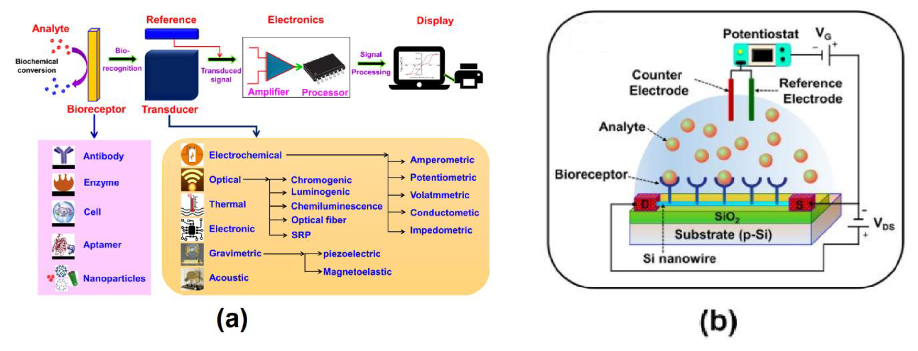
Figure 2.
Performance evaluation and integration of ISFET sensors for biochemical sensing applications.(A) Integration of an ISFET sensor through post-CMOS processing and surface modification.(B) Time-dependent surface potential behavior.(C) Surface potential variation with melibiose concentration.(D) Packaged ISFET device and corresponding transfer characteristics.(E) Long-term surface potential stability across multiple ISFET sensors[22].

Figure 3.
Fabrication, surface characterization, and electrical performance of a CNT-based biosensor for HSA detection.(A) Fabrication steps of the biosensor platform, from substrate preparation to HSA detection.(B) Surface morphology of the sensor material and assembled chip.(C) Transfer and output characteristics of the device.(D) Topographic analysis confirming nanotube immobilization.(E) FTIR spectra showing surface functionalization.(F) Electrical response to antibody binding and HSA detection.(G) Selectivity, sensitivity, and limit of detection for HSA recognition[37].
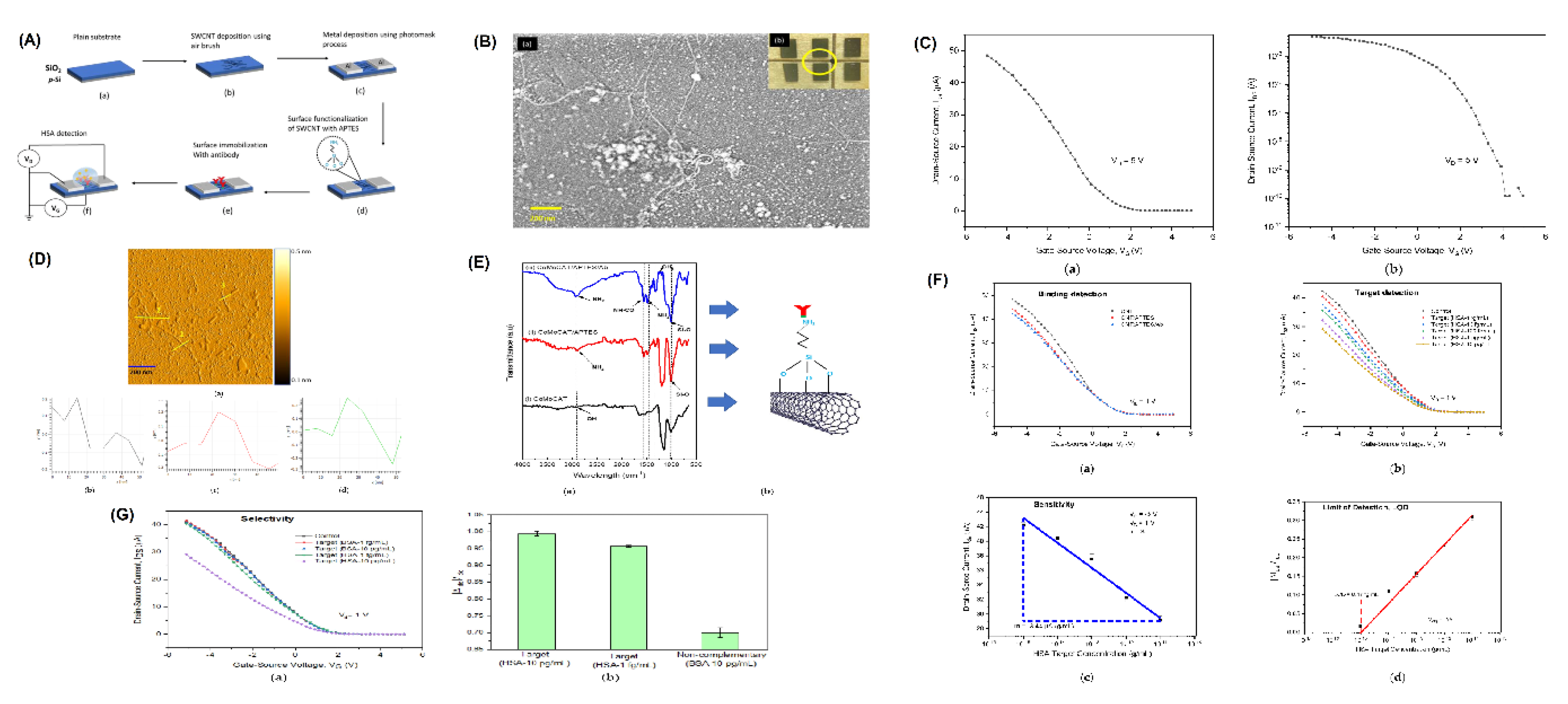
Figure 4.
Paper-based biosensing platform integrated with a custom readout system for highly sensitive detection of P90 protein in saliva samples.(A) Fabricated paper-based biosensor for P90 protein detection.(B) Custom readout system with labeled components and block diagram.(C) Output voltage response for varying P90 concentrations in saliva.(D) Functionalization steps of the gold surface with antibody immobilization.(E) Digital readout behavior across varying resistances, voltages, and capacitances.(F) Quantitative response showing sensor’s sensitivity and comparison to ELISA[40].
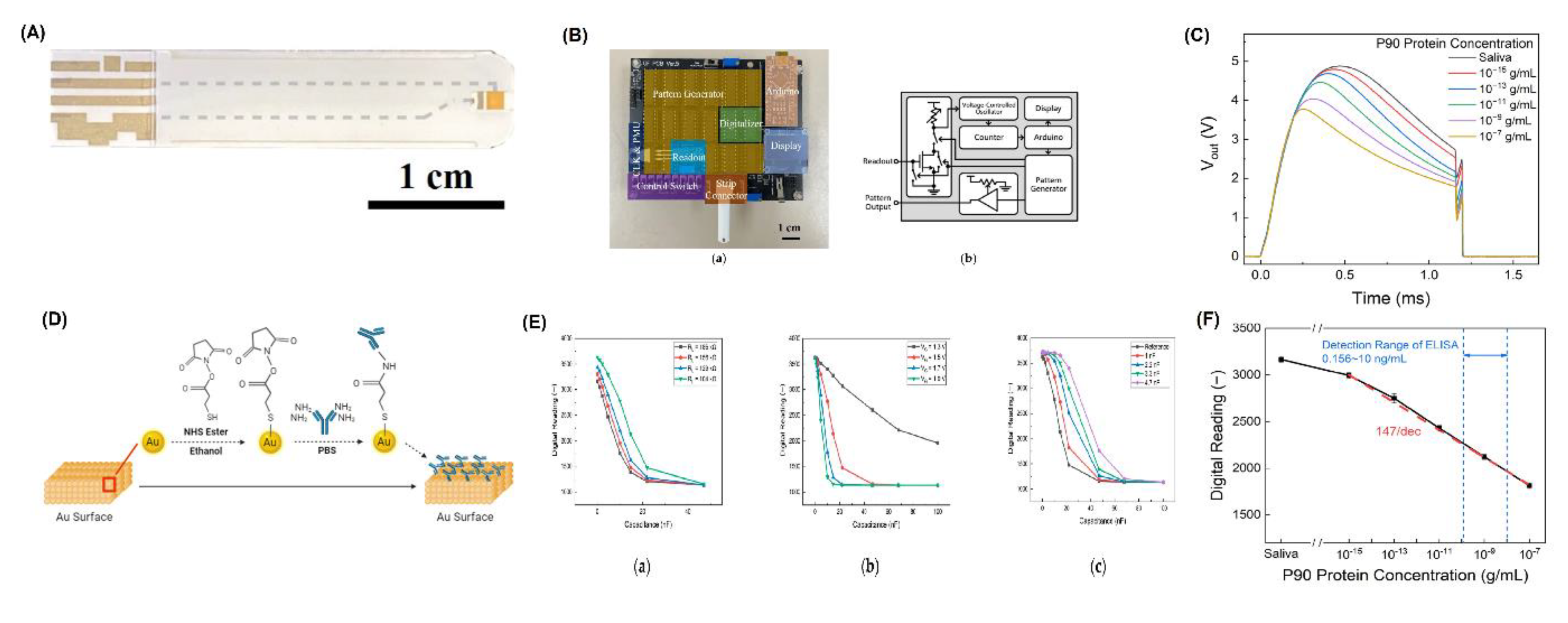
Figure 5.
Evaluation of InSe-based field-effect biosensors,high-sensitivity detection of cancer biomarkers using different gate operation modes. (A) Structural illustration of InSe-based biosensor with material characterization via SEM, AFM, Raman, and absorption spectra.(B) Device performance comparison across gate configurations during repeated measurements.(C) Photoluminescence and electrical stability of InSe biosensors over time.(D) Comparison of transfer characteristics and mobility for different gate configurations.(E) Detection of CA125 and CA199 biomarkers, with calibration curves, specificity analysis, and clinical sample comparison[63].
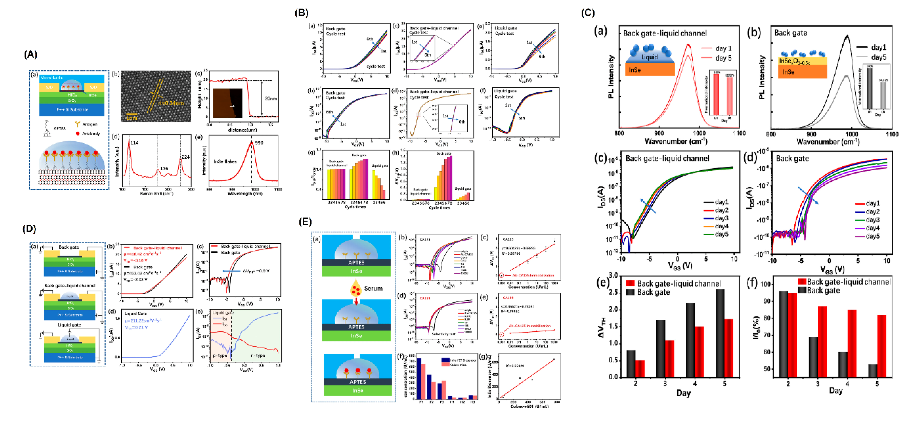

Table 1.
Overview of advanced FET-based biosensors for breast cancer detection.
| Title/Device | Detection Target | Technology Used | Simulation Tool | Key Features | Sensitivity Metrics | Reference |
|---|---|---|---|---|---|---|
| DM-DL-DMG-HJTFET | Breast cancer cell lines (healthy vs cancerous) | Dielectric modulated dual metal gate TFET | SILVACO ATLAS TCAD | - Nanogap beneath gate- Detects via dielectric property differences | Sensitivity in drain current, Vth, transconductance, ION/IOFF (exact values not given) | [54] |
| DM-PC-TFET | Breast Cancer Cells (BCCs), esp. T47D line | Dielectric modulated polarity control TFET | 2D TCAD Tool | - Works in microwave frequency band- Examines temp., geometry, charge density effects | Drain current: 7.82×10¹⁰ION/IOFF: 2.01×10⁹Transconductance: 2.32×10¹2 | [55] |
| InSe-FET | CA125 biomarker (clinical samples) | InSe-based FET with microfluidic integration | Not explicitly mentioned | - High electrical stability in liquids- Label-free detection | Detection range: 0.01–1000 U/mLTime: 20 min | [63] |
| GaN FinFET with Schottky Source/Drain | Breast cancer cells (MCF-10A, MDA-MB-231, T-47D, MCF-7, HS578t) | GaN FinFET, conductivity variation at microwave frequencies | Not specified | Studies dielectric property variation in cells; Optimized fin height, channel material, temp.; Comparative study with HEMT | 17% higher ΔIds compared to HEMT; Analysis at 900MHz and 10GHz | [64] |
| Si-doped MoS₂ TE-TFET | Breast cancer cells (Hs578T, MDA-MB-231, MCF-7, T47D) | Si-doped MoS₂ tunnel FET biosensor | Not specified | Dielectric charge modulation; Analyzes geometry variations, cavity occupancy | SVth: 0.38 (healthy), 0.77 (T47D); SION: 9.64894 (healthy), 110.37 (T47D) | [70] |
| III–V GAA TFET | Breast and prostate cancer (c-erbB-2, PSA serum) | Gate-all-around III–V TFET biosensor | Not specified | Subthreshold sensing via gm/IDS; Varies cavity lengths and BAs charge | gm/IDS: 45–61 V⁻¹; Sensitivity: 10–35; τBio: 103–419μs (varies by health condition) | [58] |
| DM-SC-JLTFET | Breast cancer cell T47D (K=32) | Dielectric modulated step channel Junctionless TFET | Not specified | Uses charge plasma; RDF reduction; Step-channel with low substrate thickness | Detection sensitivity: 2.683×10⁶; SS: 32 mV/dec | [71] |
| CP-GS-ArcTFET | C-erbB-2 protein | Charge-Plasma and Gate-Stack ArcTFET | Silvaco ATLAS TCAD | Central angle (θ) tuning; analysis of gm, gm2, gm3, fT, GBP, τ, TFP; ION/IOFF: 4.5×10⁹; SS: 35.97 mV/dec | Sensitivity to ON-current (SON), OFF-current (SOFF), and current-ratio; Impact of θ on sensitivity at 210μg/ml | [59] |
| DS-GAAE-CNTFET | MDA-MB-231 and HS578t breast cancer cells | Dual metal Gate-All-Around CNTFET with SiO2 and HfO2 stack | Not mentioned | CNT channel; dual nanocavity; early cancer detection | HS578t: 236.9 nA (Id), 285.58 mV (VT); MDA-MB-231: 343.35 nA (Id), 293.23 mV (VT) | [66] |
| Graphene FET | Estrogen receptor α (ERα) | Liquid-gated graphene FET with drug molecule as capture probe | Not mentioned | Label-free, fast detection; synthesized drug molecule as probe | LOD: 2.62 fM; Kd: 7.35 ± 0.06 pM; Response time: 30 min | [60] |
| MoS2 FET with Apta-MIP | BRCA1 ssDNA | Electrolyte-gated MoS2 FET with aptamer-MIP hybrid receptor | Not mentioned | Ag–Au@InsNCs; electropolymerized hybrid receptor | Sensitivity: 0.4851 μA/decade; LOD: 3.0 aM (buffer), 6.4 aM (serum) | [61] |
| GaN HEMT Biosensor | C-erbB2 protein | Compact GaN High Electron Mobility Transistor (HEMT) | Not mentioned | Two-finger gate (125 µm width, 5 µm length); Au–S complex; Functionalized with thioglycolic acid; High-resolution biosensing | 31% change in drain current after 6 h incubation | [65] |
| Poc-MGOTFET | C-erbB-2 in serum and saliva | Modified Gate Oxide TFET with Pocket and Dual Cavity | TCAD Sentaurus (2D simulation) | Interface charge modulation; Enhanced tunneling rate; Extended gate structure; Dual cavity under gate | Sensitivity increased by 10⁶; Improved ION/IOFF ratio | [67] |
| CNT FET Biosensor | Exosomal MUC1 protein | Polymer-sorted CNT film-based FET with AuNP-aptamer | Not mentioned | PLL-modified CNT; Aptamer-functionalized AuNPs; High-purity CNTs; Label-free detection | LOD: 0.34 fg/mL | [62] |
| IC–S-FinFET Biosensor | Various breast cancer cell lines (MCF-10A, Hs578T, MDA-MB-231, MCF-7, T47D) | Dielectrically Controlled Interconnected Multichannel Schottky FinFET | Not mentioned | Dielectric variation; Fill factor analysis; Linear and noise characterization | Improved Sn with fill factor; Evaluated drain current, SS, and ION/IOFF for different charges | [2] |
| PGFET Biosensor | Breast cancer miRNAs, SARS-CoV-2 RNA | PLL-functionalized Graphene FET | Not mentioned | PLL immobilizes DNA probes; detection from 2 μL serum/swab; 113% sensitivity enhancement over GFET | Detection limit: 1 fM; Detection time: 20 min | [68] |
| FeFET-based Muller C-element | Cancer classification via Bayesian inference | Ferroelectric FET (FeFET) | Experimentally calibrated compact model | Energy-efficient probabilistic computing; Bayesian inference; Compact (0.07 μm2); Energy: 4.1 fJ | High accuracy on Wisconsin dataset; low energy and area footprint | [69] |
| DM DPDG TFET Biosensor | SARS-CoV-2, Breast cancer cells, MCF-10A | Drain Pocket Dual Gate TFET with Nanocavity | Silvaco ATLAS TCAD | Dielectric modulation; K=22 for MDA-MB-231; optimized oxide/nanocavity thickness | Ion=0.183 mA/μm, Vth=1.712V, gm=0.581 mA/V-μm, SS=25.86 mV/dec | [56] |
| DMG-HJ-SOI-TFET | Breast Cancer Cells (BCC, e.g., T47D) | InSb/Si Heterojunction SOI TFET with Dual Material Gate | Not mentioned | Label-free detection; dielectric modulation; low power; K=32 for T47D | SS=39 mV/dec; SIon=104 | [57] |
| HJ-DL-TFET | C-erbB-2 in saliva/serum | In1-xGaxAs/Si Heterojunction Dopingless Tunnel FET | SILVACO ATLAS TCAD | Dual cavity; interface charge modulation; extended gate; x=0.2 | High ION/IOFF ratio; sensitivity ~10⁶ | [1] |
Disclaimer/Publisher’s Note: The statements, opinions and data contained in all publications are solely those of the individual author(s) and contributor(s) and not of MDPI and/or the editor(s). MDPI and/or the editor(s) disclaim responsibility for any injury to people or property resulting from any ideas, methods, instructions or products referred to in the content. |
© 2025 by the authors. Licensee MDPI, Basel, Switzerland. This article is an open access article distributed under the terms and conditions of the Creative Commons Attribution (CC BY) license (http://creativecommons.org/licenses/by/4.0/).
Copyright: This open access article is published under a Creative Commons CC BY 4.0 license, which permit the free download, distribution, and reuse, provided that the author and preprint are cited in any reuse.


