Submitted:
03 June 2025
Posted:
03 June 2025
You are already at the latest version
Abstract
New developments in biosensing technologies have transformed the entire field of diagnostics. It is possible to diagnose health problems via this technology in ways that are more accurate, faster, and less invasive than ever before. The article provides a nice overview of the newest biosensing technologies. It also discusses key advances in methods of detection, fabrication techniques, and materials used. Various detection technologies have been developed. These include biosensors, microfluidic devices and lab on a chip devices. Moreover, the studies take the pulse of progress in areas such as electrochemical and optical biosensors. In particular, indium tin oxide ranks among interesting new materials and holds promise for further advances. Moreover, various 'nano' materials, such as silver nanoparticles, are being studied along with new metalorganic frameworks. By incorporating such advances, this study can make previews more thorough and accurate. This review offers insights that are valuable to both researchers studying biosensors in industry and people practicing as doctors.

Keywords:
microfluidic devices
; electrochemical biosensors
; optical biosensors
; indium tin oxide
; silver nanoparticles
; metalorganic frameworks
; silicon materials
; nanotechnology
; diagnostic tools
1. Introduction
Biosensors turn biological responses into electrical signals. The devices had three main parts. The first part was the bioreceptor. It interacts with the target substance. Examples of bioreceptors include enzymes, antibodies, nucleic acids, and cells. The choice of a bioreceptor depends on the target analyte and the required sensitivity and specificity of the detection system. The second part was the transducer. This changed the biorecognition event into a signal. The signal can be electrical, optical, thermal, or mechanical. The third part was the signal processor. It processes the signal from the transducer and makes it readable. This involved amplifying, filtering, and converting it from analog to digital [1].
Electrochemical biosensors are a common modern chemical technique. Various types exist on the basis of measurement methods. An amperometric biosensor is one that measures the current produced from the oxidation or reduction of a substance, so it is suitable for testing the levels of glucose [2]. A potentiometric biosensor measures potential differences that are related to substance concentration, such as pH meters [3]. A conductometric biosensor detects changes in electrical conductivity caused by changes in the ionic strength or mobility of a substance [1]. An impedimetric biosensor measures changes in impedance after a recognition element has bound with a substance, which is useful for detecting large biomolecules such as proteins [4]. Electrochemical biosensors are vital in diagnostics, environmental monitoring and biomedical research. They provide high specificity and sensitivity because of diverse recognition elements, such as enzymes, antibodies or nucleic acids [2,3,4,5].
Optical biosensors use light to detect biological interactions. They offer many options on the basis of different optical methods. Fluorescence-based biosensors measure the concentration of substances by emitting light. SPR biosensors detect changes in the refractive index when substances bind. Optical waveguide-based sensors improve sensitivity by confining light. Colorimetric biosensors are simple and show color changes [1]. Biosensors using Raman technology can detect unique molecular vibrations of specific substances. These methods are especially effective when combined with SERS amplification [6]. These biosensors are crucial in diagnostic and biomedical research. They provide fast, precise, and nonintrusive analysis [7] Figure 1.
2. Biosensing Technology
This section discusses a variety of disease detection devices. The topics covered include biosensors, lab-on-a-chip devices, imaging technologies, digital PCR, microfluidic devices, assays, blood tests, liquid biopsies, BioMEMS, and genetic testing. These technologies are crucial because they are highly sensitive, fast, and portable. In addition, these methods are noninvasive. They also provide real-time monitoring and personalized treatment plans that are critical for early and accurate disease detection and the management of individual patients.
2.1. Lab On-Chip Devices
LOC technology is used to change the fluid volume includes microfluidic chips that have biological elements built into them that can detect disease biomarkers from fluids. The responses are then converted into measurable signals by a transducer, and its interpretation for the detection unit is used in analysis. The system needs only small sample sizes, which are crucial for the early detection of diseases since low biomarker concentrations are key to accurate diagnosis. In addition, the reduction in size and portability of LOC devices make them ideal for point-of-care testing, especially those suitable in remote areas lacking proper laboratory infrastructure. However, the complex fabrication processes involved in creating LOC systems can increase production costs. In addition, scalability problems exist. These limitations are being addressed by advances in technology, which is expected to reduce costs and improve scalability. Some LOC devices have difficulty detecting more than one analyte at a time; this can complicate the diagnosis of diseases with complex biomarker profiles. To overcome this, researchers are developing more sophisticated LOC systems that can handle multiple analytes, thereby improving their diagnostic capacity [8].
2.2. Biosensors
This configuration can speed up testing processes, reduce the risk of contamination and increase overall efficiency. A variety of systems include biosensors, which are devices that detect biomarkers. The device consists of a signal processing unit, transducer and biorecognition element. With a biosensor, real-time observation can be realized. Specifically, designed to be the least invasive method of sample collection. Interference from many different unwanted factors can cause the accuracy of a biosensor to decrease. The stability of the biorecognition components gradually decreases with time. This has a direct effect on the reliability of biosensors. To avoid any negative responses, it is important to evaluate the biocompatibility first. For example, a glucose biosensor for diabetes management achieves fast and accurate glucose readings but has problems with interference from other blood components [9] Figure 2.
2.3. Microfluidic Devices
These devices can test for diseases even when material is scarce, and their compactness enables them to be carried around easily. These methods are particularly suitable for emergency diagnosis. All of these microchips, biosensors, pumps, valves, chambers and microchannels are widely used in this area. Through automation, the efficient screening of large populations and examination of biomarkers in one assay are ensured. Nevertheless, compatibility, universal production standards and intricate fabrication still present major problems. For example, a study developed and demonstrated a portable, efficient microfluidic detector for rapid sepsis testing [10]. This has brought attention to the challenges of standardizing production.
2.4. Assay
An assay is a laboratory test used to look for the presence, quantity, or activity of biomarkers in samples. Assays are the most cost-effective option, especially with many samples. This saves time and money, particularly when working with extensive sample sizes. The protocols for them are standardized, making them suitable for many different environments. However, conventional assays are time-consuming and often require the expertise of skilled personnel, increasing the possibility of human error. Assays can be less portable because of the need for multiple reagents and equipment, and larger sample volumes may limit specific assays. This could limit the number of assays a person can run on any one experiment. For example, a study aknowledge the cost-effectiveness and efficiency of enzyme-linked immunosorbent assays (ELISAs) for the detection of infectious diseases is needed. However, the authors also indicated that a drawback lies in the fact that the process was time-consuming and relied on skilled personnel [11].
2.5. BioMEMS
Biomolecular devices combine biological and electronic parts and are being developed in biological microelectromechanical systems (BioMEMS). These devices, which are smaller than biosensors, are used in real-time data processing and are both useful for controlled drug delivery. Fabricating them demands highly specialized processes, making them expensive. Over time, the stability of materials can compromise the endurance and performance of devices. This creates new concerns with biocompatibility, a frequent reason that companies will have trouble obtaining FDA regulatory approval. Some studies have described the development of a BioMEMS device for controlled drug delivery, including the need for specialized manufacturing techniques and biocompatibility issues [12].
3. Material as Transducer
3.1. Gold (Au) Electrodes
Gold Nanoparticles for Biomolecule Detection
The biosensor uses gold nanoparticles (AuNPs) capped with specific antisense oligonucleotides (ssDNA) targeting the viral nucleocapsid phosphoprotein (N-gene). The sensing probes are immobilized on a paper-based electrochemical platform, creating a nucleic acid testing device suitable for widespread diagnostic use. The readout is recorded using a simple handheld reader, which is practical. The sensor was tested with SARS-CoV-2-infected Vero cells and clinical samples and produced improved output in under 5 minutes. It has a sensitivity of 231 (copies μL–1)−1 and a detection limit of 6.9 copies per micro liter without further amplification. Even during viral mutation, ssDNA-conjugated AuNPs targeting two N gene regions allow the sensor to remain functional. This dual-target strategy reduces the chance of false negatives due to viral mutations [13]. Despite advancements in nucleic acid testing, early detection of breast cancer remains a significant challenge. Achieving high specificity and sensitivity in breast cancer diagnostics is critical for improving outcomes. Early detection significantly improves treatment success, with HER2 as a key biomarker. This study introduces an electrochemical immunosensor for highly sensitive detection of HER 2 using a nanodiamond (nanoD) and gold nanoparticle (AuNP) platform. Nanodiamonds were immobilized on a glassy carbon electrode (GCE), followed by AuNP electrodeposition to increase conductivity. Techniques such as voltammetry, EIS, TEM, XRD, and FESEM confirmed the material deposition and surface morphology. The sensor achieved a detection limit of 0.29 pg mL−1 under optimal conditions, with minimal cross-reactivity [14]. Although the electrochemical immunosensor for HER 2 offers high sensitivity and specificity, its focus is limited. Broader applications are necessary to increase its versatility across different biomarkers. This study presents a method for lysozyme detection using a gold nanoparticle-based biosensor. The biosensor is integrated with decomposition Muller matrix polarimetry to increase accuracy and sensitivity. A DNA aptamer with specificity for lysozyme is used for precise binding in the detection process. Gold nanoparticles enhance biosensor sensitivity through their unique optical properties. The detected signal was processed via a Muller matrix. This focuses on depolarization, linear diattenuation, and depolarization index measurements. The results revealed a linear relationship between the optical parameters and lysozyme concentration. The dynamic range observed was between 0.01 and 500 pM with high precision. The limit of detection (LOD) was determined to be 1.24 fM, confirming the device’s sensitivity. These results highlight the potential of biosensors for expanding diagnostic applications. The use of gold nanoparticles greatly improves sensor capabilities[15]. Lysozyme detection is highly sensitive but lacks the ability for real-time biomarker monitoring, which is essential for infectious diseases. This remains a challenge. In this study, a QCM biosensor was developed to detect HBsAg, and antibodies were covalently attached to primary amines for detection. Such improvements are key. Surface modifications were performed using PEI and thiolated PEI, which improved electrode performance and enabled more efficient detection. This optimized the process. RSM optimization revealed no significant difference in immobilization yield between the modified layers used for the biosensor. Testing confirmed success. Surface analysis using FESEM, AFM, FTIR, and CA revealed increased hydrophilicity and roughness, enhancing the biosensor performance. This improved the detection reliability. The QCM biosensor demonstrated a wide dynamic range from 1 to 1 × 10³ ng/mL. The accuracy was very high. This makes it a promising tool for noninvasive and timely monitoring of HBV in human serum samples. Healthcare outcomes have improved greatly[16] Figure 3.
Electrochemical Detection in Biological Monitoring
The research focuses on creating a sensitive and flexible sensor for real-time monitoring of glucose and lactate levels in sweat. A breakthrough in flexibility. The working electrode was enhanced with gold nanopine needles (AuNNs) via electrochemical deposition, effectively amplifying signals and increasing sensor sensitivity. Precision was at every step. For enzyme immobilization, poly(ethylene glycol) diglycidylether (PEGDE) offered better enzyme activity retention than glutaraldehyde did, ensuring long-term sensor performance. Efficiency redefined in biosensing. The biosensor demonstrated excellent catalytic performance with AuNNs, achieving low detection limits of 7 μmol L−1 for glucose. Unmatched sensitivity was achieved here [17]. Although the AuNN-based sensor is sensitive and flexible, integrating sensors into wearable applications requires materials that can endure stress. The intrinsic power of gold manifests itself through its active influence. Gold is ideal for sensing because of its chemical inertness and broad electrochemical window, making it perfect for biosensing applications. This innovation meets real-world needs. Using a dry-spinning method, stretchable and conductive gold fibers fabricated that maintained conductivity even under mechanical stress. Stretchable and still powerful. These fibers were used to create lactate-sensing working electrodes, reference electrodes, and counter electrodes for integration into wearable textiles. Wearable tech with impact can be produce. The textile biosensors exhibited high sensitivity, with 19.13 μA/mM cm² in PBS and 14.6 μA/mM cm² in artificial sweat. Performance under any strain [18]. However, as biosensing applications expand, there is a growing demand for devices that can withstand stress, offering ultrawide detection ranges. There demand is rising. The development of blood glucose monitoring devices requires greater sensitivity and lower detection limits to enhance biosensor performance and reliability. These advancements have boosted diagnostics. In this study, we fabricated a high-performance flexible enzymatic glucose biosensor by electrochemically depositing dendritic gold nanostructures on a carbon cloth substrate. The addition of gold greatly improved the conductivity. Glucose oxidase was immobilized on the dendritic gold, and enzyme loading was enhanced using the enzyme precipitation coating method. This increases the detection efficiency. Scanning electron microscopy (SEM) was used to characterize the dendritic gold nanostructures in detail to confirm successful deposition and structure. Visualization aids precise measurement. Cyclic voltammetry (CV), chronoamperometry, and electrochemical impedance spectroscopy (EIS) were used to evaluate the overall electrochemical performance of the biosensor. Multiple tests ensure reliability. The biosensor exhibited high sensitivity (72.45 μA mM−1 cm−2), a low detection limit (6.7 μM), and a broad linear glucose detection range. These results indicate precision. It also demonstrated good selectivity, reproducibility, and stability, with strong accuracy when tested in real serum samples for glucose detection. It is accurate for clinical use. These results suggest that the new biosensor holds great potential for future applications in the biomedical and healthcare sectors. Promising innovations are needed ahead [19]. For comprehensive diagnostics, handling multiple analytes at once is crucial to ensure accurate and reliable results in medical tests. This is vitally important.In another study, a highly sensitive electrochemical sensor was designed for glucose detection using carbon nanotubes (CNTs) grown in situ at low temperatures. On a glass substrate. This setup involved CNTs grown on photolithographically defined gold microelectrode arrays (CNT/Au MEAs) placed on a glass substrate. Each sample is distinct. Selectivity was achieved by modifying CNT/Au MEA with the glucose oxidase (GOx) enzyme immobilized in poly(paraphenylenediamine). It improves sensor precision. The biosensor showed a linear response in the 0.2–27.5 µM range, with a sensitivity of 168.03 kΩ−1 M−1 and low detection limits. The accuracy was confirmed further. It demonstrated excellent anti-interference properties and was validated through HPLC in blood serum samples, ensuring accuracy in real-world applications. Validation through HPLC was previously reported [20]. Overall, this research highlights advancements in biosensor technology, with improved sensitivity, stability, and versatility for medical monitoring applications. It is useful for real diagnostics.
Advanced Nanostructures for Specific Detection
A biosensor system for isoprocarb detection was developed, utilizing acetylcholinesterase (AChE) inhibition by isoprocarb as the fundamental detection mechanism. Gold nanoparticles modify electrode. A gold nanoparticle-polyaniline-modified graphite pencil electrode (AuNP-PANI-GPE) was employed to detect thiocholine changes in response to isoprocarb. In the process two fabrication steps followed. The electrode was fabricated using cyclic voltammetry: electropolymerization of aniline on a graphite pencil and then electrodeposition of gold nanoparticles. Gold nanoparticles were observed. SEM–EDX revealed that gold nanoparticles ranging from 8–80 nm were successfully deposited on the modified electrode surface. A larger active area formed. Cyclic voltammetry indicated that the active surface area was 0.17019 cm², which was larger than that of both the PANI-GPE and unmodified GPE. Oxidation peak recorded. The oxidation peak of thiocholine at +0.675 V (vs. Ag/AgCl) increased with increasing acetylthiocholine concentration and decreased in the presence of isoprocarb. Linear curve observed. Under optimal conditions, a linear calibration curve for isoprocarb (0.05–1.0 μM) was generated, demonstrating high accuracy. It has impressive detection limits. The detection and quantification limits were 0.1615 nM and 0.5382 nM, respectively, with a sensitivity of 1.7771 μA/μM.mm². Also, excellent stability was achieved. The electrode showed strong stability, with an RSD of 4.87% over eight measurements, making it promising for real-world detection. The scope of future applications is vast [21]. To increase the sensitivity and broaden detection beyond isoprocarb, a new folic acid biosensor was developed in which dihydrofolic acid reductase (DHFR) was immobilized. The titanium nanoparticles were characterized. The biosensor utilized a c-MWCNT/TiO2NP-modified gold electrode and was analyzed at various stages using SEM, EIS, FTIR, and cyclic voltammetry. The pH was optimal. The biosensor exhibited a low detection limit of 11.48 nM, a wide linear range (5–50 nM), a sensitivity of 0.42 μA/nM/cm², and excellent stability. The maximum current was appear at 0.125 V . This method proved effective for folic acid detection in serum samples from pregnant women, showing potential in clinical applications. As well stability was maintained [22]. Overall, advancements from isoprocarb detection to folic acid biosensing have improved biosensor sensitivity, specificity, and application range for biomolecules Table 1.
3.2. Carbon-Based Electrodes
Electrochemical Sensors for Biological Molecules
A novel screen-printed carbon electrode (SPCE) with multiple working electrodes and a single signal output channel has been developed. This enhances immunosensing applications. The design eliminates counter and reference electrodes, reducing costs and environmental impact by excluding precious metals completely. This lowers waste. The SPCE enables simultaneous detection of various analytes through individually modified working electrodes for enhanced efficiency. Each electrode was performed independently. An independent platinum network acts as a counter electrode, improving the reproducibility and reliability of the SPCE during operation. This ensures accuracy. A hydrogel on a working electrode improves conductivity, significantly enhancing the overall sensitivity of the electrode in use. This increases the conductivity. The SPCE was applied to a multiplexed, label-free amperometric immunosensor for detecting four tumor markers with high precision. The detection limits improved. This method achieved detection limits as low as 5.5 pg mL⁻¹ for SCCA and 2.3 pg mL⁻¹ for NSE. Such low limits improve the performance[23]. Additionally, a nickel ferrite/reduced graphene oxide (NiFe₂O₄/rGO) nanocomposite was synthesized and used to fabricate a bioelectrode on a screen-printed carbon electrode (SPCE). This improved electrochemical biosensing. The uricase/nickel ferrite/reduced graphene oxide/screen-printed carbon electrode (Uricase/NiFe₂O₄/rGO/SPCE) demonstrated enhanced performance, with a linear range from 5 to 900 micromolar (μM) and a detection limit of 21.9 micromolar (μM). The sensor gets excellent stability and selectivity. The biosensor showed repeatability, making it suitable for detecting uric acid (UA) in real samples with point-of-care potential. It is used for UA disorders [24]. Screen-printed carbon electrodes (SPECs) are commonly utilized in point-of-care testing (POCT) because of their affordability, disposability, and simple design. The surface modification is challenging. Unlike gold or platinum electrodes, modifying the SPCE carbon surface is difficult because of its inherent stability properties. Carboxyl groups introduced. Oxygen plasma (O₂) treatment can enable covalent bonding by introducing carboxyl groups on the SPCE carbon surface. It has improved bonding efficiency. This research studied the effect of oxygen plasma (O₂) treatment using a novel immunosensor with gold nanoparticles (AuNPs) on electrode performance. Four modifications were compared. The modifications tested were oxygen plasma-treated/covalent-bonded antibodies (a), oxygen plasma-treated/physically adsorbed antibodies (b), bare/covalent-bonded antibodies (c), and bare/physically adsorbed antibodies (d). The results showed varying limits. The detection limits were 0.50 nanograms per milliliter (ng/mL), 9.7 nanograms per milliliter (ng/mL), 0.54 nanograms per milliliter (ng/mL), and 1.2 nanograms per milliliter (ng/mL) across the four configurations tested. Higher sensitivity was observed. The oxygen plasma-treated electrodes had an increased number of carboxyl groups, which enhanced antibody adsorption and improved sensor sensitivity and performance. Effective surface modification. Oxygen plasma (O₂) treatment was found to significantly improve surface modification, resulting in better antibody binding and sensor performance [25]. Additionally, a glucose biosensor incorporating Prussian blue nanoparticles (PBNPs) and carbon nanotubes was developed using sweat for noninvasive glucose monitoring. GOx was immobilized. Glucose oxidase (GOx) was immobilized by chitosan and encapsulated in Nafion to ensure stability and detection performance. Detection limit was 7 μM. The GOx/PBNP/MWCNT-COOH sensor demonstrated a low detection limit, high sensitivity, and excellent interference resistance across the sweat glucose range. Suitable for diabetic patients. The sensor, which was applied to screen-printed carbon electrodes (SPECs), maintained stability for two weeks, showing promise in personalized medical detection. Stability achieved was expecting in future [26]. These advancements in SPCE technologies highlight their potential for cost-effective, reliable biosensors in medical diagnostics and environmental monitoring applications. The SPCE is promising Figure 4.
Electrochemical and Optical Sensors for Environmental and Pharmaceutical Applications
A highly sensitive electrochemical sensor has been developed for detecting doxorubicin hydrochloride (DOX), a potent antitumor agent. DOX was detected well. The sensor uses a glassy carbon electrode modified with multiwalled carbon nanotubes (MWCNTs) decorated with gold nanoparticles. Nanotubes has enhanced performance. Characterization techniques, such as scanning electron microscopy (SEM), confirmed the successful decoration of the MWCNTs with gold nanoparticles. SEM confirmed the success of the study. Cyclic and linear sweep voltammetry showed efficient catalytic activity for DOX reduction, with increased peak current and reduced overpotential. It has effective catalytic activity. The sensor achieved a linear detection range from 10⁻¹¹ to 10⁻⁶ M, with a low detection limit of 6.5 pM. It has wide detection range [27].
Single-walled carbon nanotubes (SWCNTs) have shown great promise for near-infrared fluorescence-based biosensing in various applications. This study introduces a reusable sensor. The drop-cast optical biosensor uses peptide-encapsulated SWCNTs to detect low concentrations of acetic acid in air. It is reusable. SWCNTs, specifically those with (6,5) chirality, exhibit peak fluorescence in the 970–1050 nm range, which is compatible with low-cost silicon-based detectors. It is also stable . Peptide encapsulation enhances stability and sensitivity, allowing the detection of acetic acid vapor at concentrations as low as 0.05%. It is crucial. This advancement is important for cost-effective, real-time SWCNT-based gas phase detection in areas such as wine spoilage monitoring [28]. Along with this a new method has emerged. An enzymatic biosensor for the indirect detection of organophosphates (OPs) was developed that utilizes acetylcholinesterase inhibition for detection. The biosensor used the electrochemical principle. The sensor was built on a screen-printed carbon electrode modified with copper nanowires (CuNWs) and reduced graphene oxide (rGO). The current was measured. The oxidation current was measured through cyclic voltammetry (CV), which is generated by the enzymatic interaction between acetylcholinesterase and acetylthiocholine. Signal reduction happened . The biosensor response showed signal reduction due to AChE inhibition by an organophosphate inhibitor in the test sample. This enhanced the sensitivity. The CuNW/rGO nanocomposite enhanced the signal current and lowered the oxidation potential for chlorpyrifos detection in the test solution. It is highly effective. The detection range was 10–200 µg/L, with a limit of detection of 3.1 µg/L and a quantification limit of 12.5 µg/L. It detects chlorpyrifos [29]. A hydrogen peroxide (H2O2) sensor was developed using a calcium titanate-modified electrode (CaTiO3@SPE). The electrode synthesized via a hydrothermal method was characterized using a XRD, EDX, SEM, and BET, and CaTiO3 had a specific surface area of 57.6 m²/g. The sensor, which was fabricated through drop-casting, exhibited a detection limit of 0.08 µM for H2O2 with good selectivity. Stability and repeatability are also notable features [30]. These advancements highlight the continuous innovation and diversification of sensor technologies, which improve detection capabilities across a range of applications Figure 5.
The biosensor represents a significant improvement in point-of-care uric acid monitoring technology. The working electrode is based on a 3D SACNT array immobilized with uricase through precipitation and crosslinking, which increases the enzyme density and contact area with reactants while preserving the conductivity of the SACNT structure. The biosensor showed impressive sensitivity, measuring 518.8 μA/(mM⋅cm²), with an operating range of 100–1000 μM and a low detection limit of 1 μM. Dynamic uric acid monitoring was validated in serum samples, with no significant difference compared with an FDA-approved electrochemical analyzer (paired t test, p > 0.05). Its large surface area and electrocatalytic activity indicate its potential for broader point-of-care biomolecule monitoring applications [31]. The SACNT array biosensor demonstrated excellent sensitivity but focused mainly on uric acid detection. To solve this problem, a new biosensor was developed using carbon dots. These carbon dots (CDs) were synthesized from curcumin and dimethylformamide (DMF) through microwave irradiation, resulting in CDD-CDs. The CDD-CDs were functionalized with 3-(aminopropyl)-triethoxysilane (APTES), forming APT-CDs. Laccase was then covalently immobilized onto APT-CDs. This created a novel bioprobe. The CDD-CDs emitted orange fluorescence at 586 nm, APT-CDs emitted green fluorescence at 533 nm, and the bioprobe emitted blue fluorescence at 476 nm. The bioprobe could detect dopamine linearly from 0 to 30 μM, with a detection limit of 41.2 nM. For the tapered optical fibers, the detection limit improved to 46.4 nM across a range of 0-10 μM. This material showed high biocompatibility and excellent stability. This finding was validated in human serum and cerebrospinal fluid, confirming its clinical potential [32]. These new biosensors represent an innovative step forward in enhancing sensitivity and specificity for biochemical monitoring. They show promise for applications in both clinical diagnostics and environmental analysis.
3.3. Indium Tin Oxide (ITO)
Electrochemical Biosensors Using ITO for Pathogen Detection
This article explores the development of advanced electrochemical biosensors using indium tin oxide (ITO) electrodes for improved sensitivity and detection. Biosensors detect pathogens. These biosensors are crucial for detecting proteins and pathogens with high sensitivity and specificity in medical diagnostics. They are reliable tools. This study presents a electrochemical DNA biosensor for detecting Shigella flexneri via innovative detection techniques. The detection method is label-free. The sensor utilizes ITO electrodes, where a detection probe is attached to poly melamine (P-Mel) and polyglutamic acid (PGA). Probes are crucial in this method. Disuccinimidyl suberate (DSS) was used to prepare the flexible ITO electrode, and anthraquinone-2-sulfonic acid monohydrate sodium salt (AQMS) served as a signal indicator. This increases the sensitivity. This biosensor detects S. flexneri DNA at concentrations ranging from 1×10⁻⁶ to 1×10⁻²¹ mol/L, with a detection limit of 7.4×10⁻²² mol/L. The detection limit was acceptable . In real samples, it detects S. flexneri from 8×10¹⁰ to 80 cells/mL, with a detection limit of 10 cells/mL.Here, accuracy is essential [33]. Another study introduced an immunosensor for detecting GM2 activator protein (GM2A) using an ITO substrate with enhanced sensitivity. It detects GM2A. This substrate is enhanced with gold nanoparticles (GNPs) and an amino-functionalized thiophene polymer (P(ThiAmn)) multilayer. Gold enhances detection. To create this biosensor, GNPs are deposited on the substrate, and ThiAmn is electropolymerized to increase the surface area. The area increases attachment. This allows for the attachment of many anti-GM2A biorecognition elements, improving the overall detection capability and reliability in biosensing applications. The attachment is crucial. Electrochemical impedance spectroscopy (EIS) was used to study the specific interaction between anti-GM2A antibodies and GM2A antigens for targeted detection. It measures interactions. Under optimal conditions, GM2A is detected in a linear concentration range from 0.0185 to 111 pg/mL, with a detection limit (LOD) of 5.8 fg/mL. It is highly sensitive. This biosensor shows good reproducibility, long storage stability, and excellent specificity for GM2A antigens in various applications. The results are reproducible [34]. Additionally, a new platform has been developed for the detection of heat shock protein 70 (HSP70) via advanced techniques. This platform uses ITO. It employs a three-electrode system on a chip, modifying the reference and working electrodes for enhanced detection. HSP70 antibody assembly. The PS-AuNPs@Cys/Au film deposited on ITO glass provides an excellent substrate for antibody attachment, amplifying signals. This enhances detection. Under optimal conditions, the sensor shows a linear range from 0.1 ng/mL to 1000 ng/mL for protein detection. The limit was 25.7 pg/mL. This method detects HSP70 in normal human blood samples and outperforms the commonly used ELISA method for analysis. In this way,HSP70 has been detected [35]. Additionally, a novel label-free impedimetric immunosensor was developed for sensitive and selective analysis of the Aβ42 protein. Rapid detection occurs. This immunosensor uses cost-effective, disposable ITO-PET electrodes modified with 3-glycidoxypropyldimethoxymethylsilane (GPDMMS) for functionality. Functional groups are form. The interaction between anti-Aβ42 and Aβ42 was analyzed via electrochemical impedance spectroscopy (EIS) and cyclic voltammetry (CV). These changes are confirmed. Morphological changes on the electrode surface during each immobilization step were confirmed by scanning electron microscopy (SEM). The sensitivity is high. The immunosensor shows a linear detection range from 1 to 100 pg/mL, with a detection limit of 0.37 pg/mL. This method is highly selective and stable. For the first time, the kinetic behavior of the antibody‒antigen complex was analyzed via single-frequency impedance (SFI). The binding is very well. Its potential for clinical application was confirmed by analyzing Aβ42 levels in human serum, highlighting its diagnostic utility. The patient’s diagnosis was confirmed [36]. Overall, these studies highlight the advancements in ITO-based biosensors with enhanced sensitivity, specificity, and versatility for biomedical use Figure 6.
Biosensors Using ITO for Environmental and Health Applications
Recent advancements in biosensor technology have led to innovative devices made with advanced materials, significantly transforming traditional detection methods. This is revolutionary. One such advancement is the creation of a high-tech optical fiber probe designed for remote cysteine detection with precision. The design is compact. Researchers have coated optical fiber surfaces with indium tin oxide, zinc oxide, and cuprous oxide semiconductor nanomaterials for improved performance. It also increases efficiency. This setup replaces bulky macroscopic spatial light systems with a compact optical fiber path, allowing for remote light injection. The light is guided precisely. The probe acts as a working electrode, interacting with the analyte and directing light where needed for photoelectrochemical reactions. This makes the setup more effective. This fiber-optic-based cysteine biosensor has a linear detection range of 0.01-1 μM, indicating that it is promising for biochemical analysis. It has the scope in future for detection devices [37]. Another exciting development is a biosensor for detecting aflatoxin B1 (AFB1) without labels, which uses advanced nanomaterials for accurate detection. The approach used is effective. This sensor uses gold nanobipyramids placed on an indium tin oxide-coated glass substrate modified with a self-assembled APTES film. The modification is strong. These modifications ensure effective AuNBP formation and immobilization of anti-AFB1 antibodies, enabling precise electrochemical biosensing of AFB1. It is robust. The sensor has a detection limit of 0.1 nM and a recovery rate of 95–100%, demonstrating robustness in real-world applications. The results are excellent [38]. Additionally, a molecularly imprinted electrochemical sensor using Ti3C2Tx MXene has been developed for detecting bilirubin with high selectivity. It is a reliable method. Ti3C2Tx MXene is synthesized through chemical etching and applied to an ITO electrode surface using the drop-casting method. The process is simple. This modification enhances the binding properties, which are essential for molecularly imprinted polymer formation, and offers excellent electrochemical characteristics. It’s very effective. The sensor has a linear range of 10 μM to 90 μM and a detection limit of 0.197 μM, with strong reproducibility. The performance is optimal [39]. A new glucose sensor has also been introduced using a biomolecule-assisted method to deposit copper–cobalt bimetallic heteronanostructures. It is groundbreaking. This method results in a sensitive and disposable glucose-sensing electrode, solving instability issues common with transition metal films. Stability was improved. Typically, metal-based films on ITO electrodes are prone to peeling during electrocatalysis, but this innovation efficiently solves that problem. Peeling can be avoided. Researchers have used methionine as a structure-directing agent, leading to a glucose sensor with high sensitivity and selectivity. The results are accurate. This sensor showed a detection limit of 9.1 μM and a sensitivity of 1418 μA mM−1 cm−2, opening new pathways for stable electrodes. This method is highly promising [40].
Advanced Nanomaterial-Based Sensors
The current study developed a nonenzymatic glucose sensor using iodine-doped reduced graphene oxide with a titanium dioxide nanocomposite (I-rGO@TiO2). It was effective. Titanium dioxide nanoparticles and reduced graphene oxide were synthesized via sol‒gel and thermal reduction methods to ensure consistency and quality. The analysis confirmed this finding. The I-rGO@TiO2 composite was prepared through a hydrothermal technique, combining I-rGO and TiO2 in a 1:4 ratio. The results were clear. XRD and TEM analysis revealed that nanocrystalline anatase titanium was distributed across the I-rGO sheets, confirming the nanostructure of the composite. The findings were satisfactory. The sensor was fabricated by modifying an indium tin oxide electrode with I-rGO@TiO2, resulting in a wide linear detection range. It analyses well. Additionally, the nanocomposite showed positive temperature coefficient behavior with good conductivity at 200°C. Testing was performed. Antibacterial properties were tested against E. coli and Bacillus subtilis, and promising results were obtained in antimicrobial resistance studies. It shows impressive outcomes [41]. Biosensors have gained interest for the diagnosis of infectious diseases, such as tuberculosis (TB), because of their simplicity and point-of-care potential. They are useful. The incorporation of aptamer molecules and nanomaterials offers advantages such as high binding affinity and low immunogenicity, significantly enhancing aptasensor performance. Great improvements were observed. This study used microwave-synthesized copper indium tin sulfide (CITS) nanomaterials combined with the natural biopolymer chitosan for signal amplification. Better sensitivity is achieved. The optical properties of CITS include strong UV absorption, which is characteristic of kesterite semiconductor nanomaterials, confirming successful optical traits. The optical properties can be applied similar devices . X-ray diffraction confirmed the presence of the kesterite phase, with an average crystallite size of 6.188 nm, confirming the desired material phase. It was reliable. The aptasensor’s electrochemical properties were enhanced by 77.5%, and aptamer loading improved by 73.7%, increasing the overall performance. The sensor achieves good performance. The aptasensor showed sensitivity to IFN-γ concentrations, with a limit of detection of 6885 fg/mL and a wide linear range. It also have high accuracy. This sensor exhibited excellent stability, selectivity, and the ability to be applied to real-world sample testing with promising results. It achives certain durability [42]. This study integrates nanocomposites and aptamers, achieving high sensitivity and specificity in biosensors for point-of-care diagnostics.
3.4. Silver Nanoparticles
Glucose Detection Using Nanomaterials
Owing to their unique properties, nanomaterials have been extensively explored in the development of sensors, contributing to reliable sensor designs with enhanced sensitivity and specificity. This study proposes a self-powered biosensor. The fluorescent/electrochemical dual-mode biosensor uses DNA-templated silver nanoclusters (AgNCs@DNA) for advanced biosensing in glucose detection applications. AgNC@DNA serves as a probe. The study evaluated its efficacy for glucose detection, using the fluorescence emitted by AgNCs@DNA as the readout signal for glucose levels. Glucose oxidase (GOx) generates hydrogen peroxide. The fluorescence emitted by AgNCs@DNA correlates with hydrogen peroxide levels, making it highly effective for monitoring glucose. GOx facilitates detection. This biosensor is highly sensitive. The electrochemical signal uses AgNCs as charge mediators between GOx and the carbon electrode, improving the detection accuracy. The biosensor achieved low limits of detection (LODs) for optical and electrochemical signals, providing excellent sensitivity for glucose monitoring. Specifically, the LODs were 23 μM for optical readouts and 29 μM for electrochemical readouts, confirming the effectiveness of the biosensor [43]. Stretchable transparent electrodes (STEs) based on silver nanowires (AgNWs) have garnered attention for their superior optoelectronic properties in sensor development. Oxidation is a challenge. AgNWs exhibit low oxidation resistance, which limits their durability in devices utilizing stretchable transparent electrodes. A recent study developed core-sheath nanowires. Ag@Au NWs with a dual-headed matchstick morphology and a gold sheath thickness of 2.5 nm were fabricated. These nanowires enabled STEs with an optical transmittance of 78.7%, 13.0% haze, and excellent mechanical properties. The electrodes resist oxidation. The STEs showed a sheet resistance of 13.5 Ω·sq.−1 and a tensile strain capacity of 240%, which were achieved through welding. The dense gold sheath provided oxidation resistance. These Ag@Au NW STEs enabled nonenzymatic glucose biosensors, offering a high sensitivity of 967 µA·mM−1·cm−2. The detection limit was 125 µM. The performance of these materials shows potential [44]. The high level of glucose present in daily nutrition is a significant factor contributing to conditions such as diabetes (diabetes mellitus) and obesity. Glucose levels must be monitored. Determining glucose concentrations in food production processes is crucial for accurate quality control measures. Biosensors enable fast analysis. Biosensors are bioanalytical devices offering cost-effective, simple analysis and providing quick response times for glucose detection in various settings. Silver nanoparticles (AgNPs) are useful. They can be synthesized through green methods and modify electrodes for efficient glucose biosensing applications. This helps research. Agricultural waste provides a sustainable source for synthesizing these nanoparticles. In this study, an amperometric glucose biosensor was created by modifying a carbon paste electrode (CPE) with waste tea-based silver nanoparticles (WT-AgNPs). The glucose oxidase (GOx) enzyme was immobilized. This was accomplished by cross-linking onto a modified carbon paste electrode (MCPE), enabling effective glucose detection mechanisms. Detection relies on hydrogen peroxide (H₂O₂). Hydrogen peroxide reduction occurred at +0.4 V versus silver/silver chloride (Ag/AgCl), providing clear readings. The biosensor demonstrated a linear range between 0.10 and 1.0 micromolar (µM) glucose[45]. It works in juice. This sensor was successfully applied to detect glucose in commercial fruit juice samples. This method showed excellent reproducibility. Low detection limits, high reproducibility, selectivity, and long shelf-life were all demonstrated. This article explores a sensor. The development of a glossy photopaper (GPP)-based screen-printed chemiresistive interdigitate electrode (SPCIDE) for glucose sensing is highlighted. Interdigitate electrodes (IDEs) were printed. Using screen-printing methods, low-sheet resistance electrodes were made with graphene ink on the GPP. Polyaniline (PANI) was applied. Polyaniline was drop-cast onto the interdigitate electrode to form a chemiresistive matrix. Selective detection is key. The glucose oxidase (GOx) enzyme and green-synthesized silver nanoparticles (GS-AgNPs) were immobilized on PANI to increase glucose selectivity. Amperometric testing was performed. The amperometric measurements revealed a strong linear relationship between the current change and glucose concentration. The coefficient of determination (R²) was high. The sensor demonstrated a detection limit of 198 nanomolar (nM) and a sensitivity of 291.19 microamperes per millimolar per square centimeter (µA mM−1 cm−2). SPCIDE has potential. This sensor holds promise for developing affordable and eco-friendly point-of-care (PoC) diagnostic kits. Mass production is viable[46]. The use of inexpensive screen-printing techniques makes the creation of eco-friendly biosensors feasible for large-scale applications.
Detection Using AgNPs and Other Nanoparticles
In this work, silver nanoparticle (AgNP)/reduced graphene oxide (rGO) nanocomposites were electrodeposited on glassy carbon electrodes (GCEs) to develop electrochemical sensors for hydrogen peroxide (H₂O₂) and dopamine (DA) detection. AgNPs were synthesized successfully. These electrochemical sensors were designed for the sensitive detection of H₂O₂ and DA with high performance. GO reduction occurred effectively. The nanocomposites formed on the electrodes were confirmed by scanning electron microscopy (SEM) and electrochemical impedance spectroscopy (EIS). The method was reliable. The AgNP/rGO/GCE exhibited a linear response to H₂O₂ from 5 μM to 620 μM, with a sensitivity of 49 μA mM⁻¹cm⁻². The LOD was 3.19 μA. For DA, the sensor showed a linear range of 1 μM to 276 μM, with a sensitivity of 7.86 μA mM⁻¹cm⁻² and a limit of detection (LOD) of 0.18 μM. The detection was stable. These sensors could simultaneously detect DA and H₂O₂ without interference, maintaining excellent stability over time with eco-friendly fabrication. The sensors worked reliably. This fabrication method offers great potential for the sensitive detection of DA and H₂O₂ with robust and reproducible results. This is a highly effective approach [47]. A study developed an aptamer-based electrochemical sensor (AEC) for detecting STX via silver nanoparticles (AgNPs) modified with an aptamer. Aptamers were attached to the sensor. Under optimized conditions, AECs exhibited a linear response to STX between 0.04 and 0.15 μM, with high sensitivity and accuracy. Regression equation applied. The detection limit was 1 nM, which is below the regulatory limits for STX in seafood, demonstrating excellent sensitivity for practical applications. The performance was excellent. The potassium ferricyanide (K₃Fe(CN)₆)-etched AgNPs served as a signal source, providing a stable ratiometric electrochemical signal for enhanced sensitivity. STX was detected well[48]. In another study, hollow Prussian blue with ultrasmall silver nanoparticles (Ag-HPB) was fabricated via a coating-etching method for sensor development. Silver nanoparticles (Ag NPs) diffuse into Prussian blue (PB). Prussian blue (PB) was coated on silver nanoparticles (Ag NPs), allowing them to diffuse into the Prussian blue framework, enhancing sensor performance. This increased the conductivity. The biosensing platform combines the electrical conductivity of silver nanoparticles (Ag NPs) with the high enzyme loading capacity of the hollow structure. The performance was enhanced. Using glucose oxidase (GOx) and acetylcholinesterase (AChE), the sensor showed a sensitive glucose response of 24.37 μA mM−1 cm−2 and detected trichlorfon. It was highly stable. The detection limit for trichlorfon (TCF) is 2.28 pg/mL, and the system is effective for monitoring trichlorfon in apples[49]. Nanocellulose improved the sensitivity. Nanocellulose derived from hemp (HNC), combined with silver nanoparticles (AgNPs), enhances electrochemical sensing, particularly for detecting lactate in wearable sensors. The electrode performance increased. Hemp nanocellulose (HNC) was extracted through alkali treatment and acid hydrolysis, whereas silver nanoparticles (AgNPs) were nucleated via a self-reduction process to form a nanocomposite. This composite improved the conductivity. A 10 weight percentage (wt%) hemp nanocellulose/silver nanoparticle-polyvinyl alcohol (HNC/AgNP-PVA) modification achieved the highest current response with a redox couple [ferricyanide/ferrocyanide (Fe(CN)6]3−/4−]. Lactate was detected precisely. The modified screen-printed graphene electrode detected lactate concentrations in a linear range of 0–25 millimolar (mM), addressing the 12.5 millimolar (mM) cutoff for muscle fatigue [50]. Wearable sensors are promising. In summary, techniques such as electrodeposition, aptamer immobilization, coating etching, and self-reduction offer diverse approaches to achieve sensitive electrochemical sensing Figure 7.
Biosensors Using Specific Electrode Materials
Silver/silver chloride (Ag/AgCl) electrodes are widely used in electrochemical biosensing because of their stability, making them reliable reference electrode materials. This stability is essential. However, recent studies have shown that silver/silver chloride (Ag/AgCl) electrodes interact significantly with the alcohol oxidase enzyme, impacting its stability and reducing its effectiveness in biosensing applications. This affects the measurement accuracy. Screen-printed silver/silver chloride (Ag/AgCl) ink was found to negatively affect enzyme stability, as demonstrated by an optical absorbance assay, highlighting a crucial limitation of the material. It reduces enzyme stability. The Ag/AgCl electrode caused a sharp decline in enzyme activity, shortening its duration from one week to just 10 hours in a buffer solution [51]. This accelerates enzyme degradation. Understanding this interaction is essential for improving biosensors by considering alternative materials or strategies to minimize the degradation effect. A potential solution exists. A novel biosensor design features a mini-reactor with lactate oxidase (LOx) integrated with a silver amalgam screen-printed electrode for improved stability. It enhances usability. The mini-reactor, containing mesoporous silica (SBA−15) coated with covalently immobilized lactate oxidase (LOx), allows easy replacement and extended usability, enhancing performance. This improves durability. Lactate detection is based on monitoring oxygen consumption via four-electron oxygen reduction at -900 mV versus the silver (Ag) pseudo reference electrode. This minimizes interference. With 270 μg of lactate oxidase (LOx) per minireactor, 93.8% of its signal was retained after 350 measurements, and 96.9% of its signal was retained after seven months [52]. This ensures excellent stability. Successfully tested for lactate detection in saliva, wine, and dairy products, it is useful in clinical diagnostics and food quality control applications. A promising solution indeed exists Table 2.
3.5. Metal‒Organic Frameworks
Electrochemical Detection with Aptamer/Enzyme Modification
In parallel with the development of biosensors, metal‒organic frameworks (MOFs), such as MIL‒53(Al), decorated with Au@Pt nanoparticles and enzymes have led to the development of an advanced detection system for 2019‒nCoV‒NPs. The combination of horseradish peroxidase (HRP) and G-quadruplex DNAzyme, along with the thiol-modified aptamers N48 and N61 immobilized on a gold electrode (GE), formed a biosensing platform in accordance with increasing demand for sensitive COVID-19 diagnosis. The principle of using Au@Pt/MIL-53(Al) nanocomposites, HRP, and hemin/G-quadruplex DNAzymes to cocatalyze hydroquinone oxidation in the presence of hydrogen peroxide has been recognized as a signal amplification strategy. This system enabled the detection of 2019-nCoV-NPs with a detection limit of 8.33 pg/mL and a linear range of 0.025 to 50 ng/mL, thereby establishing high potential for early COVID-19 diagnosis [53]. However, the complexity and cost of the system pose challenges for wider application. The next development was an electrochemical biosensor system based on magnetic metal‒organic frameworks (MMOFs) synthesized via an in situ growth method, which employs aptamer‒biotin and streptavidin‒horseradish peroxidase for the detection of spike proteins. Unlike the earlier MOF-based system, which involved a more complex setup, this electrochemical system uses MMOFs modified on a screen-printed electrode and a smaller sample size. The MMOF-based biosensors demonstrated a detection limit of 6 pM for voltammetry and 5.12 pM for impedance spectroscopy in human serum samples. This miniaturization reduces complexity and cost, simultaneously enabling a cost-effective and sensitive detection platform for the SARS-CoV-2 spike protein during point-of-care testing [54]. In parallel with opticle sensors, noninvasive glucose detection offers a pain-free, easy, and low-cost option that can improve home glucose testing for diabetic patients. The enzyme-encapsulated MOF nanomesh, equipped with a uniform shape, regular structure, and excellent catalytic performance with high electrical conductivity, realized the development of a sensitive and stable enzymatic biosensing platform, which demonstrated a sensitivity of 86.86 µA mm−1 cm−2 and a detection limit of 16.57 µm and remained stable for 16 days. Tests with real saliva samples showed greater than 93% accuracy compared with a commercial glucose assay kit, with a Pearson's r > 0.7 correlation with blood glucose. Noninvasive glucose biosensors have strong potential for improving home glucose testing for diabetic patients, thereby establishing a promising new method for diabetic care technology [55]. The next development involved enhancing the porosity of the nanomaterial for sensitivity issues by attaching porphyrin (H2TMP) to MOF-5/CoNi2S4, improving the detection of the recombinant SARS-CoV-2 spike antigen. Atomic force microscopy (AFM) revealed a surface roughness between 0.54 and 0.80 μm, indicating strong antigen interactions. The synthesized nanomaterials demonstrated a detection limit of 5 nM for the antigen, highlighting their high sensitivity and biocompatibility. Compared with porphyrin, these nanobiosensors offer low cost, safety, and bioactivity, making them promising for future COVID-19 detection platforms [56]. These examples highlight the versatility and effectiveness of using advanced metal‒organic frameworks and nanomaterials in biosensors, emphasizing their potential to improve detection sensitivity, selectivity, and performance in various medical applications.
Electrochemical Detection with Metal/Carbon Nanocomposites
In parallel with carbon sensors, enzyme encapsulation methods equipped with ZIF-8, BC, and c-MWCNTs in combination with a self-powered EBFC platform led to the first bisphenol A (BPA) detection system, achieving a detection limit of 1.95 × 10⁻³ mM, which was developed in accordance with increasing demand for sensitive BPA detection [57]. In principle, a Cu MOF synthesized at room temperature with BTC as a ligand and copper nitrate trihydrate as a precursor has been recognized as a highly sensitive nonenzymatic glucose sensor, which was proposed by researchers in the field. The development of a Cu MOF deposited on a graphite sheet electrode sensing system made glucose detection available with a detection limit of 0.019 mM and sensitivity of 229.4 μA mM−1 cm², thereby establishing a stably growing and highly reproducible detection platform. The next development was the characterization of the Cu MOF structure using FTIR, SEM, EDX, and PXRD, confirming the electrocatalytic activity of the sensor for glucose oxidation [58]. Unlike enzyme-based BPA detection systems, which are equipped with relatively high power requirements, Cu MOF-based glucose sensors are mainly based on electrochemical principles and employ fast response times and excellent stability. In parallel with the development of glucose detection methods, measuring the levels of monoamine neurotransmitters (MNTs), such as dopamine (DA), adrenaline (Adr), norepinephrine (NE), and 5-hydroxytryptamine (5-HT), has become important for understanding MNT-related diseases such as Alzheimer's disease, Parkinson's disease, and depression. A novel electrochemical sensor equipped with a nanocomposite of multiwalled carbon nanotubes (MWCNTs) and an amine-functionalized Zr(IV) metal‒organic framework (UIO-66-NH2) was developed for detecting multiple MNTs, in accordance with increasing demand for monitoring neurotransmitter levels. The use of MWCNTs-UIO-66-NH2 for enhanced sensor performance, owing to its high surface area, low impedance, and excellent electrocatalytic activity, has been recognized as a significant advancement in MNT detection. The development of such a sensor enabled real-time monitoring of DA from PC12 and C6 cells, offering potential for diagnosing MNT-related disorders and establishing a promising platform in electrochemical sensor technology [59]. The next development could involve further integration of MOFs with other nanomaterials to expand the versatility and applicability of the sensor systems. Unlike earlier detection methods, which require complex procedures and bulky equipment, this new sensor system provides high sensitivity, a low detection limit, and real-time monitoring while reducing the cost of analysis and making the process more accessible for medical diagnostics.
Electrochemiluminescence (ECL) and Immunosensor Methods
In parallel with the development of glucose monitoring technologies, a novel sandwich paper-based electrochemiluminescence (ECL) biosensor for detecting HbA1c was introduced, utilizing an advanced nanocomposite as the tracing tag and a specialized immobilization platform for the sensing element. The biosensor was developed via screen-printed electrodes (SPEs) fabricated on a paper substrate, followed by the deposition of a thick gold layer and the electrochemical reduction of aminophenylboronic acid (APBA)-functionalized graphene oxide (GO) to form reduced graphene oxide (rGO)/APBA. This system demonstrated excellent performance by detecting HbA1c within a wide range (2%-18%) and a low detection limit of 0.072% , in accordance with the need for sensitive detection in complex biomarker applications [60]. However, this method involves complex electrode modifications, which are addressed in the subsequent approach. The development of a new sandwich-type electrochemical immunosensor based on Prussian blue (PB) as a signal indicator and functionalized metal‒organic framework nanocomposites as signal amplifiers enabled the quantitative analysis of HE4, a crucial biomarker for ovarian cancer diagnosis. Unlike previous systems, this new immunosensor displayed a wide linear range (0.1–80 ng / mL) and a lower detection limit of 0.02 ng/ mL, thereby overcoming the limitations of earlier approaches and advancing the detection capabilities for more complex biomarkers, improving both sensitivity and practicality [61] Table 3.
3.6. Silicon and Silicon Dioxide
Electrochemical Etching and Functionalization
Porous silicon (PSi) biosensors equipped with anti-HSP70 antibodies immobilized with APTES and glutaraldehyde constitute the first selective detection system for HSP70, which was developed in accordance with increasing demand for the cost-effective detection of large biomolecules. This principle, which uses the large surface area and optical properties of PSi, has been recognized as a significant advancement in label-free biosensors and was proposed as described above. The development of a mesoporous PSi biosensor system has made label-free detection of large biomolecules available for researchers, thereby establishing a stably growing research and development sector. The next development was in biosensor optimization to reduce pore clogging while maintaining high sensitivity for large biomolecules such as HSP70, with the PSi sensor having a porosity of 68% ± 3% and a thickness of 2400 ± 30 nm. Unlike other biosensing systems that face challenges with large-molecule detection, this system achieved a detection range of 3000–500000 ng/ mL and a detection limit of 1290 ± 160 ng / mL. Employing this porous structure and hand-held spectrometers, which provide results within several minutes, requires careful examination of the porosity and surface chemistry via SEM and FTIR techniques. This sensor simultaneously realized high selectivity for HSP70 detection over other proteins, such as IgG. The use of a mesoporous PSi sensor enables sensitive, label-free detection, establishing a promising platform for large biomolecule biosensing [62]. In addition to addressing pore-clogging issues in PSi, a conductive glucose sensor equipped with glucose oxidase immobilized on macroporous silicon has been studied. The macroporous silicon layer, which is fabricated on a p-type silicon substrate through an electrochemical etching process, along with two silver electrodes on the front side, serves as the sensor platform. The functionalization of the surface is carried out by the physisorption of glucose oxidase, which enables glucose detection. The current‒voltage characteristics are recorded at different glucose concentrations, revealing an initial current increase up to 1 mM, followed by a decrease as the concentration increases. The sensor response is analyzed by employing conduction mechanisms such as hopping, Poole-Frenkel, trap-assisted, and Fowler-Nordheim tunneling, each prevailing at distinct applied field ranges. This sensor demonstrates excellent performance in terms of sensitivity, response, and repeatability, effectively resolving the issues of selectivity and functionalization [63]. In parallel with the development of early glucose sensors, high-sensitivity glucose sensors face challenges in real-time applications that demand a high degree of specificity. To address this, early diagnosis and prognosis based on the monitoring of breast cancer biomarkers have become increasingly critical. In this study, gold-coated silicon microneedle arrays (Au–Si-MNAs) were employed both as a biomarker extraction platform and as an electrochemical transducer, facilitating selective immunocapture and the quantification of the breast cancer biomarker ErbB2. The device demonstrated a linear response in artificial interstitial fluid within the range of 10-250 ng/mL, with a detection limit of 4.8 ng/mL, which is lower than the levels typically observed in breast cancer patients. As a proof of concept, the immunosensor was able to extract ErbB2 from a phantom gel designed to mimic skin layers, with a linear range from 50 to 250 ng/mL and a detection limit of 25 ng/mL. This unique platform, which integrates direct transdermal biomarker extraction and quantification, offers promising new directions for developing high-performance wearable point-of-care devices [64]. These studies collectively underscore the potential of advanced fabrication techniques, including electrochemical etching, thermal oxidation, and microneedle arrays, to enhance sensor performance in various applications.
Lithography and Etching Techniques
In parallel with traditional hydrogen peroxide (H2O2) sensing technologies, a field-effect transistor (FET) based on reduced graphene oxide-polypyrrole (rGO/PPy) nanocomposites was developed, which uses lithography and etching techniques to fabricate a SiO2 trench-type structure designed for both holding and sensing H2O2. Morphological and structural analyses of the rGO/PPy nanocomposites confirmed their bonding, enabling a high-performance sensor with a fast response time of 5 s, high stability, and sensitivity, with a detection limit of 10 pM at an appropriate signal‒to‒noise ratio (S/N). However, its high selectivity toward H2O2 restricts its applicability to other uses [65]. To address this limitation, researchers have explored 3-D integrated graphene‒porous silicon (p-Si) plasmonic waveguide-based nanostructures for DNA hybridization, employing the full-vectorial finite element method. The p-Si waveguide, designed with the Maxwell–Garnett model, is sandwiched between two low-index silicon dioxide layers, incorporating a graphene layer to increase absorption, tunability, and sensitivity. This design achieves extraordinary optical transmission (EOT) through a subwavelength nanoaperture, reducing ohmic losses and improving optical transmission near the infrared region. Parametric analysis of this waveguide achieved a sensitivity of 318.5 nm/RIU, a figure of merit of 3.395/RIU, a quality factor of 17.36, and a detection accuracy of 0.01/nm. The combination of advanced fabrication techniques such as lithography and etching for H2O2 sensors, alongside sophisticated modeling and material layering in the plasmonic waveguide, highlights the critical role of innovative approaches in improving the performance and applicability of biosensors for lab-on-a-chip biological applications [66].
Microcontact and Layer-by-Layer Techniques
An electrochemical capacitance biosensor utilizing a silicon nitride substrate (Si3N4/SiO2/Si[P]/Al) was developed for the sensitive detection of tumor necrosis factor alpha (TNF-α) cytokines. The sensor was fabricated via microcontact printing to immobilize the biological recognition element onto the sensor surface, and fluorescence microscopy confirmed successful biomolecule attachment. Additionally, contact angle measurements verified biofunctionalization. TNF-α detection was carried out via Mott‒Schottky analysis across a concentration range of 1 pg/mL to 30 pg/mL. The biosensor exhibited high linearity and sensitivity, achieving responses of 4 mV·pM−1 in PBS and 4.4 mV·pM−1 in AS. The LOD values were 0.38 pg/mL in PBS and 1 pg/mL in AS. Selectivity tests revealed minimal interference from substances such as cortisol and interleukin-10 in artificial saliva [67]. A silicon-based, multilayer biosensor was reported for the selective detection of Sphingobium yanoikuyae in nonbeverage alcohols via the use of RNA aptamers as biomolecular recognition elements. The sensor was fabricated by immobilizing RNA aptamers on a silicon substrate and covalently attaching microbes to the silicon surface. This functionalization enabled specific detection of the target bacterium through iridescent color changes caused by the increased thickness of the nanolayers. The detection limit was verified as 2 × 10⁶ CFU/mL via UV‒Vis reflectance spectrophotometry, which was supported by analysis via atomic force microscopy and X-ray photoelectron spectroscopy. This approach offers a sensitive and selective method for bacterial detection, confirmed through reproducible iridescent responses [68] Figure 8.
Advanced Coating and Deposition Techniques
Compared with traditional sensors, a photonic crystal (PC)-based biosensor has been reported to demonstrate superior performance in therapeutic applications, making it suitable for use in clinical decision systems because of its enhanced sensitivity and resolution. The biosensor was constructed via the use of photonic crystals made of silicon dioxide (SiO2), gold (Au), and graphene oxide (GO) for protein analysis and immunological evaluation. The design included (i) gold plating to enhance reflectivity, (ii) GO-coated rods to increase plasmonic attraction, and (iii) improvements in biosensing by increasing the absorption of antibodies and antigens. This construction resulted in higher sensitivity and significant spectral changes for accurate detection. For this purpose, simulations based on the finite-difference time-domain (FDTD) method confirmed a spectral shift and transmission amplitude of 100 nm/RIU with a quality factor of 597, demonstrating the sensor's ability for protein analysis and immunological evaluation, which is crucial for clinical decision-making [69]. An FP-based optical fiber sensor has been reported for the detection of ultralow glucose concentrations in liquids. The optical fiber sensor was constructed using (i) phase shifted Bragg-grating lab-on-fiber (PSBG-LOF) technology, (ii) positioning a novel PSBG at the end of a single-mode fiber (SMF), (iii) deposition of 4.5 pairs of siliconoxynitrite (SiON)-doped silicon (Si) thin films via plasma-enhanced chemical vapor deposition (PECVD), and (iv) inclusion of a silica layer between the two PSBGs. This is the first instance of applying SiON-doped Si in PSBG structures for glucose detection. The sensor demonstrated notable features, such as high sensitivity (14904 nm/RIU), a low detection limit (1.98 × 10⁻⁶ RIU), and resistance to temperature variations within the range of 25°C--45°C. These properties highlight its potential for in vivo biosensing applications, where temperature stability is crucial [70]. A porous silicon (PSi) Bragg mirror (Bm)-based grayscale variation method using fluorescence imaging was proposed for quick and easy detection of pesticides, and FP optical fiber sensors are known for their high sensitivity. In this approach, CdSe/ZnS quantum dots (QDs) were employed as fluorescence labels to reduce the fluorescence intensity, with a digital imaging method applied for detection. Specifically, acetamiprid was designed to bind to its specific aptamer, which led to the replacement of the complementary strand aptamers of the QDs, resulting in a weakened fluorescence intensity. The fluorescence image of the PSi Bm surface was captured using an imaging device, and the detection of the acetamiprid pesticide was achieved by calculating the variation in the average grey value of the image. The experimental results revealed that the average grey value variation increased as the pesticide concentration increased, indicating a linear relationship within a defined range. The detection limit for the acetamiprid pesticide using this method was found to be 2.8 nM, suggesting a low-cost, fast, and simple approach for pesticide detection [71] Table 4.
Conclusions
Electrochemical sensors have secured high sensitivity, short response times, and compact designs on the market to make portable diagnostic tools feasible. They are generally affordable and can detect many analytes, but interference and fouling can affect their performance so that they have to be maintained often. By comparison, optical sensors are highly specific and capable of detecting a variety of analytes without interference. However, these methods require sophisticated instruments and are also influenced by environmental factors. When these sensors are chosen, characteristics such as sensitivity, sample type, and methodological convenience are key parameters for evaluating them. Both sensor types have strong and weak points, with research trends toward improving sensitivity, selectivity, and miniaturization. The advancement of nanomaterials and molecularly imprinted polymers offers promise for better performance. Miniaturization is essential to make biosensors in the healthcare field portable, affordable and scalable. Innovations in these areas will extend the applications and improve the performance of biosensor systems in healthcare environmental monitoring.
Author Contributions
DUR: Written original draft; conducted a survey of the literature; prepared the tables; collected the references; methodology; edited and proofread the final manuscript. BKY: Guidance on review writing; methodology; Supervision. All authors have approved the final version of the manuscript.
Funding
No funding was provided for the development of this manuscript.
Data Availability Statement
All data relevant to this review are included in the text, references, tables, and figures.
Acknowledgments
The authors would like to express sincere gratitude to the Shreenivas Deshpande Library at the Indian Institute of Technology (BHU) Varanasi for providing invaluable resources and support.
Conflicts of Interest
The authors declare that they have no competing interests.
References
- Naresh, V.; Lee, N. A review on biosensors and recent development of nanostructured materials-enabled biosensors. Sensors, 2021, 21, 1109. [Google Scholar] [CrossRef] [PubMed]
- Liu, Y.; Yang, L.; Cui, Y. Transdermal amperometric biosensors for continuous glucose monitoring in diabetes. Talanta, 2023, 253, 124033. [Google Scholar] [CrossRef]
- Pesaran, S.; Rafatmah, E.; Hemmateenejad, B. An all-in-one solid state thin-layer potentiometric sensor and biosensor based on three-dimensional origami paper microfluidics. Biosensors, 2021, 11, 44. [Google Scholar] [CrossRef] [PubMed]
- Zaccariotto, G.C.; et al. A novel method for the detection of SARS-CoV-2 based on graphene-impedimetric immunosensor. Materials, 2021, 14, 4230. [Google Scholar] [CrossRef]
- Sciuto, E.L.; et al. Miniaturized electrochemical biosensor based on whole-cell for heavy metal ions detection in water. Biotechnology and Bioengineering, 2021, 118, 1456–1465. [Google Scholar] [CrossRef]
- Serebrennikova, K.V.; et al. Raman scattering-based biosensing: new prospects and opportunities. Biosensors, 2021, 11, 512. [Google Scholar] [CrossRef]
- Asghari, A.; et al. Fast, accurate, point-of-care COVID-19 pandemic diagnosis enabled through advanced lab-on-chip optical biosensors: Opportunities and challenges. Applied physics reviews 2021, 8. [Google Scholar] [CrossRef]
- Wu, J.; et al. Lab-on-a-Chip Platforms for Detection of Cardiovascular Disease and Cancer Biomarkers. Sensors, 2017, 17, 2934. [Google Scholar] [CrossRef]
- Teymourian, H.; Barfidokht, A.; Wang, J. Electrochemical glucose sensors in diabetes management: an updated review (2010–2020). Chemical Society Reviews, 2020, 49, 7671–7709. [Google Scholar] [CrossRef]
- Bradley, Z.; Bhalla, N. Point-of-care diagnostics for sepsis using clinical biomarkers and microfluidic technology. Biosensors and Bioelectronics, 2023, 227, 115181. [Google Scholar] [CrossRef]
- Vázquez, M.; et al. Use of some cost-effective technologies for a routine clinical pathology laboratory. Lab on a Chip, 2021, 21, 4330–4351. [Google Scholar] [CrossRef] [PubMed]
- Chircov, C.; Grumezescu, A.M. Microelectromechanical systems (MEMS) for biomedical applications. Micromachines, 2022, 13, 164. [Google Scholar] [CrossRef] [PubMed]
- Alafeef, M.; et al. Rapid, ultrasensitive, and quantitative detection of SARS-CoV-2 using antisense oligonucleotides directed electrochemical biosensor chip. ACS nano, 2020, 14, 17028–17045. [Google Scholar] [CrossRef]
- Olorundare, F.O.; et al. A nanoDiamond/gold nanoparticle-based electrochemical immunosensor for the detection of HER 2 cancer biomarker. Biosensors and Bioelectronics: X, 2024, 18, 100483. [Google Scholar] [CrossRef]
- Le, T.-H.; et al. Gold nanoparticle-based biosensor for Lysozyme-DNA detection utilizing decomposition Mueller matrix polarimetry. Optics & Laser Technology, 2024, 179, 111302. [Google Scholar]
- Saffari, Z.; et al. A quartz crystal microbalance biosensor based on polyethylenimine-modified gold electrode to detect hepatitis B biomarker. Analytical Biochemistry, 2023, 661, 114981. [Google Scholar] [CrossRef]
- Yu, M.; et al. Gold nanostructure-programmed flexible electrochemical biosensor for detection of glucose and lactate in sweat. Journal of Electroanalytical Chemistry, 2021, 882, 115029. [Google Scholar] [CrossRef]
- Wang, R.; et al. Stretchable gold fiber-based wearable textile electrochemical biosensor for lactate monitoring in sweat. Talanta, 2021, 222, 121484. [Google Scholar] [CrossRef]
- Bi, R.; et al. Enzymatic biosensor based on dendritic gold nanostructure and enzyme precipitation coating for glucose sensing and detection. Enzyme and Microbial Technology, 2023, 162, 110132. [Google Scholar] [CrossRef]
- Singh, A.K.; et al. Electrochemical biosensors based on in situ grown carbon nanotubes on gold microelectrode array fabricated on glass substrate for glucose determination. Microchimica Acta, 2023, 190, 55. [Google Scholar] [CrossRef]
- Rachmawati, A.; et al. An acetylcholinesterase-based biosensor for isoprocarb using a gold nanoparticles-polyaniline modified graphite pencil electrode. Analytical Sciences, 2023, 39, 911–923. [Google Scholar] [CrossRef] [PubMed]
- Batra, B.; et al. An electrochemical biosensor for the determination of folic acid in pregnant women based on DHFR/c-MWCNTs/TiO2NPs modified gold electrode. Sensors International, 2023, 4, 100235. [Google Scholar] [CrossRef]
- Zhao, L.; Han, H.; Ma, Z. Improved screen-printed carbon electrode for multiplexed label-free amperometric immuniosensor: Addressing its conductivity and reproducibility challenges. Biosensors and Bioelectronics, 2018, 101, 304–310. [Google Scholar] [CrossRef]
- Verma, R.; et al. Uricase conjugated nanostructured NiFe2O4/rGO modified flexible screen-printed carbon electrode based interface for long-range electrochemical determination of uric acid. Surfaces and Interfaces, 2024, 49, 104406. [Google Scholar] [CrossRef]
- Osaki, S.; et al. Surface Modification of Screen-Printed Carbon Electrode through Oxygen Plasma to Enhance Biosensor Sensitivity. Biosensors, 2024, 14, 165. [Google Scholar] [CrossRef]
- Xiao, Y.; et al. Noninvasive glucose monitoring using portable GOx-Based biosensing system. Analytica Chimica Acta, 2024, 1287, 342068. [Google Scholar] [CrossRef]
- Sharifi, J.; Fayazfar, H. Highly sensitive determination of doxorubicin hydrochloride antitumor agent via a carbon nanotube/gold nanoparticle based nanocomposite biosensor. Bioelectrochemistry, 2021, 139, 107741. [Google Scholar] [CrossRef]
- Shumeiko, V.; et al. A nanoscale optical biosensor based on peptide encapsulated SWCNTs for detection of acetic acid in the gaseous phase. Sensors and Actuators B: Chemical, 2021, 327, 128832. [Google Scholar] [CrossRef]
- Suwannachat, J.; et al. An electrochemical AChE-based biosensor for organophosphate pesticides using a modified CuNWs/rGO nanocomposite on a screen-printed carbon electrode. Food Chemistry, 2024, 434, 137431. [Google Scholar] [CrossRef]
- Ahmad, K.; et al. Fabrication of CaTiO3 modified glassy carbon electrode-based hydrogen peroxide sensor. Journal of the Iranian Chemical Society 2024, 1–11. [Google Scholar] [CrossRef]
- Yang, M.; et al. A 3D electrochemical biosensor based on Super-Aligned Carbon NanoTube array for point-of-care uric acid monitoring. Biosensors and Bioelectronics, 2021, 179, 113082. [Google Scholar] [CrossRef] [PubMed]
- Sangubotla, R.; Kim, J. Fiber-optic biosensor based on the laccase immobilization on silica-functionalized fluorescent carbon dots for the detection of dopamine and multi-color imaging applications in neuroblastoma cells. Materials Science and Engineering: C, 2021, 122, 111916. [Google Scholar] [CrossRef] [PubMed]
- Ali, M.; et al. Label free flexible electrochemical DNA biosensor for selective detection of Shigella flexneri in real food samples. Talanta, 2023, 253, 123909. [Google Scholar] [CrossRef] [PubMed]
- Aydın, E.B.; Aydın, M.; Sezgintürk, M.K. A label-free electrochemical biosensor for highly sensitive detection of GM2A based on gold nanoparticles/conducting amino-functionalized thiophene polymer layer. Sensors and Actuators B: Chemical, 2023, 392, 134025. [Google Scholar] [CrossRef]
- Liu, R.; et al. Sensitive detection of HSP70 using a current-amplified biosensor based on antibody-loaded PS-AuNPs@ Cys/Au modified ITO chip. Microchimica Acta, 2024, 191, 1–10. [Google Scholar] [CrossRef]
- Altay, D.N.; Yagar, H.; Ozcan, H.M. A new ITO-based Aβ42 biosensor for early detection of Alzheimer's disease. Bioelectrochemistry, 2023, 153, 108501. [Google Scholar] [CrossRef]
- Wen, X.; et al. ZnO/Cu2O heterojunction integrated fiber-optic biosensor for remote detection of cysteine. Biosensors and Bioelectronics, 2023, 223, 115021. [Google Scholar] [CrossRef]
- Bhardwaj, H.; Sumana, G.; Marquette, C.A. Gold nanobipyramids integrated ultrasensitive optical and electrochemical biosensor for Aflatoxin B1 detection. Talanta, 2021, 222, 121578. [Google Scholar] [CrossRef]
- Ghrera, A.S. Ti3C2TxMXene modified indium tin oxide (ITO) electrode for electrochemical sensing of bilirubin based on a molecularly imprinted pyrrole polymer. Physica Scripta, 2024, 99, 055936. [Google Scholar]
- Viswanathan, P.; et al. Methionine-assisted electrodeposition of porous copper cobalt bi-metallic hetero-nanostructures on an indium tin oxide electrode: a disposable and stable electrode for non-enzymatic glucose sensing. Journal of Materials Chemistry C 2024. [Google Scholar] [CrossRef]
- Bharat, B.S.; Babu, A.R. Iodine-doped reduced graphene oxide and titanium dioxide nanocomposite as effective amperometric glucose biosensor. Materials Chemistry and Physics, 2024, 320, 129409. [Google Scholar] [CrossRef]
- Uhuo, O.; et al. Interferon gamma (IFN-γ)-sensitive TB aptasensor based on novel chitosan-indium nano-kesterite (χtCITS)-labeled DNA aptamer hairpin technology. Bioelectrochemistry, 2024, 158, 108693. [Google Scholar] [CrossRef] [PubMed]
- Gupta, A.K.; Krasnoslobodtsev, A.V. DNA-templated silver nanoclusters as dual-mode sensitive probes for self-powered biosensor fueled by glucose. Nanomaterials, 2023, 13, 1299. [Google Scholar] [CrossRef] [PubMed]
- Wang, Y.; et al. Highly stable, stretchable, and transparent electrodes based on dual-headed Ag@ Au core-sheath nanomatchsticks for non-enzymatic glucose biosensor. Nano Research, 2023, 16, 1558–1567. [Google Scholar] [CrossRef]
- Pektaş, S.Ü.; et al. Green synthesis of silver nanoparticles and designing a new amperometric biosensor to determine glucose levels. Journal of Food Composition and Analysis, 2024, 129, 106133. [Google Scholar] [CrossRef]
- Zalke, J.B.; et al. Facile chemiresistive biosensor functionalized with PANI/GOx and novel green synthesized silver nanoparticles for glucose sensing. Microchemical Journal, 2024, 200, 110339. [Google Scholar] [CrossRef]
- Zhang, Y.; et al. Hydrogen Peroxide and Dopamine Sensors Based on Electrodeposition of Reduced Graphene Oxide/Silver Nanoparticles. Sensors, 2024, 24, 355. [Google Scholar] [CrossRef]
- Zeng, W.; et al. Development of a highly sensitive aptamer-based electrochemical sensor for detecting saxitoxin based on K3Fe (CN) 6 regulated silver nanoparticles. Analytica Chimica Acta, 2024, 1287, 342134. [Google Scholar] [CrossRef]
- Li, R.; et al. Hollow Prussian blue with ultrafine silver nanoparticle agents (Ag-HPB) integrated sensitive and flexible biosensing platform with highly enzyme loading capability. Talanta, 2024, 266, 125036. [Google Scholar] [CrossRef]
- Phumma, R.; et al. Fabrication of silver nanoparticle loaded into nanocellulose derived from hemp and poly (vinyl alcohol)-based composite as an electrode for electrochemical sensors for lactate determination. ACS omega, 2024, 9, 10371–10379. [Google Scholar] [CrossRef]
- Lansdorp, B.M.; Lamberg, P.; Hamid, R. Screen-Printed Silver/Silver Chloride Electrodes Inhibit Alcohol Oxidase Activity. ECS Sensors Plus, 2023, 2, 030602. [Google Scholar] [CrossRef] [PubMed]
- Tvorynska, S.; Barek, J.; Josypcuk, B. High-performance amperometric biosensor for flow injection analysis consisting of a replaceable lactate oxidase-based mini-reactor and a silver amalgam screen-printed electrode. Electrochimica Acta, 2023, 445, 142033. [Google Scholar] [CrossRef]
- Tian, J.; et al. An electrochemical dual-aptamer biosensor based on metal-organic frameworks MIL-53 decorated with Au@ Pt nanoparticles and enzymes for detection of COVID-19 nucleocapsid protein. Electrochimica Acta, 2021, 387, 138553. [Google Scholar] [CrossRef]
- Malla, P.; et al. Magnetic metal-organic frameworks as sensitive aptasensors for coronavirus spike protein. Analytica Chimica Acta, 2024, 1309, 342671. [Google Scholar] [CrossRef]
- Wang, M.; Wang, H.; Cheng, J. An Enzyme-Encapsulated Metal-Organic Frameworks Nanomesh Biosensor for Salivary Glucose Detection. Advanced Materials Technologies, 2024, 9, 2301678. [Google Scholar] [CrossRef]
- Rabiee, N.; et al. Bioactive hybrid metal-organic framework (MOF)-based nanosensors for optical detection of recombinant SARS-CoV-2 spike antigen. Science of The Total Environment, 2022, 825, 153902. [Google Scholar] [CrossRef]
- Li, X.; et al. Encapsulation of enzyme by metal-organic framework for single-enzymatic biofuel cell-based self-powered biosensor. Nano Energy, 2020, 68, 104308. [Google Scholar] [CrossRef]
- Kumari, D.; Prajapati, M.; Kant, C.R. Highly Efficient Non-Enzymatic Electrochemical Glucose Biosensor Based on Copper Metal Organic Framework Coated on Graphite Sheet. ECS Journal of Solid State Science and Technology, 2024, 13, 047007. [Google Scholar] [CrossRef]
- Su, M.; et al. Multi-walled carbon nanotubes-metal–organic framework nanocomposite based sensor for the monitoring of multiple monoamine neurotransmitters in living cells. Bioelectrochemistry 2024, 108776. [Google Scholar] [CrossRef]
- Ma, F.; et al. Nanocluster/metal-organic framework nanosheet-based confined ECL enhancement biosensor for the extracellular vesicle detection. Analytica Chimica Acta, 2024, 1301, 342488. [Google Scholar] [CrossRef]
- Xu, X.; et al. Cooperative Amplification of Prussian Blue as a Signal Indicator and Functionalized Metal-Organic Framework-Based Electrochemical Biosensor for an Ultrasensitive HE4 Assay. Biosensors and Bioelectronics 2024, 116541. [Google Scholar] [CrossRef] [PubMed]
- Maniya, N.H.; Srivastava, D.N. Fabrication of porous silicon based label-free optical biosensor for heat shock protein 70 detection. Materials Science in Semiconductor Processing, 2020, 115, 105126. [Google Scholar] [CrossRef]
- Sarkar, T.; Mukherjee, N.; Das, J. Glucose oxidase immobilized macro porous silicon based conductive glucose sensor. Applied Physics A, 2022, 128, 336. [Google Scholar] [CrossRef]
- Dervisevic, M.; et al. Electrochemical immunosensor for breast cancer biomarker detection using high-density silicon microneedle array. Biosensors and Bioelectronics, 2021, 192, 113496. [Google Scholar] [CrossRef]
- Naorem, M.; Singh, R.; Paily, R. Detection of hydrogen peroxide using rGO/PPy nanocomposites in silicon dioxide trench embedded field effect transistor. IEEE Sensors Journal, 2021, 21, 22426–22433. [Google Scholar] [CrossRef]
- Vankalkunti, S.; Singh, M. Plasmonic Biosensor for DNA Hybridization Using Integrated Graphene-Porous Silicon Waveguide. IEEE Sensors Journal, 2023, 23, 28797–28804. [Google Scholar] [CrossRef]
- Bahri, M.; et al. Capacitance electrochemical biosensor based on silicon nitride transducer for TNF-α cytokine detection in artificial human saliva: Heart failure (HF). Talanta, 2020, 209, 120501. [Google Scholar] [CrossRef]
- Sivakumar, M.; et al. Design and Fabrication of Biosensor for a Specific Microbe by Silicon-Based Interference Color System. Micromachines, 2024, 15, 741. [Google Scholar] [CrossRef]
- Prathap, P.; Prakruthi, H.; Saara, K. SiO2-Au-GO Based 2D Photonic Crystal Biosensor For Protein Analysis And iImmunological Evaluation. Journal of Optics 2024, 1–8. [Google Scholar] [CrossRef]
- Tabaru, T.E.; Karatutlu, A.; Ortaç, B. Phase-shifted bragg-grating consisting of silicon oxynitride doped silicon and silica alternating layers lab-on-fiber for biosensors with ultrahigh sensitivity and ultralow detection limit. Optics & Laser Technology, 2023, 167, 109693. [Google Scholar]
- Bai, L.; et al. Detection of pesticide residues based on a porous silicon optical biosensor with a quantum dot fluorescence label. IEEE Sensors Journal, 2021, 21, 21441–21449. [Google Scholar] [CrossRef]
- Ahmadi, A.; et al. Electrochemiluminescence paper-based screen-printed electrode for HbA1c detection using two-dimensional zirconium metal-organic framework/Fe3O4 nanosheet composites decorated with Au nanoclusters. Microchimica Acta, 2021, 188, 296. [Google Scholar] [CrossRef]
Figure 1.
Figure of transducer made with Au electrodes, carbon electrodes, ITO, Ag nanoparticles, MOFs and Si/SiO₂ for health care and environment monitoring applications.
Figure 1.
Figure of transducer made with Au electrodes, carbon electrodes, ITO, Ag nanoparticles, MOFs and Si/SiO₂ for health care and environment monitoring applications.
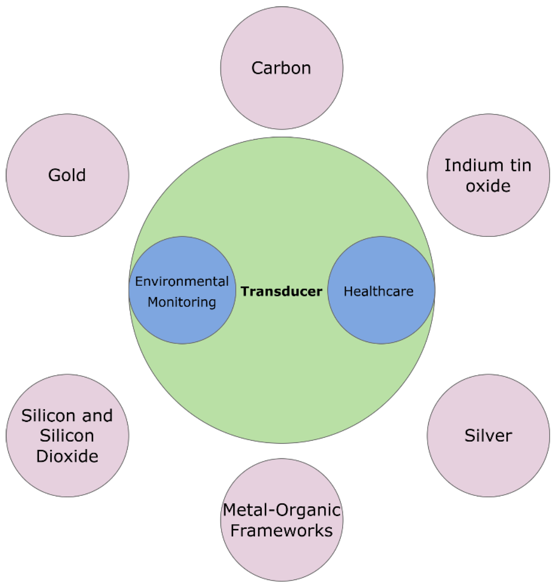
Figure 2.
A) Representation of electrochemical biosensors: (a) amperometric, (b) potentiometric, (c) conductometric, and (d) impedimetric modes.(B) Classification of nanomaterials-based biosensors including nanoparticles, nanorods, nanowires, quantum dots, carbon nanotubes, and dendrimers.(C) Dimensional classification of nanomaterials used in biosensing based on confinement direction and application domains.(D) Optical biosensor schematics: (a)chemiluminescence-based, (b) surface plasmon resonance (SPR)-based, and (c) evanescent wave-based optical biosensor[1].
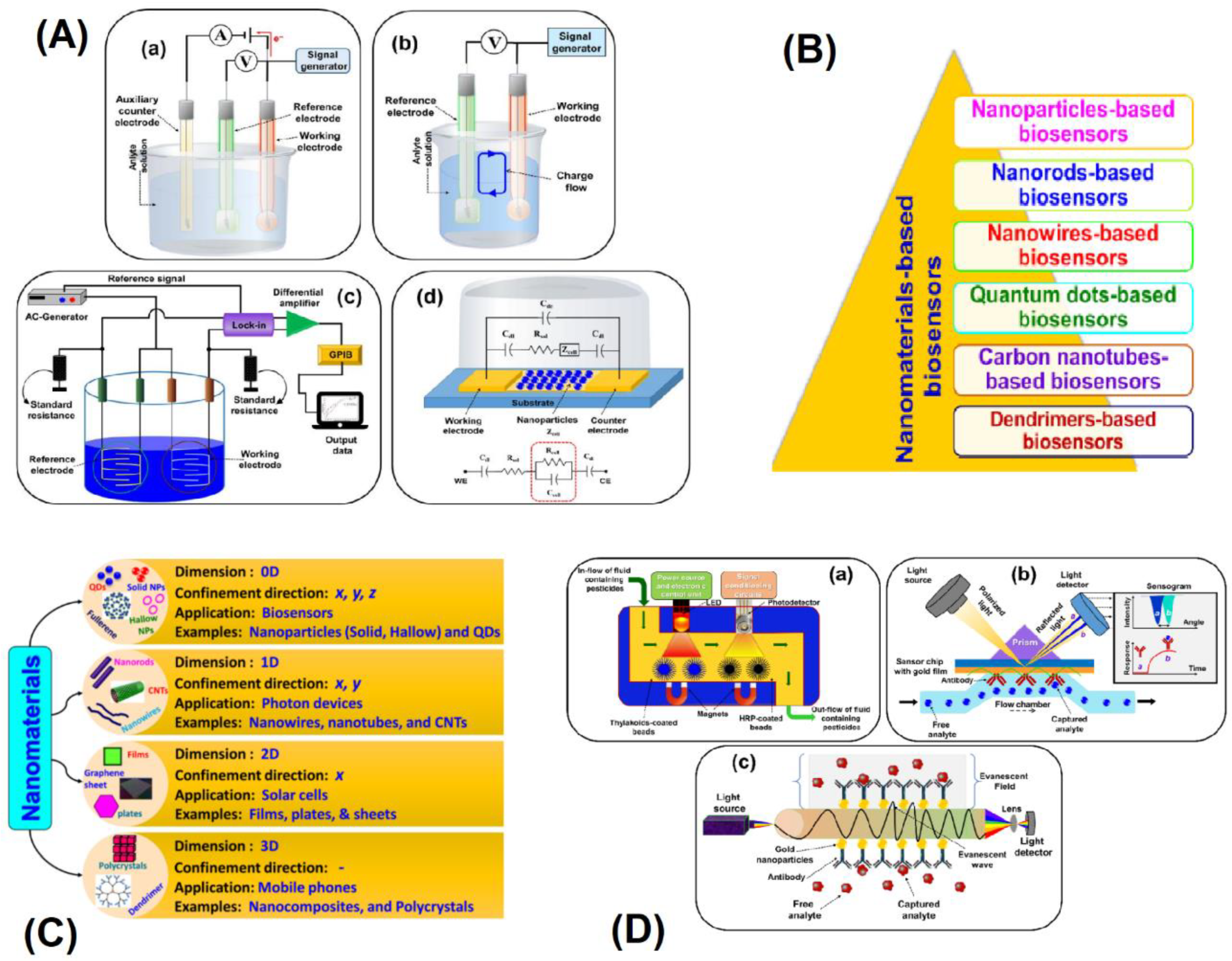
Figure 3.
Gold nanoparticle-based biosensing platforms for rapid and sensitive detection of SARS-CoV-2 N-gene, HER2, lysozyme, and HBsAg using electrochemical, optical, and QCM detection methods.
Figure 3.
Gold nanoparticle-based biosensing platforms for rapid and sensitive detection of SARS-CoV-2 N-gene, HER2, lysozyme, and HBsAg using electrochemical, optical, and QCM detection methods.
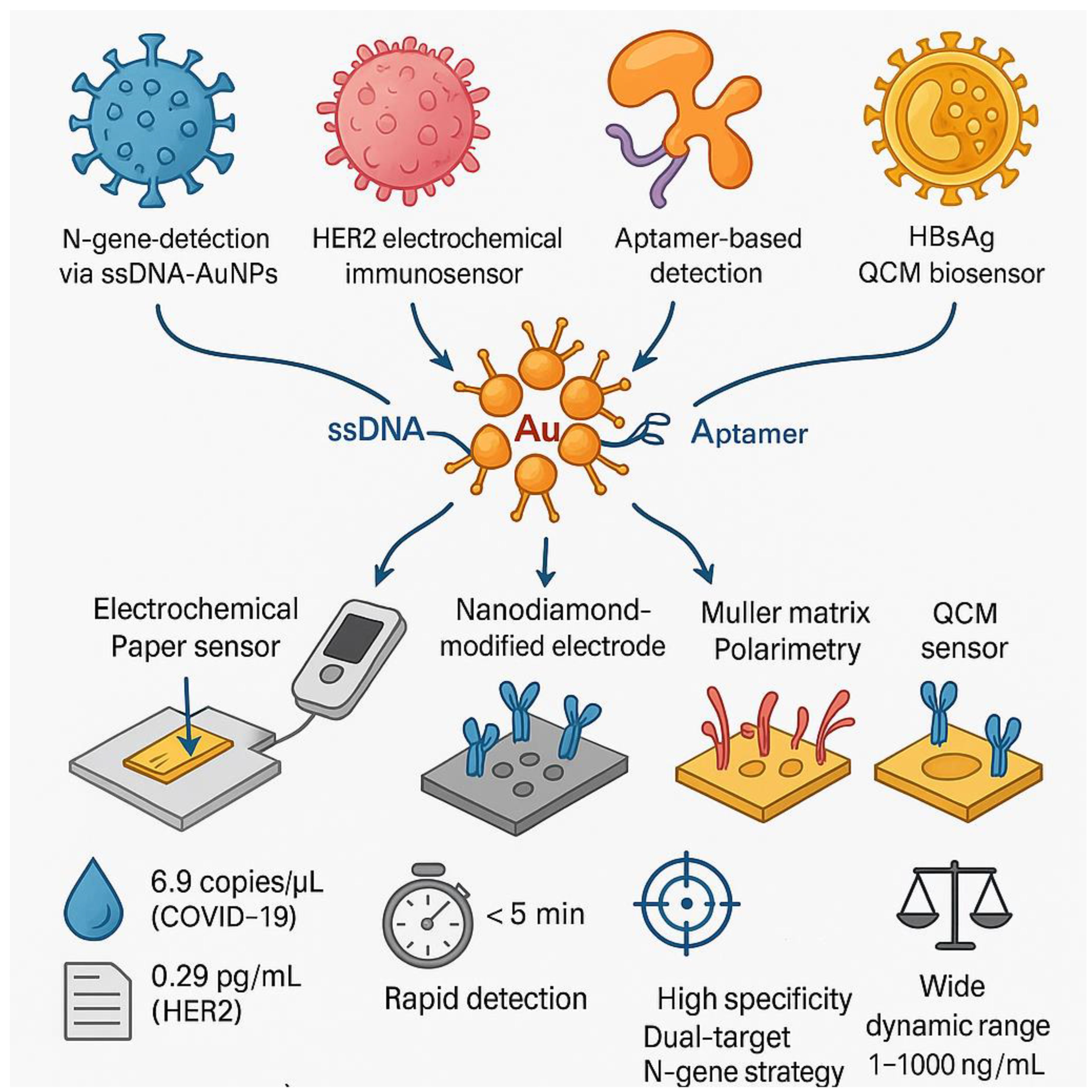
Figure 4.
Versatile SPCE platform enabling multiplexed tumor marker detection and noninvasive glucose monitoring with enhanced sensitivity and portability.
Figure 4.
Versatile SPCE platform enabling multiplexed tumor marker detection and noninvasive glucose monitoring with enhanced sensitivity and portability.
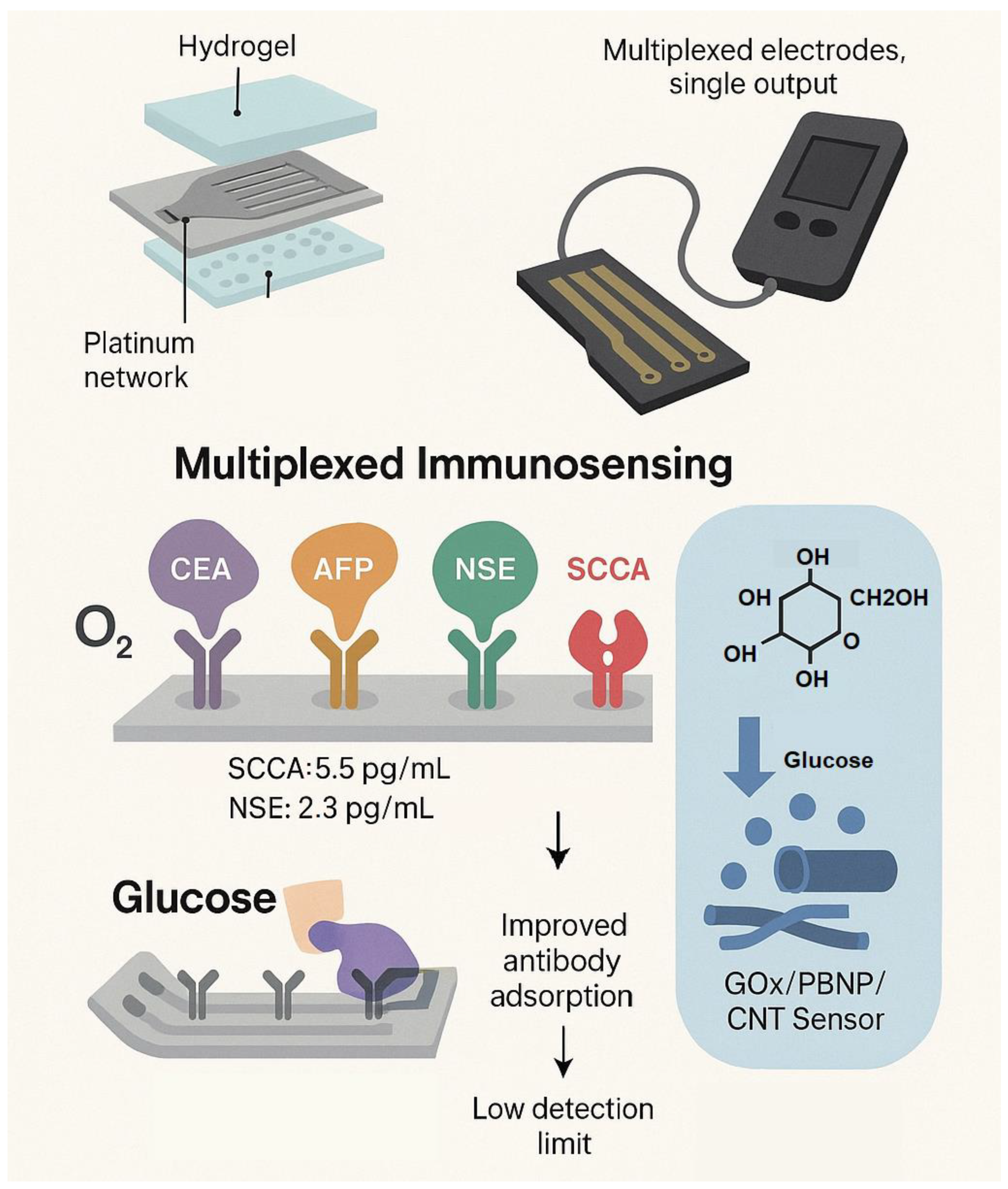
Figure 5.
A) Cyclic voltammograms of ferricyanide and ruthenium complexes on bare and plasma-treated electrodes at varying scan rates.(B) XPS spectra confirming increased surface carboxylation after plasma treatment.(C) Differential pulse voltammograms for IgA detection under four modification strategies: plasma-treated/covalent bonding, plasma-treated/physical adsorption, bare/covalent bonding, and bare/physical adsorption.(D) Electrochemical kinetics analysis showing diffusion current relationships and calculated electroactive surface areas.(E) Electrochemical impedance analysis comparing charge transfer resistance (Rct) and double-layer capacitance (Cdl) across electrode treatments.(F) Calibration curves for IgA detection demonstrating lowest limit of detection (LOD) and highest sensitivity with plasma-treated electrodes and covalent antibody immobilization.
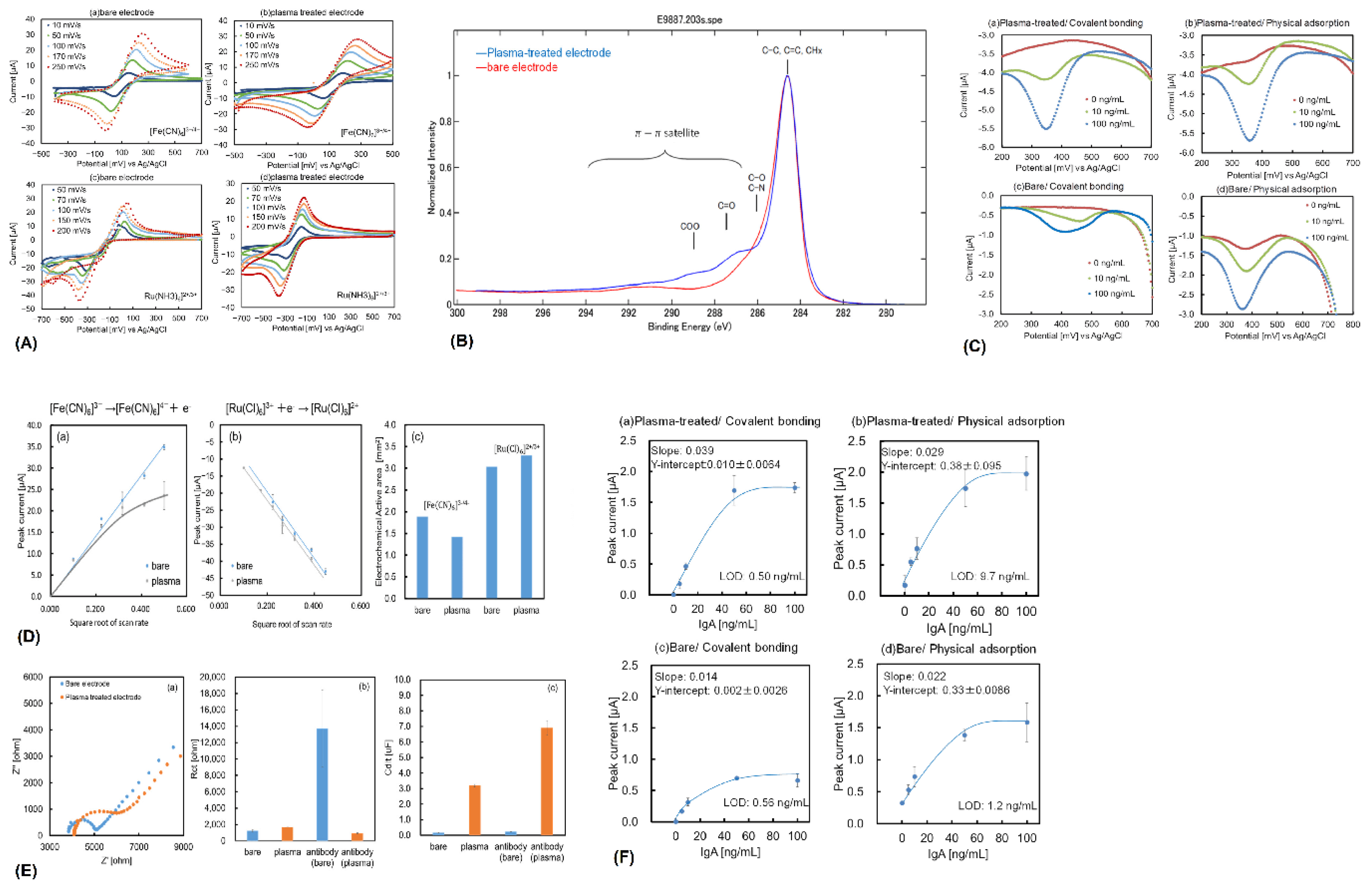
Figure 6.
Advanced ITO-based electrochemical biosensors for ultra-sensitive detection of Shigella flexneri, GM2A, HSP70, and Aβ42 in clinical diagnostics.
Figure 6.
Advanced ITO-based electrochemical biosensors for ultra-sensitive detection of Shigella flexneri, GM2A, HSP70, and Aβ42 in clinical diagnostics.
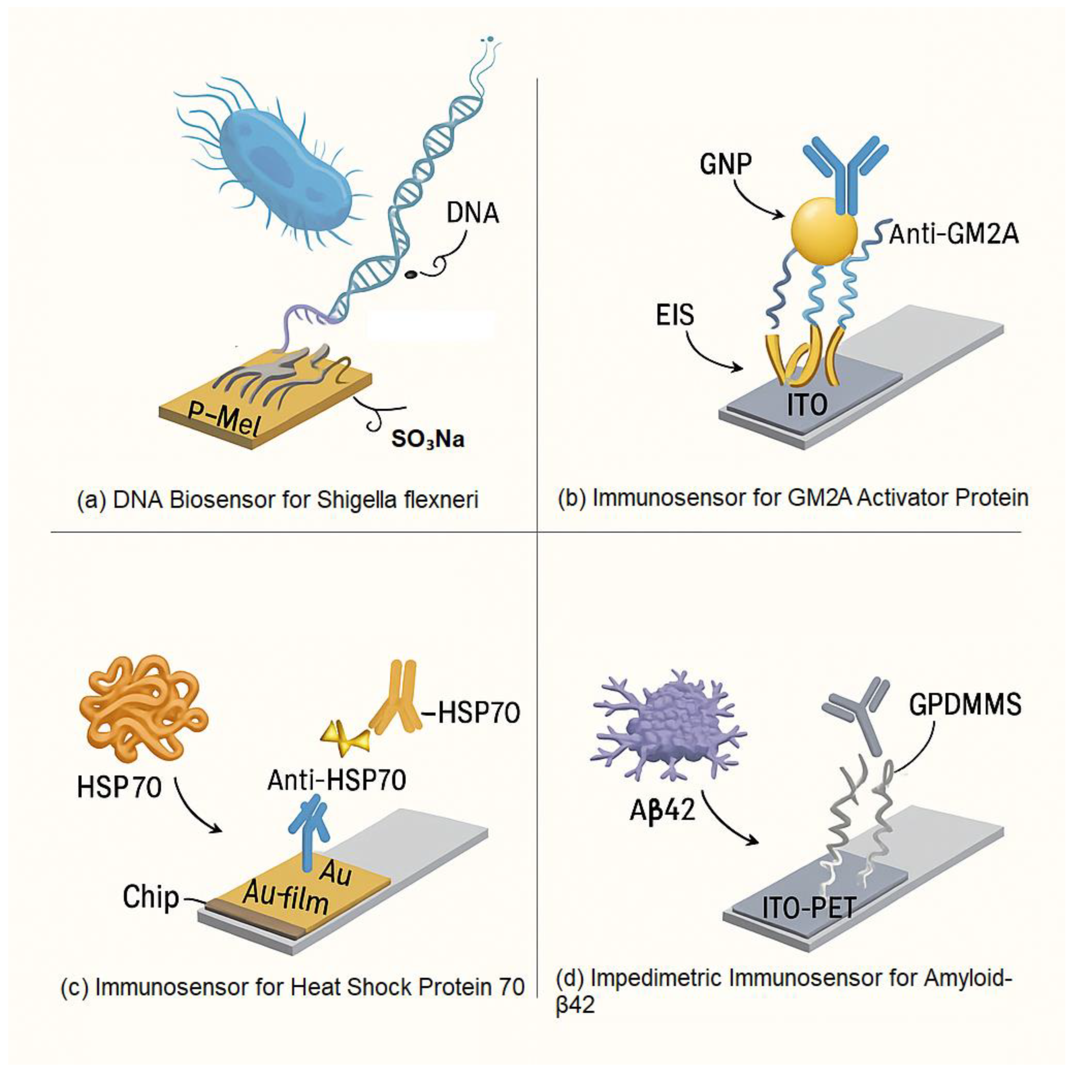
Figure 7.
Multifunctional AgNP-based electrochemical sensors for simultaneous detection of H₂O₂, dopamine, saxitoxin, and lactate with high sensitivity and stability.
Figure 7.
Multifunctional AgNP-based electrochemical sensors for simultaneous detection of H₂O₂, dopamine, saxitoxin, and lactate with high sensitivity and stability.
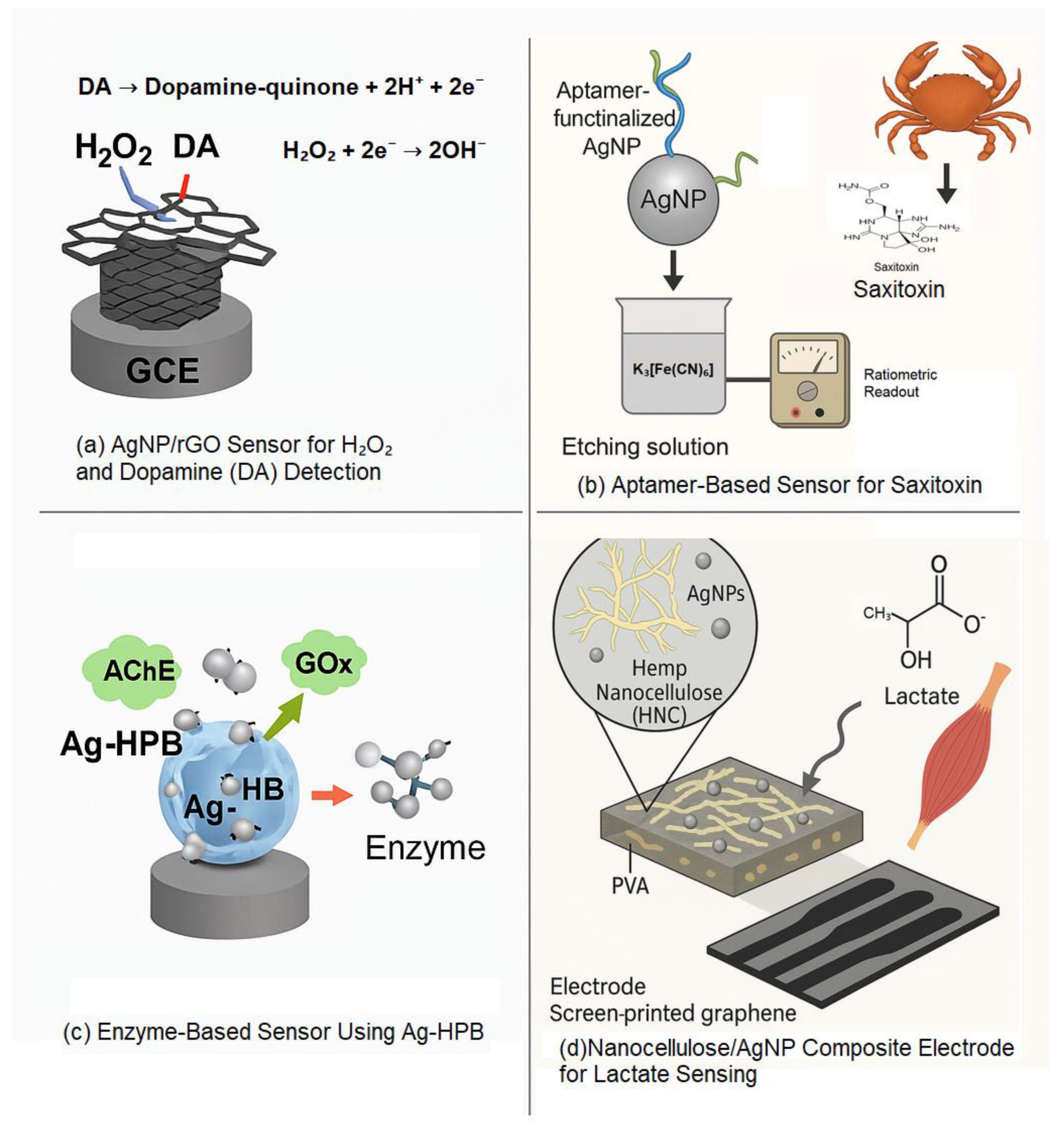
Figure 8.
A) High-resolution XPS spectra of N 1s and Si 2p for successive surface modifications: e- Biotin–dT30 and f- RNA aptamer layers.(B) (a) Binding isotherm showing Δλmin versus Sphingobium yanoikuyae concentration; (b) AFM images and corresponding optical chip color changes before and after microbial binding.(C) Schematic of multilayer surface functionalization: (a) oxidized SiO₂, (b) aminated, (c) biotinylated, (d) avidin-modified, (e) Biotin–dT30, (f) RNA aptamer layer, (g) bacterial binding, and (h) interference-based color generation.(D) XPS survey spectra for each functional layer: (a) SiO₂, (b) amine, (c) biotin, (d) avidin, (e) Biotin–dT30, (f) RNA aptamer, (g) bacteria-bound.(E) High-resolution XPS spectra for (a) C 1s, (b) O 1s, and (c) N 1s across layers (a) SiO₂, (b) amine, (c) biotin, (d) avidin, (e) Biotin–dT30, (f) RNA aptamer, (g) bacteria-bound.
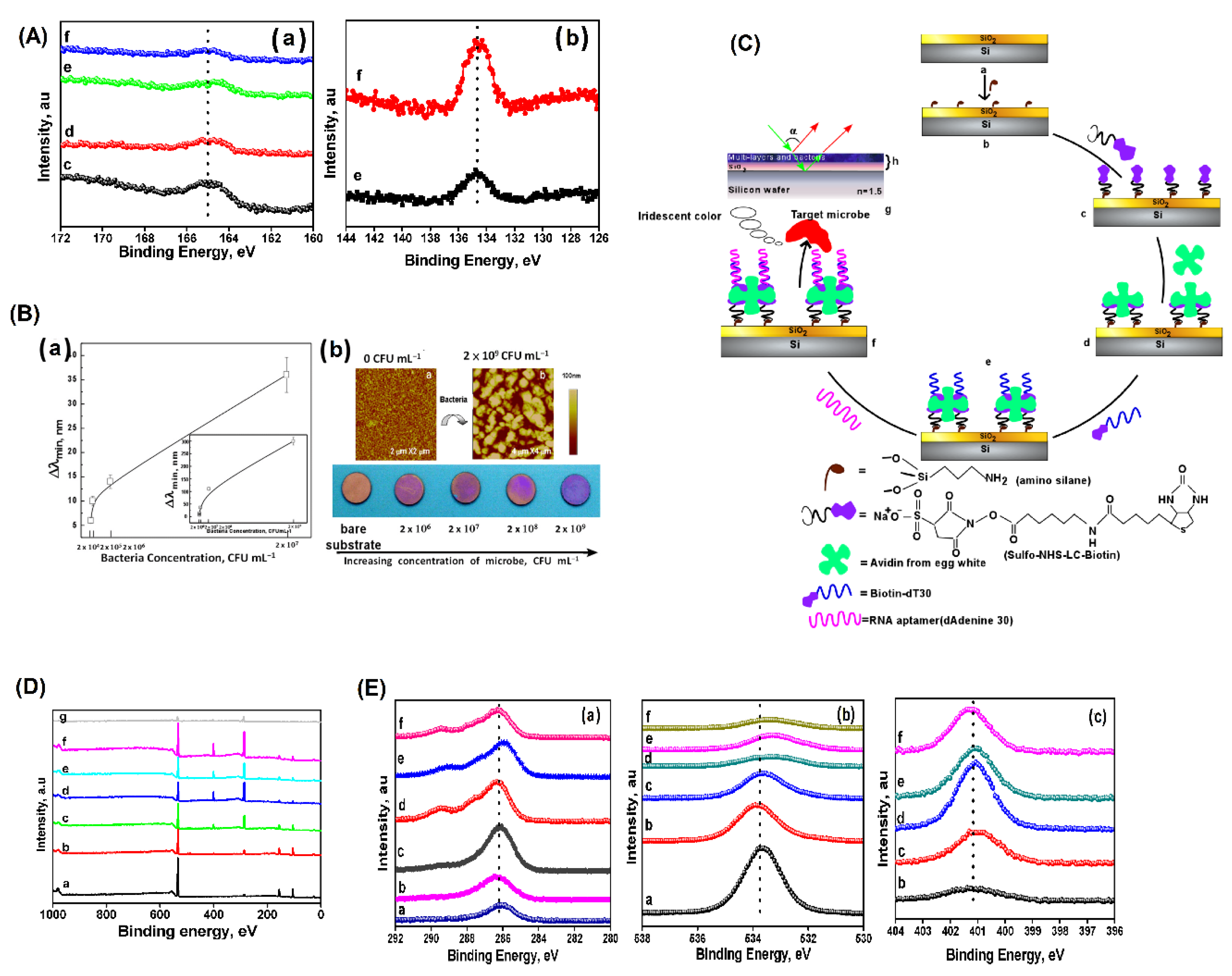

Table 1.
Gold-Based Electrodes for Biosensing Applications.
| Title | Summary | Target Analyte | Material | Technique | Detection Limit (LOD) | Sensitivity | Measurement Method | Key Features | Applications | Ref |
|---|---|---|---|---|---|---|---|---|---|---|
| Gold (Au) Electrodes for SARS-CoV-2 Detection | Biosensor using AuNPs capped with antisense ssDNA targeting the viral N-gene. | SARS-CoV-2 RNA | AuNPs, ssDNA | Paper-based electrochemical platform | 6.9 copies/μL | 231 (copies μL–1)−1 | Hand-held reader | Dual-target approach, no amplification needed, rapid detection (< 5 mins) | SARS-CoV-2 detection | [13] |
| Electrochemical Immunosensor for HER 2 Detection | Highly sensitive detection of HER 2 using a NanoDiamond and AuNP nanohybrid platform. | HER 2 | NanoDiamonds, AuNPs | GCE, DPV | 0.29 pg mL−1 | Not specified | Differential pulse voltammetry (DPV) | High specificity, low cross-reactivity, optimal at 35 °C, pH 7.2 | Breast cancer biomarker detection | [14] |
| Gold Nanoparticle-Based Biosensor for Lysozyme Detection | Detection of Lysozyme using gold nanoparticles and decomposition Muller matrix polarimetry. | Lysozyme | Gold nanoparticles, DNA aptamer | Muller matrix polarimetry | 1.24 fM | Not specified | Muller matrix calculation | High specificity, broad dynamic range (0.01 to 500 pM) | Lysozyme detection | [15] |
| Flexible Sensor for Glucose and Lactate Monitoring | Real-time monitoring of glucose and lactate in sweat using a flexible chip with AuNNs. | Glucose, Lactate | AuNNs | Electrochemical deposition, PEGDE | 7 μmol L−1 (glucose), 54 μmol L−1 (lactate) | Not specified | Electrochemical detection | High selectivity, stability, reproducibility | Glucose and lactate monitoring in sweat | [17] |
| Stretchable Gold Fiber-Based Lactate Biosensors | Fabrication of stretchable, strain-insensitive gold fibers for lactate sensing in wearable applications. | Lactate | Gold fibers | Dry-spinning, three-electrode system | Not specified | 19.13 μA/mM cm² (PBS), 14.6 μA/mM cm² (artificial sweat) | Electrochemical detection | High sensitivity, maintains performance under 100% strain | Wearable lactate monitoring | [18] |
| Flexible Enzymatic Glucose Biosensor | High-performance glucose biosensor using dendritic gold nanostructures on carbon cloth. | Glucose | Dendritic gold nanostructures, carbon cloth | Electrochemical deposition, enzyme precipitation coating | 6.7 μM | 72.45 μA mM−1 cm−2 | SEM, CV, chronoamperometry, EIS | High sensitivity, low detection limit, wide linear range, good selectivity, reproducibility, stability | Biomedical and health care | [19] |
| QCM-Biosensor for HBsAg Detection | QCM-biosensor for label-free detection of HBsAg. | HBsAg | Anti-HBsAg antibodies, gold electrode | PEI and thiolated-PEI surface modifications, RSM optimization | 3.14 ng/mL | Not specified | FESEM, AFM, ATR-FTIR, CA measurement | Broad dynamic range, high accuracy, good selectivity, stability, regenerability | Non-invasive monitoring of HBV-biomarker | [16] |
| Isoprocarb Detection Biosensor | Biosensor system based on AChE inhibition by isoprocarb using AuNPs-PANI-GPE. | Isoprocarb | Gold nanoparticles, polyaniline, graphite pencil electrode | Electro-polymerization, electro-deposition | 0.1615 nM | 1.7771 μA/μM.mm² | SEM-EDX, CV | High sensitivity, excellent stability | Real detection of isoprocarb | [21] |
| Electrochemical Sensor for Glucose Detection | Sensor using CNTs grown on Au MEA for glucose detection. | Glucose | CNTs, gold microelectrode arrays | Immobilizing GOx enzyme, poly (p-PDA) matrix | 0.2 μM | 168.03 kΩ−1 M−1 | CV, EIS | Good reproducibility, anti-interference, validated through HPLC | Multiple electroactive biomolecules detection | [20] |
| Folic Acid Biosensor | Biosensor for folic acid detection using DHFR immobilized on c-MWCNT/TiO2NPs modified Au electrode. | Folic Acid | c-MWCNT, TiO2 nanoparticles, gold electrode | TEM, XRD, FTIR, SEM, EIS, CV | 11.48 nM | 0.42 μA/nM/cm² | SEM, EIS, FTIR, CV | Wide linear range, good storage stability | Folic acid quantification in serum samples | [22] |
Table 2.
Silver Nanoparticles in Biosensing.
| Title | Summary | Target Analyte | Material | Technique | Detection Limit (LOD) | Sensitivity | Measurement Method | Key Features | Applications | Ref |
|---|---|---|---|---|---|---|---|---|---|---|
| Impact of Ag/AgCl Electrodes on Alcohol Oxidase Stability | Investigates the interaction between Ag/AgCl electrodes and Alcohol Oxidase enzyme stability. | Alcohol Oxidase enzyme | Ag/AgCl electrodes | Screen-printed Ag/AgCl ink, optical absorbance assay | N/A | N/A | Optical absorbance assay | Significant enzyme activity degradation in presence of Ag/AgCl; enzyme activity halftime reduced from ~1 week to 10 hours | Enzymatic biosensors | [51] |
| Lactate Oxidase-Based Biosensor with Silver Amalgam Electrode | Design of a biosensor with LOx-based mini-reactor and silver amalgam electrode. | Lactate | Silver amalgam electrode, lactate oxidase (LOx) | LOx mini-reactor, mesoporous silica powder, amperometric monitoring | N/A | N/A | Amperometric monitoring | High operational stability (93.8% after 350 measurements, 96.9% after 7 months); Easy replacement of mini-reactor | Clinical diagnostics, food and beverage quality control | [52] |
| Dual-Mode Biosensor Using DNA-Templated Silver Nanoclusters | Fluorescent/electrochemical dual-mode biosensor for glucose detection. | Glucose | DNA-templated silver nanoclusters (AgNCs@DNA), glucose oxidase (GOx) | Fluorescence detection, electrochemical pathway | 23 μM (optical), 29 μM (electrochemical) | N/A | Fluorescence and electrochemical | Dual-mode detection, high sensitivity, small size | Glucose monitoring in body fluids | [43] |
| Core-Sheath Ag@Au Nanowires for Stretchable Transparent Electrodes | Development of Ag@Au NWs for improved STEs with high oxidation resistance. | Glucose | Ag@Au nanowires (NWs) | Capillary-force-induced welding | 125 µM | 967 µA·mM−1·cm−2 | Electrochemical | High optical transmittance (78.7%), high tensile strain (240%), excellent robustness | Flexible electronics, glucose biosensors | [44] |
| Green Synthesis of Silver Nanoparticles for Glucose Biosensors | Amperometric glucose biosensor using green synthesized WT-AgNPs. | Glucose | Green synthesized waste tea-based silver nanoparticles (WT-AgNPs), glucose oxidase enzyme | CPE modified with WT-AgNPs, enzyme immobilization | N/A | N/A | Amperometric | Low detection limit, high reproducibility, good selectivity | Glucose detection in food, environmental applications | [45] |
| HNC/AgNPs-PVA for Lactate Detection | Uses hemp-derived nanocellulose (HNC) with silver nanoparticles (AgNPs) for lactate detection. | Lactate | HNC, AgNPs, PVA | Alkali treatment, acid hydrolysis, self-reduction | Not specified | Not specified | Cyclic voltammetry (CV), Amperometry | High current response at 10 wt% HNC/AgNPs-PVA, Linear range: 0–25 mM | Wearable lactate sensor | [50] |
| Ag-HPB Nanocomposites for Biosensing | Hollow Prussian blue with ultra-small AgNPs for glucose and trichlorfon detection. | Glucose, Trichlorfon | AgNPs, Prussian blue (PB) | Coating-etching method | 2.28 pg/mL (trichlorfon) | 24.37 μA mM−1 cm−2 (glucose) | Amperometry | High sensitivity and enzyme loading, Flexible biosensing platform | Trichlorfon monitoring in apples | [49] |
| GPP-Based SPCIDE for Glucose Sensing | Glossy Photo-Paper based sensor for glucose detection. | Glucose | Graphene, Polyaniline (PANI), GS-AgNPs, Glucose oxidase (GOx) | Screen-printing, drop-casting | 198 nM | 291.19 µA mM−1 cm−2 | Amperometry | Low-cost, eco-friendly, Linear range: 198 nM to 30 mM | Point-of-Care glucose testing | [46] |
| Aptamer-Based Electrochemical Sensor for STX | Uses aptamer and K3Fe(CN)6 regulated AgNPs for STX detection. | Saxitoxin (STX) | AgNPs, Aptamer, K3Fe(CN)6 | Aptamer immobilization, K3Fe(CN)6 etching | 1 nM | Not specified | Differential pulse voltammetry (DPV) | High sensitivity, stability, and reproducibility | STX detection in seafood | [48] |
| AgNPs/rGO Nanocomposites for H2O2 and DA Detection | Electrochemical sensors using AgNPs/rGO nanocomposites. | Hydrogen peroxide (H2O2), Dopamine (DA) | AgNPs, Reduced graphene oxide (rGO) | Hydrothermal synthesis, electrodeposition | 3.19 μA (H2O2), 0.18 μM (DA) | 49 μA mM−1cm−2 (H2O2), 7.86 μA mM−1cm−2 (DA) | Amperometry, CV | Simultaneous detection of H2O2 and DA, Good stability | Detection of H2O2 and dopamine | [47] |
Table 3.
Metal-Organic Frameworks (MOFs) in Biosensors .
| Title | Summary | Target Analyte | Material | Technique | Detection Limit (LOD) | Sensitivity | Measurement Method | Key Features | Applications | Ref |
|---|---|---|---|---|---|---|---|---|---|---|
| MOF-based Aptasensor for COVID-19 Detection | Biosensor using MIL-53(Al) with Au@Pt nanoparticles and enzymes for detecting 2019-nCoV-NP. | 2019-nCoV-NP | MIL-53(Al), Au@Pt nanoparticles, HRP, hemin, G-quadruplex DNAzyme | Thiol-modified aptamers, Au@Pt/MIL-53(Al), HRP, hemin/G-quadruplex DNAzyme | 8.33 pg/mL | Not specified | Aptamer-protein-nanoprobe sandwich electrochemical detection | Wide linear range (0.025 to 50 ng/mL), high sensitivity, selectivity, reliability | Early COVID-19 diagnosis | [53] |
| MOF-5/CoNi2S4 Nanobiosensor for SARS-CoV-2 Detection | Enhanced sensitivity for detecting SARS-CoV-2 spike antigen using MOF-5/CoNi2S4 with porphyrin. | SARS-CoV-2 spike antigen | MOF-5, CoNi2S4, H2TMP | Surface roughness measurement, cell viability assay | 5 nM | Not specified | Atomic force microscopy, MTT assay | High biocompatibility, low cytotoxicity, tunability | SARS-CoV-2 detection | [56] |
| ZIF-8 Encapsulated Laccase Electrode for BPA Detection | Encapsulation of laccase in ZIF-8 combined with BC and c-MWCNTs for detecting BPA. | Bisphenol A (BPA) | ZIF-8, BC, c-MWCNTs, laccase | Enzymatic biofuel cell | 1.95 x 10⁻³ mM | Not specified | Biofuel cell-driven sensing platform | High flexibility, excellent mechanical properties, conductivity | BPA detection | [57] |
| ECL Biosensor for HbA1c Detection | Paper-based ECL biosensor using nanocomposite tracing tag for HbA1c detection. | HbA1c | Zr-MOF, Fe3O4, TMC, AuNCs, APBA-functionalized GO | Electrochemiluminescence, cyclic voltammetry | 0.07% | Not specified | ECL and cyclic voltammetry measurements | Wide response range (2% to 18%), high sensitivity | HbA1c detection | [72] |
| Cu MOF-Based Glucose Biosensor | Non-enzymatic glucose sensor using Cu MOF for electrochemical detection. | Glucose | Cu MOF, BTC, Copper nitrate trihydrate | Electrochemical detection, cyclic voltammetry | 0.019 mM | 229.4 μAmM⁻¹ cm⁻² | Cyclic voltammetry, chronoamperometry | Exceptional stability, short response time, good repeatability | Glucose detection | [58] |
| Electrochemical Immunosensor for HE4 | A sandwich-type electrochemical immunosensor for HE4 detection using Prussian blue (PB) and functionalized MOF nanocomposites. | HE4 | PB, TiMOF-KB@AuNPs | Electrochemical immunosensor | 0.02 ng/mL | N/A | Electrochemical | PB as signal indicator, TiMOF-KB@AuNPs for signal amplification | Clinical ovarian cancer diagnosis | [61] |
| Non-Invasive Glucose Detection | An enzymatic biosensing platform using MOF nanomesh for sensitive and stable glucose detection. | Glucose | MOF nanomesh, enzyme encapsulation | Enzymatic biosensor | 16.57 µm | 86.86 µA mm−1 cm−2 | Electrochemical | High sensitivity and stability | Home-based glucose testing | [55] |
| ECL Biosensor for GC Detection | An electrochemiluminescence (ECL) biosensor for detecting microRNA in GC extracellular vesicles. | microRNA (miRNA-421) | Cu NCs, Zn MOF nanosheet, Au NPs/MXene, phospholipid layer | ECL biosensor | 0.5 fM | N/A | Electrochemiluminescence | Cu NCs/Zn MOF nanosheet for high quantum yield and stability | GC peritoneal metastasis diagnosis | [60] |
| MNT Detection with Electrochemical Sensor | A platform using MWCNTs and UIO-66-NH2 nanocomposite for detecting multiple monoamine neurotransmitters (MNTs). | Dopamine (DA), Adrenaline (Adr), Norepinephrine (NE), 5-Hydroxytryptamine (5-HT) | MWCNTs, UIO-66-NH2 | Electrochemical sensor | Low detection limit for DA, Adr, NE, 5-HT | N/A | Electrochemical | Synergistic effect between MWCNTs and UIO-66-NH2 | Clinical diagnosis of MNT-related disorders | [59] |
| MMOF-Based Electrochemical Aptasensor for SARS-CoV-2 | An MMOF-based aptasensor for detecting SARS-CoV-2 spike proteins using electrochemical methods. | SARS-CoV-2 spike proteins | MMOF, aptamer-biotin, streptavidin-HRP | Electrochemical aptasensor | 6 pM (voltammetry), 5.12 pM (impedance spectroscopy) | N/A | Electrochemical | MMOF for easy washing and deposition, high sensitivity | Point-of-care testing for SARS-CoV-2 | [54] |
Table 4.
Silicon and Silicon Dioxide-Based Biosensors .
| Title | Summary | Target Analyte | Material | Technique | Detection Limit (LOD) | Sensitivity | Measurement Method | Key Features | Applications | Ref | ||||||||||
|---|---|---|---|---|---|---|---|---|---|---|---|---|---|---|---|---|---|---|---|---|
| Porous Silicon Optical Biosensor | High sensitivity for detecting HSP70 | Heat Shock Protein 70 (HSP70) | Porous Silicon (PSi) | Electrochemical etching, thermal oxidation, antibody functionalization with APTES and glutaraldehyde | 1290 ± 160 ng/mL | N/A | Fiber Optic Spectrometer, SEM, FTIR, Contact Angle Measurement | High selectivity, -10 dB wide bandwidth, porosity: 68% ± 3%, thickness: 2400 ± 30 nm | Label-free optical biosensing | [62] | ||||||||||
| Capacitance Electrochemical Biosensor for TNF-α Detection | High sensitivity and selectivity for TNF-α cytokines | Tumor Necrosis Factor Alpha (TNF-α) | Silicon Nitride (Si3N4/SiO2/Si[P]/Al) | Micro-contact printing, fluorescence microscopy, contact angle measurement | 0.38 pg/mL (PBS), 1 pg/mL (AS) | 4 mV·pM−1 (PBS), 4.4 mV·pM−1 (AS) | Mott-Schottky analysis, Fluorescence Microscopy, Contact Angle Measurement | High linearity, selectivity against Cortisol and Interleukin-10 | Early detection of inflammatory responses | [67] | ||||||||||
| RNA Aptamer-Based Color Sensor for Pathogen Detection | Detection of specific bacteria with visual color changes | Sphingobium yanoikuyae | Silicon | Layer-by-layer deposition, RNA aptamers | 2 × 10⁶ CFU/mL | N/A | Visual color change, UV-Vis reflectance spectrophotometry, AFM, XPS | Visual detection, iridescent color changes, UV-Vis confirmation | Pathogen detection in non-beverage alcohols | [68] | ||||||||||
| Photonic Crystal-Based Biosensor for Protein Analysis | Responsive biosensor for protein analysis and immunological evaluation | Proteins, antibodies, antigens | SiO2, Gold (Au), Graphene Oxide (GO) | Gold plating, graphene oxide coating, FDTD method | N/A | 100 nm/RIU | FDTD method | High Quality factor: 597, increased plasmonic attraction | Clinical decision systems | [69] | ||||||||||
| Fabry-Perot Optical Fiber Sensor for Glucose Detection | Detecting ultralow glucose concentrations with high sensitivity | Glucose | Siliconoxynitrite (SiON) doped silicon (Si) | Plasma enhanced chemical vapor deposition (PECVD), Phase Shifted Bragg-Grating (PSBG) | 1.98 × 10⁻⁶ RIU | 14904 nm/RIU | Lab-on-Fiber (LOF) | Temperature insensitive (25°C-45°C), high sensitivity | In-vivo biosensing applications | [70] | ||||||||||
| 3-D Integrated Graphene-Porous Silicon Plasmonic Waveguide for DNA Hybridization | Studies a 3-D integrated graphene-p-Si plasmonic waveguide-based nanostructure for DNA hybridization. | DNA | Graphene, porous silicon, silicon dioxide | Full-vectorial finite element method, Maxwell Garnett model | N/A | 318.5 nm/RIU | COMSOL multiphysics software | High sensitivity, tunability, reduced ohmic losses | Lab-on-a-chip biological applications | [66] | ||||||||||
| High-Performance Hydrogen Peroxide Sensing FET | Demonstrates a high-performance H2O2 sensing FET using rGO/PPy nanocomposites. | Hydrogen peroxide (H2O2) | Reduced graphene oxide, polypyrrole, SiO2 | Lithography, etching techniques | 10:00 PM | N/A | Electrical signal measurement | Fast response, high stability, high selectivity | Liquid sensing applications | [65] | ||||||||||
| Conductive Glucose Sensor Using Macro Porous Silicon | Studies glucose sensor using glucose oxidase immobilized on macro porous silicon. | Glucose | Glucose oxidase, macro porous silicon, silver electrodes | Electrochemical etching, physisorption | N/A | N/A | Current–voltage characteristics | Good response, high sensitivity, excellent repetitive behavior | Glucose sensing | [63] | ||||||||||
| Gold-Coated Silicon Microneedle Arrays for Breast Cancer Biomarker Detection | Uses Au-Si-MNA for biomarker extraction and electrochemical transduction for breast cancer diagnosis. | Epidermal growth factor receptor 2 (ErbB2) | Gold, silicon, artificial interstitial fluid | Gold coating, electrochemical transducer | 4.8 ng/mL | N/A | Electrochemical measurement | High selectivity, simultaneous extraction and quantification | Wearable point-of-care devices | [64] | ||||||||||
| Fluorescence Image-Based Pesticide Detection Using PSi Bragg Mirrors | Proposes a grayscale variation of fluorescence image using PSi Bragg mirrors for pesticide detection. | Acetamiprid (pesticide) | CdSe/ZnS quantum dots, PSi Bragg mirrors, aptamers | Digital imaging method | 2.8 nM | N/A | Fluorescence intensity measurement | Low-cost, rapid, simple detection | Rapid and simple pesticide detection | [71] | ||||||||||
Disclaimer/Publisher’s Note: The statements, opinions and data contained in all publications are solely those of the individual author(s) and contributor(s) and not of MDPI and/or the editor(s). MDPI and/or the editor(s) disclaim responsibility for any injury to people or property resulting from any ideas, methods, instructions or products referred to in the content. |
© 2025 by the authors. Licensee MDPI, Basel, Switzerland. This article is an open access article distributed under the terms and conditions of the Creative Commons Attribution (CC BY) license (http://creativecommons.org/licenses/by/4.0/).
Copyright: This open access article is published under a Creative Commons CC BY 4.0 license, which permit the free download, distribution, and reuse, provided that the author and preprint are cited in any reuse.


