
Submitted:
02 June 2025
Posted:
02 June 2025
You are already at the latest version
Abstract
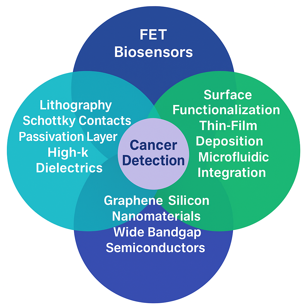
Field-effect transistor (FET)-based biosensors offer a promising platform for cancer diagnostics due to their exceptional electrical sensitivity, miniaturization potential, and ability to function in complex biological media. This review highlights comprehensive strategies for enhancing FET biosensor performance through advanced lithography, surface functionalization, thin-film deposition, and microfluidic integration. Specific emphasis is placed on the impact of materials—such as graphene, silicon nanomaterials, wide bandgap semiconductors, and high-k dielectrics—and device engineering techniques including Schottky contact optimization and passivation layers. This review compiles 57 biosensors and critically evaluates material and fabrication strategies that enhance device performance. Comparative analysis of a representative sensors shows that graphene and TiS₃-based electrolyte-gated FETs achieved the lowest detection limits (down to 10 zM and 0.04 fg/mL), while microfluidic integration and PNA/antibody functionalization significantly improved sensitivity and specificity in clinical samples. These findings highlight the critical role of 2D materials, nanolithography, and advanced surface chemistry in enabling next-generation FET biosensors for precision oncology. Future challenges include large-scale integration, multiplexed detection, and clinical translation.
Keywords:
Field-effect transistor (FET) biosensors
; cancer biomarker detection
; nanomaterials
; surface functionalization
; advanced nanofabrication
; high-k dielectrics
; graphene and 2D materials
; tunnel FET (TFET)
; microfluidic integration
; sensitivity and specificity optimization
; wide bandgap semiconductors
; Schottky barrier engineering
Introduction
Cancer, one of the leading causes of death worldwide, can be effectively treated and improved survival rates are possible only when it is caught early. Biosensors are among many diagnostic approaches to offer as promising tools for ab initio, real time analysis of cancer biomarkers. Particularly, field effect transistor (FET)-based biosensors have been focused upon for their own electrical amplification advantages, minimal detection limits and compatibility with small, label free detection platform[42].
The development of FET biosensors has been dependent on the key relationship between materials, device architectures, and functionalization, and therefore research has increasingly focused towards improving each of the design parameters. Innovations of increasing sensitivity, better stability and integration of clinical workflows blend into each layer of the biosensor stack, ranging from nanoscale lithography to thin film deposition and to chemical surface engineering. In general, material selection, particularly graphene and silicon based nanomaterials, possess high carrier mobility, high surface to volume ratio and wide band gap semiconductors such as MoS₂ and TiS₃ exhibit strong electrochemical stability in physiological conditions[28].
Additionally, recent efforts incorporated the developments of the advanced fabrication techniques such as lithographic nanocavity formation, dual gate tunneling structures and the precision patterning at the atomic scale [1,2,3,4]. On the basis of such methods, tunnel FET (TFET) devices have been developed, particularly for simulated detection of cancer cell line, and their high theoretical sensitivity is due to the electrostatic control enhanced. Additionally, usage of high-k dielectric layers and Schottky barrier engineering reduces threshold voltage modulation and increases suppression of leakage currents and application of low-abundance analyte response[46,47,48,49].
In addition to the channel and gate level engineerings, surface functionalization techniques have been of growing importance to achieving high selectivity. They are covalent immobilization of antibodies, enzymatic conjugation, and hybrid chemical layers that stabilizes the biological interface. These biosensors have transitioned towards point of care applications with microfluidic integration, along with rapid analysis using small sample volumes in the serum, saliva or plasma [17,18,19].
In this review, we explore how the materials science, fabrication engineering and biochemical interface design are being utilized to minimize the sensitivity and specificity of FET biosensors and explore the experimental and simulated platforms to describe the design rules that govern biosensor optimization and the future paths towards clinical translation.
Comprehensive Strategies for Enhanced FET Biosensor Design
Advanced Lithography and Nanofabrication Techniques
Owing to the great success in examining cardiac troponin I (cTnI), silicon nanowire field-effect transistor (SiNW FET) biosensors have received widespread attention. This is the result of numerous advanced techniques such as lithography, etching and metallization. By these means the size and dimension of silicon nanowires (SiNW) function in such a way that the biosensor become more standardized reliable [1]. However such techniques can also make it difficult to improve the electronic properties of a system.For COVID-19 detection, the SiNW-FET biosensor have a number of special techniques including controlled doping and electron-beam lithography. These methods make the sensor more sensitive and ensure uniform and finely made nanowires [2].Another difficulty is to ensure that the functionalization is both uniform and stable. By making use of advanced CMOS technology and a PDMS microfluidic channel, the silicon nanowire bio-field-effect transistor (Si-NW Bio-FET) for exosome detection overcomes this difficulty. It ensures everything is precise and very even, resulting in far better interactions between exosomes and the nanowire surface. This reduces variance and increases sensitivity [3].For bladder cancer diagnosis, the indium gallium zinc oxide field-effect transistor (IGZO FET) biosensor array solves problems, such as non-specific interactions and sensor array variation, by using advanced lithography and controlled antibody attachment. By this method the biosensor works better and has fewer defects, is more sensitive and has a better ratio of signal to noise [4].In summary, these advanced fabrication techniques are driving next steps towards how both uniform and capable work. Figure 1
Surface Functionalization and Chemical Modification
Precise fabrication techniques make rGO FET a very sensitive and specific biosensor. Techniques such as electron-beam lithography (EBL) make it possible to control important parts of the sensor, for example the length of gold contacts, the thickness of graphene, and the silicon dioxide layer. This precision is itself a condition ensuring that sensors work well and reliably [5].For larger areas, a CNT-FET biosensor uses inkjet printing to apply carbon nanotubes (CNTs) evenly onto the Si/SiO2 substrate. Doing so helps the sensor deliver consistent performance over large areas [6]. Similarly, a TiN EGFET biosensor spreads the TiN thin film evenly using DC sputtering. Rapid Thermal Annealing (RTA) then improves the film’s quality while reducing defects, it makes the sensor more stable. These steps make sure the sensor functions reliably even in large scale production [7].With fine control of these techniques and surface modifications as a basis directly enhances the sensor's precision, uniformity, and durability.
Through modern processing techniques, particularly precisely surface functionalising, biofet sensors are equipped to sense the N-protein of SARS-CoV-2 with high sensitivity and specificity.In this way, it was made sure that antibodies adhere securely to the sensor surface, forcing the specificity of binding and reducing non-specific interactions [8]. Continuing this approach requires adding a step to clean off any contaminants and improve performance.When detecting P90 (CIP2A) protein, the MOSFET-based biosensor undergoes ozone cleansing to remove carbon residue, and ammonium hydroxide was used to sweep away gold oxides. This manner insures that the electrode surface is clean for further functionalizing of the electrode [9]. More development is obviously required in making antibody attachment better with these cleaning strategies.The GFET biosensor for detecting Myoglobin (Mb) in human blood uses 3-mercaptopropionaldehyde N-hydroxysuccinimide ester (NHS ester) for bioconjugation. This chemical lets antibodies sit tightly on the sensor. By using 3D simulation software such as COMSOL Multiphysics as well as high-precision fabrication technologies, the sensor is made in the perfect sequence. This ensures that the target molecule is greatly improved in detection and antibodies are in their proper places [10]. In addition to the above steps, such careful preparation of surfaces and finesse with which antibodies are attached reduce non-specific binding and increase detection accuracy. Figure 2
Deposition Techniques and Thin-Film Processes
The inhibited gate field effect transistor (EGFET) biosensor is highly sensitive and specific. This is due to the use of high power impulse magnetron sputtering (HiPIMS), which makes a strong, well bonded HfO2 film. This film enhances the sensor's sensitivity and stability. HiPIMS also allows to control the film's thickness and quality accurately, which is important for a sensor. Consequently, the EGFET biosensor has been designed, fabricated by using the High Electron Mobility Transistor (HEMT) technology and made available for interferon-gamma (IFN-γ) detection. Extensive work was contributed toward improving material quality, micromachining, and growth of high quality by metal organic chemical vapor deposition (MOCVD), ion implantation and annealing. These methods produced very clean interfaces, which are necessary for high electron mobility as well as noise reduction. However, the process for making GaN HEMTs is complex and expensive [11].To transform these designs into reality, a dual channel biosensor for breast cancer markers uses simpler methods such as gold sputtering and careful functionalizing. It also combines it with a printed circuit board (PCB) and metal oxide semiconductor field effect transistor (MOSFET) for amplification. These methods increase the signal and sensitivity of the search data, while also making production much simpler and less expensive. The result of this project is due to fine-tuning the fabrication process to improve both sensitivity and ease of production [12].
The MNC BioFET obtains high sensitivity by employing standard CMOS processes, guaranteeing precisely manufactured equipment [13]. To further improve sensitivity, the two-gates MOSFET biosensor with MoS2 has a nanometer-scale gate dielectric gap and the configuration of dual gate. Such features help the sensor to better interact with biomolecules, reduce leakage and improve response accuracy [14]. Also, the GO-FET biosensor overcomes the problems caused by non-specific binding. By using standard procedures for the fabrication of gold electrodes, the dispersion patterns of nanoparticles and the placement of the antibodies , the sensor gets higher sensitivity [15]. At last, through advanced technologies like plasma-enhanced atomic layer deposition and RF sputtering, followed by heating at 325°C in a vacuum, the dual-gate oxide semiconductor TFT immunosensor achieves stable and reliable performance. These methods reduce defects and improve the active layer; they give a sensor very high electron mobility combined with low leakage and excellent dielectric properties. These combined techniques produce sensors of the highest sensitivity and specificity [16].
Microfluidics and Integration Techniques
As a biosensor, the Organic Electrolyte-Gated Field Effect Transistor (OEGFET) is extremely sensitive because its precise manufacturing methods. For the amyloid beta (Aß) antibodies, they are covalently bound tightly in the microchannel. This ensures that they will specifically bind to amyloid beta (Aß) peptides, lowering inadvertent binding [17]. However, some of these non-specific bindings still occur because in the complex environment high ionic concentrations (reagents) of samples being tested will cause ions to saturate all electronic sites. To tackle this problem, the Molybdenum Disulfide Field Effect Transistor (MoS2-FET) uses techniques that force antibodies securely onto the sensor surface. This not only decreases non-specific binding but also increases specificity [18]. But even after these improvements, creating smooth, highly sensitive surfaces is not easy. The single-walled carbon nanotube (SWCNT) network-based Field Effect Transistor (FET) biosensor does so through airbrushing. The method uniformly spreads SWCNTs onto the surface in a thin film. This better manufacturing process reduces defects, enhances electrical conductivity and heightens sensitivity, making the sensor more reliable [19]. Such improvements in binding and surface uniformity which enhance detection sensitivity and reliability directly come through advanced methods.
Passivation and Isolation Techniques
Using advanced fabrication techniques and sensor design the Aluminum Gallium Nitride / Gallium Nitride High Electron Mobility Transistor (AlGaN / GaN HEMT) biosensor is highly sensitive and specific. Also due to the fine inter-gate design,the sensing area is bigger and electric field distributions more even than before, letting sensors respond even to tiny changes in analyte concentration. The sensor is also enhanced by extending source-drain pads, which reduce noise while also improving contact and current measurement. The resulting signal-to-noise ratio is much better. Treated with TGA (Thioglycolic acid), the sensor surface prevents non-specific adsorption; giving a solid anchorage of antibodies. This result is further enhanced by using EDC-NHS chemistry to create strong and specific binding sites for antibodies, so as to minimize non-specific binding [20]. The Double-Gate Aluminum Gallium Nitride / Gallium Nitride Metal Oxide Semiconductor High Electron Mobility Transistor (AlGaN / GaN MOSHEMT) sensor, it goes still further with these improvements to add a nanocavity under the gate terminals. This makes it more sensitive, enhancing biomolecule interaction rates. A symmetric double-gate design brings greater control over the channel and less short-channel effects. An oxide layer between the gate and the barrier layer reduces gate leakage current, ensuring that the sensor's response is due to biomolecule interactions rather than background noise. And these improvements stem from advanced fabrication techniques that improve biomolecule interaction, electrical control, and noise reduction [21].
Optimizing Sensitivity and Specificity in FET Sensors
High-K Dielectrics for Enhanced Sensitivity and Specificity
The gate dielectric of the MI-DMTFET biosensor is a metal film. One result increases output current and, hence, sensitivity because conduction band with valence band alignment and lower barrier to tunnel density of states. However, it may be less specific. To improve this, Hafnium Dioxide (HfO2) is introduced. Its contribution is to raise both sensitivity and specificity; it provides a stable surface for molecular interaction, making the noise smaller and the signal cleaner [22]. By uniformly doping the source and drain p-type regions, researchers are able to obtain consistent electrical properties between different biomolecules. Thus, we can distinguish one molecular species from another on the grounds of their charge densities and dielectric constants [23].The Schottky barrier height is further reduced by low NiSi contacts, and this can improve sensitivity even more by increasing charge carrier generation and ON-current. Materials such as Tungsten and Palladium can be used to optimize electrical properties better, to enhance the performance [24]. These materials also allow to control precisely the effects of charge interaction and tunneling on performance of the chip.
For the biosensor module used by DM DT GE-MOSFET Biosensor, with the use of indium-doped tin oxide (ITO) as a gate, further work function and characteristics current, the result is improved response and performance leading to high sensitivity [25]. The Bi-RFET biosensor uses high-k materials such as titanium dioxide (TiO2) to improve gate control and sense changes in an unseen environment. Both sensitivity and specificity are increased with this approach [26]. The JL-DM-DG MOSFET biosensor uses hafnium dioxide (HfO2) to increase gate capacitance, which yields higher sensitivity and eliminates interference. Specificity is improved so that the interference caused by unwanted compounds does not diminish the signal [27].The JL-TFET biosensor tackles power consumption and scalability by using titanium dioxide (TiO2) to enhance gate capacitance as well as reduce gate leakage. Minimizing noise further increases specificity [28]. These dielectric materials altogether bring about a higher quality of biosensor performance, greater gate capacitance, and elimination of interference. The whole results in better receptor sensitivity and specificity. Figure 3
Graphene and Silicon Nanomaterials for Enhanced Sensitivity and Specificity
Graphene is the sensing material for a GC-CPTFET sensor. Due to its very high surface area and good electrical conductivity, the signal from attachment of target molecules can be improved at once: this means more sensitive detection [29]. In order to make use of its considerably greater surface area for interaction with biomolecules the MSM-SiNW biristor sensor uses silicon nanowires, the Schottky barrier height is optimised in the presence hafnium in order to boost its sensitivity still further [30]. In the GC-GAA-NWFET biosensor, gate capacitance is raised by using materials with high k, such as HfO2. The unique properties of silicon and its compatibility with CMOS technology ensure that accurate, scalable biomolecule detection is achievable [31]. Each succeeding sensor enhances the sensitivity and specificity of the materials. By choosing materials such as graphene, silicon nanowires, and high-k dielectrics, the result has maximum detection performance.
The Source Engineering Quad Gate Structure utilizes HfO2 in the source and SiO2 in the drain to optimize electric field distribution and barrier widths, which are necessary for distinguishing biomolecules based on their dielectric constants[32]. By employing a stack of HfO2/SiO2, the DM-TMGOS-ZHP-TFET sensor addresses these problems, reducing gate leakage and increasing capacitive coupling. This design improves the signal-to-noise ratio and lowers the subthreshold swing, overcoming previous limits [33]. The DLZ-ZHP-TFET's effectiveness lies in a design feature implemented on the triple metal gate and dual material gate-oxide stack, which increase accuracy and reduce leakage currents to different aspects of molecular recognition. Figure 4
Wide Bandgap Materials for High Sensitivity and Specificity
Biosensors in this section make use of materials such as 4H-SiC, 6H-SiC and Ge to secure both high sensitivity and specificity. The biosensor described is a 4H-SiC Schottky Barrier Field-Effect Transistor (SB-FET) with P+ implantation pockets. The 4H-SiC (about 3.2 eV) wide bandgap, coupled to its high electron mobility, makes this sensor robust and sensitive, can thus operate stably under high temperature and high voltage. This is not provided but is vital for specificity. Details about the functionalization of the surface [34] are still needed.FET with two gates improves on this by using 6H-SiC, which has a wide bandgap of 3.0 eV and high saturated drift velocity. This configuration raises sensitivity, carrier transport is faster and detection more accurate. Erbium silicide and high dielectric constant Hafnium were put in the source drain areas. [35].Germanium, with its high carrier mobility and superior tunneling characteristics, is used as the Ge-DMJ-CPTFET offers a different approach to this problem of enhancing charge transport and sensitivity when dealing with high-k HfO2 in the gate oxide layer The small rabbits made the gate button, increase the capcitant and detection. But there is still much sensitivity to be improved[36]. The Ge-source L-shaped tunnel power transistors (LTFET) produce higher sensitivity using Germanium bandgap. This allows easier band-to-band tunneling and so more drain current discharged. The use of high-k dielectric HfO2 and a thin SiO2 adhesive layer improves electrostatic control, as well as reliable bond strength[37].These materials have both high bandgaps, a carrier mobility superior to anything else available and effective dielectric properties that optimize charge transport and sensor sensitivity.
Schottky Barrier and Metal Contact Engineering for Enhanced Specificity
In order to ensure sensitivity and specificity, material is a key element choice in the Non-Uniform Tunneling Field Effect Transistor with Dual Material Source (NUTFET-DMS) sensor. Employing silicon and germanium in the source region. By making use of silicon's high bandgap (1.1 eV), the sensor has a low OFF current; whereas germanium's low bandgap (0.67 eV) means it can be a high ON chromophore. The average subthreshold swing is thus improved and sensitivity enhanced [38]. However, simply relying on silicon and germanium may not allow enough gate control to effectively reduce leakage current necessary for high-performance biosensors. To solve this problem, hafnium dioxide which has a high 22 dielectric constant is introduced into the design. It significantly improves gate control and reduces leakage current. Low concentrations of biomolecules can thus be sensed, essential to maintaining high sensitivity in a variety of biosensing applications [39]. Furthermore the gate is optimized by using 4.2 eV work function aluminum as its material, helping further control threshold voltage. This is quite important for providing more sensitive measurements. Because although aluminum has nice characteristics, environmental changes may still limit the sensor's responsiveness. To improve this, titanium dioxide, a high-permittivity dielectric which is insulated against the source/channel junction, is part of Hetero-Material with Hetero-Dielectric Region Field Effect Transistor (HM-HD-RFET) design. It has been enhanced gate capacitance so that ON current increases and sensor performance is better adapted to environmental changes. Lower-permittivity dielectric silicon dioxide (SiO2) was also used to reduce ambipolarity, further improving fidelity and sensitivity [40].Difficulties arise in maintaining high performance under varying conditions. To address this h-pHVTFET (Hetero-Material pH-Based Vertical Landau RFET) biosensor incorporates thin film oxide regions such as silicon dioxide of 1.2 nanometers (nm) and hafnium dioxide of 1 nanometer. These high dielectric constant thin film oxides both increase gate capacity and reduce leakage current. This careful selection of materials ensures that the biosensor achieves high sensitivity and maintains high specificity and long-term stability across a wide range of biomolecules and environmental conditions. Thus it is a durable choice for diverse biosensing applications [41]. The deliberate use of high-k and low-k materials enhances gate control, minimizes leakage and improves sensitivity and fidelity of detection in biomolecular interactions.
FET-Based Biosensors for Cancer Diagnostics
Pancreatic Cancer
Accurate identification of tumor derived exosomes (TD Ex) is crucial as they are crucial as biomarkers for early pancreatic cancer (PC), but their limited specificity and sensitivity make them difficult to identify. To address this, a magnetic separation in a reduced graphene oxide field-effect transistor (RGO FET) biosensor was integrated microsystem to selectively separate and detect exosomal miRNA10b from PC-derived exosomes with high selectivity and sensitivity at level down to 78 fM [42]. Although detection of microRNAs only may fail to discover other critical biomarkers, the addition of detection approaches involving multiple marker types such as DNA and protein improves diagnostic accuracy. This limitation was overcome using single molecule assay using a large transistor (SiMoT) platform able to detect simultaneously ultra low concentrations (on zM) of both KRAS DNA and MUC1 protein markers, at early stage, as noninvasively as possible [43]. Yet, such systems may be laborious to configure in fabrication, and can be simplified. Therefore, a quick and sensitive alternative based on electrolyte gated MoS₂ FET immunosensor emerged as a simple but highly selective approach towards detection of CA 19-9 antigen at detection limit of 2.8 × 10⁻¹³ U/mL offering fast with excellent selectivity [44]. An electrolyte gaped FET immunosensor utilizing TiS₃ nanoribbons achieved enhanced sensitivity and overall improved performance with excellent detection limit of 1.3×10⁻¹³ U/mL for CA 19-9 antigen. The results confirmed its superiority in selectivity with good results for a reliable outcome and validated it as a strong candidate for the accurate diagnosis and therapeutic monitoring of cancer [45]. The TiS₃ nanoribbon based approach was found to be suitable for a biosensor that should possess high sensitivity, label free detection, and a simple, scalable fabrication process.
Cancer Cell Lines Detection Using TFET
A novel POC diagnostic method used for the detection of cancerous liver cell lines based on a novel Source Extended Tunnel Field Effect Transistor (SE TFET) with Single Gate Single-Metal (SGSM) and Single Gate Dual Metal (SGDM) structures with nanocavities for immobilizing biopsy obtained liver samples and the detection of cancerous liver cell lines is introduced through drain current and on/off current ratios [46]. Sensitivity, however, depends on a number of structural parameters. In addressing this, further analysis inferred that using SiO₂ as the adhesive layer improved sensitivity over other materials, such as bare silicon or HfO₂, and varying the gate work function to 4.6 eV helped to distinguish current variation based on cancerous and non-cancerous cells [46]. However, the improvement was not sufficient and requiring the structure optimization in terms of cavity dimension and gate configuration, Dual Gate (DG) SE TFET configuration was employed. In particular, such a 10 nm source and 20 nm drain cavity setup yielded superior drain current variation and sensitivity as compared with other DG structures. Also, sensitivity and performance were further improved by using GaAs as the channel material and a gate work function of 4.2 eV, which decreased the fabrication costs [47]. To generalize applicability wider than the minority model, SM and DM structures were compared on variable proportions of cancer cells and showed both could identify presence of cancer, with DM having more variable sensitivity [47]. Based on these findings, detection strategies were propagated to breast cancer diagnosis using a dual nanocavity engraved Junctionless FET (JLFET), resulting in high sensitivity due to optimized oxide thickness and cavity occupancy [48]. However, dopingless dual metal gate heterojunction TFET (DM-DL-DMG-HJTFET) with a dielectric modulated nanogap was proposed to detect breast cancer cells even in mixed population to improve rational design and better simulate practical variability. Earlier biosensors were outclassed in terms of drain current response and transconductance, and more importantly, clear discrimination of malignant from healthy cells at low concentration was clearly demonstrated by this device [49]. The proposed DM-DL-DMG-HJTFET has a combined CMOS compatibility, affordability, and consistent performance, which makes it highly promising for scaled, early stage cancer detection in POC settings, while a sensor is easily fabricated if it is fabricated using a simple, low cost process using readily available materials and applying available scalable, CMOS compatible manufacturing techniques.
Advanced Material-Based FET Biosensors for Cancer Biomarkers
Since liver cancer biomarkers are present in low quantities in complex fluids, designing an ultrasensitive and portable noninvasive detection strategy for successful POC (point of care) liver cancer diagnosis is necessary. Although miniaturization and integration possibilities of field effect transistors (FET) are possible, the necessary combination of sensitivity and non-invasiveness has not been met. To solve this problem, a metal carbide@carbon nanotubes interdigitated FET (MC@CNT-iFET) was developed for urinary exosomal microRNA-122 detection with sensitivity (0.12 fM detection limit) that was greater than 1000 times higher than current detection methods and which correlated well (0.59) with q-PCR for distinguishing liver cancer patients from healthy controls [50]. Although this approach fulfills the need for a noninvasive method for liver cancer detection, it is restricted to nucleic acid targets. A YbTixOy based electrolyte insulator semiconductor (EIS) biosensor was introduced for genetic screening, detecting KRAS, and BRAF mutations in colorectal patients with high pH sensitivity (69.54 mV/pH), low hysteresis and stability to allow fast genetic screening [51]. However, EIS biosensors suffer from being incapable of detecting protein biomarker. To tackle this, the carcinoembryonic antigen (CEA) detection in lung cancer was addressed using a floating gate CNT-FET with undulating Y₂O₃ interface leading to increased probe binding area as well as double layer capacitance (82 Ag/mL), allowing for ultralow detection limits and stable performance in complex fluid (such as fetal bovine serum) [52]. This limitation is overcome by developing a dual material In₀.₅₃Ga₀.₄₇As/Si charge plasma extended gate TFET (DCE-HTFET) for the detection of the HE4 biomarker in ovarian cancer. The RSH strategy led to outstanding ON/OFF sensitivities (18.2 × 10⁶) and sensitivities to 95% sub-threshold swing of this device, resulting in high diagnostic accuracy in various histological subtypes [53]. Further biosensor sensitivity can be enhanced using non invasive biological samples such as urine and using hybrid material systems, such as metal carbides and carbon nanotubes, to enhance electrical conductivity and transconductance in order to allow for accurate detection at ultra low analyte concentrations.
Point-of-Care and Microfluidic Integrated FET Biosensors
Point-of-care (PoC) diagnostic devices for detecting prostate-specific antigen (PSA) exist and are affordable for resource-limited settings, but conventional methods have poor detection limits as well as require label-bearing assays, making them expensive and complex. To overcome these drawbacks, a label-free dielectrophoresis (DEP) based graphene field effect transistor (FET) with coplanar electrodes and a dielectrophoresis (DEP) based microfluidic platform on a compact disc (CD) was developed, which is simple to operate at a detection limit of 1 pg/mL and shows high selectivity to the most common interferents BSA and IgG [54]. However, although such systems are sensitive and innovative, scalability remains challenging for the fabrication. However, this limitation was addressed by wafer scale fabrication and batch processing of a platform based on a tungsten oxide (WO3) thin film FET capable of real time CYFRA 21-1 saliva detection, which provided similar low detection limit of 1.26 pg/mL with high specificity and rapid response, also in conjunction with a scalable solution for oral cancer diagnostics[55]. However, it solved scalability but left the gap of detecting intracellular biomarkers. A ZnO nanoparticle–glutathione S-transferase (GST) composite FET was introduced as a real time sensor that can detect GSH in solution and within cancerous cells with sensitivity of 210.86 nA/cell and a detection limit of 30 cells [56]. Such advancement provides a means for probing intracellular biochemical changes during the early stages of cancer. Its engineering is cellular level detection of early biomarkers like intracellular antioxidants. With specific functionalization mechanism (such as enzyme substrate interaction), it provides real time signal transduction with ultra low detection limitation and high sensitivity to capture disease related variation in small cell populations with depth beyond traditional fluid based sensing.

Table 1.
Overview of Advanced FET-Based Biosensors for Cancer Biomarker Detection.
| Biomarker | Detection Method | Sensor Type | Functionalization | Limit of Detection | Sensitivity/Selectivity | Clinical Validation | Reference |
|---|---|---|---|---|---|---|---|
| Exosomal miRNA10b from PC-derived exosomes | Magnetic separation and reduced graphene oxide (RGO) FET biosensor | RGO FET | Magnetic beads (GPC-1 & EpCAM antibodies); PNA-functionalized RGO | 78 fM | High selectivity, capable of distinguishing single-base mismatch | Tested in 40 clinical plasma samples; effective differentiation between PC patients and healthy individuals | [42] |
| KRAS DNA and MUC1 protein markers | Single-molecule assay with large transistor (SiMoT) | Organic semiconductor FET | 3D-printed gate with b-KRAS and anti-MUC1 functionalization | KRAS: 10 zM, MUC1: 40 zM | Detection down to single-molecule level | Tested in diluted human blood serum; potential for noninvasive diagnosis of pancreatic cancer precursor cysts | [43] |
| CA 19-9 antigen | MoS2-based electrolyte-gated FET immunosensor | MoS2 nanosheets FET | Covalent immobilization of antibody 19-9 | 2.8 × 10⁻¹³ U/mL | High selectivity, linear range 1.0×10⁻¹² to 1.0×10⁻⁴ U/mL | Validated with real human serum samples; suitable for early-stage pancreatic cancer diagnosis | [44] |
| CA 19-9 antigen | TiS3 nanoribbons electrolyte-gated FET immunosensor | TiS3 nanoribbons FET | Surface modification with 1-naphthylamine, glutaraldehyde, and antibody 19-9 | 1.3 × 10⁻¹³ U/mL | High sensitivity (0.04 μA/decade), selectivity; linear range 1.0 × 10⁻¹² to 1.0 × 10⁻⁵ U/mL | Compared with ELISA in spiked real human serum samples; effective for cancer diagnosis and therapeutic monitoring | [45] |
| Liver cancer cell lines (dielectric properties) | Source-Extended Tunnel FET (SE TFET) with Single-Gate Single-Metal (SGSM) and Dual-Metal (SGDM) structure | SE TFET | SiO₂ adhesive layer, modified gate oxide with nanocavities immobilizing liver samples | Not explicitly stated | Higher sensitivity in DM structure, better current differentiation in SM structure; sensitivity varies with gate work function and cavity length | Simulation-based; capable of detecting different percentages of cancerous cells in specimens | [46] |
| Exosomal microRNA-122 | Interdigitated FET incorporating metal carbide@carbon nanotubes (MC@CNT-iFETs) | MC@CNT-iFET | Metal carbide and CNT composite | 0.12 fM | High specificity, reproducibility, and stability | Differentiated 25 liver cancer patients from 25 healthy individuals; correlation with q-PCR (R²=0.8977); AUC=0.9776 | [50] |
| KRAS and BRAF gene mutations | Electrolyte-insulator-semiconductor (EIS) biosensor | YbTixOy-based EIS sensor | Functionalized with 3-aminopropyl triethoxysilane and glutaraldehyde | Sensitivity: 69.54 mV/pH; low drift and hysteresis | High sensitivity, rapid detection capability | Proposed for clinical CRC diagnostics, not yet clinically validated | [51] |
| Carcinoembryonic antigen (CEA) | Floating gate FET biosensor with semiconducting CNT film and Y₂O₃ dielectric layer | CNT-FET with Y₂O₃ dielectric | CNT film on undulating Y₂O₃ for enhanced probe-binding sites | 72 ag/mL (range: 1 fg/mL to 1 ng/mL) | Wide detection range, optimized sensitivity and detection limit | Validated in complex fetal bovine serum environment; potential for early lung cancer screening | [52] |
| Liver cancer cell lines (Hepatocellular carcinoma) | Source Extended Tunnel FET (SE TFET) | SG and DG SE-TFET | SiO₂ adhesive, etched oxide nanocavities; GaAs or SiGe channel | Not explicitly stated | Higher sensitivity in GaAs with gate work function 5.3 eV; better drain current differentiation at 4.2 eV; optimal cavity lengths (10 nm source, 20 nm drain) | Simulation-based; capable of detecting smaller percentages of malignant cells | [47] |
| Breast cancer cell lines (Hs578T, MDA-MB-231, MCF-7, T47D) | Junctionless dual nanocavity engraved FET | Dual Gate JLFET | Optimized nanocavity thickness and SiO₂ oxide length | Maximum sensitivity for T47D (ΔVTH=0.800 V, ΔION=0.165 mA/μm, Δgm=0.296 mA/V−μm, ΔSS=5.41 mV/decade) | Highly sensitive; sensitivity increases with cavity occupancy | Simulation-based; potentially useful for array-based diagnostics | [48] |
| Breast cancer cell lines (T-47D, MCF-7, MDA-MB-231, Hs 578T) | Dopingless dual metal gate heterojunction TFET | DM-DL-DMG-HJTFET | Immobilization in nanogap beneath gate electrode | Maximum sensitivity achieved at 0.55 V for T-47D; significant improvement in sensitivity (up to 72.40%) compared to healthy cells | Highly sensitive; differentiates even low cancer cell concentrations | Simulation-based; excellent candidate for early-stage breast cancer diagnostics | [49] |
| Ovarian cancer biomarker (HE4) | Dual-material In0.53Ga0.47As/Si charge-plasma-based extended gate TFET | DCE-HTFET | Optimized Ga mole fraction and gate extension length | Optimum SION/IOFF of 18.2 × 10⁶, sub-threshold swing sensitivity (SSS) of 95% for 'Mucinous' histological subgroup at 74 pmol/L | Highly effective; significantly better than state-of-the-art sensors | Simulation-based; highly efficient for detecting ovarian cancer biomarkers | [53] |
| Prostate Specific Antigen (PSA) | Label-free graphene FET integrated with coplanar electrodes and DEP microfluidics | Graphene FET | Coplanar gate electrodes, microfluidic compact disc platform | 1 pg/mL (dynamic range up to 4 ng/mL) | Highly selective against BSA and IgG interferents; improved detection limit by 3 orders compared to existing devices | Validated with commercially available systems using human serum samples | [54] |
| CYFRA 21–1 (Oral cancer biomarker) | WO₃ thin-film FET biosensor | Tungsten oxide (WO₃) FET | Antibody immobilization on WO₃ thin film | 1.26 pg/mL (range: 1 pg/mL to 1 ng/mL) | High specificity and rapid response; maximum response 122.88 at 1 pg/mL; minimal sample requirement (2 µL) | Demonstrated capability in artificial saliva samples; batch fabrication capability | [55] |
| Prostate Specific Antigen (PSA) | Microfluidic electrolyte-gated TiS₃ nanoribbon FET immunosensor | TiS₃ nanoribbon FET | Anti-PSA monoclonal antibody immobilization; microfluidic integration | 0.04 fg/mL (linear range: 0.1 fg/mL to 10 pg/mL) | High sensitivity (2.2665 nA/decade); excellent specificity; effective detection even at lower concentrations | Tested with PSA-spiked human serum; demonstrated accurate measurement in diluted serum samples | [57] |
| Glutathione (GSH) | ZnO nanoparticle-GST composite FET | ZnO nanoparticle-GST composite channel | Electrostatic binding of ZnO nanoparticles with GST enzyme | 43.96 nM; cellular detection LoD: 30 cells (HeLa cells) | Sensitivity: 60.22 μA/dec in solution, 210.86 nA/cell for cellular detection; selective differentiation between cancerous and noncancerous cells | Successfully differentiated HeLa and MCF-7 cancerous cells from noncancerous HEK cells | [56] |
Conclusion
Concurrently, FET-based biosensors develop as matting science, device engineering, and nanoscale fabrication interrelated to performance optimization. New research strives to improve the electrical interfaces by means of tuning of Schottky contact, dielectric modulation and nanocavity structuring in order to allow for better signal control as well as reduced noise. Although simulation based TFET architectures provide attractive sensitivity boosts, their realization in fabrication is still limited and there remains a need for fabrication oriented progress. Likewise, the complementary advances of passivation techniques, low voltage operation, and biocompatible coating will be necessary for fitting the sensors to operate in clinics. Realizing the multi-analyte detection and real time monitoring, the emphasis on scalable and CMOS compatible process and the use of hybrid material that bridges to inorganic and organic cases should be inevitable as the field progresses. These developments show that the field, which is maturing but still very dynamic, is seeking to understand cancer on the basis of the fusion of novel materials, precise engineering, and system-level integration to generate next-generation cancer diagnostics.
Acknowledgments
The authors would like to express sincere gratitude to the Shreenivas Deshpande Library at the Indian Institute of Technology (BHU) Varanasi for providing invaluable resources and support.
Authors’ contributions
DUR: Written original draft; conducted a survey of the literature; prepared the tables; collected the references; methodology; edited and proofread the final manuscript.
Funding
No funding was provided for the development of this manuscript.
Availability of data and material
All data relevant to this review are included in the text, references, tables, and
figures.
Ethics approval and consent to participate
Not applicable.
Consent for publication
Not applicable.
Competing interests
The authors declare that they have no competing interests
References
- Prakash, M.D., et al., A study of an ultrasensitive label free silicon nanowire FET biosensor for cardiac troponin I detection. Silicon, 2022. 14(10): p. 5683-5690. [CrossRef]
- Wasfi, A., et al., COVID-19 detection via silicon nanowire field-effect transistor: Setup and modeling of its function. Nanomaterials, 2022. 12(15): p. 2638. [CrossRef]
- Zhao, W., et al., Si nanowire Bio-FET for electrical and label-free detection of cancer cell-derived exosomes. Microsystems & Nanoengineering, 2022. 8(1): p. 57. [CrossRef]
- Yang, Y., et al., Integrated Urinalysis Devices Based on Interface-Engineered Field-Effect Transistor Biosensors Incorporated With Electronic Circuits. Advanced Materials, 2022. 34(36): p. 2203224. [CrossRef]
- Krsihna, B.V., et al., Design and development of graphene FET biosensor for the detection of SARS-CoV-2. Silicon, 2022: p. 1-9. [CrossRef]
- Zamzami, M.A., et al., Carbon nanotube field-effect transistor (CNT-FET)-based biosensor for rapid detection of SARS-CoV-2 (COVID-19) surface spike protein S1. Bioelectrochemistry, 2022. 143: p. 107982. [CrossRef]
- Pan, T.-M., et al., Rapid and label-free detection of the troponin in human serum by a TiN-based extended-gate field-effect transistor biosensor. Biosensors and Bioelectronics, 2022. 201: p. 113977. [CrossRef]
- Chen, P.-H., et al., Saliva-based COVID-19 detection: A rapid antigen test of SARS-CoV-2 nucleocapsid protein using an electrical-double-layer gated field-effect transistor-based biosensing system. Sensors and Actuators B: Chemical, 2022. 357: p. 131415. [CrossRef]
- Wan, H.-H., et al., Sensitive Detection of Oral Leukoplakia: Analyzing P90 Biomarkers in Saliva and Tissue. Biosensors, 2024. 14(6): p. 281. [CrossRef]
- Krsihna, B.V., et al., A highly sensitive graphene-based field effect transistor for the detection of myoglobin. Silicon, 2022. 14(17): p. 11741-11748. [CrossRef]
- Thakur, R.R., et al., Label-free GaN HEMT-based biosensing platform for interferon-γ detection. Materials Science in Semiconductor Processing, 2024. 178: p. 108416. [CrossRef]
- Wan, H.-H., et al., Point-of-Care Detection of HER2 and CA 15-3 in Breast Cancer Patients: Dual-Channel Biosensor Implementation. ECS Journal of Solid State Science and Technology, 2024. 13(5): p. 057003. [CrossRef]
- Samanta, S., et al., From sensing interactions to controlling the interactions: a novel approach to obtain biological transistors for specific and label-free immunosensing. Nanoscale, 2024. 16(13): p. 6648-6661. [CrossRef]
- Ghosh, R., S. Sarkhel, and P. Saha, MoS 2 based dual gate MOSFET as ultra-sensitive SARs-CoV-2 biosensor for rapid screening of respiratory syndrome. IEEE Sensors Letters, 2023.
- Wasfi, A., et al., Real-time COVID-19 detection via graphite oxide-based field-effect transistor biosensors decorated with Pt/Pd nanoparticles. Scientific reports, 2022. 12(1): p. 18155. [CrossRef]
- Kim, J., et al., An immunosensor based on a high performance dual-gate oxide semiconductor thin-film transistor for rapid detection of SARS-CoV-2. Lab on a Chip, 2022. 22(5): p. 899-907. [CrossRef]
- Johri, S., et al., ß-amyloid as a Novel Target Biomarker for the OEGFET Biosensor, Revolutionizing Non-Invasive Alzheimer's Screening. IEEE Sensors Letters, 2024.
- Wei, J., et al., Highly sensitive detection of multiple proteins from single cells by MoS2-FET biosensors. Talanta, 2022. 236: p. 122839. [CrossRef]
- Yahya, I., et al., Swcnt network-fet device for human serum albumin detection. Sensors, 2022. 22(21): p. 8212. [CrossRef]
- Kachhawa, P., et al., Antigen-antibody interaction-based GaN HEMT biosensor for C3G detection. IEEE Sensors Journal, 2022. 22(7): p. 6256-6262. [CrossRef]
- Sriramani, P., et al., Threshold and surface potential-based sensitivity analysis of symmetrical double gate AlGaN/GaN MOS-HEMT including capacitance effects for label-free biosensing. Physica Scripta, 2023. 98(11): p. 115036. [CrossRef]
- Hussian, A., et al., Metal Strip Implanted Tunneling Field-Effect Transistor Biosensor as a Label-Free Biosensor. ACS Applied Bio Materials, 2024. [CrossRef]
- Harika, P., et al., High Sensitivity of Dielectrically Modulated Tunnel Field Effect Transistor for Biosensor Applications. IEEE Transactions on NanoBioscience, 2024. [CrossRef]
- Bind, M.K. and K.K. Nigam, Sensitivity and Non-Ideal Issues Analysis of a Dielectric Modulated Electrically Doped Junctionless TFET-Based Label-Free Biosensor. IEEE Sensors Journal, 2024. [CrossRef]
- Sen, D., et al., Noise immune dielectric modulated dual trench transparent gate engineered MOSFET as a label free biosensor: proposal and investigation. Journal of Computational Electronics, 2021. 20: p. 2594-2603. [CrossRef]
- Biswas, A., C. Rajan, and D.P. Samajdar, A novel rfet sensor for label-free biomolecule detection. Silicon, 2022. 14(15): p. 9533-9541. [CrossRef]
- Chowdhury, D., et al., A Novel Dielectric Modulated Gate-Stack Double-Gate Metal-Oxide-Semiconductor Field-Effect Transistor-Based Sensor for Detecting Biomolecules. Sensors, 2023. 23(6): p. 2953. [CrossRef]
- Peesa, R.B. and D.K. Panda, Rapid detection of biomolecules in a junction less tunnel field-effect transistor (JL-TFET) biosensor. Silicon, 2022. 14(4): p. 1705-1711. [CrossRef]
- Dash, S. and G.P. Mishra, Ambipolarity sensitivity investigation using a charge-plasma TFET with graphene channel for biomolecule detection. ECS Journal of Solid State Science and Technology, 2024. 13(1): p. 011005. [CrossRef]
- Shaleen, S. Singh, and P. Kumar, Ultrasensitive label-free electrical detection of charged biomolecules using a metal–semiconductor–metal Schottky silicon nanowire biristor. Journal of Computational Electronics, 2022. 21(1): p. 86-93. [CrossRef]
- Ashima, V. Dhandapani, and B. Raj, Design and performance assessment of graded channel gate-all-around silicon nanowire FET for biosensing applications. Silicon, 2023. 15(8): p. 3535-3542. [CrossRef]
- Esakki, P., et al., Improved Dielectrically Modulated Quad Gate Schottky Barrier MOSFET Biosensor. Micromachines, 2023. 14(3): p. 685. [CrossRef]
- Reddy, N.N. and D.K. Panda, Design and investigation of dielectric modulated triple metal gate-oxide-stack Z-shaped gate horizontal pocket TFET device as a label-free biosensor. Journal of Micromechanics and Microengineering, 2022. 32(8): p. 085001. [CrossRef]
- Jia, H., et al., A P+ Pocket Doped 4H-SiC Schottky Barrier FET as Highly Sensitive Label-free Biosensor. Micro and Nanostructures, 2024: p. 207931. [CrossRef]
- Rashid, S., et al., Double gate 6h-silicon carbide schottky barrier fet as dielectrically modulated label free biosensor. Silicon, 2023. 15(8): p. 3387-3398. [CrossRef]
- Swati, J. Kaur, and A.K. Singh, Performance investigation of Ge-based dielectric modulated junctionless TFET as a label-free biosensor. Applied Physics A, 2024. 130(2): p. 133. [CrossRef]
- Chakraborti, P., A. Biswas, and A. Mallik, High sensitivity Ge-source L-shaped tunnel BioFETs for detection of high-K biomolecules. Microsystem Technologies, 2022. 28(9): p. 2131-2138. [CrossRef]
- Talukdar, J., G. Rawat, and K. Mummaneni, Highly sensitivity Non-Uniform Tunnel FET based biosensor using source engineering. Materials Science and Engineering: B, 2023. 293: p. 116455. [CrossRef]
- Biswas, A., C. Rajan, and D.P. Samajdar, A Novel HM-HD-RFET Biosensor for Label-Free Biomolecule Detection. Journal of Electronic Materials, 2022. 51(11): p. 6388-6396. [CrossRef]
- Chahardah Cherik, I. and S. Mohammadi, Fringe-fields-modulated double-gate tunnel-FET biosensor. Scientific Reports, 2024. 14(1): p. 168. [CrossRef]
- Pundir, A.K.S., et al., Electrolyte Gated based pH sensing Vertical TFET Biosensor: Design, Simulation and Noise Analysis. Micro and Nanostructures, 2024: p. 207897. [CrossRef]
- Yu, Y., et al., Integrated FET sensing microsystem for specific detection of pancreatic cancer exosomal miRNA10b. Analytica Chimica Acta, 2023. 1284: p. 341995. [CrossRef]
- Sarcina, L., et al., A large-area organic transistor with 3D-printed sensing gate for noninvasive single-molecule detection of pancreatic mucinous cyst markers. Analytical and Bioanalytical Chemistry, 2022. 414(18): p. 5657-5669. [CrossRef]
- Rahmani, H., S.M. Majd, and A. Salimi, Highly sensitive and selective detection of the pancreatic cancer biomarker CA 19-9 with the electrolyte-gated MoS2-based field-effect transistor immunosensor. Ionics, 2023. 29(9): p. 3769-3779. [CrossRef]
- Rahmani, H., et al., Ultrasensitive immunosensor for monitoring of CA 19-9 pancreatic cancer marker using electrolyte-gated TiS3 nanoribbons field-effect transistor. Talanta, 2023. 257: p. 124336. [CrossRef]
- Kolay, A. and A. Kumar, A Novel Liver Cancer POC Diagnostic Detection Technique by a Gate-engineered Source-extended TFET Device. Medical Engineering & Physics, 2024. 125: p. 104133. [CrossRef]
- Kolay, A. and A. Kumar, Liver cancer rapid-testing POC low-cost diagnostic unit using novel dual-gate source-extended TFET based biosensor. Sensors and Actuators A: Physical, 2024. 369: p. 115131. [CrossRef]
- Priyadarshani, K.N. and S. Singh, Ultra sensitive breast cancer cell lines detection using dual nanocavities engraved junctionless FET. IEEE Transactions on NanoBioscience, 2023. 22(4): p. 889-896. [CrossRef]
- Dewan, B., et al., Label-free detection of breast cancer cell lines using dopingless heterojunction TFET considering non-ideal hybridization issue. Materials Science and Engineering: B, 2024. 302: p. 117192. [CrossRef]
- Zhang, W., et al., Miniaturized and portable device for Noninvasive, ultrasensitive and point-of-care diagnosis by engineered Metal-Carbide-based field effect transistor. Chemical Engineering Journal, 2025: p. 160264. [CrossRef]
- Pan, T.-M. and P.-Y. Liao, High sensitivity and rapid detection of KRAS and BRAF gene mutations in colorectal cancer using YbTixOy electrolyte-insulator-semiconductor biosensors. Materials Today Chemistry, 2022. 25: p. 100979. [CrossRef]
- Li, L., et al., Carbon nanotube field-effect transistor biosensor with an enlarged gate area for ultra-sensitive detection of a lung cancer biomarker. ACS Applied Materials & Interfaces, 2023. 15(22): p. 27299-27306. [CrossRef]
- Bhattacharyya, A., D. De, and M. Chanda, Ovarian-cancer biomarker (HE4) recognition in serum using hetero TFET biosensor. IEEE Transactions on Nanotechnology, 2023. 22: p. 238-244. [CrossRef]
- Mandal, N., et al., PSA detection using label free graphene FET with coplanar electrodes based microfluidic point of care diagnostic device. Talanta, 2021. 222: p. 121581. [CrossRef]
- Sharma, S., et al., Tungsten oxide thin film field-effect transistor based real-time sensing system for non-invasive oral cancer biomarker detection. Sensors and Actuators B: Chemical, 2024. 407: p. 135486. [CrossRef]
- Barman, U., et al., Fabrication of Glutathione-S-Transferase–ZnO Nanoconjugate Ensemble FET Device for Detection of Glutathione. IEEE Transactions on Electron Devices, 2021. 68(3): p. 1242-1249. [CrossRef]
- Majd, S.M. and A. Salimi, Microfluidic electrolyte-gated TiS3 nanoribbons-based field-effect transistor as ultrasensitive label-free immunosensor for prostate cancer marker analysis. Sensing and Bio-Sensing Research, 2024. 43: p. 100627. [CrossRef]
Figure 1.
Label-free COVID-19 detection using SiNW-FET biosensor.(A) Structure of silicon nanowire FET with doped source/drain regions and dielectric gate.(B) COVID-19 spike protein detection using antibody-functionalized SiNW-FET and signal response upon antigen binding.(C) Conductance variation of SiNW-FET at different bias voltages for bare, antigen-bound, and antibody-antigen-bound states.(D) Interaction of spike protein with the FET channel and its spatial orientation.(E) Current response of n-type SiNW-FET before and after spike protein binding.(F) Binding configuration of spike protein and antibody complex on the FET sensing surface.
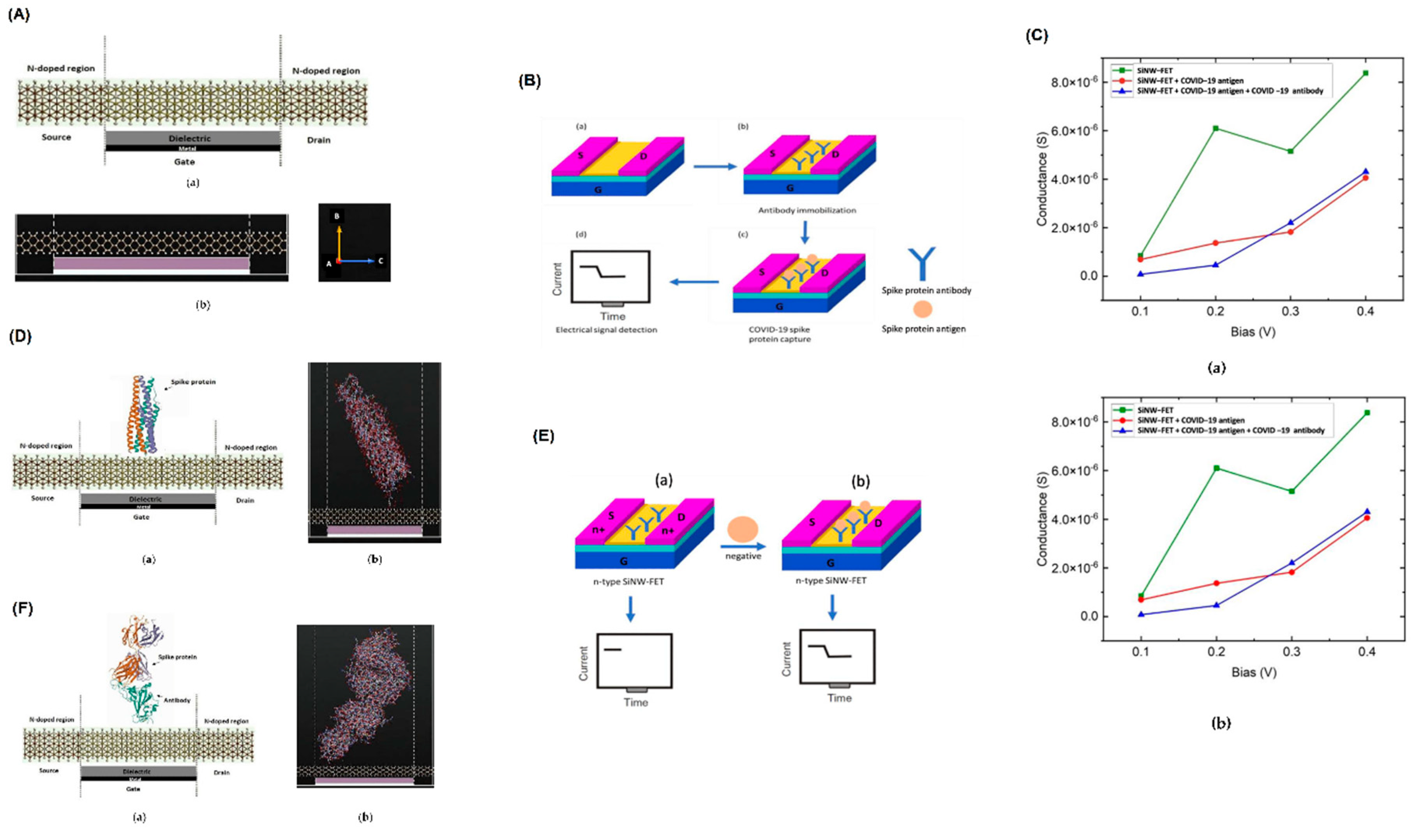
Figure 2.
Integrated biosensing platform for salivary P90 protein detection.(A) Paper-based strip with printed electrodes for biosensing.(B) Antibody functionalization on Au surface using NHS ester chemistry.(C) Portable readout unit with labeled components (a) and circuit block diagram (b).(D) Output voltage response (V_out) for varying concentrations of P90 protein in saliva.(E) Capacitance versus digital reading under different conditions: (a) varying resistance, (b) varying gate voltage, and (c) varying reference capacitance.(F) Calibration curve showing the logarithmic correlation between P90 protein concentration and digital reading, with comparison to ELISA detection range.
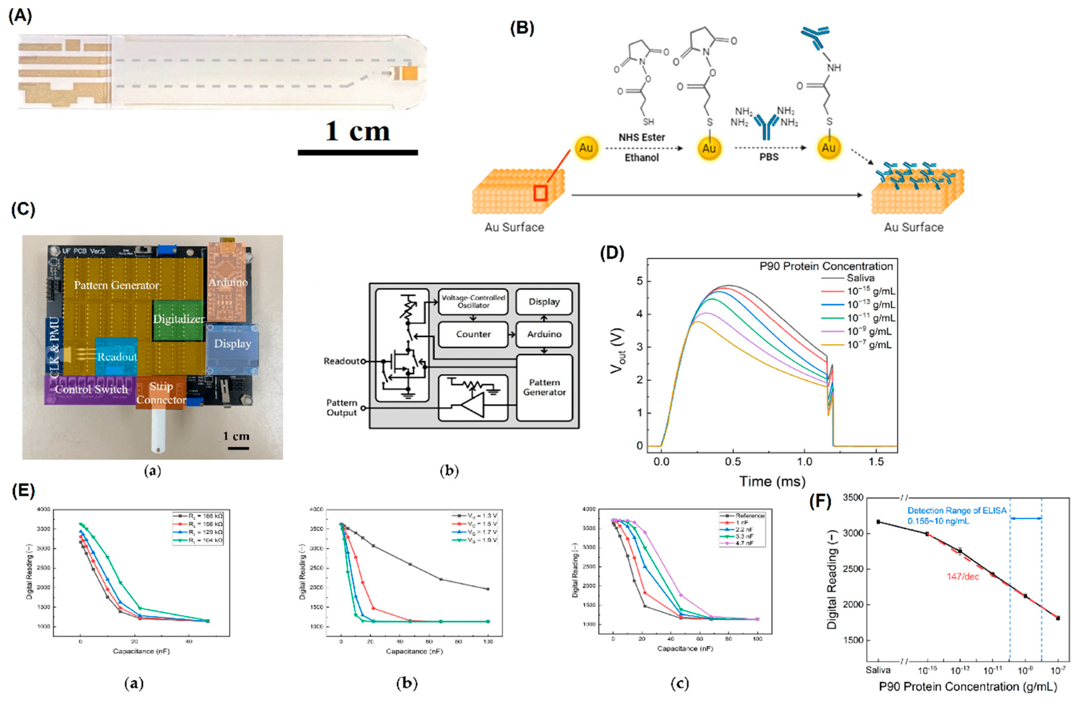
Figure 3.
Performance analysis of JL DM DG and JL DM GSDG MOSFET biosensors under dielectric and biomolecular variations.(A) Structural cross-sections of JL DM DG and JL DM GSDG MOSFETs with biomolecule placement.(B) Gate architecture comparisons across multiple MOSFET configurations.(C) Threshold voltage variation with dielectric constant (a) and biomolecule charge density (b).(D) Electric field distribution along the normalized channel length under varying dielectric constants and biomolecular densities.(E) Surface potential profiles along the channel for different dielectric constants and biomolecule densities.(F) Transfer characteristics showing drain current versus gate voltage for both MOSFET types under different sensing conditions.
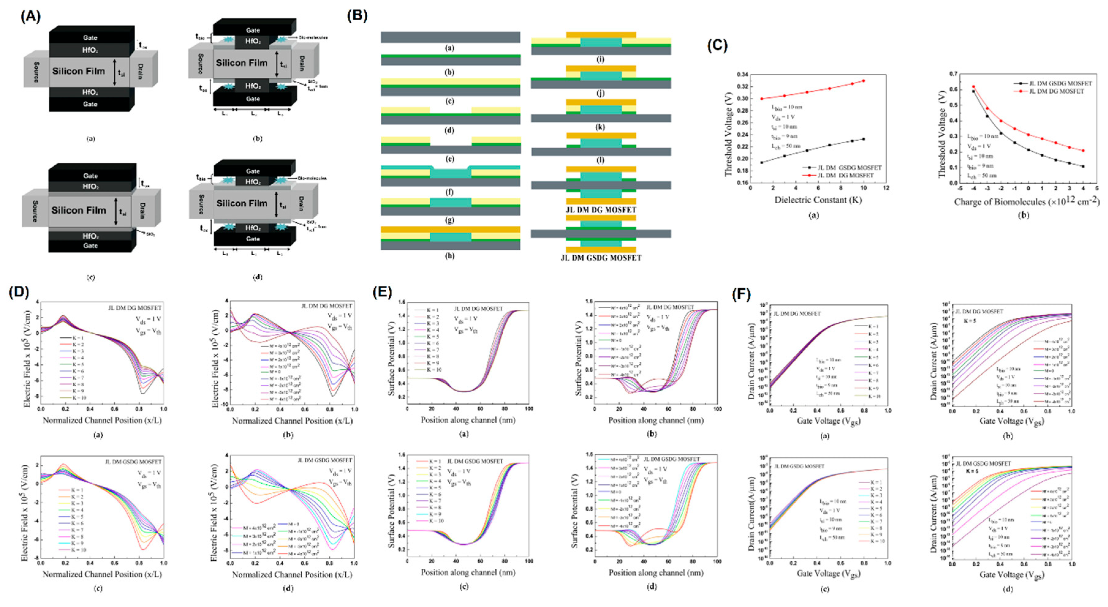
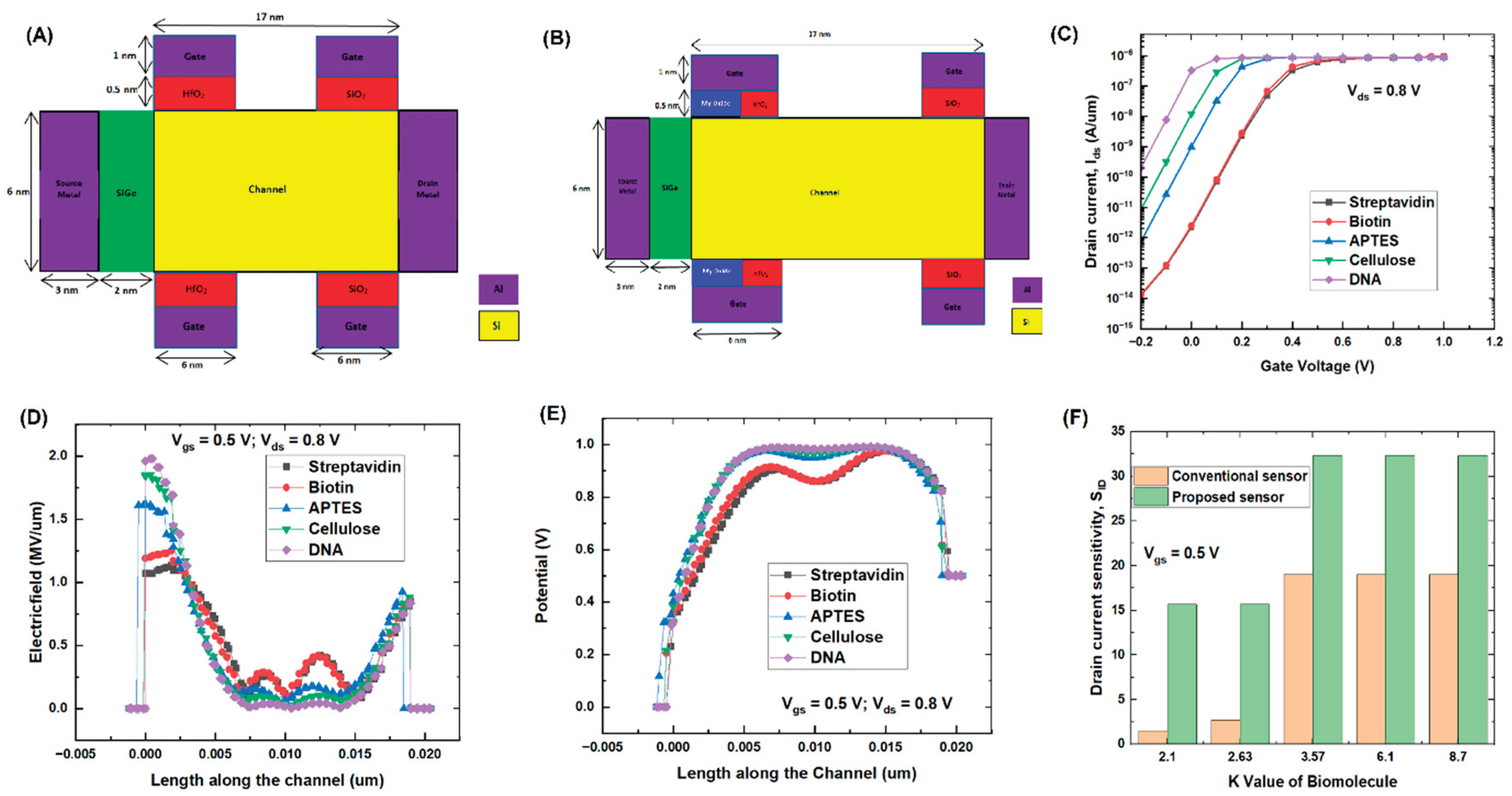
Figure 4.
Performance comparison of conventional and proposed biosensor structures using various biomolecules.(A) Cross-sectional view of the conventional biosensor structure.(B) Cross-sectional view of the proposed biosensor structure with additional oxide modification.(C) Transfer characteristics showing drain current response for different biomolecules.(D) Electric field distribution along the channel for various biomolecules.(E) Channel potential variation in presence of different biomolecules.(F) Sensitivity comparison of conventional and proposed sensors as a function of biomolecule dielectric constant.
Disclaimer/Publisher’s Note: The statements, opinions and data contained in all publications are solely those of the individual author(s) and contributor(s) and not of MDPI and/or the editor(s). MDPI and/or the editor(s) disclaim responsibility for any injury to people or property resulting from any ideas, methods, instructions or products referred to in the content. |
© 2025 by the authors. Licensee MDPI, Basel, Switzerland. This article is an open access article distributed under the terms and conditions of the Creative Commons Attribution (CC BY) license (http://creativecommons.org/licenses/by/4.0/).
Copyright: This open access article is published under a Creative Commons CC BY 4.0 license, which permit the free download, distribution, and reuse, provided that the author and preprint are cited in any reuse.


