
Submitted:
15 May 2025
Posted:
16 May 2025
You are already at the latest version
Abstract
The skin's main function is to be a physical barrier, which prevents transepidermal water loss (TEWL) and protects the body from the external environment. Changes in the structure of the stratum corneum impair the barrier function and favor the appearance of several skin diseases, including xerosis, atopic dermatitis and psoriasis. Skin health is preserved by the presence of structurally organized intercellular lipids in the stratum corneum, which are responsible for cell adhesion, preserving hydration and reducing TEWL. Changes in the lipid-protein structure or changes in the composition of the Natural Moisturizing Factor (NMF) can cause reduced hydration of the stratum corneum, resulting in dry skin with sensory symptoms such as opacity, roughness, scaling, stiffness, fissures, itching and even bleeding. Skin care cosmetics having the presence of liquid crystals resembling structurally the intercellular lipids in the stratum corneum and can beneficially affect the skin when applied topically by stimulating the skin’s natural regenerative functions of stratum corneum. We have developed an O/W emulsion for skin care whose structure observed under polarization microscopy is similar to that of the lipid distribution of the stratum corneum: the emulsion presents a lamellar crystalline phase. The formulation was composed by a blend of surfactant [lanolin derivatives plus 2EO fatty alcohol (50:50)] + liquid paraffin+ purified water. Urea was employed as hydration agent. The occlusive power of O/W was determined in vitro onto gelatin support cells (noninvasive method) and in vivo method- skin hydration, skin oiliness and skin pH evaluation. The developed formulation significantly increased the occlusive power of emulsion. Skin hydration was studied by using an in vitro non-invasive methodology and it was found that the presence of liquid crystal in the emulsion increases the skin hydration level. This was attributed to the fact that it formed the same self-organizing structure as natural stratum corneum lipids.
Keywords:
O/W emulsion
; liquid crystal
; lamellar liquid phase
; urea
; akin properties (hydration
; oiliness and pH values)
1. Introduction
The skin's function is to act as a physical barrier, preventing moisture loss and protecting the body from the external environment while maintaining elasticity and firmness. Changes in the barrier function make the skin dry and rough, sometimes associated with diseases such as xerosis, atopic dermatitis and psoriasis.
The skin is composed of three main layers: epidermis, dermis and hypodermis, with the horny layer or stratum corneum being the outermost layer of the epidermis. The stratum corneum is formed by corneocytes, flattened, densely packed and metabolically inactive cells that form an impermeable barrier. The horny layer is formed by the process of epidermal differentiation and the lipid layer has a neutral character. This continuous lipid layer formed by lipids occupies about 20% of the total volume of the horny layer and is organized into multiple lamellar structures. The skin's protective barrier is composed of corneocytes intertwined in a complex lipid-protein mixture called lamellar lipids [1,2].
The intercellular lamellar lipids in the stratum corneum form a single, coherent membrane stabilized by lipid components and the length distribution of these components in the intercellular spaces of the stratum corneum.
The healthy appearance of the skin can be attributed to the intercellular lipids of the stratum corneum that contribute to hydration, cell adhesion and reduction of TEWL. This occurs due to the structural organization of the amphiphilic molecules of the lipids of the stratum corneum [3,4,5].
A properly hydrated stratum corneum is capable of (a) maintaining the plasticity of the skin, protecting it from damage; (b) allowing hydrolytic enzymes to act adequately in the desquamation process and (c) contributing to the ideal functioning of the stratum corneum barrier.
The discovery of the NMF [5] in the stratum corneum reveals the presence of water-soluble compounds involved in the water retention process, the removal of which reduces epidermal hydration. The function of NMF is to maintain adequate skin hydration, and its components are highly efficient humectants that attract and retain water from the atmosphere, promoting corneocyte cohesion [6].
Components of NMF such as lactic acid, sugars, amino acids and urea are essential for the skin’s ability to retain water. Reduced hydration of the stratum corneum results in dry, rough-feeling skin that is prone to flaking, tightness, cracking, and itching that results in bleeding [7]. This may be caused by changes in the composition of the natural moisturizing factor.
Among the components of the skin's natural moisturizing factor, urea plays an important role in the integrity and consequent maintenance of skin hydration [5,6,7].
Due to its moisturizing capacity, urea is used to treat dry skin. It can be easily incorporated into skin care products such as creams, lotions, emulsions, ointments, gels and even shampoos and nail polishes [8,9,10]. Used in low concentrations (≤ 5%), it is very safe and effective and recommended for body products applied once or twice a day for a long period of time. A decrease in the content of NMF and especially urea can cause a reduction in the hygroscopic capacity of the skin and increase TEWL, in addition to unbalancing the epidermal reproduction process, inhibiting skin desquamation and causing hyperkeratosis and pruritus [10,11].
Skin care emulsions showing the presence of lamellar liquid crystals are structurally like the intercellular lipids of the epidermis, act as smart structures and interact positively with the stratum corneum, stimulating the skin's natural regenerative functions and accelerating epidermal renewal [12].
We therefore developed a skin care cream whose structure is similar to that found in the lipids of the stratum corneum, that is, with a lamellar crystalline phase.
2. Materials & Methods
2.1. Materials (INCI Nomenclature)
Oil phase- Paraffinum liquidum
Aqueous phase- Purified water
Surfactants:
Hydrophilic- PEG- 75 lanolin (Croda);
Lipophilic- PEG lauryl alcohol (Cognis. HLB value= 6.2 ± 0.2)
Actif- moisturizing agent – urea (Nuclear)
2.2. Methods
Development of the Emulsion
1. Ternary Diagram
To obtain the formulations it was employed the ternary diagram method. The diagram was obtained using initial concentrations that increased in 10.0% w/w in order to obtain O/W emulsions [13].
2. O/W emulsions preparation
The emulsions were prepared by the Emulsion Phase Inversion (EPI) method. The aqueous and oil phases were heated separately at 75± 2°C. Then, the aqueous phase was slowly poured over to the oily phases containing surfactants and keeping under stirring (600 rpm) (Ika RW 20 digital) at room temperature (25± 2°C) until complete cooling (28± 3 minutes). Urea aqueous solution was added at (40± 2°C) [14].
3. Macroscopic Analysis
The intrinsic stability was performed twenty-four hours after preparation and organoleptic properties and homogeneity of the formulations were observed, identifying possible signs of instability such as coalescence and creaming [15].
4. Microscopic Analysis
The microscopy analysis samples were performed using the microscope with normal and polarized light (BX 50 microscope, Olympus, Melville, NY, USA). Only emulsions with anisotropy were selected for further study [16].
5. Preliminary Stability
Macroscopically stable emulsions after twenty-four hours of preparation were subjected to the following preliminary stability tests: centrifugation, thermal stress and pH analysis [17]. All tests were performed in triplicate.
5. 1. Centrifugation Test
It was performed under the following conditions: room temperature (24± 2°C). The samples were submitted to 3000rpm in the centrifuge (Centrilab, model CE80) [18,19].
5. 2. Thermal Stress
The samples were subjected to thermal stress in a thermostated bath in the temperature range of 40± 2 to 80± 2°C, increasing the temperature by 5°C at each 30-minute interval and subsequently evaluated macroscopically. (19). For this test, the nomenclature proposed by Ribeiro et al. (20) was used to classify the samples: N = Normal; without change; LM = Slightly Modified; M = Modified; IM = Heavily Modified.
5. 3. pH Evaluation
The pH value was measured by inserting the electrode (Analion-Mod. PM608 pH meter, Analion, Ribeirão Preto, SP, Brazil) in the sample solution [18,19].
6. Accelerated Stability Test
The samples considered stable were placed in clear plastic vials and kept under different storage conditions: (25± 2°C); (4± 2°C); (40± 2°C) [20,22]. The samples were analyzed on the 1st (24 h), 7th, 15th, 30th, 60th and 90th days. The tests used to evaluate the prolonged stability was: macroscopic observation, polarized light microscopy and determination of pH values.
7. In vitro and in vivo evaluation
7.1 In vitro hydration emulsion evaluation on gelatin support cells.
Water loss was determined gravimetrically and occlusivity values were determined by the same method employed by Rocha-Filho, P.A. [23].
7.2 In vivo assessment of skin hydration, oiliness and pH (Corneometer CM 820, a Sebumeter SM 810 and a Skin-pH-meter PH 900 instruments (Courage & Khazaka, Köln, Germany)]
The biophysical properties of skin (the skin hydration evaluation, the skin oiliness evaluation and the skin pH evaluation) were evaluated by the method employed by Rocha-Filho, P.A. [24]
Environmental conditions were standardized by maintaining the temperature at 21± 1oC and relative humidity at 60± 3% (ARSEC dehumidifier). Ten healthy volunteers of both sexes aged between 20 and 40 years, without medication, free of any dermal changes in the forearm region, were part of the studies. All readings were taken in triplicate. The tests were performed on the right forearm, divided into four regions. The region identified as number 1 is kept as control (Figure 1). The products were applied (10± 1g/cm2) and measures (triplicate). were performed at the standardized times at 0, 30, 90 and 150 minutes after application
7. 2. 2. Statistical analysis
The skin hydration, oiliness and pH values were submitted to statistical treatment at the times of zero and 150 minutes after application of the product through The Wilcoxon Signed Ranks Test (two-tailed) at a significance level of 5%.
3. Results
3.1. Development of the Emulsion
1. Ternary Diagram
Table 1 shows the emulsions chosen from the ternary diagram. The emulsions had a white or slightly yellowish appearance due to the absence or presence of the lanolin derivative.
The formulations showed lamellar crystalline phase (Figures 3 and 4) and had pH values between 5.91 -sample 82 without urea and 7.63-sample 14 with urea (Table 2) and were considered suitable for skin topical application.
The centrifugation test was performed as an additional test to predict any possible instability of the emulsions [17]. After centrifugation, all formulations in Table 1 were stable and considered for studies. After 3 months of evaluation according to the accelerated stability test, no macroscopic changes and appearance were observed in the emulsions. It was found that the storage temperature can alter or eliminate the microscopic appearance and formation of liquid crystals as well as alter the pH values of all formulations.
3.1.1. In Vitro Emulsion Evaluation on Gelatin Support Cells [23]
The O/W emulsions spread easily when applied to the cells on the gelatin surface and it was observed that the water content in the emulsion progressively evaporates. This can cause the emulsion to break and therefore increase the proportion of oils and emulsifiers on the gelatin surface. According with Teeranachaideekul, V. et al. [25] this evaporation of a certain volume of water in the formulation can cause occlusion by the oil and emulsifier on the gelatin surface (Figure 2.1). For O/W emulsion with lamellar phase with a structure similar to the lamellar phase, imitating the natural lipids of the stratum corneum, occlusion can reduce the evaporation of water from the skin surface due to entrapped water onto lamellar liquid crystal structures (Figure 2.2) [12,26,27,28,29].
Figure 2.
Water content evaporation scheme: 1. from gelatin support cell; 2. from dry skin.
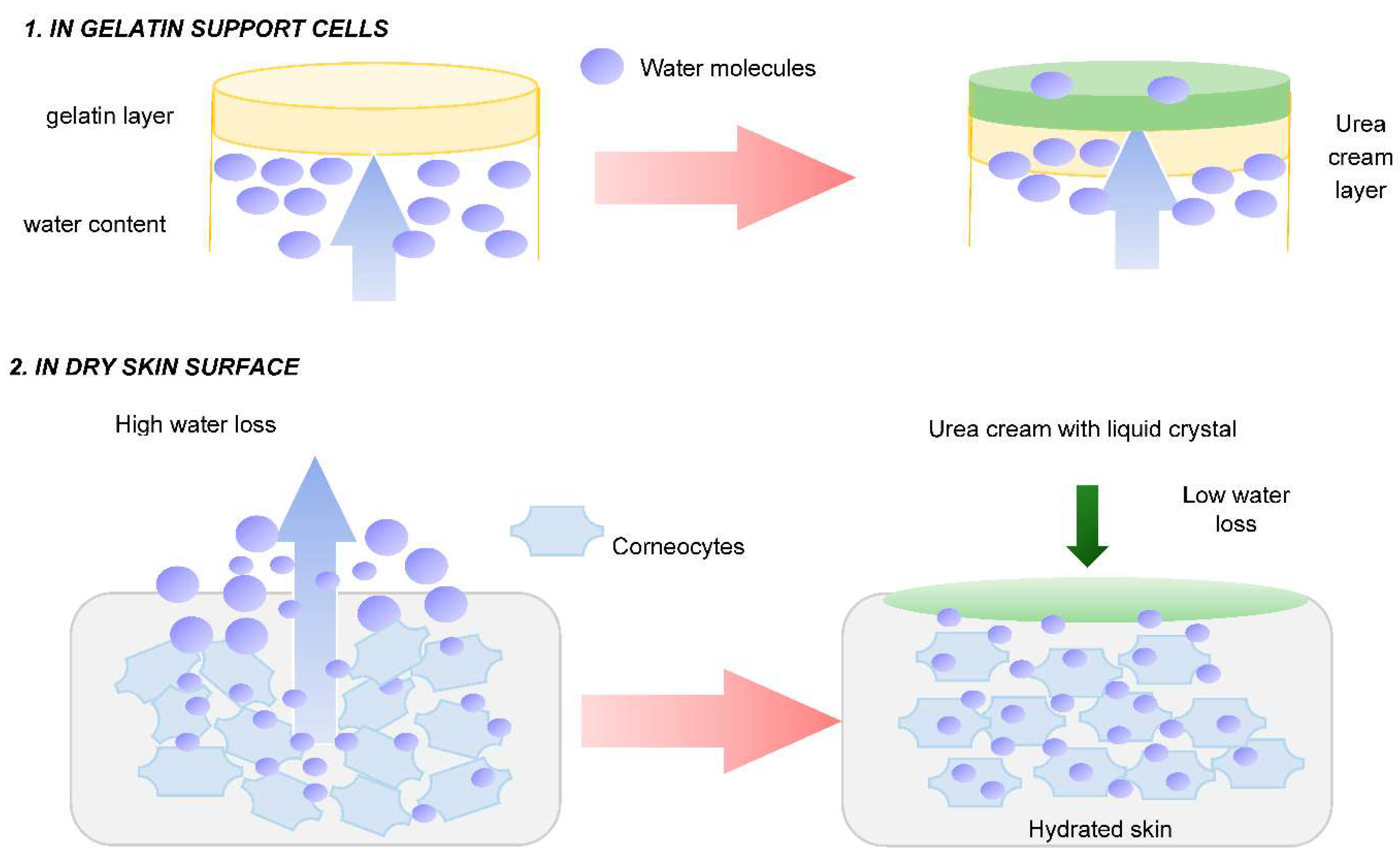
Figure 3.
Microscopic analysis (200X) of O/W emulsions samples.
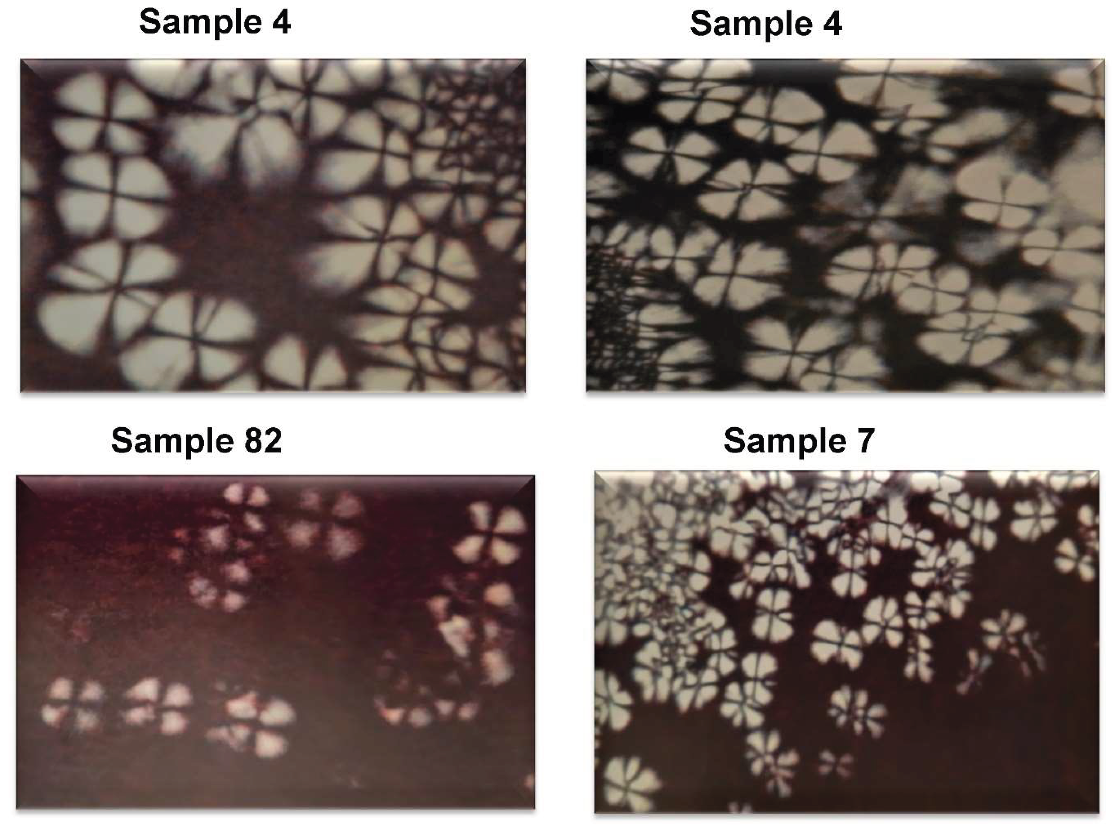
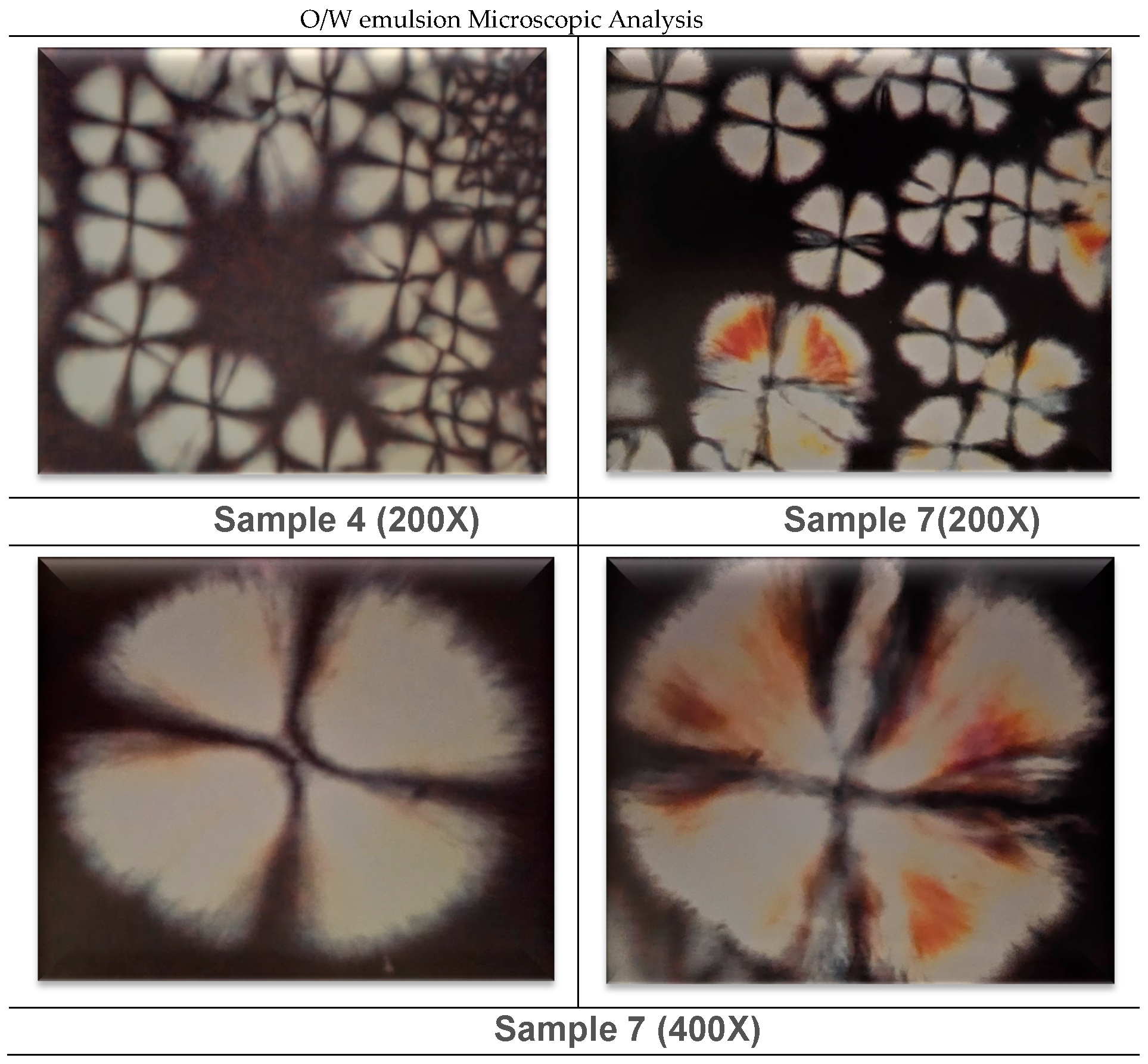
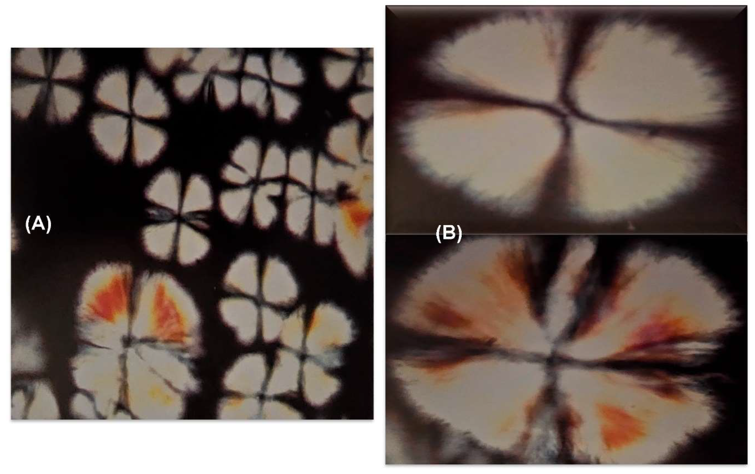
Figure 4.
Microscopic analysis of O/W emulsions sample 7 (A) 200X and (B) 400X.
The Table 3 presents the values of the angular coefficients of the lines obtained before application (25 and 50 minutes) and after application (25, 50, 100 and 150 minutes) of O/W emulsions. Comparing for example for sample 4 without urea and with urea, values of angular coefficients were minor after product application. So, the lines had minor inclinations in relation to X axis, and this show that the loss of water in gelatin support cells, was minor and the applied product avoid the water loss. In Table 4 are showed the great values of occlusive power of O/W emulsions, mainly for the presence of lamellar crystalline phase.
3.1.2. In Vivo Emulsion Evaluation- Skin Application
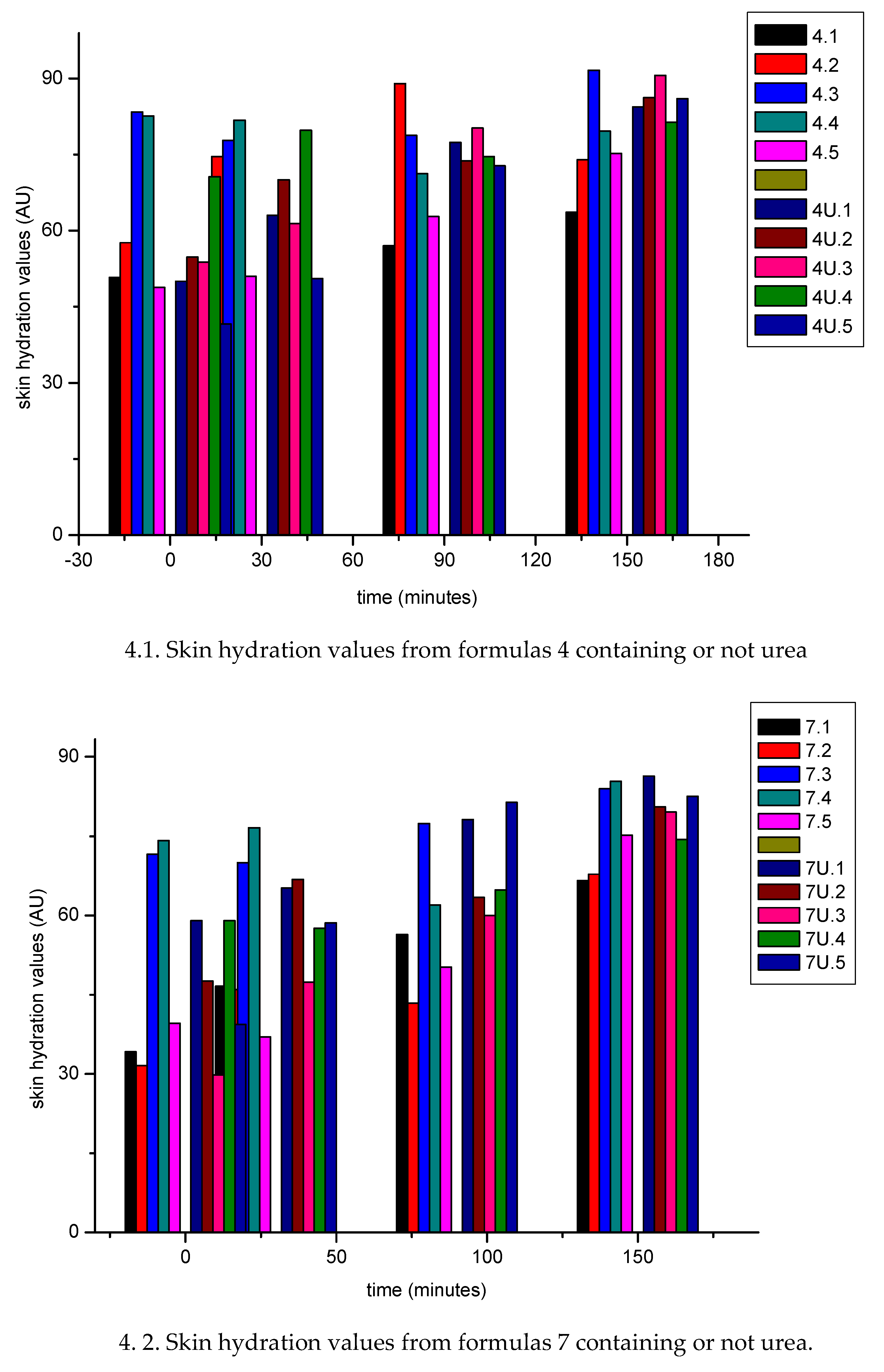
From Figure 4 (4.1;4.2; 4.3; 4.4), the results obtained for the hydration values it was observed an increase in hydration values as a function of time. In formulations containing urea, the hydration values at the final time of 150 minutes were higher than the control. These values can be explained not only by the formation of the crystalline lamellar phase, but by the presence of urea, the water retention capacity of urea and also by the formation of the lamellar crystalline phase. For formulation 37 where there was no formation of the lamellar phase, the hydration values are attributed to the presence of urea. By applying skin care products containing lamellar liquid crystal, which in itself has a water reserve, it is possible to change the resistance to transepidermal water loss and maintain and/or increase skin hydration [27].
Figure 4.
Skin hydration values (U= urea).
Regarding the oiliness of the formulations Figure 5 (5.1;5.2;5.3; 5.4) the sensorial of sample was like the application of oily formulations to the skin: the application of an O/W emulsion showed initially an increase in oiliness values. There was a gradual increase in oiliness value until the time of 90 minutes, with a decrease occurring until the final time of 150 minutes. This fact was observed for emulsions containing or not containing urea, however, the decrease in these values was smaller in relation to the values control.
The application of simple emulsion O/W showed an increase in oiliness, mainly in the initial time, despite the emulsion having low oil content. It can be observed that the presence of the urea does not present alterations to oiliness and at the end of the test, the values correspond to those of the simple O/W emulsion.
From Figure 6 (6.1; 6.2; 6.3; 6.4) it was observed that pH values measurements were between 4.5. and 7.0. With the application of O/W without urea, a slight decrease in these values was observed. When applying O/W containing urea, it was observed that the pH values were in the acidic range, which shows that there was no change in the skin's pH values.
The statistical analysis carried out at a significant level of 5% allow to conclude that the O/W showing lamellar crystalline liquid phase and urea markedly improve the hydration, maintain the oiliness the skin and do not alter skin pH values and must be indicated to individuals with dehydrated skin.
4. Conclusion
Microscopic analyses had identified lamellar crystalline phase in some formulations obtained from ternary diagram method. The hydration formulation power was studied by using an in vitro non-invasive methodology and it was observed that hydration power was increased by the presence of liquid crystal.
A high moisturizing effect was observed due to the presence of lamellar crystalline phase.
Urea.is a fundamental ingredient of cosmetic and dermatological formulations and can have several positive effects on skin function easily investigated and monitored using in vitro techniques for measuring the biophysical properties of skin.
Ethical Aspects
This study was designed in accordance with good clinical practice. All patients kindly agreed to participate in the assessments.
Author Contributions
Conceptualization, methodology, writing - review and editing P.A.R.F.
Funding
This research received no external funding.
Institutional Review Board Statement
Not applicable.
Informed Consent Statement
Not applicable.
Data Availability Statement
Not applicable.
Conflicts of Interest
The author declare no conflict of interest.
References
- Lim, K.-M. (2021). Skin Epidermis and Barrier Function. International Journal of Molecular Sciences, 22(6), 3035. [CrossRef]
- Rajkumar, J. R., Chandan, N., Lio, P., & Shi, V. Y. (2023). The skin barrier and moisturization: function, disruption, and mechanisms of repair. Skin Pharmacology and Physiology, 36(4), 174-185. [CrossRef]
- Sparr, E, Björklund, S, DatPham, Q, Mojumdar, EH, Stenqvist, Gunnarsson, BM, Topgaard, D. (2023). The stratum corneum barrier - From molecular scale to macroscopic Properties. Current Opinion in Colloid & Interface Science. 67(10) 101725. [CrossRef]
- Asada, N., Morita, R., Kamiji, R., Kuwajima, M., Komorisono, M., Yamamura, T. & Yoshikawa, S. (2022). Evaluation of intercellular lipid lamellae in the stratum corneum by polarized microscopy. Skin Research and Technology, 28(3), 391-401. [CrossRef]
- Jacobi, O. K. (1959). Moisture regulation in the skin. Drug Cosmet Ind, 84(6), 732-3.
- Fowler, J. (2012). Understanding the role of natural moisturizing factor in skin hydration. Pract. Dermatol, 9, 36-40.
- Norlén L. Skin barrier structure and function: the single gel phase model. J Invest Dermatol. 2001 Oct;117(4):830-6. [CrossRef]
- Lacarrubba, F., Nasca, M. R., Puglisi, D. F., & Micali, G. (2020). Clinical evidences of urea at low concentration. International journal of clinical practice, 74, e13626. [CrossRef]
- Celleno L. Topical urea in skincare: A review. Dermatol Ther. 2018 Nov;31(6). [CrossRef]
- Piquero-Casals, J., Morgado-Carrasco, D., Granger, C. et al. Urea in Dermatology: A Review of its Emollient, Moisturizing, Keratolytic, Skin Barrier Enhancing and Antimicrobial Properties. Dermatol Ther (Heidelb) 11, 1905–1915 (2021). [CrossRef]
- Berardesca, E., & Cameli, N. (2020). Non-invasive assessment of urea efficacy: A review. International Journal of Clinical Practice,74. [CrossRef]
- 12. Iwai H, Fukasawa J, Suzuki T. (1998). A liquid crystal application in skin care cosmetics. Int J Cosmet Sci. Apr;20(2):87-102. [CrossRef]
- Morais J.M., Rocha-Filho P.A., Burguess D.J. (2009) Influence of phase inversion on the formation and stability of one-step multiple emulsions. Langmuir. 25:7954–7961. [CrossRef]
- Kumar, M., Bishsoi, R.S., Shukla, A.K., Jain, C.P. (2019) Techniques for Formulation of Nanoemulsion Drug Delivery System: A Review. Prev. Nutr. Food Sci., 24 (3) 225- 234. Published online 2019 Sep 30. [CrossRef]
- Ferrari M., Monteiro L.C.L., Netz D.J.A., Rocha-Filho P.A. (2003) Identifying cosmetic forms and crystalline phases from ternary systems. Cosmet. Toilet. ;118: 61–70.
- Tyle P. Liquid crystals and their applications in drug delivery. In: Rosoff M., editor. Controlled Release of Drugs: Polymers and Aggregate Systems. Volume 4. VCH; New York, NY, USA: 1989. pp. 125–162.
- Andrade F.F., Santos O.D.H., Oliveira W.P., Rocha-Filho P.A. (2008); Influence of PEG-12 Dimethicone addition on stability and formation of emulsion containing liquid crystal. Int. J. Cosmet. Sci. 29:211218. https://. [CrossRef]
- BRASIL. Agência Nacional de Vigilância Sanitária (2004). Guia de estabilidade de produtos cosméticos. Séries Temáticas. Série Qualidade 1., 1, Brasília, DF.
- Davis, HM. Analysis of creams and lotions. In: Senzel, AJ. Newburger’s manual of cosmetic analysis. Washington: Association of Official Analytical Chemists. 1997, cap.4, p. 32.
- Braconi F.L., Oliveira I.S., Baroni M.N.F., Rocha-Filho P.A. Aplicação cosmética do óleo de canola; Proceedings of XII Congresso Latino Americano e Ibérico de Químicos Cosméticos; São Paulo, Brazil. 27–31 August 1995; São Paulo, Brazil: Associação Brasileira de Cosmetologia, Tecnopress; 1995. pp. 6–19. ANAIS.
- Ribeiro A.M., Khury E., Gottardi D. Validação de testes de estabilidade para produtos cosméticos; Proceedings of the 12th Congresso Nacional de Cosmetologia; São Paulo, Brazil. 30 June–2 July 1998; São Paulo, Brazil: Associação Brasileira de Cosmetologia, ANAIS Tecnopress; 1998. pp. 349–375.
- Ferrari M. Obtenção e Aplicação de Emulsões Múltiplas Contendo óleos de Andiroba e Copaíba. (Mestrado em Ciências Farmacêuticas) Dissertação, Faculdade de Ciências Farmacêuticas de Ribeirão Preto, Universidade de São Paulo; Ribeirão Preto, SP, Brazil: 1998. p. 147.
- Rocha-Filho P.A., (1997). Occlusive power evaluation of O/W/O multiples emulsions on gelatin support cells. Int. J. Cosm. Science., 19, 65-73. [CrossRef]
- P. A. Rocha-Filho & M. Maruno (2023) O/W/O multiple emulsions containing soluble collagen: in vitro and in vivo skin biophysical properties evaluation, J Disp Sci Technology. [CrossRef]
- Teeranachaideekul, V., Soontaranon, S., Sukhasem, S. et al. (2023). Influence of the emulsifier on nanostructure and clinical application of liquid crystalline emulsions. Sci Rep. 13, 4185. [CrossRef]
- Santos, ODH; Rocha- Filho, PA. (2008). Influence of Surfactant on the Thermal Behavior of Marigold Oil Emulsions with Liquid Crystal Phases. J Drug Dev Pharmacy, 33 (5) 543- 549. [CrossRef]
- Wang Y, Li J, Shang Y, Zeng X. (2018). Study on the development of wax emulsion with liquid crystal structure and its moisturizing and frictional interactions with skin. Colloids Surf B Biointerfaces. Nov 1;171: 335-342. [CrossRef]
- Tsutsumi, H., Usugi, T., Kawano, J., Ishida, A., Hayashi, S. Effect of Occlusivity of oil Film by the States of oil Film on the Skin Surface. J. Soc. Cosm. Chem., 1979,13 (2) 37-43. [CrossRef]
- Imokawa, G., Kuno, H., Kawai, M. (1991). Stratum Corneum Lipids Serve as a Bound-Water Modulator. J. Invest. Dermatology. 96 (6) 845- 851. [CrossRef]
Figure 1.
Sites for assessment of skin hydration, oiliness and pH values.
Figure 5.
Skin oiliness values (U= urea).
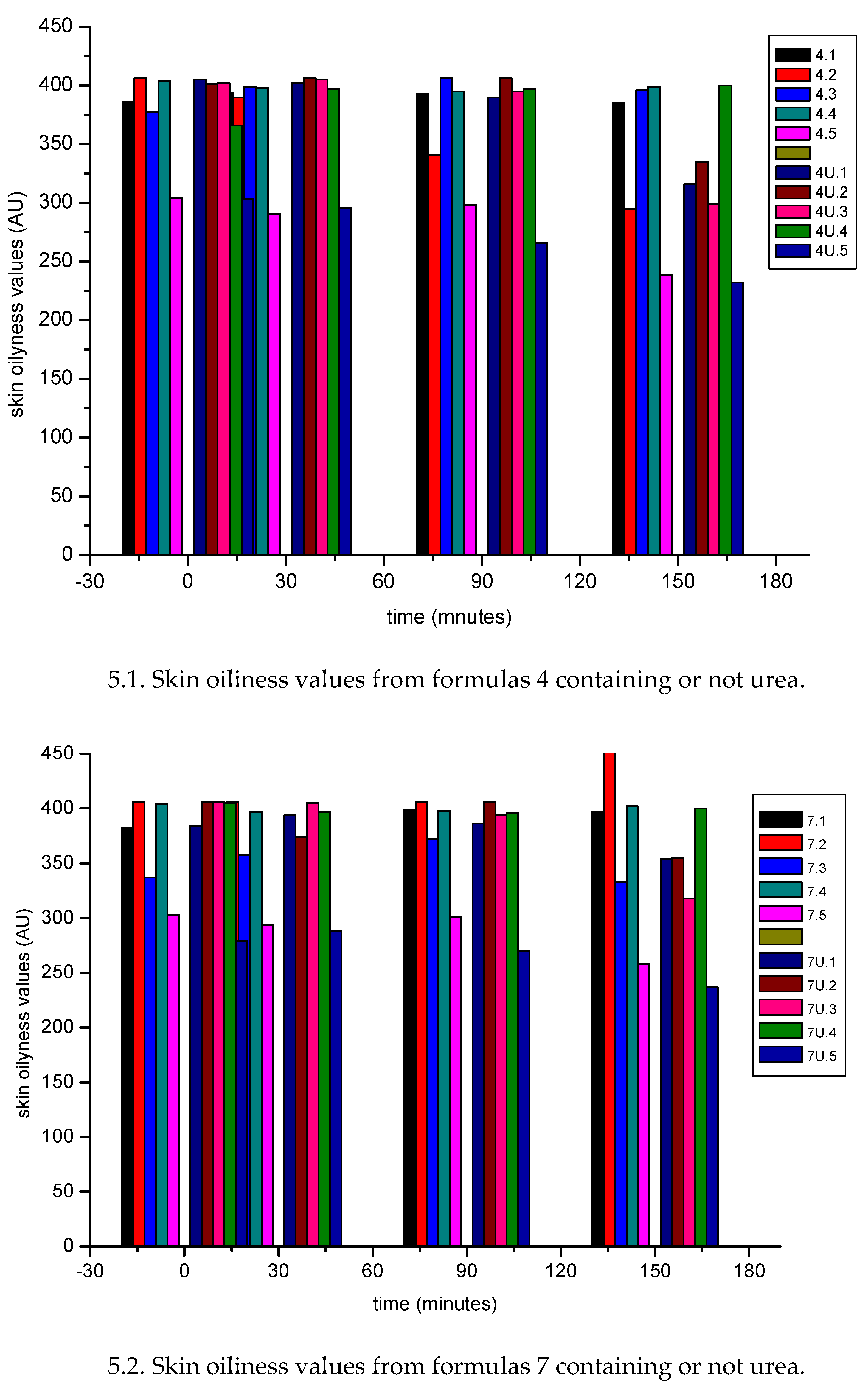
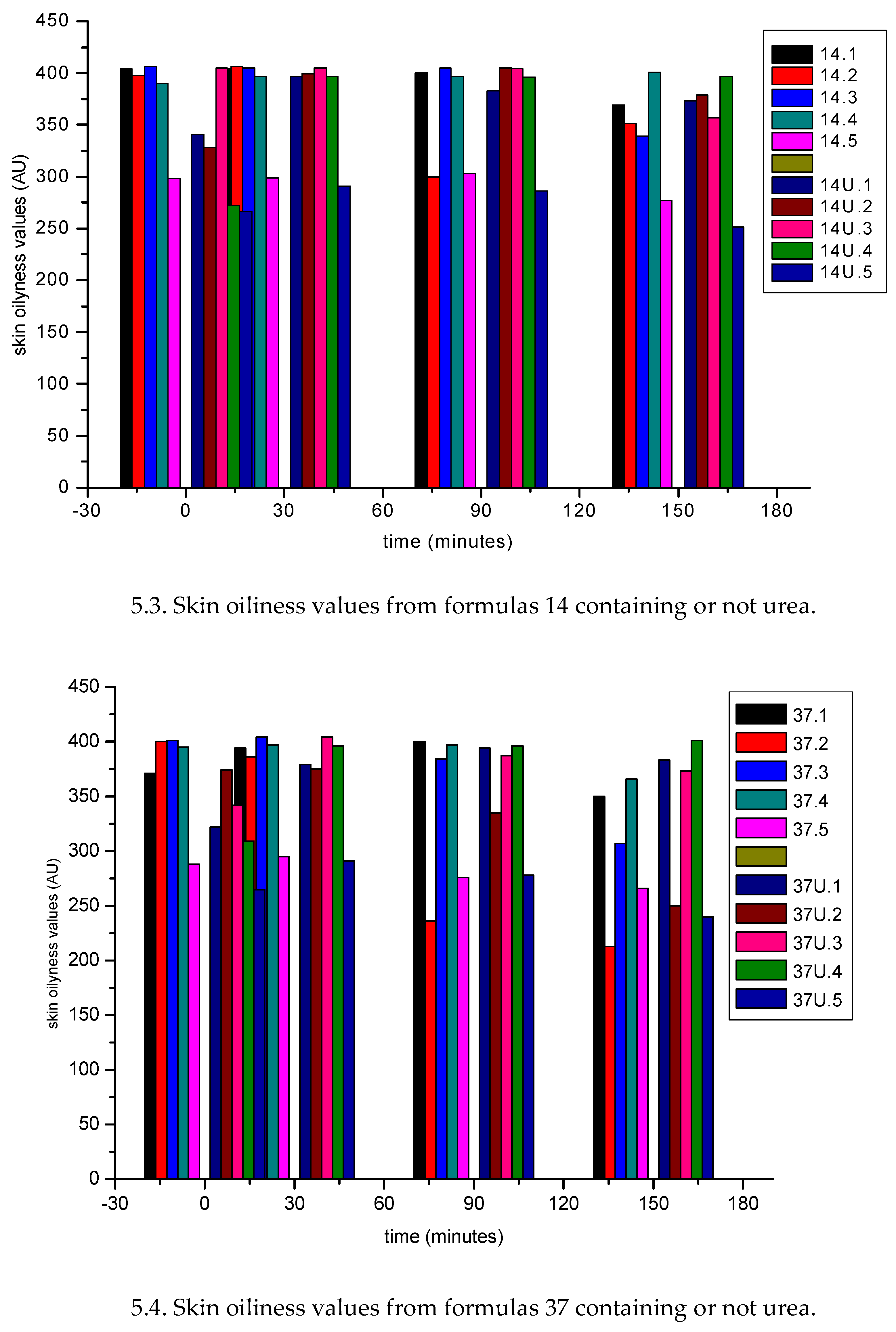
Figure 6.
Skin pH values (U= urea).
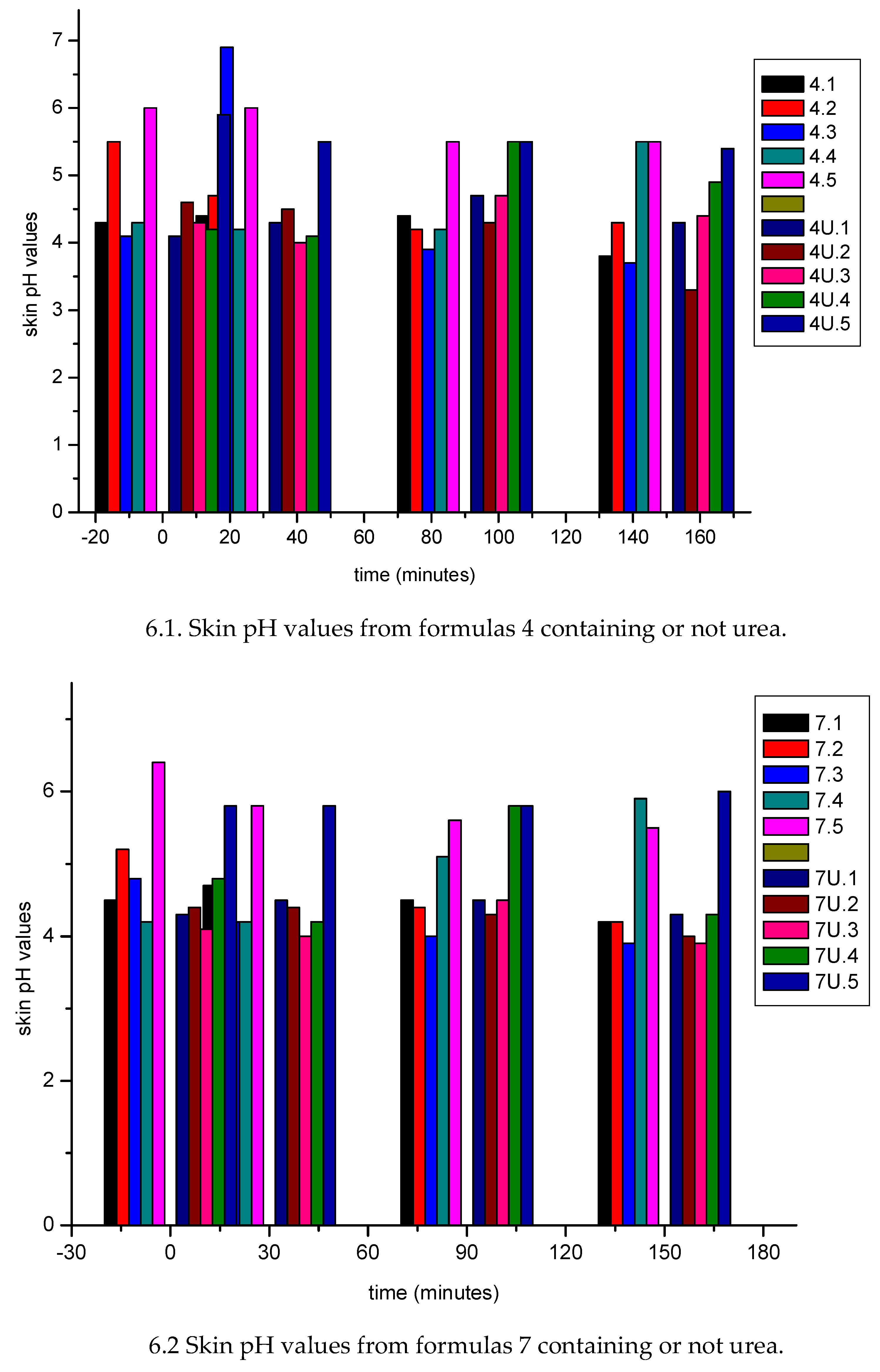
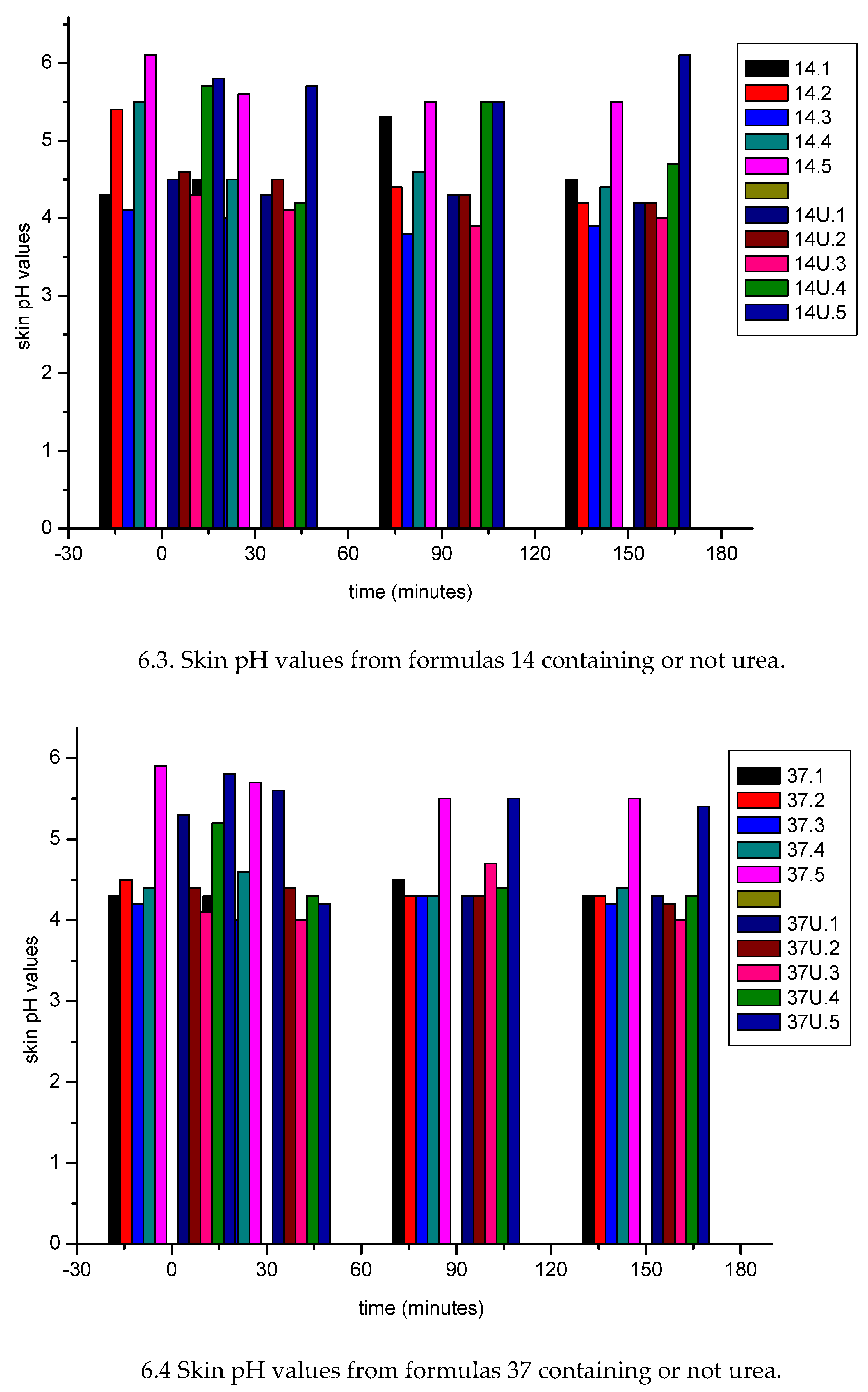

Table 1.
O/W emulsions compositions.
| Sample nr | water (% w/w) |
oil (% w/w) |
surfactant (% w/w) |
|---|---|---|---|
| 4 | 60.0 | 30.0 | 10.0 |
| 7 | 50.0 | 40.0 | 10.0 |
| 14 | 40.0 | 20.0 | 40.0 |
| 37 | 45.0 | 20.0 | 35.0 |
| 58 | 25.0 | 20.0 | 55.0 |
| 82 | 60.0 | 20.0 | 20.0 |
Table 2.
pH values of O/W emulsions.
| Sample nr↓ |
pH value | ||
|---|---|---|---|
| Without urea | With urea | difference | |
| 4 | 6.64 | 7.27 | 0.63 |
| 7 | 6.36 | 6.70 | 0.34 |
| 14 | 6.79 | 7.63 | 0.84 |
| 37 | 6.33 | 6.57 | 0.24 |
| 58 | 6.37 | - | - |
| 82 | 5.91 | 7.37 | 1.46 |
Table 3.
Angular coefficients of the lines before and after application of O/W emulsions.
|
Formula nr ↓ |
Before (B) |
After (A) application 300± 4mg |
angular coefficient difference (B- A) |
|---|---|---|---|
| 4 | 0.99774 | 0.99677 | 0.0009 |
| 4U | 0.99892 | 0.97555 | 0.0233 |
| 7 | 0.99990 | 0.99981 | 0.0009 |
| 7U | 0.99866 | 0.99327 | 0.0053 |
| 14 | 0.99753 | 0.99981 | -0.0022 |
| 14U | 0.99158 | 0.99778 | -0.0062 |
| 37 | 0.99999 | 0.99961 | 0.0004 |
| 37U | 0.99792 | 0.99950 | 0.0015 |
| 82 | 0.99958 | 0.99986 | -0.0002 |
| 82U | 0.99585 | 0.99924 | 0.0033 |
Table 4.
Occlusive power of O/W emulsions.
| Sample nr | Occlusive power (%) | ||
|---|---|---|---|
| ↓ | Without urea (C) | With urea (D) | Difference (D-C) |
| 4 | 26.85 | 77.57 | 50.72 |
| 7 | 31.49 | 57.77 | 26.28 |
| 14 | 3.81 | 23.85 | 20.04 |
| 37 | 14.94 | 18.38 | 3.44 |
| 82 | 25.11 | 43.40 | 18.29 |
Disclaimer/Publisher’s Note: The statements, opinions and data contained in all publications are solely those of the individual author(s) and contributor(s) and not of MDPI and/or the editor(s). MDPI and/or the editor(s) disclaim responsibility for any injury to people or property resulting from any ideas, methods, instructions or products referred to in the content. |
© 2025 by the authors. Licensee MDPI, Basel, Switzerland. This article is an open access article distributed under the terms and conditions of the Creative Commons Attribution (CC BY) license (http://creativecommons.org/licenses/by/4.0/).
Copyright: This open access article is published under a Creative Commons CC BY 4.0 license, which permit the free download, distribution, and reuse, provided that the author and preprint are cited in any reuse.


