Submitted:
01 May 2025
Posted:
06 May 2025
You are already at the latest version
Abstract
Lactoperoxidase (LPO) (E.C. 1.11.1.7) is a member of the superfamily of mammalian heme peroxidases that is isolated from milk, and it is the first enzyme announced to be found in milk. LPO is found in milk, saliva, tears, and airways. It contributes significantly to the self-defense of the mammal body. It catalyzes oxidation of certain molecules such as thiocyanate (SCN⁻), I⁻ and Br⁻ in the presence of hydrogen peroxide (H2O2). Therefore, it produces new molecules that have a great antimicrobial spectrum, including antibacte-rial, antiviral, and antifungal activity, especially thiocyanate (SCN⁻) and hypoiodite (OI⁻), which are coming into prominence via their high antimicrobial activity. The lactop-eroxidase system (LPOS) is the system consisting of LPO, H2O2 and SCN⁻. LPO has a great potential to be used in various areas such as preservation and shelf-life elongation of milk, milk products, meat, meat products, plants including fruits and vegetables, and oral care, diagnosis, immunomodulation, and treatment of nephrotoxicity. The LPO gene, along with LPO itself, is important for animals. In the absence of the LPO gene, an increase in the frequency of diverse diseases including inflammation, tumor formation, and obesity. In this review, we mentioned general information about the enzyme LPO and its po-tential. Chemical properties and other features of other components of LPOS, H2O2 and SCN⁻, were also touched on the review. Lastly, we discussed potential applications of LPO in different areas and left future remarks, in the light of recent studies.
Keywords:
lactoperoxidase
; milk
; antibacterial
; antiviral
; antifungal
; enzyme
; oxidation
; therapeutic
1. Introduction
Lactoperoxidase (LPO) (E.C. 1.11.1.7) is the name given to the peroxidase enzyme obtained from milk [1,2] and LPO is the first enzyme announced to be found in milk [3]. LPO is a kind of heme-containing peroxidase (HPO) enzyme, it is found in saliva, milk, tears, and airways [4]. This enzyme has an important biological role as a part of immune system; it catalyzes the oxidation of certain molecules [2] thus provides antibacterial [1,2,5,6,7,8,9,10,11,12,13,14,15], antiviral [2,16,17,18] and antifungal [2,19,20] properties to the LPO.
LPO works with hydrogen peroxide (H2O2), thiocyanate (SCN⁻), I-, and Br-. Although, it can catalyze the oxidation of some halides such as I- and Br-, however it cannot catalyze the oxidation of Cl-. The system that consists of LPO/H2O2/SCN⁻ was called Lactoperoxidase system (LPOS). In this system, the oxidation of SCN⁻ is catalyzed by LPO in the presence of H2O2 to yield hypothiocyanite (OSCN⁻) which exhibits antimicrobial actions [3,21]. Short-lived intermediary oxidation products of SCN⁻ which were formed by the reaction that was catalyzed by LPO, may be reduced back into SCN- or they can undergo further oxidation to form innocuous end products, such as ammonia, carbon dioxide, and sulfate. The primary intermediary oxidation product of the reaction is OSCN⁻ moreover, some kinetic and polarographic studies have indicated that very short-lived oxidation products, such as O2SCN⁻ and O3SCN⁻, may be produced when H2O2 is present in concentrations exceeding those of SCN⁻ [1].
SCN⁻ anions are found in many places of mammal body in tissues and secretions such as salivary, mammary, thyroid glands and their secretions; stomach and kidney; synovial, cerebral cervical and spinal fluids, lymph and plasma [21].
LPOS provides antibacterial properties to milk, OSCN⁻ ions are short lived components and the nascency of them exhibits bacteriostatic effect [2,3]. The components of the LPOS are naturally found in milk and the system continues to its bacteriostatic activity at least 1 hour after milking [6]. LPO protects the intestinal tract and lactating mammary gland of the newborn infants against harmful microorganisms. The most common method used for preventing deterioration of milk is cooling but this method is not readily available for many people [2]. The system is promising on the path of storage and transportation of the milk and milk products [2,22]. In addition to its protective effects in milk, potential contributions of LPO and LPOS to preservation of different foods such as fruit [20], meat and meat products [7,10,12] were tested and significantly beneficial results were obtained.
LPO presents in the whey fraction of milk and 0.25-0.5 percent of total protein found in milk consists of LPO [22]. In bovine milk, LPO occupies approximately 0.5% of protein in whey, concentration of LPO is around 30 mg/L and this situation makes it the most abundant enzyme next to xanthine oxidase. In bovine colostrum, LPO concentration increases during time after postpartum, reaching its maximum level after 3-5 days postpartum then decreases slowly and reaches a plateau after about 2 weeks [21].
LPO contributes significantly to the immune defense of mammals against pathogens due to its antibacterial [1,2,5,6,7,8,9,10,11,12,13,14,15], antiviral [2,16,17,18], and antifungal [2,19,20] activity. Scientists know that LPO has a great potential in various areas such as preservation of milk and milk products [6,8,11], preservation of meat and meat products [7,10,12], preservation of fruits [20], oral health [9,13,14], and even diagnosis of diseases [23]. This indicates that it is promising at a point, providing very influential benefits for human life. At this point, identification of LPO inhibitors becomes important; there are plenty of known inhibitors for LPO.
There are several types of LPO inhibitors, including drug substances such as 2,6-Dimethylphenol, 2,6-di-T-butylphenol, di(2,6-dimethylphenol), di(2,6-diisopropylphenol), di(2,6-di-T-butylphenol) [24], ketamine and bupivacaine [25]; vitamins such as L-Ascorbic acid (vitamin Q), Menadione sodium bisulfate (vitamin K3), and folic acid (vitamin B9) [26]; such as hormones, like L-adrenaline [27] and norepinephrine [28]. Additionally, in the presence of riboflavin (vitamin B2), LPO becomes very sensitive to light and can undergo photochemical inactivation. For example, when milk, whey and permeate were exposed to 6000 lux for 4 hours at 10 °C, LPO was inactivated by 55%, 75% and 97%, respectively. Although the photochemical inactivation was irreversible it could be nearly completely prevented by adding cysteine. On the other hand, in a buffer containing 50 ppm LPO at 6.7 pH, photochemical inactivation of LPO was not observed when riboflavin was not added [21].
There are various chromatographic techniques known about the LPO purification and its characterization from milk [29]. Some ways to get rid of LPO from milk are CM-Cellulose [30], CM-Sephadex ion-exchange chromatography [30,31], Sephadex G-100 gel filtration chromatography [30,32], hydrophobic affinity chromatography on Phenyl-Sepharose CL-4B [33], and Toyopearl-SP cation exchange chromatography [30]. Additionally, the affinity technique can purify LPO in one stage, using sulfanilamide as a ligand [34].
In summary, LPO is a crucial enzyme for mammals due to its high antibacterial, antifungal and antiviral activity, it can protect newborn infants from harmful microorganisms during lactation. It can also protect the tissues from the harmful effects of H2O2. In addition to its importance for the mammal body, it has great potential to be used as a beneficial material in various areas, such as the food industry, medicine, agriculture, dairy industry, and veterinary medicine. This review demonstrates the potential of LPO in these areas and compiles the general information about LPO to help future studies. Future studies should investigate the antimicrobial activity of LPO strain-specifically to understand the wide antimicrobial spectrum of LPO in more detail including determining dosage at specific conditions and it has to be more studied to develop treatments against particular diseases.
2. Components of LPOS
2.1. Lactoperoxidase
LPO is classified under the superfamily of mammalian heme peroxidases; this family involves myeloperoxidase (MPO), eosinophil peroxidase (EPO), thyroid peroxidase (TPO), and LPO [35]. All these enzymes exhibit similar functions and have similar structures, particularly on active sites [36]. LPO enzyme obtained from bovine is composed of a single polypeptide chain, it contains 612 amino acid residues with known sequence [21] including 15 cysteine residues [37]. Molecular weight of the molecule is approximately 78 kDa. Isoelectric point of bovine LPO is 9.6. Around 10% of an LPO molecule consists of carbohydrates, and it has five potential N-glycosylation sites. Each LPO molecule contains a calcium ion bonded strongly; activity of this calcium ion is crucial to the structural integrity of the LPO molecules, it stabilizes the conformation of the molecule [21]. The calcium atom of an LPO molecule is linked to Asp227, as shown in numerous peroxidases. If an LPO molecule forfeits its calcium binding ability, it entirely loses its functionality [37]. There are at least 10 different LPO fractions that have been identified [38]. Each LPO molecule contains one iron atom as a part of a heme group, and this iron content forms 0.07% mass of an LPO molecule. Different LPO fractions do not exhibit significantly different enzymatic activity compared to other fractions. Each LPO molecule has one heme group as a protoheme 9 [39]. The catalytic center of the LPO molecule contains the heme group, known as protoporphyrin IX. Also, this heme group is attached covalently to the polypeptide chain via an ester bond [40].
LPO is predominantly found in milk, saliva, tears, and airways [4]. Different species vary in the concentration of LPO and its enzymatic activity [3]. LPO content of bovine milk is about 30 mg/L [3,41] and human whey contains 0.77±0.38 mg/L LPO [32]. Cow milk has 20 times more peroxidase activity than human milk [3].

Table 1.
LPO activity in different milks.
| LPO activity (Units/mL) | Reference | |
|---|---|---|
| Cow | 1.4 | [42] |
| Ewe | 0.14-2.38 | [43] |
| Goat | 1.55 | [44] |
| 4.45 | [45] | |
| Buffalo | 0.9 | [46] |
| Guinea Pig | 22 | [42] |
| Human | 0.06-0.97 | [41] |
| Limit for bactericidal activity | 0.02 | [1] |
Human LPO and bovine LPO show similarity from many sides; their differences between their physicochemical properties, chemical activity and structure are small. Both bovine LPO and human LPO are single chains. According to mRNA studies, both bovine LPO and human LPO contain 712 amino acid residues, while according to protein analysis, human LPO contains 632 amino acid residues, including 16 cysteine residues, while bovine LPO contains 612 amino acid residues, including 15 cysteine residues. The difference between the theoretical length of the chain and length of the analyzed protein comes from the post-translational modifications, cleavage of the propeptide, and the signal peptide. Molecular mass of human LPO is 80 kDa, and it has 4 potential N-glycosylation sites [37].
In humans, the LPO gene is found on chromosome 17, while the bovine LPO gene is found on chromosome 19. Same gene produces human LPO in saliva and in other body fluids [37]. The human LPO gene has 28 kb long, it contains 12 exons and 11 introns. The gene also includes a 1.5 kb region located 5u upstream. The human LPO gene’s coding sequence can encode a protein consisting of 712 amino acids; the sequence has 2136 nucleotides. Bovine LPO and human LPO are 83% similar at the amino acid level and 86% similar at the nucleotide level [47]. mRNA of human LPO can undergo alternative splicing; in this way, 3 possible transcript variants (V1, V2, and V3) are formed. V1 includes a complete coding sequence, V2 lacks exon 3, and V3 lacks both exon 3 and 4. V1 and V3 have enzymatic activity, and the enzymatic activity of V3 is higher than that of V1 [48].
LPO’s three-dimensional structure is mostly composed of alpha helices and beta sheets. When LPO is in an active state, the heme moiety is entirely attached to the protein portion, two covalent linkages are established through Asp and Glu residues; when LPO is in an inactive state, there is a partial linkage between the heme moiety and the protein part, one covalent linkage is established through the Asp residue [49].
Studies with buffer, permeate, whey and milk indicated that denaturation of LPO due to heat starts at about 70°C. The thermal stability of LPO in permeate and buffer was lower than that of milk or whey. Additionally, concentration of calcium ion was strongly effective on the heat sensitivity of LPO. The stability of LPO decreases in acidic environment (pH 5.3), likely because of calcium ions being released from the LPO molecule [21]. LPO is considered among the most thermally stable enzymes present in milk [38,50,51]; it can undergo partial inactivation by short-time pasteurization at 74°C, and after this it still shows adequate activity to catalyze the conversion of SCN⁻ to OSCN⁻ when H2O2 is present [51]. We can use the destruction of LPO as an indicator of milk pasteurization efficiency [50]. During the standard pasteurization of cow’s milk (at 63°C for 30 minutes or at 72°C for 15 seconds), LPO maintains its activity. LPO is destroyed when exposed to 80°C for 2.5 seconds and completely inactivated in 15 seconds at 78°C [2]. In 2001, a study indicated that LPO is not inactivated by normal pasteurization of milk. The LPOS system worked against Pseudomonas aeruginosa, Staphylococcus aureus, and Streptococcus thermophilus in milk that had these bacteria added to it [2,52]. The milk had been pasteurized at 72°C for 15 seconds. LPO becomes thermally inactivated when the temperature is close to that at which the native structure of the LPO unfolds and follows first-order kinetics [21].
2.2. Thiocyanate (SCN-)
Thiocyanate (SCN⁻) is extensively found across various tissues and secretions of animals. It is detected in glands such as salivary, mammary, and thyroid glands, as well as their respective secretions. Moreover, SCN⁻ is observed in organs such as the stomach and kidneys, as well as in bodily fluids such as cerebral fluid, synovial fluid, cervical fluid, spinal fluid, lymph, and plasma [1]. In bovine milk, concentration of SCN⁻ ions depend on some factors such as breed, species, udder health and type of feed. Also, SCN⁻ concentration in bovine milk reflects blood serum levels. Additionally, colostrum contains more SCN⁻ than that in milk [21]. In bovine milk, different SCN⁻ levels are normally between 1 ppm and 10 ppm, but higher values have been reported too, especially in milk with high somatic cell count [1]. In a publishment, 10 mg of SCN⁻ per liter of fresh bovine milk was reported and this amount is not always sufficient to activate LPOS [53]. Other reports showed that the SCN⁻ concentration in human saliva is 50-300 ppm [54] and in human gastric juice 40-50 ppm [55]. These values significantly exceed the required SCN⁻ concentration (15 ppm) to activate LPOS and its bactericidal activity [54]. In human body, concentration of SCN⁻ can change by smoking, human body fluids of smokers contain more SCN⁻ than non-smokers. SCN⁻ is excreted with urine and its excretion rate increases with increasing serum concentration, the half-life of the elimination takes 2-5 days when renal functions are normal [1].
The primary dietary sources of SCN⁻ are glucosinolates and cyanogenic glucosides [51]. Hydrolysis of glucosinolates yields SCN⁻ and some other products. Some vegetables are rich in glucosinolates, for example kale, cabbage, brussel sprouts, cauliflower, rutabaga and turnips. When cyanogenic glucosides are hydrolysed, cyanide is released by them and the cyanide is in a reaction with thiosulphate (a metabolic product of sulphur-containing amino acids) is detoxified through its conversion into SCN-. The latter reaction is catalyzed by the enzyme rhodanese (EC 2.8.1.1). This is also how the metabolism of cyanide from tobacco smoke occurs. Maize, millet, cassava, sweet potatoes, sugar cane, peas, beans and the kernel of some fruits are among the sources of cyanogenic glucosides [1].
2.3. Hydrogen Peroxide (H2O2)
A covalent liquid with pale-blue color, hydrogen peroxide (H2O2) is easily miscible with water, and it easily passes through cell membranes [57]. Due to its low reactivity, H2O2 can serve as either a weak oxidizing agent or a weak reducing agent. Majority of biomolecules such as DNA, lipids, and proteins do not be oxidized by H2O2. High levels (usually ≥50 µM) of H2O2 is cytotoxic for many animals, plants and bacterial cells in culture. Cell type, physiological state, exposure time to H2O2, concentration of H2O2, and the cell culture medium used can affect LD50 values and mode of cell death induced (apoptosis or necrosis). H2O2 is highly toxic in vivo, for this reason its quickly elimination is very important. Its elimination can be accomplished through enzymes like catalases, peroxidases (especially the glutathione peroxidase enzymes) and thioredoxin-linked systems. The harmful effect of H2O2 is mainly caused by its quick transformation to reactive hydroxyl radical, this conversion can occur because of UV light exposure or through interaction with certain transition metal ions such as iron [58]. H2O2 can be produced by lactic acid bacteria via their metabolic activities, sufficient H2O2 molecule can be produced by many lactobacilli and streptococci under aerobic conditions for activation LPOS. Also, investigations showed that H2O2-producing bacteria are present in human saliva [1].
3. Biological Functions of LPO
The LPO system is a multi-functional system, and it has important roles in the continuity of life (Figure 1). The system is promising to use it in different areas in modern human life via its antibacterial [5,6,7,8,9,10,11,12,13,14,15,59], antifungal [19,20,59,60,61], antiviral [16,17,18,62,63], antiplaque [13,14], anti-inflammatory [14,64], immunomodulatory [15], antioxidant and therapeutic [65], antitumor [64], and health indicator [23] properties. There are a lot of studies in the literature about these properties, and new usage areas are found in time (Table 3).
3.1. Antibacterial Properties of LPO
Bacterial enzymes such as hexokinase and glyceraldehyde-3-phosphate dehydrogenase have -SH groups, and LPOS causes oxidation of these sulfhydryl (thiol) groups; thus, the enzymes lose their biological functions. This causes structural damage to bacterial cytoplasmic membranes. Consequently, uptake of glucose, purine, pyrimidine, and amino acids and synthesis of protein, DNA, and RNA are blocked. All these properties cause prevention of bacterial growth and bacterial proliferation [3]. OSCN⁻ targets the thiol moiety (-SH groups) of proteins and peptides to oxidize them. This stops bacteria from breaking down sugar, breathing, and transporting glucose (Figure 2). In addition to SCN⁻, LPO also can catalyze the oxidation of I⁻ to OI⁻, which targets to oxidize the SH moiety, thioether moiety, or NAD(P)H of peptides and proteins, thus inhibiting glycolysis, respiration, glucose transport, and the pentose phosphate pathway of bacteria [66]. In a study based on the LPO-iodide-hydrogen peroxide combination, the sensitivity of different Actinobacillus actinomycetemcomitans strains to the combination is compared with sensitivities to different antibiotics. Actinobacillus actinomycetemcomitans is found in periodontal pockets and whole saliva; this microorganism plays an important etiological role in localized juvenile periodontitis and progressive periodontitis. The study showed that a combination that consists of LPO (75 micrograms), I⁻ (100 nmol), H2O2 (1000 nmol) exhibited an inhibition on Actinobacillus actinomycetemcomitans similar to 2 micrograms of ampicillin [5].
In a study, antibacterial activity of LPOS consisted of goat milk LPO, SCN⁻ and H2O2 was tested on strains of Aeromonas hydrophila, Citrobacter freundi, Escherichia coli, Klebsiella pneumoniae, Proteus mirabilis, Pseudomonas aeruginosa, Salmonella enteritidis bioser Paratyphi A, Salmonella schotmuelleri, Salmonella typhi, Serratia marcescens, Shigella dysentriae, Shigella sonnei, Staphylococcus aureus, Staphylococcus citreus and Vibrio cholerae by using disc diffusion method. The system was effective on all tested strains. Inhibition zone of Aeromonas hydrophila was 22 mm, of Citrobacter freundi was 20 mm, of Escherichia coli was 22 mm, of Klebsiella pneumoniae was 23 mm, of Proteus mirabilis was 24 mm, of Pseudomonas aeruginosa was 20 mm, of Salmonella enteritidis bioser Paratyphi A was 22 mm, of Salmonella schotmuelleri was 25 mm, of Salmonella typhi was 23 mm, of Serratia marcescens was 24 mm, of Shigella dysentriae was 26 mm, of Shigella sonnei was 26 mm, of Staphylococcus aureus 24 mm, of Staphylococcus citreus was 23 mm, of Vibrio cholerae was 26mm. The MIC values of goat LPO in the system for various strains were like that 176 µg/mL for Aeromonas hydrophila, 297 µg/mL for Citrobacter freundi, 158 µg/mL for Escherichia coli, 126 µg/ml for Klebsiella pneumoniae, 196 µg/mL for Proteus mirabilis, 297 µg/mL for Pseudomonas aeruginosa, 100 µg/ml for Salmonella enteritidis bioser Paratyphi A, 100 µg/ml for Salmonella schotmuelleri, 195 µg/mL for Salmonella typhi, 84 µg/mL for Serratia marcescens, 50 µg/ml for Shigella dysentriae, 112 µg/mL for Shigella sonnei, 182 µg/mL for Staphylococcus aureus, 182 µg/ml for Staphylococcus citreus, 49 µg/ml for Vibrio cholerae [59].
In a study, the effects of camel LPO and camel LF against multidrug-resistant Acinetobacter baumannii were tested in vitro and in vivo. In the in vitro part of the study, the antibacterial activity of the purified LPO and purified LF on Acinetobacter baumannii isolates was tested by the disk diffusion method at 8, 16, 32, and 64 μg/mL concentrations, and the results of the study showed that there is a significant antibacterial activity against all isolates at all concentrations compared to control. Additionally, LPO exhibited significantly higher antibacterial activity than LF in the same conditions. In the in vivo part of the study, the antibacterial effects of imipenem, purified LF, purified LPO, and a combination of LF and LPO were tested in the lungs of mice and blood culture. LF, LPO, and LPO+LF treatments were applied on mice by injection. A significant antibacterial activity was observed in imipenem, LPO, LF, and LPO-LF combination samples in lung and blood culture compared to the control group. Comparison CFU measurements of the samples were imipenem > LF > LPO > LPO + LF in lung and imipenem > LF > LPO = LF + LPO in blood culture. In blood cultures, while LPO and LF+LPO samples inhibited the bacterial growth entirely and LF samples inhibited it almost entirely, imipenem could not act the same way, and remarkable bacterial activity was reported in the imipenem samples. In the study, immunomodulatory effects of LPO and LF were also studied. In this part of the study, proinflammatory cytokines IL-4 and IL-10 levels in the lung were determined on the seventh day of infection by A. baumannii, and the results showed that a significant increment was observed in IL-4 and IL-10 levels of the mice in groups that were treated with LPO, LF, and LPO+LF; the increment was significantly lower in the imipenem-treated group. The highest increment was observed in the LPO+LF-treated group contrasted to the control group. This situation indicated that the crude combination of LPO and LF may be used for the treatment of pneumonia caused by A. baumannii in place of imipenem. To sum up, in vitro and in vivo studies showed that camel milk-sourced LPO, LF, and the combination exhibited a significant bacterial growth inhibition on A. baumannii. Also, LPO and LF have a high synergistic potential in the increment of proinflammatory cytokines IL-4 and IL-10. Eventually, the study showed that the camel LPO and LF have the potential to reduce the amount of multi-drug-resistant A. baumannii in the lung and prevent death [15].
Studies clearly showed that LPO has a great antibacterial activity against many bacterial strains [5,6,7,8,9,10,11,12,13,14,15]such as Actinobacillus actinomycetemcomitans [5], Listeria innocua, Pseudomonas fluorescens, Staphylococcus saprophyticus [7], Streptococcus mutans [9,13], Acinetobacter baumannii [15] Aeromonas hydrophila, Citrobacter freundi, Escherichia coli, Klebsiella pneumoniae, Proteus mirabilis, Pseudomonas aeruginosa, Salmonella enteritidis bioser Paratyphi A, Salmonella schotmuelleri, Salmonella typhi, Serratia marcescens, Shigella dysentriae, Shigella sonnei, Staphylococcus aureus, Staphylococcus citreus and Vibrio cholerae [59]. Potential of LPO comes from its antibacterial effects was tested in various areas including preservation and shelf-life elongation of raw milk, pasteurized milk [6], cheese [11], pork cubes, pork ham, pâté (a kind of meat products) [7], chicken thigh meat [10], chicken breast fillet [12] and was tested in oral health studies [9,13,14].
3.1.1. Antibacterial Properties of LPO in Milk and Milk Products
Milk takes a massive part in human life due to its nutritional properties, but at the same time, milk is a suitable medium for the growth and proliferation of various microorganisms, this situation is causing its rapid deterioration [67]. Contaminant bacteria can proliferate quickly, making the milk inappropriate for processing and unsuitable for consumption by humans. Use of cooling facilitates is the most frequently used preservation method to prevent or slow down the deterioration of milk and minimize postharvest losses from farms to collection centers. Unfortunately, milk collection centers face tough challenges and obstacles such as electrical problems, the lack of available capital, bad road infrastructures, high operating expenses, regular equipment malfunctions, problems in supplying backup parts, and problems with equipment repairs [2]. The potential of LPO in the preservation and prolonging the shelf-life of milk and dairy products attracted attention [6,8,11] due to its antibacterial activity [5,6,7,8,9,10,11,12,13,14,15], and especially because it is already present naturally in milk [3,41,42,43,44,45,46].
In a recent study, a group examined the potential of LPOS to improve the storage life of raw milk and pasteurized milk in regions where there is a lack of cooling systems to store and transfer the milk. 250 milk samples, both LPOS-activated morning and LPOS-activated overnight samples, from farmers, collectors, and factories tested via total bacterial count (TBC), total coliform count (TCC), and Escherichia coli count (EC). The quality of the LPOS-activated milk samples was found to be greater than that of all the control samples, according to the results [6].
In a study, the effects of LPOS that is activated via H2O2 producing lactic acid bacteria (LAB) on the storage stability of raw camel milk at room temperature were examined and evaluated by using acidification curves, titratable acidity (TA), total bacterial count (TBC), and coliform count (CC) at 0,6,12,18,24,48 hours. E.coli growth was used as a contaminating agent in both pasteurized and boiled camel milk samples to obtain data about LPO activity and the inhibitory effect of the LPOS. Lactococcus lactis 22333, Weissella confusa 22308, W. confusa 22282, W. confusa 22296, S. Infatarius 22279, and S. lutetiensis 22319 strains were used in the experiment, and their H2O2 producing properties were examined. According to results, the W. confusa 22282 was found to be the best option to produce H2O2. In some points, exogenous H2O2 was used at different levels to optimize the LPOS activity. According to results, the LPOS activated by H2O2 producing LAB have significantly positive effects on the storage of raw camel milk [8].
In a study, LPOS and lysozyme were used to improve the shelf life of dangke, which is a kind of cheese traditional dairy product made of bovine and buffalo milk from Indonesia. The LPOS was consist of 300μL of LPO, 300μL of 0.9mM H2O2 and 300μL of 0.9mM KSCN. Quality of the product was evaluated by measuring pH value, total microbial count and hardness. The solutions that consist of LPOS, lysozyme, LPOS+lysozyme and pure water to control were used to immerse the dangke. The results showed that there is a significant inhibition of the growth of microbes in LPOS, lysozyme and LPOS+lysozyme immersed dangke samples stored for 8 hours, higher antimicrobial activity was observed in LPOS+lysozyme immersed dangke samples than other samples [11].
In conclusion, studies indicated that LPOS has great potential to be used for preservation and prolongation of shelf-life of raw milk, pasteurized milk [13], and milk products such as cheese [11]. In this way, usage of H2O2 producing lactic acid bacteria can be an alternative method to activate LPOS and elongate storage time of milk without deterioration [8]. All of these studies indicated that LPOS is promising in the dairy industry, especially in places that suffer from insufficient cooling systems for storage and transportation of milk and milk products, but more studies are needed to achieve an end product as a commonly used method.
3.1.2. Antibacterial Properties of LPO in Meat and Meat Products
There are various known chemicals used to preserve and elongate the shelf life of meat and meat products, but they are threatening due to risks of negative side consequences for human health [7]. At this point, LPO becomes well-known because it is effective against bacteria[1,2,5,6,7,8,9,10,11,12,13,14,15], viruses [2,16,17,18], and fungi [2,20]. It is also found naturally in mammals[3,4,41,42,43,44,45,46].
Another recent study showed that the LPOS can be used to prolong the shelf life of meat and meat products. In this study, the effect of LPO on selected microorganisms was tested in vitro by using liquid broth and ex vivo by using pork cubes, pork ham, and pâté in the presence of LPO. In the in vitro part of the study, the inhibitory effect of LPO on the growth of Listeria innocua, Staphylococcus saprophyticus, and Pseudomonas fluorescens was tested in liquid broth, and results showed that LPO exhibits inhibitory activity on these microorganisms due to the prolongation of lag phases. The ex vivo part of the study examined meat (pork cubes) and meat products (pork ham and pâté) as mentioned in the Table 3, in the presence of LPO solution (5 g LPO in 100 ml distilled water) + Listeria innocua for pork cubes and 0.25%, 0.50% LPO for meat products. According to results of a study on pork cubes, a significant difference was observed in total viable count (TVC) between LPO-soaked pork cube samples and control samples, TVC values of LPO-soaked pork cubes were significantly lower than the control samples. According to results of a study on pork ham and pâté, a significant lowering for values of TVC and lactic acid bacteria (LAB) was observed for both concentration of LPO, 0.25% and 0.50% compared to control samples. Also, a similar positive effect of LPO was observed on oxidation grade of the products. Totally, the results indicated that the LPO is promising to use it to elongate the shelf life of meat and meat products via its antimicrobial activity [7].
In a study, an edible coating that consists of whey protein and alginate with LPOS was tried to produce for improving the shelf life of chicken thigh meat in refrigerated conditions (4±1°C). The whey protein-alginate coatings were produced without LPOS and with concentrations of 2%, 4%, 6%, and 8% (v/v) LPOS. The LPOS that was used in this experiment contained LPO, GO, Glu, KSCN and H2O2 in the ratio of 1.00:0.35:108.70:1.09:2.17, respectively. Overall, whey protein-alginate coating incorporated with LPOS exhibits a significant antibacterial effect, and this study shows that the antibacterial effect increases if the concentration of LPOS is increased, so the highest antibacterial effect is observed in the coating that includes 8% LPOS [10].
In a study, the effects of alginate coating without LPOS and alginate coating incorporated with LPOS on the storage of chicken breast filets in cold storage were tested. LPOS-containing alginate coatings were produced with 2%, 4%, and 6% (v/v) LPOS concentrations. The coating was applied to chicken breast filets by immersing them into 2 solutions. The first solution was made of 5 g alginate, 25 g glycerol, 45 g maltodextrin, and 450 ml distilled water, and the second solution was made of 0.6 g carboxymethyl cellulose, 2.75 g calcium chloride, and 49 ml distilled water. The second solution is combined with LPOS in different concentrations: 2%, 4% and 6%. According to results of the study, the combinations of LPOS and alginate coating exhibited a positive effect on increasing the shelf life of the chicken breast filets under refrigerated conditions, the coating, which contains 6% v/v LPOS exhibited more effectiveness than the samples with %2 and %4 LPOS, especially on the bacteriological side [12].
In conclusion, LPO is a potential alternative to chemicals used for preserving meat and meat products. Especially, studies indicated that it can be used as a component of a coating to preserve and elongate the shelf life of chicken meat [10,12]. Additionally, LPO exhibited great antibacterial activity when meat and meat products were soaked in it. Additionally, LPO was successful in inhibiting Listeria innocua, Staphylococcus saprophyticus, and Pseudomonas fluorescens [7]. Despite the positive results of the studies demonstrating LPO's great potential, further research is necessary to establish LPO as a common tool in the meat industry.
3.1.3. Antibacterial Properties of LPO in Oral Health
LPOS is naturally present in human saliva, and it is one of the main defense systems against dental caries. LPOS regulates the composition of microflora and prevents pathogenic microorganisms found in periodontitis. Due to these properties of LPO, it can be a potential treatment against oral diseases such as dentinal caries and periodontitis, and it can be used in a daily oral hygiene routine. Especially, the antibacterial properties of the LPOS make it a promising tool against dentinal caries because the bacteria Streptococcus mutans is one of the most mentioned reasons for dentinal caries, and LPO can be effective against it too [37].
In a study, effects of hydroxyapatite-lysozyme-lactoferrin-LPO combination on Streptococcus mutans were examined to explore its potential for treatment of dentinal caries. S. mutans counts were determined before treatment and 24 hours, 1 month, 6 months after the treatment. In counts, a significant reduction in S. mutans was observed at 24 hours after in comparison with 1 month and 6 months after. According to results, it can be said that the combination may be used against S. mutans in dentinal caries [9].
In a clinical study, effect and potential of lozenges that contain LPOS on oral care were examined. Mouth rinse by using Listerine as positive control (A) and lozenges which contain 10 mg LPO 350 U/mg, 7.5 mg KSCN and 0.083% H2O2 in a ratio of 1:2 H2O2/SCN⁻ (B), 10 mg LPO 350 U/mg, 7.5 mg KSCN and 0.040% H2O2 in a ratio of 1:4 H2O2/SCN⁻ (C) and placebo lozenge (D) were used for 4 days instead of the typical daily oral care routine of volunteers. According to results, comparison of plaque regrowth values is D>C>B>A, bacterial count values of Streptococcus mutans is A>C>D>B, bacterial count values of Lactobacilli is A>D>B>C and the total bacterial count is C>D>B>A. Additionally, an increase in the amount of SCN ⁻ values was recorded in B and C groups day by day. The study showed that the LPOS may be used in daily oral care of humans via its plaque regrowth inhibition and cariogenic bacteria reduction properties [13].
In another clinical study, tablets which contain LF and LPO were tested on the oral health of humans. In this study, subjects were divided into 3 different groups, all groups were treated with 3 tablets per day for 12 weeks, the tablets were used by dissolving in mouth. The first group was tested with high-dosage tablets which contain 60 mg/d LF and 7.8 mg/d LPO, the second group was tested with low-dosage tablets which contain 20 mg/d LF and 2.6 mg/d LPO, the third group was tested with placebo tablets. Gingival index (GI), Plaque index (PI) and Oral Health Impact Profile (OHIP) were used to show effectiveness of the treatment. A significant reduction in GI was observed in the high-dosage group compared to the placebo group after 12 weeks treatment. A significant reduction in PI was observed in the high-dosage and the low-dosage groups at 12 weeks compared to baseline. A significant decrease in OHIP was observed at 12 weeks in the high-dosage group. The results showed that LF and LPO containing tablets may be beneficial to boost the oral health of the humans [14].
In conclusion, the studies on the effects and potential of LPO on oral health showed that LPO has a significant antibacterial effect against Streptococcus mutans [9,13] and it significantly reduces plaque regrowth [13,14]. Also, LPO can be combined with other active milk proteins such as lysozyme and LF to increase the benefits and can be used in lozenge form to boost oral health, including gingival health, and prevent plaque formation [14,37]. In vitro and in vivo studies showed that LPO reduces adhesion, biofilm viability, and plaque formation and inhibits bacteria (especially Streptococcus mutans) [37]. Due to its natural occurrence in saliva [4,37] and antibacterial properties, LPO has great potential to be a part of oral care routines of humans; for example, it can be used as a component of an oral care product such as toothpaste, mouth rinse liquid, or lozenge due to its significant effects. Although the studies have positive indications, more study is needed to use LPO as a health care product.
3.2. Antiviral Properties of LPO
Currently researchers have vaccines as a prophylactic option against some viruses, but it brings some challenges such as strain-specific-vaccination yearly, development of resistance, changes in virus related to antigenic drift and viral reassortment [16]. There is a lack of alternative options for treatments of viral diseases. At this point, LPO gains importance as an alternative approach for treatment of viral infections. LPO treatment may be cheaper than current treatments, and it may not have to deal with the challenges that vaccines and other treatments have to face.
In a study, the sensitivity of the 12 different influenza virus strains to OSCN⁻ and hypoiodite (OI⁻) was examined in vitro by using a system that contains the 3 components of the LPOS, glucose, and GO. The results showed that all strains are inactivated by OSCN⁻ and OI⁻ but at different inactivation levels and with different substrate preferences. While OSCN⁻ provided more inactivation to some strains than OI⁻, some strains were more affected by IO⁻ than OSCN⁻, and some strains had the same sensitivities to both LPO substrates. In the study, OSCN⁻/OI⁻ susceptibility ratios of the influenza A strains, and influenza B strains were examined, and the results showed that the ratio is much higher in influenza A viruses than in influenza B viruses. Eventually, the LPOS may be used against influenza A strains and influenza B strains [16].
In a study, inhibitory effects of the LPO, LF, angiogenin-1, α-lactalbumin, β-lactoglobulin, casein, lactogenin, and glycolactin on human immunodeficiency virus-1 reverse transcriptase (HIV-1RT), α-glucosidase, β-glucosidase, and β-glucuronidase were tested; also, effects of succinylation of these proteins on their inhibitory effect were tested. The comparison of inhibitory effects of the proteins on HIV-1RT was like that of bovine LF > human LF > LPO > lactogenin > angiogenin-1 > glycolactin > β-lactoglobulin, while α-lactalbumin and casein did not exhibit any inhibitory actions on HIV-1RT. According to results, succinylation improved the inhibitory effects of human lactoferrin, glycolactin, and β-lactoglobulin, and it gave inhibitory effects to α-lactalbumin and casein. Eventually, this study indicated that LPO and some other milk proteins may be used for treatment and prevention of HIV-1. Additionally, the concentrations of the proteins to reach 50% inhibition of HIV-1RT were given as 6.5 μM for lactoferrin, <55 μM for LPO, <78 μM for glycolactin, <290 μM for lactogenin, and <330 μM for angiogenin-1, in the study [17].
A study indicated that high dietary iodine intake may be beneficial against COVID-19. In this study, a compilation that includes population, vaccination ratio, infection ratio, and rate of deaths caused by COVID-19 of some countries was prepared. The compilation showed that in Asian countries that have high iodine-containing eating habits, such as Japan, South Korea, and India, death rates caused by COVID-19 were significantly lower than that of in western countries like the United States, the United Kingdom, and Sweden. This situation sparked an idea that the difference may come from the difference between iodine intake of the countries, and the high dietary iodine intake may contribute to protecting from viruses via the relation between iodine and LPO [18].
In a study, antiviral activities of bovine milk LPO, camel milk LPO, and human colostrum LPO against Herpes Simplex Virus Type 1 (HSV-1) were examined in vitro. In this study, a monolayer of vero cells was infected with 25 PFU HSV-1 pretreated with either bovine milk LPO, camel milk LPO, or human colostrum LPO at concentrations of 0.1, 0.2, 0.3, 0.4, or 0.5 mg/ml in the presence of 5 µL H2O2 and 10 mg NaSCN, then incubated at 37°C for 3 days. The results showed that the activity of bovine milk LPO against HSV-1 was 24, 38, 62, 80, and 100%, and that of human colostrum LPO was 10, 16, 30, 44, and 66%, and that of camel milk LPO was 12, 18, 34, 50, and 70% at 0.1, 0.2, 0.3, 0.4, and 0.5 mg/mL concentrations, respectively [63].
In a differnet study, antiviral protection and neutralization effects of bovine LPO, camel LPO, and human LPO against Hepatitis C virus genotype 4 were tested. To examine the protective effect, purified bovine, camel, or human LPO was added to HepG2 cells to a final concentration of 0.5 and 1.0 mg/ml, then incubated; after that, free bovine, camel, and human LPO were removed, and medium containing HCV-infected serum was added and then cultured for 7 days. To examine the neutralization effect, bovine, camel, or human LPO were incubated with HCV-infected serum at concentrations of 0.5, 1.0, and 1.5 mg/ml, and then the mixture was added to HepG2 cell culture, and the inoculated cells were incubated for 7 days. To examine the effects of bLPO, cLPO, and hLPO on the HCV replication in the HCV-infected HepG2 cells, HepG2 cells were infected by HCV, and then purified bLPO, cLPO, or hLPO samples were added to the final concentrations of 0.25, 0.50, 0.75, 1.0, 1.25, and 1.5 mg/mL. According to results, all proteins were insufficient in protecting the HepG2 cells from the HCV entry at tested concentrations. In the neutralization part, bovine and human LPO completely neutralized the HCV particles and inhibited the HCV entry at a concentration of 1.5 mg/ml, and camel LPO showed the same effect at concentrations of 1.0 and 1.5 mg/mL. In the neutralization part, bovine and human LPO failed at concentrations of 0.5 and 1.0 mg/mL, while camel LPO failed at a concentration of 0.5 mg/mL [62].
To sum up, studies indicated that LPO-containing treatments showed significant antiviral activity against many viral strains such as 12 different influenza strains [16], Human Immunodeficiency Virus-1 [17], Herpes Simplex Virus Type 1 [63], hepatitis C virus genotype 4 [62], and potentially against COVID-19 [18]. Although products of LPO-catalyzed reactions, OSCN⁻ and OI⁻, showed inhibitory effects at different levels on some influenza strains, both were significantly effective, and some strains were equally sensitive to them [16]. Additionally, studies indicated that there are differences in the effects of different animals’ LPO enzymes on tested strains [62,63]. Bovine milk LPO was more effective than camel milk LPO and human colostrum LPO on Herpes Simplex Virus Type 1; bovine milk LPO at a 0.5 mg/ml concentration completely inhibited the viral activity, while at the same concentration, camel milk LPO and human colostrum LPO could not inhibit it entirely. At lower concentrations, bovine LPO was still more effective than at the same concentration of camel milk LPO and human colostrum LPO [63]. In neutralizing the Hepatitis C virus and inhibiting its entry, camel LPO was more successful than bovine LPO and human LPO. At 1.5 mg/ml concentration, human LPO and bovine LPO completely neutralized the HCV particles and inhibited the HCV entry. Camel LPO at 1.0 mg/ml concentration showed the same effect, while human LPO and bovine LPO did not [62]. LF and LPO both had a potent inhibitory effect on human immunodeficiency virus-1 reverse transcriptase [17]. This means that the two chemicals could be used together to treat HIV infection. In conclusion, LPO is a great applicant for treatment of viral infections, but it should be studied strain-specifically.
3.3. Antifungal Properties of LPO
Postharvest crop losses, including fruits and vegetables, are a common problem that causes loss of money and sources. One of the main reasons for this situation is fungal activity. To counteract this problem, humans developed some strategies such as fungicidal chemicals [80], but it brought some challenges such as fungicide resistance [68,69,70]. Fungicides are a class of pesticides. The use of fungicides has known harmful effects, such as human toxicity and ecotoxicity [71]. At this point, LPO may be a beneficial alternative due to its wide antimicrobial spectrum [1,2,5,6,7,8,9,10,11,12,13,14,15,16,17,18,19,20,59,60,61,62,63].
In a study, candidacidal activities of the GO mediated LPOS on Candida albicans ATCC strains 18804, 10231, 11006 were tested by using bovine LPO (25 and 50 µg/mL), KSCN (1 mM), GO (1, 5, 10 and 20 units/mL), glucose (0.03, 0.3 and 3.0 mg/mL). The candidacidal activity of the system was tested without preincubation and by preincubating the system components for 30 or 60 min. After preincubation, Candida albicans cell suspension was added, and the mixture was incubated then diluted and the diluted cells were plated onto the plates. Candidacidal activity levels were varying between the strains and the activities of the systems were increasing when the preincubation times were increased. When the system consisted of 25 µg/mL of bovine LPO, 10 unit/mL of GO, and 0.03 mg/mL of glucose was tested, results showed that the loss of viability without preincubation was 13.9-27.4% and with incubation for 60 min was 28.6-34.3%. The highest candidacidal activity by the system was observed on Candida albicans ATCC strain 11006. Additionally, the usage of only bovine LPO and KSCN (in the absence of GO and glucose) or of bovine LPO, KSCN and glucose (in the absence of GO) showed candidacidal activities which were lower than those of complete systems. When 25 µg/mL of bovine LPO, 1 mM of KSCN and 10 units/mL of GO was tested at different glucose levels (0.03, 0.06, 0.3 and 3.0 mg/mL) without preincubation and with preincubation for 30 or 60 min, the results showed that candidacidal activity of the system at 3.0 mg/mL glucose was much more than that of lower glucose concentrations. At 3.0 mg/mL glucose level, the candidacidal activity reached about 100% when preincubation was applied 30 or 60 min. In conclusion, the results demonstrated that the candidacidal activity of the system depends on the strain and preincubation [19].
In a study, the influence of chitosan coating with or without LPOS on postharvest mangoes was tested. Iodine containing LPOS and LPOS without iodine were tested in the experiment. Chitosan at concentrations of 0.5, 1.0 and 1.5% was tested. LPOS was prepared with LPO, GO, Glu, KSCN and KI in weight ratios of 0.35, 1.00, 1.09, 2.17 and 108.70 respectively. Strains of Colletotrichum gloeosporioides, Phomopsis sp. RP257, Pestalotiopsis sp. and Lasiodiplodia Theobromae ngr 05 A were used in the study. When LPOS or LPOSI incorporated with chitosan, the inhibitory effects increased. A significant difference between the effects of LPOS and LPOSI at the same concentration of chitosan was not observed so the iodine content in the LPOS could not significantly affect the antifungal performance of LPOS. Inhibition by the coatings was increased by LPOS, in all cases. Moreover, some chitosan-resistant strains became more susceptible to LPOS. For example, when 1% chitosan alone was used on Phomopsis, the inhibition was 58% while that was 100% when LPOS is present. The findings suggested the presence of a synergistic effect of LPOS and chitosan [60].
In a study, antifungal activity of LPOS consisted of goat milk LPO, SCN⁻ and H2O2 was tested on strains of Aspergillus niger, Pencillium chrysogeum, Aspergillus flavus, Alternaria sp., Trichoderma sp., Corynespora cassiicola, Phytopthora meadii, Claviceps sp., Corticium salmonicolor, Candida albicans and Pythium sp. Candida albicans and Pythium sp. were resistant to the goat milk LPO-SCN⁻-H2O2 system. MIC values of goat LPO in the system against the strains tested were like that 475 µg/mL for Aspergillus niger, 490 µg/mL for Pencillium chrysogeum, 484 µg/mL for Aspergillus flavus, 493 µg/ml for Alternaria sp., 244 µg/mL for Trichoderma sp., 483 µg/mL for Corynespora cassiicola, 119 µg/mL for Phytopthora meadii, 62 µg/mL for Claviceps sp., and 242 µg/mL for Corticium salmonicolor. [59].
In a study from 2020, the antifungal activity of LPOS against two strains of fungi, Phomopsis sp. RP257 and Pestalotiopsis sp., isolated from mango, was tested by using edible coating, which is made of chitosan and LPOS. The coating solution is tested in vitro and in vivo. LPOS solution used in coating was prepared with 15.5 mg of LPO, 10.6 mg of GO, 3.36 g of glucose, 32 g of potassium thiocyanate, and 55 mg of potassium iodide in 50 ml of buffer phosphate at pH 6.2 (0.1 M). Two LPOS solutions were prepared in the study: one was prepared without iodide, and the other one was prepared with iodide. Different chitosan solutions were prepared by dissolving the chitosan in distilled water containing 0.7% lactic acid (v/v) at pH 5.5 by setting 0.46M K2HPO4, in different concentrations of 0.5, 1.0, and 1.5% (w/v). Then glycerol (25% w/w of chitosan) was added into the solution. After that, LPOS solutions were incorporated with the chitosan solution in a way that the LPOS solution concentration of the mixture was 5% (v/v). In the end, three solutions were obtained for each chitosan concentration: chitosan solution containing LPOS solution without iodide, chitosan solution containing LPO solution with iodide, and chitosan solution without LPOS solution. In-vitro part of the study, disk diffusion method was used to test the antifungal activity. According to results of the in vitro part, 0.5% chitosan without LPOS exhibited a low inhibitory effect against Pestalotiopsis sp., while 1.0% and 1.5% ones inhibited the growth totally, but when LPOS with or without iodide was used with 0.5% chitosan, the inhibition reached 93% from 26%, while LPOS with or without iodide did not significantly influence the inhibition level of 1.0% and 1.5% chitosan. On the other hand, Phomopsis sp. RP257 was resistant to chitosan without LPOS, but the resistance was decreasing when chitosan concentration was increasing. Also, the incorporation of chitosan with LPOS without iodide increased the inhibition, while chitosan with LPOS with iodide exhibited a low inhibition level. In the in vivo part of the study, in both strains, an increment was observed in the inhibition level when chitosan concentration increased or LPOS with or without iodide was added. The Phomopsis RP257 strain was more resistant to coating than the Pestalotiopsis sp. strain. Pestalotiopsis sp. was inhibited at 100% in chitosan concentrations of 1.0 and 1.5% while Phomopsis RP257 was inhibited at about 50% at the same concentrations. Additionally, chitosan solutions that contained LPOS without iodide exhibited higher inhibition than chitosan solutions that contained LPOS with iodide against Phomopsis RP257. In conclusion, this study showed that LPOS incorporated with chitosan can be used to prevent postharvest issues such as stem and rot caused by fungal microorganisms, specifically Pestalotiopsis sp. and Phomopsis RP257, especially LPOS without iodide [20].
In a study, synergistic anti-candida activities of LF and LPOS on Candida albicans were tested by using bovine LF and bovine LPO. LPOS was prepared with 43 mg/g bovine LPO, 430 mg/g GO, 450 mg/g glucose, 24 mg/g citric acid, and 53 mg/g sodium citrate. LPOS and LF both affected the growth morphology of Candida albicans. The mycelial volume of Candida albicans was not reduced by bovine LF alone at 500 µg/ml, while, morphologically, the hyphal length of Candida albicans was shorter than those of the control. On the other hand, the LPOS alone at the same concentration morphologically changed the hyphal shape of Candida albicans to a more isolated and smaller colony-like appearance. The combination of 125 µg/ml of bovine LF and 125 µg/ml LPOS changed the size and shape of cells and notably reduced the mycelial volume. The cellular metabolic activity of Candida albicans was not affected by the LPOS and LF when they were used alone at a concentration of 125 µg/ml. LF alone did not exhibit an inhibitory effect, even at 2000 µg/mg. Additionally, LPOS alone at 500 µg/ml showed a weak inhibitory effect that was 30.7%. Other than that, the metabolic activity of Candida albicans was completely stopped when LF at concentrations higher than 7.8 µg/ml and LPOS at concentrations higher than 31 µg/mL were mixed together. LF and the LPOS both significantly affected the adhesive hyphal form of Candida albicans. The IC50 of LF alone was 1000 µg/ml, while that of LPOS was 400 µg/mL. On the other hand, the combination of LF at 7.8 µg/mL and the LPOS at 50 µg/mL showed 82.6% inhibition. The effect of the combination of 1000 µg/ml LF and 600 µg/mL LPOS was 90.8%. In conclusion, LF and LPOS can significantly influence Candida albicans, and they have a significant synergistic relationship against fungi [61].
To sum up, studies indicated that LPO has great antifungal activity on many strains such as Phomopsis sp. RP257, Pestalotiopsis sp. [20,60], Colletotrichum gloeosporioides, and Lasiodiplodia theobromae ngr 05A [60] Aspergillus niger, Pencillium chrysogeum, Aspergillus flavus, Alternaria sp., Trichoderma sp., Corynespora cassiicola, Phytopthora meadii, Claviceps sp., Corticium salmonicolor, Candida albicans and Pythium sp. [59] and Candida albicans ATCC strains 18804, 10231, 11006 [19]. Researchers prepared a coating using LPO and chitosan, a substance known for its antimicrobial properties, including its ability to combat fungi. According to the results, often, LPO increased the inhibitory effect of the coating on the fungal strains tested [20,60]. In conclusion, the use of LPO against fungi that cause plant diseases, particularly to maintain crop yield, holds great potential. Additionally, combining chitosan and LPO can enhance its effectiveness against various fungal strains.
3.4. Other Properties of LPO
Various functions of LPO were reported and identified [1,2,5,6,7,8,9,10,11,12,13,14,15,16,17,18,19,20,59,60,61,62,63] thanks to that LPO has been investigating for decades [1,6,7,39,72,73]. In addition to its well-known antibacterial, antiviral and antifungal properties, new properties and functions of LPO and new strains which are sensitive to LPO were still being discovered.
In a study from 2024, researchers used adult male rats to examine antioxidant and therapeutic effects of LPO on nephrotoxicity caused by aflatoxin B1 (AFB1). This study involved 40 adult male rats, divided into four groups in a way that each involved 10 rats. The first group was the control group; the second group was treated intraperitoneally with LPO 50 mg/kg/day for 6 weeks; the third group was intoxicated orally by giving AFB1 80 µg/kg/day for 6 weeks; the fourth group was treated intraperitoneally with LPO 50 mg/kg/day for 6 weeks after being intoxicated with AFB1 80 µg/kg/day for 6 weeks. The researchers measured the markers of kidney function such as creatinine, blood urea, urea nitrogen, sodium, potassium, magnesium, total calcium, phosphorus, and albumin. Measurements showed that there are significant differences between groups 1-3 and between groups 3-4. According to results, values of group 4 were significantly closer to values of group 1 compared to group 3. This situation indicated that LPO treatment after AFB1 intoxication in group 4 approached all the values of group 1, which consists of healthy individuals. Researchers also measured kidney values such as malondialdehyde (MDA), nitric oxide (NO), reduced glutathione (GSH), superoxide dismutase (SOD), and catalase (CAT). The same situation was observed in this part as well. There were significant differences between groups 1-3 and between groups 3-4; furthermore, results indicated that LPO treatment after AFB1 intoxication ameliorated the deteriorations, so it approached all values to group 1. Other parameters that were examined in the study were immune system-related components such as TNFα, IL1β, CD4, and DNA fragmentation. When these parameters were examined, the same results were observed as well. Significant differences were observed between the groups 1-3 and between the groups 3-4 for these 4 parameters; furthermore, LPO treatment ameliorated the deteriorations caused by AFB1 intoxication as well as in previously mentioned results of other measurements. In conclusion, the study showed that LPO has a therapeutic potential for treatment of nephrotoxicity and tissue degeneration caused by AFB1 [65].
In a study from 2021, researchers deleted the LPO gene from mice, and they obtained interesting results that help to understand LPO and its importance. Researchers observed some diseases in LPO gene-deleted mice at significantly higher frequencies than in wild-type mice. Results showed that the deletion of the LPO gene caused significantly higher rates of cardiomyopathy, carditis, arteriosclerosis, airway inflammation, glomerulonephritis, and inflammation in digestive system organs such as the small intestine, colon, liver, and pancreas. Researchers also reported that they observed significant brain pathology, including ventriculomegaly, degenerative changes, and neuroaxonal dystrophy. Another striking result was that tumor presence was detected in 7 mice from a group of mice consisting of 19 one-year-old LPO gene-deleted mice; the tumors were various, including carcinoma within the lung, lymphoma adjacent or attached to the heart, mesentery, pancreas, salivary glands, or lung; and in the spleen or small intestines; pleomorphic sarcoma in the skin; histiocytic sarcoma in the spleen, liver, or bone marrow, while none of them were detected in wild-type mice used in the study. Additionally, some LPO gene-deleted mice were overweight or even obese. This study showed the importance of LPO genes and that there are still some facts about LPO waiting to be revealed [64].
A study from 2022 showed that LPO activity may be measured to determine mammary gland health of cows due to LPO activity increases when mastitis occurs in cows [31]. These results indicated that LPO has a great potential in other various areas due to its other properties in addition to its antibacterial, antifungal and antiviral activity. LPO may be a common tool, but more studies are needed to clarify its potential.
4. Potential Applications of Lactoperoxidase
LPO has a great antibacterial effect on many bacterial strains, and it can be an alternative treatment against diseases caused by bacteria [5,6,7,8,9,10,11,12,13,14,15]. It can be used instead of antibiotics [5], which have many known side effects, including diarrhea, Clostridium difficile infection (CDI), selection of antibiotic-resistant microorganisms, allergy, and obesity [74]. LPO-catalyzed reactions can be used even against drug-resistant bacteria [15].
One of the most suitable areas of use for LPO is the dairy industry. LPO is already found in milk; thus, it is a safer option for maintaining the quality of milk and milk products [3,41,42,43,44,45,46]. Currently, one of the most commonly used techniques for preservation and shelf-life elongation of milk in the dairy industry is heat treatment, which causes a significant reduction in the nutritional values of milk, especially proteins and water-soluble vitamins [75]. LPO could be a good alternative at this point, or it could let pasteurization processes happen at lower temperatures, which would mean that milk loses fewer of its nutritional properties during pasteurization. There would be no difference in the amount of microbes present in milk that was pasteurized at higher temperatures compared to milk that was pasteurized at lower temperatures. For this purpose, LPO/H2O2/SCN⁻ and LPO/H2O2/I⁻ systems, which have known antimicrobial activity, can be used; their components can be added into the milk to boost the benefits of the LPO (Figure 3).
Additionally, when mother’s milk is not present or not enough, the best option is to get donor milk. The milk obtained from human milk banks is usually pasteurized by using Holder pasteurization (performed at 62.5°C for 30 minutes), which is a technique recommended in international guidelines. Although it is performed at lower temperatures than those of other common heat-based pasteurization procedures, it causes a significant reduction in the nutritional potential of the milk. Studies indicated that it causes a significant reduction for vitamins such as ascorbic acid + dehydroascorbic acid, ascorbic acid, and vitamin B6; hormones such as insulin and adiponectin; growth factors such as IGF-1, IGF-2, IGFBP-2, IGFBP-3, EPO, HB-EGF, and HGF; enzymes such as lipase, alkaline phosphatase, and amylase; cytokines such as MIP-1β and MCAF/MCP-1; and immunoglobulin G4 [76]. LPO can be a great alternative to provide better nutrition to newborn infants when mother’s milk is not available or is not enough.
Another potential usage area for LPO is the meat industry. Due to its great antimicrobial spectrum, LPO is a suitable alternative to chemicals used in the meat industry to preserve and elongate the shelf life of meat and meat products. It can be used as an edible coating material, or a method may be developed that allows the preservation of meat and meat products by soaking them in an LPO-containing solution.
Due to its natural occurrence in the human body, including saliva [4,37], and its antimicrobial properties [1,2,5,6,7,8,9,10,11,12,13,14,15,16,17,18,19,20,59,60,61,63], especially antibacterial [1,2,5,6,7,8,9,10,11,12,13,14,15,19], LPO has a great potential to be used to support oral health and treat many oral diseases, including gingival diseases and dental diseases, or to prevent carious lesion formation in teeth. It may be used in oral hygiene products such as toothpaste and mouth rinse liquids and can be used in lozenge form. Additionally, it may be combined with other beneficial proteins such as LF to boost antimicrobial effect. LPO has known antiviral activity against many virus strains, such as 12 different influenza strains [16], Human Immunodeficiency Virus-1 [17], Herpes Simplex Virus Type 1 [63], hepatitis C virus genotype 4 [62], and potentially against COVID-19 [18]. According to these data, LPO is a potential enzyme to be used against various viruses; it can inhibit and neutralize them, thus many viral diseases may be prevented or treated.
Currently, to maintain crop yield and reduce postharvest losses, chemicals (especially fungicides) are commonly used to prevent or treat diseases in plants, including fruits and vegetables [77]. Fungicides are a class of pesticides, and the usage of fungicides contributes to human toxicity and ecotoxicity [71]. Fungicides can have adverse health effects, particularly when misused. They can cause allergic dermatitis and skin or mucous membrane irritation. Pesticides may potentially cause acute and chronic toxic reactions, mutagenicity, carcinogenicity, and reproductive dysfunction [78]. On the other hand, pesticides, especially fungicides, have negative impacts on agriculturally important insect pollinators. Fungicides cause lethal and sublethal outcomes for pollinators exposed to fungicides [79]. Due to its antimicrobial properties, especially antifungal activity, LPO can be an alternative to pesticides in agriculture.
5. Conclusion and Future Remarks
LPO is an important enzyme which is promising in various fields, including the food industry (milk, dairy products, meat, fruits and vegetables), agriculture, and medicine. In medicine, LPO can be used in oral health as an oral care product or as a treatment product against bacterial, viral, fungal infections and toxicity. Furthermore, it has potential uses in industrial cleaning and disinfectant products, veterinary (including diagnosis), and many other areas, owing to its antibacterial, antiviral, antifungal, antioxidant, anti-inflammatory, anti-plaque and therapeutic properties. The enzyme’s diverse applications continue to expand with ongoing research.
However, there are some limitations and points that may be improved. New biotechnological methods for more efficient production and purification of LPO may be developed for the future. Further, effects of different pasteurization techniques on LPO content of milk should be studied in more detail to determine the best technique. Furthermore, interactions of LPO with other biological molecules should be investigated, especially for use in biological areas. Studies on LPO highlight the need for research on its stability in different environments and its effects on various microorganisms. Further investigations into the enzyme’s biological interactions may enable the development of new treatment methods and biotechnological applications. Moreover, genetic engineering applications and biotechnological innovations may be used to improve the enzyme’s production efficacy, enzymatic activity and spectrum. Also, further genetic studies and biotechnological developments may make LPO more suitable for broader industrial use. In the future, LPO may be applied in a wider range of fields, as its potential becomes more evident. Additionally, there are some other important points that may be improved such as commercial production of LPO and cost-effectiveness.
In conclusion, LPO has a continually growing and evolving potential in scientific and industrial research. As a result, it is expected that this enzyme will find even more innovative and efficient applications in the future.
Author Contributions
Conceptualization, S.K., M.B.; writing—original draft preparation, H.D., H.K.Ö; writing—review and editing, S.K., M.B., H.D., H.K.Ö, visualization, H.K.Ö. All authors have read and agreed to the published version of the manuscript.
Funding
This research received no external funding.
Institutional Review Board Statement
Not applicable.
Informed Consent Statement
Not applicable.
Data Availability Statement
Not applicable.
Conflicts of Interest
The authors declare no conflicts of interest.
References
- Reiter, B.; Harnulv, G. Lactoperoxidase Antibacterial System: Natural Occurrence, Biological Functions and Practical Applications; 1984; Vol. 47. [Google Scholar]
- Seifu, E.; Buys, E.M.; Donkin, E. Significance of the lactoperoxidase system in the dairy industry and its potential applications: a review. Trends Food Sci. Technol. 2005, 16, 137–154. [Google Scholar] [CrossRef]
- Koksal, Z.; Gulcin, I.; Ozdemir, H. An Important Milk Enzyme: Lactoperoxidase. In Milk Proteins - From Structure to Biological Properties and Health Aspects; 2016. [Google Scholar]
- Sharma, S.; Singh, A.K.; Kaushik, S.; Sinha, M.; Singh, R.P.; Sharma, P.; Sirsohi, H.; Kaur, P.; Singh, T.P. Lactoperoxidase: Structural Insights into the Function, Ligand Binding and Inhibition. Int J Biochem Mol Biol 2013, 4. [Google Scholar]
- Ihalin, R.; Pienihäkkinen, K.; Lenander, M.; Tenovuo, J.; Jousimies-Somer, H. Susceptibilities of different Actinobacillus actinomycetemcomitans strains to lactoperoxidase–iodide–hydrogen peroxide combination and different antibiotics. Int. J. Antimicrob. Agents 2003, 21, 434–440. [Google Scholar] [CrossRef] [PubMed]
- Ashenafi, T.; Zebib, H.; Woldegiorgis, A.Z. Improvement of Raw and Pasteurized Milk Quality through the Use of Lactoperoxidase Systems (LPSs) along the Dairy Value Chain, under Real Conditions in Ethiopia. Foods 2024, 13, 1272. [Google Scholar] [CrossRef]
- Beňo, F.; Velková, A.; Hruška, F.; Ševčík, R. Use of Lactoperoxidase Inhibitory Effects to Extend the Shelf Life of Meat and Meat Products. Microorganisms 2024, 12, 1010. [Google Scholar] [CrossRef] [PubMed]
- Dashe, D.; Hansen, E.B.; Kurtu, M.Y.; Berhe, T.; Eshetu, M.; Hailu, Y.; Waktola, A.; Shegaw, A. Preservation of Raw Camel Milk by Lactoperoxidase System Using Hydrogen Peroxide Producing Lactic Acid Bacteria. Open J. Anim. Sci. 2020, 10, 387–401. [Google Scholar] [CrossRef]
- Pinheiro, S.; da Silva, C.C.; da Silva, L.; Cicotti, M.P. Antimicrobial Capacity of a Hydroxyapatite–Lysozyme–Lactoferrin–Lactoperoxidase Combination Against Streptococcus mutans for the Treatment of Dentinal Caries. Indian J. Dent. Res. 2020, 31, 916–920. [Google Scholar] [CrossRef]
- Molayi, R.; Ehsani, A.; Yousefi, M. The antibacterial effect of whey protein–alginate coating incorporated with the lactoperoxidase system on chicken thigh meat. Food Sci. Nutr. 2018, 6, 878–883. [Google Scholar] [CrossRef]
- Al-Baarri, A.N.M.; Legowo, A.M.; Arum, S.K.; Hayakawa, S. Extending Shelf Life of Indonesian Soft Milk Cheese (Dangke) by Lactoperoxidase System and Lysozyme. Int J Food Sci 2018, 2018. [Google Scholar] [CrossRef]
- Yousefi, M.; Farshidi, M.; Ehsani, A. Effects of lactoperoxidase system-alginate coating on chemical, microbial, and sensory properties of chicken breast fillets during cold storage. J. Food Saf. 2018, 38, e12449. [Google Scholar] [CrossRef]
- Welk, A.; Patjek, S.; Gärtner, M.; Baguhl, R.; Schwahn, C.; Below, H. Antibacterial and antiplaque efficacy of a lactoperoxidase-thiocyanate-hydrogen-peroxide-system-containing lozenge. BMC Microbiol. 2021, 21, 1–12. [Google Scholar] [CrossRef] [PubMed]
- Nakano, M.; Yoshida, A.; Wakabayashi, H.; Tanaka, M.; Yamauchi, K.; Abe, F.; Masuda, Y. Effect of tablets containing lactoferrin and lactoperoxidase on gingival health in adults: A randomized, double-blind, placebo-controlled clinical trial. J. Periodontal Res. 2019, 54, 702–708. [Google Scholar] [CrossRef]
- Mahdi, L.; Mahdi, N.; Al-Kakei, S.; Musafer, H.; Al-Joofy, I.; Essa, R.; Zwain, L.; Salman, I.; Mater, H.; Al-Alak, S.; et al. Treatment strategy by lactoperoxidase and lactoferrin combination: Immunomodulatory and antibacterial activity against multidrug-resistant Acinetobacter baumannii. Microb. Pathog. 2018, 114, 147–152. [Google Scholar] [CrossRef]
- Patel, U.; Gingerich, A.; Widman, L.; Sarr, D.; Tripp, R.A.; Rada, B. Susceptibility of influenza viruses to hypothiocyanite and hypoiodite produced by lactoperoxidase in a cell-free system. PLOS ONE 2018, 13, e0199167. [Google Scholar] [CrossRef]
- Wang, H.; Ye, X.; Ng, T.B. Pharmacology Letters Accelerated Communication First Demonstration of an Inhibitory Activity of Milk Proteins against Human Immunodeficiency Virus-1 Reverse Transcriptase and the Effect of Succinylation; 2000; Vol. 67.
- Smith, M.L.; Sharma, S.; Singh, T.P. High dietary iodine intake may contribute to the low death rate from COVID-19 infection in Japan with activation by the lactoperoxidase system. Scand. J. Immunol. 2023, 98, e13269. [Google Scholar] [CrossRef] [PubMed]
- Kho, H.-S.; Kim, Y.-Y.; Chang, J.-Y.; Kim, M.-J.; Lee, S.-G. Candidacidal activities of the glucose oxidase-mediated lactoperoxidase system. Arch. Oral Biol. 2012, 57, 684–688. [Google Scholar] [CrossRef] [PubMed]
- Mohamed, C.; Elise, N.; Etienne, T.V.; Loiseau, G.; Montet, D. Antifungal activity of edible coating made from chitosan and lactoperoxidase system against Phomopsis sp. RP257 and Pestalotiopsis sp. isolated from mango. J. Food Saf. 2020, 40. [Google Scholar] [CrossRef]
- Kussendrager, K.D.; Van Hooijdonk, A.C.M. Lactoperoxidase: Physico-Chemical Properties, Occurrence, Mechanism of Action and Applications. British Journal of Nutrition 2000, 84. [Google Scholar] [CrossRef]
- Marshall, K. Therapeutic applications of whey protein. Alternative Medicine Review 2004, 9, 136–56. [Google Scholar]
- Silva, E.P.E.; Moraes, E.P.; Anaya, K.; Silva, Y.M.O.; Lopes, H.A.P.; Neto, J.C.A.; Oliveira, J.P.F.; Oliveira, J.B.; Rangel, A.H.N. Lactoperoxidase potential in diagnosing subclinical mastitis in cows via image processing. PLOS ONE 2022, 17, e0263714. [Google Scholar] [CrossRef]
- Köksal, Z.; Usanmaz, H.; Özdemir, H.; Gülçin, I.; Güney, M. Inhibition effects of some phenolic and dimeric phenolic compounds on bovine lactoperoxidase (LPO) enzyme. Int. J. Acad. Res. 2014, 6, 27–32. [Google Scholar] [CrossRef]
- Özdemi̇r, H.; Uğuz, M.T. In vitroeffects of some anaesthetic drugs on lactoperoxidase enzyme activity. J. Enzym. Inhib. Med. Chem. 2005, 20, 491–495. [Google Scholar] [CrossRef] [PubMed]
- Sisecioglu, M.; Cankaya, M.; Ozdemir, H. Effects of Some Vitamins on Lactoperoxidase Enzyme Activity. Int. J. Vitam. Nutr. Res. 2009, 79, 188–194. [Google Scholar] [CrossRef]
- Şisecioglu, M.; Gülçin, I.; Çankaya, M.; Ozdemir, H. The Inhibitory Effects of L-Adrenaline on Lactoperoxidase Enzyme Purified from Bovine Milk. Int. J. Food Prop. 2012, 15, 1190–1199. [Google Scholar] [CrossRef]
- Gülçin, I.; Çankaya, M.; Sisecioglu, M.; Gulcin, I.; Cankaya, M.; Atasever, A.; Ozdemir, H. The Effects of Norepinephrine on Lactoperoxidase Enzyme (LPO). Scientific Research and Essays 2010, 5, 1351–1356. [Google Scholar]
- Yata, M.; Sato, K.; Ohtsuki, K.; Kawabata, M. Fractionation of Peptides in Protease Digests of Proteins by Preparative Isoelectric Focusing in the Absence of Added Ampholyte: A Biocompatible and Low-Cost Approach Referred to as Autofocusing. J. Agric. Food Chem. 1996, 44, 76–79. [Google Scholar] [CrossRef]
- Shimazaki, K.; Nishio, M.; Kawano, N. Separation of Biologically Active Proteins from Whey. Rakuno Kagaku, Shokuhin no Kenkyu 1988, 37, 45–51. [Google Scholar]
- Uguz, M.T.; Ozdemir, H. Purification of Bovine Milk Lactoperoxidase and Investigation of Antibacterial Properties at Different Thiocyanate Mediated. Appl. Biochem. Microbiol. 2005, 41, 349–353. [Google Scholar] [CrossRef]
- Shin, K.; Hayasawa, H.; Lönnerdal, B. Purification and quantification of lactoperoxidase in human milk with use of immunoadsorbents with antibodies against recombinant human lactoperoxidase. Am. J. Clin. Nutr. 2001, 73, 984–989. [Google Scholar] [CrossRef]
- Langbakk, B.; Flatmark, T. Lactoperoxidase from human colostrum. Biochem. J. 1989, 259, 627–631. [Google Scholar] [CrossRef]
- Atasever, A.; Ozdemir, H.; Gulcin, I.; Kufrevioglu, O.I. One-step purification of lactoperoxidase from bovine milk by affinity chromatography. Food Chem. 2013, 136, 864–870. [Google Scholar] [CrossRef] [PubMed]
- Singh, P.K.; Pandey, S.; Rani, C.; Ahmad, N.; Viswanathan, V.; Sharma, P.; Kaur, P.; Sharma, S.; Singh, T.P. Potassium-induced partial inhibition of lactoperoxidase: structure of the complex of lactoperoxidase with potassium ion at 2.20 Å resolution. JBIC J. Biol. Inorg. Chem. 2021, 26, 149–159. [Google Scholar] [CrossRef]
- Singh, A.K.; Smith, M.L.; Yamini, S.; Ohlsson, P.-I.; Sinha, M.; Kaur, P.; Sharma, S.; Paul, J.A.K.; Singh, T.P.; Paul, K.-G. Bovine Carbonyl Lactoperoxidase Structure at 2.0Å Resolution and Infrared Spectra as a Function of pH. Protein J. 2012, 31, 598–608. [Google Scholar] [CrossRef]
- Magacz, M.; Kędziora, K.; Sapa, J.; Krzyściak, W. The Significance of Lactoperoxidase System in Oral Health: Application and Efficacy in Oral Hygiene Products. Int. J. Mol. Sci. 2019, 20, 1443. [Google Scholar] [CrossRef]
- Shakeel-ur-Rehman; Farkye, N.Y. Lactoperoxidase. In Encyclopedia of Dairy Science; Roginski, R., Fuquay, J.W., Fox, P.F., Eds.; Academic Press, 2003; pp. 939–941. [Google Scholar]
- Paul, K.G.; Ohlsson, P.I. The Chemical Structure of Lactoperoxidase. In The lactoperoxidase system: Chemistry and biological significance; Pruitt, K.M., Tenovuo, J.O., Eds.; Marcel Dekker, 1985; pp. 15–29. [Google Scholar]
- Colas, C.; Kuo, J.M.; de Montellano, P.R.O. Asp-225 and Glu-375 in Autocatalytic Attachment of the Prosthetic Heme Group of Lactoperoxidase. Journal of Biological Chemistry 2002, 277, 7191–7200. [Google Scholar] [CrossRef]
- Reiter, B. Lactoperoxidase System of Bovine Milk. In The Lactoperoxidase System: Chemistry and Biological Significance; Pruitt, K.M., Tenovuo, J.O., Eds.; Marcel Dekker, 1985; pp. 123–141. [Google Scholar]
- Stephens, S.; Harkness, R.; Cockle, S. Lactoperoxidase Activity in Guinea-Pig Milk and Saliva: Correlation in Milk of Lactoperoxidase with Bactericidal Activity against Escherichia coli. Br J Exp Pathol 1979, 60, 252–258. [Google Scholar] [PubMed]
- Medina, M.; Gaya, P.; Nuñez, M. The lactoperoxidase system in ewes' milk: levels of lactoperoxidase and thiocyanate. Lett. Appl. Microbiol. 1989, 8, 147–149. [Google Scholar] [CrossRef]
- Zapico, P.; Gaya, P.; Nuñez, M.; Medina, M. Lactoperoxidase and thiocyanate contents of goats' milk during lactation. Lett. Appl. Microbiol. 1990, 11, 90–92. [Google Scholar] [CrossRef]
- de Schoos, S.S.; Oliver, G.; Fernandez, F. Relationships between lactoperoxidase system components in Creole goat milk. Small Rumin. Res. 1999, 32, 69–75. [Google Scholar] [CrossRef]
- Härnulv, G.; Kandasamy, C. Increasing the Keeping Quality of Milk by Activation of Its Lactoperoxidase System: Results from Sri Lanka. Milchwissenschaft 1982, 37, 454–457. [Google Scholar]
- Ueda, T.; Sakamaki, K.; Kuroki, T.; Yano, I.; Nagata, S. Molecular Cloning and Characterization of the Chromosomal Gene for Human Lactoperoxidase. Eur. J. Biochem. 1997, 243, 32–41. [Google Scholar] [CrossRef] [PubMed]
- Fragoso, M.A.; Torbati, A.; Fregien, N.; Conner, G.E. Molecular heterogeneity and alternative splicing of human lactoperoxidase. Arch. Biochem. Biophys. 2008, 482, 52–57. [Google Scholar] [CrossRef] [PubMed]
- Sheikh, I.A.; Jiffri, E.H.; Ashraf, G.M.; Kamal, M.A. Structural insights into the camel milk lactoperoxidase: Homology modeling and molecular dynamics simulation studies. J. Mol. Graph. Model. 2019, 86, 43–51. [Google Scholar] [CrossRef] [PubMed]
- Griffiths, M.W. Copyright® International Association of Miik; 1986; Vol. 49.
- Wolfson, L.M.; Sumner, S.S. Copyright©, International Association of Milk; 1993; Vol. 56.
- Marks, N.; Grandison, A.; Lewis, M. Challenge testing of the lactoperoxidase system in pasteurized milk. J. Appl. Microbiol. 2001, 91, 735–741. [Google Scholar] [CrossRef]
- Davidson, P.M. Chemical Preservatives and Natural Antimicrobial Compounds. In Food Microbiology: Fundamentals and Frontiers; Doyle, M.P., Beuchat, L.R., Montville, T.J., Eds.; ASM Press, 1997; pp. 520–556. [Google Scholar]
- Björck, L.; Claesson, O.; Schuthes, W. The Lactoperoxidase-Thiocyanate-Hydrogen Peroxide System as a Temporary Preservative for Raw Milk in Developing Countries. Milchwissenschaft 1979, 34, 726–729. [Google Scholar]
- Korhonen, H. A New Method for Preserving Raw Milk—The Lactoperoxidase Antibacterial System. World Animal Review 1980, 35, 23–29. [Google Scholar]
- Metwally, M.M.K.; Nasr, M.M. Preservation of Raw Milk by Activation of the Lactoperoxidase System: Review Article. Egyptian Journal of Food Science 1992, 20, 175–196. [Google Scholar]
- Halliwell, B.; Gutteridge, J.M.C. Free Radicals in Biology and Medicine, 3rd ed.; Clarendon Press, 1999. [Google Scholar]
- Halliwell, B.; Clement, M.V.; Long, L.H. Hydrogen peroxide in the human body. FEBS Lett. 2000, 486, 10–13. [Google Scholar] [CrossRef]
- Jacob, B.M.; Antony, E.; Haridas’, M. THIOCYANATE MEDIATED ANTIFUNGAL AND ANTIBACTERIAL PROPERTY OF GOAT MILK LACTOPEROXIDASE; Ehvier S&m Inc, 2000; Vol. 66.
- Cissé, M.; Polidori, J.; Montet, D.; Loiseau, G.; Ducamp-Collin, M.N. Preservation of mango quality by using functional chitosan-lactoperoxidase systems coatings. Postharvest Biol. Technol. 2015, 101, 10–14. [Google Scholar] [CrossRef]
- Nakano, M.; Suzuki, M.; Wakabayashi, H.; Hayama, K.; Yamauchi, K.; Abe, F.; Abe, S. Synergistic anti-candida activities of lactoferrin and the lactoperoxidase system. Drug Discov. Ther. 2019, 13, 28–33. [Google Scholar] [CrossRef]
- Redwan, E.M.; Almehdar, H.A.; El-Fakharany, E.M.; Baig, A.-W.K.; Uversky, V.N. Potential antiviral activities of camel, bovine, and human lactoperoxidases against hepatitis C virus genotype 4. RSC Adv. 2015, 5, 60441–60452. [Google Scholar] [CrossRef]
- El-Fakharany, E.M.; Uversky, V.N.; Redwan, E.M. Comparative Analysis of the Antiviral Activity of Camel, Bovine, and Human Lactoperoxidases Against Herpes Simplex Virus Type 1. Appl. Biochem. Biotechnol. 2016, 182, 294–310. [Google Scholar] [CrossRef] [PubMed]
- Yamakaze, J.; Lu, Z. Deletion of the lactoperoxidase gene causes multisystem inflammation and tumors in mice. Sci. Rep. 2021, 11, 1–16. [Google Scholar] [CrossRef]
- Ismail, A.E.M.; Soliman, S.M.; Ashry, M. Therapeutic and antioxidant effects of lactoperoxidase on aflatoxin B1-induced nephrotoxicity in adult male rats. Egypt. Pharm. J. 2024, 23, 437–444. [Google Scholar] [CrossRef]
- Bafort, F.; Parisi, O.; Perraudin, J.-P.; Jijakli, M.H. Mode of Action of Lactoperoxidase as Related to Its Antimicrobial Activity: A Review. Enzym. Res. 2014, 2014, 1–13. [Google Scholar] [CrossRef]
- Sarkar, S. Microbiological Considerations: Pasteurized Milk. Int. J. Dairy Sci. 2015, 10, 206–218. [Google Scholar] [CrossRef]
- Deising, H.B.; Reimann, S.; Sérgio; Pascholati, S.F. MECHANISMS AND SIGNIFICANCE OF FUNGICIDE RESISTANCE. Brazilian Journal of Microbiology 2008, 39, 286–295. [Google Scholar] [CrossRef]
- Lucas, J.A.; Hawkins, N.J.; Fraaije, B.A. The Evolution of Fungicide Resistance. Adv Appl Microbiol 2015, 90, 29–92. [Google Scholar] [CrossRef]
- Hollomon, D.W. Fungicide resistance: facing the challenge - a review. Plant Prot. Sci. 2015, 51, 170–176. [Google Scholar] [CrossRef]
- Tsalidis, G.A. Human Health and Ecosystem Quality Benefits with Life Cycle Assessment Due to Fungicides Elimination in Agriculture. Sustainability 2022, 14, 846. [Google Scholar] [CrossRef]
- Morell, D.B. Derivatives and kinetics of lactoperoxidase. Biochem. J. 1954, 56, 683–690. [Google Scholar] [CrossRef] [PubMed]
- Morrison, M.; Hamilton, H.; Stotz, E. The Isolation And Purification Of Lactoperoxidase By Ion Exchange Chromatography. J. Biol. Chem. 1957, 228, 767–776. [Google Scholar] [CrossRef]
- de Gunzburg, J.; Ghozlane, A.; Ducher, A.; Le Chatelier, E.; Duval, X.; Ruppé, E.; Armand-Lefevre, L.; Sablier-Gallis, F.; Burdet, C.; Alavoine, L.; et al. Protection of the Human Gut Microbiome From Antibiotics. J. Infect. Dis. 2017, 217, 628–636. [Google Scholar] [CrossRef] [PubMed]
- Bezie, A. The Effect of Different Heat Treatment on the Nutritional Value of Milk and Milk Products and Shelf-Life of Milk Products. A Review. J. Dairy Veter- Sci. 2019, 11. [Google Scholar] [CrossRef]
- Peila, C.; Moro, G.E.; Bertino, E.; Cavallarin, L.; Giribaldi, M.; Giuliani, F.; Cresi, F.; Coscia, A. The Effect of Holder Pasteurization on Nutrients and Biologically-Active Components in Donor Human Milk: A Review. Nutrients 2016, 8, 477. [Google Scholar] [CrossRef]
- Coates, L.; Johnson, G. 33 POSTHARVEST DISEASES OF FRUIT AND VEGETABLES.
- Schneider, E.P.; Dickert, K.J. Health Costs and Benefits of Fungicide Use in Agricultur:. J. Agromedicine 1994, 1, 19–37. [Google Scholar] [CrossRef]
- Fisher, A.; DeGrandi-Hoffman, G.; Liao, L.-H.; Tadei, R.; Harrison, J.F. The Challenge of Balancing Fungicide Use and Pollinator Health. In Advances in Insect Physiology; 2023; Vol. 64. [Google Scholar]
Figure 1.
Known functions of LPO [5,6,7,8,9,10,11,12,13,14,15,16,17,18,19,20,23,59,60,61,62,63,64,65].
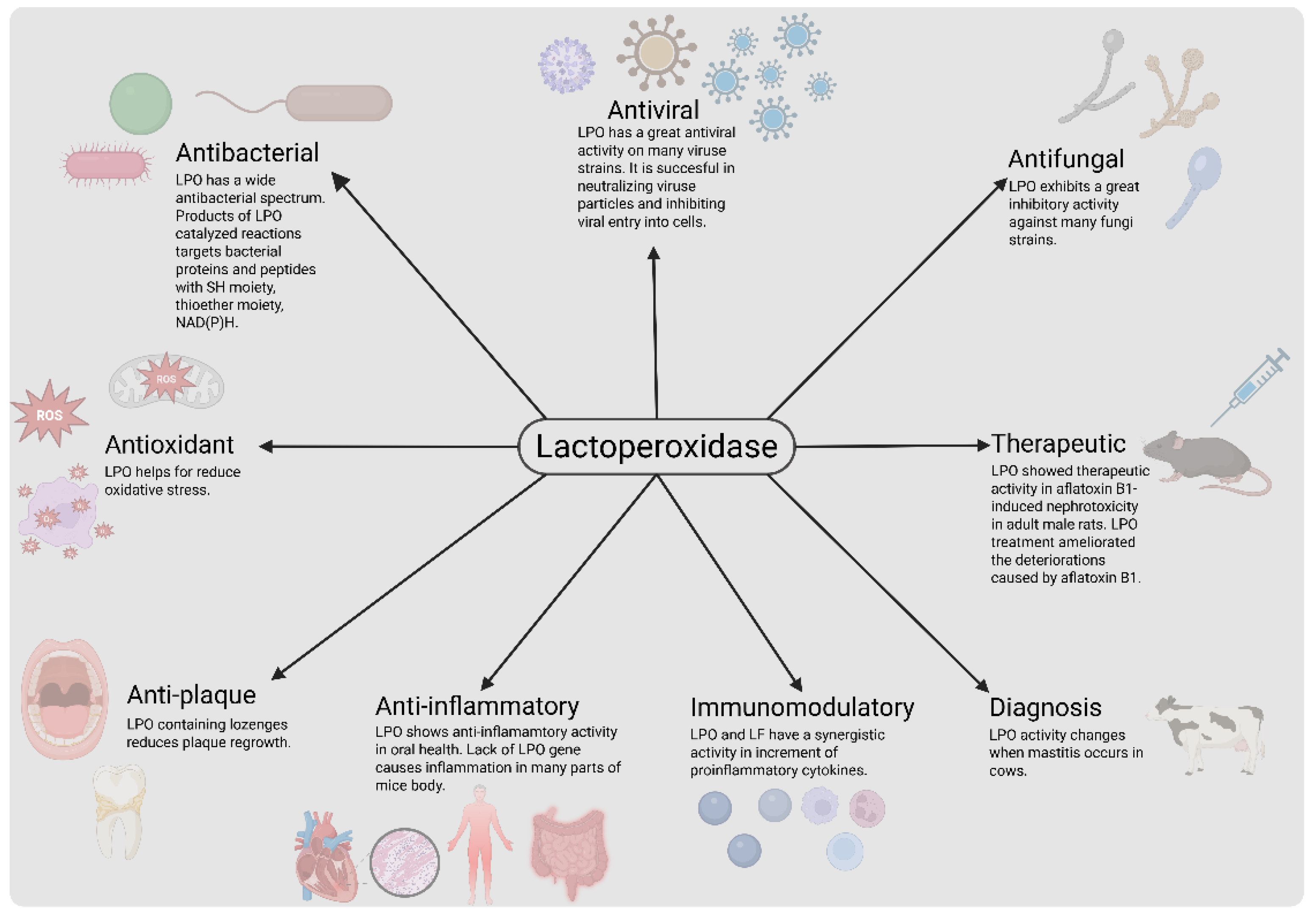
Figure 2.
Production of OSCN- and OI- by LPO catalyzed reactions [3,21] and their antibacterial activity [66].
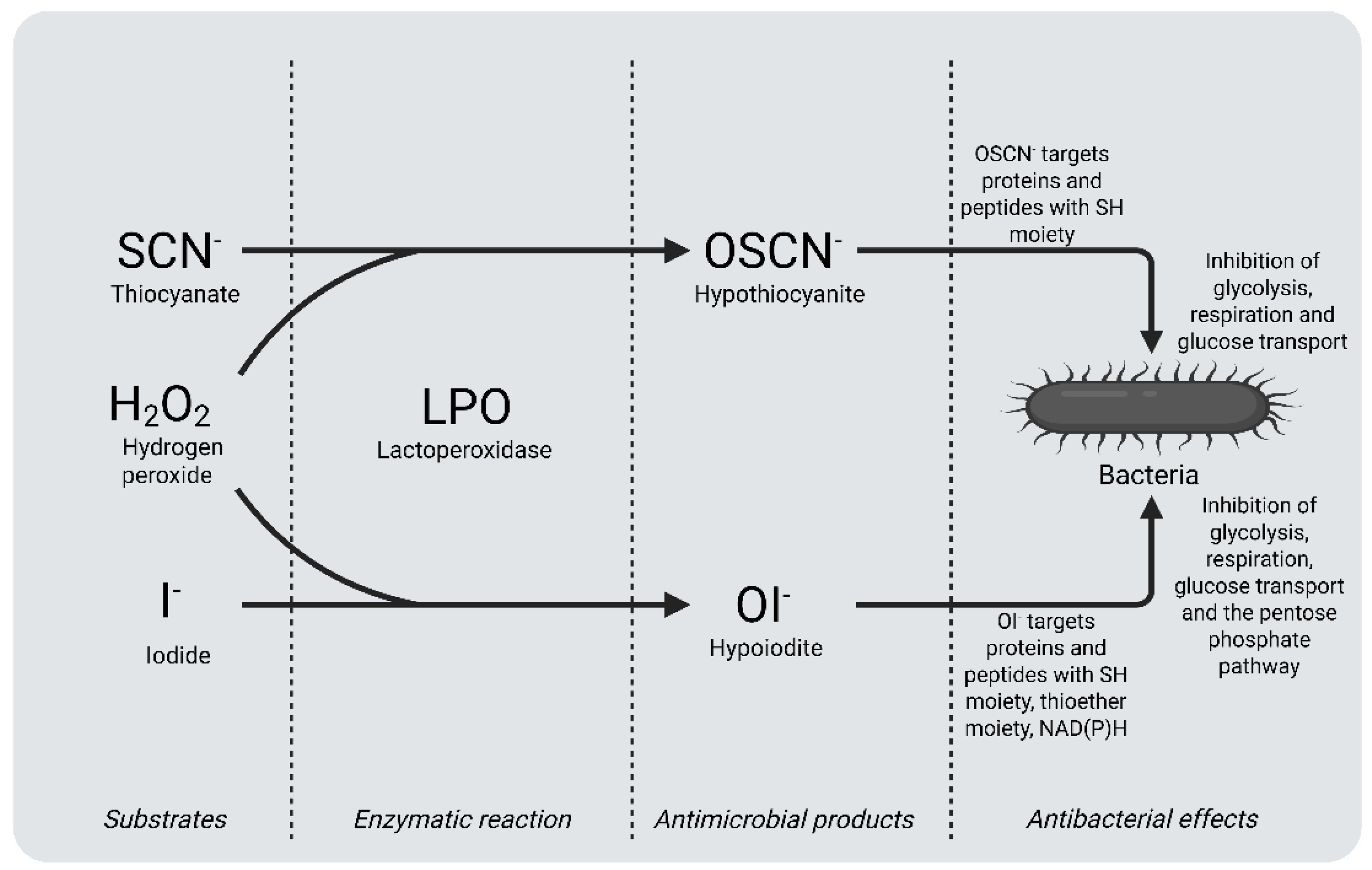
Figure 3.
A demonstration shows possible applications of LPO.
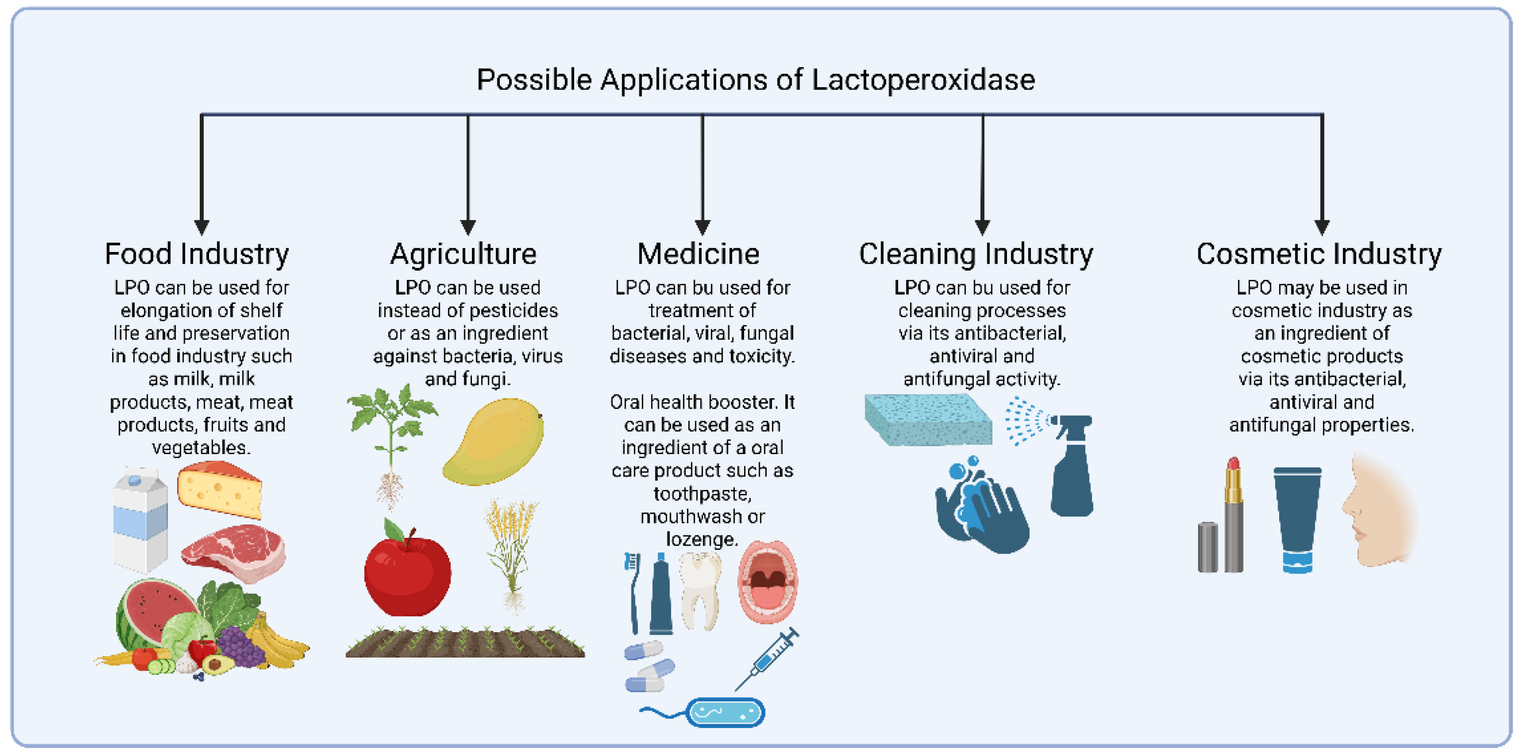
Table 3.
A comparison of studies which show functions of LPO.
| Function | Study Design | Treatment | Results | Reference |
| Antibacterial | In vitro | Susceptibilities of clinical isolates (n=12) and type strains (n=5) of Actinobacillus actinomycetemcomitans to LPO-I⁻-H2O2 combination and different antibiotics were tested. In combination 75 µg LPO, 100 nmol I⁻ and 1000 nmol H2O2 were used. |
The combination exhibited an inhibition on Actinobacillus actinomycetemcomitans similar to 2 microgram ampicillins. | [5] |
| Antibacterial | In vitro | Milk samples (n=250) consisting of morning and overnight samples, were tested. LPO in raw milk was activated by addition of 14 mL of freshly made NaSCN (1 mg/mL) solution per liter of milk and in total 10 mL of freshly made 1 mg/ mL H2O2 solution. |
The quality of the all LPOS-activated milk samples was found to be higher than all the control samples. | [6] |
| Antibacterial | In vitro and ex vivo |
In vitro: 14 mL TSB broth, 1 mL inoculum (Listeria innocua, Pseudomonas fluorescens, or Staphylococcus saprophyticus), 1 mL 1% LPO (sterile distilled water). Ex-vivo meat: 3 groups of pork (shoulder) cubes (2x2x2cm) were soaked in distilled water, TSB broth with Listeria innocua, and LPO solution (5 g LPO in 100 ml distilled water). 0.25% and 0.50% LPO solutions were used to make pork ham and pâté ex vivo. |
LPO has significant inhibitory effects on growth of Listeria innocua, Pseudomonas fluorescens and Staphylococcus saprophyticus. Study showed that LPO can be used to elongate shelf life of meat and meat products. | [7] |
| Antibacterial | In vitro | Specific dosage of LPOS components was not mentioned. Raw camel milk, pasteurized raw camel milk and boiled camel milk each were tested by itself, with addition of exogenous H2O2 or with addition of H2O2 producing lactic acid bacteria. | LPOS activated by H2O2 producing lactic acid bacteria have significantly positive effects on storage of raw camel milk. Weissella confusa 22282 was determined as the best strain of the strains tested in the study to produce H2O2. | [8] |
| Antibacterial | In vitro | Challenge 20 permanent third molars to replicate caries-affected dentin. Before sealing, five samples were treated with LPO, LF, lysozyme, and hydroxyapatite. The combination contains 0.018 mg LPO, LF, lysozyme, and hydroxyapatite powders. The total viable Streptococcus mutans count was measured before, 24h, 1 month, and 6 months after therapy. | LPO may be combined with LF, lysozyme and hydroxyapatite for treatment of dentinal caries. A significant reduction in Streptococcus mutans was observed 24 hours after treatment by the combination. | [9] |
| Antibacterial | In vitro | A whey protein-alginate coating with LPOS was created and tested for its antibacterial properties on chicken thigh meat. The coatings were produced with varying concentrations of LPOS, including LPO, GO, Glu, KSCN, and H2O2. | A whey protein-alginate coating with LPOS has a substantial antibacterial impact, and this study found that the effect increases with LPOS content, with the largest effect at 8% LPOS. A study found that LPOS coating can extend chicken thigh meat shelf life. | [10] |
| Antibacterial | In vitro | Dangke samples were immersed in solutions of LPOS, lysozyme or LPOS+lysozyme at 30°C for 4 hours. Dangke which was immersed in distilled water was used as a control. LPOS was prepared with 300μL of LPO, 300μL of 0.9mM H2O2 and 300μL of 0.9mM KSCN. | The results showed that there is a significant inhibition of the growth of microbes in LPOS, lysozyme and LPOS+lysozyme immersed dangke samples stored for 8 hours, higher antimicrobial activity was observed in LPOS+lysozyme immersed dangke samples than other samples | [11] |
| Antibacterial | In vitro | Effects of alginate coatings with and without LPOS on shelf life of chicken breast fillet were tested. LPOS-containing alginate coatings were produced with 2%, 4% and 6% (v/v) LPOS concentrations. LPOS was made of LPO, GO, Glu, KSCN, KI at ratios of 1.00:0.35:108,70:1.09:2.17 (w/w), respectively. 15.5 mg LPO was used. | The combinations of LPOS and alginate coating exhibited a significant positive effect on increasing the shelf life of the chicken breast fillets under refrigerated conditions. Higher effect was observed at higher LPOS concentrations. | [12] |
| Antibacterial | In vitro | The antibacterial activity of a system containing goat LPO, H2O2, and KSCN was tested on various bacteria by using the disc diffusion method | The system was effective on all tested strains. Inhibition zones were ranging from 22 mm to 26 mm and MIC values for goat milk LPO in the system were ranging from 49 µg/ml to 297 µg/ml | [59] |
| Antibacterial and Antiplaque | Four-replicate crossover study design | A study involving 16 volunteers was conducted to test four different oral hygiene treatments. The treatments were applied in five different sequences. The control group received a mouth rinse with Listerine twice daily. The B treatment involved lozenges containing 10 mg LPO 350 U/mg, 7.5 mg KSCN, and 0.083% H2O2 in a 1:2 H2O2/SCN⁻ ratio, while the C treatment used lozenges with 10 mg LPO 350 U/mg, 7.5 mg KSCN, and 0.040% H2O2 in a 1:2 H2O2/SCN⁻ ratio | The study found that LPOS-containing lozenges can be used in daily oral care for humans due to their anti-plaque regrowth and cariogenic bacteria reduction properties. Treatment B reduced more Streptococcus mutans than treatment C and D, while treatment A showed the most significant antiplaque-regrowth activity. Treatment C showed more reduction on Lactobacilli. | [13] |
| Antibacterial, antiplaque and anti-inflammation | A randomized, double-blind, placebo controlled clinical trial study design |
150 adults were divided into 3 groups and treated by tablets in addition to their daily oral hygiene routine for 12 weeks. The first group was treated by high-dosage tablets that contained 60 mg/d bovine LF and 7.8 mg/d bovine LPO, the second group was treated by low-dosage tablets that contained 20 mg/d bovine LF and 2.6 mg/d bovine LPO, the third group was treated by placebo tablets. | After 12 weeks of treatment, the high dosage group had considerably lower gingival index (GI) than the placebo group. Both high and low dosage groups had considerably lower plaque index (PlI) at 12 weeks than baseline. The high dosage group had a considerably lower 12-week OHIP score. Results demonstrated that bovine LPO and bovine LF pills may improve oral health in healthy persons. |
[14] |
| Antibacterial and immunomodulatory | In vitro and in vivo | Camel colostrum LPO and camel colostrum LF were tested against 14 multidrug-resistant Acinetobacter baumannii isolates from patients with nosocomial pneumonia. The study used the agar well diffusion method to examine their inhibitory effect. In vivo, 5-6 weeks old mice were divided into five groups and exposed to experimental treatments. The mice were injected with different doses of PBS, imipenem, LF, LPO, and LPO, and exposed to different treatments. The results showed promising results in treating nosocomial pneumonia. | Studies have demonstrated that camel colostrum LPO and camel colostrum LF have significant antibacterial activity against Acinetobacter baumannii isolates. LPO showed higher activity than LF at the same concentrations. In vivo, a combination of imipenem, LPO, LF, and a crude combination significantly reduced bacteria in lung and blood cultures. LPO and LF also synergistically affected proinflammatory cytokines in treated mice | [15] |
| Antiviral | In vitro | The study tested influenza virus susceptibility to LPO products and substrate specificity in a cell-free system. Madin-Darby canine kidney cells were used to evaluate viral inactivation. The system used for virus inactivation included LPO, SCN⁻/I⁻, glucose, and glucose oxidase | All tested influenza strains were inactivated by LPO. LPO did not prefer substrates to inactivate H1N1 and H1N2 viruses. H3N2 strains were inactivated better using iodide than thiocyanate as the LPO substrate. |
[16] |
| Antiviral | In vitro | Milk proteins LF, angiogenin-1, α-lactalbumin, β-lactoglobulin, LPO, casein, lactogenin, and glycolactin inhibited HIV-1 RT, α-glucosidase, β-glucosidase, and β-glucuronidase, with succinylation effect examined using ELISA | LPO, both unmodified and succinylated, showed significant inhibitory activity on HIV-1 RT. Succinylated LPO showed small inhibitory activity on α-glucosidase and β-glucosidase. Unmodified proteins, except α-lactalbumin and casein, showed significant inhibition. Succinylation increased inhibitory activity of human LF, glycolactin β-lactoglobulin, and α-lactalbumin and casein. | [17] |
| Antiviral | The study is an evaluation of datas from different sources as a completion | A compilation that includes population, vaccination ratio, infection ratio, and rate of deaths caused by COVID-19 of some countries was prepared. | The study found that Asian countries with high iodine-containing diets, like Japan, South Korea, and India, had significantly lower COVID-19 death rates compared to western countries, possibly due to differences in iodine intake | [18] |
| Antiviral | In vitro | The antiviral activities of human LPO, bovine LPO, and camel LPO against Herpes Simplex Virus Type 1 (HSV-1) were evaluated using cytotoxic effect assay and plaque assay. The viruses were treated with LPO at different concentrations and infected with vero cells. | At concentrations of 0.1, 0.2, 0.3, 0.4, and 0.5 mg/ml, bovine milk LPO had 24, 38, 62, 80, 100% anti-HSV-1 activity, human colostrum had 10, 16, 30, 44, 66%, and camel milk had 12, 18, 34, 50, 70%. | [63] |
| Antiviral | In vitro | The study investigated the protective and neutralizing effects of bLPO, cLPO, and hLPO on Hepatitis C virus (HVC) genotype 4. HepG2 cells were pre-incubated with purified LPO samples and infected with 2% HCV infected serum. The effects of bLPO, cLPO, and hLPO on HCV replication were also examined. LF was used as a control due to its strong activity against HCV G4. | The study revealed that LPO, including bLPO, cLPO, and hLPO, did not protect cells from HCV entry, but they effectively neutralized HCV particles and inhibited entry into HepG2 cells. | [62] |
| Antifungal | In vitro | LPO-containing systems were investigated for candidacidal action against Candida albicans ATCC strains 18804, 10231, and 11006, comparing CFU and cell viability loss. The study also explored how component concentrations affected candidacidal action. | The system of bovine LPO, GO, glucose, and KSCN showed varying candidacidal activity on different strains. The system's activity increased with preincubation time, with the highest activity on Candida albicans ATCC strain 11006. | [19] |
| Antifungal | In vitro | The antifungal activity of LPOS was tested on various fungi, using a mixture of H2O2, KSCN, and goat milk LPO, with concentrations ranging from 30 to 1 mg/mL | Candida albicans and Pythium sp. were resistant to the goat milk LPO-H2O2-KSCN system. MIC values of goat LPO in the system against the other strains tested were determined in the study. | [59] |
| Antifungal | In vitro | The anti-candida activities of LF and LPOS were tested using bovine LF, bovine LPO, and Candida albicans. OSCN⁻ solution was produced by using the enzyme system that consisted of 0.16 mg/ml LPO with 7.5 mM NaSCN and 3.75 mM H2O2 in a buffer | LF and LPOS exhibit significant anti-candida activity on Candida albicans, impacting growth morphology, metabolic activity, and adhesive hyphal form, affecting cellular metabolism | [61] |
| Antifungal | In vitro | Postharvest mangoes were treated by chitosan coatings with or without LPOS. Two LPOS solutions were used, one of them containing iodine (LPOSI) while the other one did not. Antifungal activity was tested by using strains of Colletotrichum gloeosporioides, Phomopsis sp. RP257, Pestalotiopsis sp. and Lasiodiplodia Theobromae ngr 05 A | According to results, there is a synergistic effect of chitosan and LPOS. Inhibitory effect of the coatings was improved by LPOS in all cases. Some strains showed a resistance to chitosan while they were more sensitive to LPOS. | [60] |
| Antifungal | In vitro and in vivo | A coating made of chitosan solutions and LPOS was produced, tested in vitro and in vivo against fungal strains, with a 5% concentration in the film-forming solution | In vitro results showed that chitosan solutions at 1% and 1.5% without LPOS completely inhibited Pestalotiopsis sp. growth, while low inhibition was observed at 0.5%. In vivo results showed increased inhibition, with Phomopsis sp. RP257 less sensitive. LPOS with or without iodide increased inhibition at 0.5% chitosan. | [20] |
| Antioxidant and Therapeutic | In vivo | The study involved 40 adult male albino rats divided into four groups, each with 10 rats, and exposed to experimental treatments to investigate the effects of LPO on aflatoxin B1-induced nephrotoxicity | Measurements indicated that LPO treatment after AFB1 intoxication in group 4 approached all of the values to the values of group 1 which consist of healthy individuals. LPO treatment ameliorated the deteriorations caused by AFB1 | [65] |
| Antitumor and Anti-inflammation | In vivo | Researchers deleted the LPO gene from mice | LPO gene deletion in mice leads to higher rates of diseases like cardiomyopathy, carditis, arteriosclerosis, airway inflammation, glomerulonephritis, digestive system inflammation, and brain pathology. Tumors are also observed in 7 out of 19 one-year-old mice, some of which are overweight or obese | [64] |
| Diagnosis | In vitro and in silico | LPO activity in cow milk was measured to determine mastitis in dairy cows | LPO activity level can be used as a parameter to determine mammary gland health of cows due to LPO activity increases when mastitis occurs in cows. LPO activity reflects the mammary gland health of cows | [23] |
LPO: lactoperoxidase, LPOS: lactoperoxidase system, LF: lactoferrin, LAB: lactic acid bacteria, GO: glucose oxidase, Glu: α-D-glucose.
Disclaimer/Publisher’s Note: The statements, opinions and data contained in all publications are solely those of the individual author(s) and contributor(s) and not of MDPI and/or the editor(s). MDPI and/or the editor(s) disclaim responsibility for any injury to people or property resulting from any ideas, methods, instructions or products referred to in the content. |
© 2025 by the authors. Licensee MDPI, Basel, Switzerland. This article is an open access article distributed under the terms and conditions of the Creative Commons Attribution (CC BY) license (http://creativecommons.org/licenses/by/4.0/).
Copyright: This open access article is published under a Creative Commons CC BY 4.0 license, which permit the free download, distribution, and reuse, provided that the author and preprint are cited in any reuse.


