Submitted:
30 April 2025
Posted:
30 April 2025
You are already at the latest version
Abstract
Periodontal abscesses are a common health problem in rabbits, which can lead to severe complications of infection, including life-threatening conditions. Trueperella pyogenes is an opportunistic pathogen isolated from pyogenic infections in different animal species, most often ruminants and pigs. Here, we report the first case of isolation and molecular characterization of T. pyogenes from periodontal abscess in the rabbit. We also present a narrative review concerning the bacteriological aetiology of odontogenic abscesses in rabbits. The identification of T. pyogenes isolate was based on the phenotypic properties and confirmed by molecular biology methods, such as the presence of species-specific pyolysin gene and the 16S rRNA gene sequencing. Furthermore, six out of eight studied virulence genes were detected, confirming the pathogenic potential of the isolate. This report, as well as literature data, indicate that fastidious bacteria, including T. pyogenes, are one of the etiological factors of periodontal abscesses in rabbits. However, inappropriate laboratory procedures may lead to underestimation of their occurrence. Although periodontal abscesses are a common problem in rabbits, the limited literature data is based on different bacteriologic culture methods. This study showed the need for standardization of the laboratory methods, including extended microbial culture for the clinical samples from periodontal abscesses in rabbits. These procedures should also include the isolation of fastidious pathogens like T. pyogenes.
Keywords:
Trueperella pyogenes
; rabbit
; periodontal abscess
; pyolysin
; virulence factors
1. Introduction
Periodontal abscesses, a common health issue in pet rabbits, are caused by different factors, such as diet, trauma, infections, metabolic bone diseases or neoplasma. However, they are usually related to dental problems [1,2,3]. Despite the frequent occurrence of such cases, the literature data regarding bacteriological aetiology is sparse. Available publications indicate that both aerobic and anaerobic bacteria may be isolated from rabbit abscesses in rabbits. The most frequently obtained bacteria are Pasteurella multocida, Staphylococcus aureus, Streptococcus spp. and Pseudomonas spp., while Bacteroides spp. and Fusobacterium spp. predominate among anaerobic bacteria [1,4,5,6]. Trueperella pyogenes is a facultatively anaerobic, opportunistic pathogen isolated from purulent infections in different animal species, occasionally from humans. These infections are reported in farm and free-living animals, with most documented cases concerning cattle and pigs [7,8,9,10]. Recently, there have been reports regarding the isolation of T. pyogenes from unusual animal hosts such as grey slender lorises (Loris lydekkerianus nordicus), okapi (Okapia johnstoni) and royal python (Python regius) [11,12].
The paper aimed to present an unusual aetiology of periodontal abscess in a pet rabbit. To our knowledge, this is the first description of a T. pyogenes isolate obtained from a rabbit, including its phenotypic characterization, molecular species identification, and detection of virulence genes.
2. Materials and Methods
2.1. Isolation and Phenotypic Identification of T. pyogenes
The clinical material for bacteriological examination was obtained from a 6-year-old male intact French lop rabbit (Oryctolagus cuniculus). Visible head deformations in the maxillary and mandibular area were the reason for the visit to the veterinary clinic. The rabbit was kept in a household with no other animals, in a standard cage, with ad libitum access to commercial feed and hay. Clinical examination revealed the presence of two deformations of the right cheek and left mandibular area. Oral examination showed instability of the left lower premolar (P1). After its extraction, a fistula into the subcutaneous abscess in the left mandibular area was detected. The abscess was swabbed and the material was submitted for routine bacteriological examination. Treatment included local procedures (curettage and drainage) and antibiotic therapy according to the results of antibiotic susceptibility testing for isolated bacteria.
A swab taken from the abscess was inoculated on Columbia Agar with 5% sheep blood (CA) (Graso Biotech, Poland) and McConkey Agar (Graso Biotech, Poland). Incubation was carried out in aerobic conditions and 5% CO2 atmosphere at 37°C for four days. Additionally, to isolate anaerobic bacteria Schaedler Broth medium with paraffin was inoculated and incubated for 48 h. The isolated microorganism was subjected to a routine bacteriological identification, i.e., Gram staining method, catalase test, CAMP test with the reference strain Staphylococcus aureus ATCC 25923 and API Coryne test (bioMérieux).
2.2. Antimicrobial Susceptibility Testing
The isolate obtained from the rabbit was subjected to antimicrobial susceptibility testing (AST) using the Kirby-Bauer disk diffusion method [13]. The suspension density of bacterial cells in sterile saline was adjusted to a 0,5 McFarland standard and spread evenly on Mueller-Hinton agar (Graso Biotech, Poland) supplemented with 5% sheep blood. The disks with the following antimicrobial agents were applied: penicillin (P), cefpodoxime (CPD), clindamycin (CC), tetracycline (CC), enrofloxacin (ENR), chloramphenicol (C) and sulfamethoxazole with trimethoprim (SXT). After incubating at 37°C with 5% CO2 for 24 h, the diameters of growth inhibition were measured with a calliper. The recommendations for the interpretation of AST are not available for T. pyogenes. Thus, breakpoints for Streptococcus spp. were used, and Streptococcus pneumoniae ATCC 49619 was used as a quality control [14].
2.3.16. S rRNA Gene Sequence Analysis
A genomic DNA for molecular biology tests was obtained using the boiling method. A few colonies from a pure culture of the isolate cultured on CA were suspended in 200 µL of nuclease-free water. Then, the suspension was boiled for 10 minutes, cooled on ice and centrifuged (5 min, 8000× g). The supernatant was used as a DNA template for PCR. The 16S rRNA gene sequencing confirmed the phenotypic identification of the isolate. A fragment of the 16S rRNA (843 bp) gene was amplified using universal primers UNF (5’-GAGTTTGATCCTGGCTCAG-3’) and UNR (5’-GGACTACCAGGGTATCTAAT-3’) [15].
In order to perform phylogenetic analysis, the 16S rRNA sequence obtained from the isolate described in this study was compared with 18 sequences of the 16S rRNA of T. pyogenes isolates isolated from different hosts (pig, calf, goat, sheep, deer, Bison bonasus, Naemorhedus goral) in various countries (Poland, China, Japan), along with three type strains deposited in the GeneBank, i.e.: T. pyogenes ATCC 19411, Trueperella bernardiae ATCC BAA-441 and Trueperella bialowiezensis DSM 17162. The analysis also included the sequence of Arcanobacterium haemolyticum DSM 20595 as additional references. All sequences were aligned using The MEGA 12.0 package [16]. Multiple sequence alignment of the partial 16S rRNA gene sequences from 19 T. pyogenes strains showed similarity ranging from 100% to 99,08%. The phylogenetic tree was reconstructed using the neighbour-joining method [17], with bootstrap analysis based on 1000 replications [18]. The evolutionary distances were computed using the maximum composite likelihood method [19].
2.4. Detection of Virulence Genes
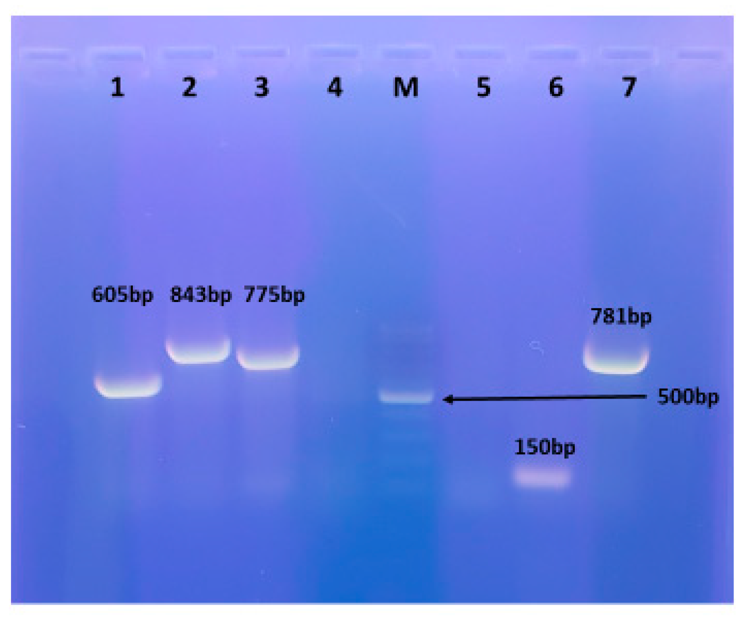
The presence of the following virulence determinants was investigated: pyolysin, neuraminidase H, neuraminidase P, four fimbrial subunits (A, C, E, and G), and collagen-binding protein. The primers and amplification conditions were described by Silva et al. (2008) [7]. T. pyogenes ATCC 49698 was used as a reference strain. All amplification products were visualised by electrophoresis in 1,5% (w/v) agarose gel.
3. Results
3.1. Isolation and Phenotypic Identification of T. pyogenes
After 48 h of incubation on a CA in 5% CO2 atmospheric conditions, tiny β-hemolytic colonies were detected (Figure 1). After 72 h of incubation, the colonies were approximately 1 mm in diameter, white, round and showing β-hemolysis. Small colonies on CA medium incubated in aerobic conditions appeared after 4 days of incubation. After 2 days of incubation, slight turbidity of the Schaedler's medium under the paraffin was observed. Subsequently, a subculture was made from Schaedler's medium to CA medium, on which, after 48 h of incubation in 5% CO2 atmosphere at 37°C, tiny β-hemolytic colonies were found. No microbial growth was observed on McConkey Agar. No other bacteria were cultured.
The morphology of bacterial cells was assessed using the Gram staining method revealed Gram-positive irregular rods. The test for catalase production was negative. In the CAMP test with the reference strain Staphylococcus aureus ATCC 25923 a positive result was observed, visible as enhancement of β-hemolysis. The isolated microorganism was identified using the API Coryne test as T. pyogenes.
3.2. Antimicrobial Susceptibility Testing
The examined T. pyogenes isolate showed a high level of susceptibility to antimicrobial agents, i.e. penicillin, cefpodoxime, clindamycin, tetracycline, enrofloxacin, and chloramphenicol. Resistance was shown only to sulfamethoxazole with trimethoprim.
3.3. 16S rRNA Gene Sequence Analysis
The obtained 16S rRNA gene sequence has been deposited in the NCBI GenBank (accession number PV022409). GenBank BLAST analysis of the sequence showed 100,00% identity with sequences of several T. pyogenes strains deposited in the NCBI database, including T. pyogenes strain ATCC 19411 (GeneBank accession number NR117537.1), finally confirming the identification of the tested isolate as T. pyogenes. Based on partial 16S rRNA gene sequences, the phylogenetic analysis indicated a close genetic relationship between the different T. pyogenes isolates included in the analysis. T. pyogenes isolate obtained from a rabbit was clustered with other isolates included in the analysis and with the reference strain, T. pyogenes ATCC 19411 (Figure 2).
3.4. Detection of Virulence Genes
Six of eight tested virulence determinants were detected in the T. pyogenes isolate of rabbit origin. The presence of the pyolysin gene and tree fimbrial subunits genes (fimA, fimC, and fimE) was confirmed. Additionally, both neuraminidase genes (nanH and nanP) were found. The fimG gene and collagen-binding protein gene were not detected (Figure 3).
4. Discussion
This study presented an abscess in a rabbit of an unusual aetiology. To the best of our knowledge, there are no reports describing the isolation of T. pyogenes from rabbits, along with molecular identification and characterization of the isolated strain. Five reports on the isolation of T. pyogenes from clinical materials obtained from rabbits are available, but the identification was based only on phenotypic properties. One publication described a case of severe infection with the formation of multiple abscesses around the stifle joint and the sternum. The authors could not determine the source of this unusual infection, even though this rabbit was kept in a laboratory animal husbandry, where contact with the external environment is limited. The bacteriological examination of material collected from abscesses showed the presence of T. pyogenes, although the identification was based only on phenotypic methods [20]. Two further publications concern the retrospective analysis of microbiological culture results. One of them is a retrospective analysis of the results of bacteriological examinations of clinical materials collected from rabbits over the years 2010-2021 in Spain, which mentions T. pyogenes as one of the rarely isolated species from abscesses and dental diseases, in 32 (1.1%) out of 3596 cases. The authors performed a study on results obtained from a private diagnostic laboratory, and the detailed culture methods were not disclosed in this publication [21]. Moreover, two reports were published demonstrating the isolation of T. pyogenes from suppurative disorders in rabbits. One concerning lung lesion in an animal with septicemia, in the other, bacteria were isolated from the uterine content in rabbits with pyometra [22,23]. The latest report from 2024 provided an analysis of the medical record databases regarding rabbits with dental diseases (2013-2023), in which 51 cases were included. Bacterial culture conditions were not described, only one T. pyogenes isolate was found [24].
In our study T. pyogenes was the only bacterial species isolated from a rabbit abscess, indicating its involvement in the infection. The result of bacterial culture in the CO2 atmosphere was visible after 48 h in the form of tiny colonies. The interpretation of culture results on Schaedler's broth under paraffin was possible only after subculture onto a CA agar, i.e. after 4 days of incubation. Bacterial growth in aerobic conditions was delayed and detectable after 4 days of incubation. These results confirm that the use of 24 h or 48 h of aerobic incubation is not effective for the culture of T. pyogenes, as well as other fastidious microorganisms.
The obtained isolate was characterized by the presence of genes encoding virulence factors, among which the plo gene is regarded as the most important in pathogenicity and also species-specific [8]. Thus, the detection of the plo gene confirms the phenotypic identification. Other virulence genes detected in the isolate include fimbrial subunits genes, fimA, fimC, and fimE, and two neuraminidase genes, nanH and nanP. Two virulence factor genes were not detected: the fimG and the cbpA. T. pyogenes isolates obtained from different animals, and from various types of infections expressed different virulence factors genes, except the plo gene, which is always detected. Different profiles of virulence genes were observed in T. pyogenes, with the fimG, cbpA, and neuraminidase genes being detected the least often [7,9]. The detection of six out of eight virulence factors in the isolate described in this study indicates its high pathogenic potential.
The phylogenetic analysis showed high homology of the 16S rRNA sequence in the tested isolate obtained from the rabbit and in isolates belonging to this species originating from different hosts and isolated in different countries as well as to the reference strain T. pyogenes ATCC 19411. This is consistent with the results indicating high similarity of the 16S rRNA sequences and the lack of specificity of T. pyogenes for different host species [25,26].
Although odontogenic abscesses in rabbits are a common health problem leading to morbidity and in case of complications even to mortality, the literature data regarding this issue are relatively scarce. Available research results on the bacterial aetiology of periodontal abscesses in rabbits indicate that they are often mixed infections involving both aerobic and anaerobic bacteria. Some discrepancies in the literature result from differences in methods used to collect clinical materials and microbiological culture conditions [5,6,27]. Different culture bacterial methods are used in individual studies, which makes it impossible to compare the results. If aerobic culture conditions are used, Pasteurella multocida, Staphylococcus spp., Streptococcus spp. and Pseudomonas spp. are most frequently found. The anaerobic species isolated from these lesions were predominantly Bacteroides spp. and Fusobacterium spp. [1,4,5]. Moreover, the bacteriological culture results interpretation may be hampered by isolating a few bacterial species, including faecal microorganisms. Coprophagia, a physiological behaviour in rabbits, leads to the presence of faecal bacteria in the oral cavity, most of which cause opportunistic infections [27]. However, if several bacterial species are isolated from an abscess, distinguishing whether they solely represent contamination of the clinical material becomes challenging. Furthermore, antibiotic therapy against faecal bacteria may lead to gastrointestinal tract dysbiosis and severe diarrhoea. Another essential element during the collection of the clinical materials from the oral cavity is to avoid contamination by gingival biota or dental plaque bacteria. Incorrect collection of clinical specimens for microbiological culture may lead to inconclusive results [4]. An additional factor that may influence the effectiveness of treatment procedures is the thick consistency of pus. Lysosomal enzymes involved in the digestion of dead cells and purulent material liquefaction are not produced by rabbits. The aspiration or drainage of pus from the abscess may be difficult, as the penetration of antimicrobials [4].
In the study of abscesses from 12 rabbits described by Tyrrell et al. (2002), aerobic and anaerobic culture conditions were used, and two samples from each rabbit were examined: a biopsy specimen with the abscess margin and bone (if available) [4]. The second specimen was pus from the centre of the abscess. In this study, Fusobacterium nucleatum (6/12), Micromonas micra (5/12) and Streptococcus intermedius (6/12) were most frequently isolated, and in individual cases, various species of aerobic and anaerobic bacteria were obtained. Interestingly, P. multocida, S. aureus and Bacteroides fragilis were not detected. Another publication described the results of a retrospective analysis of microbiological examinations of 48 rabbits with dental abscesses [5]. In most cases (73.3%), aerobic and anaerobic bacteria were isolated, and in 50.8% of cases, more than three bacterial species were obtained. A total of 185 isolates were obtained, including 52 aerobic bacteria and 133 anaerobic species. The unusual domination of anaerobic species may result from the accidental isolation of contaminating faecal bacteria. The most common bacteria isolated in this study included Pseudomonas aeruginosa (14/52), Pasteurella spp. (10/52), Streptococcus spp. (8/52) Staphylococcus spp. (7/52), Fusobacterium spp. (36/133), Peptostreptococcus spp. (27/133) and Bacteroides spp. (27/133). Comparison of the results presented in the works of Tyrrell et al. (2002) and Gardhouse et al. (2017) is complicated by the different methods used for collecting clinical samples [4,5]. Tyrell et al. (2002) employed percutaneous excision of abscesses to avoid contamination with gingival bacteria. In the second study, swabs were collected from the oral cavity and the inside of the abscesses [4].
A study describing the outcome of wound-packing treatment of dental abscesses in 13 rabbits was published by Taylor et al. (2010) [1]. Additionally, the bacteriological examination of purulent materials was performed using culture conditions for aerobic and anaerobic bacteria. Mixed infections caused by aerobic and anaerobic bacteria were generally found, but the authors did not provide detailed culture conditions or incubation time. However, there was no bacterial growth in 4 out of 14 (28.6%) abscesses despite their presence in direct smears. It can be assumed that appropriate methods of culture for the fastidious microorganisms, including T. pyogenes, were not used T. pyogenes.
Periodontal infections often are caused by endogenous oral bacteria that penetrate the surrounding tissues [27]. Thus, it is advisable to determine the composition of the natural oral microbiota in healthy animals, but only one publication concerning rabbits is available in the literature. In research conducted by Flenghi et al. (2023) bacterial microbiota of 33 healthy pet rabbits was evaluated using culture methods for aerobic and anaerobic bacteria [27]. However, an essential drawback of this study is the short incubation time of only 24 h, which is insufficient for some microorganisms, especially anaerobic ones and T. pyogenes. This may explain the results obtained, where aerobic bacteria, such as Streptococcus spp, were most often isolated. (19.8%), Rothia spp. (17.9%), Enterobacter spp. (7%), and Staphylococcus spp. (6.6%). The genera of bacteria isolated from healthy individuals were mainly in concordance with those obtained from abscesses by other authors, except for Rothia spp. and Enterobacter spp., found only in healthy rabbits [27]. Another study concerning the aetiology of bacterial infection in rabbits (n=170), where the majority were dental diseases (n=60). However, conditions for the isolation of fastidious and anaerobic bacteria were not included [6].
There is a significant limitation of our study. Unfortunately, due to the owner's decision, the follow-up visit did not take place. Thus, it was impossible to re-examine the rabbit and assess the effectiveness of the applied treatment procedures.
5. Conclusions
This study demonstrated that T. pyogenes may be isolated from periodontal abscesses in rabbits. However, further studies are required to confirm its prevalence in rabbits. Moreover, the review of the sparse and inconsistent literature data indicates the need for standardization of microbiological culture methods regarding the clinical samples from periodontal abscesses in rabbits. The methodology should include culture conditions for aerobic and anaerobic bacteria, as well as the prolonged time of incubation for T. pyogenes and some other fastidious bacterial species.
Author Contributions
Conceptualization, M.K.Ś. and W.B.; methodology, M.K.Ś., E.K., I.S., D.C.C., M R. and W.B.; software, M.K.Ś.; validation, M.K.Ś.; formal analysis, M.K.Ś. and W.B.; investigation, M.K.Ś., E.K., I.S., D.C.C., M R. and W.B.; resources, M.K.Ś. and W.B.; data curation, M.K.Ś. and W.B.; writing—original draft preparation, M.K.Ś. and W.B.; writing—review and editing M.K.Ś., E.K., I.S., D.C.C., M R. and W.B.; visualization, M.K.Ś..; supervision, M.K.Ś. and W.B.; project administration, M.K.Ś. and W.B. All authors have read and agreed to the published version of the manuscript.
Funding
This research received no external funding.
Institutional Review Board Statement
Not applicable.
Informed Consent Statement
Informed consent has been obtained from the patient’s owner to publish this paper.
Data Availability Statement
The data generated and analyzed during the current study are available in the NCBI GenBank repository, under the accession number: PV022409.
Acknowledgments
We thank the technical staff at the Department of Preclinical Sciences (Microbiology), Institute of Veterinary Medicine, Warsaw University of Life Sciences Warsaw, Poland, for their support in this study.
Conflicts of Interest
The authors declare no conflicts of interest.
References
- Taylor, W.M.; Beaufrère, H.; Mans, C.; Smith, D.A. Long-term outcome of treatment of dental abscesses with a wound-packing technique in pet rabbits: 13 cases (1998-2007). J. Am. Vet. Med. Assoc. 2010, 237, 1444-1449. [CrossRef]
- Levy, I.; Mans, C. Diagnosis and outcome of odontogenic abscesses in client-owned rabbits (Oryctolagus cuniculus): 72 cases (2011-2022). J. Am. Vet. Med. Assoc. 2024, 262, 658-664. [CrossRef]
- Rätsep, E.; Ludwig, L.; Dobromylskyj, M. Orofacial masses in domestic rabbits: a retrospective review of 120 cases from 2 institutions, 2000-2023. J. Vet. Diagn. Invest. 2024, 36, 724-729. [CrossRef]
- Tyrrell, K.L.; Citron, D.M.; Jenkins, J.R.; Goldstein, E.J. Periodontal bacteria in rabbit mandibular and maxillary abscesses. J. Clin. Microbiol. 2002, 40, 1044-1047. [CrossRef]
- Gardhouse, S.; Sanchez-Migallon, Guzman, D.; Paul-Murphy, J.; Byrne, B.A.; Hawkins, M.G. Bacterial isolates and antimicrobial susceptibilities from odontogenic abscesses in rabbits: 48 cases. Vet. Rec. 2017, 181, 538. [CrossRef]
- Crăciun, S.; Novac, C.Ş.; Fiţ, N.I.; Bouari, C.M.; Bel, L.V.; Nadăş, G.C. Bacterial Diversity in Pet Rabbits: Implications for Public Health, Zoonotic Risks, and Antimicrobial Resistance. Microorganisms 2025, 13, 653. [CrossRef]
- Silva, E.; Gaivão, M.; Leitão, S.; Jost, B.H.; Carneiro, C.; Vilela, C.L.; Lopes, da Costa L.; Mateus, L. Genomic characterization of Arcanobacterium pyogenes isolates recovered from the uterus of dairy cows with normal puerperium or clinical metritis. Vet. Microbiol. 2008, 132, 111-118. [CrossRef]
- Rzewuska, M.; Kwiecień, E.; Chrobak-Chmiel, D.; Kizerwetter-Świda, M.; Stefańska, I.; Gieryńska, M. Pathogenicity and Virulence of Trueperella pyogenes: A Review. Int. J. Mol. Sci. 2019, 20, 2737. [CrossRef]
- Kwiecień, E.; Stefańska, I.; Kizerwetter-Świda, M.; Chrobak-Chmiel, D.; Didkowska, A.; Bielecki, W.; Olech, W.; Krzysztof Anusz, K.; Rzewuska, M. Prevalence and Genetic Diversity of Trueperella pyogenes Isolated from Infections in European Bison (Bison bonasus). Animals (Basel) 2022, 12, 1825. [CrossRef]
- Kwiecień, E.; Stefańska, I.; Kizerwetter-Świda, M.; Chrobak-Chmiel, D.; Czopowicz, M.; Moroz-Fik, A.; Mickiewicz, M.; Biernacka, K.; Bagnicka, E.; Kaba, J.; Rzewuska, M. Genetic diversity and virulence properties of caprine Trueperella pyogenes isolates. BMC Vet. Res. 2024, 20, 395. [CrossRef]
- Nagib, S.; Glaeser, S.P.; Eisenberg; T.; Sammra, O.; Lämmler, C.; Kämpfer, P.; Schauerte, N.; Geiger, C.; Kaim, U.; Prenger-Berninghoff, E.; Becker, A.; Abdulmawjood, A. Fatal infection in three Grey Slender Lorises (Loris lydekkerianus nordicus) caused by clonally related Trueperella pyogenes. BMC Vet. Res. 2017, 13, 273. [CrossRef]
- Ahmed, M.F.E.; Alssahen, M.; Lämmler, C.; Eisenberg, T.; Plötz, M.; Abdulmawjood, A. Studies on Trueperella pyogenes isolated from an okapi (Okapia johnstoni) and a royal python (Python regius). BMC Vet. Res. 2020, 16, 292. https://doi:10.1186/s12917-020-02508-y.
- CLSI, 2017. Methods for Antimicrobial Susceptibility Testing of Infrequently Isolated or Fastidious Bacteria Isolated From Animals. 1st ed. CLSI supplement VET06. Clinical and Laboratory Standards Institute; 2017, Wayne, PA.
- CLSI, 2024. Performance Standards for Antimicrobial Disk and Dilution Susceptibility Tests for Bacteria Isolated from Animals, 6th ed. CLSI standard VET01. Clinical and Laboratory Standards Institute, 2024, Wayne PA.
- Alexeeva, I.; Elliott, E.J.; Rollins, S.; Gasparich, G.E.; Lazar, J.; Rohwer, R.G. Absence of Spiroplasma or other bacterial 16s rRNA genes in brain tissue of hamsters with scrapie. J. Clin. Microbiol. 2006, 44, 91-97. [CrossRef]
- Kumar, S.; Stecher, G.; Suleski, M.; Sanderford, M.; Sharm,. S.; Tamura, K. MEGA12: Molecular Evolutionary Genetic Analysis version 12 for adaptive and green computing. Mol. Biol. Evol. 2024. [CrossRef]
- Saitou, N.; Nei, M. The neighbor-joining method: a new method for reconstructing phylogenetic trees. Mol. Biol. Evol. 1987, 4, 406-425. [CrossRef]
- Felsenstein, J. Confidence Limits in Phylogenies: An Approach Using the Bootstrap. Evolution 1985, 39, 783-791. [CrossRef]
- Tamura, K.; Nei, M.; Kumar, S. Prospects for inferring very large phylogenies by using the neighbor-joining method. Proc. Natl. Acad. Sci. USA 2004, 101, 30, 11030-11035. [CrossRef]
- Shahbazfar, A.A.; Kolahian, S.; Mohammadpour, H.; Helan, J. Multi abscessation with multinodular abscesses in a New Zealand white rabbit (Oryctolagus cuniculus) following Arcanobacterium pyogenes infection. Revue. Méd. Vét. 2013;164, 23-26.
- Fernández, M.; Garcias, B.; Duran, I.; Molina-López, R.A.; Darwich ,L. Current Situation of Bacterial Infections and Antimicrobial Resistance Profiles in Pet Rabbits in Spain. Vet. Sci. 2023, 10, 352. [CrossRef]
- Hijazin, M.; Ulbegi-Mohyla, H.; Alber, J.; Lämmler, C.; Hassan, A.A.; Abdulmawjood, A.; Prenger-Berninghoff, E.; Weiss, R.; Zschöck, M. Molecular identification and further characterization of Arcanobacterium pyogenes isolated from bovine mastitis and from various other origins. J. Dairy Sci. 2011, 94, 1813-9. [CrossRef]
- Planas, J.; Ester, Pintado, E.; Verdés, J.; Lourdes, Abarca, M.; Martorell, J. Rabbit with polyuria and polydipsia. 2020. J. Exot. Pet. Med. 2020, 35, 4-8. [CrossRef]
- Minich, D.J.; Marrow, J.C.; Sadar, M.J.; Borsdorf, M.C. High incidence of complications following intraoral extractions and treatment of periapical infections in the management of domestic rabbit (Oryctolagus cuniculus) dental disease (51 cases). J. Am. Vet. Med. Assoc. 2024, 263, 193-198. [CrossRef]
- Rogovskyy, A.S.; Lawhon, S.; Kuczmanski, K.; Gillis, D.C.; Wu, J.; Hurley, H.; Rogovska, Y.V.; Konganti, K.; Yang, C.Y.; Duncan, K. Phenotypic and genotypic characteristics of Trueperella pyogenes isolated from ruminants. J. Vet. Diagn. Invest. 2018, 30, 348-353. https://doi:10.1177/1040638718762479.
- Magossi, G.; Gzyl, K.E.; Holman, D.B.; Nagaraja, T.G.; Amachawadi, R.; Amat, S. Genomic and metabolic characterization of Trueperella pyogenes isolated from domestic and wild animals. Appl. Environ. Microbiol. 2025, 91, e0172524. [CrossRef]
- Flenghi, L.; Mazouffre, M.; Le, Loc'h, A.; Le, Loc'h, G.; Bulliot, C. Normal bacterial flora of the oral cavity in healthy pet rabbits (Oryctolagus cuniculus). Vet. Med. Sci. 2023, 9, 1621-1626. [CrossRef]
Figure 1.
Colony morphology of Trueperella pyogenes isolated from the rabbit on Columbia Agar with 5% sheep blood, 48 h of incubation in CO2 atmosphere at 35°C.
Figure 1.
Colony morphology of Trueperella pyogenes isolated from the rabbit on Columbia Agar with 5% sheep blood, 48 h of incubation in CO2 atmosphere at 35°C.
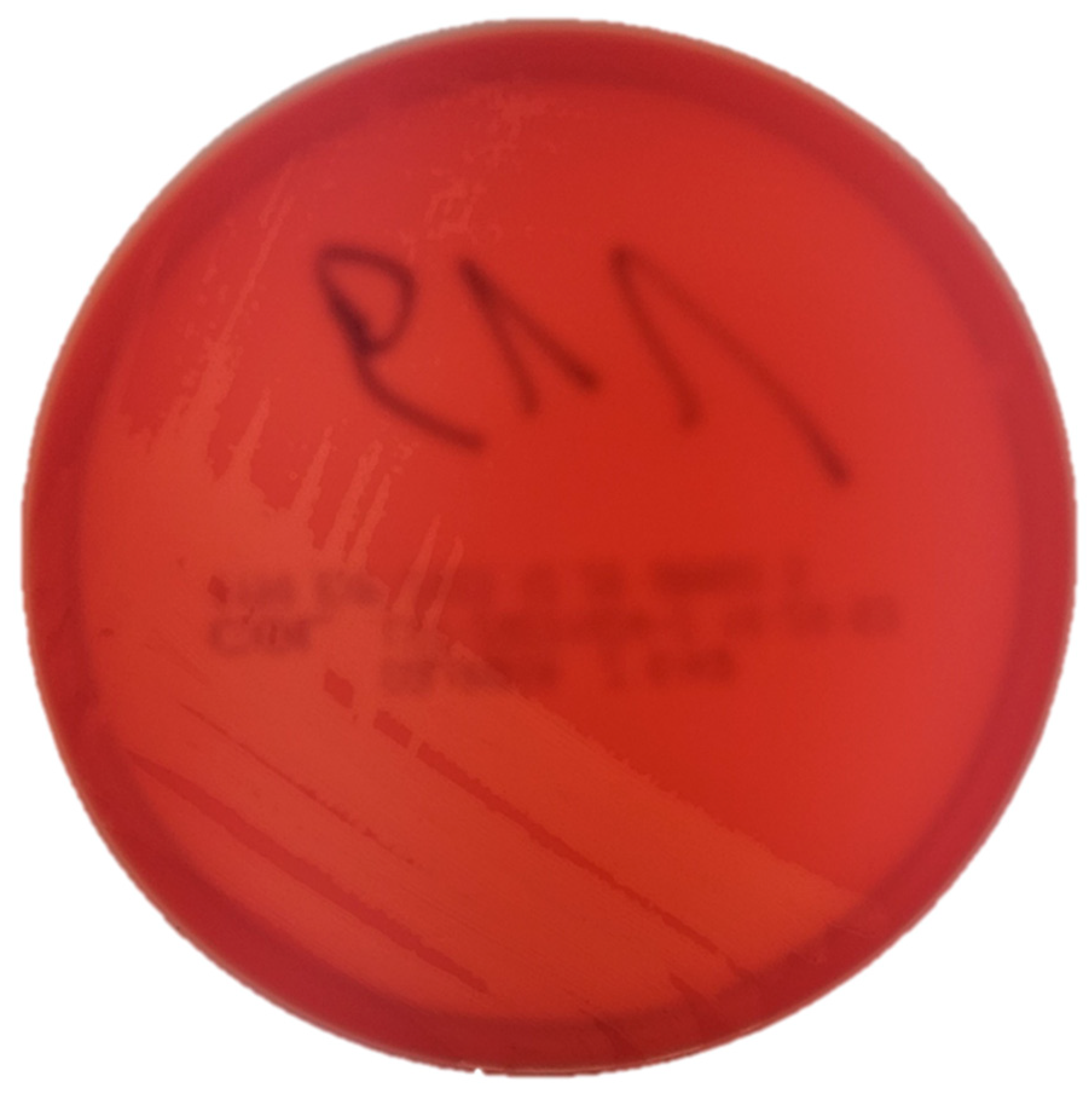
Figure 2.
Phylogenetic tree based on partial sequences of 16S rRNA of the T. pyogenes isolate obtained from rabbit (marked with a dot), T. pyogenes obtained from different hosts in various countries deposited in the GenBank (NCBI) and reference strains of T. pyogenes ATCC 19411, Trueperella bernardiae ATCC BAA-441 and Trueperella bialowiezensis DSM 17162. The tree was constructed using the neighbour-joining method of 16S rRNA gene sequences. Numbers at branch nodes represent the percentage of replicate trees in which the associated taxa clustered together in bootstrap tests (1000 replicates). Arcanobacterium haemolyticum DSM 20595 was used as an outgroup. Bootstrap values below 70 are not shown. The scale bar represents 0.01-nucleotide substitutes per position.
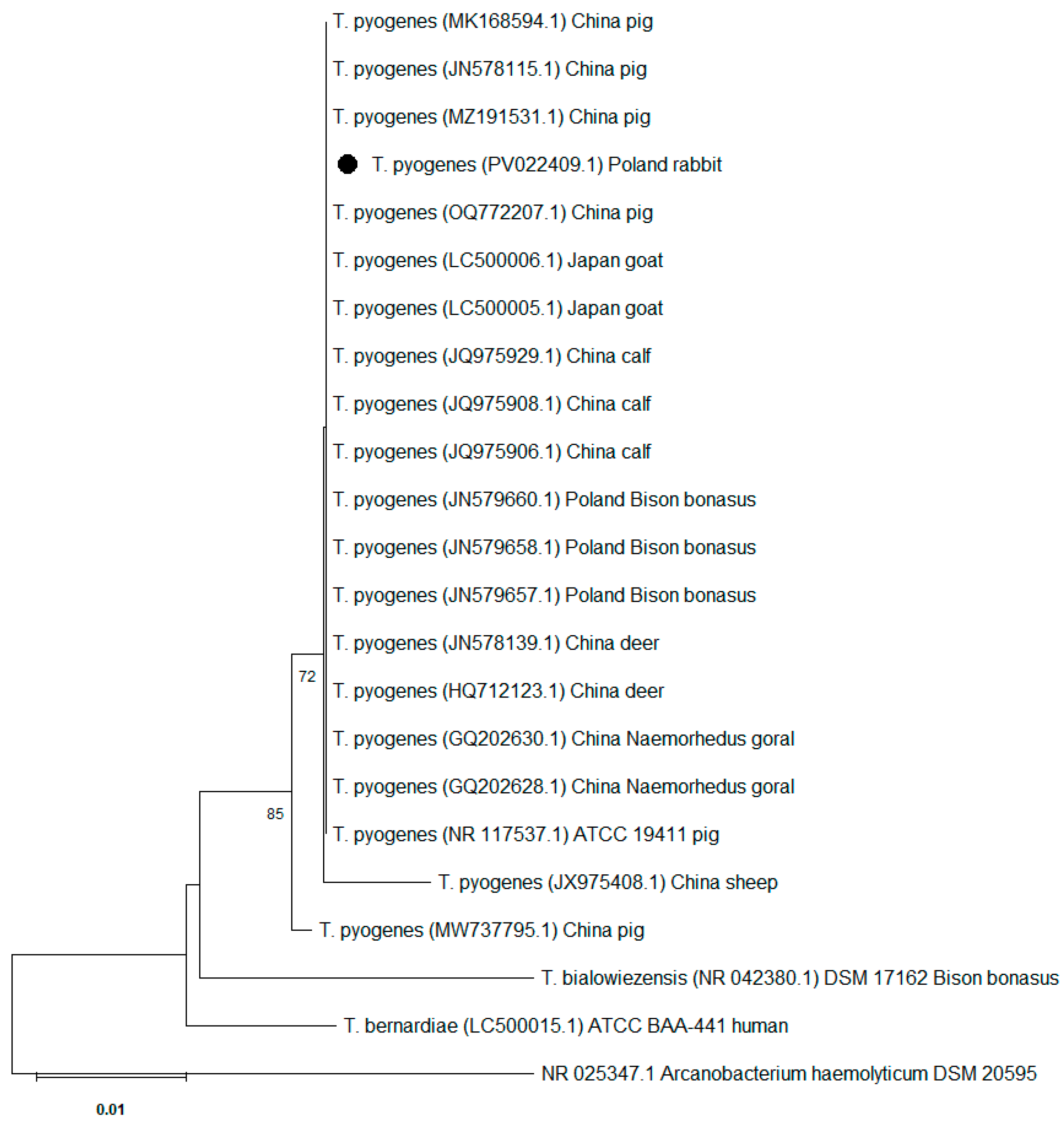
Figure 3.
Agarose gel electrophoresis of the virulence genes in Trueperella pyogenes isolate obtained from a rabbit. Line 1: fimA (605bp); Line 2: fimC (843bp); Line 3: fimE (775bp); Line 4: fimG (negative); M - molecular weight marker; Line 5: cbpA (negative); Line 6: nanP (150bp); Line 7: nanH (781bp).
Disclaimer/Publisher’s Note: The statements, opinions and data contained in all publications are solely those of the individual author(s) and contributor(s) and not of MDPI and/or the editor(s). MDPI and/or the editor(s) disclaim responsibility for any injury to people or property resulting from any ideas, methods, instructions or products referred to in the content. |
© 2025 by the authors. Licensee MDPI, Basel, Switzerland. This article is an open access article distributed under the terms and conditions of the Creative Commons Attribution (CC BY) license (http://creativecommons.org/licenses/by/4.0/).
Copyright: This open access article is published under a Creative Commons CC BY 4.0 license, which permit the free download, distribution, and reuse, provided that the author and preprint are cited in any reuse.


