Submitted:
27 April 2025
Posted:
28 April 2025
You are already at the latest version
Abstract
The consumption of nutraceuticals may help prevent and treat cancer. Tropical plants are packed with copious nutraceuticals, including alkaloids, terpenoids, and phenolic compounds, such as flavonoids, lignans, and phenolic acids, and can be used as preventive and therapeutic phytochemical agents against cancer. For example, bael fruit, soybean, garlic, ginger, olive oil, pomegranate, common wheat, beetroot, green tea, cardoon, neem, turmeric, ashwagandha, and their main nutraceuticals are reviewed in this article for their cancer prevention and therapeutic properties. Various cell signaling pathways and genes involved in different types of cancers, such as breast cancer, colon cancer, skin cancer, prostate cancer, and oral cancer, are potential targets of nutraceutical anticancer effects and are discussed in this review.
Keywords:
nutraceutical
; functional food
; preventive
; therapeutic
; cancer
; cell signaling
Introduction
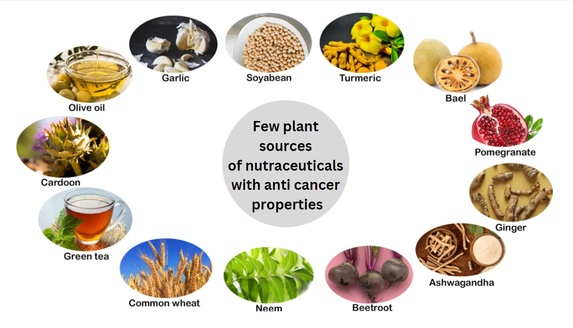
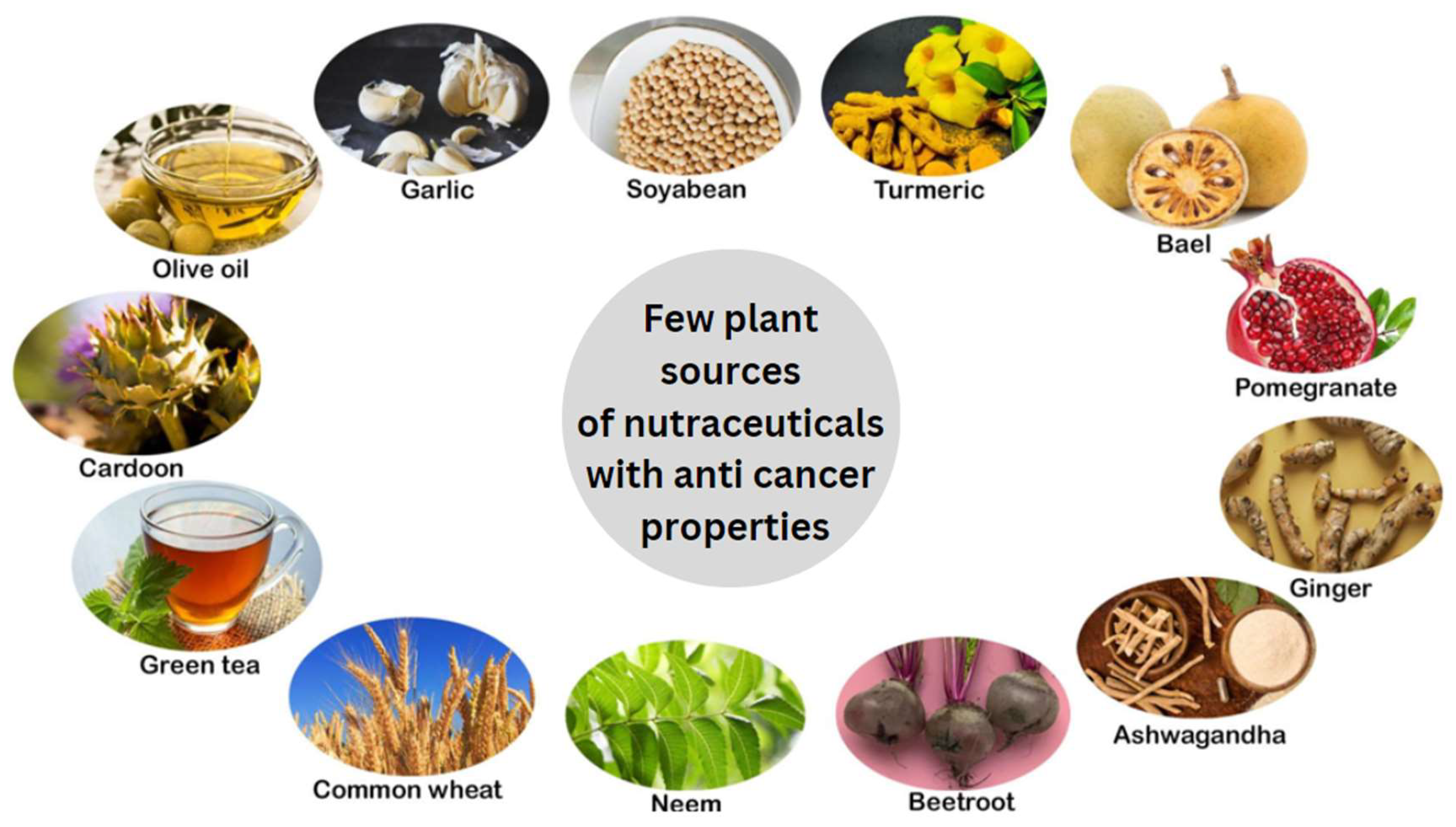
Nutraceuticals are foods, or parts of foods, that provide medical or health benefits, including the prevention and treatment of diseases. The most commonly used nutraceutical compounds, which usually have antioxidant or anti-inflammatory properties, are derived from fruits and vegetables (Figure 1). Epidemiological studies have suggested that nutraceutical compounds prevent chronic diseases and disorders such as cardiovascular disease, diabetes, and cancer [1]. Chemoprevention, which involves the use of dietary compounds, is being explored for the prevention and treatment of cancers [2].
Flavonoids such as luteolin [3], fisetin [4], acetylapoaranotin [5], astaxanthin, lycopene from fruits and vegetables such as tomatoes, grapes and papaya [6], gamma-aminobutyric acid (GABA) [7] and resveratrol [8] are some examples of secondary metabolites that exhibit potential cancer preventive and therapeutic properties. Compared with nutraceuticals alone or in combination with drug delivery systems, anticancer nutraceuticals loaded with biodegradable polymeric nanoparticles demonstrate maximum solubility, absorption, bioavailability, and anticancer potential [9]. Various phytochemicals, such as quercetin, genistein, curcumin, and epigallocatechin gallate, have been shown to affect the ability of polymeric nanoparticles to prevent cancer [9].
The combined treatment of low doses of PEITC (phenethyl isothiocyanate) and curcumin, apple extracts and quercetin 3-O-β-glucuronide (Q3G), epigallocatechin gallate (EGCG) with genistein and luteolin, pomegranate fruit extract and diallyl sulfide from garlic [10] has shown promising anticancer activity. Nutraceuticals combined with various anticancer drugs, such as adriamycin and cisplatin, among others, have been shown to enhance anticancer effects [10].
Although nutraceuticals promise various health benefits, they also need to be evaluated for safety [1]. Metabolites of EGCG may increase oxidative stress and have been associated with liver injury [11]. Soybean isoflavones have estrogenic properties, produce uterine hypertrophy and reproductive tract malformations, inhibit androgen production and steroidogenesis in Leydig cells [12], and stimulate estrogen-dependent tumor growth [13]. The use of soy-derived genistein and daidzein by menopausal women has led to the development of endometriosis [11]. They also face an increased risk of estrogen-sensitive cancers. Extensive clinical trials must be conducted to obtain more data on nutraceuticals and their adverse effects on humans.
On the other hand, the nutraceutical trade encompasses the production and distribution of products derived from food sources that offer additional health benefits beyond basic nutrition. This industry has experienced significant growth in recent years due to increasing consumer interest in preventive healthcare, wellness, and natural products that support health wellness. Nutraceuticals, including dietary supplements, functional foods, and fortified beverages, have become popular for addressing health concerns such as immunity, heart health, cognitive function, aging and cancer.
Nutraceuticals such as bael fruit, soybean, garlic, ginger, olive oil, pomegranate, common wheat, beetroot, green tea, cardoon, neem, and turmeric show potential anticancer properties (Table 1). In the following sections of this review article, we discuss some of the above sources and their nutraceutical compounds with cancer prevention and therapeutic properties.
Bael Fruit (Aegle marmelos Correa)
Aegles marmelos Correa or Stone Apple, belonging to the Rutaceae family, is widely found in India, Sri Lanka, Bangladesh, and Thailand. A. marmelos has several beneficial activities, such as antimicrobial [14], antioxidant [15] and hypoglycemic [16] activities. It is widely used in Ayurveda [17] as an agent that decreases and delays tumor growth [18]. The extract of A. marmelos increased the total white blood cell count, neutrophil percentage, and degree of macrophage activation. It has also been shown to decrease the volume of ascites, indicating that it is better than conventional anticancer treatments such as 5-fluorouracil. A. marmelos has been shown to have antitumor properties when used as a combination therapy to ameliorate Dalton’s lymphoma ascites and Ehrlich’s ascites carcinoma in murine models [19].
Disorders such as diabetes, Alzheimer’s disease, cancer, and cardiovascular disorders are caused mainly by the overproduction of reactive oxygen species (ROS). Compounds such as marmelosin and deferoxamine, which are found in A. marmelos (Table 1), have been shown to suppress ROS production. They also inhibit cellular and DNA damage and dysregulate cell growth under oxidative stress conditions. Marmelosin also inhibits tyrosinase and hence is a good anti-inflammatory and antioxidant agent for fighting cancer [20]. According to previous studies [21], among the different varieties of leaves of A. marmelos, the variety Pant Aparna has the maximum capacity to inhibit superoxide radical production.
A. marmelos has the potential to decrease the levels of certain inflammatory cytokines, such as IL-1β and IL-6, and antiapoptotic genes, such as Bcl-2 and c-jun (see Table 2). Hence, A. marmelos may be used in cancer therapy since it has hepatoprotective and immunomodulatory effects [22].
Bael fruit has long been used to delay the appearance of tumors. It has apoptotic, immunomodulatory, hepatoprotective, antiproliferative, antioxidant, and anti-inflammatory activities. Owing to its potential anticancer properties, bael fruit can be used as an alternative therapeutic compound for cancer. The incorporation of bael fruit into food products has gained popularity not only because of its nutritional benefits but also because of its potential as a functional ingredient with anticancer properties. Some examples of commercial food products that use bael fruit for its health-promoting attributes, particularly its anticancer potential, are described below.
Bael fruits contain several compounds, such as tannins, sterols, and essential oils, which are listed in Table 1 [23,24]. Bael leaf extract containing butyl p-tolyl sulfide, 6-methyl-4-chromanone, and butylated hydroxyanisole was shown to be effective in tumor suppression. However, cisplatin and 5-fluorouracil had better effects than did 5,6-dimethoxy-1-indanone, palmitic acid, methyl linoleate, and 5-methoxypsoralen. Marmelin triggers apoptosis through tumor necrosis factor α (TNFα) [25]. Eugenol also induces apoptosis and has strong effects on salivary gland tumors [26]. Citral and cineole are effective against hematopoietic cancer cells [27] and human leukemia cells [28], respectively. Hydroalcoholic extracts of A. marmelos have been shown to help prevent radiation-induced sickness [29]. The sesquiterpenes in A. marmelos oil, such as β-caryophyllene, γ-murolene, α-humulene, and curcumene, play key roles in inhibiting tumor cell proliferation [30].
Bael fruit is often processed into juices or beverages, which are marketed as health drinks that support digestion and overall immunity. Juice contains high levels of flavonoids and tannins, which help reduce the risk of cancer by protecting cells from genotoxic damage. Various companies have commercialized Bael fruit, such as Organic India Bael Juice, the Heera Ayurvedic Research Foundation, and Krishna’s Herbal and Ayurveda. Companies such as Himalaya Wellness, Bixa Botanical, Patanjali, and Baidyanath produce Bael capsules, powders, and jams, and the polyphenols and alkaloids present are claimed to support the body’s natural defense mechanisms against cancer by inducing apoptosis in cancerous cells. Phytochemical compounds such as alkaloids, flavonoids, and phenols exhibit prominent antidiabetic activity by inhibiting the enzymes alpha amylase and alpha-glucosidase in rat fibroblast lines [31]. The nutraceutical components present in bael fruit offer promising anticancer benefits, making it a valuable ingredient in commercial food products.
Soybean (Glycine max)
Fermented soybean is widely consumed across Asian countries such as Japan, South Korea, and China. The progression of cancer can be prevented by using soybean for a prolonged period of time [32]. The bioactive compounds in soybean, such as saponins, phenolic compounds, trypsin, lunasin and phytic acid, are known to exhibit anticancer and anti-inflammatory activities against matrix metalloproteinases (MMPs)-2 and -9 [33] (see Tables 1 and 2). Genistein is known to inhibit tyrosine kinase enzymes and topoisomerase II, which are usually upregulated in cancer cells. It arrests the cell cycle between the G2 and M phases and induces apoptosis. Genistein is usually used in combination with chemotherapeutic drugs such as B43 murine monoclonal antibodies to kill cancer cells [34].
Natto, which is popularly consumed in Japan, exhibits antitumor activity because it contains a lipopeptide biosurfactant. Korean soybean sauce, tofu, and soy milk are all said to contain Lunasin, encoded by the GM2S-1 (Glycine max 2S albumin (soybean)) gene of soybean, which induces apoptosis. Lunasin mediates anticancer and anti-inflammatory effects by suppressing the nuclear factor-κB (NF-κB) pathway [35]. The expression of JMJD5 (Jumonji domain containing 5), an epigenetic molecule involved in the progression of breast cancer, is inhibited by soybean nutraceuticals [33]. Black soybean extract attenuated the expression of BRAC1 and TNFα in a rat model of breast cancer because of the presence of catechin, daidzein, genistein, and glycitein [36].
Soybeans, which are rich in various bioactive compounds, hold promise as nutraceuticals with potential anticancer properties (Table 1). While research is ongoing, incorporating soy products into a balanced diet may provide health benefits, particularly in cancer prevention. There are various commercial products derived from soybean, including soybean protein isolate, tempeh, edamame, tofu, soy milk, soy flour, soy nuts, and soybean oils. Various companies that are involved in the production of the abovementioned soybean products are Solae and Soy Life. Tata Nutrikraft, Soyfresh, Nutri Soy, Patanjali, Himalayan Food International, etc., among various other companies. Thus, soy products play a significant role in the prevention and treatment of various cancers.
Garlic (Allium sativum)
In silico studies have shown that S-allyl cysteine, p-coumaric acid, phloroglucinol, kaempferol, isobutyl isothiocyanate, quercetin, γGSAC (gamma-glutamyl-S-allyl-cysteine), S-allyl-mercapto cysteine, ferulic acid, taurine and apigenin from garlic possess anticancer activity by specifically targeting breast cancer biomarkers [37], as indicated in Table 1. Diallyl trisulfide (DATS), a major organosulfur compound, exerts anticancer effects on the MCF-7 breast cancer cell line by decreasing steroid synthetase gene expression [38]. Additionally, DATS is a nutraceutical found in garlic against skin [39], liver [40], prostate [41], stomach [42] and colon [43] cancers.
DATS treatment has been shown to suppress neoangiogenesis, decrease Bcl-2 protein levels, reduce lipopolysaccharide-induced expression of inducible nitric oxide synthase and nitric oxide production, increase the activation of T cells, enhance the antitumor function of macrophages [44], activate the NF-κB transcription factor, modify membrane rigidity in tumor cells and reverse cancer chemotherapy drug resistance [45]. See Table 2. It can be used in combination with selenium to decrease cancer morbidity rates without any major harmful side effects [46].
Nutraceuticals from garlic may also induce autophagy or type-II programmed cell death in cancer cells [47]. Organosulfur compounds such as allicin, which are extracted from garlic, contain N-nitroso compounds (see Table 1) and have been shown to reduce tumor initiation. It has been shown that the injection of raw garlic extract has better effects than does ingestion by preventing the regrowth of ascites [48]. The growth of human prostate cancer cells is suppressed by allicin [49] and S-allylcysteine [50], as they induce both caspase-dependent and caspase-independent apoptosis.
The increasing prevalence of lifestyle-related diseases, including cancer, has led to a greater focus on preventive healthcare. Garlic, a natural and widely accepted ingredient, has attracted increasing attention in the nutraceutical space. Numerous companies have stepped into the commercialization of various products, such as capsules, extracts, and supplements, by Himalayan Wellness, Patanjali, Dabur, NutraBlast, Kerala Ayurveda, and Baidyanath. These companies leverage garlic’s health benefits to create a diverse range of products aimed at promoting wellness and preventing health issues, including cancer. As the demand for natural health solutions grows, these offerings highlight the potential of garlic in the nutraceutical market worldwide.
Ginger (Zingiber officinale)
Ginger and its compounds display a variety of properties, such as anti-inflammatory, antioxidant, antimetastatic, and anticancer effects. However, ginger compounds also play a significant role in treating gastrointestinal complications, diarrhea, rheumatic disorders, nausea, common colds, fever, and dizziness, among other conditions [51]. Studies based on immunohistochemistry techniques have indicated that ginger extract may have a chemotherapeutic effect in the treatment of liver cancer, as it blocks NF-κB activation, resulting in suppressed production of TNFα [52] (Table 2). Ginger extract has an anticancer effect on pancreatic cancer cells through ROS-mediated autotic cell death [53].
6-Gingerol (6G) [54], 8-gingerol (8G) [55], 10-gingerol (10G) [56] and 6-shogaol (6S) [57] are collectively referred to as ginger phenolics (Table 1). Pharmacokinetic-pharmacodynamic studies have indicated that the free forms of ginger phenolics are the active components responsible for their anticancer efficacy and not the conjugated forms [58]. In vivo and in vitro studies have established that 6-paradol, 6-gingerol, and 6-shogaol induce apoptosis and inhibit the metastasis of cancer cells [59]. Zerumbone, a sesquiterpene, is known to modulate NF-κB, p53, VEGF, p21, and CXCR4 expression in gastrointestinal cancer [60] (Table 2).
Olive Oil (Olea europaeae)
Olive oil is a major component of the Mediterranean diet. Hydroxytyrosol, a polyphenol found in olive oil (Table 1), acts as a chemopreventive agent against colorectal cancer. It is known to reduce cell proliferation, adhesion, migration, and invasion; arrest the cell cycle at the G2/M phase; induce apoptosis; decrease the expression of cyclins B, D1, and E and CDK2, CDK4, and CDK6; and increase the expression of the CDK inhibitors p21 and p27 [61] (Table 2).
Olive oil consumption has been proven to influence the composition of the intestinal and colonic microbiota, showing modulatory effects on cancer [62]. Oleoflurane inhibited the enzymatic activity of mTOR in breast cancer cells [63]. Hydroxytyrosol, oleuropein, and tyrosol protect cells against oxidative DNA damage [64]. They have shown a reduction in the drug cytotoxicity of mitomycin C by inhibiting ROS production and increasing apoptotic cell death when it is combined with paclitaxel [65].
In vivo experimental models have shown the anti-inflammatory and anticancer effects of extra virgin olive oil [66]. (-)-Oleocanthal, which is isolated from extra virgin olive oil, rapidly and selectively induces cancer cell death via lysosomal membrane permeabilization [67]. However, in vitro and in vivo studies suggest that a high olive oil diet aggravates cervical cancer progression, linking dietary fat and carcinogenesis [68].
Pomegranate (Punica granatum)
Pomegranate seeds possess strong antioxidant and anti-inflammatory properties because of their high content of hydrolyzable tannins and anthocyanins and are edible sources of these compounds (Table 1) [69]. The oral consumption of pomegranate extract inhibited the growth of lung, skin, colon, and prostate tumors.
Compared with fresh pomegranate juice, fermented pomegranate juice is much more beneficial for cancer treatment, as polyphenols from fermented pomegranate juice have twofold greater antiproliferative effects than polyphenols from fresh pomegranate juice [69]. Additionally, pomegranate seed oil (PGO; 100 µg/ml of medium) resulted in 90% inhibition of the proliferation of MCF-7 cells. It was also shown to potentially increase the effectiveness of existing cancer chemotherapy treatments [69].
Similar results have been reported in various studies, including studies on liver cancer, brain tumors, bladder cancer, leukemia, skin cancer, and pancreatic cancer. Pomegranates slow the proliferation of cancer cells and may hasten their death, help reduce the blood supply to tumors, starve them and reduce their size [70]. Anticancer activities are driven mainly by processes such as cell cycle arrest, microRNA modulation, and inhibition of breast cancer growth (Table 2).
Beetroot (Beta vulgaris)
Betavulgarin, an isoflavone, is a type of beta-glucoside (Table 1). Its structure is a glucose moiety linked to a phenolic aglycone. Betayulgarin isolated from beetroot suppressed the proliferation of breast cancer cells and reduced the size of the CD44+/CD24- subpopulation and the expression of the self-renewal-related genes C-Myc, Nanog, and Oct4 (Table 2). Betavulgarin also suppressed the proliferation, migration, colony formation, and mammosphere formation of breast cancer cells [71].
Vitexin-2-O-xyloside [72], betaxanthins, and betacyanins [73] from beetroot can be used in combination with conventional anticancer drugs to reduce their toxicity and overcome the multidrug resistance of cancer cells. The combination of galangin and berberine might provide a promising treatment for patients with esophageal carcinoma, and betanin/isobetanin concentrate significantly decreases cancer cell proliferation and viability [74].
Autophagic cell death upon betanin/isobetanin treatment was also induced, whereas the betanin-enriched extract did not affect normal cell lines [75]. Additionally, organic and conventionally produced beetroots and fermented beetroot juices have different chemical properties and different impacts on cancer cells [76,77]. Moreover, aqueous and ethanolic extracts of beetroot peel powder exhibited antioxidant and anti-proliferative effects on the MCF-7 and MDA-MB-231 breast cancer cell lines, which was due to the presence of betacyanin and betaxanthins.
Green Tea (Camellia sinensis)
One of the oldest and most popular drinks in the world is tea or Camellia sinensis. The most common kind of polyphenol in green tea is catechins (Table 1). Catechins possess antioxidant properties because they can neutralize ROS and bind metal ions (chelators), particularly copper ions, which are key in the Fenton and Haver-Weiss reactions. Tea has been shown to have chemopreventive properties in a variety of human epidemiological and clinical studies; these findings have been supported by cell-based and animal research [78]. The ability of EGCG ((-)-epigallocatechin-3-gallate) to cause apoptosis and end the cell cycle has been extensively studied. The use of HCT-116 cells, which are utilized to study colon cancer, is one example [79,80].
It is generally accepted that the main anticancer mechanism of EGCG is the inhibition of metalloproteinase activity. This theory has been supported by a study that revealed that taking green tea catechins orally reduced the number of metastases produced by prostate cancer [81]. In addition, EGCG taken orally as a supplement affects H1299 human non-small cell lung cancer xenografts in mice, an animal model. The results of this study imply that apoptosis, which causes cancer cells to die, increases and that tumor growth in lung cancer is suppressed [82,83,84].
Clinical evaluation of the role of green tea or green tea components in ovarian cancer prevention and treatment is underway [85]. Green tea extracts [especially EGCG] and resveratrol have been studied for the treatment of oral cancer to determine whether they have potential anticancer effects [86].
The results of structure‒activity relationship (SAR) studies have also greatly enhanced the discovery of novel tea polyphenol analogs as potential anticancer and cancer-preventive agents. Green tea polyphenols such as EGCG have the potential to affect multiple biological pathways, including gene expression, growth factor-mediated pathways, the mitogen-activated protein kinase-dependent pathway, and the ubiquitin/proteasome degradation pathway [87].
Efforts to enhance and evaluate more analogs of green tea catechins should continue to lead to the discovery of more potent, stable, and selective tea polyphenol analogs as potential innovative anticancer medications. However, studies on the structural modification of EGCG have produced encouraging results in terms of its anticancer capabilities. One of the greatest obstacles to cancer prevention is incorporating new molecular findings into therapeutic practice. Identifying additional molecular targets or biomarkers for green tea polyphenols is crucial if synthetic EGCG analogs are to be used efficiently to prevent and treat cancer. Additionally, these findings will aid in our comprehension of the modes of action of anticancer agents [78]. A recent discovery on the anticancer properties of green tea in combination with rosemary extracts revealed an antiproliferative effect on triple-negative cancer cell lines (MDA-MB-231), which was further enhanced when green tea was combined with synthetic drugs, such as cisplatin and paclitaxel [88].
The commercial use of green tea nutraceuticals in India is on the rise, fueled by increasing health consciousness among consumers. A variety of companies are tapping into this market, offering diverse products that cater to health-oriented consumers such as Patanjali Ayurved, Lipton, Tata Tea, Himalayan Wellness, Organic India, Dharani Tea, etc. As regulatory frameworks evolve and consumer preferences shift toward natural and functional foods, the nutraceutical landscape for green tea is likely to expand further. With numerous companies entering the market and consumer interest in health and wellness on the rise, the potential for growth in this sector is significant.
Artichoke (Cynara cardunculus L.)
Artichoke and its phenolic extracts have been shown to exhibit anticancer properties (Table 1). When an artichoke phenolic extract is added to oral squamous carcinoma cell lines, it has apoptotic and cytotoxic effects on cancer cell lines and promotes cell cycle arrest at the G2/M phase. There is an increase in Bax and caspase-9 gene expression in cancer cell lines [89] (Table 2).
Artichoke extracts with paclitaxel increase the production of ROS, which are responsible for obstructing breast cancer progression [90,91]. Silver nanoparticle (AgNP) synthesis is carried out by utilizing Cynara scolymus (Artichoke) leaf extract samples (Artichoke), which is an eco-friendly and cost-effective synthetic technique [92,93] along with photodynamic therapy (PDT). Compared with conventional therapy techniques, the PDT technique results in the generation of ROS, which are responsible for the death of cancer cells with fewer side effects and greater efficiency [94,95]. The generation of ROS results in the activation of the proapoptotic protein Bax and the inhibition of the antiapoptotic protein Bcl-2 in breast cancer cells (MCF7) [96]. This approach of incorporating AgNPs with PDT has potential in breast cancer therapeutics.
Neem (Azadirachta indica)
Oral squamous cell carcinoma (OSCC) is repressed by neem-leaf aqueous extract, which is promoted by 7,12-dimethylbenz[a]anthracene (Table 1) [97]. This extract also effectively decreases the levels of procancer inflammatory cytokines and cell migration pathways [98]. Decreases in various protumor inflammatory mediators and modifications of cellular signaling in OSCC cell lines were shown by the use of supercritical CO2 neem leaf extract (SCNE) and its bioactive compound, nimbolide (NIM) [98].
A nanodelivery system consisting of the polymeric nanoparticle poly(lactic-co-glycolic acid) (PLGA), which is an important nanocarrier with NIM-nano, was formulated and shown to be more effective than free NIM in breast and pancreatic cancer cell lines. This finding indicated that this NIM-nanoformulation approach can be used for the targeted delivery of NIM for cancer treatment [99].
Turmeric (Curcuma longa)
Turmeric contains curcumin, a major polyphenolic compound (Table 1), and is widely used as a therapeutic for many diseases. When supplemented with copper, curcumin effectively inhibited the viability and migration of oral cancer cells, resulting in increased levels of E-cadherin and reduced vimentin. Moreover, Nrf2 levels were elevated, and intracellular ROS were induced in OSCC cells treated with curcumin and copper. This combined treatment also led to early apoptosis [100]. Curcumin has been shown to reduce miR-21 expression levels via a transcriptional mechanism involving binding to the promoter region of the miR-21 gene. Various signaling pathways, such as programmed cell death protein 4 (PDCD4) and NF-κB, are affected by miR-21, which is controlled by curcumin in different types of cancer [101] (Table 2). Curcumin has been shown to decrease the expression of DNA methyltransferase I and induce DNA hypomethylation [102]. Combining temozolomide with dimethoxy curcumin has been shown to have antitumor effects on glioblastoma [103].
Curcumin, an active polyphenol, possesses anti-inflammatory and antitumor properties. However, in one study, it reduced the migration and invasion of breast cancer cells as well as lung metastasis. Moreover, curcumin reduced the migration and invasion of BC cells as well as the lung metastasis of BC in nude mice via TEA domain transcription factor 4 (TEAD4) induction. TEAD4 can regulate the transcription level of the adhesion molecule fibronectin (FN1), which is an important component of the extracellular matrix that participates in tumor cell adhesion and migration processes, and the binding of TEAD4 to the FN1 promoter is suppressed by curcumin [104].
Curcumin has been shown to suppress the proliferation of a wide variety of tumor cells, including those in breast carcinoma, colon carcinoma, renal cell carcinoma, and hepatocellular carcinoma [105]. NF-κB plays an important role in immune system cells because it is rapidly activated by a wide variety of pathogenic signals, and intervention in its activation can be beneficial in suppressing harmful inflammatory reactions. Studies have shown that curcumin completely blocks the TNFα-dependent activation of NF-κB. The activation induced by various other agents, including phorbol ester and H2O2, was also inhibited by curcumin [106]. One of the limiting factors of using curcumin as a drug candidate is that it has poor pharmacodynamic and pharmacokinetic properties [107].
The combination of curcumin nanoparticles with plasma proteins is a favorable approach for cancer therapeutics and helps increase the bioavailability of curcumin [108]. Curcuminoids such as curcumin, dimethoxy curcumin and noncurcuminoids such as α-turmerone significantly inhibit the proliferation of the human cancer cell lines HepG2, MCF-7 and MDA-MB-231. Noncurcuminoids primarily found in turmeric oil, such as elemanes, tumerones, furanodiene and bisacurone, have been shown to have significant anticancer properties. α-Turmerone causes cell death via activation of the caspase cascade and apoptosis [109].
Elemene has been shown to inhibit tumor growth in ovarian, prostate, breast, brain, leukemia, and glioblastoma cells [110]. Elemene induces apoptosis in tumorous cells by causing cell cycle arrest between the S and G2M phases [111]. Via ERK1/2- and AMP-activated protein kinase-mediated inhibition, β-elemene has the potential to inhibit human lung cancer [112] (Table 2). When used in combination with paclitaxel, furanodiene has antiproliferative effects on lung cancer cells [113]. Studies on the anticancer activity of bisacurone indicate that it inhibits the adhesion of cancer cells to endothelial cells by downregulating the expression of VCAM-1 via TNFα stimulation in human oral cancer [114].
Ashwagandha (Withania somnifera)
Ashwagandha water extract (ASH-WEX) has been shown to activate numerous proapoptotic pathways, leading to the inhibition of the tumor-promoting proteins p-NF-κB, p-Akt, heat shock protein 70 (HSP70), cyclin D1 and VEGF (Table 2). The intracranial tumor volumes were also decreased in the rat model of orthotopic glioma allograft. The antiglioma potency of ASH-WEX can be established because it leads to a decrease in glial fibrillary acidic protein (GFAP) in tumor-bearing tissues [115]. However, a recent report revealed that withaferin A downregulated the expression of glycolytic enzymes such as glut 1 (glucose transporter 1), HK2 (hexokinase isoform 2) and PKM (pyruvate kinase isoform M2). This in turn decreases glucose uptake, lactate production and ATP generation. Taken together, these findings suggest that aferin A deregulates metabolism in breast cancer models and acts as an anticancer agent [116].
Laying hens with ovarian cancer (OVCA) supplemented with dietary Ashwagandha presented an increase in intratumoral and stromal NK cells and a reduction in OVCA development [117]. Ashwagandha is an herb that has been extensively used in Ayurveda as a traditional medicine. A withanolide from Withania somnifera (WS), Withaferin A (Table 1), has been shown to potentially inhibit angiogenesis [118] and to exhibit COX-2 inhibitory activity [119].
The expression of a number of genes under the transcriptional control of NF-κB, including angiogenic and inflammation-associated proteins, was decreased upon withaferin A treatment. Interestingly, the expression of these genes is also negatively regulated by the nuclear receptor liver X receptor-α (LXR-α). A novel mechanism by which withaferin-A-activated LXR-α inhibits NF-κB transcriptional activity and suppresses the proliferation, migration, invasion, and anchorage-independent growth of these HCC cells has been previously reported. These data suggest that withaferin A is a potent anticancer compound that suppresses various angiogenesis and inflammatory markers associated with the development and progression of HCC. This beneficial and potential therapeutic property of withaferin A will be very useful for the treatment of HCC.
Conclusion
Nutraceuticals show promising health benefits because of their antioxidant, anti-inflammatory, antidiabetic, antimicrobial, and anticancer properties. Nutraceuticals contained in food include various dietary chemicals, such as alkaloids, terpenoids,, and phenolic compounds, such as flavonoids, lignans, and phenolic acids, which can be used as chemopreventive and therapeutic agents against cancer. Although nutraceuticals have various beneficial effects, extensive research must be carried out to determine the possible adverse and secondary effects in humans.
The expansion of the nutraceutical trade can be achieved through a combination of innovation, market diversification, and enhanced consumer engagement. By investing in research and development, companies can create personalized, science-backed products that cater to specific health needs, such as cognitive health, mental well-being, and disease prevention. Leveraging advancements in biotechnology, nanotechnology, and artificial intelligence will improve the effectiveness and accessibility of nutraceuticals, while sustainable and ethical sourcing practices will attract environmentally conscious consumers. Additionally, expanding into emerging markets, especially in Asia, Africa, and Latin America, will open new growth avenues, supported by strategic partnerships and e-commerce platforms.
The future success of the nutraceutical industry depends on continued research and innovation. The key areas of future studies include clinical validation, which involves conducting large-scale clinical trials to substantiate the efficacy and safety of nutraceuticals, ensuring that they meet regulatory standards and build consumer trust and regulatory harmonization, which means that future studies should focus on creating standardized frameworks for labeling, quality control, and health claims to ensure global regulatory compliance and product consistency. New ingredients and formulations, including plant-based, algae-derived, or laboratory-grown ingredients, should be researched to lead to the development of more effective and sustainable nutraceuticals.
Overall, by focusing on scientific research, regulatory compliance, technological advancements, and global market expansion, the nutraceutical trade is poised for continued growth and innovation in the coming years. Finally, enhancing consumer education around the benefits of nutraceuticals, combined with transparent labeling and regulatory compliance, will build trust and drive further adoption.
Supplementary Materials
The following supporting information can be downloaded at the website of this paper posted on Preprints.org.
Acknowledgments
CHK and VVM acknowledge DSCE management for their kind support and help with the preparation of this manuscript. DCR and SEGM are researchers supported by the National Bureau of Science and Technology (CONICET), Ministerio de Capital Humano, Gobierno de la República Argentina.
Abbreviations
PEITC (phenethyl isothiocyanate), Q3G (quercetin 3-O-β-glucuronide), EGCG (epigallocatechin gallate), JMJD5 (jumonji domain containing 5), GM2S-1 (glycine max 2S albumin (soybean)), γGSAC (gamma-glutamyl-S-allylcysteine), FWGE (fermented wheat germ extract), photodynamic therapy (PDT), OSCC (oral squamous cell carcinoma), SCNE (supercritical CO2 neem leaf extract), ASH-WEX (ashwagandha water extract), VEGF (vascular endothelial growth factor), GFAP (glial fibrillary acidic protein), and OVCA (ovarian cancer). Diallyl trisulfide (DATS), ROS, reactive oxygen species, WS (Withania somnifera).
References
- Ronis, Martin J J et al. “Adverse Effects of Nutraceuticals and Dietary Supplements.” Annual review of pharmacology and toxicology vol. 58 (2018): 583-601. [CrossRef]
- Salami, A., Seydi, E., & Pourahmad, J. (2013). Use of nutraceuticals for prevention and treatment of cancer. Iranian journal of pharmaceutical research : IJPR, 12(3), 219–220.
- Yoshida, T., Maoka, T., Das, S. K., Kanazawa, K., Horinaka, M., Wakada, M., Satomi, Y., Nishino, H., & Sakai, T. (2007). Halocynthiaxanthin and peridinin sensitize colon cancer cell lines to tumor necrosis factor-related apoptosis-inducing ligand. Molecular cancer research : MCR, 5(6), 615–625. [CrossRef]
- Suh, Y., Afaq, F., Johnson, J. J., & Mukhtar, H. (2009). A plant flavonoid fisetin induces apoptosis in colon cancer cells by inhibition of COX2 and Wnt/EGFR/NF-kappaB-signaling pathways. Carcinogenesis, 30(2), 300–307. [CrossRef]
- Choi, E. J., Park, J. S., Kim, Y. J., Jung, J. H., Lee, J. K., Kwon, H. C., & Yang, H. O. (2011). Apoptosis-inducing effect of diketopiperazine disulfides produced by Aspergillus sp. KMD 901 isolated from marine sediment on HCT116 colon cancer cell lines. Journal of applied microbiology, 110(1), 304–313. [CrossRef]
- Miller, E. C., Giovannucci, E., Erdman, J. W., Jr, Bahnson, R., Schwartz, S. J., & Clinton, S. K. (2002). Tomato products, lycopene, and prostate cancer risk. The Urologic clinics of North America, 29(1), 83–93. [CrossRef]
- Al-Wadei, H. A., Ullah, M. F., & Al-Wadei, M. (2011). GABA (γ-aminobutyric acid), a nonprotein amino acid counters the β-adrenergic cascade-activated oncogenic signaling in pancreatic cancer: a review of experimental evidence. Molecular nutrition & food research, 55(12), 1745–1758. [CrossRef]
- Jang, M., Cai, L., Udeani, G. O., Slowing, K. V., Thomas, C. F., Beecher, C. W., Fong, H. H., Farnsworth, N. R., Kinghorn, A. D., Mehta, R. G., Moon, R. C., & Pezzuto, J. M. (1997). Cancer chemopreventive activity of resveratrol, a natural product derived from grapes. Science (New York, N.Y.), 275(5297), 218–220. [CrossRef]
- Illahi, A. F., Muhammad, F., & Akhtar, B. (2019). Nanoformulations of Nutraceuticals for Cancer Treatment. Critical reviews in eukaryotic gene expression, 29(5), 449–460. [CrossRef]
- Saldanha, S. N., & Tollefsbol, T. O. (2012). The role of nutraceuticals in chemoprevention and chemotherapy and their clinical outcomes. Journal of oncology, 2012, 192464. [CrossRef]
- Mazzanti, G., Menniti-Ippolito, F., Moro, P. A., Cassetti, F., Raschetti, R., Santuccio, C., & Mastrangelo, S. (2009). Hepatotoxicity from green tea: a review of the literature and two unpublished cases. European journal of clinical pharmacology, 65(4), 331–341. [CrossRef]
- Akingbemi, B. T., Braden, T. D., Kemppainen, B. W., Hancock, K. D., Sherrill, J. D., Cook, S. J., He, X., & Supko, J. G. (2007). Exposure to phytoestrogens in the perinatal period affects androgen secretion by testicular Leydig cells in the adult rat. Endocrinology, 148(9), 4475–4488. [CrossRef]
- Allred, C. D., Allred, K. F., Ju, Y. H., Virant, S. M., & Helferich, W. G. (2001). Soy diets containing varying amounts of genistein stimulate growth of estrogen-dependent (MCF-7) tumors in a dose-dependent manner. Cancer research, 61(13), 5045–5050.
- Kothari, S., Mishra, V., Bharat, S., & Tonpay, S. D. (2011). Antimicrobial activity and phytochemical screening of serial extracts from leaves of Aegle marmelos (Linn.). Acta poloniae pharmaceutica, 68(5), 687–692.
- Chockalingam, V., Kadali, S. S., & Gnanasambantham, P. (2012). Antiproliferative and antioxidant activity of Aegle marmelos (Linn.) leaves in Dalton's Lymphoma Ascites transplanted mice. Indian journal of pharmacology, 44(2), 225–229. [CrossRef]
- Narender, T., Shweta, S., Tiwari, P., Papi Reddy, K., Khaliq, T., Prathipati, P., Puri, A., Srivastava, A. K., Chander, R., Agarwal, S. C., & Raj, K. (2007). Antihyperglycemic and antidyslipidemic agent from Aegle marmelos. Bioorganic & medicinal chemistry letters, 17(6), 1808–1811. [CrossRef]
- Sharma RK, Bhagwan D. Agnivesa’s Charaka Samhita. Vol. 3. Varanasi, India: Chaukhambha Orientalia; 1988.
- Agrawal, A., Jahan, S., & Goyal, P. K. (2011). Chemically induced skin carcinogenesis in mice and its prevention by Aegle marmelos (an Indian medicinal plant) fruit extract. Journal of environmental pathology, toxicology and oncology : official organ of the International Society for Environmental Toxicology and Cancer, 30(3), 251–259. [CrossRef]
- George, Suraj & Radhakrishnan, Rajesh & Kumar S, Sunil & TT, Sreelekha & Balaram, Prabha. (2014). Chemopreventive Efficacy of Aegle marmelos on Murine Transplantable Tumors. Integrative Cancer Therapies. 1. 10.1177/1534735413490234.
- Pynam, H., & Dharmesh, S. M. (2018). Antioxidant and anti-inflammatory properties of marmelosin from Bael (Aegle marmelos L.); Inhibition of TNF-α mediated inflammatory/tumor markers. Biomedicine & pharmacotherapy = Biomedecine & pharmacotherapie, 106, 98–108. [CrossRef]
- Mujeeb, F., Khan, A. F., Bajpai, P., & Pathak, N. (2018). Phytochemical Study of Aegle marmelos: Chromatographic Elucidation of Polyphenolics and Assessment of Antioxidant and Cytotoxic Potential. Pharmacognosy magazine, 13(Suppl 4), S791–S800. [CrossRef]
- Verma, S., Bahorun, T., Singh, R. K., Aruoma, O. I., & Kumar, A. (2013). Effect of Aegle marmelos leaf extract on N-methyl N-nitrosourea-induced hepatocarcinogensis in BALB/c mice. Pharmaceutical biology, 51(10), 1272–1281. [CrossRef]
- Karawya, M. S., Mirhom, Y. W., Shehata, IA. (1980). Sterols, triterpenes, coumarins and alkaloids of Aegle marmelos Correa, cultivated in Egypt. Egypt Journal of Pharmaceutical Science, 1:388-389.
- Maity, P., Hansda, D., Bandyopadhyay, U., & Mishra, D. K. (2009). Biological activities of crude extracts and chemical constituents of Bael, Aegle marmelos (L.) Corr. Indian journal of experimental biology, 47(11), 849–861.
- Subramaniam, D., Giridharan, P., Murmu, N., Shankaranarayanan, N. P., May, R., Houchen, C. W., Ramanujam, R. P., Balakrishnan, A., Vishwakarma, R. A., & Anant, S. (2008). Activation of apoptosis by 1-hydroxy-5,7-dimethoxy-2-naphthalene-carboxaldehyde, a novel compound from Aegle marmelos. Cancer research, 68(20), 8573–8581. [CrossRef]
- Atsumi, T., Fujisawa, S., Satoh, K., Sakagami, H., Iwakura, I., Ueha, T., Sugita, Y., & Yokoe, I. (2000). Cytotoxicity and radical intensity of eugenol, isoeugenol or related dimers. Anticancer research, 20(4), 2519–2524.
- Dudai, N., Weinstein, Y., Krup, M., Rabinski, T., & Ofir, R. (2005). Citral is a new inducer of caspase-3 in tumor cell lines. Planta medica, 71(5), 484–488. [CrossRef]
- Moteki, H., Hibasami, H., Yamada, Y., Katsuzaki, H., Imai, K., & Komiya, T. (2002). Specific induction of apoptosis by 1,8-cineole in two human leukemia cell lines, but not an in human stomach cancer cell line. Oncology reports, 9(4), 757–760.
- Jagetia, G. C., Venkatesh, P., & Baliga, M. S. (2004). Evaluation of the radioprotective effect of bael leaf (Aegle marmelos) extract in mice. International journal of radiation biology, 80(4), 281–290. [CrossRef]
- Quassinti, L., Maggi, F., Barboni, L., Ricciutelli, M., Cortese, M., Papa, F., Garulli, C., Kalogris, C., Vittori, S., & Bramucci, M. (2014). Wild celery (Smyrnium olusatrum L.) oil and isofuranodiene induce apoptosis in human colon carcinoma cells. Fitoterapia, 97, 133–141. [CrossRef]
- Saravankumar V., Anusha R., Balasubramanyam M., Vishwanathan M. Phytochemical analysis and evaluation of antioxidants, antidiabetic and anti-inflammatory properties of Aegle marmelos and its validation and its validation in an in vitro cell model. Cureus. 2024;16(9);e70491. [CrossRef]
- Wang, Y., Liu, L., Ji, F., Jiang, J., Yu, Y., Sheng, S., & Li, H. (2018). Soybean (Glycine max) prevents the progression of breast cancer cells by downregulating the level of histone demethylase JMJD5. Journal of cancer research and therapeutics, 14(Supplement), S609–S615. [CrossRef]
- Yang, J., Pi, C., & Wang, G. (2018). Inhibition of PI3K/Akt/mTOR pathway by apigenin induces apoptosis and autophagy in hepatocellular carcinoma cells. Biomedicine & pharmacotherapy = Biomedecine & pharmacotherapie, 103, 699–707. [CrossRef]
- Ravindranath, M. H., Muthugounder, S., Presser, N., & Viswanathan, S. (2004). Anticancer therapeutic potential of soy isoflavone, genistein. Advances in experimental medicine and biology, 546, 121–165. [CrossRef]
- de Mejia, E. G., & Dia, V. P. (2009). Lunasin and lunasin-like peptides inhibit inflammation through suppression of NF-kappaB pathway in the macrophage. Peptides, 30(12), 2388–2398. [CrossRef]
- Pratama DAO, Annesia, F. Riccadonna, R, Fazar SP, and Muhammad LN. Black soybean extract inhibits rat mammary carcinogenesis through BRCA1 and TNF-α expression: In silico and in vivo study. Open Vet J. 2024 Oct;14(10):2678-2686. [CrossRef]
- Roy, N., Davis, S., Narayanankutty, A., Nazeem, P., Babu, T., Abida, P., Valsala, P., & Raghavamenon, A. C. (2016). Garlic Phytocompounds Possess Anticancer Activity by Specifically Targeting Breast Cancer Biomarkers - an in Silico Study. Asian Pacific journal of cancer prevention : APJCP, 17(6), 2883–2888.
- Akira Sato, Ayano Yabuki, Genta Sato, Hina Nemoto, Yuta Ogawa and Makoto Ohira. Garlic (Allium sativum L.) Organosulfur compounds inhibit breast cancer cell proliferation by decreasing steroid sulfatase levels. Anticancer Res.2025 Jan; 45(1):145-152;. [CrossRef]
- Perchellet, J. P., Perchellet, E. M., & Belman, S. (1990). Inhibition of DMBA-induced mouse skin tumorigenesis by garlic oil and inhibition of two tumor-promotion stages by garlic and onion oils. Nutrition and cancer, 14(3-4), 183–193. [CrossRef]
- Wu, C. C., Chung, J. G., Tsai, S. J., Yang, J. H., & Sheen, L. Y. (2004). Differential effects of allyl sulfides from garlic essential oil on cell cycle regulation in human liver tumor cells. Food and chemical toxicology : an international journal published for the British Industrial Biological Research Association, 42(12), 1937–1947. [CrossRef]
- Xiao, D., Herman-Antosiewicz, A., Antosiewicz, J., Xiao, H., Brisson, M., Lazo, J. S., & Singh, S. V. (2005). Diallyl trisulfide-induced G(2)-M phase cell cycle arrest in human prostate cancer cells is caused by reactive oxygen species-dependent destruction and hyperphosphorylation of Cdc 25 C. Oncogene, 24(41), 6256–6268. [CrossRef]
- Hu, X., Benson, P. J., Srivastava, S. K., Xia, H., Bleicher, R. J., Zaren, H. A., Awasthi, S., Awasthi, Y. C., & Singh, S. V. (1997). Induction of glutathione S-transferase pi as a bioassay for the evaluation of potency of inhibitors of benzo(a)pyrene-induced cancer in a murine model. International journal of cancer, 73(6), 897–902. [CrossRef]
- Wu, P. P., Liu, K. C., Huang, W. W., Chueh, F. S., Ko, Y. C., Chiu, T. H., Lin, J. P., Kuo, J. H., Yang, J. S., & Chung, J. G. (2011). Diallyl trisulfide (DATS) inhibits mouse colon tumor in mouse CT-26 cells allograft model in vivo. Phytomedicine : international journal of phytotherapy and phytopharmacology, 18(8-9), 672–676. [CrossRef]
- Feng, Z. H., Zhang, G. M., Hao, T. L., Zhou, B., Zhang, H., & Jiang, Z. Y. (1994). Effect of diallyl trisulfide on the activation of T-cell and macrophage-mediated cytotoxicity. Journal of Tongji Medical University = Tong ji yi ke da xue xue bao, 14(3), 142–147. [CrossRef]
- Liu, K. L., Chen, H. W., Wang, R. Y., Lei, Y. P., Sheen, L. Y., & Lii, C. K. (2006). DATS reduces LPS-induced iNOS expression, NO production, oxidative stress, and NF-kappaB activation in RAW 264.7 macrophages. Journal of agricultural and food chemistry, 54(9), 3472–3478. [CrossRef]
- Li, H., Li, H. Q., Wang, Y., Xu, H. X., Fan, W. T., Wang, M. L., Sun, P. H., & Xie, X. Y. (2004). An intervention study to prevent gastric cancer by microselenium and large dose of allitridum. Chinese medical journal, 117(8), 1155–1160.
- Xiao, J., Ching, Y. P., Liong, E. C., Nanji, A. A., Fung, M. L., & Tipoe, G. L. (2013). Garlic-derived S-allylmercaptocysteine is a hepato-protective agent in nonalcoholic fatty liver disease in vivo animal model. European journal of nutrition, 52(1), 179–191. [CrossRef]
- Liu, Z., Xia, Y., Zhang, X., Liu, L., Tu, S., Zhu, W., Yu, L., Wan, H., Yu, B., & Wan, F. (2018). Roles of the MST1-JNK signaling pathway in apoptosis of colorectal cancer cells induced by Taurine. The Libyan journal of medicine, 13(1), 1500346. [CrossRef]
- Oommen, S., Anto, R. J., Srinivas, G., & Karunagaran, D. (2004). Allicin (from garlic) induces caspase-mediated apoptosis in cancer cells. European journal of pharmacology, 485(1-3), 97–103. [CrossRef]
- Chu, Q., Lee, D. T., Tsao, S. W., Wang, X., & Wong, Y. C. (2007). S-allylcysteine, a water-soluble garlic derivative, suppresses the growth of a human androgen-independent prostate cancer xenograft, CWR22R, under in vivo conditions. BJU international, 99(4), 925–932. [CrossRef]
- Pereira, M. M., Haniadka, R., Chacko, P. P., Palatty, P. L., & Baliga, M. S. (2011). Zingiber officinale Roscoe (ginger) as an adjuvant in cancer treatment: a review. Journal of B.U.ON. : official journal of the Balkan Union of Oncology, 16(3), 414–424.
- Habib, S. H., Makpol, S., Abdul Hamid, N. A., Das, S., Ngah, W. Z., & Yusof, Y. A. (2008). Ginger extract (Zingiber officinale) has anticancer and anti-inflammatory effects on ethionine-induced hepatoma rats. Clinics (Sao Paulo, Brazil), 63(6), 807–813. [CrossRef]
- Akimoto, M., Iizuka, M., Kanematsu, R., Yoshida, M., & Takenaga, K. (2015). Anticancer Effect of Ginger Extract against Pancreatic Cancer Cells Mainly through Reactive Oxygen Species-Mediated Autotic Cell Death. PloS one, 10(5), e0126605. [CrossRef]
- Bode, A. M., & Dong, Z. (2011). The Amazing and Mighty Ginger. In I. Benzie (Eds.) et al., Herbal Medicine: Biomolecular and Clinical Aspects. (2nd ed.). CRC Press/Taylor & Francis.
- Zick SM, Djuric Z, Ruffin MT, Litzinger AJ, Normolle DP, Alrawi S, Feng MR, Brenner DE. Pharmacokinetics of 6-gingerol, 8-gingerol, 10-gingerol, and 6-shogaol and conjugate metabolites in healthy human subjects. Cancer Epidemiol Biomarkers Prev. 2008 Aug;17(8):1930-6. PMID: 18708382; PMCID: PMC2676573. [CrossRef]
- Chen, C. Y., Li, Y. W., & Kuo, S. Y. (2009). Effect of [10]-gingerol on [ca2+]i and cell death in human colorectal cancer cells. Molecules (Basel, Switzerland), 14(3), 959–969. [CrossRef]
- Wu, H., Hsieh, M. C., Lo, C. Y., Liu, C. B., Sang, S., Ho, C. T., & Pan, M. H. (2010). 6-Shogaol is more effective than 6-gingerol and curcumin in inhibiting 12-O-tetradecanoylphorbol 13-acetate-induced tumor promotion in mice. Molecular nutrition & food research, 54(9), 1296–1306. [CrossRef]
- Mukkavilli, R., Yang, C., Tanwar, R. S., Saxena, R., Gundala, S. R., Zhang, Y., Ghareeb, A., Floyd, S. D., Vangala, S., Kuo, W. W., Rida, P., & Aneja, R. (2018). Pharmacokinetic-pharmacodynamic correlations in the development of ginger extract as an anticancer agent. Scientific reports, 8(1), 3056. [CrossRef]
- Ishiguro, K., Ando, T., Maeda, O., Ohmiya, N., Niwa, Y., Kadomatsu, K., & Goto, H. (2007). Ginger ingredients reduce viability of gastric cancer cells via distinct mechanisms. Biochemical and biophysical research communications, 362(1), 218–223. [CrossRef]
- Prasad, S., & Tyagi, A. K. (2015). Ginger and its constituents: role in prevention and treatment of gastrointestinal cancer. Gastroenterology research and practice, 2015, 142979. [CrossRef]
- Terzuoli, E., Nannelli, G., Frosini, M., Giachetti, A., Ziche, M., & Donnini, S. (2017). Inhibition of cell cycle progression by the hydroxytyrosol-cetuximab combination yields enhanced chemotherapeutic efficacy in colon cancer cells. Oncotarget, 8(47), 83207–83224. [CrossRef]
- Macdonald, R. S., & Wagner, K. (2012). Influence of dietary phytochemicals and microbiota on colon cancer risk. Journal of agricultural and food chemistry, 60(27), 6728–6735. [CrossRef]
- Khanfar, M. A., Bardaweel, S. K., Akl, M. R., & El Sayed, K. A. (2015). Olive Oil-derived Oleocanthal as Potent Inhibitor of Mammalian Target of Rapamycin: Biological Evaluation and Molecular Modeling Studies. Phytotherapy research : PTR, 29(11), 1776–1782. [CrossRef]
- Fabiani, R., Rosignoli, P., De Bartolomeo, A., Fuccelli, R., Servili, M., Montedoro, G. F., & Morozzi, G. (2008). Oxidative DNA damage is prevented by extracts of olive oil, hydroxytyrosol, and other olive phenolic compounds in human blood mononuclear cells and HL60 cells. The Journal of nutrition, 138(8), 1411–1416. [CrossRef]
- Coccia, A., Mosca, L., Puca, R., Mangino, G., Rossi, A., & Lendaro, E. (2016). Extravirgin olive oil phenols block cell cycle progression and modulate chemotherapeutic toxicity in bladder cancer cells. Oncology reports, 36(6), 3095–3104. [CrossRef]
- Fezai, M., Senovilla, L., Jemaà, M., & Ben-Attia, M. (2013). Analgesic, anti-inflammatory and anticancer activities of extra virgin olive oil. Journal of lipids, 2013, 129736. [CrossRef]
- LeGendre, O., Breslin, P. A., & Foster, D. A. (2015). (-)-Oleocanthal rapidly and selectively induces cancer cell death via lysosomal membrane permeabilization. Molecular & cellular oncology, 2(4), e1006077. [CrossRef]
- Zhang, X., Yang, P., Luo, X., Su, C., Chen, Y., Zhao, L., Wei, L., Zeng, H., Varghese, Z., Moorhead, J. F., Ruan, X. Z., & Chen, Y. (2019). High olive oil diets enhance cervical tumor growth in mice: transcriptome analysis for potential candidate genes and pathways. Lipids in health and disease, 18(1), 76. [CrossRef]
- Martos, M. V., López, J. F., Pérez-Álvarez, J. A. (2010). .Pomegranate and its Many Functional Components as Related to Human Health: A Review. Comprehensive Review in Food Science and Food Safety, 9(6), 635-654. [CrossRef]
- Adhami, V. M., Khan, N., & Mukhtar, H. (2009). Cancer chemoprevention by pomegranate: laboratory and clinical evidence. Nutrition and cancer, 61(6), 811–815. [CrossRef]
- Liu, R., Choi, H. S., Zhen, X., Kim, S. L., Kim, J. H., Ko, Y. C., Yun, B. S., & Lee, D. S. (2020). Betavulgarin Isolated from Sugar Beet (Beta vulgaris) Suppresses Breast Cancer Stem Cells through Stat3 Signaling. Molecules (Basel, Switzerland), 25(13), 2999. [CrossRef]
- Ninfali, P., Antonini, E., Frati, A., & Scarpa, E. S. (2017). C-Glycosyl Flavonoids from Beta vulgaris Cicla and Betalains from Beta vulgaris rubra: Antioxidant, Anticancer and Antiinflammatory Activities-A Review. Phytotherapy research : PTR, 31(6), 871–884. [CrossRef]
- Esatbeyoglu, T., Huebbe, P., Ernst, I. M., Chin, D., Wagner, A. E., & Rimbach, G. (2012). Curcumin--from molecule to biological function. Angewandte Chemie (International ed. in English), 51(22), 5308–5332. [CrossRef]
- Ren, K., Zhang, W., Wu, G., Ren, J., Lu, H., Li, Z., & Han, X. (2016). Synergistic anticancer effects of galangin and berberine through apoptosis induction and proliferation inhibition in esophageal carcinoma cells. Biomedicine & pharmacotherapy = Biomedecine & pharmacotherapie, 84, 1748–1759. [CrossRef]
- Nowacki, L., Vigneron, P., Rotellini, L., Cazzola, H., Merlier, F., Prost, E., Ralanairina, R., Gadonna, J. P., Rossi, C., & Vayssade, M. (2015). Betanin-Enriched Red Beetroot (Beta vulgaris L.) Extract Induces Apoptosis and Autophagic Cell Death in MCF-7 Cells. Phytotherapy research : PTR, 29(12), 1964–1973. [CrossRef]
- Kazimierczak, R., Hallmann, E., Lipowski, J., Drela, N., Kowalik, A., Püssa, T., Matt, D., Luik, A., Gozdowski, D., & Rembiałkowska, E. (2014). Beetroot (Beta vulgaris L.) and naturally fermented beetroot juices from organic and conventional production: metabolomics, antioxidant levels and anticancer activity. Journal of the science of food and agriculture, 94(13), 2618–2629. [CrossRef]
- Coimbra PPS, Silva-e-Silva ACAG, Silva Antonio A, Pereira HMG, Veiga Junior VF, Felzenszwalb I, Araujo-Lima CF, and Teodoro AJ. Antioxidant capacity, antitumour activity, and metabolomic profile of a beetroot peel flour. Metabolites.2023,13(2):277:. [CrossRef]
- Farhan, Mohd. “Green Tea Catechins: Nature's Way of Preventing and Treating Cancer.” International journal of molecular sciences vol. 23,18 10713. 14 Sep. 2022. [CrossRef]
- Farhan M., Rizvi A., Naseem I., Hadi S.M., Ahmad A. Targeting increased copper levels in diethylnitrosamine induced hepatocellular carcinoma cells in rats by epigallocatechin-3-gallate. Tumor Biol. 2015;36:8861–8867. [CrossRef]
- Toden S., Tran H.M., Tovar-Camargo O.A., Okugawa Y., Goel A. Epigallocatechin-3-gallate targets cancer stem-like cells and enhances 5-fluorouracil chemosensitivity in colorectal cancer. Oncotarget. 2016;7:16158–16170. [CrossRef]
- Bonuccelli G., Sotgia F., Lisanti M.P. Matcha green tea (MGT) inhibits the propagation of cancer stem cells (CSCs), by targeting mitochondrial metabolism, glycolysis, and multiple cell signaling pathways. Aging. 2018;10:1867–1883. [CrossRef]
- Khan N., Mukhtar H. Tea and health: Studies in humans. Curr. Pharm. Des. 2013;19:6141–6147. [CrossRef]
- Fujiki H., Watanabe T., Sueoka E., Rawangkan A., Suganuma M. Cancer Prevention with Green Tea and Its Principal Constituent, EGCG: From Early Investigations to Current Focus on Human Cancer Stem Cells. Mol. Cells. 2018;41:73–82.
- Fu H., He J., Mei F., Zhang Q., Hara Y., Ryota S. Lung cancer inhibitory effect of epigallocatechin-3-gallate is dependent on its presence in a complex mixture (polyphenon E) Cancer Prev. Res. 2009;2:531–537. [CrossRef]
- Mahboub, F. A., Khorshid, F.A. (2010). The Role of Green Tea Extract on the Proliferation of Human Ovarian Cancer Cells (in vitro) Study. International Journal of Cancer Research, 6(2), 78-88. [CrossRef]
- Gupta, S. C., Kim, J. H., Prasad, S., & Aggarwal, B. B. (2010). Regulation of survival, proliferation, invasion, angiogenesis, and metastasis of tumor cells through modulation of inflammatory pathways by nutraceuticals. Cancer metastasis reviews, 29(3), 405–434. [CrossRef]
- Chen, D., Wan, S. B., Yang, H., Yuan, J., Chan, T. H., & Dou, Q. P. (2011). EGCG, green tea polyphenols and their synthetic analogs and prodrugs for human cancer prevention and treatment. Advances in clinical chemistry, 53, 155–177. [CrossRef]
- Raad C, Raad A and Pandey S. Green Tea Leaves and Rosemary Extracts Selectively Induce Cell Death in Triple-Negative Breast Cancer Cells and Cancer Stem Cells and Enhance the Efficacy of Common Chemotherapeutics. Evid Based Complement Alternat Med. 2024 Jan 25:2024:9458716. [CrossRef]
- Hassabou, N. F., & Farag, A. F. (2020). Anticancer effects induced by artichoke extract in oral squamous carcinoma cell lines. Journal of the Egyptian National Cancer Institute, 32(1), 17. [CrossRef]
- Park, B. H., Lim, J. E., Jeon, H. G., Seo, S. I., Lee, H. M., Choi, H. Y., Jeon, S. S., & Jeong, B. C. (2016). Curcumin potentiates antitumor activity of cisplatin in bladder cancer cell lines via ROS-mediated activation of ERK1/2. Oncotarget, 7(39), 63870–63886. [CrossRef]
- Chan, M. M., Soprano, K. J., Weinstein, K., & Fong, D. (2006). Epigallocatechin-3-gallate delivers hydrogen peroxide to induce death of ovarian cancer cells and enhances their cisplatin susceptibility. Journal of cellular physiology, 207(2), 389–396. [CrossRef]
- Senthil, B., Devasena, T., Prakash, B., & Rajasekar, A. (2017). Noncytotoxic effect of green synthesized silver nanoparticles and its antibacterial activity. Journal of photochemistry and photobiology. B, Biology, 177, 1–7. [CrossRef]
- Samuggam, S., Vasanthi, S., Sivadasan, S., 3, Chinni, S. V., 1, Gopinath, S. C. B., Kathiresan, S., Anbu, P., Ravichandran, V. (2019). Durio zibethinus rind extract mediated green synthesis of silver nanoparticles: Characterization and biomedical applications.Pharmacognosy Magazine, 15(30), 52-58. [CrossRef]
- Antonenko, Y. N., Kotova, E. A., Omarova, E. O., Rokitskaya, T. I., Ol'shevskaya, V. A., Kalinin, V. N., Nikitina, R. G., Osipchuk, J. S., Kaplan, M. A., Ramonova, A. A., Moisenovich, M. M., Agapov, I. I., & Kirpichnikov, M. P. (2014). Photodynamic activity of the boronated chlorin e6 amide in artificial and cellular membranes. Biochimica et biophysica acta, 1838(3), 793–801. [CrossRef]
- Braathen, L. R., Szeimies, R. M., Basset-Seguin, N., Bissonnette, R., Foley, P., Pariser, D., Roelandts, R., Wennberg, A. M., Morton, C. A., & International Society for Photodynamic Therapy in Dermatology (2007). Guidelines on the use of photodynamic therapy for nonmelanoma skin cancer: an international consensus. International Society for Photodynamic Therapy in Dermatology, 2005. Journal of the American Academy of Dermatology, 56(1), 125–143. [CrossRef]
- Abel, F., Sjöberg, R. M., Nilsson, S., Kogner, P., & Martinsson, T. (2005). Imbalance of the mitochondrial pro- and anti-apoptotic mediators in neuroblastoma tumors with unfavorable biology. European journal of cancer (Oxford, England : 1990), 41(4), 635–646. [CrossRef]
- Balasenthil, S., Arivazhagan, S., Ramachandran, C. R., Ramachandran, V., & Nagini, S. (1999). Chemopreventive potential of neem (Azadirachta indica) on 7,12-dimethylbenz[a]anthracene (DMBA)-induced hamster buccal pouch carcinogenesis. Journal of ethnopharmacology, 67(2), 189–195. [CrossRef]
- Morris, J., Gonzales, C. B., De La Chapa, J. J., Cabang, A. B., Fountzilas, C., Patel, M., Orozco, S., & Wargovich, M. J. (2019). The Highly Pure Neem Leaf Extract, SCNE, Inhibits Tumorigenesis in Oral Squamous Cell Carcinoma via Disruption of Pro-tumor Inflammatory Cytokines and Cell Signaling. Frontiers in oncology, 9, 890. [CrossRef]
- Patra, A., Satpathy, S., & Hussain, M. D. (2019). Nanodelivery and anticancer effect of a limonoid, nimbolide, in breast and pancreatic cancer cells. International journal of nanomedicine, 14, 8095–8104. [CrossRef]
- Lee, H. M., Patel, V., Shyur, L. F., & Lee, W. L. (2016). Copper supplementation amplifies the anti-tumor effect of curcumin in oral cancer cells. Phytomedicine : international journal of phytotherapy and phytopharmacology, 23(12), 1535–1544. [CrossRef]
- Chen, J., Xu, T., & Chen, C. (2015). The critical roles of miR-21 in anticancer effects of curcumin. Annals of translational medicine, 3(21), 330. [CrossRef]
- Yu, J., Peng, Y., Wu, L. C., Xie, Z., Deng, Y., Hughes, T., He, S., Mo, X., Chiu, M., Wang, Q. E., He, X., Liu, S., Grever, M. R., Chan, K. K., & Liu, Z. (2013). Curcumin downregulates DNA methyltransferase 1 and plays an anti-leukemic role in acute myeloid leukemia. PloS one, 8(2), e55934. [CrossRef]
- Shi, L., Fei, X., & Wang, Z. (2015). Demethoxycurcumin was prior to temozolomide on inhibiting proliferation and induced apoptosis of glioblastoma stem cells. Tumor biology : the journal of the International Society for Oncodevelopmental Biology and Medicine, 36(9), 7107–7119. [CrossRef]
- Mengjie Li, Lihua Chen, Miao Wang, Xia Huang, Qiaodan Ke and Chenxia Hu. Curcumin alleviates the aggressiveness of breast cancer through inhibiting cell adhesion mediated by TEAD4-fibronectin axis. Biochemical Pharmacol.2024 Nov.29:232:116690. [CrossRef]
- Huang, H. C., Jan, T. R., & Yeh, S. F. (1992). Inhibitory effect of curcumin, an anti-inflammatory agent, on vascular smooth muscle cell proliferation. European journal of pharmacology, 221(2-3), 381–384. [CrossRef]
- Singh, S., & Aggarwal, B. B. (1995). Activation of transcription factor NF-kappa B is suppressed by curcumin (diferuloylmethane) [corrected]. The Journal of biological chemistry, 270(42), 24995–25000. [CrossRef]
- Heger, M., van Golen, R. F., Broekgaarden, M., & Michel, M. C. (2013). The molecular basis for the pharmacokinetics and pharmacodynamics of curcumin and its metabolites in relation to cancer. Pharmacological reviews, 66(1), 222–307. [CrossRef]
- Yallapu, M. M., Jaggi, M., & Chauhan, S. C. (2012). Curcumin nanoformulations: a future nanomedicine for cancer. Drug discovery today, 17(1-2), 71–80. [CrossRef]
- Yue, G. G., Chan, B. C., Hon, P. M., Lee, M. Y., Fung, K. P., Leung, P. C., & Lau, C. B. (2010). Evaluation of in vitro anti-proliferative and immunomodulatory activities of compounds isolated from Curcuma longa. Food and chemical toxicology : an international journal published for the British Industrial Biological Research Association, 48(8-9), 2011–2020. [CrossRef]
- Mukunthan, K. S., Satyan, R. S., & Patel, T. N. (2017). Pharmacological evaluation of phytochemicals from South Indian Black Turmeric (Curcuma caesia Roxb.) to target cancer apoptosis. Journal of ethnopharmacology, 209, 82–90. [CrossRef]
- Yang, H., Wang, X., & Yu, L. (1996). Zhonghua zhong liu za zhi [Chinese journal of oncology], 18(3), 169–172.
- Zhao, S., Wu, J., Zheng, F., Tang, Q., Yang, L., Li, L., Wu, W., & Hann, S. S. (2015). β-elemene inhibited expression of DNA methyltransferase 1 through activation of ERK1/2 and AMPKα signaling pathways in human lung cancer cells: the role of Sp1. Journal of cellular and molecular medicine, 19(3), 630–641. [CrossRef]
- Xu, W. S., Dang, Y. Y., Guo, J. J., Wu, G. S., Lu, J. J., Chen, X. P., & Wang, Y. T. (2012). Furanodiene induces endoplasmic reticulum stress and presents antiproliferative activities in lung cancer cells. Evidence-based complementary and alternative medicine : eCAM, 2012, 426521. [CrossRef]
- Sun, D.I., Nizamutdinova I. T., Kim Y. M., et al. (2008). Bisacurone inhibits adhesion of inflammatory monocytes or cancer cells to endothelial cells through downregulation of VCAM-1 expression. International Immunopharmacology, 8(9):1272-1281. [CrossRef]
- Kataria, H., Kumar, S., Chaudhary, H., & Kaur, G. (2016). Withania somnifera Suppresses Tumor Growth of Intracranial Allograft of Glioma Cells. Molecular neurobiology, 53(6), 4143–4158. [CrossRef]
- Asifa Khan, Asad ur Rehman, Shumaila Siddiqui, Jiyauddin Khan, Sheersh Massey, Prithvi Singh, Daman Saluja, Syed Akhtar Husain and Mohammad Askandar Iqbal. Withaferin A decreases glycolytic reprogramming in breast cancer. Scientific Reports 2024(14) -23147. [CrossRef]
- Barua, A., Bradaric, M. J., Bitterman, P., Abramowicz, J. S., Sharma, S., Basu, S., Lopez, H., & Bahr, J. M. (2013). Dietary supplementation of Ashwagandha (Withania somnifera, Dunal) enhances NK cell function in ovarian tumors in the laying hen model of spontaneous ovarian cancer. American journal of reproductive immunology (New York, N.Y. : 1989), 70(6), 538–550. [CrossRef]
- Mohan, R., Hammers, H. J., Bargagna-Mohan, P., Zhan, X. H., Herbstritt, C. J., Ruiz, A., Zhang, L., Hanson, A. D., Conner, B. P., Rougas, J., & Pribluda, V. S. (2004). Withaferin A is a potent inhibitor of angiogenesis. Angiogenesis, 7(2), 115–122. [CrossRef]
- Min, K. J., Choi, K., & Kwon, T. K. (2011). Withaferin A downregulates lipopolysaccharide-induced cyclooxygenase-2 expression and PGE2 production through the inhibition of STAT1/3 activation in microglia. International immunopharmacology, 11(8), 1137–1142. [CrossRef]
- Chen, X., & Anderson, J. J. (2001). Isoflavones inhibit proliferation of ovarian cancer cells in vitro via an estrogen receptor-dependent pathway. Nutrition and cancer, 41(1-2), 165–171. [CrossRef]
- He, Y., Wu, X., Cao, Y., Hou, Y., Chen, H., Wu, L., Lu, L., Zhu, W., & Gu, Y. (2016). Daidzein exerts anti-tumor activity against bladder cancer cells via inhibition of FGFR3 pathway. Neoplasma, 63(4), 523–531. [CrossRef]
- Zang, Y. Q., Feng, Y. Y., Luo, Y. H., Zhai, Y. Q., Ju, X. Y., Feng, Y. C., Wang, J. R., Yu, C. Q., & Jin, C. H. (2019). Glycitein induces reactive oxygen species-dependent apoptosis and G0/G1 cell cycle arrest through the MAPK/STAT3/NF-κB pathway in human gastric cancer cells. Drug development research, 80(5), 573–584. [CrossRef]
- Zhou, P., Wang, C., Hu, Z., Chen, W., Qi, W., & Li, A. (2017). Genistein induces apoptosis of colon cancer cells by reversal of epithelial-to-mesenchymal via a Notch1/NF-κB/slug/E-cadherin pathway. BMC cancer, 17(1), 813. [CrossRef]
- Sharma, S. H., Rajamanickam, V., & Nagarajan, S. (2018). Antiproliferative effect of p-Coumaric acid targets UPR activation by downregulating Grp78 in colon cancer. Chemico-biological interactions, 291, 16–28. [CrossRef]
- Kim, T. W., Lee, S. Y., Kim, M., Cheon, C., & Ko, S. G. (2018). Kaempferol induces autophagic cell death via IRE1-JNK-CHOP pathway and inhibition of G9a in gastric cancer cells. Cell death & disease, 9(9), 875. [CrossRef]
- Liu, Z., Xia, Y., Zhang, X., Liu, L., Tu, S., Zhu, W., Yu, L., Wan, H., Yu, B., & Wan, F. (2018). Roles of the MST1-JNK signaling pathway in apoptosis of colorectal cancer cells induced by Taurine. The Libyan journal of medicine, 13(1), 1500346. [CrossRef]
- Yang, J., Pi, C., & Wang, G. (2018). Inhibition of PI3K/Akt/mTOR pathway by apigenin induces apoptosis and autophagy in hepatocellular carcinoma cells. Biomedicine & pharmacotherapy = Biomedecine & pharmacotherapie, 103, 699–707. [CrossRef]
- Xu, S., Zhang, H., Liu, T., Yang, W., Lv, W., He, D., Guo, P., & Li, L. (2020). 6-Gingerol induces cell-cycle G1-phase arrest through AKT-GSK 3β-cyclin D1 pathway in renal-cell carcinoma. Cancer chemotherapy and pharmacology, 85(2), 379–390. [CrossRef]
- Hu, S. M., Yao, X. H., Hao, Y. H., Pan, A. H., & Zhou, X. W. (2020). 8-Gingerol regulates colorectal cancer cell proliferation and migration through the EGFR/STAT/ERK pathway. International journal of oncology, 56(1), 390–397. [CrossRef]
- Ediriweera, M. K., Moon, J. Y., Nguyen, Y. T., & Cho, S. K. (2020). 10-Gingerol Targets Lipid Rafts Associated PI3K/Akt Signaling in Radio-Resistant Triple Negative Breast Cancer Cells. Molecules (Basel, Switzerland), 25(14), 3164. [CrossRef]
- Bawadood, A. S., Al-Abbasi, F. A., Anwar, F., El-Halawany, A. M., & Al-Abd, A. M. (2020). 6-Shogaol suppresses the growth of breast cancer cells by inducing apoptosis and suppressing autophagy by targeting notch signaling pathway. Biomedicine & pharmacotherapy = Biomedecine & pharmacotherapie, 128, 110302. [CrossRef]
- Hong, M. Y., Seeram, N. P., & Heber, D. (2008). Pomegranate polyphenols downregulate expression of androgen-synthesizing genes in human prostate cancer cells overexpressing the androgen receptor. The Journal of nutritional biochemistry, 19(12), 848–855. [CrossRef]
- Pacheco-Palencia, L. A., Noratto, G., Hingorani, L., Talcott, S. T., & Mertens-Talcott, S. U. (2008). Protective effects of standardized pomegranate (Punica granatum L.) polyphenolic extract in ultraviolet-irradiated human skin fibroblasts. Journal of agricultural and food chemistry, 56(18), 8434–8441. [CrossRef]
- Costantini, S., Rusolo, F., De Vito, V., Moccia, S., Picariello, G., Capone, F., Guerriero, E., Castello, G., & Volpe, M. G. (2014). Potential anti-inflammatory effects of the hydrophilic fraction of pomegranate (Punica granatum L.) seed oil on breast cancer cell lines. Molecules (Basel, Switzerland), 19(6), 8644–8660. [CrossRef]
- Boussetta, T., Raad, H., Lettéron, P., Gougerot-Pocidalo, M. A., Marie, J. C., Driss, F., & El-Benna, J. (2009). Punicic acid a conjugated linolenic acid inhibits TNFalpha-induced neutrophil hyperactivation and protects from experimental colon inflammation in rats. PloS one, 4(7), e6458. [CrossRef]
- Liu, R., Choi, H. S., Zhen, X., Kim, S. L., Kim, J. H., Ko, Y. C., Yun, B. S., & Lee, D. S. (2020). Betavulgarin Isolated from Sugar Beet (Beta vulgaris) Suppresses Breast Cancer Stem Cells through Stat3 Signaling. Molecules (Basel, Switzerland), 25(13), 2999. [CrossRef]
- Liu, D., You, P., Luo, Y., Yang, M., & Liu, Y. (2018). Galangin Induces Apoptosis in MCF-7 Human Breast Cancer Cells Through Mitochondrial Pathway and Phosphatidylinositol 3-Kinase/Akt Inhibition. Pharmacology, 102(1-2), 58–66. [CrossRef]
- Liu, Y., Hua, W., Li, Y., Xian, X., Zhao, Z., Liu, C., Zou, J., Li, J., Fang, X., & Zhu, Y. (2020). Berberine suppresses colon cancer cell proliferation by inhibiting the SCAP/SREBP-1 signaling pathway-mediated lipogenesis. Biochemical pharmacology, 174, 113776. [CrossRef]
- Sun, X., Song, J., Li, E., Geng, H., Li, Y., Yu, D., & Zhong, C. (2019). (-)-Epigallocatechin-3-gallate inhibits bladder cancer stem cells via suppression of sonic hedgehog pathway. Oncology reports, 42(1), 425–435. [CrossRef]
- Yuan, L., Zhou, M., Huang, D., Wasan, H. S., Zhang, K., Sun, L., Huang, H., Ma, S., Shen, M., & Ruan, S. (2019). Resveratrol inhibits the invasion and metastasis of colon cancer through reversal of epithelial- mesenchymal transition via the AKT/GSK-3β/Snail signaling pathway. Molecular medicine reports, 20(3), 2783–2795. [CrossRef]
- Elumalai, Perumal, and Jagadeesan Arunakaran. “Review on molecular and chemopreventive potential of nimbolide in cancer.” Genomics & informatics vol. 12,4 (2014): 156-64. [CrossRef]
- Sophia, J., Kowshik, J., Dwivedi, A., Bhutia, S. K., Manavathi, B., Mishra, R., & Nagini, S. (2018). Nimbolide, a neem limonoid inhibits cytoprotective autophagy to activate apoptosis via modulation of the PI3K/Akt/GSK-3β signaling pathway in oral cancer. Cell death & disease, 9(11), 1087. [CrossRef]
- Subramani, R., Gonzalez, E., Nandy, S. B., Arumugam, A., Camacho, F., Medel, J., Alabi, D., & Lakshmanaswamy, R. (2017). Gedunin inhibits pancreatic cancer by altering sonic hedgehog signaling pathway. Oncotarget, 8(7), 10891–10904. [CrossRef]
- Song, X., Zhang, M., Dai, E., & Luo, Y. (2019). Molecular targets of curcumin in breast cancer (Review). Molecular medicine reports, 19(1), 23–29. [CrossRef]
- Soni, D., & Salh, B. (2012). A neutraceutical by design: the clinical application of curcumin in colonic inflammation and cancer. Scientifica, 2012, 757890. [CrossRef]
- Zhong, Z., Chen, X., Tan, W., Xu, Z., Zhou, K., Wu, T., Cui, L., & Wang, Y. (2011). Germacrone inhibits the proliferation of breast cancer cell lines by inducing cell cycle arrest and promoting apoptosis. European journal of pharmacology, 667(1-3), 50–55. [CrossRef]
- Yu, Z., Xu, J., Shao, M., & Zou, J. (2020). Germacrone Induces Apoptosis as Well as Protective Autophagy in Human Prostate Cancer Cells. Cancer management and research, 12, 4009–4016. [CrossRef]
- Tyagi, A. K., Prasad, S., Majeed, M., & Aggarwal, B. B. (2016). Calebin A downregulates osteoclastogenesis through suppression of RANKL signaling. Archives of biochemistry and biophysics, 593, 80–89. [CrossRef]
- Li, Y., Li, S., Han, Y., Liu, J., Zhang, J., Li, F., Wang, Y., Liu, X., & Yao, L. (2008). Calebin-A induces apoptosis and modulates MAPK family activity in drug resistant human gastric cancer cells. European journal of pharmacology, 591(1-3), 252–258. [CrossRef]
- Tang, Q., Ren, L., Liu, J., Li, W., Zheng, X., Wang, J., & Du, G. (2020). Withaferin A triggers G2/M arrest and intrinsic apoptosis in glioblastoma cells via ATF4-ATF3-CHOP axis. Cell proliferation, 53(1), e12706. [CrossRef]
- Widodo, N., Priyandoko, D., Shah, N., Wadhwa, R., & Kaul, S. C. (2010). Selective killing of cancer cells by Ashwagandha leaf extract and its component Withanone involves ROS signaling. PloS one, 5(10), e13536. [CrossRef]
- Cook M. T. (2018). Mechanism of metastasis suppression by luteolin in breast cancer. Breast cancer (Dove Medical Press), 10, 89–100. [CrossRef]
- Rafiq, S., Raza, M. H., Younas, M., Naeem, F., Adeeb, R., Iqbal, J., Anwar, P., Sajid, U. & Manzoor, H. M. (2018) Molecular Targets of Curcumin and Future Therapeutic Role in Leukemia. Journal of Biosciences and Medicines, 6, 33-50. [CrossRef]
- Zang, Y. Q., Feng, Y. Y., Luo, Y. H., Zhai, Y. Q., Ju, X. Y., Feng, Y. C., Wang, J. R., Yu, C. Q., & Jin, C. H. (2019). Glycitein induces reactive oxygen species-dependent apoptosis and G0/G1 cell cycle arrest through the MAPK/STAT3/NF-κB pathway in human gastric cancer cells. Drug development research, 80(5), 573–584. [CrossRef]
- Bishayee A. (2009). Cancer prevention and treatment with resveratrol: from rodent studies to clinical trials. Cancer prevention research (Philadelphia, Pa.), 2(5), 409–418. [CrossRef]
Figure 1.
Plant sources of nutraceuticals with potential anticancer properties.
Disclaimer/Publisher’s Note: The statements, opinions and data contained in all publications are solely those of the individual author(s) and contributor(s) and not of MDPI and/or the editor(s). MDPI and/or the editor(s) disclaim responsibility for any injury to people or property resulting from any ideas, methods, instructions or products referred to in the content. |
© 2025 by the authors. Licensee MDPI, Basel, Switzerland. This article is an open access article distributed under the terms and conditions of the Creative Commons Attribution (CC BY) license (http://creativecommons.org/licenses/by/4.0/).
Copyright: This open access article is published under a Creative Commons CC BY 4.0 license, which permit the free download, distribution, and reuse, provided that the author and preprint are cited in any reuse.


