
Submitted:
25 April 2025
Posted:
25 April 2025
You are already at the latest version
Abstract
Systematic reviews and original research articles should have a structured ab-stract of around 250 words and contain the following headings: Background/Objectives, Methods, Results, and Conclusions. Background/Objectives: This study aimed to assess and compare the elimination of the smear layer and microhardness of dentin in root ca-nals after sequential versus continuous chelation using different agitation techniques. Methods: Sixty-four palatal roots of upper first molars were instrumented to size X3 (Protaper Next files). According to the irrigant solution, samples were assigned to two groups (N=32/group), 3% NaOCl irrigation followed by 17% EDTA (sequential chelation (SC)), or Dual Rinse (3% NaOCl/9% HEDP) irrigation (continuous chelation (CC)). Each group has been divided into four subgroups (n=8/subgroup), based on agitation tech-niques used: conventional needle (CN)(control group), EndoActivator (EA), ultrasonic ag-itation (UAI), and Er.Cr.YSGG 2780 nm (laser). SEM images assessed the smear layer, and Vicker microhardness (VHN) was performed at 50 and 100 µm depths. Data was ana-lyzed using: Kruskal-Wallis, Wilcoxon, and Mann-Whitney U test. Statistical significance was set at p< 0.05. Results: In the UAI and laser agitation, CC significantly reduced the smear layer presence compared to SC in the apical and coronal thirds, respectively (p< 0.05) and no significant differences were observed in the CN and EA groups between SC and CC (p>0.05). There were significantly higher VHNs of dentine in CC groups than in SC groups in all sections and depths except in the apical of the CN group at 50 µm and the coronal section of EA and UAI groups at 100 µm. Conclusions: CC was comparable to SC in smear layer removal. CC had a less detrimental effect on dentin compared with SC.
Keywords:
Agitation
; Dual rinse HEDP
; EDTA
; microhardness
; smear layer
1. Introduction
The main objective of root canal therapy is to treat or prevent apical periodontitis, which is an immunological response to microbial infection of the root canal system [1]. by removing or inhibiting the growth of microorganisms via cleaning, shaping, and tri-dimensional obturation [2]. On mechanically prepared dentine surfaces, an amorphous layer of organic and inorganic detritus known as the "smear layer" is created [3]. Research has shown that removing the smear layer enhances the fluid-tight seal of filled root canals, and bacteria eradication facilitating the penetration of antimicrobials, sealants, and intracanal medications into dentinal tubules [4]. Sodium hypochlorite (NaOCl) and ethylene diamine tetraacetic acid (EDTA) solutions are usually utilized among the many root canal irrigants for the removal of organic and inorganic components of the smear layer respectively [5]. NaOCl and EDTA are used alternately or subsequently; hence, this approach necessitates two distinct solutions called "sequential chelation "[6]. However, several serious drawbacks persist, such as the rapid removal of active chlorine upon NaOCl interaction with EDTA [7]. Research has indicated that the biomechanical characteristics of root dentin can be negatively impacted by prolonged usage of strong chelators like EDTA, as demonstrated by a decrease in microhardness and flexural strength [8]. While reports of irrigant interaction exist, applying this "sequential chelation" technique prevents irrigants from being mixed simultaneously, which might exacerbate their interaction [9].
Etidronic acid, also known as "1-Hydroxyethylidene-1, 1-Bisphosphonate" HEBP, or HEDP, is the soft biocompatible chelator used in direct conjunction with NaOCL to create a comprehensive solution that provides deproteinization, disinfection, and chelation functionalities [10]. A concept of continuous chelation uses the single solution of a weak chelator with sodium hypochlorite during the root canal instrumentation process without lowering NaOCL's antibacterial and proteolytic activity [11]. The only available chelator presently offered as an approved commercial product, "Dual Rinse HEDP", is acceptable for clinical usage in endodontic treatment [12]. The Dual Rinse HEDP product (Medcem, GmbH, Weinfelden, Switzerland) had been approved clinically based on this chemistry. The product comes in a 0.9 g etidronate capsule that needs to be combined with a 10 mL NaOCL solution [13].
The chemo-mechanical preparation markedly lowers bacterial load by directly targeting the root canal's wall and facilitating the diffusion of antibacterial solutions into the dentinal tubules [14]. To enhance canal irrigation effectiveness and reduce the presence of refractory bacteria, various irrigant agitation methodologies have been devised [15]. Hand files, gutta-percha cones, plastic tools, sonic and ultrasonic devices, apical negative pressure irrigant administration, and photon-initiated photoacoustic streaming are some of these methods [16]. The sonically generated vibration of a polymer tip in a well-formed and fluid-filled channel causes a hydrodynamic phenomenon and the generation of intracanal waves [17]. In passive ultrasonic irrigation (PUI), energy is conveyed from an oscillating smooth wire through ultrasonic waves that generate acoustic streaming and may create cavitation in the irrigant, facilitating dynamic and comprehensive movement of the solution within the canal system [18]. The dentinal walls of the root canal can be effectively cleaned of smear layers and debris using Er, Cr-YSGG lasers operating at 2780 nm and Er: YAG lasers operating at 2940 nm. Both can cut hard tissue and allow for direct ablation of the root canal's wall [19]. The development of numerous clinical protocols for liquid agitation using lasers has resulted from investigating the application of laser energy to improve the irrigant flow rate and create physical pressures against canal walls that can remove the smear layer, even in canals that have been minimally prepared [20]. Although applying a chelator and NaOCl together may have therapeutic advantages, the root dentin matrix may potentially be adversely affected. Prior studies have demonstrated that the use of NaOCl followed by EDTA or HEDP decreases the microhardness, flexural strength, fracture resistance, and erosion of the root canal dentin [9].
Little information in the literature about the impact of Dual Rinse HEDP irrigation solution with agitation on smear layer removal and the microhardness of dentin . There is some researches has been done to evaluate how various agitation techniques using HEDP irrigant affect the the smear layer removal [21]. The present study was conducted to assess and compare the efficacy of smear layer removal and the microhardness of prepared root canal dentin using sequential chelation (3% NaOCl followed by 17% EDTA) versus continuous chelation (3% NaOCl / 9% HEDP) using a conventional needle, endoactivator, ultrasonic and laser agitation techniques. The null hypothesis proposed no difference in the smear layer removal and microhardness of the root canal between different agitation techniques used.
2. Materials and Methods
The study involved sixty-four human maxillary 1st molars with age range (18 to 35 years-old) patients to minimize variations in dentin hardness that were extracted. Power calculation of the study sample was based on 95% power with an alpha error of 5% and an effect size of 1.16, an 8 sample was required for each group [22]. The research ethics committee of the College XXXXX, reviewed and approved the research project (project number 851523, ref. number 851 on 23/11/2023). The study was conducted in accordance with the Declaration of Helsinki. Every tooth had a straight palatal root , the roots were free from cracks, external root resorption, or fractures. Calculus was removed using an ultrasonic scaler. The teeth were then submerged for 48 hours in a 1% thymol solution (Sigma-Aldrich, Steinheim, Germany) and kept in deionized water until the designated procedure was initiated. After sectioning the palatal roots to a length of 12mm, a size 10 K-file (Dentsply Maillefer, Ballaigues, Switzerland) was inserted into each root canal until it became visible at the apex. The working length was established by deducting 1 mm from the measured value. Size 10 of K-file (Dentsply Maillefer, Ballaigues, Switzerland) was used to achieve root canal accessibility.
The Endo Motor (E-CONNECT, Eighteeth, Changzhou Sifary Medical Technology Co., Ltd., China) with a rotation speed of 300 RPM and 2 Ncm torque with Protaper Next® rotary files (Dentsply Maillefer, Ballaigues, Switzerland) were used to shape the root canal to the working length, up to X3 (30/.07 size/taper) file, following the specifications provided by the manufacturer. During the shaping process, a 30-gauge 2-sided needle (SinaliDent, China) was kept 2 mm less than the working length utilized for irrigating the solution between each instrumentation file. The samples were grouped into two primary groups (SC and CC), with 32 samples each, based on the irrigation solution, NaOCl/ EDTA (2 mL 3% NaOCl (Modern medical equipment LLC,Dubai,UAE )) for 1 minute after each instrument change, 5 mL 17% EDTA (Cerkamed, stalwa Wola, Poland) for 1 minute as a final rinse . then, 5 mL of distilled water for 1 minute) in group SC [6] and NaOCl/ HEDP (2 mL 3% NaOCl/9% Dual Rinse HEDP (Medcem, GmbH, Weinfelden, Switzerland)) for 1 minute after each instrument change, 5 mL 3% NaOCl/9%Dual Rinse HEDP for 1 minute and final rinse of 5 mL distilled water for 1 minute) in group CC [6]. Immediately prior to instrumentation, 10 mL of the 3% NaOCl was combined with one capsule (0.9 g of etidronate powder) to create the NaOCl/HEDP mixture.
Subsequently, the samples were further divided into four subgroups, each with a total of 8 samples, based on the agitation technique used. First, a conventional needle (CN) using a 30-gauge needle 2-sided vented (SinaliDent, China) was used throughout all phases of irrigation. The needle was placed 2mm short of the established working length and moved inside the apical portion of the canal up and down motion. Second, a sonic-driven Endoactivator (EA) (Dentsply Maillefer, Switzerland) was used to agitate the irrigant using a medium-sized polymer tip (25/ .04) (Dentsply, Maillefer, Ballaigues, Switzerland). It is operated at 10,000 revolutions per minute for 60 seconds, with the tip passively placed two millimeters shorter than the working length into the canal. (UAI) is employed to agitate the irrigant, silver activator tip (Ultra X, Eighteeth, Changzhou Sifary Medical Technology Co., Ltd., China) with a size of 20/0.02 and a length of 21mm was attached to an ultrasonic activator (Ultra X, Eighteeth, Changzhou Sifary Medical Technology Co., Ltd., China) at "High Output Power Mode" (frequency 45 kHz). The tip was maintained at 1 mm from the established working length at the center of the canal, and at 2-3 mm, apical-coronal operating motions were executed. Each canal underwent 60 seconds of ultrasonic agitation. Fourth, The irrigant was agitated with the Er:Cr: YSGG laser using the Er:Cr: YSGG laser (Biolase, Waterlase, Iplus, CA, USA) operating at a wavelength of 2780 nm. A 200 µm diameter radial firing tip from Biolase Technology, with a 25 mm long, was used following the instructions of the manufacturer. The absolute calibration factor was 0.85. Panel configuration was set at power=1.25 W, repetition rate 50 hertz, pulse energy 25 millijoules, pulse duration 60 microseconds. According to the instructions of the manufacturer, the laser fiber's tip was positioned 2 mm distant from the apex. A helicoidal motion was performed in contact mode, moving from the apex to the coronal direction at a velocity of 1 mm/s. This movement was carried out in three cycles, with each cycle lasting 20 seconds. In total, the irradiation time was 60 seconds. For SC groups, the agitation was done with EDTA irrigant. For all agitation techniques, the irrigation was performed only once before agitation. The volume utilized and the sequence of irrigation and agitation are summarized in Table 1.
Root sectioning and preparation for SEM and microhardness test
The samples were sectioned using diamond discs (22 x 0.4) (Komet Dental.Germany) held under dental loops with a magnification of 3.5X. Longitudinal pre-fracture groves were created on the buccal and palatal surfaces using a diamond disc with water irrigation. A passive master cone size X3 was inserted in the root canals to prevent contamination during the splitting process. The roots were bisected with a hammer and blade number 11. The samples underwent dryness by vaccum of sputtering coater with gold sputter coating and were examined at 2500x using an Axia Chemisem scanning electron microscope (Thermo Scientific Fisher, USA, 2021) at an accelerated voltage of 30 kV. The samples were examined in the coronal, middle, and apical canal sections. Using a permanent marker, horizontal lines were created at the midpoint of each section. This was done to objectively locate each section's center when inspected under an SEM.
The images were evaluated by two calibrated examiners based on Hulsmann's criteria, which assessed the smear layer's prevalence, amount, and dispersion as follows: score 1 was assigned if no smear layer was present and all dentinal tubules were open, score 2 if there was a smear layer and more than 50% of the tubules were open, score 3 if there was smear layer with fewer than 50% of the tubules open, and score 4 if more than 75% of the tubules were occluded by the smear layer [23].
A Vickers microhardness tester (model: HVS-1000, power:220V, 50HZ, Sr. No. 20221240, Date:2023.01, China) was used to measure the surface hardness of the dentin for another halves of each sample which are horizontally embeddedin autopolymerizing acrylic disc. After removing any surface scratches with a series of fine carbide papers (800, 1200, and 2400 grit), the mounted specimen's dentin surface was ground flat and smooth. Finally, it was polished using fine grades of composite polishing kit (Shofu Dental, Japan), which used alumina suspension with a size of 50 μm on a rotating felt disc.
The indentations were created using a Vickers diamond indenter in at least six distinct locations on each specimen. The locations were selected in three regions of the canal wall: Apical, middle, and cervical. Measurements were done at each region at 50 and 100 µm depth from the line angle between the lumen of the root canal and the sectioned surface of the canal wall. Indentations were created on the sectioned surface of each sample using a 100 g load for 10 seconds. Data were analyzed using SPSS version 26 (SPSS Inc., Chicago, IL,USA). Shapiro-Wilks test was used to assess the normality of data. Kruskal Wallis, Wilcoxon, and Mann Whitney U tests were used to compare groups, depths, and sections. The significance level was set at p<0.05.
3. Results
Smear Layer
There was a good agreement between the two examiners (weighted kappa = 0.89). Table 2 shows the scoring results of the smear layer, including the median and mean rank. Representative scanning electron microscope (SEM) images of all thirds of SC groups are shown in Figure 1 and CC groups are shown in Figure 2. A Kruskal-Wallis analysis revealed a statistically significant difference between the different groups (p<0.05).
Among SC groups, the smear layer scores in the CN group were higher than the Laser group in the coronal third (p<0.05). Among CC groups, the smear layer scores in the CN group were higher than those in the EA and Laser groups at the apical third. Also, the smear layer scores in UAI were higher than the laser group at apical third (p<0.05). For chelators' comparisons, in the UAI groups, the smear layer scores in the CC were higher than those of SC in the apical third (p<0.05). Also, in Laser agitation groups, there was a considerably higher mean rank of scores in CC compared to SC in the coronal third (p<0.05). (Figure 3).
Microhardness
Vickers microhardness values (mean ± S.D.) following the different irrigating procedures are presented in Table 3. For SC groups, there were significant differences in VHN between groups in coronal, middle, and apical sections at both 50 and 100 µm depths (p<0.05). When comparing 50 and 100 µm values of Microhardness, there were no significant differences among all subgroups (p>0.05), except in the middle third of laser groups, there was significantly higher VHN at 50-µm depth compared to 100-µm depth (p<0.05).
For CC groups, there were significant differences in VHN between subgroups in coronal and apical sections at both 50 and 100 µm depths (p<0.05). There were no significant differences in VHN between subgroups in the middle sections at both 50 and 100 µm depths (p>0.05). Comparison of VHN between 50 and 100-µm depth showed no significant differences in all subgroups (p>0.05), except in the apical third of CN and laser groups, there was significantly higher VHN at 50-µm depth compared to 100-µm depth (p<0.05).
For chelators' comparisons, in the CN subgroups, there were significantly higher VHNs of dentine with CC compared to SC in all sections and depths (p<0.05) except at the 50 µm depth in the apical section (p>0.05). Also, There were significantly higher VHNs of dentine with CC compared to SC in all sections and depths (p<0.05) except at the 100 µm depth in the coronal section in both EA and UAI (p>0.05). In laser activation, there were significantly higher VHNs of dentine with CC than SC in all sections and depths (p<0.05). (Figure 4).
4. Discussion
The smear layer produced during root canal preparation forms on the canal walls, impeding three-dimensional sealing and efficient cleaning of the root canal system [24]. The study's results indicated no significant variation in smear layer removal comparing SC versus CC in CN, Similar to a prior study [6]. In contrast, another study showed that HEDP removed the smear layers more effectively than EDTA [25]. This can be attributed to the efficiency of HEDP in reducing the development of the smear layer and decreasing the hard tissue debris that accumulates during the root canal preparation [13]. Conversely, in another study, HEDP was less efficient at a neutral pH than EDTA in eliminating the smear layer, the elevated pH and diminished stability constant of HEDP relative to EDTA resulted in less smear layer clearance [26].
The agitation of irrigation solutions is essential for enhancing the cleanliness of the root canal system. Statistically significant differences in smear layer removal were seen between different agitation methods in the present study (Table 2). The groups of (SC + CN) and (CC + CN) exhibited a substantial smear layer throughout the total canal length compared to other agitation groups. Similar to a prior study with other irrigants [22]. The present study revealed that all other agitation methods showed much greater efficacy compared to conventional needle applications in eliminating debris and smear layers (Table 2). The major drawback of the conventional needle is that irrigants are frequently fail to penetrate to the whole root canal. Also, the hydrodynamic shear stresses created by conventional irrigation are not enough to remove the tissue or microbial biofilm that is adhered to the root canal wall [27].
Sonic agitation did not exhibit a significantly different effect compared with ultrasonic agitation, congruent with the another findings, which observed comparable efficacy in removing the smear layer utilizing a #20 ultrasonic tip and a #15 EndoActivator polymer tip, along with a canal featuring a #40/0.02 apical diameter [28]. This finding may be attributed to the restricted capacity of the ultrasonic tip in the root canal, causing agitation of the irrigant solution [29]. In the subgroups of EA and UAI, the variances between coronal and apical thirds may be associated with the problem of restricted apical space for oscillation and agitation of the solutions in the apical third. The finding of this study is consistent with the another study [30], where removing the smear layer was significantly more effective in the coronal third than in the apical third, disregarding the specific type of agitation technique. Also, there are fewer, smaller-diameter dentinal tubules in the apical third, which were only partially obscured by a thin smear layer [31].
This study's findings revealed that the EA and UAI enhance irrigation efficiency considerably more effectively than CN. The laser group showed superior efficacy in removing smear layers than other groups on all thirds, with some variations between thirds. The CC group with laser agitation shows the lowest mean rank in the apical third among all others (8.88). This is attributed to that the irrigant receives energy from the laser during agitation, which results in pressure waves and cavitation effects, this results in the irrigant penetrating deeper into the dentinal tubules, which is believed to enhance canal cleaning [32]. The shockwaves generated by dental lasers in root canals can effectively remove the smear layer by promoting the formation of vapor bubbles, these bubbles grow when the laser pulse is initiated and then collapse when the pulse is terminated [33]. During endodontic therapy, a bubble is generated within a restricted geometry, constrained by the walls of the root canal. The expansions and collapses of the bubble generate fluid streaming, which may explain the cleaning efficacy of laser-induced irrigation [34].
The microhardness test is used to measure the surface hardness of the dentin. Significant differences in VHN between groups in coronal, middle, and apical sections at 50 and 100 µm depths (p<0.05) were observed between groups. Tested specimens treated with HEDP exhibited markedly greater microhardness values than those treated with EDTA, corroborating a previous study [9]. Regardless of the agitation technique used, there was significantly higher VHN after CC compared to SC irrigation protocol; this reference to CC retains greater mechanical integrity of dentin than SC, probably because it removes less calcium from the dentin. An investigation was conducted to assess the effects of chelating agents on calcium depletion and their subsequent effects on the microhardness of dentin. The results indicated that an increase in calcium loss from root dentin led to a decrease in the microhardness of root dentin [35].
In this study, the application of EDTA resulted in a significant decrease in dentin surface microhardness compared to HEDP. This reduction can be attributed to the considerable loss of mineral content and hydroxyapatite in the intertubular dentin, which adversely affects the hardness of the human dentin structure [8], while partial degradation of the surface collagen fibers is exposed by HEDP and proteolyzed by sodium hypochlorite [36].
Regarding the impact of the agitation method on the demineralization efficacy of each chelating irrigant, the root dentin exposed to CN and laser agitation showed greater hardness compared to EA and UAI with the highest hardness recorded in the roots of the laser agitation groups. The agitating irrigants with Er: Yag laser (2-watt power, 15_ hertz frequency) led to a lower decrease in micro-hardness compared to agitation without laser, which agrees with our findings [37]. Also, another study revealed that the agitation with PIPS did not result in any further reduction in dentin microhardness compared to non-agitated samples [38]. This infers that the irrigation solution, not the agitation technique, changed dentin mineral content and microhardness. The degree of these alterations depends upon the composition of the laser energy and the tissue's density and absorption properties [39]. According to our findings, Irrigation solutions activated with sonic and ultrasonic agitation decreased microhardness; because ultrasonic agitation improves the ability of irrigant solutions to penetrate the dentinal tubules [40].
This study is constrained by the in vitro environmental conditions. Thus, the acquired findings cannot be readily applied to clinical situations. The tests were conducted at room temperature rather than at body temperature. Moreover, it is important to remember that SEM images are obtained from a specific area of the canals and may not be representative of the whole root canal wall surface. A further limitation of SEM technology is that it yields a two-dimensional image, which precludes the assessment of the thickness of the smear layer and debris, Ideally, a longitudinal observation of the canal using micro-CT is regarded as a fundamental requirement to study the smear layer removal procedures. Other possible limitations are variations in parameters such as the range of stresses applied for microhardness testing, individual variability in the dentin characteristics among specimens, method of delivering the irrigating solution, and differences in measuring procedures, which might lead to discrepancies in the results. Future in vivo studies are necessary to investigate the significance in clinical practice of using these irrigation procedures on the elimination of smear layers and ultrastructure of dentin.
5. Conclusions
The results indicate that CC combined with laser agitation is the most effective technique for smear layer removal. CC was comparable to SC in removing the smear layer from the root canal dentin wall. Nevertheless, neither solutions nor agitation methods could fully eradicate the smear layer from root canals. CC had significantly lower detremental effect on dentin microhardness, mainly with CN and laser agitation, compared to the more traditional SC. In this study, CC resulted in significantly higher dentin microhardness than SC, which may encourage CC in clinical practice and improve the outcome of endodontic treatment.
Author Contributions
Conceptualization, A.A.K. and A.H.A.; methodology, A.A.K. and A.H.A..; software A.A.K. and A.H.A..; validation, A.A.K. and A.H.A.; formal analysis, A.A.K. and A.H.A. investigation, A.A.K. and A.H.A.; resources, A.A.K. and A.H.A.; data curation, A.A.K. and A.H.A.; writing—original draft preparation A.A.K. and A.H.A.,; writing—review and editing, A.A.K., A.H.A., F.F. and F.M.; visualization, A.H.A., F.F. and F.M.; supervision, A.H.A., F.F. and F.M.; project administration, A.A.K. and A.H.A.; funding acquisition, A.A.K All authors have read and agreed to the published version of the manuscript.
Funding
This research received no external funding.
Institutional Review Board Statement
The study was conducted in accordance with the Declaration of Helsinki, The research ethics committee of the College of Dentistry, University of Baghdad, reviewed and approved the research project (protocol code 851523, ref. number 851 on 23/11/2023).
Informed Consent Statement
Not applicable.
Acknowledgments
The authors have reviewed and edited the output and take full responsibility for the content of this publication.
Conflicts of Interest
The authors declare no conflicts of interest.
References
- Ali, A.H.; Mahdee, A.F.; Fadhil, N.H.; Shihab, D.M. Prevalence of periapical lesions in non-endodontically and endodontically treated teeth in an urban Iraqi adult subpopulation: A retrospective CBCT analysis. Journal of Clinical and Experimental Dentistry 2022, 14, e953. [Google Scholar] [CrossRef] [PubMed]
- Fadhil, N.H.; Ahmed Hamid, A.; AL-HASHIMIRA; Al-Qathi, O.S.; Foschi, F. Assessment of Treatment Quality Risk Factors Influencing the Radiographic Detection of Apical Periodontitis in Root-Filled Teeth: A Retrospective CBCT Analysis. European endodontic journal 2024, 9, 252–259.
- Saleh, A.; Ettman, W. Effect of endodontic irrigation solutions on microhardness of root canal dentine. Journal of dentistry 1999, 27, 43–46. [Google Scholar] [CrossRef]
- Ballal, N.V.; Jain, I.; Tay, F.R. Evaluation of the smear layer removal and decalcification effect of QMix, maleic acid and EDTA on root canal dentine. Journal of dentistry 2016, 51, 62–68. [Google Scholar] [CrossRef]
- Dineshkumar, M.K.; Vinothkumar, T.S.; Arathi, G.; Shanthisree, P.; Kandaswamy, D. Effect of ethylene diamine tetra-acetic acid, MTADTM, and HEBP as a final rinse on the microhardness of root dentin. Journal of Conservative Dentistry and Endodontics 2012, 15, 170–173. [Google Scholar]
- Adham, A.; Ali, A. The effectiveness of continuous versus sequential chelation in the removal of smear layer and their influence on push-out bond strength of Bio-C sealer (An in vitro study). Cumhuriyet Dental Journal 2023, 26, 112–120. [Google Scholar] [CrossRef]
- Grawehr, M.; Sener, B.; Waltimo, T.; Zehnder, M. Interactions of ethylenediamine tetraacetic acid with sodium hypochlorite in aqueous solutions. International endodontic journal 2003, 36, 411–415. [Google Scholar] [CrossRef]
- El-Banna, A.; Elmesellawy, M.Y.; Elsayed, M.A. Flexural strength and microhardness of human radicular dentin sticks after conditioning with different endodontic chelating agents. Journal of Conservative Dentistry and Endodontics 2023, 26, 344–348. [Google Scholar] [CrossRef]
- Ballal, N.V.; Ray, A.B.; Narkedamalli, R.; Urala, A.S.; Patel, V.; Harris, M.; et al. Effect of continuous vs sequential chelation on the mechanical properties of root dentin: An ex vivo study. Journal of Dentistry 2024, 148, 105214. [Google Scholar] [CrossRef]
- Zehnder, M.; Schmidlin, P.; Sener, B.; Waltimo, T. Chelation in root canal therapy reconsidered. Journal of endodontics 2005, 31, 817–820. [Google Scholar] [CrossRef]
- Neelakantan, P.; Varughese, A.; Sharma, S.; Subbarao, C.; Zehnder, M.; De-Deus, G. Continuous chelation irrigation improves the adhesion of epoxy resin-based root canal sealer to root dentine. International endodontic journal 2012, 45, 1097–1102. [Google Scholar] [CrossRef] [PubMed]
- Adham, A.H.; Ali, A.H.; Mannocci, F. Continuous Chelation Concept in Endodontics. Journal of Baghdad College of Dentistry 2022, 34, 59–69. [Google Scholar] [CrossRef]
- Zollinger, A.; Mohn, D.; Zeltner, M.; Zehnder, M. Short-term storage stability of Na OC l solutions when combined with Dual Rinse HEDP. International endodontic journal 2018, 51, 691–696. [Google Scholar] [CrossRef] [PubMed]
- Tonini, R.; Salvadori, M.; Audino, E.; Sauro, S.; Garo, M.L.; Salgarello, S. Irrigating solutions and activation methods used in clinical endodontics: a systematic review. Frontiers in oral health 2022, 3, 838043. [Google Scholar] [CrossRef]
- Zeng, C.; Everett, J.; Sidow, S.; Bergeron, B.E.; Tian, F.; Ma, J.; et al. In vitro evaluation of efficacy of two endodontic sonic-powered irrigant agitation systems in killing single-species intracanal biofilms. Journal of Dentistry 2021, 115, 103859. [Google Scholar] [CrossRef]
- Niu L-n Luo X-j Li G-h Bortoluzzi, E.A.; Mao, J.; Chen J-h et, a.l. Effects of different sonic activation protocols on debridement efficacy in teeth with single-rooted canals. Journal of dentistry 2014, 42, 1001–1009. [Google Scholar]
- Ruddle, C.J. Endodontic triad for success: the role of minimally invasive technology. Dent Today 2015, 34, 76–80. [Google Scholar]
- Ballal, N.V.; Gandhi, P.; Shenoy, P.A.; Dummer, P.M. Evaluation of various irrigation activation systems to eliminate bacteria from the root canal system: a randomized controlled single blinded trial. Journal of Dentistry 2020, 99, 103412. [Google Scholar] [CrossRef]
- Fahad, F.; Al-Hashimi, R.A.; Hussain, M.J. The Advancement in irrigation solution within the field of endodontics, A Review. Journal of Baghdad College of Dentistry 2024, 36, 54–69. [Google Scholar] [CrossRef]
- McGillivray, A.; Dutta, A. The influence of laser-activated irrigation on post-operative pain following root canal treatment: A systematic review. Journal of Dentistry. 2024, 104928. [Google Scholar] [CrossRef]
- D’Souza, N.; Santhosh, A.; Kumari, A.; Murthy, C.S.; Gowda, V.; Mallandur, S. Effect of different irrigation activation systems using continuous chelating irrigation protocol on the removal of smear layer in the apical third of root canals: An in vitro scanning electron microscope study. Endodontology 2024, 36, 49–53. [Google Scholar] [CrossRef]
- Al-baker, H.S.; Al-Huwaizi, H.F. Efficacy of Smear Layer Removal from Root Canal Surface Using: Sonic, Ultrasonic, Different Lasers as Activation Methods of Irrigant (SEM study). Journal of Research in Medical and Dental Science 2021, 9, 65–72. [Google Scholar]
- Hülsmann, M.; Rümmelin, C.; Schäfers, F. Root canal cleanliness after preparation with different endodontic handpieces and hand instruments: a comparative SEM investigation. Journal of endodontics 1997, 23, 301–306. [Google Scholar] [CrossRef]
- Mankeliya, S.; Singhal, R.K.; Gupta, A.; Jaiswal, N.; Pathak, V.K.; Kushwah, A. A comparative evaluation of smear layer removal by using four different irrigation solutions like root canal irrigants: An in vitro SEM study. The Journal of Contemporary Dental Practice 2021, 22. [Google Scholar] [CrossRef]
- Ulusoy, Ö.İ.; Zeyrek, S.; Çelik, B. E valuation of smear layer removal and marginal adaptation of root canal sealer after final irrigation using ethylenediaminetetraacetic, peracetic, and etidronic acids with different concentrations. Microscopy research and technique 2017, 80, 687–692. [Google Scholar] [CrossRef]
- Deari, S.; Mohn, D.; Zehnder, M. Dentine decalcification and smear layer removal by different ethylenediaminetetraacetic acid and 1-hydroxyethane-1, 1-diphosphonic acid species. International endodontic journal 2019, 52, 237–243. [Google Scholar] [CrossRef]
- Chodankar, S.S.; Ashwini, P.; Meena, N.; Gowda, V.; D’souza, N. Assessment of smear layer removing efficacy of different irrigation activation devices in mandibular premolar teeth using a scanning electron microscope: An in vitro comparative study. Endodontology 2023, 35, 210–216. [Google Scholar] [CrossRef]
- Urban, K.; Donnermeyer, D.; Schäfer, E.; Bürklein, S. Canal cleanliness using different irrigation activation systems: a SEM evaluation. Clinical oral investigations 2017, 21, 2681–2687. [Google Scholar] [CrossRef]
- Liu, C.; Li, Q.; Yue, L.; Zou, X. Evaluation of sonic, ultrasonic, and laser irrigation activation systems to eliminate bacteria from the dentinal tubules of the root canal system. Journal of Applied Oral Science 2022, 30, e20220199. [Google Scholar] [CrossRef]
- Haupt, F.; Meinel, M.; Gunawardana, A.; Hülsmann, M. Effectiveness of different activated irrigation techniques on debris and smear layer removal from curved root canals: a SEM evaluation. Australian endodontic journal 2020, 46, 40–46. [Google Scholar] [CrossRef]
- Foschi, F.; Nucci, C.; Montebugnoli, L.; Marchionni, S.; Breschi, L.; Malagnino, V.; et al. SEM evaluation of canal wall dentine following use of Mtwo and ProTaper NiTi rotary instruments. International Endodontic Journal 2004, 37, 832–839. [Google Scholar] [CrossRef] [PubMed]
- Huynh, A.; Poly, A.; Yamaguchi, M.; Jalali, P. Efficacy of Laser Activated vs Sonic Activated Irrigation for Debris Removal in Conservatively Instrumented Root Canals. Journal of Endodontics. 2024. [Google Scholar]
- Meire, M.; De Moor, R.J. Principle and antimicrobial efficacy of laser-activated irrigation: A narrative review. International Endodontic Journal. 2024. [Google Scholar] [CrossRef]
- Gregorčič, P.; Lukač, N.; Možina, J.; Jezeršek, M. In vitro study of the erbium: yttrium aluminum garnet laser cleaning of root canal by the use of shadow photography. Journal of biomedical optics 2016, 21, 015008. [Google Scholar] [CrossRef]
- Taneja, S.; Kumari, M.; Anand, S. Effect of QMix, peracetic acid and ethylenediaminetetraacetic acid on calcium loss and microhardness of root dentine. Journal of Conservative Dentistry and Endodontics 2014, 17, 155–158. [Google Scholar] [CrossRef]
- Rath, P.P.; Yiu, C.K.; Matinlinna, J.P.; Kishen, A.; Neelakantan, P. The effects of sequential and continuous chelation on dentin. Dental materials 2020, 36, 1655–1665. [Google Scholar] [CrossRef]
- Quteifani, M.; Madarati, A.A.; Layous, K.; Tayyan, M. A comparative ex-vivo study of effects of different irrigation protocols with/without laser activation on the root dentine’s micro-hardness. Eur Endod J 2019, 4, 127–132. [Google Scholar] [CrossRef]
- Akbulut, M.B.; Terlemez, A. Does the photon-induced photoacoustic streaming activation of irrigation solutions alter the dentin microhardness? Photobiomodulation, Photomedicine, and Laser Surgery 2019, 37, 38–44. [Google Scholar] [CrossRef]
- Wu, L.; Jiang, S.; Ge, H.; Cai, Z.; Huang, X.; Zhang, C. Effect of optimized irrigation with photon-induced photoacoustic streaming on smear layer removal, dentin microhardness, attachment morphology, and survival of the stem cells of apical papilla. Lasers in Surgery and Medicine 2021, 53, 1105–1112. [Google Scholar] [CrossRef]
- Souza, M.A.; Ricci, R.; Bischoff, K.F.; Reuter, E.; Ferreira, E.R.; Dallepiane, F.G.; et al. Effectiveness of ultrasonic activation over glycolic acid on microhardness, cohesive strength, flexural strength, and fracture resistance of the root dentin. Clinical Oral Investigations 2023, 27, 1659–1664. [Google Scholar] [CrossRef]
Figure 1.
Representative SEM images (magnification 2500 X) showing root canal lumen at coronal, middle, and apical sections using SC (sequential chelation ) + CN (Conventional needle irrigation, EA (EndoActivator), UAI (Ultrasonic activated irrigation) and Er:Cr: YSGG laser.
Figure 1.
Representative SEM images (magnification 2500 X) showing root canal lumen at coronal, middle, and apical sections using SC (sequential chelation ) + CN (Conventional needle irrigation, EA (EndoActivator), UAI (Ultrasonic activated irrigation) and Er:Cr: YSGG laser.
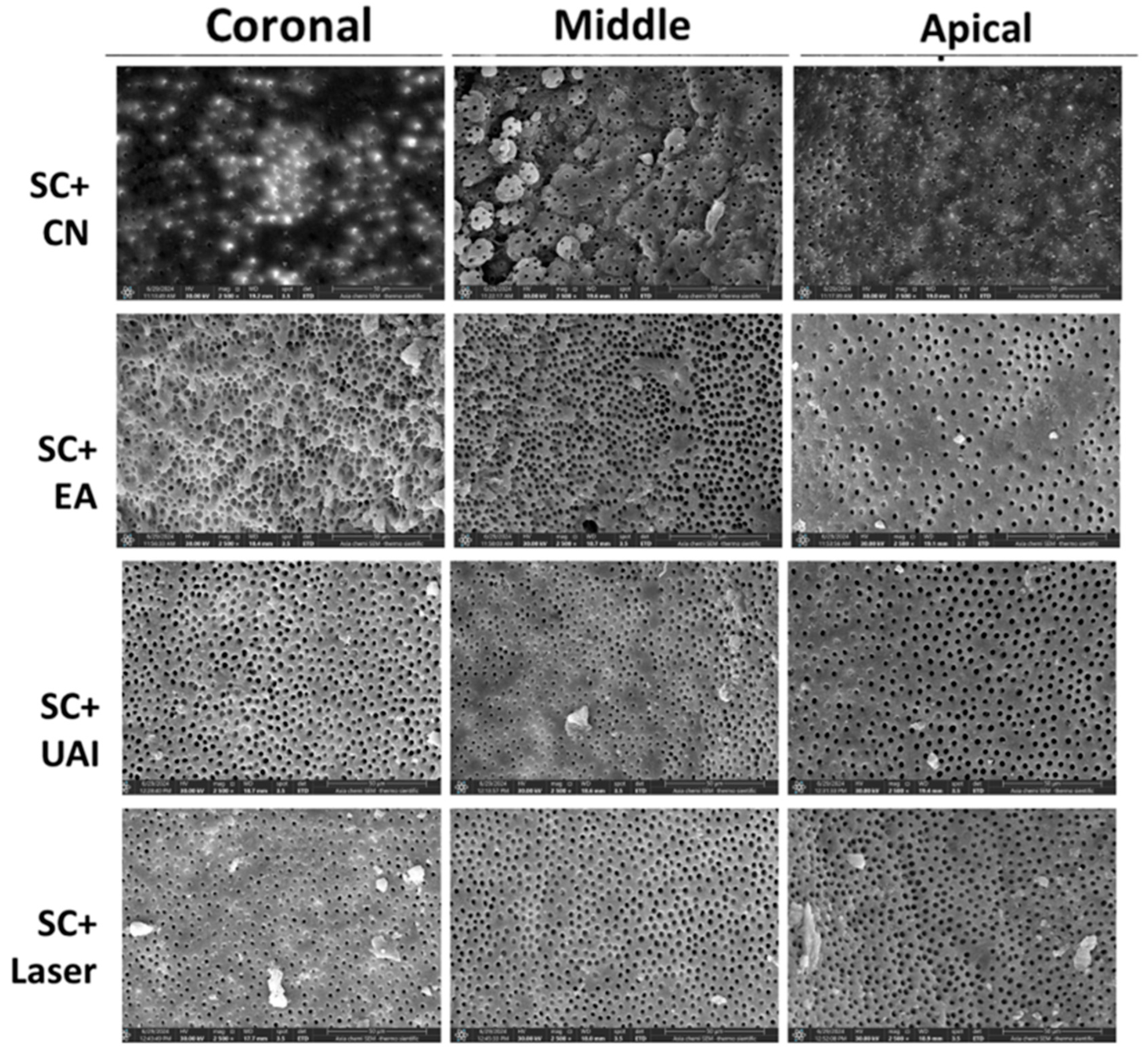
Figure 2.
Representative SEM images (magnification 2500 X) showing root canal lumen at coronal, middle, and apical sections using CC (continuous chelation) + CN (Conventional needle irrigation, EA (EndoActivator), UAI (Ultrasonic activated irrigation) and Er:Cr: YSGG laser.
Figure 2.
Representative SEM images (magnification 2500 X) showing root canal lumen at coronal, middle, and apical sections using CC (continuous chelation) + CN (Conventional needle irrigation, EA (EndoActivator), UAI (Ultrasonic activated irrigation) and Er:Cr: YSGG laser.
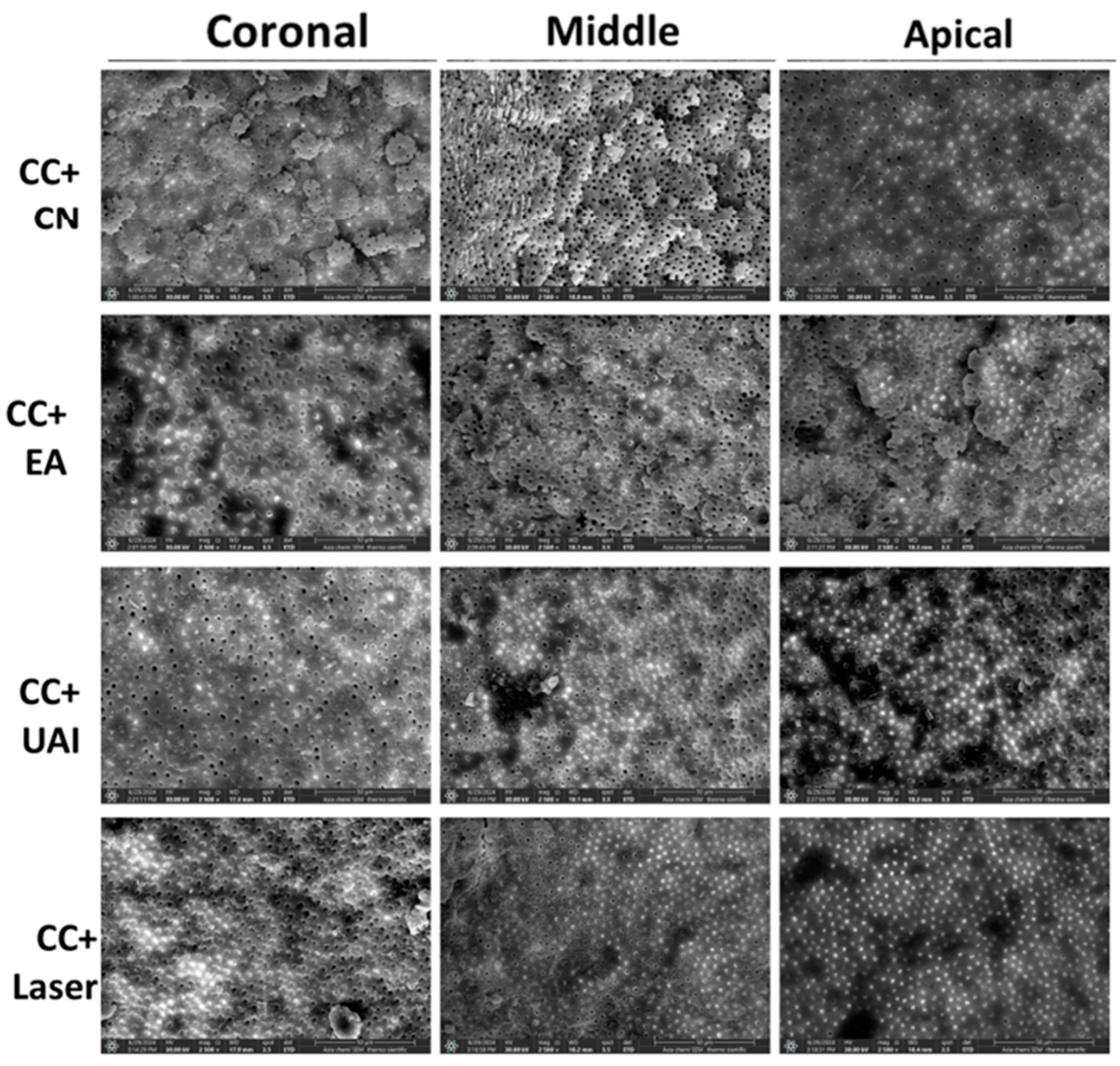
Figure 3.
Bar chart illustrating the mean rank of smear layer after SC (sequential chelation ) and CC (continuous chelation) in coronal, middle, and apical sections using CN (Conventional needle irrigation), EA (EndoActivator), UAI (Ultrasonic activated irrigation) and Er:Cr: YSGG laser.
Figure 3.
Bar chart illustrating the mean rank of smear layer after SC (sequential chelation ) and CC (continuous chelation) in coronal, middle, and apical sections using CN (Conventional needle irrigation), EA (EndoActivator), UAI (Ultrasonic activated irrigation) and Er:Cr: YSGG laser.

Figure 4.
Bar chart illustrating the mean microhardness of root canal dentin after sequential and continuous chelation in coronal, middle, and apical sections at 50 and 100 µm from the canal surface using: A (Conventional needle irrigation), B (EndoActivator), C (Ultrasonic activated irrigation) D (Er:Cr: YSGG laser).


Table 1.
The protocols of irrigation and agitation in each group of the study.
| Group | the sequence of irrigation and agitation protocol |
|---|---|
| SC+CN |
1. 2 mL 3% NaOCl for 1 minute after each instrument change. 2. 5 mL 17% EDTA for 1 minute. 3. final rinse of 5 mL distilled water for 1 minute. |
| SC+EA |
1. 2 mL 3% NaOCl for 1 minute after each instrument change. 2. 5 mL 17% EDTA for 1 minute with 3 cycles of agitation for 20 seconds each. 3. final rinse of 5 mL distilled water for 1 minute. |
| SC+UAI |
1. 2 mL 5.25% NaOCl for 1 minute after each instrument change. 2. 5 mL 17% EDTA for 1 minute with 3 cycles of agitation for 20 seconds each. 3. final rinse of 5 mL distilled water for 1 minute. |
| SC+Laser |
1. 2 mL 3% NaOCl for 1 minute after each instrument change. 2. 5 mL 17% EDTA for 1 minute with 3 cycles of agitation for 20 seconds each. 3. final rinse of 5 mL distilled water for 1 minute. |
| CC+CN |
1. 2 mL 3% NaOCl/9% Dual Rinse HEDP for 1 minute after each instrument change. 2. 5 mL 3% NaOCl/9%Dual Rinse HEDP for 1 minute. 3. Final rinse of 5 mL distilled water for 1 minute. |
| CC+EA |
1. 2 mL 3% NaOCl/9% Dual Rinse HEDP for 1 minute after each instrument change. 2. 5 mL 3% NaOCl/9%Dual Rinse HEDP for 1 minute with 3 cycles of agitation for 20 seconds each. 3. Final rinse of 5 mL distilled water for 1 minute. |
| CC+UAI |
1. 2 mL 3% NaOCl/9% Dual Rinse HEDP for 1 minute after each instrument change. 2. 5 mL 3% NaOCl/9%Dual Rinse HEDP for 1 minute with 3 cycles of agitation for 20 seconds each. 3. Final rinse of 5 mL distilled water for 1 minute. |
| CC+Laser |
1. 2 mL 3% NaOCl/9% Dual Rinse HEDP for 1 minute after each instrument change. 2. 5 mL 3% NaOCl/9%Dual Rinse HEDP for 1 minute with 3 cycles of agitation for 20 seconds each. 3. Final rinse of 5 mL distilled water for 1 minute. |
SC= sequential chelation, CC= continuous chelation, CN= conventional needle, EA= Endoactivator, UAI= ultrasonic activated irrigation.
Table 2.
Median and mean rank of smear layer scoring in the coronal, middle, and apical thirds of the root canal for all groups.
Table 2.
Median and mean rank of smear layer scoring in the coronal, middle, and apical thirds of the root canal for all groups.
| Group | Coronal | Middle | Apical | |||
|---|---|---|---|---|---|---|
| Median | Mean rank | Median | Mean rank | Median | Mean rank | |
| SC+CN | 2 | 23.00a | 2 | 20.44 | 2.5 | 22.19 |
| SC+EA | 1.5 | 17 | 1.5 | 13.5 | 2 | 15 |
| SC+UAI | 1.5 | 17 | 2 | 18.56 | 2 | 13.56 |
| SC+Laser | 1 | 9.00a | 1.5 | 13.5 | 2 | 15.25 |
| CC+CN | 2 | 17.63 | 2 | 16.81 | 3 | 24.88a,b |
| CC+EA | 2 | 16.13 | 2 | 15.25 | 2 | 11.75a |
| CC+UAI | 2 | 18.06 | 2 | 18.69 | 3 | 20.50c |
| CC+Laser | 2 | 14.19 | 2 | 15.25 | 2 | 8.88b,c |
*Kruskal Wallis test. Identical superscript lowercase letters indicate significant differences among relevant groups in each chelator (Mann-Whitney U test for pairwise comparison). Identical superscript lowercase letters indicate significant differences among relevant groups in between chelators (Mann-Whitney U test for pairwise comparison (p<0.05)). SC sequential chelation, CC continuous chelation, CN conventional needle, EA Endoactivator, UAI ultrasonic activated irrigation.
Table 3.
Average Vickers microhardness values (mean ± standard deviation) of dentin after different irrigating protocols. .
Table 3.
Average Vickers microhardness values (mean ± standard deviation) of dentin after different irrigating protocols. .
| Section and depth | Group | Mean (VHN) | SD | Group | Mean (VHN) | SD |
|---|---|---|---|---|---|---|
| Sequential Chelation | Continuous Chelation | |||||
| Coronal_50 | SC+CN | 58.84a | 18.69 | CC+CN | 79.45 | 6.87 |
| SC+EA | 34.34a,b | 6.49 | CC+EA | 59.96a | 18.85 | |
| SC+UAI | 43.72 | 8.99 | CC+UAI | 71.06b | 25.56 | |
| SC+Laser | 59.08b | 7.61 | CC+Laser | 87.48a,b | 14.81 | |
| Middle_50 | SC+CN | 59.45a | 14.93 | CC+CN | 75.57 | 6.84 |
| SC+EA | 33.04a,b | 9.87 | CC+EA | 61.15 | 22.87 | |
| SC+UAI | 43.39 | 6.71 | CC+UAI | 69.77 | 20.67 | |
| SC+Laser | 56.18b | 6.26 | CC+Laser | 85.40 | 8.62 | |
| Apical_50 | SC+CN | 60.29a | 16.79 | CC+CN | 78.66 | 0.51 |
| SC+EA | 36.42a,b | 6.46 | CC+EA | 64.16a | 13.10 | |
| SC+UAI | 44.07 | 1.94 | CC+UAI | 70.56b | 23.00 | |
| SC+Laser | 58.76b | 10.36 | CC+Laser | 90.06a,b | 20.64 | |
| Coronal_100 | SC+CN | 51.66a | 14.81 | CC+CN | 75.44 | 3.08 |
| SC+EA | 34.59a,b | 8.95 | CC+EA | 51.46a | 22.49 | |
| SC+UAI | 42.24 | 9.74 | CC+UAI | 58.97b | 24.97 | |
| SC+Laser | 53.34b | 7.23 | CC+Laser | 82.17a,b | 12.24 | |
| Middle_100 | SC+CN | 53.91a | 13.09 | CC+CN | 78.25 | 5.84 |
| SC+EA | 35.64a,b | 4.76 | CC+EA | 54.52 | 25.22 | |
| SC+UAI | 40.08 | 11.24 | CC+UAI | 66.30 | 19.32 | |
| SC+Laser | 50.62b | 5.18 | CC+Laser | 79.70 | 10.34 | |
| Apical_100 | SC+CN | 50.52a | 11.10 | CC+CN | 76.62a | 1.41 |
| SC+EA | 34.24a,b | 10.99 | CC+EA | 56.58a,b | 15.60 | |
| SC+UAI | 42.61 | 7.52 | CC+UAI | 66.11 | 21.94 | |
| SC+Laser | 52.84b | 5.05 | CC+Laser | 81.14b | 22.42 | |
*Kruskal Wallis test. Identical superscript lowercase letters represent significant differences among the relevant groups in each section and depth (Mann Whitney U test (p<0.05). SC sequential chelation, CC continuous chelation, CN conventional needle, EA Endoactivator, UAI ultrasonic activated irrigation.
Disclaimer/Publisher’s Note: The statements, opinions and data contained in all publications are solely those of the individual author(s) and contributor(s) and not of MDPI and/or the editor(s). MDPI and/or the editor(s) disclaim responsibility for any injury to people or property resulting from any ideas, methods, instructions or products referred to in the content. |
© 2025 by the authors. Licensee MDPI, Basel, Switzerland. This article is an open access article distributed under the terms and conditions of the Creative Commons Attribution (CC BY) license (http://creativecommons.org/licenses/by/4.0/).
Copyright: This open access article is published under a Creative Commons CC BY 4.0 license, which permit the free download, distribution, and reuse, provided that the author and preprint are cited in any reuse.


