Submitted:
23 April 2025
Posted:
23 April 2025
You are already at the latest version
Abstract
Background/Objectives: Beyond clinical areas, photon-counting detector (PCD) CT is innovatively applied to study paleontological specimens such as dinosaur fossils. The aim of this study was to compare PCD-CT with conventional energy-integrating detector (EID) CT for dinosaur fossil analysis. Methods: The lower jaw of a Tyrannosaurus and the head of a Camarasaurus were imaged using a PCD-CT in ultra high-resolution mode and an EID-CT. The PCD-CT and EID-CT image quality of the dinosaurs were visually assessed. Results: Compared with EID-CT, PCD-CT yielded higher-resolution anatomical images free of image deterioration; achieving better definition of the Tyrannosaurus mandibular canal and the three semicircular canals of a Camarasaurus. Conclusions: PCD-CT clearly depicts the internal structure and morphology of dinosaur fossils without damaging them and also provides spectral information allowing researchers to gain insights into fossil mineral composition and preservation state in the future.
Keywords:
PCT-CT
; paleontology
; dinosaur fossil
1. Introduction
Photon-counting detector (PCD) computed tomography (CT) is a next-generation imaging technology measuring X-ray photon energy levels that are converted into an electrical signal [1,2]. PCD-CT has many advantages over conventional energy-integrating detector CT (EID-CT) such as high spatial resolution, low electronic noise, improved contrast-to-noise ratio, and spectral information acquisition. There are many clinical applications of PCD-CT, from detection of minute calcifications in cardiovascular disease to precise cancer tumor mapping [3].
Beyond clinical areas, CT is innovatively applied to study paleontological specimens such as dinosaur fossils [4,5]. This non-destructive imaging technique enables internal fossil examination without requiring physical sectioning, preserving the integrity of these invaluable specimens. By leveraging PCD-CT’s high-resolution capabilities, researchers will gain insights into the internal morphology and pathology of fossils, shedding light on the life and environment of ancient organisms.
This paper reports our initial experience using PCD-CT in ultra high-resolution (UHR) mode for dinosaur fossil imaging, highlighting the synergistic benefits of this technology for both medical and paleontological research. Through an interdisciplinary approach, we illustrate PCD-CT’s potential for new discoveries in the field of paleontology.
2. Materials and Methods
2.1. Dinosaur Fossils
This study was approved by the Ethics Committee of the University of Fukui, Faculty of Medical Sciences (study protocol # 20220056, 29 June 2023). Two dinosaur fossils (the lower jaw of a Tyrannosaurus and the head of a Camarasaurus), stored in Fukui Prefectural Dinosaur Museum (Katsuyama City, Fukui, Japan) were carefully transported to University of Fukui Hospital.
The carnivorous Tyrannosaurus rex, often referred to as T. rex, lived during the late Cretaceous period, around 68 to 66 million years ago [6,7]. It is one of the most well-known dinosaurs, recognized for its large size and fearsome appearance. Adult T. rex specimens are recorded at lengths up to 40 feet (12 meters) and weighing up to 9 tons. The T. rex had a massive body, strong legs, and powerful jaws with sharp teeth. Studies of T. rex’s teeth and jaws suggest that it possessed a crushing bite force that indicates a diet likely including both flesh and bone. Paleontologists believe it was an apex predator, preying upon herbivorous dinosaurs and scavenging as well.
The Camarasaurus, often referred to as the ‘chambered lizard’, is a sauropod (long-necked) dinosaur that lived during the Late Jurassic period, around 155 to 145 million years ago [8,9]. This dinosaur is recognized for its large size and is the most common North American sauropod fossil. Adult Camarasaurus specimens are recorded at lengths up to 50 feet (15 meters) with estimated weights up to 20 tons. The Camarasaurus had a robust build, with a distinctive small, square head balanced by a long, heavy tail. Paleontologists believe that it fed on plant material by the shape and wear patterns of its teeth.
2.2. CT Systems
The first clinical PCD-CT system (NAEOTOM Alpha, Siemens Healthineers) with a large 82 cm aperture uses a dual-source geometry and 0.25-second gantry rotation time to provide 66-msec temporal resolution (isocenter) [10,11,12]. The two detector arrays consist of dedicated PCDs with 1.6 mm thick CdTe and have 50-cm and 36-cm scanning field of views, respectively. The detector pixel layout shows 0.151 × 0.176 mm2 subpixels (measured at isocenter). Due to the presence of collimator blades every 6 subpixels in the z-direction, the minimum effective section thickness is 0.2 mm in UHR mode and 0.4 mm in standard mode. The UHR mode features 120 detector rows (120 × 0.2 mm collimation) while the standard mode features 144 detector rows (144 × 0.4 mm collimation).
The Aquilion Lightning Helios i Edition, developed by Canon Medical Systems, is an 80-slice/160-slice EID-CT scanner with a 78 cm aperture and enables a high-speed helical scan of 0.5 mm × 80 slices.
3. Results
Imaging parameters for PCD-CT and EID-CT are listed in Table 1. The large 82 cm gantry bore of the PCD-CT was large enough to photograph part of the lower jaw bone of a Tyrannosaurus and the entire head of a Camarasaurus (Figure. 1A,B, respectively). Figure 1C shows a 3D image of the entire Camarasaurus head taken using PCD-CT.
Figure 2A,B show the sagittal sections of the Tyrannosaurus lower jawbone imaged with PCD-CT and EID-CT, respectively. Compared with EID-CT, PCD-CT more clearly depicts the cross-sectional patterns and the mandibular canal including a hyper-dense structure.
Figure 3A,B show the cross-sections of the Camarasaurus temporal bone imaged with PCD-CT and EID-CT, respectively. Compared with EID-CT, PCD-CT more clearly depicts the selected section of the semicircular canals.
4. Discussion
To the best of our knowledge, this is the first photon-counting CT imaging study of dinosaur fossils. This study demonstrates the significant advantages of PCD-CT over conventional EID-CT in imaging dinosaur fossils. The superior spatial resolution and contrast-to-noise ratio of PCD-CT allow for more detailed visualization of fossil internal structures such as the Tyrannosaurus mandibular canal and the semicircular canals of the Camarasaurus. Visualizing fine anatomical details with PCD-CT creates new possi-bilities for paleontological research. For example, detailed images of the mandibular canal in the Tyrannosaurus fossil may provide new information on the sensory capabilities and feeding behavior of this iconic dinosaur [13]. Similarly, the clear imaging of the semicir-cular canals in the Camarasaurus fossil may shed light on the balance and locomotion of this large sauropod.
Non-destructive PCD-CT imaging ensures that valuable fossils are studied in detail without the risk of damage. One additional benefit of PCD-CT is its ability to create effec-tive atomic number images from spectral information, which may be used for future analysis of fossil composition (i.e., the weathering phenomenon known as pyrite disease that affects fossils). Pyrite disease occurs when fossils are extracted from rock and placed in oxygen-rich environments or exposed to rising temperatures, which accelerates oxida-tion [14]. As a result, crystals of pyrite or marcasite (FeS2) grow within the fossil, causing it to deteriorate from the inside. Pyrite disease is a significant concern for the preservation of specimens, as it often affects dinosaur fossils. However, the progression of pyrite disease within fossils and the ultimate destruction it causes have not been thoroughly investigat-ed. The ability of PCD-CT to create effective atomic number images from spectral infor-mation provides great potential for non-destructive testing and analysis in paleontology, allowing researchers to gain insights into fossil mineral composition and preservation state without damaging them.
Despite these advantages, there are some limitations for PCD-CT in paleontology. The high cost and limited availability of PCD-CT scanners may restrict their use to well-funded research institutions. Additionally, PCD-CT image interpretation requires specialized knowledge and expertise, which may not be readily available in all paleonto-logical research settings. Thus, it is important for radiologists and paleontologists to col-laborate and complement each other’s knowledge to advance photon-counting CT re-search on dinosaur fossils.
5. Conclusions
This study highlights PCD-CT as a powerful tool for non-destructive imaging of di-nosaur fossils. The superior image quality and additional spectral information provided by PCD-CT enhances our understanding of fossil internal structure and composition, and paves the way for new discoveries in paleontology. Future research should focus on ex-panding the use of PCD-CT and developing standardized protocols for its application as a paleontological tool of discovery.
Author Contributions
Soichiro Kawabe, Hideki Hyodoh and Tetsuya Tsujikawa contributed to the study conception and design. Material preparation, data collection and analysis were performed by Tasuku Wakabayashi, Kenji Takata, Soichiro Kawabe, Masato Shimada, Takeshi Mugitani, Takuya Yachida, Rikiya Maruyama and Toshiki Tateishi. The first draft of the manuscript was written by Wakabayashi and Kenji Takata, and all authors commented on previous versions of the manuscript. All authors read and approved the final manuscript.
Funding
This research received no external funding.
Institutional Review Board Statement
The study was conducted in accordance with the Declaration of Helsinki, and approved by the Institutional Review Board (or Ethics Committee) of the University of Fukui, Faculty of Medical Sciences (study protocol # 20220056, 29 June 2023).
Informed Consent Statement
Not applicable.
Data Availability Statement
The files/data used to support the findings of this study are available from the corresponding author upon request.
Conflicts of Interest
The authors declare no conflicts of interest.
References
- Nakamura Y, Higaki T, Kondo S, Kawashita I, Takahashi I, Awai K. An introduction to photon-counting detector CT (PCD CT) for radiologists. Jpn J Radiol. 2023 Mar;41(3):266-282. [CrossRef]
- McCollough CH, Rajendran K, Leng S, Yu L, Fletcher JG, Stierstorfer K, Flohr TG. The technical development of photon-counting detector CT. Eur Radiol. 2023 Aug;33(8):5321-5330. [CrossRef]
- McCollough CH, Rajendran K, Baffour FI, Diehn FE, Ferrero A, Glazebrook KN, Horst KK, Johnson TF, Leng S, Mileto A, Rajiah PS, Schmidt B, Yu L, Flohr TG, Fletcher JG. Clinical applications of photon counting detector CT. Eur Radiol. 2023 Aug;33(8):5309-5320. [CrossRef]
- Schilling R, Jastram B, Wings O, Schwarz-Wings D, Issever AS. Reviving the dinosaur: virtual reconstruction and three-dimensional printing of a dinosaur vertebra. Radiology. 2014 Mar;270(3):864-71. [CrossRef]
- Bolliger SA, Ross S, Thali MJ, Hostettler B, Menkveld-Gfeller U. Scenes from the past: initial investigation of early jurassic vertebrate fossils with multidetector CT. Radiographics. 2012 Sep-Oct;32(5):1553-9. [CrossRef]
- Meiri, S. Population sizes of T. rex cannot be precisely estimated. Frontiers of Biogeography 2022, 14.2, e53781. [Google Scholar] [CrossRef]
- Marshall CR, Latorre DV, Wilson CJ, Frank TM, Magoulick KM, Zimmt JB, Poust AW. Absolute abundance and preservation rate of Tyrannosaurus rex. Science. 2021 Apr 16;372(6539):284-287. [CrossRef]
- Upchurch, P. The phylogenetic relationships of sauropod dinosaurs. Zool J Linn Soc-Lond 1998;124(1):43-103. [CrossRef]
- Ikejiri, T. Distribution and biochronology of Camarasaurus (Dinosaria, Sauropoda) from the Jurassic Morrison Formation of the Rocky Mountain Region. 2005 New Mexico Geological Society, Guidebook, 56th Field Conference, pp. 367-379.
- Rajendran K, Petersilka M, Henning A, Shanblatt ER, Schmidt B, Flohr TG, Ferrero A, Baffour F, Diehn FE, Yu L, Rajiah P, Fletcher JG, Leng S, McCollough CH. First Clinical Photon-counting Detector CT System: Technical Evaluation. Radiology. 2022 Apr;303(1):130-138. [CrossRef]
- Sartoretti T, Wildberger JE, Flohr T, Alkadhi H. Photon-counting detector CT: early clinical experience review. Br J Radiol. 2023 Jul;96(1147):20220544. [CrossRef]
- Sartoretti T, Landsmann A, Nakhostin D, Eberhard M, Roeren C, Mergen V, Higashigaito K, Raupach R, Alkadhi H, Euler A. Quantum Iterative Reconstruction for Abdominal Photon-counting Detector CT Improves Image Quality. Radiology. 2022 May;303(2):339-348. [CrossRef]
- Kawabe S, Hattori S. Complex neurovascular system in the dentary of Tyrannosaurus. Historical Biology, 2021;34(7),1137-1145. [CrossRef]
- Tacker, R. A review of “pyrite disease” for paleontologists, with potential focused interventions. Palaeontologia Electronica 2020;23:a44. [CrossRef]
Figure 1.
A part of the lower jawbone fossil of a Tyrannosaurus (a) and the entire head fossil of a Camarasaurus (b) placed on the PCD-CT bed, a 3D image of the entire head fossil of a Camarasaurus taken with PCD-CT (c).
Figure 1.
A part of the lower jawbone fossil of a Tyrannosaurus (a) and the entire head fossil of a Camarasaurus (b) placed on the PCD-CT bed, a 3D image of the entire head fossil of a Camarasaurus taken with PCD-CT (c).
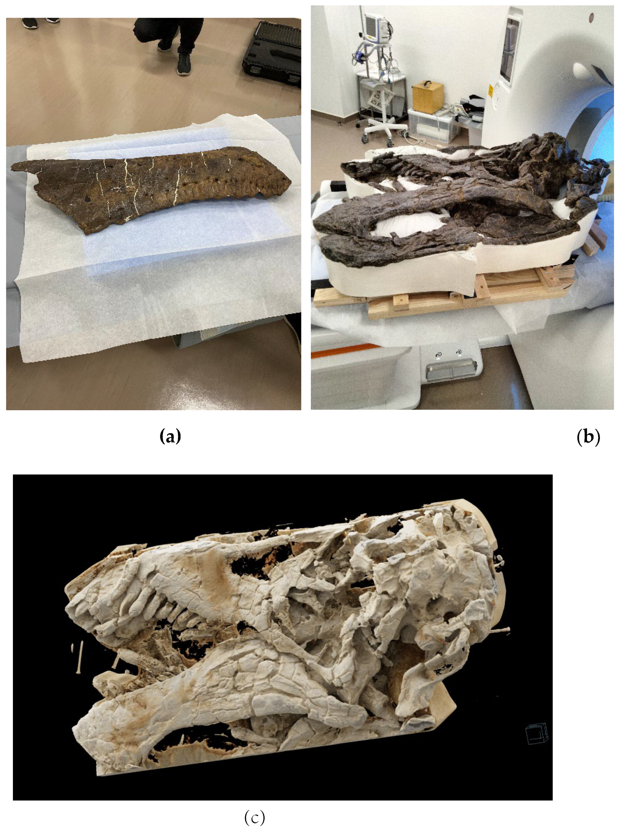
Figure 2.
Sagittal sections of the Tyrannosaurus lower jawbone imaged with PCD-CT and EID-CT.
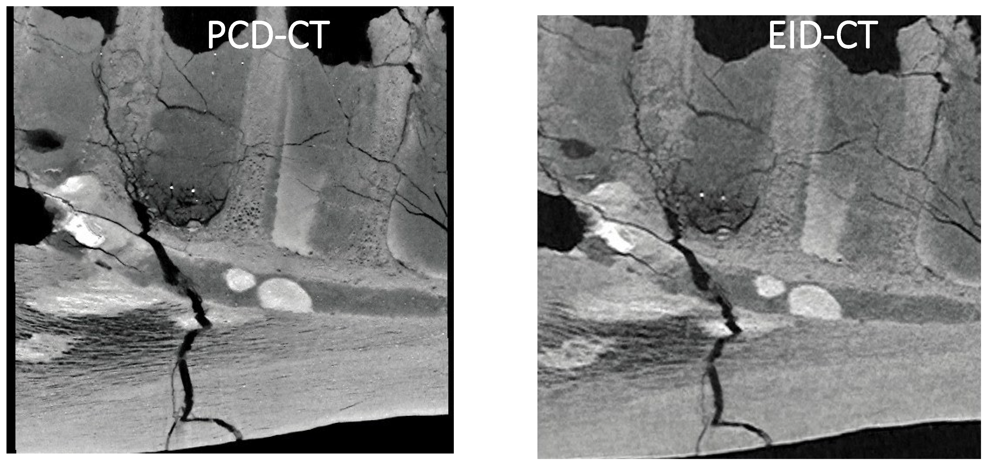
Figure 3.
Cross sections of the Camarasaurus temporal bone imaged with PCD-CT and EID-CT.
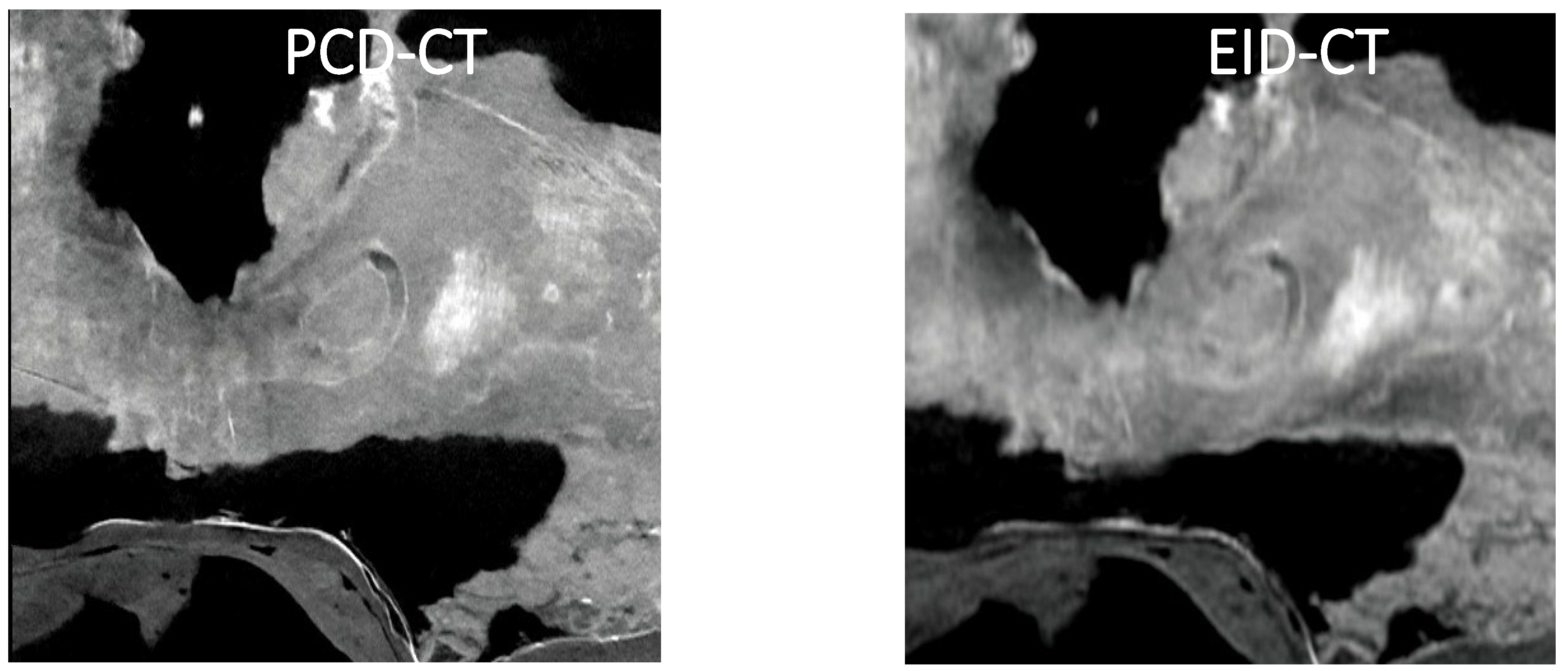

Table 1.
Experimental Details for dinosaur CT scans.
| T. rex | Camarasaurus | |||
| Parameters | PCD CT | EID CT | PCD CT | EID CT |
| Slice thickness (mm) | 0.2 | 0.5 | 0.2 | 0.5 |
| Slice interval (mm) | 0.15 | 0.3 | 0.15 | 0.3 |
| Rows | 120 | 80 | 120 | 80 |
| Rotation time (sec) | 1 | 0.75 | 1 | 1 |
| Helical pitch | 0.8 | 0.813 | 0.8 | 0.813 |
| kVp setting | 140 | 120 | 140 | 120 |
| CTDI vol (mGy) | 9.06 | 6.3 | 36.4 | 19.4 |
| DLP (mGy/cm) | 851 | 624.9 | 4509 | 2461.9 |
| Focus size | S | L | S | L |
| Output image | 70 keV | 120 kVp | 70 keV | 120 kVp |
| Matrix size | 1024 x 1024 | 512 x 512 | 1024 x 1024 | 512 x 512 |
| FOV (mm) | 320 | 320 | 500 | 500 |
Disclaimer/Publisher’s Note: The statements, opinions and data contained in all publications are solely those of the individual author(s) and contributor(s) and not of MDPI and/or the editor(s). MDPI and/or the editor(s) disclaim responsibility for any injury to people or property resulting from any ideas, methods, instructions or products referred to in the content. |
© 2025 by the authors. Licensee MDPI, Basel, Switzerland. This article is an open access article distributed under the terms and conditions of the Creative Commons Attribution (CC BY) license (http://creativecommons.org/licenses/by/4.0/).
Copyright: This open access article is published under a Creative Commons CC BY 4.0 license, which permit the free download, distribution, and reuse, provided that the author and preprint are cited in any reuse.


