Submitted:
21 April 2025
Posted:
27 April 2025
You are already at the latest version
Abstract
Among the several leech saliva extracts, around 20 bioactive molecules are widely known and identified with their mechanisms of action and therapeutic importance. Some of are Hirudin, Calin, Destabilase, Hyaluronidase, Apyrase, Hirustatin, Saratin, and Eglin. Thus, this review provides valuable insights into the most commonly known bioactive molecules in leech saliva, their putative medical importance, current status, and potential implication for future medical use. The practice of using leech in medical treatment, called hirudotherapy, has been in use for several centuries and can be drawn back to ancient Egyptians. They used it to treat various diseases like skin diseases, inflammation, and dental problems. Nowadays, these invaluable bioactive molecules of leeches have been utilized in modern medicine for the treatment of angina pectoris, tinnitus, phlebitis, gout, ischemic heart disease, asthma, cancer, hypertension, and migraines. Additionally, it is very effective in the treatment of mastitis, diabetic foot ulcers, cutaneous leishmaniosis, alopecia, and digital gangrene. The contribution of leeches in cosmetic and plastic surgery is also gaining popularity. At present time, the world’s most advanced nations with the most sophisticated in medical science have become practitioners. Despite its long history of medical application, there is still much to discover about the specific substance responsible for the valuable effects of leech saliva. Therefore, leech saliva holds enormous promise, and continued research will unlock its full potential.
Keywords:
bioactive substance
; leech
; leech therapy
; leech saliva
; putative medical importance
1. Introduction
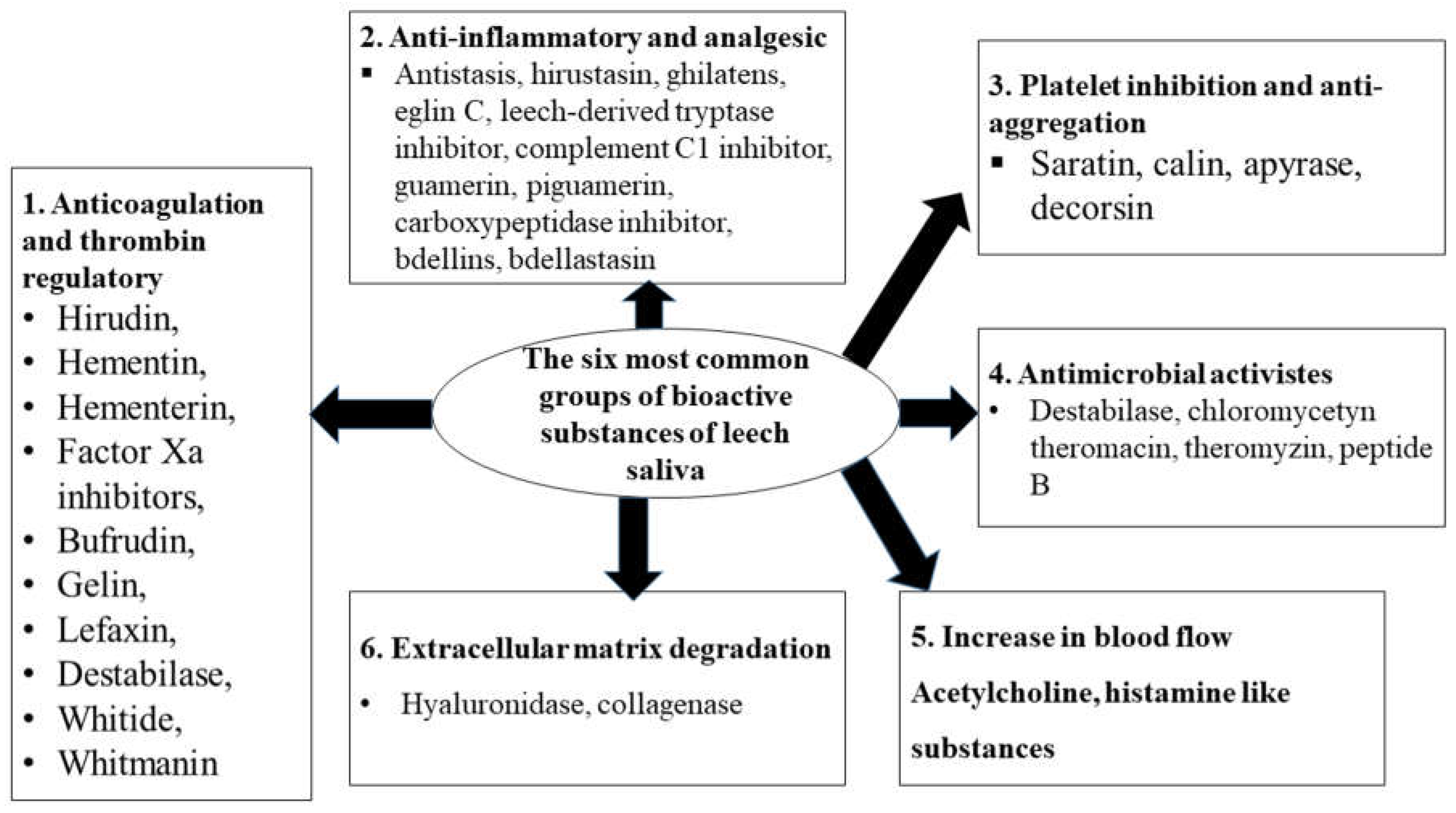
Leech saliva has been widely researched for its exceptional bioactive properties. Over the years, it has been discovered to contain a diverse collection of bioactive substances released during feeding (Hildebrandt and Lemke, 2011). More than 100 of these substances have been identified, although most remain poorly understood regarding their chemical structures, mechanisms of action, and therapeutic functions (Baskova et al., 2004, 2008; Liu et al., 2019). However, over 20 bioactive substances in leech saliva have been recognized for their significant therapeutic applications and mechanisms of action (Eldor et al., 1996; Sig et al., 2017). These substances include hirudin, apyrase, begins, bdellins, decorsin, hirustatin, saratin, hyaluronidase, calin, histamine-like substances, complement inhibitors, carboxypeptidase A inhibitors, destabilise, collagenase, piguamerin, tryptase inhibitors, hemetin, guamerin and hementerin (Alaama et al., 2024; Eldor et al., 1996; Zaidi et al., 2011). Generally, Based on their modes of action and therapeutic purposes, these bioactive molecules are divided into six kinds, which include analgesic and anti-inflammatory properties, anticoagulant effects, platelet inhibitory and thrombin regulatory roles, antibacterial actions, and extracellular matrix degradation (Hildebrandt and Lemke, 2011; Sudhadevi, 2021).
The bioactive compounds present in leech saliva have made leeches as valuable tools in medical treatment (Malik et al., 2022). The therapeutic application of leeches dates back to ancient times, with Egyptian, Indian, Greek, and Arab physicians employing them to treat various conditions ranging from bleeding to systemic ailments such as skin diseases, abnormalities, urinary infections, reproductive disorders, inflammation, and dental diseases (Munshi et al., 2008; Sharma and Jagdhane, 2020).
In contemporary medicine, clinicians and researchers cultivate leeches in sterile laboratory conditions. The bioactive substances in leech saliva are particularly favored in developed countries, including Germany, Israel, Russia, France, China, India, the UK, and the USA (Houschyar et al., 2015). Leech therapy has seen resurgence as an effective treatment for chronic and life-threatening diseases, including cancer, infectious diseases, and cardiovascular diseases. Additionally, it has become a valuable tool in plastic and microsurgery (Houschyar et al., 2015; Varsha et al., 2020).
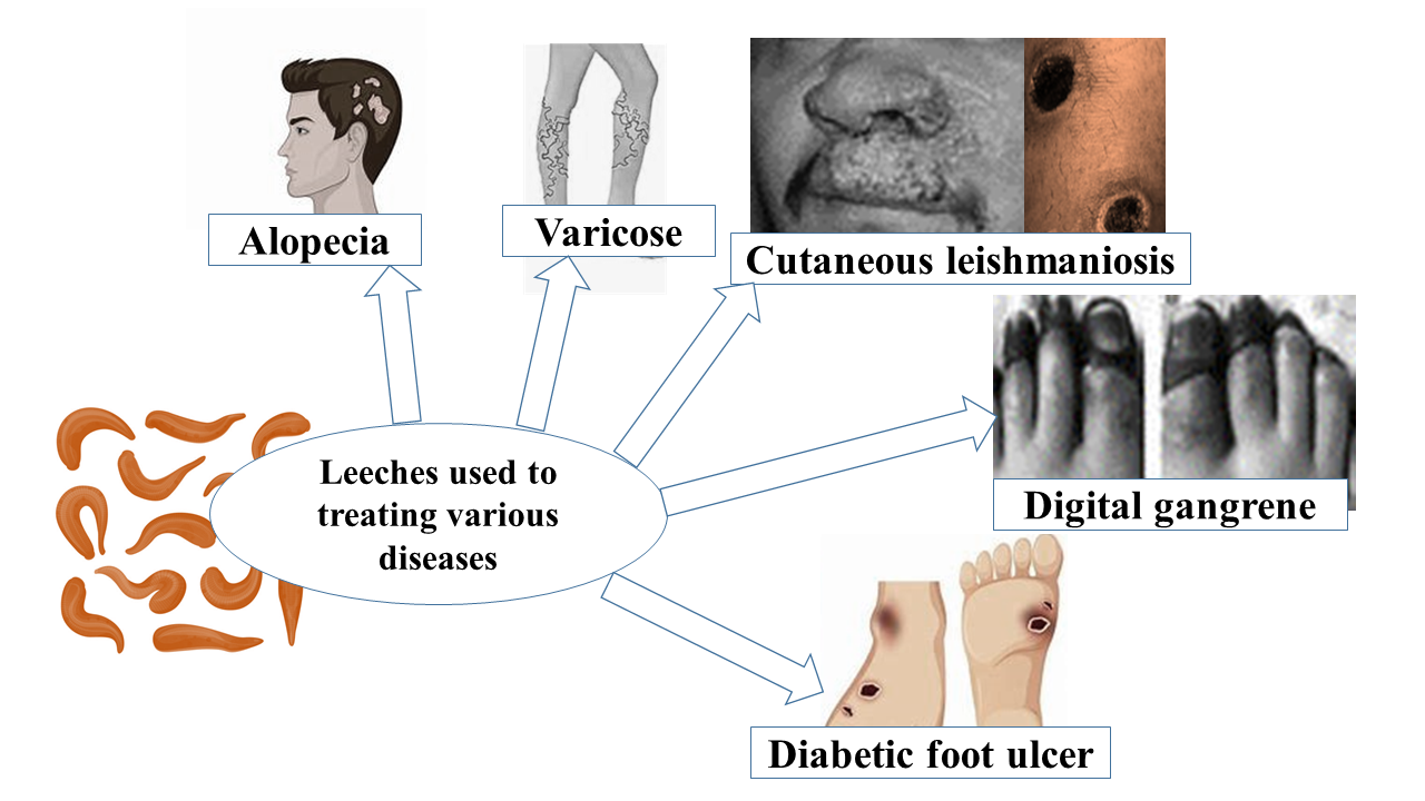
Moreover, leech therapy has proven effective in managing mastitis on farms (Malik et al., 2022). Currently, hirudotherapy plays a significant role in dermatology and cosmetology, particularly in anti-aging and anti-psoriatic treatments, contributing to youthful skin appearance (Abkowska et al., 2022). Additionally, it is employed to address nervous system abnormalities, various dental pains and issues, skin problems, and a range of painful conditions such as arthritis, migraines, and lower back pain (Kaliyaperumal et al., 2023). Recent studies have demonstrated the significance of hirudotherapy in treating diabetic foot ulcers, cutaneous leishmaniasis, alopecia, and digital gangrene (Dudhrejiya et al., 2023). In the 21st century, numerous clinics and healthcare professionals globally are incorporating this therapy in disease treatment, as well as in cosmetic and plastic surgery.
Leech therapy, utilizing leech bioactive substances, has a long-standing history in medicine. Despite its resurgence, there is still a need to fully understand the specific bioactive substances responsible for their therapeutic effects. A comprehensive understanding of these bioactive components can lead to novel therapeutic opportunities and optimized treatment outcomes. This review aims to bridge this gap by examining the most commonly studied bioactive compounds in leech saliva, consolidating existing knowledge, and providing a valuable resource for researchers, clinicians, and the global medical community. The current review intends to offer a thorough overview of the key bioactive components in leech saliva, their putative medical applications, and potential implications for current and future medical treatments.
2. Literature Review
2.1. General Description of Bioactive Substances of Leech Saliva and Their Putative Therapeutic Use
A complex mixture of chemicals with pharmacological and biological activity makes up leech saliva (Hildebrandt and Lemke, 2011). These substances facilitate leech feeding by anesthetizing the wound area and dilating blood vessels to enhance blood flow. Notable examples include acetylcholine substances, carboxypeptidase-A inhibitors together with histamine-like substances. Consequently, leech saliva has become a valuable tool in medicine (Gronwald et al., 2008; Malik et al., 2022). The therapeutic benefits of these compounds include anticoagulant (hirudin), vasodilatory, thrombolytic, anti-inflammatory, analgesic, anti-adhesive, anti-edematous effects, immune system enhancement, microcirculation improvement, tissue permeability restoration, hypoxia elimination, infarct and stroke relief, detoxification, extracellular matrix degradation, and antimicrobial effects (Eldor et al., 1996; Gileva and Mumcuoglu, 2013).
Most of the bioactive compounds in leech saliva are proteins and peptides (Alaama et al., 2011; Baskova et al., 2008). Over a hundred unique bioactive substances have been discovered through transcriptomic, genomic, and proteomic studies, adding to expanding knowledge of gene expression, regulation, and molecular processes. Besides, techniques such as two-dimensional SDS-PAGE, mass spectrometry (MS), and chromatography have been instrumental in characterizing the profiles of leech salivary gland secretions. However, the chemical structures and mode of action of many of these substances remain unclear (Baskova et al., 2004, 2008; Liu et al., 2019). Nonetheless, over 20 bioactive substances and their therapeutic applications have been identified and described (Abdualkader et al., 2013; Hildebrandt and Lemke, 2011). Based on their modes of action and therapeutic importance, these substances are divided into six kinds, as illustrated and modified by (Abkowska et al., 2022; Dudhrejiya et al., 2023; Hildebrandt and Lemke, 2011; Junren et al., 2021; Lemke and Vilcinskas, 2020; Sig et al ., 2017) in (Figure 1) below.
2.1.1. Hirudin
Hirudin is a highly powerful polypeptide, distinguished by its exceptional efficiency as a thrombin inhibitor when compared to other peptides with similar functions. It was originally purified from the saliva of Hirudo medicinalis, it comprises sixty-four to sixty-six amino acids with a molecular mass of approximately 7kDa. Hirudin's anticoagulant properties were first identified by Haycraft in 1884, and the word hirudin was assigned to Jacoby in 1904 (Dodt et al., 1984; Markwardt, 1991).
According to Nowak (2002), the N-terminus of natural hirudin consists of five neutral aquaphobic amino acid sequences, while the C-terminus contains 25 amino acid residues. Additionally, Tyr63' is a sulfated amino acid that increases the number of negatively charged amino acids. The polypeptide features three pairs of disulfide bridges (Cys6-Cys14, Cys16-Cys28, Cys22-Cys39) that form a compact structure capable of binding to the thrombin's active site. The C-terminal acidic amino acid residues bind to thrombin's fibrinogen binding site, thereby inhibiting thrombin's anticoagulant function (Nowak, 2002). Hirudin exhibits a high affinity and specificity in binding to thrombin, thereby neutralizing its enzymatic activity. Markwardt 1994 demonstrated that the inhibition of thrombin obstructs the conversion of fibrinogen to fibrin, preventing clot formation. Moreover, by inhibiting thrombin, hirudin also impedes thrombin-induced platelet activation through Protease-Activated Receptors (PARs), leading to a reduction in platelet aggregation at sites of vascular injury (Markwardt, 1994). The inhibition of thrombin by hirudin disrupts the feedback loops within the coagulation cascade. Thrombin, acting downstream of factor Xa, is essential for converting prothrombin to thrombin. By blocking thrombin, hirudin indirectly impacts the activation of factor Xa, as thrombin generation is a pivotal step in the propagation phase of coagulation involving factor Xa (Junren et al., 2021)
2.1.1.1. Putative Medical Use
Hirudin is the most commonly recognized and utilized bioactive substance, particularly for treating patients with disseminated intravascular coagulation syndrome and platelet abnormalities (Markwardt, 2002). Recent studies have significantly expanded the understanding and applications of hirudin and its derivatives. These include its roles in wound repair, antifibrosis, antidiabetic medication, antithrombotic agent, anti-hyperuricemia, and treatments for cerebral hemorrhage, IgA nephropathy, acute lung injury, and myocardial infarction (Junren et al., 2021). Furthermore, hirudin has demonstrated anti-tumor effects in various cancers, including liver, lung, breast, laryngeal, malignant glioma, and prostate cancer (Chuang et al., 2023; Esser et al., 2009; Green and Karpatkin, 2010).
2.1.2. Antistasin
Antistasin was first isolated from the salivary glands of the Mexican leech Haementeria officinalis with 119-amino acid and a molecular weight of 15 kDa by Tuszynski et al. (1987). However, recently identified in the saliva of the leech species Helobdella austinensis by Kwak et al. (2019). Antistasin includes five conserved cysteine residues and two glycine residues or six cysteines and two glycine residues (Kwak et al., 2019). It exerts anticoagulant effects by inhibiting the conversion of prothrombin to thrombin through its action on coagulation factor Xa (Kwak et al., 2019).
2.1.2.1. Putative Medical Use
Antistasin is a crucial component of leech saliva extract, known anti-inflammatory properties. The presence of platelet aggregation inhibitors, anticoagulants, and anti-proteolytic enzymes in the Haementeria officinalis saliva made it antistasin have antimetastatic activity (Tuszynski et al., 1987). Additionally, antistasin inhibits the kinin–kallikrein system, which is linked to the coagulation cascade and plays a significant role in the inflammatory response (Kashuba et al., 2013; Nutt et al., 1991). Beyond its role alongside hirudin, antistasin has been demonstrated to have antimetastatic effects, as well as anti-platelet and anti-thrombin effects (Tuszynski et al., 1987; Ammar et al., 2015).
2.1.3. Saratin
Saratin is a protein with a molecular mass of 12 kDa, consisting of 103 amino acids and three disulfide bridges, extracted from the saliva of the leech Hirudo medicinalis (Barnes et al., 2001). This bioactive molecule prevents thrombocyte aggregation by disrupting the initial binding stage of thrombocytes to the von Willebrand factor (vWF). It achieves this by binding to exposed type I and II collagen, thus preventing platelet aggregation. Consequently, saratin reduces thrombocyte aggregation to vessel walls and subsequent thrombus formation (Gronwald et al., 2008).
2.1.3.1. Putative Medical Use
Saratin is a potent therapeutic agent that locally prevents coagulation without affecting normal hemostatic functions (Vilahur et al., 2004). He demonstrated saratin's have antithrombotic effect on human atherosclerotic plaques. Additionally, animal studies suggest that recombinant saratin could be a promising treatment option (Gronwald et al., 2008).
2.1.4. Tryptase inhibitor
The leech-derived tryptase inhibitor (LDTI), originally isolated from the European medicinal leech Hirudo medicinalis, consists of 46 amino acids and has a molecular weight of 4.7 kDa (Di Marco and Priestle, 1997; Pohlig et al., 1996). Tryptase inhibitors have been shown to counteract the effects of various proteolytic enzymes, including chymosin, mast cell chymotrypsin, subtilisin protease, human blood neutrophil proteases, elastase, and cathepsin G. The complex structure of LDTI with inhibited proteinases serves as a model for developing low molecular weight tryptase inhibitors (Di Marco and Priestle, 1997). Campos et al. (2004) elucidated that LDTI functions by inhibiting mast cell proteolytic enzymes, particularly mast cell tryptase, with additional inhibitory effects on trypsin and chymotrypsin.
2.1.4.1. Putative Medical Use
The tryptase inhibitor holds promising effects for allergic and inflammatory diseases, including alleviating allergic reactions and anaphylaxis symptoms such as itching, swelling, and respiratory distress; and managing conditions like asthma, arthritis, and inflammatory diseases (Di Marco and Priestle, 1997; Pantojauceda et al., 2009). It is also important for wound healing and tissue repair processes and for alleviating associated pain from various conditions (Sig et al., 2017; Di Marco and Priestle, 1997).
2.1.5. Hyaluronidase
A hyaluronidase activity was first described in Hirudo medicinalis by Linker et al. (1957). It has a molecular weight of 27.5 Da and exhibits hydrolase activity, targeting glycosidic bonds (Hovingh and Linker, 1999). Its primary function involves the degradation of hyaluronan, a multifunctional polysaccharide found in the extracellular matrix of connective tissues (Shakouri et al., 2019). Menzel and Farr, (1998) have demonstrated that hyaluronidase acts as a spreading factor by digesting tissue hyaluronic acid and exhibiting antimicrobial activity, thereby increasing the viscosity of interstitial walls and leading to antibiotic effects. Consequently, this facilitates the infiltration and diffusion of other leech components into deep or congested tissues (Hovingh and Linker, 1999).
2.1.5.1. Putative Medical Use
In terms of medical applications, hyaluronidase plays a role in various biological processes, including wound healing by hyaluronidase cleaves hyaluronan and hence decreases the amount of hyaluronan. Its degradation of hyaluronan enhances connective tissue permeability and reduces body fluid viscosity. Hyaluronic acid, a polymer associated with tumor metastasis, may be influenced by hyaluronidase, potentially exerting anticancer activity by inhibiting tumor-promoting substances and slowing cancer progression (Hovingh and Linker, 1999; Shakouri et al., 2019). Furthermore, hyaluronidase facilitates the diffusion and penetration of pharmacologically active substances into tissues, particularly in joint pain management, and possesses antibiotic properties (Eldor et al., 1996; Glyova, 2005). Additionally, it is utilized to decrease venous congestion and exhibits antithrombotic, thrombolytic, hypotensive, anti-inflammatory, and bacteriostatic activities (Salzet, 2002).
2.1.6. Collagenase
Collagenase, a well-known bioactive compound extracted from the saliva of the medicinal leech Hirudo medicinalis by Rigb et al. 1987, possesses a molecular weight of approximately 100 kDa. As an enzyme, it exerts inhibitory effects on platelet functions and the coagulation cascade, with a specific target on collagen. Its primary function lies in facilitating tissue penetration and the dispersion of other bioactive molecules secreted by the leech (Sig et al., 2017).
2.1.6.1. Putative Medical Use
Collagenase aids in removing dead tissue from chronic wounds like ulcers, promoting healing by cleaning the wound bed. It also treats scars and keloids by breaking down excess collagen, improving their appearance, and reducing discomfort (Rigb et al., 1987; Sig et al., 2017).
2.1.7. Calin
Calin, an enzymatic protein secreted by the salivary glands of the medicinal leech Hirudo medicinalis, possesses a molecular weight of approximately 65,000 Da (Munro et al., 1991). Functionally analogous to saratin, this bioactive substance primarily binds to type I collagen thereby impeding von Willebrand factor (vWF) from binding to exposed collagen and inhibiting platelet aggregation (Harsfalvi et al., 1995).
2.1.7.1. Putative Medical Use
Calin exhibits significant therapeutic potential by extending bite wound duration to approximately 12 hours. This prolonged bleeding duration aids in wound cleansing, mitigating the risk of sepsis, particularly beneficial for the patient or victim animal (Gross and Roth, 2007). Bioactive compounds like calin in leech saliva can disrupt various aspects of clot formation or inhibition of platelet aggregation (Deckmyn et al., 1995). Overall, calin likely contributes significantly to inhibiting platelet aggregation as well as regulating blood clotting, improving blood flow, and restoring vascular permeability (Munro et al., 1991; Deckmyn et al., 1995).
2.1.8. Destabilase
Destabilase, an enzyme with a molecular weight of approximately 12.3 kDa, is produced by the medicinal leech Hirudo medicinalis and functions as a highly specific endo-ε-(γ-Glu)-Lys- isopeptidase (Zavalova et al., 1996). It uniquely hydrolyzes endo-isopeptide bonds formed by transglutaminases between the γ-carbamide group of glutamine and the ε-amino group of lysine (Aeschlimann and Paulsson, 1994). Notably, destabilase exhibits a distinct ability to monomerize D-dimers by cleaving Lys-isopeptide bonds between the γ chains of ε-(γ-Glu) D-dimers, altering the fibrinogen-fibrin product balance during fibrin degradation. This phenomenon promotes the formation of fibrin degradation products while inhibiting fibrinolysis activation, rendering destabilase a specific and effective modulator of the fibrinolysis process (Abdisa, 2018; Baskova et al., 2001).
2.1.8.1. Putative Medical Use
Destabilase displays antimicrobial activity by disrupting bacterial cellular components (Hildebrandt and Lemke, 2011) exerting beta glycosidase activity that directly targets beta 1-4 bonds crucial in the peptidoglycan layer of bacterial cell walls (Baskova et al., 2001; Zavalova et al., 2006). Furthermore, the enzyme's various isoforms with distinct capabilities and degradation actions on stabilized fibrin render it a potential candidate for evaluation as an anticoagulant agent (Abdisa, 2018; Baskova et al., 2001)
2.1.9. Apyrase
Apyrase, an enzyme first isolated from the saliva of the leech Hirudo medicinalis in 1987 by Rigbi et al. boasts a molecular weight of 45 kDa. Its primary function lies in strongly inhibiting ADP-induced platelet aggregation by acting as an ectonucleotidase. This enzymatic action involves the hydrolysis of nucleotide triphosphates (such as ATP and ADP) into their respective diphosphates (ADP and AMP), and further into monophosphates (AMP), thereby regulating platelet aggregation, blood clotting, and vascular tone. By reducing the concentration of ATP and ADP, both crucial in platelet activation and aggregation, apyrase indirectly counteracts the activities of ADP, arachidonic acid, platelet-activating factor, and epinephrine (Rigbi et al., 1987).
2.1.9.1. Putative Medical Use
Apyrase was used to enhancing tissue permeability, preventing platelet aggregation and excessive clotting during surgical procedures (Rigbi et al., 1987; Eldor et al., 1996). By preventing platelets from producing thromboxane, it inhibits the generation of thrombin-induced platelet aggregation and platelet-activating factor (Rigbi et al., 1996).
2.1.10. Eglin
Eglin C, the principal isoform within the natural protein family, comprises 70 amino acids with an approximate molecular weight of 8.1 kDa. It was purified from the saliva of the leech Hirudo medicinalis, as detailed by Rink et al. (1984). It classified within the potato type I proteinase inhibitor family, Eglin C vigorously inhibits various enzymes, including chymase, alpha chymotrypsin, subtilisin, elastase, and cathepsin G, with particular emphasis on its inhibition of human neutrophil elastase and cathepsin G—potent inflammatory mediators ( Rink et al., 1984).
2.1.10.1. Putative Medical Use
Eglin demonstrates a pivotal role in mitigating the levels of free oxygen radicals in neutrophils, thereby curbing tissue inflammation and damage (Rigbi et al., 1987). In an experimental animal model, Eglin has proven beneficial in managing shock and emphysema and has great potential as a therapeutic agent for inflammatory illnesses (Siebeck et al., 1992). Moreover, Eglin shows promise in managing diseases associated with neutral granulocytic proteinases (Seemuller et al., 1986). Leech therapy treats diabetic neuropathy by applying an anti-inflammatory effect on nerves through the saliva of Bdellins and Eglins (Dudhrejiya et al., 2023).
2.1.11. Bdellins
Bdellins, ranging from 8 to 10 kDa, are a group of proteinaceous enzymes secreted by leeches, notably Limnatis nilotica and Hirudo medicinalis, initially identified by (Fritz et al., 1969). Their amino acid sequence varies across leech species, influencing their functional properties and interactions with other molecules. Bdellins interfere with blood clotting by inhibiting enzymes involved in the coagulation cascade and exhibit potent inhibitory activity against the trypsin-like proteinase acrosin (Fritz et al., 1969; Seemuller et al., 1986).
2.1.11.1. Putative Medical Use
Leech-derived bdellins possess anti-inflammatory effects, aiding in reducing inflammation in conditions like joint pain or arthritis (Fritz et al., 1970). It plays a crucial role in inhibiting clotting during blood feeding and employs various mechanisms to prevent blood coagulation (Cheng et al., 2019). A case study done on diabetic foot ulcer patient by Dudhrejiya et al. (2023) showed that, Bdellins have an anti-inflammatory effect on nerves, which is used to correct Diabetic Neuropathy.
2.1.12. Decorsin
Decorsin, a protein identified from the American medicinal leech Macrobdella decora in 1990 by Seymour et al., has a molecular weight of around 4 kDa and consists of 39 amino acids. The mechanism of action is a strong inhibitor of platelet aggregation and an antagonist of platelet glycoprotein II b-III receptors (Seymour et al., 1990).
2.1.12.1. Putative Medical Use
Because of activated platelet glycoprotein IIb, decorsin acts as a fibrinogen receptor and von Willebrand factor, which makes it a potential drug for the treatment of acute coronary syndrome (Seymour et al., 1990). It has anticoagulant properties, effectively preventing the formation of blood clots and promoting improved blood circulation by regulating clotting processes (Krezel et al., 1994; Sig et al., 2017).
2.1.13. Hirustasin
Hirustasin, derived from the saliva of the leech species Hirudo medicinalis, is a protein consisting of 55 amino acids and possessing a molecular weight of 5866 Da. It was isolated by Sollner et al. (1994) and stands as the initial inhibitor of tissue kallikrein identified in leeches. Hirustasin exhibits robust inhibitory activity against trypsin, chymotrypsin, and neutrophil cathepsin G, in addition to its tight-binding inhibition of tissue kallikrein (Sollner et al., 1994). Despite its potent effects on tissue kallikrein, hirustasin lacks anticoagulant properties but demonstrates analgesic and anti-inflammatory attributes (Sig et al., 2017).
2.1.13. Putative Medical Use
The potential of hirustasin as a novel inhibitor of tissue kallikrein opens avenues for therapeutic interventions in various conditions. The tissue kallikrein/kinin system, pivotal in regulating normal blood pressure (Sollner et al., 1994; Ware and Luck, 2017).
2.1.14. Piguamerin
Piguamerin, a peptide initially identified in the blood-feeding Korean leech Hirudo nipponia, comprises 48 amino acid residues with a molecular mass of 5090 Da (Kim and Kang, 1998). They elucidated its role as a serine protease inhibitor, demonstrating its inhibitory effects on plasma and tissue kallikrein, as well as trypsin. Notably, it shares similarities with antistasin-type inhibitors in the spacing of ten cysteine residues but diverges from hirustasin and antistasin at residues surrounding Arg27, a common P1 reactive residue for these inhibitors (Kim and Kang, 1998)
2.1.14. Putative Medical Use
Piguamerin is a pivotal enzyme in the blood clotting cascade, rendering it valuable in preventing clot formation. It has anticoagulant properties and can enhance blood flow by impeding thrombosis and fostering vasodilation (Kim and Kang, 1998). It is a potential bioactive substances in leech secretions which has analgesic and ant-inflammatory effects Sig et al. (2017), which mean it modifying inflammatory responses.
2.1.15. Histamine-like Substances
Histamine-like substances, identified in the saliva of the medicinal leech Hirudo medicinalis with molecular weights of 111.1 Da (Baskova et al., 2008). While the specific amino acid sequences of these substances remain undisclosed, their bioactive nature facilitates the removal of congested blood from wounds. In addition to this, histamine-like substances acts as a vasodilator (Rahul et al., 2014).
2.1.15.1. Putative Medical Use
Histamine-like substances exert vasodilatory effects, augmenting blood flow to tissues. This property proves advantageous in conditions necessitating enhanced blood circulation, such as ischemic diseases and wound-healing processes (Baskova et al., 2008). Furthermore, a case study involving patients with diabetic foot ulcers receiving leech therapy revealed that the patient's wound healed in 30 days as a result of the presence of acetylcholine, carboxypeptidase A inhibitors, and histamine-like substances, which improve blood flow and reduce congestion to treat diabetic microangiopathy (Dudhrejiya et al., 2023).
2.1.16. Carboxypeptidase A Inhibitors
Carboxypeptidase A inhibitors, discovered from the saliva of the leech Hirudo medicinalis in 1998 by Reverter et al. a target enzymes like carboxypeptidases N and M, involved in kinin degradation, leading to B receptor agonism and subsequent bradykinin-induced inflammation (Kashuba et al., 2013). Although inhibition of carboxypeptidases by leech secretions does not affect bradykinin action via B2 receptors, it may be impeded by B1 receptors, which are associated with chronic inflammation, in contrast to B2 receptors linked to acute inflammation (Braun et al., 1987; Reverter et al., 1998).
2.1.16.1. Putative Medical Use
Carboxypeptidase-A inhibitors exhibit anti-inflammatory effects, particularly relevant in conditions like multiple sclerosis, asthma, and rheumatoid arthritis, associated with B1 receptor activity (Braun et al., 1987). This bioactive substance has a vasodilator effect which increase inflow of blood to wound site, that mean it reduce local swelling (Das, 2015). Therefore, it can get rid of collected blood in the wound which prevent flow of fresh oxygenated arterial blood from incoming area and providing wound with oxygen and nutrients, then tissue nearby wound will be delivered with oxygenated blood and nutrients (Rahul et al., 2014). According to a case study conducted by Dudhrejiya et al. (2023) on diabetic ulcers patients showed a correcting diabetic microangiopathy through an increasing blood circulation and lowering congestion, due to the presence of Carboxypeptidase A inhibitors together with histamine-like compounds in leech saliva.
2.1.17. Guamerin
Guamerin is a bioactive substance isolated from the saliva of the Korean medicinal leech Hirudo nipponia by (Kim and Lee, 1997) and it exhibits varying molecular weights across leech species and the amino acid sequence also varies among species (Jung et al., 1995). Guamerin selectively inhibits human leukocyte elastase and porcine pancreatic elastase (Jung et al., 1995; Kim and Lee, 1997).
2.1.17.1. Putative Medical Use
Guamerin's elastase inhibition presents a promising avenue for managing inflammatory conditions and potentially reducing tissue damage (Kim and Lee, 1997). It has analgesic and ant-inflammatory effects Sig et al. (2017), by modulating inflammatory responses, it may also contribute to pain relief, offering potential applications in managing certain vascular disorders.
2.1.18. Ghilantens
Ghilantens, identified by Blankenship et al. in 1990 from studies on Haementeria ghilianii saliva, exhibit varying molecular weights depending on the isoform, typically falling within the 10-20 kDa range. These bioactive substances, akin to antistasin, display anticoagulant properties by impeding factors crucial in blood clotting (Blankenship et al., 1990).
2.1.18.1. Putative Medical Use
Ghilanten is one of the most important bioactive substances of leech saliva which is used to suppress metastasis of melanoma, breast cancer, lung cancer, and prostate cancer in the experimental animal model (Cardin and Sunkara, 1994). it plays a pivotal role in the therapeutic mechanisms of leech therapy for inflammatory conditions because this bioactive molecule has both analgesic and anti-inflammatory importance as reviewed by Sig et al. (2017). Though, a small number of investigations indicated this bioactive substances has anticoagulant importance and the others its possible significance are debated due to absence of further findings (Blankenship et al., 1990; Moser et al., 1998).
2.1.19. Hementerin
Kelen and Rosenfeld successfully isolated Hementerin in 1975 from the saliva of Haementeria depressa with a molecular weight of approximately 80 kDa. Its precise amino acid sequence remains undisclosed. Hementerin exerts its anticoagulant effect by cleaving peptide bonds in fibrinogen and inhibiting thrombin activity, a pivotal enzyme in the coagulation cascade. This property facilitates prolonged blood-feeding by the leech from its host (Chudzinski-Tavassi et al., 1998).
2.1.19.1. Putative Medical Use
Hementerin helps to prevent blood clots by inhibiting platelet aggregation and the coagulation cascade (Chudzinski-Tavassi et al., 1998). Similarly, it is an effective inhibitor of human platelet aggregation, presumably through activation of the platelet’s nitridergic pathway (Chudzinski-Tavassi et al., 2003).
2.1.20. Complement C1 Inhibitor
In a significant study conducted by Baskova et al. (1988), a protein C1 component inhibitor was isolated and characterized from the saliva of the medicinal leech, Hirudo medicinalis. This protein was identified as a potent inhibitor of the complement system, particularly targeting the C1 component factor (Baskova and Zavalova, 2001), which typically has a molecular weight ranging from 60 to 70 kDa. The inhibition mechanism extends to several factors within the coagulation cascade and the kinin–kallikrein system, as observed by (Baskova and Zavalova, 2001).
2.1.20.1. Putative Medical Use
The complement C1 inhibitor can reduce inflammation by inhibiting the activation of the complement system, which plays a critical role in the body's inflammatory response. This makes it useful in treating inflammatory conditions such as rheumatoid arthritis and others where excessive inflammation is a problem (Baskova et al., 1988).
2.1.21. Poeciguamerin
Poeciguamerin was a bioactive peptide, purified from the saliva of leech Poecilobdella manillensis, with an estimated molecular weight of 6000 daltons and 57 amino acids with the sequence (Wang et al., 2023). Poeciguamerin serves as an anticoagulant by inhibiting thrombin, thus impeding clot formation and facilitating blood flow during leech feeding. Featuring an antistatic-like domain, it acts as a serine protease inhibitor, particularly targeting elastase while also moderately inhibiting FXIIa and kallikrein. Remarkably, it exerts analgesic effects through elastase inhibition and curtails coagulation via FXIIa and kallikrein inhibition (Wang et al., 2023).
1.1.1.1. Putative Medical Use
Poeciguamerin's anticoagulant properties make it promising for preventing thrombosis, enhancing blood circulation, and supporting wound healing processes (Wang et al., 2023).

Table 1.
The most known and identified bioactive molecules of leech with their mechanisms of action, therapeutic importance, and species of leeches to be extracted.
Table 1.
The most known and identified bioactive molecules of leech with their mechanisms of action, therapeutic importance, and species of leeches to be extracted.
| Name of bioactive substances of leech | Species of leech | Mode of action | Significances/effects |
| Hirudin | Hirudo medicinalis | Act on thrombin | Anticoagulant effect |
| Antistasin | Haementeria officinalis | Inhibits Factor Xa, antimetastatic | Analgesic and anti-inflammatory effects |
| Saratin | Hirudo medicinalis | Inhibits the binding of von Willebrand factor to collagen | Inhibition of platelet function |
| Leech-derived tryptase inhibitor | Hirudo medicinalis | Inhibit proteolytic host mast cells | Analgesic and anti-inflammatory effects |
| Hyaluronidase | Hirudo medicinalis | Hydrolase activity, acting on glycosylic bonds | Extracellular matrix degradation |
| Collagenase | Hirudo medicinalis | Reduces collagen | Extracellular matrix degradation |
| Calin | Hirudo medicinalis | Prevents the binding of von Willebrand factor to collagen | Inhibition of platelet function |
| Destabilase | Hirudo medicinalis | Cleavage of isopeptide bonds in stabilized fibrin, thrombolysis | Anticoagulant effect Antimicrobial effect |
| Apyrase | Hirudo medicinalis | Action on adenosine 5’ diphosphate, arachidonic acid, platelet-activating factor, and epinephrine | Inhibition of platelet function |
| Eglin | Hirudo medicinalis | Inhibit alpha-chymotrypsin, chymase, subtilisin, elastase, and cathepsin-G | Analgesic and anti-inflammatory effects |
| Bdellins | Limnatis nilotica | Inhibits trypsin, plasmin, acrosin | Analgesic and anti-inflammatory effects |
| Decorsin | Macrobdella decora | Acts as an antagonist of platelet glycoprotein II b-III a | Inhibition of platelet function |
| Hirustasin | Hirudo medicinalis | Act on kallikrein, trypsin, chymotrypsin | Analgesic and anti-inflammatory effects |
| Piguamerin | Hirudo Nippon. | Inhibits plasma, tissue kallikrein, and trypsin | Analgesic and anti-inflammatory effects |
| Histamine-like substance | Hirudo medicinalis | Act as a vasodilator | Increases the inflow of blood |
| Carboxypeptidase A inhibitor | Hirudo medicinalis | Act in kinin degradation, resulting in agonism of B receptors | Analgesic and anti-inflammatory effects |
| Acetylcholine | Hirudo medicinalis | Act as a vasodilator | Increases the inflow of blood |
| Guamerin | HirudoNippon | Inhibit elastase | Analgesic and anti-inflammatory effects |
| Ghilantens | Haementeria ghilianii | Inhibits Factor Xa, antimetastatic | Analgesic and anti-inflammatory effects |
| Hementerin | Haementeria depressed | Plasminogen activator | Anticoagulant effect |
| Theromin | Theromyzon tessulatum | Acts on thrombin | Anticoagulant effect |
| Complement C1 inhibitor | Hirudo medicinalis | Blocks the activation of the classical pathway of the complement system | Analgesic and anti-inflammatory effects |
| Poeciguamerin | Poecilobdella manillensis | Acts as a serine protease inhibitor | Anticoagulant effect |
| Haemadin | Haemadipsa sylvestris | Acts on thrombin | Anticoagulant effect |
| Hementin | Haementeria ghilianii | Degrades fibrinogen and fibrin Inhibits tumor spread and metastasis | Anticoagulant effect |
Modified from (Gross and Roth, 2007:Liu et al., 2019; Sig et al., 2017; Zaidi et al., 2011).
2.2. Current Status and Future Prospective
The ancient practice of utilizing leeches in medical treatments, known as hirudotherapy, dates back centuries, with roots tracing to civilizations like the Egyptians, Indians, Greeks, and Arabs. Over time, this therapeutic approach has evolved to encompass a wide range of conditions, from bleeding disorders to systemic ailments affecting the skin, nervous system, urinary and reproductive systems, inflammation, and even dental issues (Munshi et al., 2008; Sharma andJagdhane, 2020).
In contemporary times, the breeding of leeches under controlled, hygienic conditions in laboratory settings have become commonplace. This has been spurred by the recognition of the beneficial bioactive compounds present in leech saliva across many nations. Regulatory bodies in various countries oversee the use of medicinal leeches, with notable examples including the Food and Drug Administration (FDA) in the United States and healthcare regulators in Germany, which classified leeches as medical devices and medicinal products, respectively, in 2004 (Gileva and Mumcuoglu, 2013).
More recently, countries like Turkey have introduced regulations to govern the practice of traditional and complementary medicine, including hippotherapy. This reflects a growing recognition of the therapeutic potential of leeches in modern medical treatments (Yapici et al., 2017). Advanced economies such as Germany, Israel, Russia, France, China, the United Kingdom (UK), and the USA are at the forefront of leveraging the benefits of leeches in medical interventions (Houschyar et al., 2015).
Various investigations have explored the therapeutic potential of bioactive substances found in leech saliva across different studies and years. Michalsen et al. (2007) illustrated the efficacy of leech therapy in rapidly alleviating pain associated with knee osteoarthritis. Subsequent research on 52 outpatients with active knee osteoarthritis revealed significant improvements in function and pain reduction following leech therapy compared to transcutaneous electrical nerve stimulation (Stange et al., 2012).
Moreover, studies have highlighted the antimicrobial properties of leech saliva against a spectrum of pathogens, including Gram-negative and Gram-positive bacteria. Tasiemski and Salzet (2012) reported potent antibacterial effects against pathogens like Shewanella and Aerococcus viridans, with therapeutic implications for arthritis, foodborne illnesses, and hospital-acquired infections. In surgical contexts, leech therapy finds application in addressing venous congestions in microvascular replantation, reconstructive surgery, traumatology, neurology, dermatology, and gynecology, yielding favorable outcomes (Abdullah et al., 2012). Plastic surgeons utilize leech therapy in post-reconstructive surgery to mitigate venous congestion in skin grafting and manage polycythemia, effectively resolving accumulated blood pooling in wounds, which can otherwise impede fresh blood flow and elevate venous blood pressure (Buote, 2014).
Leech therapy extends its benefits to microcirculatory disorders, correcting vascular permeability issues in organs and tissues, alongside inducing vasodilation, analgesia, and anti-inflammatory, and anticoagulant effects (Yapici et al., 2017). Additionally, (Abdisa, 2018) highlighted its successful application in animal wound healing postoperative surgeries and skin grafting in dogs, cats, and horses, with notable anti-tumor effects observed in vitro and animal experiments, effectively impeding tumor growth, invasion, and metastasis (Shakouri et al., 2019).
Moreover, leech therapy has emerged as a promising treatment modality for chronic and life-threatening diseases, including cancer, infectious diseases, cardiovascular issues, and metastasis (Varsha et al., 2020). Its integration into plastic and microsurgery has positioned it as a pivotal tool for enhancing patient outcomes (Sudhadevi, 2021). Modern medicine has expanded the application of medicinal leeches to address various venous ailments such as thrombophlebitis, angina pectoris, arthritis, hematomas, and tinnitus (Sudhadevi, 2021). Notably, leech therapy has found utility in mastitis control on farms due to the antimicrobial and antioxidant properties of leech saliva, effectively managing subclinical mastitis and other animal ailments (Malik et al., 2022). Additionally, its significance in dermatology and cosmetology is underscored by its potential in anti-aging and anti-psoriatic therapies, contributing to youthful skin maintenance (Abkowska et al., 2022).
Recent research highlights novel applications of leech therapy, such as inhibiting breast cancer cell growth through liposome-leech protein targeting, achieving remarkable efficacy with a 97% kill rate (Chuang et al., 2023). Furthermore, it is employed in managing nervous system abnormalities, various dental issues, skin disorders, and infections, offering relief from migraine, headaches, and upper back pain (Kaliyaperumal et al., 2023). Poeciguamerin, a bioactive peptide identified from the salivary secretions of Poecilobdella manillensis recently, exhibits promising elastase inhibition, along with potent analgesic and antithrombotic properties, making it a valuable asset for post-surgical pain and thrombosis management (Wang et al., 2023). Similarly, recent studies endorse leech therapy as a time-saving, cost-effective, and well-received intervention for diabetic foot ulcers, cutaneous leishmaniasis, alopecia, and digital gangrene (Dudhrejiya et al., 2023). As a result, leech extract stands as a valuable repository of bioactive compounds with wide-ranging therapeutic potential for both mild and severe medical conditions (Alaama et al., 2024).
Generally, Leech therapy, once a widely embraced practice, has traversed a trajectory from prominence to inconspicuousness and back again. Compared to conventional pharmaceutical interventions, leech therapy offers a swift learning curve and holds the potential for mitigating the complications associated with synthetic drugs. Presently, extensive research endeavors are underway to unveil the therapeutic prowess of leeches across diverse medical scenarios, making them a focal point for global scientific inquiry. As scientists delve deeper into the intricate composition of leech saliva and its therapeutic implications, the landscape of leech therapy continues to evolve. Recent applications in cosmetics and plastic surgery underscore the promising prospects of leech therapy in contemporary medicine. This ongoing exploration underscores the untapped potential of leech saliva and its bioactive constituents, signaling a pathway for future advancements in medical therapeutics.
3. Conclusion and Recommendations
Leech saliva harbors a numerous of profoundly impactful bioactive compounds, each possessing invaluable therapeutic potential across a spectrum of medical conditions. While more than 20 of these substances have been identified and elucidated for their mechanisms of action and clinical utility, countless others await discovery. Among the well-established entities are Hirudin, Calin, Destabilase, and Hyaluronidase, which have traversed centuries of medicinal practice and persist in contemporary medicine. As ongoing research endeavors shed light on the multifaceted capabilities of these bioactive agents, their significance in addressing diverse diseases and health concerns becomes increasingly evident. Presently, economically developed nations leverage these therapies in the management of ailments ranging from cancer and tinnitus to ischemic heart disease, migraines, diabetic foot ulcers, cutaneous leishmaniosis, alopecia, and digital gangrene, even extending to mastitis. However, the utilization of leech therapy remains largely untapped in developing nations, presenting a significant limitation. Despite this, the profound potential of leech saliva remains undisputed, with continued exploration poised to unlock its full therapeutic ability.
Based on the above conclusion the following recommendation should forwarded

- The researchers and physicians should prepare guidelines for leech therapy protocols, dosage, and safety measures.
- The clinician and researchers should conduct rigorous clinical trials to validate the efficacy of therapeutic applications of leech saliva.
- Recently, the therapeutic applications of leech have been practiced only in advanced countries, so it should be distributed and applied in other countries, particularly in developing ones.
- The researcher should do further investigation on the mechanisms of action, pharmaceutical activities, and therapeutic applications of all other bioactive substances in leech saliva.
Author contributions
Seid Kassaw Ali: Methodology, Searching, Supervision, Conceptualization, Writing – Review and Editing, Writing – Original Draft. Ahmed Geto Wunu: Writing – Review and Editing, Writing – Original Draft. All authors read and approved the final manuscript.
Funding
This review article did not receive any specific grant from funding agencies in the public, commercial, or not-for-profit sectors.
Availability of data and materials
The datasets for the current study are available from the corresponding author upon request.
Consent for publication
Not applicable
Acknowledgments
Not applicable
Conflicts of interest
The authors declare that there are no conflicts of interest.
References
- Abdisa, T. (2018). Therapeutic importance of leech and impact of leech in domestic animals. MOJ Drug Design Development and Therapy, 2(6), 235–242. [CrossRef]
- Abdualkader, A. M., Ghawi, A. M., Alaama, M., Awang, M., and Merzouk, A. (2013). Leech Therapeutic Applications. Indian J Pharma.Sci., 127–137.
- Abdullah, S., Dar, L. M., Rashid, A., and Tewari, A. (2012). Hirudotherapy / leech therapy : applications and indications in surgery. Archives of Clin. Experi. Surgery, 1, 172–180. [CrossRef]
- Abkowska, E. Z., Olga, C. ´nska-L., Magdalena, B., and Anna, P. (2022). Case reports and experts opinions about current use of leech therapy in dermatology and cosmetology. Cosmetics Case, 9(137), 1–11. [CrossRef]
- Aeschlimann, D., & Paulsson, M. (1994). Thromb. Haemostasis. 71, 402–415.
- Alaama, M., AlNajjar, M., and Abdualkader, A. (2011). Isolation and analytical characterization of local Malaysian leech saliva. IIUM Engineering J., 12(4), 32–40.
- Alaama, M., Kucuk, O., Bilir, B., and Merzouk, A. (2024). Pharmacological research - modern Chinese medicine development of leech extract as a therapeutic agent : A chronological review. Pharma. Res. Modern Chinese Med., 10, 1–8. [CrossRef]
- Ammar, A., Hassona, M., Meckling, G., Chan, L., Chin, M., and Abdualkader, A. (2015). Assessment of the antitumor activity of leech (huridinaria manillensis) saliva extract in prostate cancer. Cancer Res., 75(15). [CrossRef]
- Barnes, C., Krafft, B., and Frech, M. (2001). Production and characterization of saratin, an inhibitor of von Willebrand factor-dependent platelet adhesion to collagen. Semin. Thromb .Hemost., 27, 337–348.
- Baskova, I., Kostrjukova, E., Vlasova, M., Kharitonova, O., Levitskiy, S., Zavalova, L., Moshkovskii, S., and Lazarev, V. (2008). Proteins and peptides of the salivary gland secretion of medicinal leeches Hirudo verbana, H. medicinalis, and H. orientalis,. Biochem. Mosc., 73(3), 315–320.
- Baskova, I. P., Ferner, Z., Balkina, A. S., Kozin, S. A., Kharitonova, O. V., Zavalova, L. L., and Zgoda, V. G. (2008). Steroids, histamine, and serotonin in the medicinal leech salivary gland secretion. Biochemistry (Moscow) Supplement Series B: Biomed. Chem., 2(3), 215–225. [CrossRef]
- Baskova, I. P., Nikonov, G. I., and Khalil, S. (1988). Thrombolytic agents of the preparation from the medicinal leeches. Folia Haematol, 115, 166–170.
- Baskova, I. P., Zavalova, L. L., Basanova, A. V, and Sass, A. V. (2001). Separation of monomerizing and lysozyme activities of destabilase from medicinal leech salivary gland secretion. Biochem.(Moscow), 66(12), 1368–1373.
- Baskova, I., and Zavalova, L. (2001). Proteinase inhibitors from the medicinal leech Hirudo medicinalis. Biochem. (Mosc), 66, 703–714.
- Baskova, I., Zavalova, Z., Basanova, A., Moshkovskii, S., and Zgoda, V. (2004). Protein profiling of the medicinal leech salivary gland secretion by proteomic analytical methods. Biochem. Mosc., 69(7), 770–775.
- Blankenship, D., Brankamp, R., Manley, G., and Cardin, A. (1990). Amino acid sequence of ghilanten: anticoagulant-antimetastic principle of the south American leech, Haementeria ghilianii. B. Iochemical and Biophysical Res. Communucation, 166, 1384–1389.
- Braun, N. J., Bodmer, J. L., Virca, G. D., Μvirca, G., Maschler, R., C., and Schnebli, H. P. (1987). Kinetic studies on the interaction of eglin c with human leukocyte elastase and cathepsin G. 368, 299–308.
- Buote, N. (2014). The use of medical leeches for venous congestion. Vet. Comparative Orthopedics and Traumatology., 27(3), 173‒178.
- Campos, I., Silva, M., Azzolini, S., Souza, A., Sampaio, C., and Fritz, H. (2004). Evaluation of phage display system and leech-derived tryptase inhibitor as a tool for understanding the serine proteinase specificities. Arch Bioche. Biophys., 425, 87–94.
- Cardin, A., and Sunkara, S. (1994). Ghilanten antimetastatic principle from the south American leech Haementeria ghilianii. European Patent No. EP 0404055. Munich, European Patent Office, issued March 2, 1994. Germany.
- Chuang, C., Lingling, M., Xiangshen, L. I., Li, P., Azmir, N., Hashim, A., and Jumuddin, F. A. (2023). The actions of leech saliva components and their mechanisms in antitumor activity. Journal of Propulsion Techno., 44, 3092–3101.
- Chudzinski-Tavassi, A. M., Bermejo, E., Rosenstein, R. E., Faria, F., Keller Sarmiento, M. I., Alberto, F., Sapaio, M. U., and Lazzari, M. A. (2003). Nitridergic platelet pathway activation by hementerin, a metalloprotease from the leech Haementeria depressa. Biologi. Chem., 384(9). 1333–1339. [CrossRef]
- Chudzinski-Tavassi, A., Kelen, E., de Paula, R. A., Loyau, S., Sampaio, C., and Bon, E. (1998). Angles-Cano, Fibrino (geno) lytic properties of purified hementerin, a metalloproteinase from the leech Haementeria depressa,. Thromb.Haemost., 80(07), 155–160.
- Das, B. K. (2015). An Overview on Hirudotherapy / leech therapy. Indian J. Pharma. Sci., 1(1), 32–45.
- Di Marco, S., and Priestle, J. P. (1997). Structure of the complex of leech-derived tryptase inhibitor (LDTI) with trypsin and modeling of the LDTI-tryptase system. Structure, 5(11), 1465–1474. [CrossRef]
- Dodt, J., Müller, H. P., Seemüller, U., and Chang, J. Y. (1984). The complete amino acid sequence of hirudin, a thrombin specific inhibitor: Application of color carboxymethylation. Febs Lett. 165, 180–184. [CrossRef]
- Dudhrejiya, A. V, Pithadiya, S. B., Patel, A. B., and Amitkumar, J. (2023). Medicinal leech therapy and related case study : Overview in current medical field. J. Pharmacogn. Phytochem., 12(1), 21–31.
- Eldor, A., Orevi, M., and Rigbi, M. (1996). The role of the leech in medical therapeutics. Blood Reviews, 10(201–209), 201–209.
- Esser, D., Lange, W., Schröder, J., and Wiswedel, I. (2009). Antitumor activity of leech therapy in an animal model of prostate cancer. J. Complem. Integ. Med., 6(1).
- Fritz, H., Gebhardt, M., Meister, R., and Fink, E. (1970). Trypsin-plasmin inhibitors from leeches isolation, amino acid composition, inhibitory characteristics. Proceedings of the International Research Conference on Proteinase Inhibitors, Munich, November 4–6, 271–280. [CrossRef]
- Fritz, H., Oppitz, K., and Gebhardt, M. (1969). On the presence of a trypsin-plasmin inhibitor in hirudin. Hoppe Seylers Z Physiol. Chem., 350, 91-92.
- Gileva, O., and Mumcuoglu, K. (2013). Hirudotherapy in: Grassberger, M Sherman, RA Gileva, OS Kim, CMH (eds)., Mumcuoglu KYBiotherapy - Histo- ry, Principles and Practice: A Practical Guide to the Diagnosis and Treatment of Disease using Living Organisms. London, Springer.
- Glyova, O. (2005). Modern hirudotherapy — a review. (Biotherapeutics, Education and Research Foundation). he (BeTER) LeTER. 2, 1–3.
- Green, D., and Karpatkin, S. (2010). Role of thrombin as a tumor growth factor. Cell Cycle, 9(4), 656–661. [CrossRef]
- Gronwald, W., Maurer, T., Kremer, W., Frech, M., Bomke, J., Domogalla, B., Hubber, F., Schumann, F., Fink, F., Rysiok, T., and Robert, H. K. (2008). Structure of the Leech Protein Saratin and Characterization of Its Binding to Collagen. J. Molecular Bio., 381, 913–927. [CrossRef]
- Gross, U., and Roth, M. (2007). The Biochemistry of Leech Saliva. In: Medicinal Leech Therapy. Michaelsen, A., M. Roth and G. Dobos, Georg Thieme Verlag, Stuttgart, Germany.
- Hackenberger, P. N., and Janis, J. (2019). A comprehensive review of medicinal leeches in plastic and reconstructive surgery. PRS Global Open, 1–6. [CrossRef]
- Harsfalvi, B. J., Stassen, J. M., Hoylaerts, M. F., Houtte, E. Van, Sawyer, R. T., Vermylen, J., and Deckmyn, H. (1995). Calin from hirudo medicinalis, an inhibitor of von willebrand factor binding to collagen under static and flow conditions. Blood, 85, 705–711.
- Hildebrandt, J., and Lemke, S. (2011). Small bite , large impact – saliva and salivary molecules in the medicinal leech , Hirudo medicinalis. Naturwissenschaften, 98, 995–1008. [CrossRef]
- Houschyar, K., Momeni, A., Maan, Z., Pyles, M., Jew, O., and Strathe, M. (2015). Medical leech therapy in plastic reconstructive surgery. Wien. Med .Wochen- Schr., 165, 419-425.
- Hovingh, P., and Linker, A. (1999). Hyaluronidase activity in leeches (Hirudinea). Comparative Biochem. Physiology Part B, 124, 319–326.
- Jung, H. Il, Kim, S. Il, Ha, K. S., Joe, C. O., and Kang, K. W. (1995). Isolation and characterization of guamerin, a new human leukocyte elastase inhibitor from Hirudo nipponia. J. Biologi. Chem., 270 (23), 13879–13884). [CrossRef]
- Junren, C., Xiaofang, X., Huiqiong, Z., Gangmin, L., and Yanpeng, Y. (2021). Pharmacological activities and mechanisms of hirudin and its derivatives - a review. Frontiers in Pharmacology, 12, 1–23. [CrossRef]
- Kaliyaperumal, K., Elamin, A., Dohare, S., And Katiyar, K. (2023). A study on leech ’ s therapeutic applications a study on leech ’ s therapeutic. Eur. Chem. Bull, 12, 2643–2647.
- Kashuba, E., Bailey, J., Allsup, D., and Cawkwell, L. (2013). The kinin–kallikrein system: physiological roles, pathophysiology and its relationship to cancer biomarkers. Biomarker, 5804, 1–18. [CrossRef]
- Kelen, E., and Rosenfeld, G. (1975). Fibrinogenolytic substance (Hementerin) of Brazilian blood sucking lechhes. Haemostasis, 4, 51–64.
- Kim, D., and Kang, K. (1998). Amino acid sequence of piguamerin, an antistasin-type protease inhibitor from the blood sucking leech Hirudo nipponia. Eur J Biochem, 254, 692–697.
- Kim, I. J., and Lee, M. K. (1997). Guamerin, a novel protease inhibitor from the medicinal leech, Hirudo nipponia. Biochemical and Biophysical Res. Communications, 235(2), 302-306.
- Krezel, A., Wagner, G., Seymour-Ulmer, J., and Lazarus, R. (1994). Structure of the RGD protein decorsin: conserved motif anddistinct function in leech proteins that affect blood clotting. Sci., 264, 1944–1948.
- Kwak, H., Park, J., Irene, B., Jim, M., Park, S. C., and Cho, S. (2019). Spatiotemporal expression of anticoagulation factor antistasin in freshwater leeches. Int. J Molecular Sci., 20, 1–12.
- Lemke, S., and Vilcinskas, A. (2020). European medicinal leeches — new roles in modern medicine. Biomed., 8(99), 1–12.
- Linker, A., Hoffma, P., and Meyer, K. (1957). The hyaluronidase of the leech: an endoglucuronidase. Nature, 180:810–811.
- Liu, Z., Tong, X., Su, Y., Wang, D., Du, X., and Zhao, F. (2019). In-depth profiles of bioactive large molecules in saliva secretions of leeches determined by combining salivary gland proteome and transcriptome data. J. Proteomics, 1–8. [CrossRef]
- Malik, B., Djuanda, U., Jayanegara, A., and Rahminiwati, M. (2022). Leech saliva and its potential use in animal health and production : a review. Int. J. Dairy Sci., 17(2), 41–53. [CrossRef]
- Markwardt, F. (1991). Past, present and future of hirudin. Haemostasis. 21(1)21, 11–26. [CrossRef]
- Markwardt, F. (1994). The development of hirudin as an antithrombotic drug. Thromb. Res., 74, 1. [CrossRef]
- Markwardt, F. (2002). of Haemostasis historical perspective of the development of thrombin inhibitors. Pathophysiol. Haemost. Thromb., 32, 15–22. [CrossRef]
- Menzel, E. J., and Farr, C. (1998). Hyaluronidase and its substrate hyaluronan : biochemistry , biological activities and therapeutic uses. Cancer Letters, 131, 3–11.
- Michalsen, A., Roth, M., Dobos, G., and Aurich, M. (2007). Medicinal leech therapy. Stattgurt: Apple Wemding; 2007.
- Moser, M., Auerswald, E., Mentele, R., Eckerskorn, C., Frit, H., and Fink, E. (1998). Bdellastasin, a serine protease inhibitor of the antistasin family from the medical leech (Hirudo medicinalis). Eur. J .Biochem., 253, 212–20.
- Munoro, R., Powell, C. J., and Sawyer, R. T. (1991). Calin: a platelet adhesion inhibitor from the saliva of the medical leech. Blood Coagulation and Fibrinolysis, 2, 179–184.
- Munshi, Y., Ara, I., Rafique, H., and Ahmed, Z. (2008). Leeching in the History-A Review. Pakistan J. Biological Sci., 11(13), 1650–1653. [CrossRef]
- Nowak, G. (2002). Pharmacology of Recombinant Hirudin. Seminars in thrombosis and hemostasis. 28 (5), 415–423.
- Nutt, E., Jain, D., Lenny, A., Schaffe, L., Siegl, P., and Dunwiddie, C. (1991). Purification and characterization of recombinant antistasin: a leech-derived inhibitor of coagulation factor Xa. Arch.Biochem. Biophys, 285, 37–44.
- Pantojauceda, D., Arolas, J. L., Aviles, F. X., Santoro, J., Ventura, S., and Sommerhoff, C. P. (2009). Deciphering the structural basis that guides the oxidative folding of leech-derived tryptase inhibitor. 284(51), 35612–35620. [CrossRef]
- Pohlig, G., Fendrich, G., Knecht, R., Eder, B., Piechottka, G., Sommerhoff, & Heim, J. (1996). Purification, characterization and biological evaluation of recombinant leech-derived tryptase inhibitor (rLDTI) expressed at high level in the yeast. Eur. J. Biochem., 626, 619–626.
- Rahul, S., Swarnasmita, P., and Janhavi, D. (2014). Hirudotherapy-a holistic natural healer. IJOCR. 2(6), 59-69.
- Reverter, D., Vendrell, J., Canals, F., Horstmann, J., Avile, F. X., Fritz, H., and Sommerhoff, C. P. (1998). A Carboxypeptidase Inhibitor from the Medical Leech Hirudo medicinalis. J. Biologi. chem., 273(49), 32927–32933.
- Rigbi, M., Levy, H., Iraqi, F., and Teitelbaum, M. (1987). The saliva of the medicinal leech hirudo medicinalis. Biochemical characterization of the high molecular weight fraction. Comp. Biochem. Physiol, 87B(3), 567–573.
- Rink, H., Liersch, M., Sieber, P., and Meyer, F. (1984). A large fragment approach to DNA synlthesis: total synthesis of a gene for the protease inhibitor eglin c from the leech Hirudo medicinalis and its expression in E. coli. Nucleic Acids Res., 12(16), 6369–6387.
- Salzet, M. (2002). Leech thrombin inhibitors. Current pharmaceutical design. 8(7), 493‒503.
- Seemuller, U., Dodt, J., Fink, E., and Fritz, H. (1986). Proteinase inhibitors of the leech Hirudo medicinalis (hirudins, bdellins, eglins). In: Barettand AJ, Salvesen G, eds. Proteinase Inhibitors. New York, NY: Elsevier Science Ltd.
- Seymour, L., Francisco, S., Henzelg, J., Stultsq, T., and Lazarus, A. (1990). Decorsin: A potentglycoprotein IIb-IIIa antagonist and platelet aggregation inhibitor from the leech macrobdella decora. J. Biologi. Chem., 265(17), 10143–10147. [CrossRef]
- Shakouri, A., Parvan, R., Adliouy, N., and Abdolalizadeh, J. (2019). Purification of hyaluronidase as an anticancer agent inhibiting. Biomed. Chromatography, 1–9. [CrossRef]
- Shakouri, A., and Wollina, U. (2021). Time to change theory ; medical leech from a molecular medicine perspective leech salivary proteins playing a potential role in medicine. Tabriz University of Medical Sciences, 11, 261–266. [CrossRef]
- Sharma, V. M., & Jagdhane, V. C. (2020). Role of leech therapy to encounter heel pain in associated conditions of retrocalcaneal bursitis, plantar fascitis with tenosynovitis: a case study. retrieved from https://wjpr.s3.ap-south- 1.amazonaws.com/article.
- Siebeck, M., Spannag, M., Hoffmann, H., and Fink, E. (1992). Effects of protease inhibitors in experimental septic shock. Agents actions suppl. 38, 421–427.
- Sig, A., Guney, M., and UskudarGuclu, A. (2017). Medicinal leech therapy-an overall perspective. J. Integr. Med. Res., 6(4), 337–343.
- Sig, A. k., Guney, M., Uskudar, A. G., and Ozmen, E. (2017). Medicinal leech therapy an overall perspective. Integr.Med. Res., 6(4), 337–343. [CrossRef]
- Singh, A. P. (2010). Complementary therapies in clinical practice medicinal leech therapy ( hirudotherapy ) : a brief overview. Complementary therapies in clinical practice, 16(4), 213–215. [CrossRef]
- Sollner, C., Mentele, R., Eckerskorn, C., Fritz, H., and Sommerhoffl, C. P. (1994). Isolation and characterization of hirustasin , an antistasin-type serine-proteinase inhibitor from the medical leech Hirudo medicinalis. Eur. J. Biochem. 219, 943, 937–943.
- Stange, R., Moser, C., Hopfenmueller, W., Mansmann, U., Buehring, M., and Uehleke, B. (2012). Randomised controlled trial with medical leeches for osteoarthritis of the knee. Complement Ther. Med., 20, 1–7.
- Sudhadevi, M. (2021). Leech therapy - a holistic treatment international journal of advance research in nursing leech therapy : a holistic treatment. Int. J. Advance Res. in Nursing, 3, 130–132. [CrossRef]
- Tasiemski, A., and Salzet, M. (2012). Use of extract of leeches as antibacterial agent. U.S. Patent No. 20,120,251,625. Washington, DC: U.S. Patent and trademark office, issued October 4, 2012. WO 2011/045427 A1, 2011.
- Tuszynski, G., Gasic, T., and Gasic, G. (1987). Isolation and characterization of antistasin. An inhibitor of metastasis and coagulation. J. Biol. Chem., 262, 9718–9723.
- Varsha, S., Niraj, S., and Pradeep, K. (2020). Leeth therapy (Jalaukavacharana)-A novel gift from Ayurveda for treatment of medico-surgical diseases. Indian J. Public Health Res. Dev., 11(6), 845-852.
- Vilahur, G., Duran, X., Juan-Babot, O., Casani, L., and Badimon, L. (2004). Antithrombotic effects of saratin on human atherosclerotic plaques. Thromb. Haemostasis. 92, 191–200.
- Wang, C., Chen, M., Lu, X., Yang, S., Yang, M., Fang, Y., Lai, R., and Duan, Z. (2023). Isolation and characterization of Poeciguamerin , a peptide with dual analgesic and anti-Thrombotic activity from the Poecilobdella manillensis Leech. Int. J. Molecular Sci., 24, 1–13. [CrossRef]
- Ware, F. L., and Luck, M. R. (2017). Evolution of salivary secretions in haematophagous animals. 10.
- Yapici, K., Durmus, M., Training, G., Arsenishvilli, A., and Group, M. H. (2017). Hirudo medicinalis - historical and biological background and their role in microsurgery: Review article. Hand and Microsurgery, 6, 34–38. [CrossRef]
- Zaidi, S., Jameel, S., and Zaman, F. (2011). A systematic overview of the medicinal importance of sanguivorous leeches. Altern. Med. Rev., 16(1), 59–65.
- Zaidi, S. M. A., Juhi, S. J., Sultana, A., and Khan, S. (2011). A Systematic overview of the medicinal importance of Sanguivorous Leeches. Alternative Medicine Review: A J. Clinical Therapeutic, 16, 60–64.
- Zavalova, L. L., Yudina, T. G., Artamonova, I. I., and Baskova, I. P. (2006). Antibacterial Non-Glycosidase Activity of Invertebrate Destabilase-Lysozyme and of Its Helical Amphipathic Peptides. 117997, 158–160. doi.org/10.1159/000092904.
- Zavalova, L., Lukyanov, S., Baskova, I., Snezhkov, E., Berezhnoy, S., Bogdanova, E., Barsova, E., and Sverdlov, E. (1996). Mol. Gen. Genet. 253, 20–25.
Figure 1.
The six main groups of common bioactive substances of leech saliva along with its mechanisms of action and therapeutic applications.
Figure 1.
The six main groups of common bioactive substances of leech saliva along with its mechanisms of action and therapeutic applications.
Disclaimer/Publisher’s Note: The statements, opinions and data contained in all publications are solely those of the individual author(s) and contributor(s) and not of MDPI and/or the editor(s). MDPI and/or the editor(s) disclaim responsibility for any injury to people or property resulting from any ideas, methods, instructions or products referred to in the content. |
© 2025 by the authors. Licensee MDPI, Basel, Switzerland. This article is an open access article distributed under the terms and conditions of the Creative Commons Attribution (CC BY) license (http://creativecommons.org/licenses/by/4.0/).
Copyright: This open access article is published under a Creative Commons CC BY 4.0 license, which permit the free download, distribution, and reuse, provided that the author and preprint are cited in any reuse.


