
Submitted:
16 April 2025
Posted:
17 April 2025
You are already at the latest version
Abstract
Dental restorations play a vital role in restoring teeth affected by caries, with stress distribution being a key factor in their durability. Finite element analysis is numerical method which commonly applied in dentistry to calculate and predict stress distribution. The study aimed to calculate and evaluate the von Mises stress distribution in Class V restorations of a mandibular first premolar using three different materials under varying occlusal loads. The 3D model of the mandibular first premolar was used in this study. Computer-aided design (version 4.0 SR8, USA), MIMICS®, 3-MATIC® software (3-Matic Medical 13.0, Materialise NV, Belgium) were used for preparation of the 3D model. ANSYS 16.0 (2020) program was to calculate and evaluate the distance and stress distribution four different applied forces ranging from 100N to 250N. To simulate and replicate the impact of tooth contact during a lateral excursive movement, the applied forces were positioned inside the buccal cusp tip (0.4 mm, right angles).Activa™ BioActive-Restorative (BIO) showed improved stress distribution, its stress concentrations remained higher than the two tested restorative materials. Among the restorative materials tested, Cention40 dental composite exhibited the lowest stress concentrations, especially at 100N (28.02 MPa). Stress distribution was similar across all materials, with no significant differences between them (p = 0.202). This suggests that all materials performed similarly in terms of stress distribution. There were statistically significant differences between loads in term of stress (p = 0.004). Stress values increased significantly with higher occlusal loads for all groups (p < 0.05).All the restorative materials exhibited comparable stress distribution patterns. Load intensity is the dominant factor influencing stress distribution in Class V restorations.
Keywords:
Activa™ BioActive-Restorative
; Cention40
; finite element analysis
; glass ionomer cement
; strain
; stress distribution
1. Introduction
Dental caries is a prevalent and preventable condition that, in untreated cases, can lead in toothache, tooth loss, and gradual damage to tooth tissue[1]. Consequently, choosing appropriate restorative materials is crucial, as it aid in reduces biofilm, caries formation, periodontal disease risk, and stress on dental tissues [2] and [1]. Improper stress distribution and biofilm can lead to restoration detachment, leakage, and retention failure [3,4].
Dentists have used many restorative materials over time including amalgam, gold, ceramics, composite resins, and resin-modified glass ionomers, each has its advantages and disadvantages [5,6,7]. Amalgam, and gold are biocompatible but seldom used due to cosmetic concerns, but ceramics can accumulate plaque at the margins of the restoration [8,9].
Teeth and supporting structures are exposed to chewing and biting forces continuously, as a result this creates stresses within the tooth [10]. The high durability of restorations is essential since the varying material properties of filling and tooth substrate affect their mechanical behavior [11].Many studied aims to calculate and predict stress distribution of restored teeth since their durability will be dependent on the various mechanical stresses due to the biting forces[12,13,14].
The quality and performance of dental restorations are shaped by the physical and chemical properties of used material, as it impact the pattern of stress distribution and reduce the stress concentration[15,16]. Restoration durability and strength of surrounding tooth tissue is significantly related to the elastic modulus of material, a similar elasticity to dental tissue ensure perfect stress distribution but it is challenging as enamel and dentin have different elastic properties [17,18]. A numerical based methods such as finite element analysis (FEA) usually provides applicable computerized methods and software to understand, calculate, predict, and evaluate strain, stress distribution, and deformations in target restorative materials [10,19,20,21].
An acid-base dental material, such as Glass ionomer cement (GIC), is created through the reaction between weak polymeric acids and aluminofluorosilicate glass [22]. GIC is widely used in procedures like restorations, luting, cavity lining, and root caries treatment as a result of its capacity to bond to teeth, color match, and fluoride release [22,23]. Recently, Activa™ BioActive-Restorative (Pulpdent, USA) was launched as a bioactive material that uniting the strength and aesthetics of composites with the benefits of glass ionomers, serving as a hybrid of resin-modified glass ionomer cement (RMGI) and resin composite [24,25]. It is a novel bioactive dental material that mimics the physical and chemical characteristics of natural teeth by combining bioactive fillers, an ionic resin matrix, and a shock-absorbing resin component [25]. The alkaline restorative material Cention N is based on urethane dimethacrylate and contains glass fillers that release hydroxide, calcium, and fluoride ions, giving full-volume restorations a high density of polymer networks [26,27].
This study aimed to evaluate von Mises stress and analyze stress distribution patterns in Class V dental restorations from three distinct restorative materials under occlusal forces ranging from 100N to 250N. Using finite element analysis (FEA), the research linked between observed stress behaviors and the properties of materials.
2. Materials and Methods
2.1. Study Design and Setting
The research was conducted in Erbil, situated in the Kurdistan Region of Iraq, from December 2024 to March 2025. Ethical approval for the experimental protocol was granted by the ethics committee at Hawler Medical University - College of Dentistry, ensuring alignment with the ethical standards established in the Declaration of Helsinki. A three-dimensional finite element model of the mandibular first premolar was developed and analyzed to simulate biomechanical responses.
2.2. 3D Model and Mesh
The 3D model of the mandibular first premolar was sourced from the Sketchfab database, originally created by the University of Dundee, School of Dentistry. After preparing the model using the computer-aided design (CAD) program (version 4.0 SR8, McNeel North America, Seattle, WA, USA), the file was moved to MIMICS® for additional processing. A standardized Class V cavity preparation was modeled within the tooth structure, maintaining fixed dimensions of 3 mm mesiodistally, 2 mm gingivo-occlusally, and 1.5 mm in depth. The occlusal boundary was positioned within the enamel layer, while the gingival margin extended into the dentin. To avoid the concentration of stress, the cavity's internal line angles were designed to be round [28].
In MIMICS®, floating pixels were fixed, and a smoothing mask filter was applied to enhance the definition of the borders. Additionally, finer details were added to ensure a more accurate representation of the virtual mandibular premolar model.
The edited 3D model was imported into 3-MATIC® (3-Matic Medical 13.0, Materialise NV, Belgium) for further preparation. The working area was isolated using rectangular clipping, and smoothing was applied with the lasso tool. The diagnostic fix wizard corrected any orientation issues with the triangle mesh. A volume mesh was created using a tetrahedral mesh (4-node element), and the internal mesh structures were checked. Finally, the mandibular premolar was sectioned into appropriate volumetric parts for finite element analysis. The details are represented in Figure 1 and Figure 2.
2.3. Materials Properties and Finite Element Analysis
The materials selected for the study included three restorative materials and one group without restorative material, to accurately replicate clinical conditions and evaluate stress distribution in restored teeth, mechanical properties were considered for each materials, elastic modulus and Poisson's ratio, Table 1. Additionally, occlusal stresses in the absence of any restorative material were considered as the baseline.
The analysis was performed to simulate stress distribution in restored and intact teeth under occlusal loads of 100N, 150N, 200N, and 250N. Forces were applied perpendicularly 0.4mm from the buccal cusp tip to replicate lateral excursive movement conditions [28].
The von Mises stress criterion was used to analyze the distribution of stress. Boundary conditions were applied, and stress concentrations were assessed, particularly at the interfaces between the tooth structure and restorative materials, using FEA software, ANSYS APDL (ANSYS Parametric Design Language 16.0, ANSYS Inc.; Pennsylvania, USA). The results were subsequently compared to evaluate the mechanical performance and durability of each restorative material.
3. Results
3.1. Stress Distribution in Mandibular Premolars
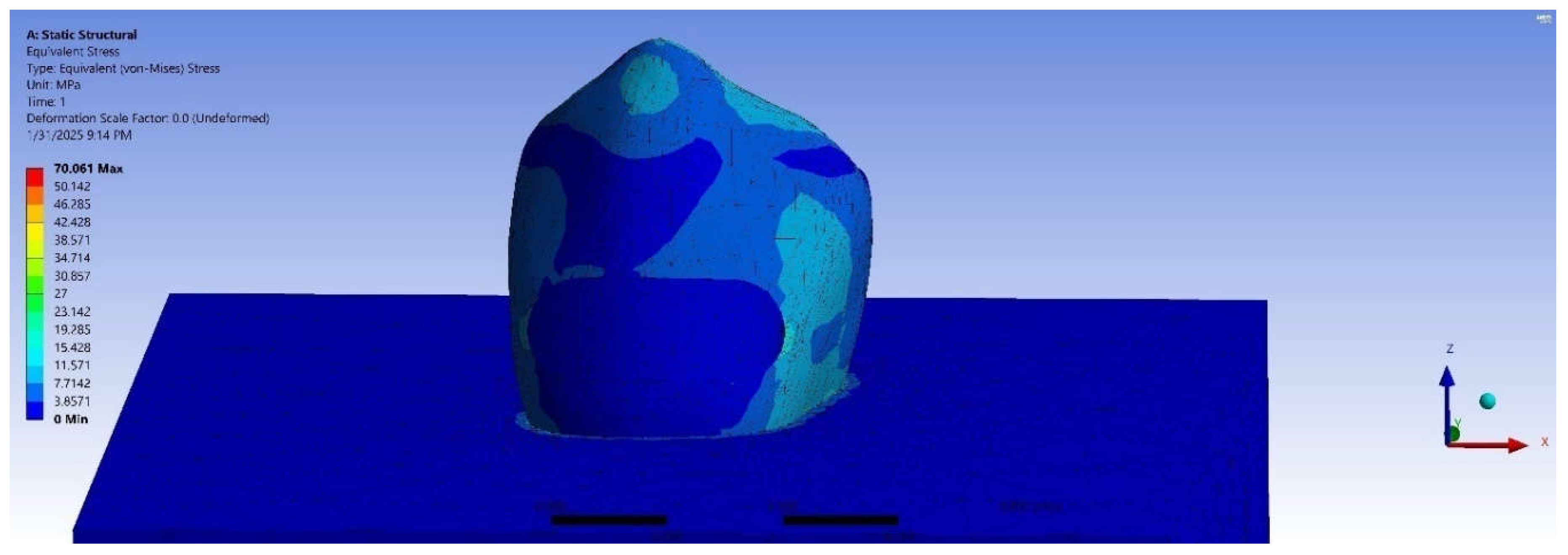
The stress distribution in mandibular premolars with a Class V cavity, both without restoration and with different restorative materials, was evaluated under varying occlusal loads applied at the buccal cusp, Figure (3) to (6). The von Mises stress values in megapascals (MPa) for each condition are presented in Table (2). The stress values in the un restored tooth increased progressively with the applied load, ranging from 49.23 MPa at 100N to 123.15 MPa at 250N. Stress concentrations in the GIC restoration were lower compared to the unrestored tooth at all load levels, with values starting at 35.00 MPa at 100N and rising to 87.51 MPa at 250N. While the Activa™ BioActive-Restorative (BIO) showed improved stress distribution, its stress concentrations remained higher than those of the other restorative materials. Among the restorative materials tested, Cention40 dental composite exhibited the lowest stress concentrations, especially at 100N (28.02 MPa).

Table 2.
von Mises Stress in Mandibular Premolars Under Varying Loads.
| Applied Load (N) | A1 (MPa) | A2 (MPa) | A3 (MPa) | A4 (MPa) |
| 100 | 49.23 | 35.00 | 47.10 | 28.02 |
| 150 | 73.88 | 52.50 | 70.66 | 42.03 |
| 200 | 98.51 | 70.01 | 94.21 | 56.04 |
| 250 | 123.15 | 87.51 | 117.77 | 70.06 |
A1: without restoration, A2: Glass Ionomer Cement, A3: Activa™ BioActive-Restorative, A4: Cention 40.
Figure 3.
Stress distribution in the tooth without restoration, 250 N.
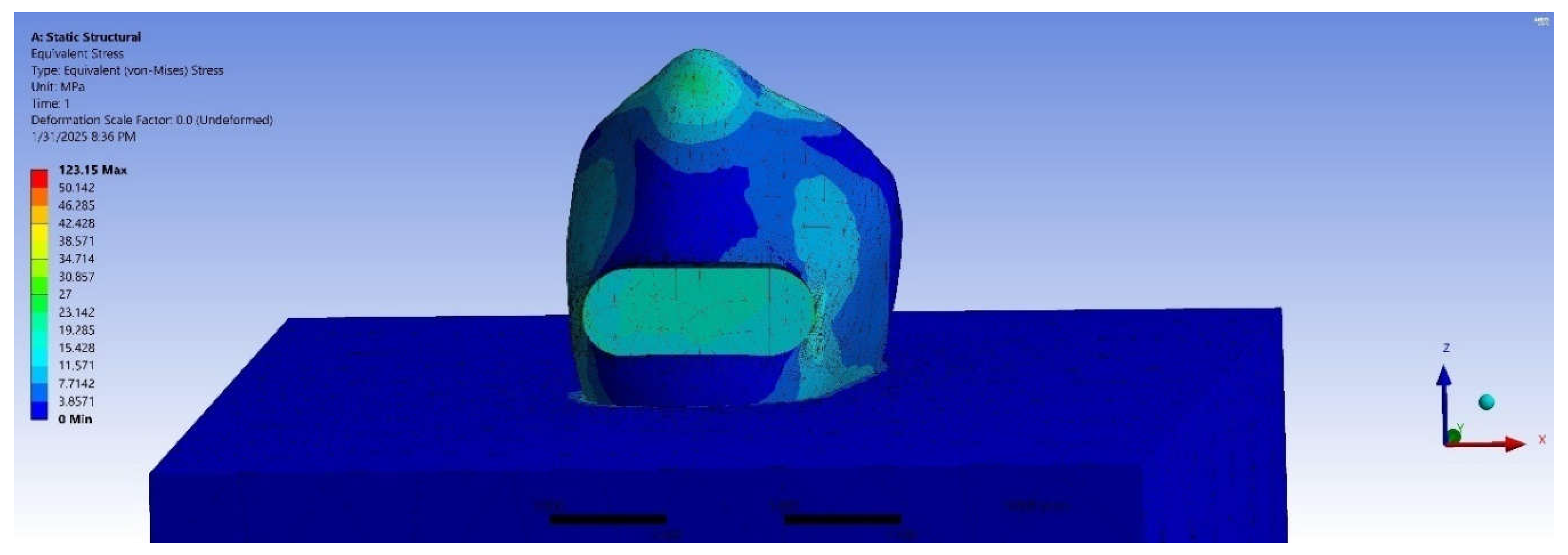
Figure 4.
Stress distribution in the GIC-restored tooth,250 N.
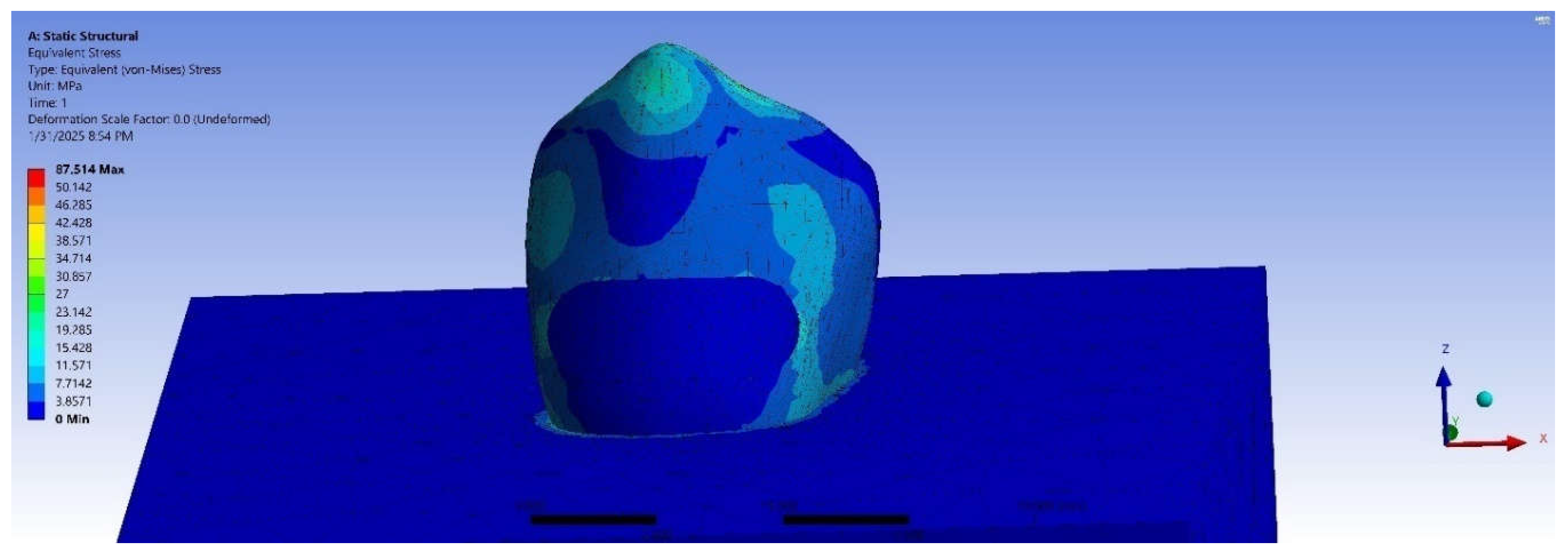
Figure 5.
Stress distribution in the bioactive-restored tooth, 250 N.
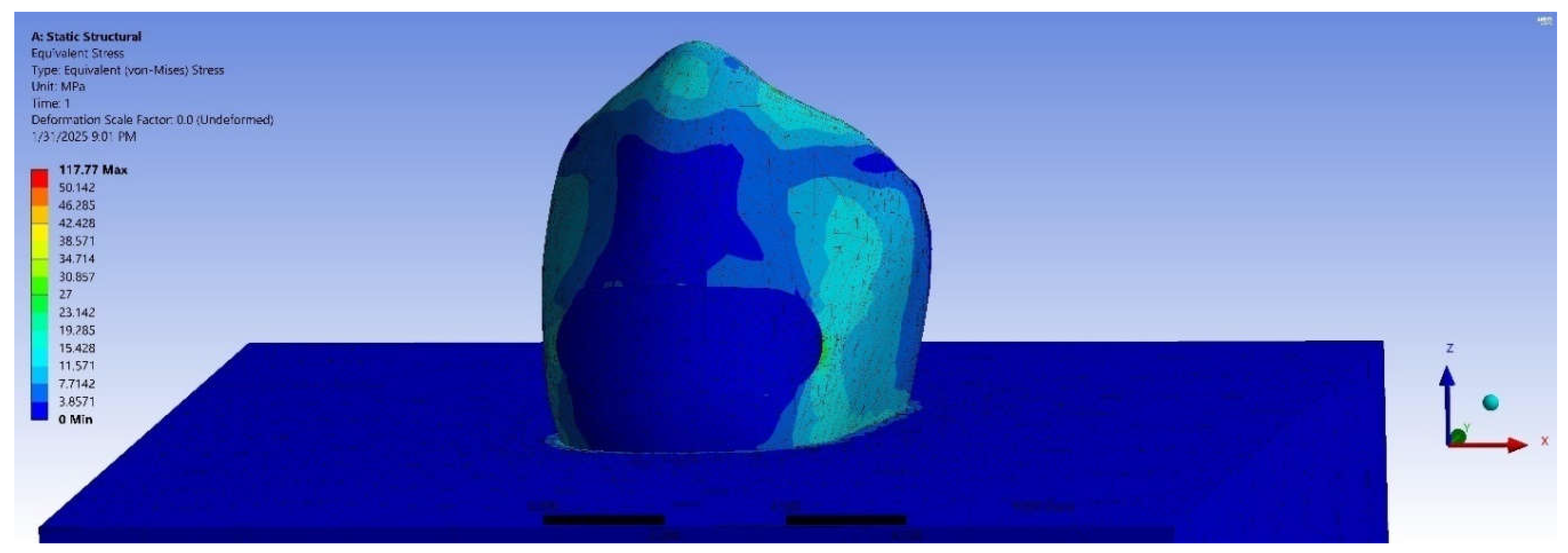
Figure 6.
Stress distribution in the Cention40-restored tooth, 250 N.
3.2. Stress Distribution by Restoration Type and Load Levels
Statistical analysis (ANOVA: F = 1.792, p = 0.202) revealed no statistically significant variation in stress levels among the three restorative material groups.This suggests that all materials performed similarly in terms of stress distribution.
On the other hand, the ANOVA test revealed a significant difference in stress values between the different load levels (100N, 150N, 200N, and 250N) (F-statistic: 7.72, p-value: 0.004), with higher loads resulting in greater stress concentration.
3.3. Compare Stress Values at Different Load Levels (Paired t-Tests)
Statistical analysis was performed to further explore the differences in stress values at various load levels, Table (3). According to the tests, a significant difference in stress was observed at various load levels (p < 0.05) for all materials.
Table 3.
Comparison of Stress Values at Different Loads.
| Pair | Mean Difference | t-value | df | p-value |
| 100N vs. 150N | 24.94 | 5.42 | 3 | 0.012 |
| 100N vs. 200N | 3.76 | 5.38 | 3 | 0.013 |
| 100N vs. 250N | 37.16 | 5.42 | 3 | 0.012 |
| 150N vs. 200N | -21.18 | -5.42 | 3 | 0.012 |
| 150N vs. 250N | 12.22 | 5.42 | 3 | 0.012 |
| 200N vs. 250N | 33.40 | 5.42 | 3 | 0.012 |
4. Discussion
Usually, cervical lesions are very common and difficult to treat and achieving long-lasting results is a challenge [31]. A study reported the prevalence of noncarious cervical lesions (NCCLs) was 30.91%, mostly in premolars, followed by first molars, canines, and incisors,after orthodontic treatments [32]. It is prevalence reach as high as(77.78%)which commonly observed in premolars [33]. According to estimates, between 31% and 56% of people have noncarious Class V lesions, while 85% of people have some degree of tooth loss in the cervical region [28,34].
A three-dimensional finite element analysis study, showed that stress concentrated at the cervical margin of restorations in abfraction lesions, with maximum stress values recorded in different models at the cervical region, regardless of the presence of occlusal restoration [35].Additionally, Restorations in the mandibular arch have a higher failure rate than in the maxillary arch due to the lingual orientation of the mandibular teeth, which concentrates tensile stresses at the cervical area, especially in the premolars, leading to failure under stress [29,36].
In this study, finite element analysis (FEA) using ANSYS 16.0 (2020) assessed von Mises stress and distribution in Class V restorations (mandibular first premolar) for three materials under occlusal loads of 100–250N. Forces were applied perpendicularly 0.4mm from the buccal cusp tip, aligning with standard biomechanical protocols [37,38,39].
In this study, a separated layer for enamel and dentin was used, this unique feature of finite element analysis usually neglected in previous studies. Additionally, investigation performed to determine the distribution of stress in various parts of the tooth based on the biomechanical properties of used materials such as the modulus of elasticity and the Poisson’s ratio, which ensure more predictable clinical outcomes as outlined by [40]. Moreover, numerous studies have highlighted that the characteristics of dental materials significantly influence stress patterns in teeth restored under various occlusal forces [28,29,30,31,35,41]. Thus, this study, evaluate the von Mises stress in Class V restorations of three different restorative materials, Glass–ionomer cement, Activa™ BioActive-Restorative (Pulpdent, USA), and Cention40.
The previous study used a human mandibular first molar with a Class II cavity restored with EverX Posterior (Elastic modulus: 11.4 GPa) and ActivaTM Bioactive (Elastic modulus: 2.35 GPa) materials, applying a 600 N static occlusal load at a 60° angle. FEA results showed that ActivaTM Bioactive, with its lower elastic modulus, absorbed more stress within the material itself, leading to higher deformation and stress concentration compared to EverX Posterior, which had a higher elastic modulus [30]. In comparison, the present study found that while ActivaTM Bioactive, with its lower elastic modulus (2.35 GPa), showed improved stress distribution, its stress concentrations were still higher than those of other restorative materials, such as Cention40 dental composite, which exhibited the lowest stress concentrations at 100N.A study by Shubhashini et al. [28], evaluated stress distribution in Class V restorations of mandibular premolars restored with microfilled composite, flowable composite, glass-ionomer cement (GIC), and resin-modified glass ionomer cement. They applied occlusal loads of 100N, 150N, 200N, and 250N to the restored cavities using Finite Element Modeling (FEM). The study found that GIC exhibited Von Mises stress values ranging from 36.0 MPa at 100N to 90.1 MPa at 250N. In comparison, the present analysis also showed GIC's stress concentrations, with values starting at 35.00 MPa at 100N and rising to 87.51 MPa at 250N. While both studies observed similar trends in stress concentration, with GIC showing moderate stress levels, the results of this investigation highlight that Cention40 exhibited the lowest stress concentrations across all applied loads compared to GIC. Another study by Swathi et al. [29], evaluated stress distribution in Class V cervical lesions of mandibular premolars restored with Cention N, glass-ionomer cement (GIC), and dental amalgam, which differed in elastic modulus, with Cention N having the lowest, followed by GIC, and amalgam having the highest (35000 MPa). In the study, occlusal pressure loads of 100, 150, 200, and 250 N were applied to the restored cavities, and it was found that GIC had the lowest Von Mises stress values. Cention N showed stress concentrations similar to GIC, while amalgam had the highest stress values. However, these results differ from ours, where Cention40 showed the lowest stress concentrations across all applied loads, outperforming GIC.
The ANOVA test revealed no statistically significant difference in stress values among the three restoration groups (F-statistic: 1.792, p-value: 0.202), indicating that the materials performed similarly in terms of stress distribution. However, significant differences were found between load levels (F-statistic: 7.72, p-value: 0.004), with stress increasing as the load increased. Additionally, the paired t-tests show that changes in applied load significantly affect von Mises stress values. It is important to note that most FEA studies do not use statistical analysis to assess these differences.
5. Conclusions
All materials exhibited similar stress distribution, with no significant differences observed (p = 0.202). However, significant differences were noted in stress distribution across varying load levels (p = 0.004), which was consistent across all materials tested (p < 0.05).
6. Limitations
The investigation of only three restorative materials and a single mandibular first premolar model, which might not accurately represent the variety of clinical settings. Additionally, von Mises stress was the only focus of the analysis, other factors such as (wear, material fatigue, and long-term performance) in clinical settings were not taken into account.
Funding
This research received no external funding.
Conflicts of Interest
The author declare no conflicts of interest.
References
- Gönder HY, Mohammadi R, Harmankaya A, Yüksel İB, Fidancıoğlu YD, Karabekiroğlu SJP. Teeth Restored with Bulk–Fill Composites and Conventional Resin Composites; Investigation of Stress Distribution and Fracture Lifespan on Enamel, Dentin, and Restorative Materials via Three-Dimensional Finite Element Analysis. 2023;15(7):1637. [CrossRef]
- Sabbagh J, Fahd JC, McConnell RJJDU. Post-operative sensitivity and posterior composite resin restorations: a review. 2018;45(3):207-13. [CrossRef]
- Mjör IA, Toffentti FJQi. Secondary caries: a literature review with case reports. 2000;31(3).
- Brambilla E, Ionescu ACJOB, Bioactivity MDMAT. Oral biofilms and secondary caries formation. 2021:19-35. [CrossRef]
- Jafari F, Jafari S, Etesamnia PJIej. Genotoxicity, bioactivity and clinical properties of calcium silicate based sealers: a literature review. 2017;12(4):407. [CrossRef]
- Sarfati A, Tirlet GJIJED. Deep margin elevation versus crown lengthening: biologic width revisited. 2018;13(3):334-56.
- Parirokh M, Torabinejad M, Dummer PJIej. Mineral trioxide aggregate and other bioactive endodontic cements: an updated overview–part I: vital pulp therapy. 2018;51(2):177-205. [CrossRef]
- Castelo-Baz P, Argibay-Lorenzo O, Muñoz F, Martin-Biedma B, Darriba IL, Miguéns-Vila R, et al. Periodontal response to a tricalcium silicate material or resin composite placed in close contact to the supracrestal tissue attachment: A histomorphometric comparative study. 2021;25:5743-53. [CrossRef]
- Suhag D. Dental Biomaterials. Handbook of Biomaterials for Medical Applications, Volume 2: Applications: Springer; 2024. p. 235-79.
- Syed AUY, Rokaya D, Shahrbaf S, Martin NJAS. Three-dimensional finite element analysis of stress distribution in a tooth restored with full coverage machined polymer crown. 2021;11(3):1220. [CrossRef]
- Babaei B, Shouha P, Birman V, Farrar P, Prentice L, Prusty GJJotMBoBM. The effect of dental restoration geometry and material properties on biomechanical behaviour of a treated molar tooth: A 3D finite element analysis. 2022;125:104892. [CrossRef]
- Yaman S, Şahin M, Aydin CJJoor. Finite element analysis of strength characteristics of various resin based restorative materials in Class V cavities. 2003;30(6):630-41. [CrossRef]
- Boschian Pest L, Guidotti S, Pietrabissa R, Gagliani MJJoor. Stress distribution in a post-restored tooth using the three-dimensional finite element method. 2006;33(9):690-7. [CrossRef]
- Huang L, Nemoto R, Okada D, Shin C, Saleh O, Oishi Y, et al. Investigation of stress distribution within an endodontically treated tooth restored with different restorations. 2022;17(3):1115-24. [CrossRef]
- Yamanel K, Çaglar A, Gülsahi K, Özden UAJDmj. Effects of different ceramic and composite materials on stress distribution in inlay and onlay cavities: 3-D finite element analysis. 2009;28(6):661-70. [CrossRef]
- Guler M, Guler C, Cakici F, Cakici E, Sen SJNJoCP. Finite element analysis of thermal stress distribution in different restorative materials used in class V cavities. 2016;19(1):30-4. [CrossRef]
- Chung SM, Yap AUJ, Koh WK, Tsai KT, Lim CTJB. Measurement of Poisson's ratio of dental composite restorative materials. 2004;25(13):2455-60. [CrossRef]
- Mesquita RV, Axmann D, Geis-Gerstorfer JJDM. Dynamic visco-elastic properties of dental composite resins. 2006;22(3):258-67. [CrossRef]
- Wang Y-T, Chen C-H, Wang P-F, Chen C-T, Lin C-LJAS. Design of a metal 3D printing patient-specific repairing thin implant for zygomaticomaxillary complex bone fracture based on buttress theory using finite element analysis. 2020;10(14):4738. [CrossRef]
- Dawood SN, Al-Zahawi AR, Sabri LAJAS. Mechanical and thermal stress behavior of a conservative proposed veneer preparation design for restoring misaligned anterior teeth: A 3D finite element analysis. 2020;10(17):5814. [CrossRef]
- Lin P-J, Su K-CJAS. Biomechanical design application on the effect of different occlusion conditions on dental implants with different positions—A finite element analysis. 2020;10(17):5826. [CrossRef]
- Saridena USNG, Sanka GSSJ, Alla RK, AV R, Mc SS, Mantena SRJIJoDM. An overview of advances in glass ionomer cements. 2022;4(4):89-94.
- Park EY, Kang SJYUjom. Current aspects and prospects of glass ionomer cements for clinical dentistry. 2020;37(3):169-78. [CrossRef]
- Lardani L, Derchi G, Marchio V, Carli EJC. One-year clinical performance of Activa™ bioactive-restorative composite in primary molars. 2022;9(3):433. [CrossRef]
- Martínez-Sabio L, Peñate L, Arregui M, Veloso Duran A, Blanco JR, Guinot FJP. Comparison of shear bond strength and microleakage between activa™ bioactive restorative™ and bulk-fill composites—An in vitro study. 2023;15(13):2840. [CrossRef]
- Adsul PS, Dhawan P, Tuli A, Khanduri N, Singh AJIJoCPD. Evaluation and comparison of physical properties of cention n with other restorative materials in artificial saliva: an in vitro study. 2022;15(3):350. [CrossRef]
- Singbal K, Shan MKW, Dutta S, Kacharaju KRJB, Journal P. Cention N compared to other contemporary tooth-colored restorative materials in terms of fluoride ion releasing efficacy: Validation of a novel caries-prevention-initiative by the Ministry of Health, Malaysia. 2022;15(2):669-76. [CrossRef]
- Narayanaswamy S, Meena N, Shetty A, Kumari A, Naveen DJJoCD. Finite element analysis of stress concentration in Class V restorations of four groups of restorative materials in mandibular premolar. 2008;11(3):121-6. [CrossRef]
- Pai S, Naik N, Patil V, Kaur J, Awasti S, Nayak NJJoISoP, et al. Evaluation and comparison of stress distribution in restored cervical lesions of mandibular premolars: three-dimensional finite element analysis. 2019;9(6):605-11. [CrossRef]
- Bansal P, Seth T, Kumar M, Bhatt M, Arora P, Gupta I, et al. Comparative Evaluation of Stress Distribution and Deformation in Class II Cavities Restored With Two Different Biomimetic Restorative Materials: A Three-Dimensional Finite Element Analysis. 2024;16(9). [CrossRef]
- Pai S, Bhat V, Patil V, Naik N, Awasthi S, Nayak NJJoISoP, et al. Numerical three-dimensional finite element modeling of cavity shape and optimal material selection by analysis of stress distribution on class V cavities of mandibular premolars. 2020;10(3):279-85. [CrossRef]
- Gomes RR, Zeola LF, Barbosa TAQ, Fernandes Neto AJ, de Araujo Almeida G, Soares PVJPiO. Prevalence of non-carious cervical lesions and orthodontic treatment: a retrospective study. 2022;23(1):17. [CrossRef]
- Villamayor KGG, Codas-Duarte D, Ramirez I, Souza-Gabriel AE, Sousa-Neto MD, Candemil APJAoOB. Morphological characteristics of non-carious cervical lesions. A systematic review. 2024:106050. [CrossRef]
- Browning W, Brackett WW, Gilpatrick RJOD. Two-year clinical comparison of a micro filled and a hybrid resin-based composite in non-carious class V lesions. 2000;25:46-50.
- Srirekha A, Bashetty KJJoCD, Endodontics. A comparative analysis of restorative materials used in abfraction lesions in tooth with and without occlusal restoration: Three-dimensional finite element analysis. 2013;16(2):157-61. [CrossRef]
- Lee WC, Eakle WSJTJopd. Stress-induced cervical lesions: review of advances in the past 10 years. 1996;75(5):487-94. [CrossRef]
- Kantardžić I, Vasiljević D, Blažić L, Lužanin OJCmj. Influence of cavity design preparation on stress values in maxillary premolar: a finite element analysis. 2012;53(6):568-76. [CrossRef]
- Benazzi S, Grosse IR, Gruppioni G, Weber GW, Kullmer OJCoi. Comparison of occlusal loading conditions in a lower second premolar using three-dimensional finite element analysis. 2014;18:369-75. [CrossRef]
- Wang Z-f, Fu B-pJTJoPD. Minimum residual root dentin thickness of mandibular premolars restored with a post: a finite element analysis study. 2024;131(5):878-85. [CrossRef]
- Rajagopal S, Sharma S, Sharma CSJC. Finite element analysis and clinical applications of transverse post for the rehabilitation of endodontically treated teeth. 2024;16(7). [CrossRef]
- Soares PV, Machado AC, Zeola LF, Souza P, Galvão A, Montes TC, et al. Loading and composite restoration assessment of various non-carious cervical lesions morphologies–3D finite element analysis. 2015;60(3):309-16. [CrossRef]
Figure 1.
Model of the mandibular premolar from different perspectives.
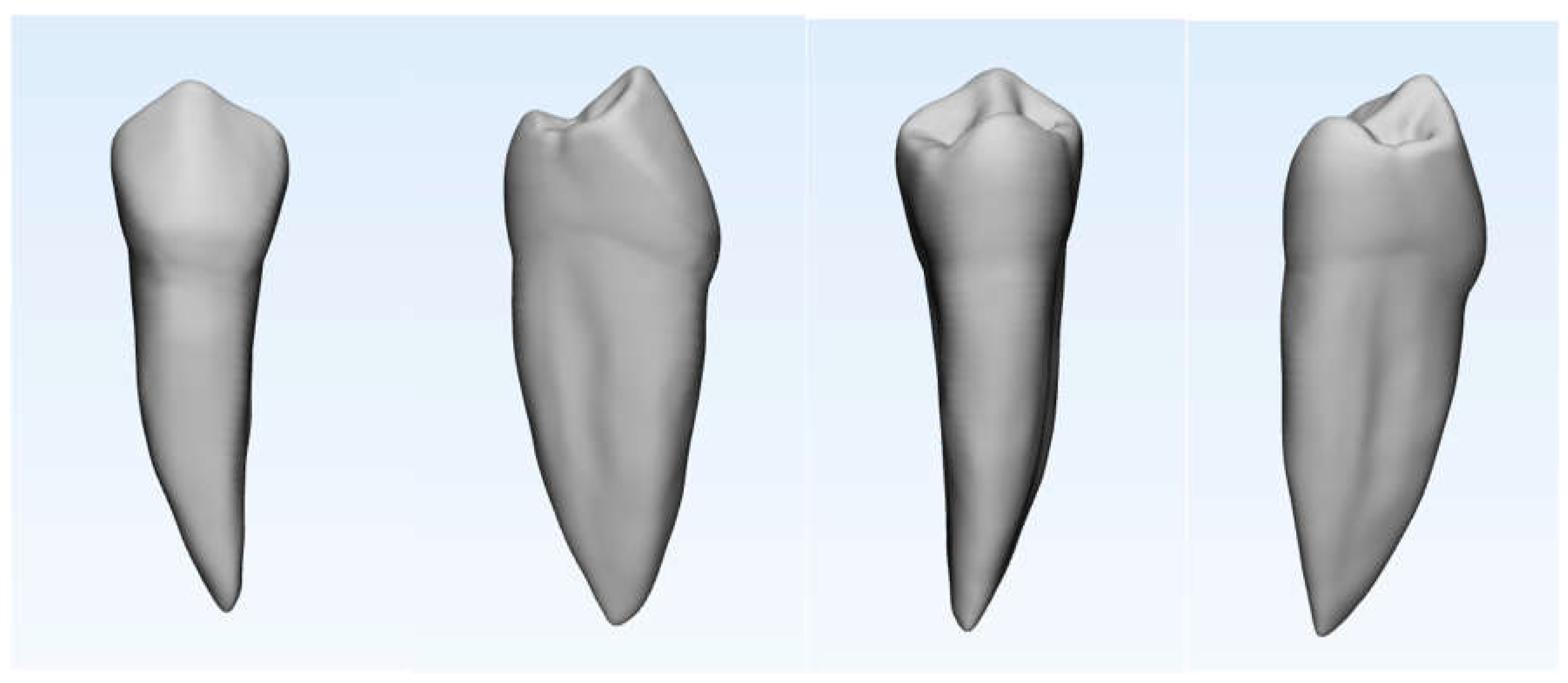
Figure 2.
Virtual model after meshing. A: mandibular premolar with prepared cavity, B: restored material, C: bone block.
Figure 2.
Virtual model after meshing. A: mandibular premolar with prepared cavity, B: restored material, C: bone block.
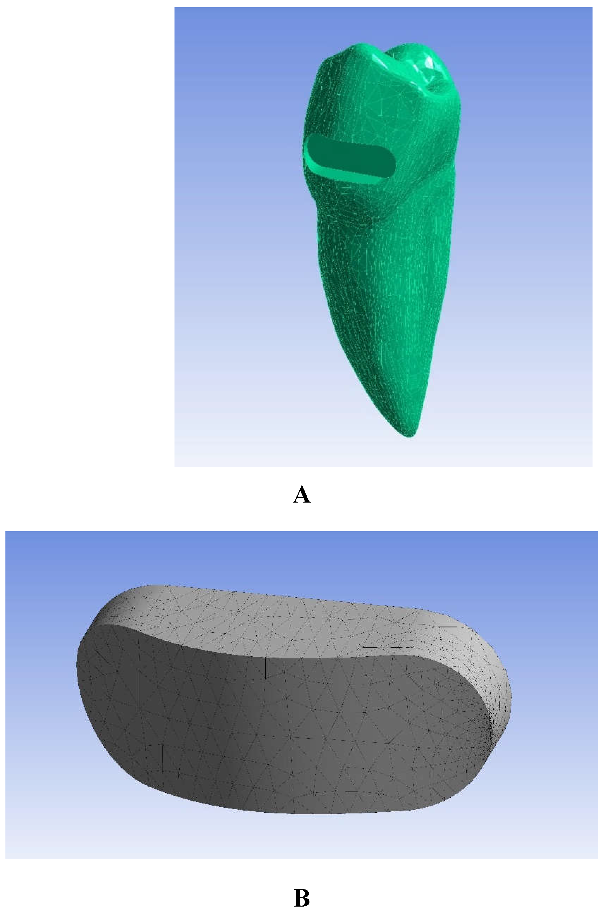
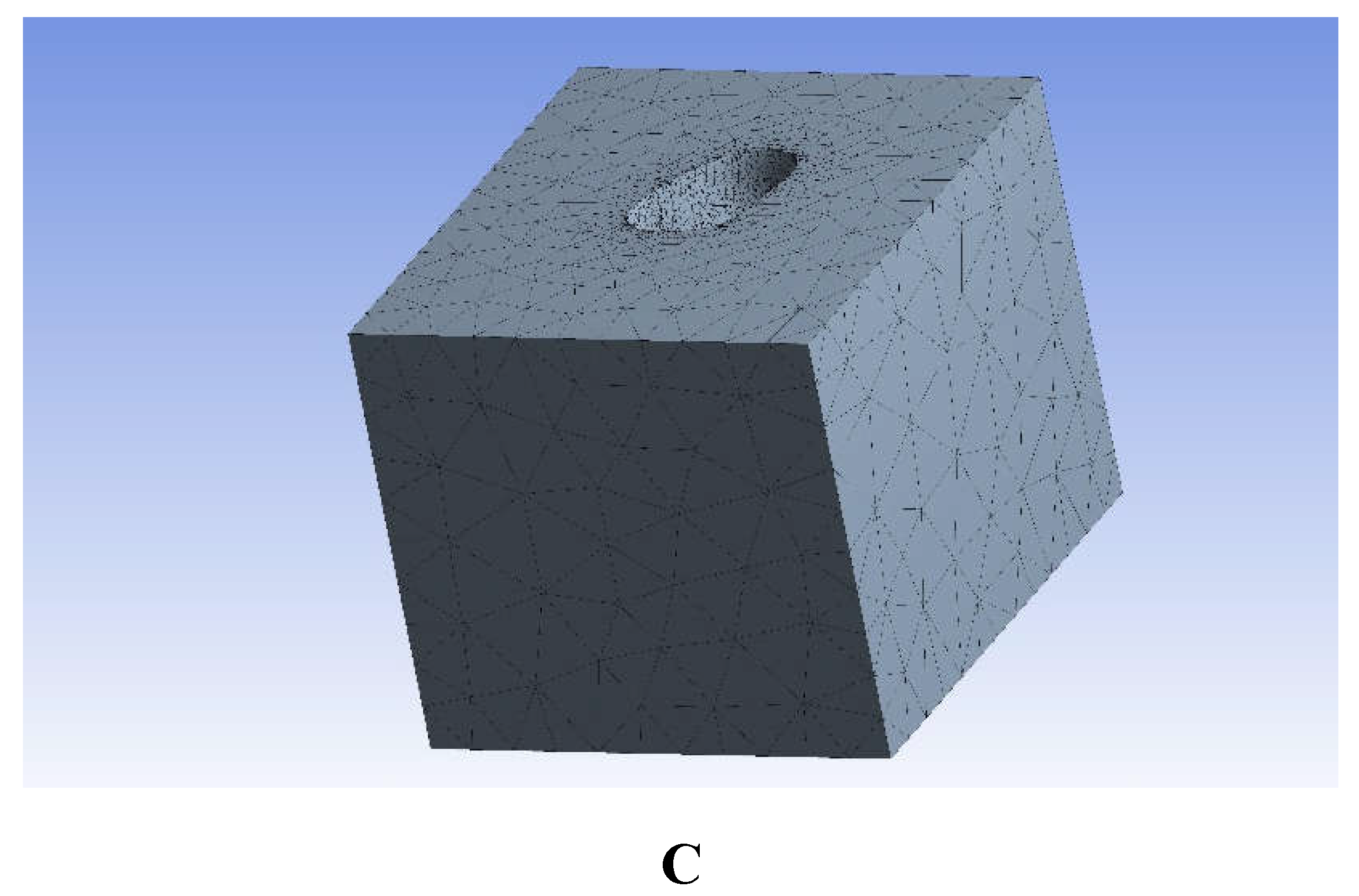
Table 1.
Biomechanical parameters of dental tissues and anatomical regions incorporated in the finite element analysis.
Table 1.
Biomechanical parameters of dental tissues and anatomical regions incorporated in the finite element analysis.
| Materials | Modulus of elasticity (MPa) |
Poisson’s ratio(μ) |
Reference |
|---|---|---|---|
| Enamel | 84100 | 0.33 | [29] |
| Dentin | 13700 | 0.31 | [29] |
| Glass–ionomer cement | 10800 | [29] | |
| Activa™ BioActive-Restorative | 2350 | 0.25 | [30] |
| Cention40 | 13000 | 0.3 | [29] |
Disclaimer/Publisher’s Note: The statements, opinions and data contained in all publications are solely those of the individual author(s) and contributor(s) and not of MDPI and/or the editor(s). MDPI and/or the editor(s) disclaim responsibility for any injury to people or property resulting from any ideas, methods, instructions or products referred to in the content. |
© 2025 by the authors. Licensee MDPI, Basel, Switzerland. This article is an open access article distributed under the terms and conditions of the Creative Commons Attribution (CC BY) license (http://creativecommons.org/licenses/by/4.0/).
Copyright: This open access article is published under a Creative Commons CC BY 4.0 license, which permit the free download, distribution, and reuse, provided that the author and preprint are cited in any reuse.


