
Submitted:
15 April 2025
Posted:
15 April 2025
You are already at the latest version
Abstract
Background/Objectives: Obstructive salivary gland disorders—primarily sialolithiasis and ductal stenosis—remain a significant source of morbidity, often requiring surgical intervention. Sialendoscopy has emerged as a minimally invasive, gland-preserving technique for both diagnosis and treatment. This retrospective study aimed to evaluate diagnostic and interventional sialendoscopy outcomes in a Romanian patient cohort and to identify gland-specific considerations in the management of salivary gland obstruction; Methods: A total of 89 patients with confirmed obstructive salivary gland disease (parotid or submandibular) were included. All underwent clinical evaluation, imaging (ultrasound, CBCT, CT, MRI), and sialendoscopic treatment between 2021 and 2025 in two centers. Data on demographics, imaging, calculus size, procedural technique, anesthesia, and complications were collected and analyzed using descriptive and inferential statistics; Results: The submandibular gland was more frequently involved (70.8%), with larger calculi compared to the parotid (mean 7.57 mm vs. 5.07 mm; P = 0.004). Minimally invasive techniques were predominantly used: sialolithotomy and intracorporeal lithotripsy were each performed in 32.6% of cases. Submandibulectomy was required in only 5.6% of patients. Most procedures (93.3%) were conducted under local anesthesia. Complication rates were low and primarily minor and self-limiting; Conclusions: Sialendoscopy is a safe and effective gland-preserving approach in managing obstructive salivary gland disorders. Gland-specific anatomy influences diagnostic pathways and therapeutic choices. These findings support broader adoption of sialendoscopy in routine practice and highlight the need for tailored management protocols based on gland involvement and stone characteristics.
Keywords:
sialendoscopy
; salivary gland obstruction
; sialolithiasis
; ductal stenosis
; submandibular gland
; parotid gland
; lithotripsy
; minimally invasive surgery
; gland preservation
1. Introduction
Obstructive disorders of the major salivary glands represent a significant clinical burden, frequently presenting as recurrent episodes of painful glandular swelling, often exacerbated during meals [1,2]. The most common etiologies are sialolithiasis (salivary stone formation) and ductal stenosis (narrowing of the salivary ducts), which collectively account for approximately 50% of benign salivary gland diseases [3]. Sialolithiasis alone affects an estimated 1–2% of the general population, making it the most prevalent form of obstructive salivary pathology [4]. Clinically, these conditions result in impaired salivary flow, gland enlargement, and recurrent or chronic sialadenitis—a bacterial infection of the gland. If left untreated, such pathologies can significantly compromise patients’ quality of life and often necessitate therapeutic intervention [5].
Both the submandibular and parotid glands are commonly affected, although they differ in disease distribution and pathogenesis. Approximately 80–90% of salivary calculi form in the submandibular gland, likely due to its higher mucin and mineral content and the anatomical configuration of Wharton’s duct. In contrast, ductal strictures are more frequently observed in the parotid gland, with 70–75% of cases occurring in Stensen’s duct, often secondary to autoimmune conditions or prior infections [6].
Despite these anatomical and pathological differences, both glands are susceptible to recurrent obstruction and sialadenitis [7,8]. A comprehensive evaluation of obstructive salivary gland disorders must, therefore, account for gland-specific characteristics while maintaining a primary focus on relieving ductal obstruction and restoring normal salivary function.
Over the past two decades, sialendoscopy has emerged as a minimally invasive, gland-preserving technique for both the diagnosis and treatment of obstructive salivary gland disorders. First introduced in the early 1990s, sialendoscopy involves the insertion of a miniature endoscope (typically 0.8–1.6 mm in diameter) via the natural ductal orifice, enabling real-time, direct visualization of the salivary ductal system [9,10,11,12]. Compared to traditional open surgical excision—which carries risks such as nerve injury and xerostomia [13], sialendoscopy offers a safe and effective alternative with significantly reduced morbidity [14,15]. It also allows for accurate diagnosis without the use of ionizing radiation [16], and its diagnostic performance is further enhanced when combined with imaging modalities such as ultrasound and cone-beam computed tomography (CBCT) [17,18]. Particularly, CBCT has been shown to be a complementary tool in the detection of salivary calculi or ductal abnormalities that may not be visible on conventional ultrasound or sialography alone [19].
Minimally invasive instruments for the removal of salivary calculi have gained increasing popularity, particularly with the advent and widespread adoption of interventional sialendoscopy as an alternative to traditional open surgical approaches [16]. Certain types of stones—particularly those less than 7 mm in diameter—can be effectively managed endoscopically, and are therefore less likely to require invasive transoral sialolithotomy or gland excision [3]. For larger or impacted calculi, sialendoscopy-assisted lithotripsy offers a valuable approach, allowing stone fragmentation and safe removal while preserving gland function [20,21,22]. Despite its advantages, sialendoscopy is not without limitations, such as failure of stone retrieval or challenges in managing severe ductal stenoses. In such cases, auxiliary techniques such as ductal dilation or stenting may be necessary [23,24]. Nonetheless, the risk of complications remains low, and sialendoscopy has increasingly been recognized as the gold standard in managing obstructive salivary gland disease [25].
Accurate diagnosis is essential for guiding appropriate treatment. In this regard, diagnostic and interventional sialendoscopy serves as a reference standard, offering a direct and minimally invasive assessment of the salivary ductal system [24] and facilitating the identification of underlying pathologies such as calculi, strictures, mucous plugs, or anatomical anomalies [26].
While the efficacy of sialendoscopy has been demonstrated in multiple international studies [24,27], there remains a notable lack of data from Eastern Europe, including Romania, where clinical experience with this technique is still limited. In the absence of local evidence, traditional surgical excision continues to be widely practiced, despite the availability of less invasive, gland-sparing alternatives.
To address this gap, our retrospective study evaluates the clinical outcomes of diagnostic and interventional sialendoscopy in a cohort of 89 Romanian patients presenting with obstructive salivary gland disorders. This is one of the few studies conducted in Eastern Europe to analyze the use of sialendoscopy in clinical practice. Our objectives include assessing demographic and imaging data, procedural techniques, and therapeutic outcomes in order to support the broader implementation of this technique and enhance the quality of care for patients with salivary gland obstructions.
2. Materials and Methods
This retrospective clinical study was conducted in accordance with international ethical guidelines, including the Declaration of Helsinki, the Belmont Report, CIOMS-WHO guidelines, ICH-GCP standards, and EU Regulation 2016/679 (GDPR). Ethical approval was obtained from the Research Ethics Committee of the “Carol Davila” University of Medicine and Pharmacy, Bucharest (Approval No. 19772/2023). Written informed consent was obtained from all participants for both procedures and data collection.
The study included patients diagnosed with obstructive salivary gland disorders (parotid and/or submandibular glands) who underwent consultation and treatment between January 2021 and January 2025 in two centers affiliated with “Carol Davila” University of Medicine and Pharmacy from Bucharest, Romania: a stationary hospital unit and a private outpatient clinic, both equipped with diagnostic and therapeutic sialendoscopy facilities.
2.1. Inclusion and Exclusion Criteria
Patients were eligible if they:
- -
- Had a confirmed diagnosis of obstructive disorder of the major salivary glands (parotid or submandibular);
- -
- Had complete demographic and clinical documentation, including sex, age, affected gland, diagnostic approach, treatment method, clinical evolution, and any complications.
A structured diagnostic protocol was applied to most patients, beginning with anamnesis and review of prior investigations, followed by soft tissue ultrasound, which was the primary imaging modality due to its non-invasive nature. When needed, cone-beam computed tomography (CBCT) was used to assess ductal pathology—particularly in suspected submandibular sialolithiasis—or to detect calculi not visualized by ultrasound. Magnetic resonance imaging (MRI) was selectively employed in cases where ductal stenosis or extrinsic compression of Stensen’s duct was suspected. In rare instances, sialography–CBCT with contrast was used for cases with unclear etiology.
Patients were excluded if they:
- -
- Had systemic or locoregional conditions contraindicating sialendoscopy, such as hematologic disorders, coagulopathies, severe cardiovascular diseases, or systemic infections;
- -
- Presented with acute suppurative sialadenitis or other high-risk comorbidities that significantly increased the potential for intra- or post-procedural complications.
2.2. Data Collection and Analysis
Data were extracted from patient medical records, operative reports, and follow-up documentation. For each patient, anamnesis data were reviewed, with particular attention to the onset, duration, and progression of symptoms, as well as a detailed description of associated clinical signs. Symptom duration was analyzed as a potential contributing factor to the severity and clinical presentation of obstructive salivary gland disease.
Clinical examination findings were documented, including visual inspection and bimanual palpation of the affected area. Evaluated structures included the facial soft tissues, overlying skin, oral mucosa, and salivary papilla. Bimanual palpation, especially in cases of submandibular lithiasis, often provided critical information for treatment planning. For instance, if a stone was palpable within Wharton’s duct, transoral sialolithotomy under endoscopic guidance was commonly indicated.
Imaging data were also reviewed retrospectively. Ultrasound was the most frequently employed imaging modality, both pre- and intra-operatively, due to its non-invasive nature and ability to guide real-time decision-making. In selected cases, CBCT, MRI, or sialography–CBCT were used based on clinical suspicion and anatomical considerations.
Technical aspects of the procedure were evaluated through operative reports. Particular attention was paid to papilla instrumentation and endoscope insertion, which were frequently challenging. Lacrimal or metallic probes were typically used for ductal access, and magnification tools (4.5× surgical loupes or operating microscope) were employed in many cases to reduce the risk of complications such as false passage or trauma to peripapillary tissues.
Diagnostic sialendoscopy was primarily performed using a 0.89 mm endoscope, which permitted access to second- and occasionally third-order ducts. For interventional procedures, endoscopes with diameters of 1.1 mm and 1.6 mm were used, with the narrower 1.1 mm scope preferred for the parotid (Stensen’s) duct. All procedures involved intermittent saline irrigation to maintain hydrostatic pressure, improve endoscope navigation, and clear the ductal lumen of fibrin debris.
Most procedures were conducted under local anesthesia, as noted in the records. Cases requiring general anesthesia were identified and justified by the patient’s medical condition, psychological status, or complex diagnostic needs.
2.3. Statistical Analysis
All collected data were entered into Microsoft Excel and subsequently analyzed using IBM® SPSS® Statistics version 25.0 (IBM Corp., Armonk, NY, USA). Statistical analysis was conducted to explore associations between demographic variables, diagnostic strategies, therapeutic interventions, and clinical outcomes in patients undergoing salivary gland evaluation and treatment.
Descriptive statistics were used to summarize continuous variables such as patient age, number of calculi extracted, and stone size. These results were expressed as means and standard deviations (SD). Descriptive evaluations were stratified by gender and salivary gland involvement (parotid vs submandibular).
Inferential statistical analysis included Welch’s t-test (independent-samples t-test) for continuous variables with unequal variances (e.g., age and calculus size comparisons by gender or gland type) and Chi-square (χ²) test to examine associations between categorical variables, such as gender, gland type, anesthesia use, diagnostic method, pathology type, and treatment approach.
Where appropriate, logistic regression models were attempted to predict binary outcomes (e.g., submandibulectomy as treatment failure), based on predictors including age, gender, calculus size, anesthesia, diagnostic tools, and pathology. However, perfect separation was detected in some models, limiting their reliability. Descriptive and inferential comparisons were therefore prioritized in these cases.
These methods enabled a structured evaluation of demographic characteristics, glandular distribution, diagnostic investigations, stone characteristics, and therapeutic modalities, contributing to the overall interpretation of clinical outcomes.
Statistical significance was defined as P < .05.
3. Results
A total of 89 patients were included in this retrospective study. The mean age of the cohort was 45 ± 16 years (range: 14–78 years). Of the total participants, 61.8% were female and 38.2% were male (Figure 1). The follow-up period was between two months and four years, with a mean of 28 months. The stratification by gender and salivary gland involvement (parotid vs submandibular) is presented in Table 1.
3.1. Gland Involvement
The submandibular gland was the most frequently affected site, accounting for 70.8% of all cases (Figure 1). When analyzed by gender, parotid gland involvement was observed in 32.7% of female patients and 26.5% of male patients, indicating a lower incidence of parotid involvement among males compared to females (Table 1).
3.2. Imaging Investigations
Various imaging modalities were employed to aid in diagnosis and assess the feasibility of calculus removal, and were frequently used alongside diagnostic sialendoscopy.
Ultrasound was the most frequently employed, performed in 77 patients (86.5%). It was used both prior to and during the initial consultation and showed consistent value across equipment and institutions (hospital/private clinic) in detecting obstructive pathology.
CBCT was performed in 54 patients (60.7%), with a statistically significant predominance in cases involving the submandibular gland (74.2% of all investigations), compared to 29.6% in parotid-related conditions (Table 1). CBCT proved valuable by providing essential anatomical detail for precise calculus localization and procedural planning.
Conventional medical CT was conducted in 6 patients (6.7%), with a statistically significant predominance in cases involving the parotid gland (Table 1). Although it provides superior image quality, its use in the evaluation of salivary gland obstruction is limited by the higher radiation dose compared to CBCT.
MRI was used in 5 patients (5.6%), primarily for non-lithiasic parotid obstruction. Its utility in visualizing ductal anatomy was noted, although cost and accessibility remain limiting factors.
CBCT Sialography was performed in 3 patients (3.4%), particularly when sialendoscopic access was not possible. It provided enhanced visualization of strictures or deep-seated calculi, aiding in treatment planning. Despite its diagnostic utility, its invasiveness and potential for allergic reaction were acknowledged as limitations [28].
3.3. Diagnosis and Pathology Distribution
The most prevalent pathology was sialolithiasis, observed in 64 patients (69.8%), with an additional 5 patients (5.6%) presenting with both lithiasis and ductal stenosis. Combined, lithiasic conditions accounted for over 75% of all cases. The submandibular gland was the most frequently affected site, with a statistically significant predominance compared to the parotid gland, which accounted for only 37.1% of all gland-related pathologies (Table 1).
Sialadenitis was identified in 10 patients (11.2%) (Figure 1). Among these, parotid gland involvement was observed in 5 patients, representing 18.5% of all parotid cases, while submandibular gland sialadenitis was recorded in 5 patients, corresponding to 8.1% of submandibular cases (Table 1).
Ductal stenosis, in the absence of calculi, was observed in 10 patients (11.2%), but when including patients with combined lithiasis and stenosis, the overall incidence of ductal stenosis was 16.8% (15 out of 89 patients) (Table 1).
3.4. Salivary Calculus Characteristics
The mean diameter of the salivary calculi was 7.04±4.56 mm, with a range of 2 to 29 mm. In terms of stone burden:
- 61 patients had a single calculus,
- 4 patients had 2 calculi,
- 2 patients had 3 calculi, and
- 1 patient presented with 10 calculi.
For statistical purposes, the largest stone size per patient was recorded in the database.
3.5. Anesthesia
The vast majority of procedures (83 patients, 93.26%) were performed under local anesthesia. Benefits included reduced procedural cost, avoidance of general anesthesia-related risks, shorter recovery time, and the feasibility of conducting treatment in an outpatient setting. Only 6 patients (6.74%) required general anesthesia due to clinical or psychological considerations (Table 1).
3.6. Therapeutic Interventions
The treatment approach in this study emphasized minimally invasive, gland-preserving techniques. The main interventions were as follows:
- Transoral sialolithotomy: performed in 29 patients (32.6%) (Figure 2);
- Intracorporeal lithotripsy: also in 29 patients (32.6%) (Figure 3);
- Dormia basket extraction: used in 8 patients (9%) (Figure 4);
- Ductal dilation and stenting were indicated in 7 patients (7.9%), particularly in those with strictures or after papillotomy to enable removal of calculi (Figure 5);
- Submandibulectomy, as a last-resort surgical intervention (Figure 6), was conducted in only 5 patients (5.6%), typically in cases where minimally invasive techniques were unsuccessful or anatomical conditions precluded other options.
The use of sialolitotmy was significantly associated with submandibular gland (P < 0.001). Submandibulectomy was rare (n = 5); logistic regression indicated a trend toward higher odds in males, but it was not statistically significant (P = 0.06).
In this retrospective analysis, the recorded incidence of complications was low, supporting the favorable safety profile of the minimally invasive techniques employed. Documented complications were generally minor and self-limiting, with the most frequently reported being localized postoperative edema, moderate pain, and, in a few cases, secondary ductal infection. These infections responded well to oral antibiotic therapy with amoxicillin with clavulanic acid, 1 g, administered twice daily for five days. Importantly, no cases of iatrogenic ductal stenosis were identified in the reviewed records. Additionally, three cases of transient paresthesia were noted following transoral sialolithotomy, all of which resolved within a period of up to five months.
4. Discussion
To our knowledge, this is the first study published on a Romanian patient cohort evaluating the clinical utility of sialendoscopy as both a diagnostic and interventional tool for the management of obstructive salivary gland disorders. In this retrospective analysis, we reviewed 89 cases of endoscopically assisted, transoral sialolith removal conducted over a four-year period. The primary aim was not only to assess the effectiveness of sialendoscopy in real-world clinical practice but also to begin shaping a standardized treatment protocol that reflects the epidemiological, anatomical, and procedural particularities of patients from this geographic region.
An important aspect of this study is the emphasis on minimally invasive, gland-preserving approaches, most of which were successfully performed under local anesthesia and in an outpatient setting, eliminating the need for hospitalization in the vast majority of cases. This demonstrates not only the feasibility of sialendoscopy in standard practice, but also its suitability for broader implementation across both hospital-based units and private clinical facilities, as reflected in the dual-center nature of this study.
With regard to demographic aspects, among the 89 patients included in this study, 55 (61.8%) were female, which contrasts with findings from previous studies [29]. While some authors have reported a balanced gender distribution [29,30], others have noted a male predominance [31]. The predominance of female patients observed in our cohort may be partially explained by hormonal imbalances associated with menopause, which can lead to reduced salivary flow and xerostomia [32]—both recognized risk factors for obstructive salivary gland disorders.
In terms of age, Escudier et al. reported a higher incidence of sialolithiasis in individuals aged 25 to 50 years [33], while Lustmann et al. identified peak prevalence between the third and sixth decades of life [30]. The findings of the present study are consistent with these observations, with a mean patient age of 45 ± 16 years (range: 14–78 years). Notably, three patients (3.4%) were under 18 years of age, aligning with existing data that pediatric sialolithiasis accounts for approximately 3% of all cases [30].
In the present study, the type of salivary gland involved was shown to have a significant impact on both diagnostic strategies and therapeutic outcomes. Submandibular gland involvement was significantly associated with a higher prevalence of sialolithiasis and larger calculus size (P < 0.05), a finding that aligns with previous literature [1,34]. This predilection is largely attributed to several anatomical and physiological factors, including the uphill direction of salivary flow against gravity, and the higher calcium and mucin content of submandibular saliva, all of which promote stone formation [35]. Consistent with other reports, calculi in submandibular cases were more frequently located within Wharton’s duct, rather than at the hilum or intraglandular regions [34,36].
In contrast, parotid gland involvement was more commonly associated with the need for advanced imaging modalities, such as CBCT and MRI, to support diagnosis and treatment planning. This is in line with existing evidence emphasizing the utility of high-resolution imaging techniques in parotid-related pathologies, as these modalities enable detailed visualization of ductal anatomy—extending to second and third-order branches—without the need for duct cannulation [37].
Furthermore, sialolithotomy was significantly more frequent in submandibular cases (P < 0.05), reflecting both the larger stone sizes and the more accessible ductal anatomy. These findings highlight the importance of gland-specific diagnostic and therapeutic approaches, as submandibular gland disease typically necessitates more invasive interventions, whereas parotid gland conditions often rely on imaging precision and endoscopic techniques [16].
These observations reinforce the need for personalized clinical pathways, where the anatomical and pathological context of the affected gland directly informs the choice of imaging, treatment modality, and postoperative care.
Calculus size varied significantly based on salivary gland involvement, with submandibular calculi being notably larger than those in the parotid gland (mean 7.57 mm vs. 5.07 mm; P = 0.004). This observation is consistent with established anatomical and physiological distinctions between the glands, particularly the longer and more tortuous course of Wharton’s duct and the higher viscosity and mineral content of submandibular saliva, which collectively predispose to the formation of larger and more obstructive calculi. These results are in concordance with previously published studies that report average stone diameters ranging from 5 to 10 mm in submandibular cases [29,30].
The clinical implication is substantial, as larger calculi often necessitate more invasive interventions, including sialolitotmy or submandibulectomy, particularly when minimally invasive techniques fail. These findings support a tailored management approach based on calculus size and gland location.
Over the past three decades, the advancement and widespread adoption of minimally invasive and gland-preserving techniques—including extracorporeal and intracorporeal lithotripsy, interventional sialendoscopy, and sialendoscopy-assisted transoral or transfacial surgical approaches—have significantly reduced the need for traditional procedures such as parotidectomy or submandibular sialadenectomy. These developments have shifted clinical practice away from radical surgical excision, even in cases where gland removal was previously considered standard management [7].
The primary objective of the present study was to evaluate therapeutic interventions based on minimally invasive techniques aimed at preserving the function of the affected salivary gland. In accordance with this approach, transoral sialolithotomy and intracorporeal lithotripsy were each performed in 32.6% of cases, yielding favorable outcomes and allowing for the avoidance of more radical procedures. Additionally, the use of a Dormia-type extraction device was documented in 9% of cases, while ductal stenting—indicated for maintaining ductal patency following dilation—was required in 7.9% of patients with stenotic lesions.
Importantly, submandibulectomy was performed in only 5.6% of cases, and exclusively as a last-resort intervention when minimally invasive methods failed or were not feasible. This supports the broader clinical trend toward conservative, gland-preserving management of obstructive salivary gland disease.
In line with current recommendations from the literature, the therapeutic protocol in our study followed a gradual, stepwise approach, beginning with the least invasive options and escalating as needed. In more complex or refractory cases, a multimodal strategy—combining interventional sialendoscopy with adjunctive procedures such as lithotripsy, stenting, or sialolithotomy—was implemented to optimize outcomes. This approach is consistent with published data indicating that multimodal therapy offers higher success rates and a lower risk of recurrence compared to single-modality interventions [38].
Particular attention in this study was also given to the evaluation of procedural complications. The techniques employed demonstrated a favorable safety profile, and challenges such as papilla instrumentation and navigation of the Stensen duct were effectively managed through the use of magnification tools, including surgical loupes and microscopes. These strategies minimized the risk of soft tissue trauma and helped reduce both intraoperative and postoperative complications.
These results are consistent with findings in the current literature, which indicate that interventional sialendoscopy, when performed by an experienced team, is associated with a significantly lower complication rate compared to traditional surgical approaches such as gland excision [39]. Moreover, the conservative nature of this technique allows for salivary gland preservation and functional recovery, which positively impacts patient quality of life [40,41].
However, in two cases treated via sialolithotomy, complete stone removal was not possible, and only partial extraction of the calculi could be achieved. Due to persistent symptoms and the risk of recurrent infection, definitive surgical treatment via submandibulectomy was subsequently indicated. These cases highlight the limitations of conservative approaches in the context of complex or large calculi, where ductal anatomy or deep stone positioning may hinder complete removal through minimally invasive techniques. They also emphasize the importance of rigorous case selection and comprehensive preoperative assessment, including the use of high-resolution imaging to guide treatment planning.
In our cohort study, the overwhelming majority of sialendoscopic procedures (93.26%) were successfully performed under local anesthesia, with only 6 patients (6.74%) requiring general anesthesia due to specific clinical or psychological indications. This finding reinforces the growing body of evidence supporting the feasibility, safety, and efficiency of performing diagnostic and even interventional sialendoscopy under local anesthesia in appropriately selected patients. Studies have shown that local anesthesia is well tolerated, particularly in adult patients, and offers multiple benefits, including shorter procedure times, faster recovery, reduced costs, and avoidance of risks associated with general anesthesia [42,43].
Bawazeer et al. reported that there was no statistically significant difference in success or complication rates between procedures performed under general anesthesia and those under conscious sedation, with high levels of patient satisfaction in both groups [42]. Similarly, Trujillo et al. demonstrated that procedures performed with monitored anesthesia care were associated with shorter hospital stays and comparable outcomes relative to those under general anesthesia [43]. These observations are consistent with our findings and suggest that local anesthesia should be considered the default approach for sialendoscopy, particularly in straightforward cases or in outpatient settings. Nevertheless, general anesthesia may still be warranted in specific situations, such as complex intra-glandular stone removal, cooperative challenges, or in pediatric patients, where patient movement must be minimized to ensure procedural safety and efficacy [27,44].
Our results further support a tailored, patient-centered approach to anesthesia, balancing procedural complexity, patient comorbidities, and tolerance levels. Future prospective studies comparing outcomes, patient satisfaction, and cost-effectiveness between anesthesia modalities may help optimize decision-making algorithms for sialendoscopic practice.
A binary logistic regression analysis was performed to attempt identify independent predictors of intervention failure, defined as the need for submandibulectomy following unsuccessful sialendoscopic management. Although none of the analyzed variables reached statistical significance, trends were observed suggesting that male gender and the use of general anesthesia may be associated with increased odds of treatment failure. General anesthesia, in particular, approached the significance threshold (P = 0.055), potentially reflecting the complexity or severity of cases necessitating more invasive management. Calculus size and patient age did not appear to independently predict failure. These findings underline the importance of individualized patient assessment, while also highlighting the need for further investigation using larger datasets.
Despite the valuable insights provided, this study has several limitations that should be acknowledged. Firstly, although the study cohort of 89 patients is among the largest from Romania, it still represents a relatively small sample size when stratified by variables such as pathology type, gland involvement, and intervention. This may limit the statistical power to detect subtle differences across subgroups. Also, the low number of submandibulectomy cases (n = 5), which restricted the statistical power of the logistic regression model and may have contributed to convergence issues and unstable estimates.
Also, long-term follow-up data were not uniformly available for all patients, which may have impacted the ability to assess recurrence or delayed complications.
Future prospective multicenter studies with larger and more diverse cohorts are warranted to validate these results, assess cost-effectiveness, and optimize treatment algorithms.
5. Conclusions
This retrospective study is the first to evaluate the outcomes of diagnostic and interventional sialendoscopy in a Romanian patient cohort and adds valuable data to the limited literature available from Eastern Europe. The findings support the safety, feasibility, and clinical utility of minimally invasive techniques for the management of obstructive salivary gland disorders. Submandibular gland involvement was significantly associated with larger calculi and a greater need for invasive interventions, while parotid gland cases more often required advanced imaging.
Most procedures were successfully performed under local anesthesia, demonstrating the potential for efficient, cost-effective outpatient management. The overall low complication rate, high gland preservation, and favorable outcomes underscore the importance of personalized, gland-specific treatment protocols. These results advocate for the broader adoption of sialendoscopy as a first-line diagnostic and therapeutic tool in specialized salivary gland centers across the region.
Funding
This research received no external funding.
Institutional Review Board Statement
The study was conducted in accordance with the Declaration of Helsinki, and approved by Research Ethics Committee of the “Carol Davila” University of Medicine and Pharmacy, Bucharest (Approval No. 19772/2023).
Informed Consent Statement
Written informed consent was obtained from all subjects involved in the study.
Data Availability Statement
Data supporting reported results are available from the corresponding author upon request.
Acknowledgments
Publication of this paper was partially supported by the University of Medicine and Pharmacy Carol Davila, through the institutional program Publish not Perish.
Conflicts of Interest
The authors declare no conflicts of interest.
Abbreviations
The following abbreviations are used in this manuscript:
| CBCT | Cone-beam computed tomography |
| Medical CT | Medical computed tomography |
| MRI | Magnetic resonance imaging |
| SD | Standard deviation |
References
- Karwowska, N.N.; Turner, M.D. Etiology, diagnosis, and surgical management of obstructive salivary gland disease. Front. Oral Maxillofac. Med. 2021, 3, 17. [Google Scholar] [CrossRef]
- Subha, S.T.; Osman, M.; Narayanan, P. Obstructive Salivary Gland Disorders - A Malaysian Patient Series. Int. Arch. Otorhinolaryngol. 2024, 28, e608–e613. [Google Scholar] [CrossRef] [PubMed]
- Capaccio, P.; Torretta, S.; Ottaviani, F.; Sambataro, G.; Pignataro, L. Modern management of obstructive salivary diseases L’attuale orientamento terapeutico nelle patologie ostruttive salivari. Otorhinolaryngol. Ital. 2007, 27, 161–172. [Google Scholar]
- Wang, Y.-H.; Chen, Y.-T.; Chiu, Y.-W.; Yu, H.-C.; Chang, Y.-C. Time trends in the prevalence of diagnosed sialolithiasis from Taiwanese nationwide health insurance dental dataset. J. Dent. Sci. 2019, 14, 365–369. [Google Scholar] [CrossRef]
- Sánchez Barrueco, A.; Alcalá Rueda, I.; Ordoñez González, C.; Sobrino Guijarro, B.; Santillán Coello, J.; Tapia, G.D.; Guerra Gutiérrez, F.; Campos González, A.; Brenna, A.; Cenjor Españo, C.; et al. Transoral removal of submandibular hilar lithiasis: results on the salivary duct system, glandular parenchyma, and quality-of-life recovery. Eur. Arch. Oto-Rhino-Laryngology 2023, 280, 5031–5037. [Google Scholar] [CrossRef] [PubMed]
- Jensen, S.B.; Vissink, A.; Firth, N. Salivary Gland Disorders and Diseases. In Contemporary Oral Medicine; Springer International Publishing: Cham, 2019; pp. 1437–1521. ISBN 978-3-319-72303-7. [Google Scholar]
- Capaccio, P.; Gaffuri, M.; Canzi, P.; Pignataro, L. Recurrent obstructive salivary disease after sialendoscopy. A narrative literature review. Acta Otorhinolaryngol. Ital. 2023, 43, S95. [Google Scholar] [CrossRef]
- Filipov, I.; Cristache, C.M.; Săndulescu, M. Minimally-invasive definitive treatment of recurrent sialadenitis due to obstructive sialolithiasis - a case report. Germs 2023, 13, 288–291. [Google Scholar] [CrossRef]
- Singh, P.P.; Gupta, V. Sialendoscopy: Introduction, Indications and Technique. Indian J. Otolaryngol. Head Neck Surg. 2013, 66, 74. [Google Scholar] [CrossRef]
- Gundlach, P.; Hopf, J.; Linnarz, M. Introduction of a new diagnostic procedure: salivary duct endoscopy (sialendoscopy) clinical evaluation of sialendoscopy, sialography, and X-ray imaging. Endosc. Surg. Allied Technol. 1994, 2, 294–296. [Google Scholar]
- Konigsberger, R.; Feyh, J.; Goetz, A.; Schilling, V.; Kastenbauer, E. [Endoscopic controlled laser lithotripsy in the treatment of sialolithiasis]. Laryngorhinootologie. 1990, 69, 322–323. [Google Scholar] [CrossRef]
- Katz, P. [Endoscopy of the salivary glands]. Ann. Radiol. (Paris). 1991, 34, 110–113. [Google Scholar] [PubMed]
- Iro, H.; Zenk, J.; Escudier, M.P.; Nahlieli, O.; Capaccio, P.; Katz, P.; Brown, J.; Mcgurk, M. Outcome of minimally invasive management of salivary calculi in 4,691 patients. Laryngoscope 2009, 119, 263–268. [Google Scholar] [CrossRef]
- Cox, D.; Chan, L.; Veivers, D. Prognostic factors for therapeutic sialendoscopy. J. Laryngol. Otol. 2018, 132, 275–278. [Google Scholar] [CrossRef]
- Al-Abri, R.; Marchal, F. New era of Endoscopic Approach for Sialolithiasis: Sialendoscopy. Sultan Qaboos Univ. Med. J. 2010, 10, 382. [Google Scholar] [CrossRef]
- Koch, M.; Mantsopoulos, K.; Müller, S.; Sievert, M.; Iro, H. Treatment of Sialolithiasis: What Has Changed? An Update of the Treatment Algorithms and a Review of the Literature. J. Clin. Med. 2022, 11. [Google Scholar] [CrossRef] [PubMed]
- Keshet, N.; Aricha, A.; Friedlander-Barenboim, S.; Aframian, D.J.; Nadler, C. Novel parotid sialo-cone-beam computerized tomography features in patients with suspected Sjogren’s syndrome. Oral Dis. 2019, 25, 126–132. [Google Scholar] [CrossRef] [PubMed]
- Thomas, W.W.; Douglas, J.E.; Rassekh, C.H. Accuracy of Ultrasonography and Computed Tomography in the Evaluation of Patients Undergoing Sialendoscopy for Sialolithiasis. Otolaryngol. Head. Neck Surg. 2017, 156, 834–839. [Google Scholar] [CrossRef]
- Bertin, H.; Bonnet, R.; Le Thuaut, A.; Huon, J.F.; Corre, P.; Frampas, E.; Langlois, E.M.; Chesneau, A.S.D. A comparative study of three-dimensional cone-beam CT sialography and MR sialography for the detection of non-tumorous salivary pathologies. BMC Oral Health 2023, 23, 463. [Google Scholar] [CrossRef]
- Capaccio, P.; Torretta, S.; Pignataro, L.; Koch, M. Salivary lithotripsy in the era of sialendoscopy. Acta Otorhinolaryngol. Ital. 2017, 37, 113–121. [Google Scholar] [CrossRef]
- Strychowsky, J.E.; Sommer, D.D.; Gupta, M.K.; Cohen, N.; Nahlieli, O. Sialendoscopy for the management of obstructive salivary gland disease: a systematic review and meta-analysis. Arch. Otolaryngol. Head. Neck Surg. 2012, 138, 541–547. [Google Scholar] [CrossRef]
- Filipov, I.; Chirila, L.; Sandulescu, M.; Cristache, G.; Cristache, C.M. Clinical Efficacy and Outcomes of Electro-Pneumatic Intracorporeal Lithotripsy in the Management of Sialolithiasis. OTO Open 2025, 9, e70080. [Google Scholar] [CrossRef]
- Koch, M.; Iro, H. Salivary duct stenosis: diagnosis and treatment. Acta Otorhinolaryngol. Ital. 2017, 37, 132. [Google Scholar] [CrossRef] [PubMed]
- Yadav, N.; Khorate, M.M.; Chinam, N. Efficacy of sialendoscopy in treatment of obstructive salivary gland diseases: A systematic review and meta-analysis. J. Oral Maxillofac. Surgery, Med. Pathol. 2024, 36, 570–578. [Google Scholar] [CrossRef]
- Gallo, A.; Benazzo, M.; Capaccio, P.; De Campora, L.; De Vincentiis, M.; Fusconi, M.; Martellucci, S.; Paludetti, G.; Pasquini, E.; Puxeddu, R.; et al. Sialoendoscopy: state of the art, challenges and further perspectives. Round Table, 101st SIO National Congress, Catania 2014. Acta Otorhinolaryngol. Ital. 2015, 35, 217. [Google Scholar] [PubMed]
- Vanden Daele, A.; Drubbel, J.; Van Lierde, C.; Meulemans, J.; Delaere, P.; Vander Poorten, V. Long-term outcome of a cohort of 272 patients undergoing sialendoscopy. Clin. Otolaryngol. 2022, 47, 138–145. [Google Scholar] [CrossRef]
- Kallas-Silva, L.; Azevedo, M.F.D.; de Matos, F.C.M.; Petrarrolha, S.P.; Dedivitis, R.A.; Kulcsar, M.A.V.; Matos, L.L. Sialendoscopy for treatment of major salivary glands diseases: a comprehensive analysis of published systematic reviews and meta-analyses. Brazilian Journal of Otorhinolaryngology, 2023; 89, 101293. [Google Scholar] [CrossRef]
- Kroll, T.; May, A.; Wittekindt, C.; Kähling, C.; Sharma, S.J.; Howaldt, H.P.; Klussmann, J.P.; Streckbein, P. Cone beam computed tomography (CBCT) sialography--an adjunct to salivary gland ultrasonography in the evaluation of recurrent salivary gland swelling. Oral Surg. Oral Med. Oral Pathol. Oral Radiol. 2015, 120, 771–775. [Google Scholar] [CrossRef]
- Avishai, G.; Ben-Zvi, Y.; Ghanaiem, O.; Chaushu, G.; Gilat, H. Sialolithiasis—Do Early Diagnosis and Removal Minimize Post-Operative Morbidity? Medicina (B. Aires). 2020, 56, 332. [Google Scholar] [CrossRef]
- Lustmann, J.; Regev, E.; Melamed, Y. Sialolithiasis. A survey on 245 patients and a review of the literature. Int. J. Oral Maxillofac. Surg. 1990, 19, 135–138. [Google Scholar] [CrossRef]
- Borner, U.; Anschuetz, L.; Caversaccio, M.; von Werdt, M.; Panosetti, E.; Keghian, J.; Remacle, M. A Retrospective Analysis of Multiple Affected Salivary Gland Diseases: Diagnostic and Therapeutic Benefits of Interventional Sialendoscopy. Ear. Nose. Throat J. 2024, 103. [Google Scholar] [CrossRef]
- Agrawal, A.T.; Hande, A.; Reche, A.; Paul, P. Appraisal of Saliva and Its Sensory Perception in Reproductive Transitions of Women: A Review. Cureus 2022, 14, e31614. [Google Scholar] [CrossRef]
- Escudier, M.P.; McGurk, M. Symptomatic sialoadenitis and sialolithiasis in the English population, an estimate of the cost of hospital treatment. Br. Dent. J. 1999, 186, 463–466. [Google Scholar] [CrossRef] [PubMed]
- Pachisia, S.; Mandal, G.; Sahu, S.; Ghosh, S. Submandibular Sialolithiasis: A Series of Three Case Reports with Review of Literature. Clin. Pract. 2019, Vol. 9, Page 1119 2019, 9, 1119. [Google Scholar] [CrossRef]
- Drage, N.A.; Brown, J.E.; Makdissi, J.; Townend, J. Migrating salivary stones: Report of three cases. Br. J. Oral Maxillofac. Surg. 2005, 43, 180–182. [Google Scholar] [CrossRef]
- Duong, L.T.; Kakiche, T.; Ferré, F.; Nawrocki, L.; Bouattour, A. Management of anterior submandibular sialolithiasis. J. Oral Med. Oral Surg. 2019, 25, 16. [Google Scholar] [CrossRef]
- Gaudino, C.; Cassoni, A.; Pisciotti, M.L.; Pucci, R.; Veneroso, C.; Di Gioia, C.R.T.; De Felice, F.; Pantano, P.; Valentini, V. High Field MRI in Parotid Gland Tumors: A Diagnostic Algorithm. Cancers (Basel). 2024, 17. [Google Scholar] [CrossRef]
- Koch, M.; Schapher, M.; Mantsopoulos, K.; von Scotti, F.; Goncalves, M.; Iro, H. Multimodal treatment in difficult sialolithiasis: Role of extracorporeal shock-wave lithotripsy and intraductal pneumatic lithotripsy. Laryngoscope 2018, 128, E332–E338. [Google Scholar] [CrossRef] [PubMed]
- Jokela, J.; Tapiovaara, L.; Lundberg, M.; Haapaniemi, A.; Bäck, L.; Saarinen, R. A Prospective Observational Study of Complications in 140 Sialendoscopies. Otolaryngol. Head. Neck Surg. 2018, 159, 650–655. [Google Scholar] [CrossRef]
- de Paiva Leite, S.; de Oliveira, M.M.R.; Ahmad, Z.; Morton, R.P. Impact on quality of life in obstructive sialadenitis predicting outcomes after sialendoscopy. Am. J. Otolaryngol. 2022, 43. [Google Scholar] [CrossRef] [PubMed]
- Terhaard, C. Salivary Glands and Quality of Life BT - Functional Preservation and Quality of Life in Head and Neck Radiotherapy. In; Harari, P.M., Connor, N.P., Grau, C., Eds.; Springer Berlin Heidelberg: Berlin, Heidelberg, 2009; pp. 89–101. ISBN 978-3-540-73232-7. [Google Scholar]
- Bawazeer, N.; Carvalho, J.; Djennaoui, I.; Charpiot, A. Sialendoscopy under conscious sedation versus general anesthesia. A comparative study. Am. J. Otolaryngol. 2018, 39, 754–758. [Google Scholar] [CrossRef]
- Trujillo, O.; Drusin, M.A.; Pagano, P.P.; Askin, G.; Rahmati, R. Evaluation of Monitored Anesthesia Care in Sialendoscopy. JAMA Otolaryngol. Head Neck Surg. 2017, 143, 769. [Google Scholar] [CrossRef]
- Nahlieli, O.; Shacham, R.; Shlesinger, M.; Eliav, E. Juvenile recurrent parotitis: a new method of diagnosis and treatment. Pediatrics 2004, 114, 9–12. [Google Scholar] [CrossRef] [PubMed]
Figure 1.
Descriptive data on demographic characteristics, gland involvement, and pathology in the included participants. The artwork used in this figure was adapted from Servier Medical Art (http://https://smart.servier.com/ accessed on 05.04.2025). Servier Medical Art by Servier is licensed under CC BY 4.0.
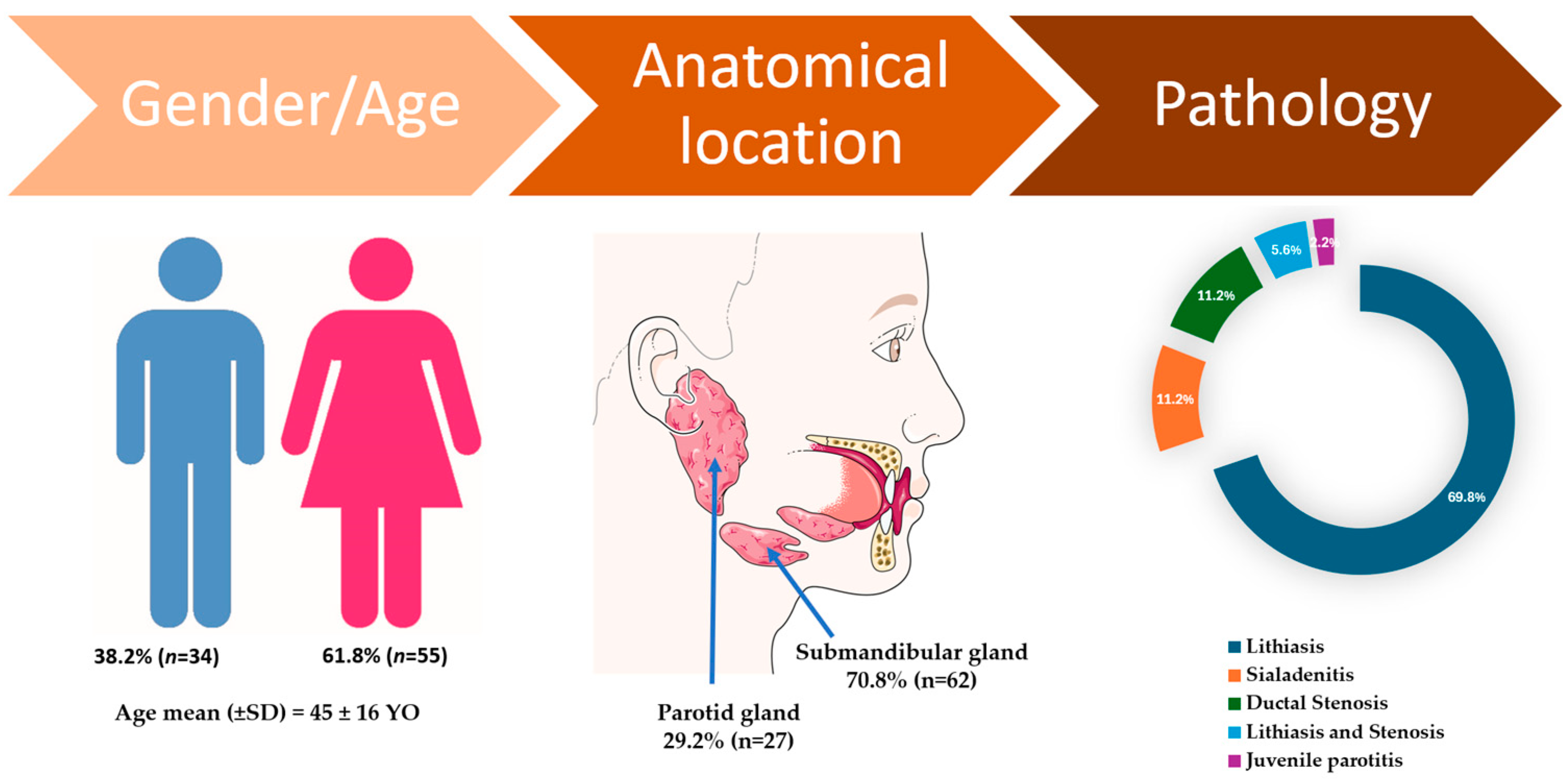
Figure 2.
Sialolithotomy of a 9 mm calculus: (a) CBCT axial view showing the radiopaque calculus; (b) Intraoperative view; (c) Calculus removed from Wharton’s duct; (d) The calculus following sialolithotomy.
Figure 2.
Sialolithotomy of a 9 mm calculus: (a) CBCT axial view showing the radiopaque calculus; (b) Intraoperative view; (c) Calculus removed from Wharton’s duct; (d) The calculus following sialolithotomy.
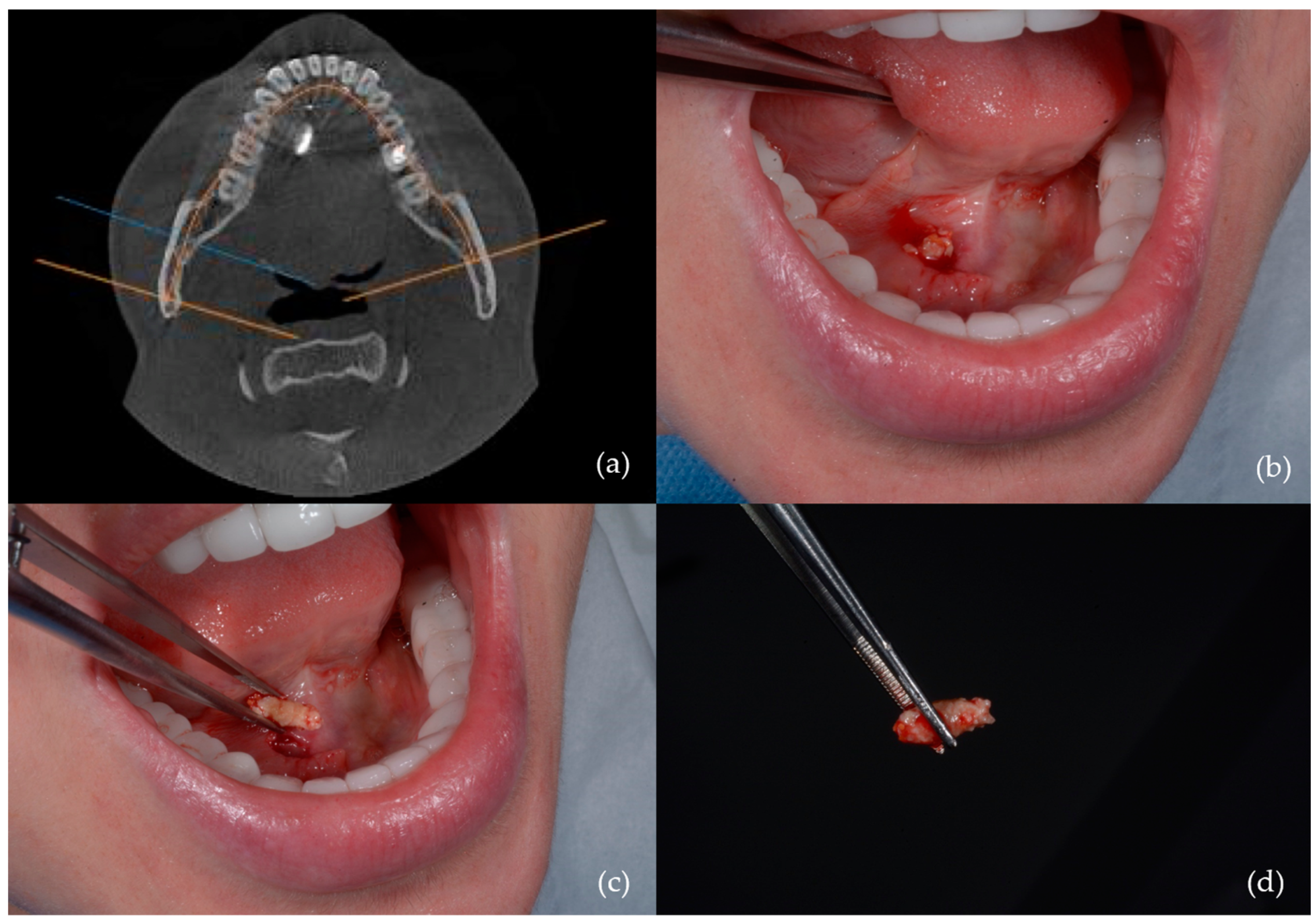
Figure 3.
Calculus removal by sialolithotripsy – procedural steps: (a) Initial appearance; (b) Identification of a longitudinal fissure; (c) Appearance after removal of a fragment from the sialolith; (d) Fragmentation of the sialolith into two distinct portions.
Figure 3.
Calculus removal by sialolithotripsy – procedural steps: (a) Initial appearance; (b) Identification of a longitudinal fissure; (c) Appearance after removal of a fragment from the sialolith; (d) Fragmentation of the sialolith into two distinct portions.
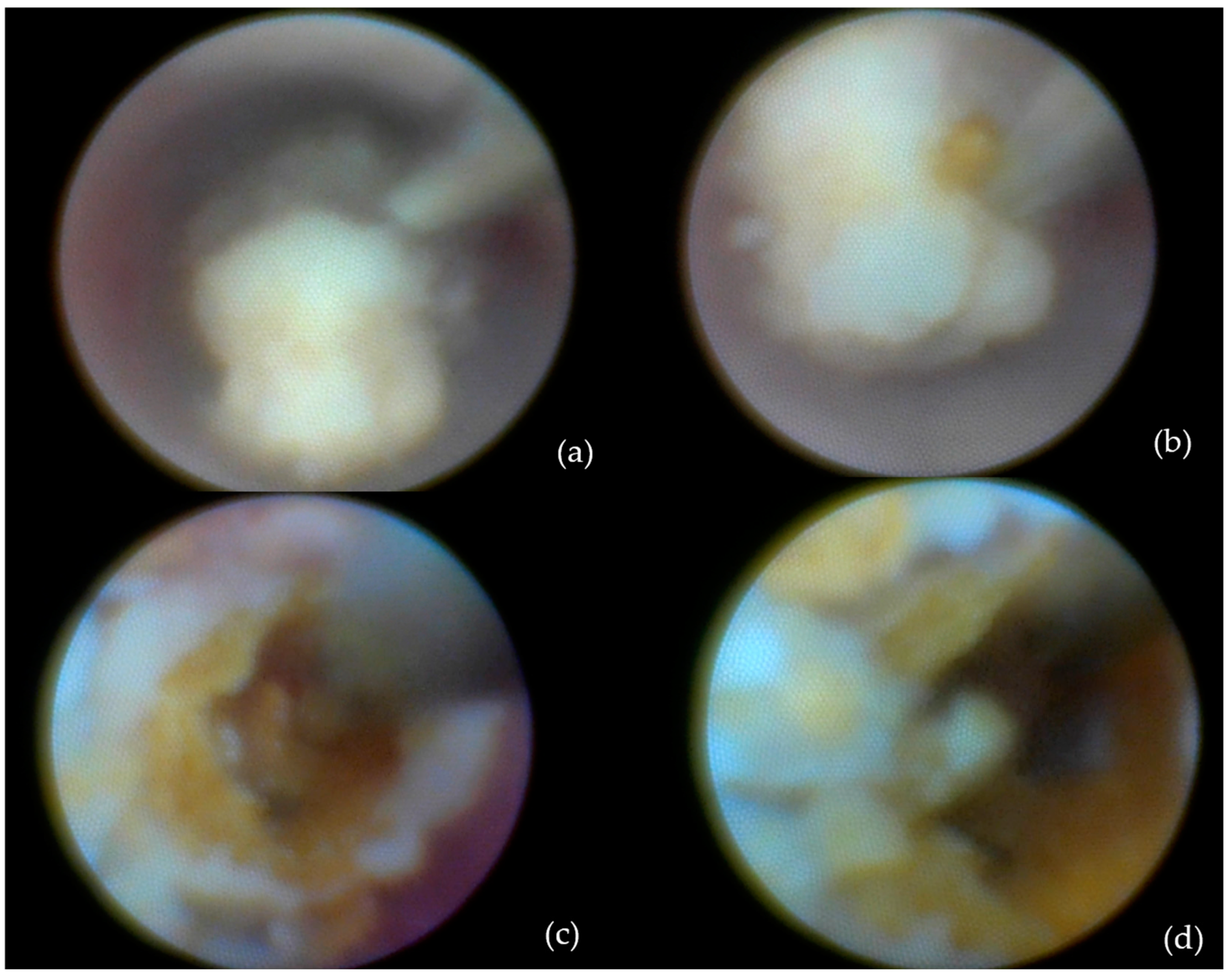
Figure 4.
Interventional sialendoscopy using a Dormia basket: (a) Stone captured with the extraction probe; (b) Stone secured within the Dormia-type device (macro lens view).
Figure 4.
Interventional sialendoscopy using a Dormia basket: (a) Stone captured with the extraction probe; (b) Stone secured within the Dormia-type device (macro lens view).
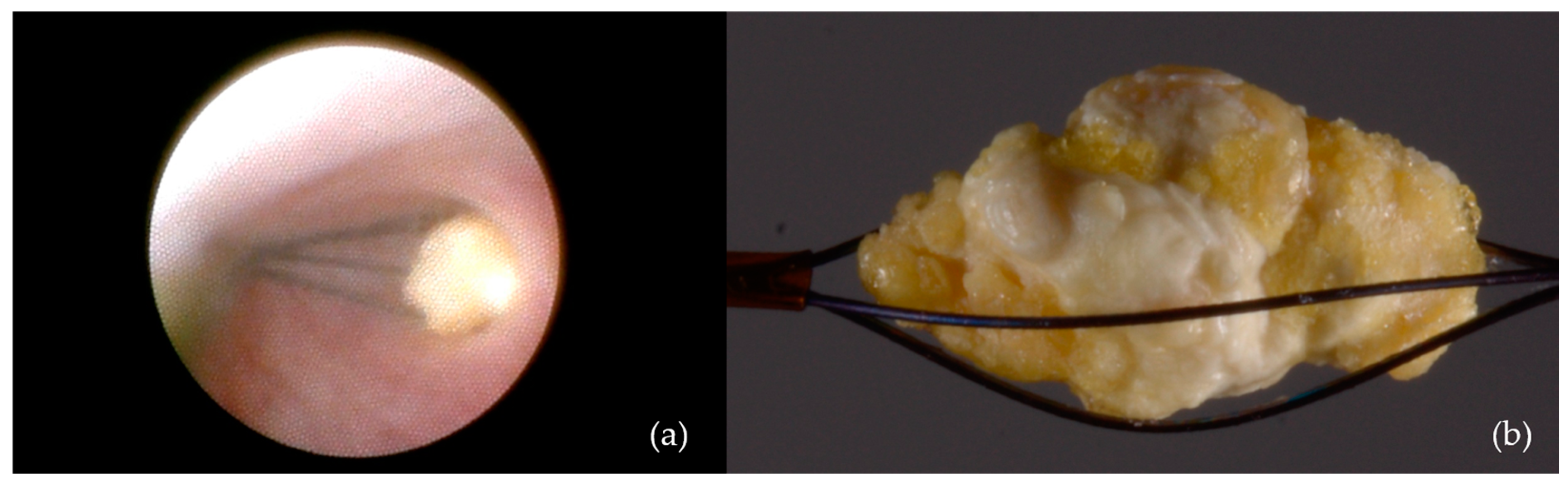
Figure 5.
Ductal dilation and stenting: (a) Balloon used for dilation of the stenotic duct segment; (b) Progressive ductal expansion; (c) Stent used for maintaining ductal patency; (d) Stent positioned and secured within Stensen’s duct.
Figure 5.
Ductal dilation and stenting: (a) Balloon used for dilation of the stenotic duct segment; (b) Progressive ductal expansion; (c) Stent used for maintaining ductal patency; (d) Stent positioned and secured within Stensen’s duct.

Figure 6.
One of the cases requiring submandibulectomy involved a patient with 10 calculi: (a) Submandibular gland and sialoliths following submandibulectomy; (b) Clinical aspect 48 hours post-submandibulectomy.
Figure 6.
One of the cases requiring submandibulectomy involved a patient with 10 calculi: (a) Submandibular gland and sialoliths following submandibulectomy; (b) Clinical aspect 48 hours post-submandibulectomy.
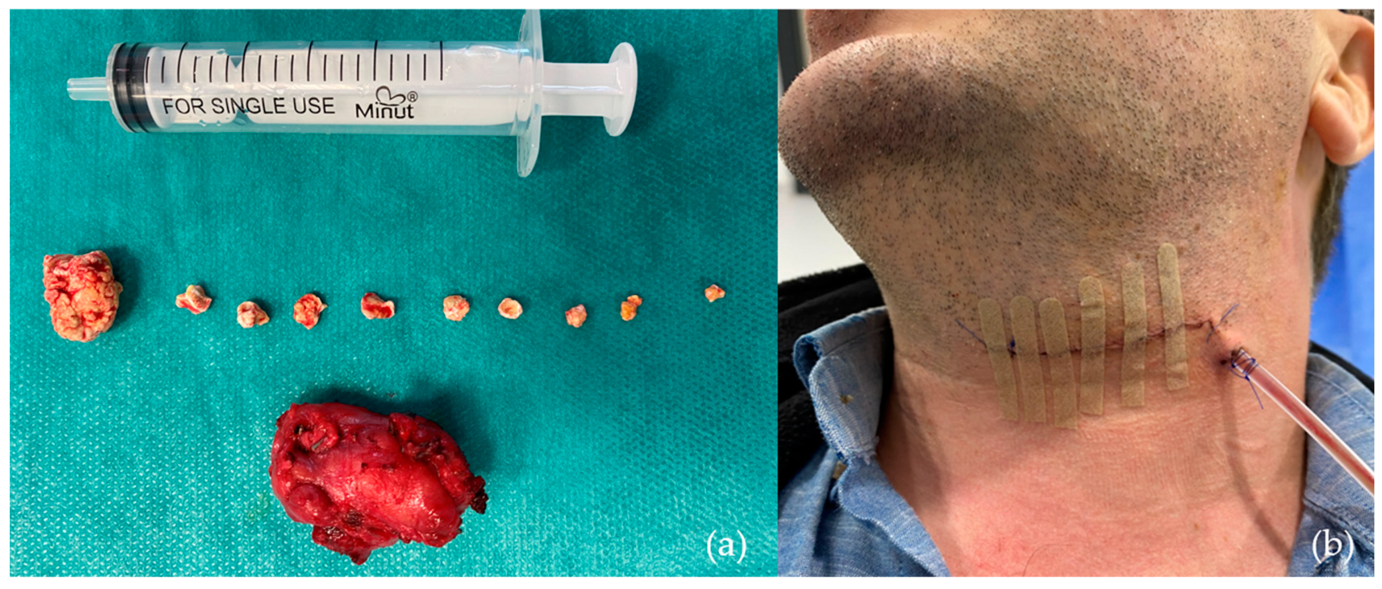

Table 1.
Descriptive evaluations stratified by gender and salivary gland involvement (parotid vs submandibular).
Table 1.
Descriptive evaluations stratified by gender and salivary gland involvement (parotid vs submandibular).
| Variable | Female (n=55) | Male (n=34) | P-value | Parotid | Submandibular | P-value | Total (n=89) |
|---|---|---|---|---|---|---|---|
| Age (mean ± SD) | 43.7±17.0 | 46.2±14.8 | 0.46 | 41.89±16.50 | 45.85±15.96 | 0.29 | 45 ± 16 |
| Gland involved | |||||||
| Parotid | 18 (32.7%) | 9 (26.5%) | 0.67 | 27 (29.2%) | - | - | 27 (29.2%) |
| Submandibular | 37 (67.3%) | 25 (73.5%) | 0.67 | - | 62 (70.8%) | - | 62 (70.8%) |
| Calculus size (mm) | 7.19±4.17 | 6.80±5.24 | 0.75 | 5.07±1.82 | 7.57±4.92 | 0.00* | 7.04±4.56 |
| Pathology | |||||||
| Lithiasis | 38 (69.1%) | 24 (70.6%) | 1.00 | 10 (37.1%) | 52 (83.9%) | 0.00* | 64 (69.8%) |
| Sialadenitis | 7 (12.7%) | 3 (8.8%) | 0.83 | 5 (18.5%) | 5 (8.1%) | 0.28 | 10 (11.2%) |
| Ductal Stenosis | 6 (10.9%) | 4 (11.8%) | 1.00 | 6 (22.2%) | 4 (6.5%) | 0.07 | 10 (11.2%) |
| Lithiasis and stenosis | 4 (7.3%) | 1 (2.9%) | 0.69 | 4 (14.8%) | 1 (1.5%) | 0.05* | 5 (5.6%) |
| Juvenile parotitis | 0 | 2 (5.9%) | 0.28 | 2 (7.4%) | 0 | - | 2 (2.2%) |
| Imaging Investigations | |||||||
| Ultrasound | 47 (85.5%) | 30 (88.2%) | 0.96 | 25 (92.59%) | 52 (83.87%) | 0.44 | 77(86.5%) |
| CBCT | 34 (61.8%) | 20 (58.8%) | 0.95 | 8 (29.6%) | 46 (74.19%) | 0.00* | 54 (60.7%) |
| Medical CT | 2 (3.6%) | 4 (11.8%) | 0.29 | 5 (18.52%) | 1 (1.61%) | 0.01* | 6 (6.7%) |
| MRI | 3 (5.5%) | 2 (5.9%) | 1.0 | 2 (3.23%) | 0.32 | 5 (5.6%) | |
| CBCT Sialography | 2 (3.6%) | 1 (2.9%) | 1.0 | 2 (7.40%) | 1 (1.61%) | 0.45 | 3 (3.4%) |
| Anesthesia | |||||||
| Local | 53 (96.4%) | 30 (88.2%) | 0.29 | 27 (100%) | 56 (90.32%) | 0.22 | 83 (93.26%) |
| General | 2 (3.6%) | 4 (11.8%) | 0.29 | 0 | 6 (9.68%) | 0.22 | 6 (6.74%) |
| Therapeutic Interventions | |||||||
| Transoral sialolithotomy | 19 (34.5%) | 10 (29.4%) | 0.79 | 0 | 29 (8.06%) | 0.46 | 29 (32.6%) |
| Intracorporeal lithotripsy | 18 (32.7%) | 11 (32.4%) | 1.0 | 12 (44.4%) | 17 (27.42%) | 0.18 | 29 (32.6%) |
| Dormia basket extraction | 6 (10.9%) | 2 (5.9%) | 0.67 | 3 (11.11%) | 5 (8.06%) | 0.95 | 8 (9.0%) |
| Ductal dilation and stenting | 4 (7.3%) | 3 (8.8%) | 1.0 | 4 (14.81%) | 3 (4.84%) | 0.24 | 7 (7.9%) |
| Submandibulectomy | 1 (1.8%) | 4 (11.8%) | 0.13 | 0 | 5 (46.77%) | 0.31 | 5 (5.6%) |
CBCT = cone beam computed tomography; Medical CT = medical computed tomography; MRI = magnetic resonance imaging; * = statistically significant.
Disclaimer/Publisher’s Note: The statements, opinions and data contained in all publications are solely those of the individual author(s) and contributor(s) and not of MDPI and/or the editor(s). MDPI and/or the editor(s) disclaim responsibility for any injury to people or property resulting from any ideas, methods, instructions or products referred to in the content. |
© 2025 by the authors. Licensee MDPI, Basel, Switzerland. This article is an open access article distributed under the terms and conditions of the Creative Commons Attribution (CC BY) license (http://creativecommons.org/licenses/by/4.0/).
Copyright: This open access article is published under a Creative Commons CC BY 4.0 license, which permit the free download, distribution, and reuse, provided that the author and preprint are cited in any reuse.


