Submitted:
20 March 2025
Posted:
21 March 2025
You are already at the latest version
Abstract
Nanoparticles (NPs) have revolutionized biomedical and pharmaceutical applications due to their unique physicochemical properties. However, their widespread use has raised concerns regarding their potential toxicity, particularly through oxidative stress mechanisms. Oxidative stress, primarily driven by reactive oxygen species (ROS) overproduction, plays a central role in NP-induced toxicity, leading to cellular dysfunction, inflammation, apoptosis, and genotoxicity. Zebrafish (Danio rerio) has emerged as a powerful in vivo model for nanotoxicology, offering advantages such as genetic similarity to humans, rapid development, and optical transparency, allowing real-time monitoring of oxidative damage. This review synthesizes current findings on NP-induced oxidative stress in zebrafish, highlighting key toxicity mechanisms and case studies involving metallic (gold, silver, copper), metal oxide (zinc oxide, titanium dioxide, iron oxide), polymeric, and lipid-based NPs. The influence of NP physicochemical properties, such as size, surface charge, and functionalization on oxidative stress responses is explored. Additionally, experimental approaches used to assess ROS generation, antioxidant enzyme activity, and oxidative damage biomarkers in zebrafish models are examined. In addition to toxicity concerns, pharmaceutical applications of antioxidant-modified NPs are evaluated, particularly their potential in drug delivery, neuroprotection, and disease therapeutics. Challenges in zebrafish-based nanotoxicology, the need for standardized methodologies, and future directions for optimizing NP design to minimize oxidative stress-related risks are also discussed. By integrating insights from toxicity mechanisms, case studies, and pharmaceutical strategies, this review supports the development of safer and more effective nanoparticle-based therapies while addressing the challenges of oxidative stress-related toxicity.
Keywords:
nanoparticles
; oxidative stress
; zebrafish
; pharmaceutical applications
1. Introduction
Nanoparticles (NPs) are increasingly utilized across multiple industries, including pharmaceuticals, biomedical imaging, drug delivery, cosmetics, diagnostics, and environmental applications [1]. Due to their small size and high surface area-to-volume ratio, NPs exhibit unique physicochemical properties that enhance their reactivity and bioavailability, making them ideal for various biomedical and technological applications [2]. In the pharmaceutical field, NPs serve as efficient drug carriers, improving drug solubility, targeted delivery, and controlled release, reducing side effects and enhancing therapeutic efficacy [3]. Metallic NPs, such as gold (Au) and silver (Ag), are extensively applied in biomedical imaging and diagnostics due to their optical and electronic properties [4]. Additionally, lipid-based and polymeric NPs are commonly employed in drug delivery systems, offering enhanced biocompatibility and reduced toxicity [5]. Beyond biomedical applications, NPs are widely used in cosmetics and skincare products, where they enhance product stability and skin penetration [6]. Titanium dioxide (TiO₂) and zinc oxide (ZnO) NPs, for example, are key ingredients in sunscreens, providing superior UV protection [7]. However, concerns regarding the potential toxicity of NPs, especially their ability to generate oxidative stress, have increased [8]. Environmental exposure to NPs through industrial waste, air pollution, and contaminated water sources has raised significant health concerns, necessitating further research into their long-term effects on biological systems [9].
One of the most critical toxicological effects associated with NPs is oxidative stress. This occurs when NPs induce an imbalance between the production of reactive oxygen species (ROS) and the antioxidant defense mechanisms within biological systems [10]. ROS, including superoxide anion (O₂⁻), hydrogen peroxide (H₂O₂), and hydroxyl radicals (OH⁻), play a crucial role in cellular signaling but become harmful when generated excessively [11]. Overproduction of ROS leads to oxidative damage to lipids, proteins, and nucleic acids, contributing to cellular dysfunction and various pathological conditions [12].
Numerous studies have demonstrated that exposure to metal and metal oxide NPs, such as AgNPs, CuO NPs, and ZnO NPs, results in increased ROS production, leading to oxidative stress-induced apoptosis and genotoxicity [13]. The oxidative stress triggered by NPs can activate multiple pathways, including mitochondrial dysfunction, DNA damage, and inflammatory responses [14]. Prolonged oxidative stress has been linked to chronic inflammation, which plays a crucial role in the pathogenesis of various diseases, including cancer, neurodegeneration, and cardiovascular disorders [15]. Understanding oxidative stress as a central mechanism of NP toxicity is essential for developing safer nanomaterials with reduced adverse effects.
Zebrafish (Danio rerio) has emerged as a widely used vertebrate model in nanotoxicology due to its genetic similarity to humans (~70%), rapid development, and ease of maintenance in laboratory settings [16]. One of the key advantages of zebrafish is the optical transparency of embryos, allowing real-time visualization of NP interactions and oxidative stress responses in developing tissues [17]. This feature makes zebrafish an excellent in vivo model for assessing NP-induced oxidative damage, mitochondrial dysfunction, and inflammatory responses [15].
Studies have shown that zebrafish embryos exposed to AgNPs, TiO₂ NPs, and CdSe quantum dots exhibit significant ROS generation, leading to apoptosis and developmental abnormalities [18]. In particular, AgNPs have been reported to induce oxidative stress-mediated neurotoxicity and cardiovascular dysfunction in zebrafish models [12]. The use of zebrafish in NP toxicity assessments provides valuable insights into NP biodistribution, bioaccumulation, and their effects on different organ systems. Furthermore, zebrafish models are increasingly employed in high-throughput screening assays to evaluate pharmaceutical formulations and antioxidant-modified NPs designed to mitigate oxidative stress (Figure 1) [19].
This review aims to provide a comprehensive analysis of NP-induced oxidative stress in zebrafish, focusing on its toxicological implications and pharmaceutical applications. The study highlights key mechanisms of oxidative stress, evaluates case studies involving different NP types, and explores the role of zebrafish as a model for assessing NP safety and therapeutic potential. Additionally, the review addresses challenges in nanotoxicology and the need for standardized methodologies to improve NP risk assessment and pharmaceutical development.
2. Physicochemical Properties of Nanoparticles Influencing Oxidative Stress
2.1. NP Size and Surface Area
The size of nanoparticles plays a crucial role in their toxicity potential, as smaller NPs exhibit higher surface-area-to-volume ratios, increasing their reactivity and potential to induce oxidative stress [20]. Studies have demonstrated that reduced NP size correlates with increased ROS generation and toxicity in aquatic organisms, including zebrafish. For instance, silver nanoparticles (AgNPs) smaller than 10 nm have been shown to trigger higher oxidative stress levels compared to larger particles due to their enhanced cellular penetration and bioavailability [21,22]. Similarly, titanium dioxide (TiO₂) NPs of <20 nm have been reported to induce significant ROS production and mitochondrial dysfunction in zebrafish embryos [23,24]. Table 1 presents a synthesis of studies on the size of various NPs and their induced oxidative stress.
Silver Nanoparticles (Ag NPs): Zebrafish embryos exposed to AgNPs of varying sizes demonstrated that smaller AgNPs (10 nm) triggered more severe ROS production and developmental toxicity compared to larger particles (50 nm) [21]. The study indicated that smaller AgNPs disrupted the antioxidant defense system, downregulating antioxidant enzymes such as superoxide dismutase (SOD) and catalase (CAT) [44].
Gold Nanoparticles (Au NPs): Investigations on AuNPs found that particles smaller than 20 nm induced more oxidative stress than larger ones due to their ability to accumulate in tissues and penetrate cellular compartments [45]. Additionally, smaller AuNPs enhanced pro-inflammatory cytokine expression, further exacerbating ROS generation [46,47].
Copper Nanoparticles (Cu NPs) have gained attention in nanotoxicology due to their unique physicochemical properties and potential biomedical applications [36]. Their small size (≤20 nm) leads to increased oxidative stress, causing significant DNA damage and mitochondrial dysfunction in biological systems [48]. Studies have demonstrated that Cu NPs induce oxidative stress through ROS overproduction, which can trigger inflammation and apoptosis in zebrafish tissues [35,49].
Iron Nanoparticles (Fe NPs) are widely used in environmental remediation and biomedical applications. Their unique magnetic properties make them valuable for drug delivery, hyperthermia treatment, and MRI contrast agents [39]. Studies show that Fe NPs generate ROS through Fenton reactions, causing cellular damage and mitochondrial dysfunction in zebrafish models [50]. Exposure to Fe NPs in zebrafish embryos has been associated with oxidative stress-induced apoptosis, DNA damage, and developmental abnormalities [51].
Zinc Oxide Nanoparticles: Different sizes were evaluated for oxidative stress induction in zebrafish larvae, revealing that smaller ZnO NPs led to significantly higher ROS production and mitochondrial damage [31,33]. The study suggested that the smaller NPs had increased bioavailability, leading to prolonged oxidative stress and apoptosis [34,52].
Titanium Dioxide Nanoparticles (TiO₂ NPs): A recent study demonstrated that TiO₂ NPs induce oxidative stress and neurotoxic effects in zebrafish embryos, disrupting behavioral responses and mitochondrial function [24]. These findings suggest that TiO₂ NPs contribute to ROS accumulation and affect neural pathways, highlighting their potential long-term toxicity in aquatic organisms and human health implications [23].
Copper Oxide Nanoparticles (CuO NPs) are widely utilized in industrial and biomedical applications due to their antimicrobial properties and catalytic activity [36]. Studies have shown that CuO NPs induce substantial ROS production, causing lipid peroxidation, mitochondrial dysfunction, and DNA fragmentation in zebrafish models. Exposure to CuO NPs in zebrafish embryos has been linked to apoptotic cell death and developmental toxicity due to their ability to disrupt redox homeostasis [28].
Iron(III) Oxide Nanoparticles (Fe₂O₃ NPs) have superparamagnetic properties. Studies have indicated that Fe₂O₃ NPs can induce oxidative DNA fragmentation, lipid peroxidation, and mitochondrial dysfunction in zebrafish models, leading to apoptosis and neurotoxicity [53]. Additionally, zebrafish embryos exposed to Fe₂O₃ NPs exhibit inflammatory responses and oxidative stress in gill and liver tissues due to ROS-induced damage [50,54].
Cadmium Selenide Quantum Dots (CdSe QDs) have optoelectronic applications due to their fluorescent properties. Studies have demonstrated that CdSe QDs induce DNA fragmentation, lipid peroxidation, and apoptosis in zebrafish models, leading to developmental abnormalities and neurotoxicity [55]. Exposure to CdSe QDs has also been linked to mitochondrial dysfunction, as the nanoparticles interfere with electron transport chain activity, leading to increased ROS generation and cellular damage [56].
Zinc Sulfide Quantum Dots (ZnS QDs) have photonic applications due to their unique optical properties. Studies have shown that ZnS QDs can induce lipid peroxidation, oxidative DNA damage, and apoptosis in zebrafish embryos, particularly under illumination conditions [42,57]. Exposure to ZnS QDs has also been linked to mitochondrial stress and inflammatory responses, contributing to oxidative toxicity [43].
These findings reinforce the idea that NP size plays a crucial role in determining oxidative stress outcomes and toxicity levels. Future research should focus on optimizing NP size to balance efficacy in pharmaceutical applications while minimizing oxidative damage.
2.2. Surface Charge and Coating Effects
The charge and surface modifications of NPs significantly influence their cellular uptake, bioavailability, and oxidative stress potential. Positively charged (cationic) NPs tend to exhibit greater cytotoxicity due to enhanced cellular uptake and stronger electrostatic interactions with negatively charged cell membranes [58]. Conversely, surface coatings such as polyethylene glycol (PEG) can modify NP interactions and mitigate oxidative stress [59]. Figure 2 represents a schematic of the importance of the surface charge and the coating effects.
Cationic NPs show stronger ROS production and cytotoxicity compared to neutral or anionic NPs [60]. These nanoparticles tend to interact more aggressively with negatively charged cell membranes, facilitating their internalization and causing significant oxidative stress. Studies have demonstrated that cationic NPs exhibit increased genotoxicity and inflammatory responses due to their ability to disrupt cell membranes, induce mitochondrial dysfunction, and activate apoptosis pathways [61]. For example, research on AgNPs has shown that positively charged silver nanoparticles are more cytotoxic than their neutral or negatively charged counterparts, leading to increased ROS production and DNA damage in zebrafish embryos [11,62]. Similarly, CuNPs exhibit a strong capacity to generate oxidative stress, leading to lipid peroxidation and cell membrane rupture [63]. Furthermore, cationic NPs have been linked to neuroinflammation and cardiovascular dysfunction in zebrafish models, highlighting the need for careful assessment of surface charge when designing nanoparticle-based applications in biomedical and pharmaceutical sciences [64,65,66].
Anionic and Neutral NPs tend to show reduced oxidative stress responses, as their charge properties limit cellular interactions and membrane permeability, leading to lower toxicity [67]. Studies have previously demonstrated that negatively charged NPs have lower cytotoxicity due to their repulsion from negatively charged cell membranes, reducing internalization and subsequent ROS production. Neutral lipid-based nanoparticles like liposomes have shown that they do not significantly induce ROS generation, making them promising candidates for drug delivery applications [68,69,70]. In another study, negatively charged Au NPs exhibited reduced inflammatory response and minimal oxidative damage compared to positively charged counterparts [71]. The reduced interaction of anionic and neutral NPs with cellular components suggests their potential as safer alternatives in biomedical applications, particularly in drug delivery and antioxidant therapy.
Surface coatings and modifications of NPs play a crucial role in influencing their interactions with biological systems, particularly in mitigating oxidative stress [61]. Functionalizing NPs with polymers such as polyethylene glycol (PEG) or albumin can reduce ROS production by decreasing NP aggregation and limiting cellular internalization. For instance, PEGylation of AuNPs has been shown to diminish oxidative stress and inflammatory responses [72,73]. Similarly, coating AgNPs with biocompatible polymers has been demonstrated to reduce their ROS generation and improve stability in biological environments [74]. These findings underscore the importance of controlled NP surface modifications to optimize biocompatibility and minimize oxidative stress-related toxicity in biological systems.
3. Oxidative Stress Pathways Activated by Nanoparticles in Zebrafish
NP-induced oxidative stress in zebrafish occurs either through direct ROS generation or secondary mechanisms like inflammation [16]. Excess ROS production disrupts cellular homeostasis, leading to biomolecular damage. One significant effect of NP exposure is the inhibition of key antioxidant enzymes, such as SOD, CAT, and GPx, weakening the cellular defense system [29]. Exposure to TiO₂ NPs has been shown to suppress antioxidant defenses in zebrafish liver and gill tissues, resulting in oxidative damage and inflammation [24]. Similarly, AgNPs significantly reduce SOD and CAT activity in zebrafish larvae, increasing oxidative stress and lipid peroxidation [44]. CuO NPs further disrupt glutathione homeostasis, impairing detoxification pathways and exacerbating ROS accumulation [75]. Also, ZnO NP exposure depleted antioxidant enzyme levels in zebrafish embryos, correlating with mitochondrial dysfunction and apoptosis [36]. These effects are synthetized in Table 2.
3.1. Mitochondrial Dysfunction and Apoptotic Pathways
Mitochondria are highly susceptible to oxidative stress caused by NP exposure. ROS accumulation in mitochondria disrupts electron transport chain (ETC) function, reduces ATP production, and activates pro-apoptotic signaling pathways [5,36]. NP-induced oxidative damage can trigger p53 tumor suppressor activation, leading to caspase-3-mediated apoptosis. A study on zebrafish embryos exposed to CuO NPs demonstrated significant mitochondrial swelling, cytochrome c release, and p53 activation, which triggers caspase-3-mediated apoptosis. This process has been associated with neurotoxicity in zebrafish brain tissue [80]. Another study found that AgNP exposure led to mitochondrial membrane potential collapse in zebrafish neurons, accelerating neurotoxicity through p53 upregulation and caspase-3 activation [81]. Additionally, TiO₂ NPs were reported to induce oxidative damage in zebrafish liver cells, impairing mitochondrial respiration and promoting apoptotic cell death [82]. Furthermore, CdSe quantum dots disrupted mitochondrial membrane integrity in zebrafish cardiomyocytes, contributing to cardiac dysfunction and increased apoptotic cell death [83].
3.2. Inflammation and Immune Dysregulation
NP exposure in zebrafish has been linked to immune system dysregulation through the activation of pro-inflammatory cytokines such as tumor necrosis factor-alpha (TNF-α), interleukin-6 (IL-6), and nuclear factor-kappa B (NF-κB) [29]. These cytokines are key mediators of inflammation, leading to immune overactivation and subsequent tissue damage. Chronic immune activation contributes to prolonged oxidative stress, affecting multiple organ systems, including the gills, liver, and nervous system [54]. Studies indicate that NP-induced inflammation can also disrupt hematopoiesis and immune cell homeostasis, further exacerbating toxicity. Recent studies have shown that Fe₂O₃ NPs not only activate NF-κB pathways in zebrafish gills, leading to oxidative stress and impaired respiratory function but also increase the expression of IL-8 and IL-1β, leading to macrophage recruitment and localized inflammation [10]. Another study demonstrated that AgNPs cause upregulation of TNF-α and IL-6 in zebrafish liver cells, promoting hepatotoxicity and oxidative damage [84]. Additionally, exposure to TiO₂ NPs in zebrafish larvae resulted in increased leukocyte infiltration and cytokine secretion, demonstrating NP-induced immune activation in early developmental stages [85].
3.3. Genotoxicity and Lipid Peroxidation
Oxidative stress induced by NPs can lead to severe genotoxic effects, including DNA fragmentation, chromosomal aberrations, and mutagenic potential, which may contribute to long-term developmental and reproductive toxicity in zebrafish. DNA damage is commonly assessed through comet assays and micronucleus tests, which have revealed increased genotoxic markers upon exposure to metal-based and semiconductor NPs. Additionally, lipid peroxidation—caused by excessive ROS attacking membrane lipids—compromises cellular integrity, disrupts signaling pathways, and initiates apoptotic cascades [8]. Studies have shown that oxidative stress-driven lipid peroxidation is a key mechanism in NP-mediated toxicity, particularly in organs with high lipid content such as the brain and liver. CdSe QDs were found to induce DNA strand breaks in zebrafish larvae, leading to chromosomal fragmentation and developmental abnormalities [86]. A study using fluorescent in situ hybridization (FISH) confirmed increased nuclear damage following CdSe QD exposure, indicating their potential to cause long-term genetic instability [87]. Another study demonstrated that exposure to ZnO NPs in zebrafish embryos resulted in both DNA double-strand breaks and increased lipid peroxidation in liver and muscle tissues, further confirming the dual genotoxic and oxidative stress-related impacts of these NPs. Histopathological examination showed mitochondrial swelling and nuclear fragmentation, consistent with apoptotic progression [88].
4. Experimental Approaches for Assessing Oxidative Stress in Zebrafish
4.1. Biochemical Assays for ROS and Antioxidant Activity
Biochemical assays are fundamental for quantifying oxidative stress markers in zebrafish tissues, providing insights into the impact of nanoparticles on redox balance. The most commonly used assays include:
Lipid Peroxidation Assay: Measures malondialdehyde (MDA) levels, a key indicator of lipid peroxidation. Elevated MDA levels correlate with membrane lipid degradation and oxidative stress in NP-exposed zebrafish. Studies have shown increased MDA production in zebrafish embryos exposed to AgNPs and ZnO NPs, linking lipid damage to NP-induced toxicity [89].
Total ROS Quantification: Fluorometric and chemiluminescent methods detect ROS accumulation in tissues. The DCFDA assay is widely used for quantifying intracellular ROS levels. In zebrafish models, increased ROS production has been documented following CuO NP exposure, with ROS levels peaking at 24 hours post-exposure [90].
Enzyme Activity Assays: Assess antioxidant defense mechanisms through measurement of SOD and CAT enzyme activity. Studies indicate that exposure to TiO₂ NPs leads to a significant reduction in SOD and CAT activity, suggesting antioxidant depletion and impaired oxidative defense in zebrafish liver and gill tissues [82].
4.2. Gene expression Analysis of Oxidative Stress Pathways
Examining gene expression changes in oxidative stress-related pathways provides insights into NP-induced molecular alterations. Quantitative PCR (qPCR) and RNA sequencing (RNA-seq) are commonly used to analyze differential gene expression in zebrafish exposed to various nanoparticles [10]. Key oxidative stress-related genes include:
nrf2: Master regulator of antioxidant response, upregulated in response to oxidative stress. Studies have shown increased nrf2 expression in zebrafish larvae following exposure to AgNPs, correlating with an upregulation of downstream detoxifying enzymes [3,91].
sod1: Encodes superoxide dismutase 1, responsible for neutralizing superoxide radicals. Zebrafish embryos exposed to ZnO NPs demonstrated sod1 overexpression as an early defense mechanism against ROS accumulation [34].
gpx1a: Encodes glutathione peroxidase, a key enzyme in detoxifying hydrogen peroxide and lipid peroxides. Research has demonstrated that CdSe quantum dots induce gpx1a suppression in zebrafish liver, leading to increased oxidative damage and apoptosis [92].
Studies comparing metal oxides, polymeric NPs, and quantum dots have revealed distinct gene expression profiles. For example, TiO₂ NPs predominantly upregulate nrf2 and sod1, while AgNPs induce a more systemic stress response, affecting inflammatory and oxidative stress-related genes. Polymeric NPs tend to induce a more moderate response, with lower activation of detoxification genes, suggesting reduced toxicity compared to metal-based NPs [93].
4.3. Histopathological and Imaging Techniques
Advanced imaging techniques enable direct visualization of oxidative stress-induced damage caused by nanoparticles in zebrafish tissues (Table 3). These approaches are essential for assessing nanoparticle localization, oxidative damage, and cellular alterations at the histopathological level.
4.4. Behavioral and Physiological Endpoints
Oxidative stress can manifest as behavioral and physiological abnormalities in zebrafish, providing crucial indicators of NP-induced toxicity. Behavioral changes and physiological responses serve as non-invasive biomarkers of oxidative stress and neurotoxicity.
Swimming Activity: Reduced motility, erratic swimming patterns, and hypoactivity in NP-exposed zebrafish indicate neurotoxic effects associated with oxidative stress. A study on AgNP exposure demonstrated decreased locomotion and anxiety-like behavior in zebrafish larvae, correlating with increased ROS levels in the brain [100].
Heart Rate Variability: Cardiotoxicity is evaluated by measuring bradycardia or tachycardia in NP-exposed embryos. Zebrafish exposed to CuO NPs exhibited significant alterations in heart rate, attributed to mitochondrial dysfunction and ROS accumulation in cardiac tissues [75,101].
Developmental Malformations: Craniofacial deformities, pericardial edema, and spinal curvature are common teratogenic effects linked to NP-induced oxidative stress. Research on TiO₂ NPs exposure in zebrafish embryos revealed increased malformations, with ROS-mediated DNA damage being a key underlying mechanism [10,81].
5. Pharmaceutical Applications of Nanoparticles Using Zebrafish (Danio rerio) Models
Zebrafish have become an essential vertebrate model in nanomedicine research due to their high genetic homology to humans (~70%), rapid organ development, and affordability in large-scale experiments. Their high fecundity enables large-scale pharmacological screenings, and their transparent embryos allow real-time tracking of NP biodistribution and interactions in vivo. Zebrafish have been successfully used for toxicity screening, drug efficacy evaluations, and nanoparticle-mediated therapeutic interventions [102]. Studies indicate that selenium nanoparticles (SeNPs), quercetin-functionalized AuNPs, and curcumin-loaded NPs significantly enhance antioxidant enzyme activity in zebrafish tissues, mitigating ROS-related toxicity [103,104,105]. Recent findings highlight the potential of nanoencapsulated polyphenols in promoting cellular defense mechanisms against oxidative stress [106]. AuNPs associated with curcumin have been shown to exert neuroprotective effects in zebrafish models of oxidative stress, reducing neuronal apoptosis by 40% and significantly upregulating antioxidant enzymes (SOD, GPx, and CAT) in brain tissues [107]. Another study demonstrated that curcumin-loaded polymeric nanoparticles increased zebrafish survival rates in oxidative stress models by 50% compared to free curcumin administration [108]. Additionally, lipid-based antioxidant NPs loaded with resveratrol demonstrated enhanced bioavailability and anti-inflammatory effects in zebrafish models of cardiovascular disease, reducing oxidative stress markers and preventing endothelial dysfunction [109,110].
5.1. Nanoparticles as Antioxidant Therapeutics in Pharmaceutical Sciences
Nanoparticles infused with antioxidants show significant potential in the treatment of oxidative stress-related diseases, including neurodegenerative disorders, cardiovascular conditions, and cancer. By acting as ROS scavengers, these NPs can mitigate oxidative damage, prevent inflammatory cascades, and improve cellular resilience. Numerous studies indicate that NP-based antioxidant systems efficiently neutralize ROS, thereby protecting cells from oxidative damage [8]. For example, cerium oxide (CeO₂) NPs exhibit intrinsic catalytic activity that mimics SOD and CAT, making them highly effective in oxidative stress models [111]. A study on zebrafish oxidative stress models found that CeO₂ NPs reduced lipid peroxidation by 45% and significantly improved neuronal survival rates. The catalytic nature of CeO₂ NPs helped restore normal antioxidant enzyme function, effectively reducing neurotoxicity in zebrafish larvae exposed to oxidative stress-inducing compounds [112]. Other research has demonstrated that selenium nanoparticles (SeNPs) and AuNPs functionalized with flavonoids provide neuroprotection in zebrafish models of Parkinson’s disease and Alzheimer’s disease by preventing oxidative damage and modulating inflammatory pathways [113,114].
5.2. Natural Antioxidants Used in NP Formulations
The incorporation of natural antioxidants into NP formulations enhances their therapeutic potential while reducing toxicity. These information are detailed in Table 4.
5.3. Drug Delivery and Biocompatibility Testing
Advanced imaging techniques, such as fluorescence imaging, confocal microscopy, and bioluminescence tracking, allow for real-time visualization of NP distribution in zebrafish models. Studies using quantum dots (QDs) and radiolabeled NPs have demonstrated their ability to monitor NP uptake, accumulation, and clearance in different organs, offering valuable pharmacokinetic insights [120].
Zebrafish models have been extensively used to analyze NP absorption, circulation, metabolism, and excretion. Studies on AuNPs and polymeric nanocarriers have shown their prolonged circulation times and biodistribution in zebrafish embryos, mimicking pharmacokinetic profiles observed in mammalian models [121]. Research on lipid-based NPs has also indicated improved bioavailability, making them suitable carriers for hydrophobic drugs [122,123].
Polylactic-co-glycolic acid (PLGA) NPs loaded with antioxidants have exhibited controlled drug release in zebrafish models, demonstrating sustained therapeutic effects, enhanced bioavailability, and reduced systemic toxicity. A study on curcumin-loaded PLGA NPs reported prolonged circulation and targeted accumulation in zebrafish liver, leading to a 50% reduction in ROS levels compared to free curcumin administration. Furthermore, chitosan-coated PLGA nanoparticles encapsulating vitamin E exhibited sustained release and antioxidant protection, significantly improving zebrafish survival rates under oxidative stress conditions and restoring mitochondrial integrity [108,124,125].
5.4. Balancing Toxicity versus Therapeutic Potential
Zebrafish-based oxidative stress models allow researchers to assess how different nanoparticle compositions and dosages affect biological systems and provide insights into their long-term safety profiles.
Studies have shown that determining the safe concentration range for therapeutic nanoparticles (NPs) is crucial to minimize off-target effects. Research involving AuNPs and polymeric nanocarriers in zebrafish models has demonstrated that precise dosing can reduce cytotoxicity while enhancing drug delivery efficiency. For instance, a study on the toxicity of various nanomedicine materials, including gold and iron oxide nanoparticles, evaluated their effects in zebrafish embryos, highlighting the importance of assessing safe concentration ranges to minimize adverse effects [54]. Moreover, during our own experience with silver NPs we observed a striking contrast in the biological effects of plant extracts when administered alone versus when incorporated into silver nanoparticle formulations. When used independently, the extracts demonstrated significant antioxidant and neuroprotective effects without any detectable toxicity. However, upon integration within silver NPs, at the same dose and under identical experimental conditions, we noticed an unexpected increase in toxicity. These findings highlight the complexity of NPs-mediated substance delivery, where interactions between the nanocarrier and the bioactive compound can alter pharmacodynamics and toxicity. Based on such discrepancies, comprehensive pharmacokinetics studies are essential in order to elucidate how NPs formulations can influence absorption, distribution and metabolism of therapeutic compounds. Understanding these mechanisms is imperative in order to properly balance the therapeutic potential of NPs formulations while mitigating possible unexpected and unintended toxicological effects.
Other studies on chronic NP exposure in zebrafish revealed potential risks associated with prolonged usage. AgNPs and TiO₂ NPs have been found to accumulate in the liver and the brain of zebrafish, leading to oxidative damage and inflammatory responses over extended periods [24,126]. Research on lipid-based NPs suggests that their biodegradable nature may reduce long-term toxicity risks, making them promising candidates for pharmaceutical applications [127].
Furthermore, a study on iron oxide (Fe₃O₄) NPs demonstrated that chronic exposure in zebrafish induced significant oxidative stress and neurotoxicity, highlighting the need for careful evaluation of NP formulations before their use in clinical settings [128].
6. Conclusions
The findings of this review highlight the significant role of oxidative stress in NP-induced toxicity, particularly in zebrafish models. Studies have demonstrated that various nanoparticles, including metal-based (AgNPs, TiO₂ NPs, Fe₂O₃ NPs), polymeric, lipid-based, and quantum dots, can induce oxidative stress through ROS overproduction, mitochondrial dysfunction, inflammation, and genotoxicity. Zebrafish have emerged as a versatile and reliable model organism for evaluating NP-induced oxidative stress, offering advantages such as genetic similarity to humans, rapid development, and transparent embryos that allow real-time assessment of oxidative damage and pharmacological responses.
From a pharmaceutical perspective, NPs functionalized with antioxidants, such as curcumin, resveratrol, quercetin, and selenium nanoparticles, have shown promising therapeutic applications in neuroprotection, hepatoprotection, and cardiovascular disease management. Zebrafish models have been instrumental in screening NP formulations, assessing biodistribution, and optimizing drug delivery systems to balance efficacy with safety. Additionally, zebrafish-based toxicity studies have provided valuable insights into the long-term risks associated with NP accumulation, guiding the development of safer nanopharmaceuticals. Despite these advances, standardized methodologies for oxidative stress assessment in zebrafish models remain a critical need. Future research should focus on long-term NP exposure studies, multi-generational toxicity assessments, and the integration of omics technologies (transcriptomics, proteomics, and metabolomics) to deepen the understanding of NP interactions with biological systems.
Furthermore, advancing the biodegradability and biocompatibility of NP formulations will be essential in mitigating potential toxic effects while maximizing therapeutic benefits. In conclusion, zebrafish models provide an effective and high-throughput platform for evaluating both the toxicity and therapeutic potential of nanoparticles. Understanding NP-induced oxidative stress mechanisms will not only facilitate the design of safer nanomaterials but also accelerate the development of novel antioxidant-based nanomedicines for various biomedical applications.
Future studies should consider expanding multi-generational studies (long-term exposure studies on zebrafish should be conducted to assess how NP-induced oxidative stress affects successive generations, particularly in the context of epigenetic modifications and inherited toxicity), personalized nanomedicine (develop patient-specific nanotherapeutics, particularly for neurodegenerative diseases, cancer, and metabolic disorders), green nanotechnology approaches (develop environmentally friendly and biocompatible NPs using sustainable materials to reduce toxicity risks while maintaining therapeutic efficacy)
Author Contributions
Conceptualization, D.B.-M., O.C. and M.B.; Data Curation, M.B.; Formal Analysis, A-M.M. and C.M.; Funding Acquisition, D.B.-M.; Investigation, D.B.-M., M.B., A.F.B., O.C., C.M., I.I.L., A-M.M. and M.H.; Methodology, M.B. and C.M.; Project Administration, M.H.; Resources, M.H.; Software, G-A.M.; Supervision, O.C. and M.H.; Validation, M.B.; Visualization, A.F.B. and G-A.M.; Writing—Original Draft, D.B.-M., M.B., A-M.M. and O.C.; Writing—Review and Editing, M.H. and C.M. All authors have read and agreed to the published version of the manuscript.
Funding
This research received no external funding.
Institutional Review Board Statement
Not applicable.
Data Availability Statement
No new data were created.
Conflicts of Interest
The authors declare no conflicts of interest and all images included in this article are original and were created using BIORENDER software.
Abbreviations
The following abbreviations are used in this manuscript:
| NPs | Nanoparticles |
| ROS | Reactive oxygen species |
| CAT | Catalase |
| SOD | Superoxide dismutase |
| GPx | Glutathione peroxidase |
| DNA | Deoxyribonucleic acid |
| MDA | Malondialdehyde |
| DCFDA | 2′,7′-dichlorofluorescin diacetate |
| RNA | Ribonucleic acid |
| ATP | Adenosine triphosphate |
| IL | Interleukin |
| TNF-α | Tumor necrosis factor alpha |
| PCR | Polymerase chain reaction |
References
- Sies, H. Oxidative Stress: Eustress and Distress in Redox Homeostasis; Elsevier Inc., 2019; ISBN 9780128131466.
- Nikalje, A. Nanotechnology and Its Applications in Medicine. Med chem. 2015, 5, 81–89. [Google Scholar] [CrossRef]
- Morales-González, J. A Master Regulator of Oxidative Stress - The Transcription Factor Nrf2; 2016; ISBN 978-953-51-2838-0.
- Etheridge, M.L.; Campbell, S.A.; Erdman, A.G.; Haynes, C.L.; Wolf, S.M.; McCullough, J. The Big Picture on Nanomedicine: The State of Investigational and Approved Nanomedicine Products. Nanomedicine Nanotechnology, Biol. Med. 2013, 9, 1–14. [Google Scholar] [CrossRef] [PubMed]
- Patel, T.A.; Kevadiya, B.D.; Bajwa, N.; Singh, P.A.; Zheng, H.; Kirabo, A.; Li, Y.L.; Patel, K.P. Role of Nanoparticle-Conjugates and Nanotheranostics in Abrogating Oxidative Stress and Ameliorating Neuroinflammation. Antioxidants 2023, 12. [Google Scholar] [CrossRef] [PubMed]
- Boroumand, Z.; Golmakani, N.; Boroumand, S. Clinical Trials on Silver Nanoparticles for Wound Healing. Nanomed. J 2018, 5, 186–191. [Google Scholar] [CrossRef]
- Chowdhury, S.; Saikia, S.K. Oxidative Stress in Fish: A Review. J. Sci. Res. 2020, 12, 145–160. [Google Scholar] [CrossRef]
- Elsayed Azab, A.; A Adwas, Almokhtar; Ibrahim Elsayed, A. S.; A Adwas, A.; Ibrahim Elsayed, Ata Sedik; Quwaydir, F.A. Oxidative Stress and Antioxidant Mechanisms in Human Body. J. Appl. Biotechnol. Bioeng. 2019, 6, 43–47. [Google Scholar] [CrossRef]
- Haque, E.; Ward, A.C. Zebrafish as a Model to Evaluate Nanoparticle Toxicity. Nanomaterials 2018, 8, 1–18. [Google Scholar] [CrossRef]
- Mutalik, C.; Nivedita; Sneka, C. ; Krisnawati, D.I.; Yougbaré, S.; Hsu, C.C.; Kuo, T.R. Zebrafish Insights into Nanomaterial Toxicity: A Focused Exploration on Metallic, Metal Oxide, Semiconductor, and Mixed-Metal Nanoparticles. Int. J. Mol. Sci. 2024, 25. [Google Scholar] [CrossRef]
- Bonfanti, P.; Colombo, A.; Bengalli, R.; Gualtieri, M.; Zanoni, I.; Blosi, M.; Costa, A.; Mantecca, P. Functional Silver-Based Nanomaterials Affecting Zebrafish Development: The Adverse Outcomes in Relation to the Nanoparticle Physical and Chemical Structure. Environ. Sci. Nano 2024, 11, 2521–2540. [Google Scholar] [CrossRef]
- Mugoni, V.; Camporeale, A.; Santoro, M.M. Analysis of Oxidative Stress in Zebrafish Embryos. J. Vis. Exp. 2014. [Google Scholar] [CrossRef]
- Dormousoglou, M.; Efthimiou, I.; Antonopoulou, M.; Fetzer, D.L.; Hamerski, F.; Corazza, M.L.; Papadaki, M.; Santzouk, S.; Dailianis, S.; Vlastos, D. Investigation of the Genotoxic, Antigenotoxic and Antioxidant Profile of Different Extracts from Equisetum Arvense L. Antioxidants 2022, 11, 1393. [Google Scholar] [CrossRef] [PubMed]
- Mourabit, S.; Fitzgerald, J.A.; Ellis, R.P.; Takesono, A.; Porteus, C.S.; Trznadel, M.; Metz, J.; Winter, M.J.; Kudoh, T.; Tyler, C.R. New Insights into Organ-Specific Oxidative Stress Mechanisms Using a Novel Biosensor Zebrafish. Environ. Int. 2019, 133, 105138. [Google Scholar] [CrossRef] [PubMed]
- Lungu-Mitea, S.; Oskarsson, A.; Lundqvist, J. Development of an Oxidative Stress in Vitro Assay in Zebrafish (Danio Rerio) Cell Lines. Sci. Rep. 2018, 8, 1–11. [Google Scholar] [CrossRef]
- Zhao, W.; Chen, Y.; Hu, N.; Long, D.; Cao, Y. The Uses of Zebrafish (Danio Rerio) as an in Vivo Model for Toxicological Studies: A Review Based on Bibliometrics. Ecotoxicol. Environ. Saf. 2024, 272, 116023. [Google Scholar] [CrossRef]
- Murugasan Kuppuswamy, J.; Seetharaman, B. Monocrotophos Based Pesticide Alters the Behavior Response Associated with Oxidative Indices and Transcription of Genes Related to Apoptosis in Adult Zebrafish (Danio Rerio) Brain. Biomed. Pharmacol. J. 2020, 13, 1291–1304. [Google Scholar] [CrossRef]
- Asharani, P.V.; Lian Wu, Y.; Gong, Z.; Valiyaveettil, S. Toxicity of Silver Nanoparticles in Zebrafish Models. Nanotechnology 2008, 19. [Google Scholar] [CrossRef]
- YEŞİLBUDAK, B. Toxicological Aspects and Bioanalysis of Nanoparticles: Zebrafish Model. Open J. Nano 2023, 8, 22–35. [Google Scholar] [CrossRef]
- Oberdörster, G.; Stone, V.; Donaldson, K. Toxicology of Nanoparticles: A Historical Perspective. Nanotoxicology 2007, 1, 2–25. [Google Scholar] [CrossRef]
- Singh, S.P.; Bhargava, C.S.; Dubey, V.; Mishra, A.; Singh, Y. Silver Nanoparticles: Biomedical Applications, Toxicity, and Safety Issues. Int. J. Res. Pharm. Pharm. Sci. 2017, 2, 2455–2698. [Google Scholar]
- Caloudova, H.; Hodkovicova, N.; Sehonova, P.; Blahova, J.; Marsalek, B.; Panacek, A.; Svobodova, Z. The Effect of Silver Nanoparticles and Silver Ions on Zebrafish Embryos (Danio Rerio). Neuroendocrinol. Lett. 2018, 39, 299–304. [Google Scholar]
- Teulon, J.-M.; Godon, C.; Chantalat, L.; Moriscot, C.; Cambedouzou, J.; Odorico, M.; Ravaux, J.; Podor, R.; Gerdil, A.; Habert, A.; et al. On the Operational Aspects of Measuring Nanoparticle Sizes. Nanomaterials 2018, 9, 18. [Google Scholar] [CrossRef]
- Caruso, G.; Scalisi, E.M.; Pecoraro, R.; Cardaci, V.; Privitera, A.; Truglio, E.; Capparucci, F.; Jarosova, R.; Salvaggio, A.; Caraci, F.; et al. Effects of Carnosine on the Embryonic Development and TiO2 Nanoparticles-Induced Oxidative Stress on Zebrafish. Front. Vet. Sci. 2023, 10. [Google Scholar] [CrossRef] [PubMed]
- Qiang, L.; Arabeyyat, Z.H.; Xin, Q.; Paunov, V.N.; Dale, I.J.F.; Mills, R.I.L.; Rotchell, J.M.; Cheng, J. Silver Nanoparticles in Zebrafish (Danio Rerio) Embryos: Uptake, Growth and Molecular Responses. Int. J. Mol. Sci. 2020, 21, 1–14. [Google Scholar] [CrossRef] [PubMed]
- Chakraborty, C.; Sharma, A.R.; Sharma, G.; Lee, S.S. Zebrafish: A Complete Animal Model to Enumerate the Nanoparticle Toxicity. J. Nanobiotechnology 2016, 14, 1–13. [Google Scholar] [CrossRef]
- Bar-Ilan, O.; Albrecht, R.M.; Fako, V.E.; Furgeson, D.Y. Toxicity Assessments of Multisized Gold and Silver Nanoparticles in Zebrafish Embryos. Small 2009, 5, 1897–1910. [Google Scholar] [CrossRef] [PubMed]
- Bondarenko, O.; Juganson, K.; Ivask, A.; Kasemets, K.; Mortimer, M.; Kahru, A. Toxicity of Ag, CuO and ZnO Nanoparticles to Selected Environmentally Relevant Test Organisms and Mammalian Cells in Vitro: A Critical Review. Arch. Toxicol. 2013, 87, 1181–1200. [Google Scholar] [CrossRef]
- Kumar, N.; Thorat, S.T.; Gunaware, M.A.; Kumar, P.; Reddy, K.S. Unraveling Gene Regulation Mechanisms in Fish: Insights into Multistress Responses and Mitigation through Iron Nanoparticles. Front. Immunol. 2024, 15, 1–19. [Google Scholar] [CrossRef]
- Vargas-Ortiz, J.R.; Gonzalez, C.; Esquivel, K. Magnetic Iron Nanoparticles: Synthesis, Surface Enhancements, and Biological Challenges. Processes 2022, 10, 2282. [Google Scholar] [CrossRef]
- Bai, W.; Zhang, Z.; Tian, W.; He, X.; Ma, Y.; Zhao, Y.; Chai, Z. Toxicity of Zinc Oxide Nanoparticles to Zebrafish Embryo: A Physicochemical Study of Toxicity Mechanism. J. Nanoparticle Res. 2010, 12, 1645–1654. [Google Scholar] [CrossRef]
- Sirelkhatim, A.; Mahmud, S.; Seeni, A.; Kaus, N.H.M.; Ann, L.C.; Bakhori, S.K.M.; Hasan, H.; Mohamad, D. Review on Zinc Oxide Nanoparticles: Antibacterial Activity and Toxicity Mechanism. Nano-Micro Lett. 2015, 7, 219–242. [Google Scholar] [CrossRef]
- Zavitri, N.G.; Syahbaniati, A.P.; Primastuti, R.K.; Putri, R.M.; Damayanti, S.; Wibowo, I. Toxicity Evaluation of Zinc Oxide Nanoparticles Green Synthesized Using Papaya Extract in Zebrafish. Biomed. Reports 2023, 19, 1–11. [Google Scholar] [CrossRef] [PubMed]
- Al-Zahaby, S.A.; Farag, M.R.; Alagawany, M.; Taha, H.S.A.; Varoni, M.V.; Crescenzo, G.; Mawed, S.A. Zinc Oxide Nanoparticles (ZnO-NPs) Induce Cytotoxicity in the Zebrafish Olfactory Organs via Activating Oxidative Stress and Apoptosis at the Ultrastructure and Genetic Levels. Animals 2023, 13. [Google Scholar] [CrossRef]
- Kovrižnych, J.A.; Sotńikóva, R.; Zeljenková, D.; Rollerová, E.; Szabová, E.; Wimmerov́a, S. Acute Toxicity of 31 Different Nanoparticles to Zebrafish (Danio Rerio) Tested in Adulthood and in Early Life Stages - Comparative Study. Interdiscip. Toxicol. 2013, 6, 67–73. [Google Scholar] [CrossRef] [PubMed]
- Cameron, S.J.; Sheng, J.; Hosseinian, F.; Willmore, W.G. Nanoparticle Effects on Stress Response Pathways and Nanoparticle–Protein Interactions. Int. J. Mol. Sci. 2022, 23. [Google Scholar] [CrossRef]
- Karlsson, H.L.; Cronholm, P.; Gustafsson, J.; Möller, L. Copper Oxide Nanoparticles Are Highly Toxic: A Comparison between Metal Oxide Nanoparticles and Carbon Nanotubes. Chem. Res. Toxicol. 2008, 21, 1726–1732. [Google Scholar] [CrossRef] [PubMed]
- Kooi, M.E.; Cappendijk, V.C.; Cleutjens, K.B.J.M.; Kessels, A.G.H.; Kitslaar, P.J.E.H.M.; Borgers, M.; Frederik, P.M.; Daemen, M.J.A.P.; van Engelshoven, J.M.A. Accumulation of Ultrasmall Superparamagnetic Particles of Iron Oxide in Human Atherosclerotic Plaques Can Be Detected by in Vivo Magnetic Resonance Imaging. Circulation 2003, 107, 2453–2458. [Google Scholar] [CrossRef]
- Malhotra, N.; Lee, J.-S.; Liman, R.A.D.; Ruallo, J.M.S.; Villaflores, O.B.; Ger, T.-R.; Hsiao, C.-D. Potential Toxicity of Iron Oxide Magnetic Nanoparticles: A Review. Molecules 2020, 25, 3159. [Google Scholar] [CrossRef]
- Johnson, J.M.; Mohamed, A.S.R.; Ding, Y.; Wang, J.; Lai, S.Y.; Fuller, C.D.; Shah, R.; Butler, R.T.; Weber, R.S. Ultra-small Superparamagnetic Iron Oxide (USPIO) Magnetic Resonance Imaging in Benign Mixed Tumor of the Parotid Gland. Clin. Case Reports 2021, 9, 123–127. [Google Scholar] [CrossRef]
- Ferrucci, J.T.; Stark, D.D. Iron Oxide-Enhanced MR Imaging of the Liver and Spleen: Review of the First 5 Years. Am. J. Roentgenol. 1990, 155, 943–950. [Google Scholar] [CrossRef]
- Le, N.; Zhang, M.; Kim, K. Quantum Dots and Their Interaction with Biological Systems. Int. J. Mol. Sci. 2022, 23, 10763. [Google Scholar] [CrossRef]
- Jia, H.R.; Zhu, Y.X.; Duan, Q.Y.; Chen, Z.; Wu, F.G. Nanomaterials Meet Zebrafish: Toxicity Evaluation and Drug Delivery Applications. J. Control. Release 2019, 311–312, 301–318. [Google Scholar] [CrossRef] [PubMed]
- Yan, Z.; Zhou, Y.; Zhu, P.; Bao, X.; Su, P. Polystyrene Nanoplastics Mediated the Toxicity of Silver Nanoparticles in Zebrafish Embryos. Front. Mar. Sci. 2023, 10, 1–11. [Google Scholar] [CrossRef]
- Ielo, I.; Rando, G.; Giacobello, F.; Sfameni, S.; Castellano, A.; Galletta, M.; Drommi, D.; Rosace, G.; Plutino, M.R. Synthesis, Shemical–Physical Characterization, and Biomedical Applications of Functional Gold Nanoparticles: A Review. Molecules 2021, 26, 5823. [Google Scholar] [CrossRef]
- Ramalingam, V.; Varunkumar, K.; Ravikumar, V.; Rajaram, R. Target Delivery of Doxorubicin Tethered with PVP Stabilized Gold Nanoparticles for Effective Treatment of Lung Cancer. Sci. Rep. 2018, 8, 3815. [Google Scholar] [CrossRef] [PubMed]
- Kus-Liśkiewicz, M.; Fickers, P.; Ben Tahar, I. Biocompatibility and Cytotoxicity of Gold Nanoparticles: Recent Advances in Methodologies and Regulations. Int. J. Mol. Sci. 2021, 22, 10952. [Google Scholar] [CrossRef]
- Lei, R.; Yang, B.; Wu, C.; Liao, M.; Ding, R.; Wang, Q. Mitochondrial Dysfunction and Oxidative Damage in the Liver and Kidney of Rats Following Exposure to Copper Nanoparticles for Five Consecutive Days. Toxicol. Res. (Camb). 2015, 4, 351–364. [Google Scholar] [CrossRef]
- Jing, Y.Y.; Tai, Z.P.; Liu, J.X. Copper Nanoparticles and Silver Nanoparticles Impair Lymphangiogenesis in Zebrafish. Cell Commun. Signal. 2024, 22, 1–15. [Google Scholar] [CrossRef]
- Gaharwar, U.S.; Meena, R.; Rajamani, P. Iron Oxide Nanoparticles Induced Cytotoxicity, Oxidative Stress and DNA Damage in Lymphocytes. J. Appl. Toxicol. 2017, 37, 1232–1244. [Google Scholar] [CrossRef] [PubMed]
- Singh, N.; Jenkins, G.J.S.; Asadi, R.; Doak, S.H. Potential Toxicity of Superparamagnetic Iron Oxide Nanoparticles (SPION). Nano Rev. 2010, 1, 5358. [Google Scholar] [CrossRef]
- Agarwal, H.; Shanmugam, V. A Review on Anti-Inflammatory Activity of Green Synthesized Zinc Oxide Nanoparticle: Mechanism-Based Approach. Bioorg. Chem. 2020, 94, 103423. [Google Scholar] [CrossRef]
- Tran, S.; DeGiovanni, P.; Piel, B.; Rai, P. Cancer Nanomedicine: A Review of Recent Success in Drug Delivery. Clin. Transl. Med. 2017, 6. [Google Scholar] [CrossRef]
- D’Amora, M.; Schmidt, T.J.N.; Konstantinidou, S.; Raffa, V.; De Angelis, F.; Tantussi, F. Effects of Metal Oxide Nanoparticles in Zebrafish. Oxid. Med. Cell. Longev. 2022, 2022. [Google Scholar] [CrossRef]
- Derfus, A.M.; Chan, W.C.W.; Bhatia, S.N. Probing the Cytotoxicity of Semiconductor Quantum Dots. Nano Lett. 2004, 4, 11–18. [Google Scholar] [CrossRef] [PubMed]
- Fischer, A.; Brodziak-Dopierała, B.; Loska, K.; Stojko, J. The Assessment of Toxic Metals in Plants Used in Cosmetics and Cosmetology. Int. J. Environ. Res. Public Health 2017, 14. [Google Scholar] [CrossRef] [PubMed]
- Lungu, I.-I. Catechin-Zinc-Complex: Synthesis, Characterization and Biological Assessment. Farmacia 2023, 71, 755–763. [Google Scholar] [CrossRef]
- Marana, M.H.; Poulsen, R.; Thormar, E.A.; Clausen, C.G.; Thit, A.; Mathiessen, H.; Jaafar, R.; Korbut, R.; Hansen, A.M.B.; Hansen, M.; et al. Plastic Nanoparticles Cause Mild Inflammation, Disrupt Metabolic Pathways, Change the Gut Microbiota and Affect Reproduction in Zebrafish: A Full Generation Multi-Omics Study. J. Hazard. Mater. 2022, 424, 127705. [Google Scholar] [CrossRef]
- Sen, G.T.; Ozkemahli, G.; Shahbazi, R.; Erkekoglu, P.; Ulubayram, K.; Kocer-Gumusel, B. The Effects of Polymer Coating of Gold Nanoparticles on Oxidative Stress and DNA Damage. Int. J. Toxicol. 2020, 39, 328–340. [Google Scholar] [CrossRef]
- Loos, C.; Simmet, T.; Syrovets, T. Role of Nanoparticle Surface Charge in Their Toxicity. E3S Web Conf. 2024, 575, 02009. [Google Scholar] [CrossRef]
- Auclair, J.; Turcotte, P.; Gagnon, C.; Peyrot, C.; Wilkinson, K.J.; Gagne, F. The Influence of Surface Coatings of Silver Nanoparticles on the Bioavailability and Toxicity to Elliptio Complanata Mussels. J. Nanomater. 2019, 2019. [Google Scholar] [CrossRef]
- D’amora, M.; Raffa, V.; De Angelis, F.; Tantussi, F. Toxicological Profile of Plasmonic Nanoparticles in Zebrafish Model. Int. J. Mol. Sci. 2021, 22, 1–32. [Google Scholar] [CrossRef]
- Eskandarinezhad, S.; Wani, I.A.; Nourollahileilan, M.; Khosla, A.; Ahmad, T. Review—Metal and Metal Oxide Nanoparticles/Nanocomposites as Electrochemical Biosensors for Cancer Detection. J. Electrochem. Soc. 2022, 169, 047504. [Google Scholar] [CrossRef]
- Batir-Marin, D.; Boev, M.; Cioanca, O.; Mircea, C.; Burlec, A.F.; Beppe, G.J.; Spac, A.; Corciova, A.; Hritcu, L.; Hancianu, M. Neuroprotective and Antioxidant Enhancing Properties of Selective Equisetum Extracts. Molecules 2021, 26, 2565. [Google Scholar] [CrossRef]
- Paatero, I.; Casals, E.; Niemi, R.; Özliseli, E.; Rosenholm, J.M.; Sahlgren, C. Analyses in Zebrafish Embryos Reveal That Nanotoxicity Profiles Are Dependent on Surface-Functionalization Controlled Penetrance of Biological Membranes. Sci. Rep. 2017, 7, 1–13. [Google Scholar] [CrossRef] [PubMed]
- Fang, C.; Kievit, F.M.; Cho, Y.-C.; Mok, H.; Press, O.W.; Zhang, M. Effect of Cationic Side-Chains on Intracellular Delivery and Cytotoxicity of PH Sensitive Polymer–Doxorubicin Nanocarriers. Nanoscale 2012, 4, 7012. [Google Scholar] [CrossRef]
- Liaqat, N.; Jahan, N.; Khalil-ur-Rahman; Anwar, T. ; Qureshi, H. Green Synthesized Silver Nanoparticles: Optimization, Characterization, Antimicrobial Activity, and Cytotoxicity Study by Hemolysis Assay. Front. Chem. 2022, 10. [Google Scholar] [CrossRef]
- Evensen, L.; Johansen, P.L.; Koster, G.; Zhu, K.; Herfindal, L.; Speth, M.; Fenaroli, F.; Hildahl, J.; Bagherifam, S.; Tulotta, C.; et al. Zebrafish as a Model System for Characterization of Nanoparticles against Cancer. Nanoscale 2016, 8, 862–877. [Google Scholar] [CrossRef]
- Sercombe, L.; Veerati, T.; Moheimani, F.; Wu, S.Y.; Sood, A.K.; Hua, S. Advances and Challenges of Liposome Assisted Drug Delivery. Front. Pharmacol. 2015, 6. [Google Scholar] [CrossRef] [PubMed]
- Daraee, H.; Etemadi, A.; Kouhi, M.; Alimirzalu, S.; Akbarzadeh, A. Application of Liposomes in Medicine and Drug Delivery. Artif. Cells, Nanomedicine, Biotechnol. 2016, 44, 381–391. [Google Scholar] [CrossRef] [PubMed]
- TYAl-Abdullah, Z.; Al-Shawi, A.A.A.; Aboud, M.N.; Abdulaziz, B.A.A.; Al-Furaiji, H.Q.M.; Luaibi, I.N. Synthesis and Analytical Characterization of Gold Nanoparticles Using Microwave-Assisted Extraction System and Study Their Application in Degradation. J. Nanostructures 2020, 10, 682–690. [Google Scholar] [CrossRef]
- Amatya, R.; Hwang, S.; Park, T.; Min, K.A.; Shin, M.C. In Vitro and in Vivo Evaluation of PEGylated Starch-Coated Iron Oxide Nanoparticles for Enhanced Photothermal Cancer Therapy. Pharmaceutics 2021, 13, 871. [Google Scholar] [CrossRef]
- Mostovei, A. Practical Aspects of the Use of Acrylic Biomaterials in Dental Medical Practice. Med. Mater. 2022, 2, 25–30. [Google Scholar] [CrossRef]
- Mallick, K.; Witcomb, M.J.; Scurrell, M.S. Polymer Stabilized Silver Nanoparticles: A Photochemical Synthesis Route. J. Mater. Sci. 2004, 39, 4459–4463. [Google Scholar] [CrossRef]
- Liu, N.; Tong, L.; Li, K.; Dong, Q.; Jing, J. Copper-Nanoparticle-Induced Neurotoxic Effect and Oxidative Stress in the Early Developmental Stage of Zebrafish (Danio Rerio). Molecules 2024, 29. [Google Scholar] [CrossRef] [PubMed]
- Naghibi, F.; Khalaj, A.; Mosaddegh, M.; Malekmohamadi, M.; Hamzeloo-Moghadam, M. Cytotoxic Activity Evaluation of Some Medicinal Plants, Selected from Iranian Traditional Medicine Pharmacopoeia to Treat Cancer and Related Disorders. J. Ethnopharmacol. 2014, 155, 230–239. [Google Scholar] [CrossRef] [PubMed]
- Xiong, G.; Deng, Y.; Liao, X.; Zhang, J.; Cheng, B.; Cao, Z.; Lu, H. Graphene Oxide Nanoparticles Induce Hepatic Dysfunction through the Regulation of Innate Immune Signaling in Zebrafish (Danio Rerio). Nanotoxicology 2020, 14, 667–682. [Google Scholar] [CrossRef]
- Johnston, H.J.; Gillies, S.L.J.; Verdon, R.; Stone, V.; Henry, T.; Tran, L.; Tucker, C.; Rossi, A.G.; Tyler, C.R. Application of Transgenic Zebrafish for Investigating Inflammatory Responses to Nanomaterials: Recommendations for New Users. F1000Research 2023, 12, 51. [Google Scholar] [CrossRef]
- Almasoud, H.A.; Ali, D.; Yaseen, K.N.; Almukhlafi, H.; Alothman, N.S.; Almutairi, B.; Almeer, R.; Alyami, N.; Alkahtani, S.; Alarifi, S. Dose-Dependent Variation in Anticancer Activity of Hexane and Chloroform Extracts of Field Horsetail Plant on Human Hepatocarcinoma Cells. Biomed Res. Int. 2022, 2022, 1–8. [Google Scholar] [CrossRef]
- Li, Y.; Sun, Y.; Zhang, G.; He, Z.; Wang, Y.; Cui, J. Effects of Copper Oxide Nanoparticles on Developing Zebrafish Embryos and Larvae. Int. J. Nanomedicine 2016, 905. [Google Scholar] [CrossRef]
- Rochaa*, A.C.P.T.G.M.R.F.M.T.L. The Zebrafish Embryotoxicity Test (ZET) for Nanotoxicity Assessment: From Morphological to Molecular Approach. 2019, 110, 10–11.
- Tang, T.; Zhang, Z.; Zhu, X. Toxic Effects of TiO2 NPs on Zebrafish. Int. J. Environ. Res. Public Health 2019, 16, 523. [Google Scholar] [CrossRef]
- Wang, Y.; Tang, M. Dysfunction of Various Organelles Provokes Multiple Cell Death after Quantum Dot Exposure. Int. J. Nanomedicine 2018, Volume 13, 2729–2742. [Google Scholar] [CrossRef]
- Zheng, Y.; Song, J.; Qian, Q.; Wang, H. Silver Nanoparticles Induce Liver Inflammation through Ferroptosis in Zebrafish. Chemosphere 2024, 362, 142673. [Google Scholar] [CrossRef] [PubMed]
- Mamboungou, J.; Canedo, A.; Qualhato, G.; Rocha, T.L.; Vieira, L.G. Environmental Risk of Titanium Dioxide Nanoparticle and Cadmium Mixture: Developmental Toxicity Assessment in Zebrafish (Danio Rerio). J. Nanoparticle Res. 2022, 24, 186. [Google Scholar] [CrossRef]
- Bosch, S.; Botha, T.L.; Wepener, V. Influence of Different Functionalized CdTe Quantum Dots on the Accumulation of Metals, Developmental Toxicity and Respiration in Different Development Stages of the Zebrafish (Danio Rerio). Front. Toxicol. 2023, 5. [Google Scholar] [CrossRef]
- Pensado-López, A.; Fernández-Rey, J.; Reimunde, P.; Crecente-Campo, J.; Sánchez, L.; Torres Andón, F. Zebrafish Models for the Safety and Therapeutic Testing of Nanoparticles with a Focus on Macrophages. Nanomaterials 2021, 11, 1–34. [Google Scholar] [CrossRef] [PubMed]
- Zhao, X.; Ren, X.; Zhu, R.; Luo, Z.; Ren, B. Zinc Oxide Nanoparticles Induce Oxidative DNA Damage and ROS-Triggered Mitochondria-Mediated Apoptosis in Zebrafish Embryos. Aquat. Toxicol. 2016, 180, 56–70. [Google Scholar] [CrossRef]
- Carrillo, Y.; Torres-Duarte, C.; Oviedo, M.J.; Hirata, G.A.; Huerta-Saquero, A.; Vazquez-Duhalt, R. Lipid Peroxidation and Protein Oxidation Induced by Different Nanoparticles in Zebrafish Organs. Appl. Ecol. Environ. Res. 2015, 13, 709–723. [Google Scholar] [CrossRef]
- Denluck, L.; Wu, F.; Crandon, L.E.; Harper, B.J.; Harper, S.L. Reactive Oxygen Species Generation Is Likely a Driver of Copper Based Nanomaterial Toxicity. Environ. Sci. Nano 2018, 5, 1473–1481. [Google Scholar] [CrossRef]
- Mao, B.H.; Chen, Z.Y.; Wang, Y.J.; Yan, S.J. Silver Nanoparticles Have Lethal and Sublethal Adverse Effects on Development and Longevity by Inducing ROS-Mediated Stress Responses. Sci. Rep. 2018, 8, 1–16. [Google Scholar] [CrossRef]
- Matos, B.; Martins, M.; Samamed, A.C.; Sousa, D.; Ferreira, I.; Diniz, M.S. Toxicity Evaluation of Quantum Dots (ZnS and CdS) Singly and Combined in Zebrafish (Danio Rerio). Int. J. Environ. Res. Public Health 2020, 17, 1–18. [Google Scholar] [CrossRef]
- Vicario-Parés, U.; Castañaga, L.; Lacave, J.M.; Oron, M.; Reip, P.; Berhanu, D.; Valsami-Jones, E.; Cajaraville, M.P.; Orbea, A. Comparative Toxicity of Metal Oxide Nanoparticles (CuO, ZnO and TiO2) to Developing Zebrafish Embryos. J. Nanoparticle Res. 2014, 16, 2550. [Google Scholar] [CrossRef]
- Erdem, T.; Demir, H.V. Color Science of Nanocrystal Quantum Dots for Lighting and Displays. Nanophotonics 2013, 2, 57–81. [Google Scholar] [CrossRef]
- Figueroa, D.; Signore, A.; Araneda, O.; Contreras, H.R.; Concha, M.; García, C. Toxicity and Differential Oxidative Stress Effects on Zebrafish Larvae Following Exposure to Toxins from the Okadaic Acid Group. J. Toxicol. Environ. Heal. Part A 2020, 83, 573–588. [Google Scholar] [CrossRef]
- Lackmann, C.; Santos, M.M.; Rainieri, S.; Barranco, A.; Hollert, H.; Spirhanzlova, P.; Velki, M.; Seiler, T.-B. Novel Procedures for Whole Organism Detection and Quantification of Fluorescence as a Measurement for Oxidative Stress in Zebrafish (Danio Rerio) Larvae. Chemosphere 2018, 197, 200–209. [Google Scholar] [CrossRef] [PubMed]
- Zou, Y.; Zhang, Y.; Han, L.; He, Q.; Hou, H.; Han, J.; Wang, X.; Li, C.; Cen, J.; Liu, K. Oxidative Stress-mediated Developmental Toxicity Induced by Isoniazide in Zebrafish Embryos and Larvae. J. Appl. Toxicol. 2017, 37, 842–852. [Google Scholar] [CrossRef] [PubMed]
- Sorrells, S.; Toruno, C.; Stewart, R.A.; Jette, C. Analysis of Apoptosis in Zebrafish Embryos by Whole-Mount Immunofluorescence to Detect Activated Caspase 3. J. Vis. Exp. 2013. [Google Scholar] [CrossRef]
- Bai, C.; Tang, M. Progress on the Toxicity of Quantum Dots to Model Organism-zebrafish. J. Appl. Toxicol. 2023, 43, 89–106. [Google Scholar] [CrossRef]
- Duan, J.; Yu, Y.; Shi, H.; Tian, L.; Guo, C.; Huang, P.; Zhou, X.; Peng, S.; Sun, Z. Toxic Effects of Silica Nanoparticles on Zebrafish Embryos and Larvae. PLoS One 2013, 8, 4–12. [Google Scholar] [CrossRef]
- Saputra, F.; Uapipatanakul, B.; Lee, J.-S.; Hung, S.-M.; Huang, J.-C.; Pang, Y.-C.; Muñoz, J.E.R.; Macabeo, A.P.G.; Chen, K.H.-C.; Hsiao, C.-D. Co-Treatment of Copper Oxide Nanoparticle and Carbofuran Enhances Cardiotoxicity in Zebrafish Embryos. Int. J. Mol. Sci. 2021, 22, 8259. [Google Scholar] [CrossRef]
- Cascallar, M.; Alijas, S.; Pensado-López, A.; Vázquez-Ríos, A.J.; Sánchez, L.; Piñeiro, R.; de la Fuente, M. What Zebrafish and Nanotechnology Can Offer for Cancer Treatments in the Age of Personalized Medicine. Cancers (Basel). 2022, 14, 1–28. [Google Scholar] [CrossRef]
- Hariharan, S.; Chauhan, S.; Marcharla, E.; Alphonse, C.R.W.; Rajaretinam, R.K.; Ganesan, S. Developmental Toxicity and Neurobehavioral Effects of Sodium Selenite and Selenium Nanoparticles on Zebrafish Embryos. Aquat. Toxicol. 2024, 266, 106791. [Google Scholar] [CrossRef]
- Balakrishnan, S.; Bhat, F.A.; Raja Singh, P.; Mukherjee, S.; Elumalai, P.; Das, S.; Patra, C.R.; Arunakaran, J. Gold Nanoparticle-Conjugated Quercetin Inhibits Epithelial-Mesenchymal Transition, Angiogenesis and Invasiveness via EGFR/VEGFR-2-Mediated Pathway in Breast Cancer. Cell Prolif. 2016, 49, 678–697. [Google Scholar] [CrossRef]
- Cioanca, O.; Lungu, I.; Batir-marin, D.; Lungu, A.; Marin, G.; Huzum, R.; Stefanache, A.; Sekeroglu, N.; Hancianu, M. Modulating Polyphenol Activity with Metal Ions: Insights into Dermatological Applications. 2025, 1–18.
- Agraharam, G.; Girigoswami, A.; Girigoswami, K. Nanoencapsulated Myricetin to Improve Antioxidant Activity and Bioavailability: A Study on Zebrafish Embryos. Chemistry (Easton). 2021, 4, 1–17. [Google Scholar] [CrossRef]
- Benatti Justino, A.; Prado Bittar, V.; Luiza Borges, A.; Sol Peña Carrillo, M.; Sommerfeld, S.; Aparecida Cunha Araújo, I.; Maria da Silva, N.; Beatriz Fonseca, B.; Christine Almeida, A.; Salmen Espindola, F. Curcumin-Functionalized Gold Nanoparticles Attenuate AAPH-Induced Acute Cardiotoxicity via Reduction of Lipid Peroxidation and Modulation of Antioxidant Parameters in a Chicken Embryo Model. Int. J. Pharm. 2023, 646, 123486. [Google Scholar] [CrossRef]
- Singha, A.; Harshitha, M.; Kalladka, K.; Chakraborty, G.; Maiti, B. Exploring the Potential of Curcumin - Loaded PLGA Nanoparticles for Angiogenesis and Antioxidant Proficiency in Zebrafish Embryo ( Danio Rerio ). Futur. J. Pharm. Sci. 2024. [Google Scholar] [CrossRef]
- Buse, J.; El-Aneed, A. Properties, Engineering and Applications of Lipid-Based Nanoparticle Drug-Delivery Systems: Current Research and Advances. Nanomedicine 2010, 5, 1237–1260. [Google Scholar] [CrossRef] [PubMed]
- Reis, S. ; Neves; Lúcio; Martins; Lima Novel Resveratrol Nanodelivery Systems Based on Lipid Nanoparticles to Enhance Its Oral Bioavailability. Int. J. Nanomedicine 2013, 177. [Google Scholar] [CrossRef] [PubMed]
- Nelson, B.; Johnson, M.; Walker, M.; Riley, K.; Sims, C. Antioxidant Cerium Oxide Nanoparticles in Biology and Medicine. Antioxidants 2016, 5, 15. [Google Scholar] [CrossRef]
- Das, S.; Dowding, J.M.; Klump, K.E.; McGinnis, J.F.; Self, W.; Seal, S. Cerium Oxide Nanoparticles: Applications and Prospects in Nanomedicine. Nanomedicine 2013, 8, 1483–1508. [Google Scholar] [CrossRef]
- Soleimani Asl, S.; Amiri, I.; Samzadeh- kermani, A.; Abbasalipourkabir, R.; Gholamigeravand, B.; Shahidi, S. Chitosan-Coated Selenium Nanoparticles Enhance the Efficiency of Stem Cells in the Neuroprotection of Streptozotocin-Induced Neurotoxicity in Male Rats. Int. J. Biochem. Cell Biol. 2021, 141, 106089. [Google Scholar] [CrossRef]
- Yadav, V.K.; Dhanasekaran, S.; Choudhary, N.; Nathiya, D.; Thakur, V.; Gupta, R.; Pramanik, S.; Kumar, P.; Gupta, N.; Patel, A. Recent Advances in Nanotechnology for Parkinson’s Disease: Diagnosis, Treatment, and Future Perspectives. Front. Med. 2025, 12. [Google Scholar] [CrossRef]
- Humulescu, I.; Flutur, M.M.; Cioancă, O.; Mircea, C.; Robu, S.; Marin-Batir, D.; Spac, A.; Corciova, A.; Hăncianu, M. Comparative Chemical and Biological Activity of Selective Herbal Extracts. Farmacia 2021, 69, 861–866. [Google Scholar] [CrossRef]
- Abdi Syahputra, R.; Dalimunthe, A.; Utari, Z.D.; Halim, P.; Sukarno, M.A.; Zainalabidin, S.; Salim, E.; Gunawan, M.; Nurkolis, F.; Park, M.N.; et al. Nanotechnology and Flavonoids: Current Research and Future Perspectives on Cardiovascular Health. J. Funct. Foods 2024, 120, 106355. [Google Scholar] [CrossRef]
- Radeva, L.; Yoncheva, K. Resveratrol—A Promising Therapeutic Agent with Problematic Properties. Pharmaceutics 2025, 17, 134. [Google Scholar] [CrossRef]
- Tărăboanță, I.; Burlec, A.F.; Stoleriu, S.; Corciovă, A.; Fifere, A.; Batir-Marin, D.; Hăncianu, M.; Mircea, C.; Nica, I.; Tărăboanță-Gamen, A.C.; et al. Influence of the Loading with Newly Green Silver Nanoparticles Synthesized Using Equisetum Sylvaticum on the Antibacterial Activity and Surface Hardness of a Composite Resin. J. Funct. Biomater. 2023, 14. [Google Scholar] [CrossRef] [PubMed]
- Wang, J.; Xue, X.; Miao, X. Antioxidant Effects of Quercetin Nanocrystals in Nanosuspension against Hydrogen Peroxide-Induced Oxidative Stress in a Zebrafish Model. Pharmaceuticals 2023, 16, 1209. [Google Scholar] [CrossRef]
- Rizvi, S.A.A.; Saleh, A.M. Applications of Nanoparticle Systems in Drug Delivery Technology. Saudi Pharm. J. 2018, 26, 64–70. [Google Scholar] [CrossRef]
- Sabourian, P.; Yazdani, G.; Ashraf, S.S.; Frounchi, M.; Mashayekhan, S.; Kiani, S.; Kakkar, A. Effect of Physico-Chemical Properties of Nanoparticles on Their Intracellular Uptake. Int. J. Mol. Sci. 2020, 21, 8019. [Google Scholar] [CrossRef]
- Wang, G.; Wang, J.; Wu, W.; Tony To, S.S.; Zhao, H.; Wang, J. Advances in Lipid-Based Drug Delivery: Enhancing Efficiency for Hydrophobic Drugs. Expert Opin. Drug Deliv. 2015, 12, 1475–1499. [Google Scholar] [CrossRef] [PubMed]
- Ashfaq, R.; Rasul, A.; Asghar, S.; Kovács, A.; Berkó, S.; Budai-Szűcs, M. Lipid Nanoparticles: An Effective Tool to Improve the Bioavailability of Nutraceuticals. Int. J. Mol. Sci. 2023, 24, 15764. [Google Scholar] [CrossRef]
- Al-Thani, H.F.; Shurbaji, S.; Zakaria, Z.Z.; Hasan, M.H.; Goracinova, K.; Korashy, H.M.; Yalcin, H.C. Reduced Cardiotoxicity of Ponatinib-Loaded PLGA-PEG-PLGA Nanoparticles in Zebrafish Xenograft Model. Materials (Basel). 2022, 15. [Google Scholar] [CrossRef]
- Tabatabaei Mirakabad, F.S.; Nejati-Koshki, K.; Akbarzadeh, A.; Yamchi, M.R.; Milani, M.; Zarghami, N.; Zeighamian, V.; Rahimzadeh, A.; Alimohammadi, S.; Hanifehpour, Y.; et al. PLGA-Based Nanoparticles as Cancer Drug Delivery Systems. Asian Pacific J. Cancer Prev. 2014, 15, 517–535. [Google Scholar] [CrossRef] [PubMed]
- Deenathayalan, U.; Nandita, R.; Kavithaa, K.; Kavitha, V.S.; Govindasamy, C.; Al-Numair, K.S.; Alsaif, M.A.; Cheon, Y.P.; Arul, N.; Brindha, D. Evaluation of Developmental Toxicity and Oxidative Stress Caused by Zinc Oxide Nanoparticles in Zebra Fish Embryos/ Larvae. Appl. Biochem. Biotechnol. 2024, 196, 4954–4973. [Google Scholar] [CrossRef]
- Kučuk, N.; Primožič, M.; Knez, Ž.; Leitgeb, M. Sustainable Biodegradable Biopolymer-Based Nanoparticles for Healthcare Applications. Int. J. Mol. Sci. 2023, 24, 3188. [Google Scholar] [CrossRef] [PubMed]
- Zhu, X.; Tian, S.; Cai, Z. Toxicity Assessment of Iron Oxide Nanoparticles in Zebrafish (Danio Rerio) Early Life Stages. PLoS One 2012, 7, 1–6. [Google Scholar] [CrossRef] [PubMed]
Figure 1.
Nanoparticles and Oxidative Stress: Zebrafish as a Model for Toxicity and Pharmaceutical Applications.
Figure 1.
Nanoparticles and Oxidative Stress: Zebrafish as a Model for Toxicity and Pharmaceutical Applications.

Figure 2.
Impact of NP surface charge on cellular uptake and toxicity.
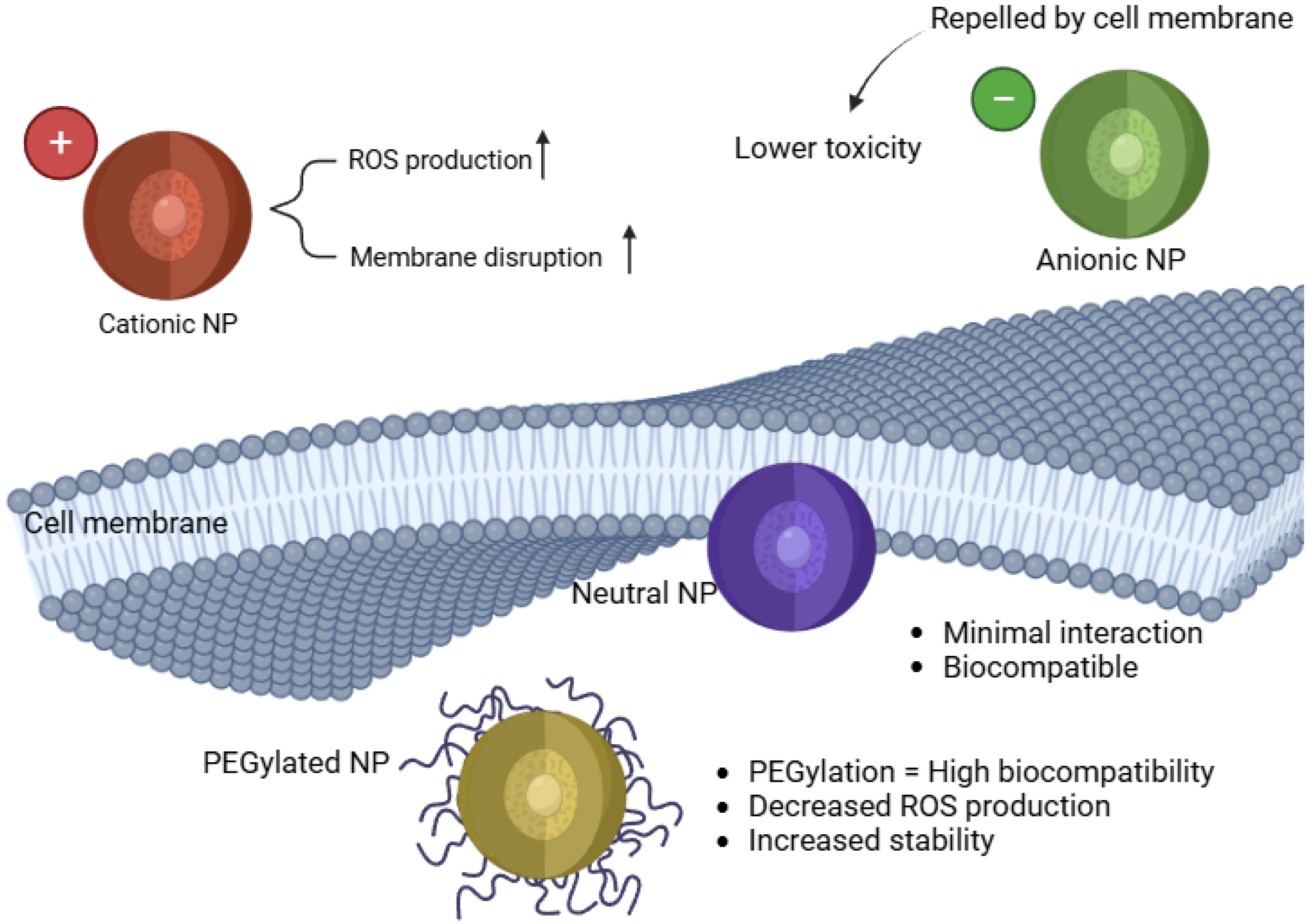

Table 1.
Impact of NP Size on Oxidative Stress and Toxicity.
| Nanoparticle Type | Small Size (≤20 nm) | Medium Size (20–50 nm) | Large Size (>50 nm) | Reference |
|---|---|---|---|---|
| Ag NPs | Mitochondrial Dysfunction | Moderate ROS | Minimal ROS | [11,19,22,25] |
| Au NPs | Pro-inflammatory Effects | Minimal Effects | Low ROS | [19,26,27] |
| Cu NPs | DNA Damage | Inflammation | Low ROS | [28] |
| Fe NPs | Cellular Damage | Mitochondrial Dysfunction | Low ROS | [29,30] |
| ZnO NPs | Apoptosis | Moderate ROS | Low ROS | [31,32,33,34] |
| TiO₂ NPs | Neurotoxicity, Behavioral Disruptions | Moderate ROS | Minimal ROS | [23,24] |
| CuO NPs | Genotoxicity | Cellular Stress | Minimal ROS | [35,36,37] |
| Fe₂O₃ NPs | DNA Fragmentation | Cellular Inflammation | Low ROS | [36,38,39,40,41] |
| CdSe QDs | DNA Damage, Apoptosis | Mitochondrial Dysfunction | Low ROS | [42,43] |
| ZnS QDs | Phototoxicity, Lipid Peroxidation | DNA Damage | Limited Cytotoxicity | [42,43] |
Table 2.
Effects of Nanoparticles on Oxidative Stress Mechanisms in Zebrafish.
| Nanoparticle Type | Affected Organ/System | Mechanism of Toxicity | Key Biomarkers/ Cytokines | Observed Effects | Case Study | Reference |
|---|---|---|---|---|---|---|
| TiO₂ NPs | Liver, Gills | ROS Overproduction, Antioxidant Enzyme Suppression | ↓ SOD, ↓ CAT, ↑ MDA | Oxidative stress-mediated toxicity | TiO₂ NPs reduced SOD and CAT activity in zebrafish liver cells | [24,48] |
| CuO NPs | Brain | Mitochondrial Dysfunction, Apoptotic Pathways | ↑ p53, ↑ Caspase-3, ↓ ATP | ROS-induced neurotoxicity | CuO NPs triggered apoptosis in zebrafish brain tissue via mitochondrial damage | [48,75,76] |
| Fe₂O₃ NPs | Gills, Immune System | NF-κB Activation, Inflammatory Response | ↑ IL-8, ↑ IL-1β, ↑ NF-κB | Respiratory dysfunction, chronic inflammation | Fe₂O₃ NPs caused NF-κB-mediated inflammation in zebrafish gills | [29,77] |
| AgNPs | Liver | Hepatotoxicity, Cytokine Overexpression | ↑ TNF-α, ↑ IL-6 | Liver inflammation, oxidative damage | AgNPs upregulated TNF-α and IL-6 in zebrafish liver cells | [29,77,78] |
| ZnO NPs | Muscle, Liver | DNA Damage, Lipid Peroxidation | ↑ DNA Strand Breaks, ↑ MDA | Chromosomal instability, apoptosis | ZnO NPs induced DNA double-strand breaks in zebrafish embryos | [8,34] |
| CdSe QDs | Brain, Muscle | Genotoxicity, Lipid Peroxidation | ↑ XRCC1, ↑ p53 | DNA fragmentation, neuronal dysfunction | CdSe QDs caused DNA strand breaks in zebrafish larvae | [9,26,79] |
Table 3.
Imaging and Histopathological Techniques for Oxidative Stress Assessment.
| Technique | Purpose | Application in Zebrafish | Key Findings | Reference |
|---|---|---|---|---|
| Fluorescent ROS Detection (DCFDA Assay) | Measures intracellular ROS levels | Used to quantify oxidative stress in zebrafish tissues | AgNP-exposed zebrafish embryos show significant ROS increase in brain and liver | [87,94,95] |
| Transmission Electron Microscopy (TEM) | Ultrastructural analysis of NP localization and organelle damage | Identifies mitochondrial swelling, cristae disruption, and vacuolization | TiO₂ NPs accumulate in hepatic mitochondria, leading to apoptosis | [72,96] |
| Hematoxylin and Eosin (H&E) Staining | Histopathological assessment of tissue damage and inflammation | Detects necrosis, epithelial degeneration, and immune cell infiltration | CuO NP exposure causes epithelial damage and chronic inflammation in zebrafish gills | [97] |
| TUNEL Assay (Terminal deoxynucleotidyl transferase dUTP Nick End Labeling) | Identifies apoptotic DNA fragmentation | Used to assess neurotoxicity and genotoxicity in zebrafish tissues | CdSe QD exposure leads to increased TUNEL-positive cells in zebrafish brain | [98,99] |
Table 4.
Natural Antioxidants in Nanoparticle Formulations for Oxidative Stress Reduction.
| Type of Antioxidant-Functionalized NP | Mechanism of Action | Effect in Zebrafish Models | Reference |
|---|---|---|---|
| Polyphenol-coated NPs (quercetin, catechins) | Enhance oxidative stress resistance by scavenging ROS | Reduce ROS levels and increase antioxidant enzyme activity in zebrafish tissues | [57,115,116] |
| Resveratrol-loaded liposomes | Improve bioavailability and stability of antioxidant compounds | Prevent lipid peroxidation and DNA damage in oxidative stress-exposed zebrafish embryos | [110,117] |
| Plant-derived antioxidant NPs | Provide biocompatible approaches for reducing oxidative toxicity | Decrease apoptosis and inflammatory response in zebrafish liver and brain tissues | [118,119] |
| Liposomal curcumin NPs | Target oxidative damage by decreasing ROS accumulation and restoring mitochondrial function | Reduce oxidative damage in zebrafish liver tissue and improve metabolic activity | [108,109] |
Disclaimer/Publisher’s Note: The statements, opinions and data contained in all publications are solely those of the individual author(s) and contributor(s) and not of MDPI and/or the editor(s). MDPI and/or the editor(s) disclaim responsibility for any injury to people or property resulting from any ideas, methods, instructions or products referred to in the content. |
© 2025 by the authors. Licensee MDPI, Basel, Switzerland. This article is an open access article distributed under the terms and conditions of the Creative Commons Attribution (CC BY) license (http://creativecommons.org/licenses/by/4.0/).
Copyright: This open access article is published under a Creative Commons CC BY 4.0 license, which permit the free download, distribution, and reuse, provided that the author and preprint are cited in any reuse.


