Submitted:
18 March 2025
Posted:
18 March 2025
You are already at the latest version
Abstract
Background/Objectives: Galeopsis spp. (Lamiaceae) are widely distributed across extensive areas in Romania, being used mainly for their sedative, neuroprotective, antioxidant, anti-inflammatory, expectorant, astringent and diuretic properties. The paper reports, for the first time, the investigation of total phenolic content (TPC), total flavonoid content (TFC) and phenolic acids profile in roots, aerial parts and leaves from three wild-grown Galeopsis spp. (G. bifida Boenn., G. speciosa Mill. and G. tetrahit L.), along with their . Methods: Ultra-high-performance liquid chromatography/ultraviolet/mass spectrometry (HPLC/UV/MS) method was used for the identification and quantification of key phenolic acids. The spectrophotometric method was applied for the determination of TPC, TFC, 2,2-diphenyl-1-picrylhydrazyl (DPPH) and 2,2’-azino-bis(3-ethylbenzothiazoline-6-sulfonic acid) (ABTS) radical scavenging activities, and also ferric-reducing antioxidant power (FRAP). High-performance thin-layer chromatography (HPTLC) was employed for the assessment of in situ antioxidant (DPPH assay) and AChE inhibitory potential. Results: Galeopsis spp. significantly contributes to polyphenol accumulation. Chlorogenic acid was the most abundant compound, with the highest levels detected in G. tetrahit leaves, followed by G. tetrahit aerial parts and G. speciosa leaves. G. tetrahit consistently showed the highest antioxidant potential, followed by G. speciosa, while G. bifida exhibited the weakest activity. AChE inhibition activity increases progressively from roots to aerial parts to leaves, with leaves consistently exhibiting the strongest inhibitory effects across all Galeopsis spp. Conclusions: Our study provides novel and valuable insights into the bioactivity of Galeopsis spp., a group of plants that has been largely overlooked in phytochemical and pharmacological research.
Keywords:
Galeopsis spp.
; Lamiaceae
; Romania flora
; phenolic acids
; UHPLC/UV/MS analysis
; antioxidant activity
; acetylcholinesterase inhibitory activity
1. Introduction
Galeopsis genus comprises annual herbaceous species distributed throughout Europe and Asia but naturalized in other regions of the world (North America) [1,2,3]. In Europe, nine Galeopsis spp. are documented, of which seven occur in Romania (G. angustifolia Ehrh., G. bifida Boenn., G. ladanum L., G. pubescens Besser, G. segetum Neck., G. speciosa Mill., G. tetrahit L.) [1,2,4,5,6].
The nomenclature of the Galeopsis genus is linked to the helmet-like shape of the corolla and derives from the fusion of two words, one Latin (galea) and one Greek (opsis), with galea meaning “helmet” and opsis signifying “aspect” [2].
Plants of the Galeopsis genus feature four-sided, highly branched stems which, in certain species, may exhibit rigid, appressed trichomes and sub-nodal swellings. The leaves may be ovate, elliptical, lanceolate, or linear-lanceolate, arranged in a decussate opposite pattern. The flowers are aggregated in verticillasters positioned on the superior portions of the stems and branches. Each verticillaster comprises 6–10 (up to 16) flowers, subtended by rigid bracts that are shorter than the calyx. The flowers possess a calyx with spiny-toothed margins and a bilabiate corolla, lacking an internal ring of trichomes, with the superior lip vaulted in a helmet-like form and the inferior lip trilobed. The stamens are ascending and concealed by the superior lip, while the style bears nearly equal stigmatic lobes [1,2,4,6].
Species of the Galeopsis genus are known by various Romanian vernacular names, including “zabră” (applied to G. pubescens and G. speciosa), “lungurică” (G. tetrahit), “tapoşnic” (G. ladanum), “cânepiţă” (G. tetrahit), and “faţa mâţei” (G. angustifolia). Some of these taxa are considered to be highly melliferous (notably G. ladanum and G. tetrahit), while others, such as G. ladanum, are also recognized as noxious weeds in cereal crop cultivation [2,4,6].
Depending on the species, plants of the Galeopsis genus are widely distributed across extensive areas in Romania, with some taxa being particularly common (G. ladanum, G. speciosa, G. tetrahit). In Romania, these species are found in ruderal habitats, forest edges, and shrubby areas (G. bifida); in forests, forest clearings, along tracks, and within hedgerows (G. speciosa); and along roadsides, in gardens, orchards, shrublands, forest clearings, or other ruderal sites (G. tetrahit) [2,4,6].
In the chemical composition of Galeopsis spp., flavonoids [3,7,8,9,10,11,12,13,14], phenolic acids [3,7,8], phenylpropanoid glycosides [3,7,8,15], iridoids [3,7,16,17,18], diterpenoids [3,7,19,20,21], triterpenoid compounds [3,7], essential oil [3,7,22,23], and fatty acids [3,7,24,25,26,27,28] have been identified.
Although few studies have examined the pharmacological activities of these plants, the Galeopsis genus is noted for its sedative, anticholinesterase, neuroprotective [3,7,29,30], antioxidant [3,7,31,32,33], anti-inflammatory [3,7], expectorant [3,7], astringent [3,7], diuretic [3,7], antianemic [3,7], and remineralizing [3,7] properties.
Some of these plants are employed in traditional medicine in certain regions. G. ladanum is reported to be used in Italy for the treatment of respiratory disorders through an infusion prepared from its leaves or flowers [7]. G. bifida is cited in Asian phytotherapy among various ethnic groups. In Tibet, the aerial parts of G. bifida are utilized in the form of a decoction to treat oral afflictions (stomatitis) and gastrointestinal disorders, including gastritis, ulcers, gastroenteritis, and inflammations affecting the esophagus, stomach, or intestines, as well as conjunctivitis, cystitis, and inflammatory conditions of the genital organs. In the Far East, a tincture derived from the aerial parts of G. bifida is employed to stimulate appetite, manage gastric ailments, and address epilepsy, while nomadic populations in northern Asia use the plant for the treatment of hepatic diseases [3].
There is evidence suggesting the potential for intoxication—manifesting as transient limb paralysis—following the consumption of fruits (with seed oil even being implicated) from Galeopsis spp. (G. bifida, G. ladanum, G. speciosa, G. tetrahit) [3,34].
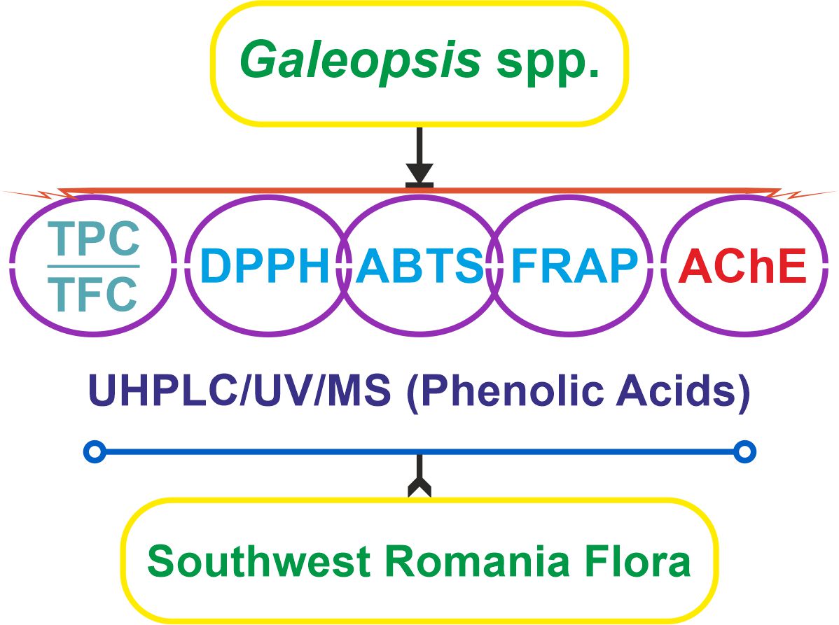
Our paper aimed to investigate, for the first time, total phenolic content (TPC), total flavonoid content (TFC) and phenolic acids profile in roots, aerial parts and leaves from three wild-grown Galeopsis spp., collected from the southwestern region of Romania, along with their antioxidant and acetylcholinesterase (AChE) inhibitory potential. Also, the research provides new data for a better understanding of Galeopsis spp. in the context of therapeutic perspective.
2. Results
2.1. Total Polyphenols and Flavonoids
The TPC and TFC were evaluated across the three Galeopsis spp. (G. bifida, G. speciosa, and G. tetrahit) and plant parts (roots, aerial parts, and leaves) (Table S1; Figure 1, a and b). The results from the two-way analysis of variance (ANOVA) demonstrated that both species and plant part significantly influenced TPC and TFC levels, with plant part showing a particularly strong effect on TFC.
2.1.1. Effect of Species and Plant Parts on TPC
The two-way ANOVA for TPC confirmed that species identity accounted for 47.57% (p<0.0001) of the total variation, while plant part explained 41.98% (p<0.0001). A significant interaction effect (8.31%) between species and plant part (p<0.0001) indicated that the differences in TPC between roots, aerial parts, and leaves were not consistent across species.
Post-hoc analysis using Tukey’s test provided further insights into the differences among plant parts. Leaves exhibited the highest TPC, significantly exceeding both aerial parts and roots (p<0.0001), while the aerial parts contained significantly more polyphenols than roots (p<0.0001). The greatest contrast was observed between roots and leaves (mean difference: -356.2 mg GAE/g), highlighting the substantial polyphenol accumulation in the leaf tissue (Figure 1a).
Among species, G. tetrahit consistently displayed the highest TPC across all plant parts, reaching 971.203±60.377 mg GAE/g in leaves, while G. bifida exhibited the lowest values, particularly in roots (152.674±12.412 mg GAE/g) (Figure 1a).
These results confirm that both species and plant part significantly contribute to polyphenol accumulation, with leaves being the richest source of polyphenols across all three species. The significant interaction effect suggests that the influence of plant part on TPC varies depending on species, emphasizing the importance of selecting the appropriate plant material for bioactive compound extraction.
2.1.2. Effect of Species and Plant Parts on TFC
A similar pattern was observed for TFC. The two-way ANOVA revealed that the plant part had the strongest influence on TFC levels, explaining 51.99% of the total variation (p<0.0001), while species accounted for 38.43% (p<0.0001). A significant interaction effect (8.99%) was also detected, confirming that the impact of plant part on flavonoid content was species dependent.
Tukey’s post-hoc test further highlighted the significant differences between plant parts. Leaves contained the highest TFC, significantly higher than both aerial parts and roots (p<0.0001). Aerial parts had significantly more flavonoids than roots (p<0.0001). The largest difference was between roots and leaves (mean difference: -290.3 mg QE/g), further confirming that flavonoid accumulation is predominantly in the leaves (Figure 1b).
Among species, G. tetrahit had the highest flavonoid content across all plant parts, particularly in leaves (568.543±24.174 mg QE/g), while G. bifida exhibited the lowest values, particularly in roots (22.78±0.663 mg QE/g) (Figure 1b).
These findings indicate that flavonoids are strongly localized in the leaves, aligning with their known role in plant defense and ultraviolet (UV) protection. The significant differences between species further emphasize the genetic variation in flavonoid biosynthesis among Galeopsis spp.
The results clearly demonstrate that both species and plant part significantly influence polyphenol and flavonoid content, with leaves consistently exhibiting the highest levels across all three species. The significant interaction effect suggests that species-specific differences influence how polyphenols and flavonoids are distributed within different plant parts. These findings highlight the importance of selecting the appropriate species and plant material for maximizing the extraction of bioactive compounds with potential antioxidant and neuroprotective applications.
2.2. Antioxidant Activity (DPPH, ABTS, FRAP)
The antioxidant activity of Galeopsis spp. was assessed using 2,2-diphenyl-1-picrylhydrazyl (DPPH) radical scavenging (IC50), 2,2’-azino-bis(3-ethylbenzothiazoline-6-sulfonic acid) (ABTS) radical scavenging (IC50), and ferric-reducing antioxidant power (FRAP) assays (Table S1; Figure 2, a–c). The effects of species and plant part were analyzed using two-way ANOVA, which confirmed significant differences across both factors, along with an interaction effect.
2.2.1. DPPH Radical Scavenging Activity
The DPPH radical scavenging assay revealed substantial differences in activity based on both species and plant part. Species had the strongest influence, accounting for 48.64% of the total variation (p<0.0001), while plant part contributed 25.02% (p<0.0001), with an interaction effect of 25.97% (p<0.0001), confirming that antioxidant potential varied not only between species but also among different plant parts within each species.
Among all samples, G. bifida roots exhibited the weakest DPPH radical scavenging activity, with an IC50 value of 16.84±0.97 mg/mL, indicating a lower antioxidant potential. Conversely, G. tetrahit leaves had the highest radical scavenging activity, with an IC50 value of 0.458±0.03 mg/mL, demonstrating a markedly stronger antioxidant effect. Leaves consistently showed the strongest DPPH activity across all species, with G. speciosa leaves also displaying a low IC50 value of 0.789±0.03 mg/mL, comparable to G. tetrahit. Aerial parts exhibited an intermediate effect, with G. bifida aerial parts showing an IC50 of 8.102±0.49 mg/mL, while G. tetrahit aerial parts demonstrated a stronger activity at 1.511±0.11 mg/mL (Figure 2a).
Post-hoc comparisons confirmed that leaves had significantly higher radical scavenging activity than aerial parts and roots (p<0.0001), and aerial parts showed stronger activity than roots (p<0.0001).
2.2.2. ABTS Radical Scavenging Activity
A similar trend was observed for the ABTS assay, where species explained 47.01% of the total variation (p<0.0001), plant part accounted for 36.86% (p<0.0001), and an interaction effect of 15.27% was found (p<0.0001). These results confirm that both genetic and morphological factors influence the ABTS radical scavenging potential of Galeopsis spp.
The lowest ABTS radical scavenging activity was observed in G. bifida roots, which had an IC50 value of 4.772±0.30 mg/mL, while the strongest activity was found in G. tetrahit leaves, with an IC50 of 0.328±0.003 mg/mL. Among aerial parts, G. speciosa exhibited a slightly stronger effect (1.665±0.10 mg/mL) compared to G. bifida (3.2±0.25 mg/mL) (Figure 2b).
Post-hoc analysis showed that leaves exhibited significantly stronger ABTS radical scavenging activity than aerial parts and roots (p<0.0001), with aerial parts also showing significantly higher activity than roots (p<0.0001).
2.2.3. FRAP Assay
Unlike DPPH and ABTS, where lower IC50 values indicate stronger radical scavenging, FRAP measures reducing power, meaning higher values correspond to stronger antioxidant activity. The two-way ANOVA results demonstrated that species accounted for 55.42% of the total variation (p<0.0001), plant part explained 34.28% (p<0.0001), and the interaction effect contributed 7.861% (p<0.0001), making species the dominant determinant of reducing power.
The highest FRAP value was recorded in G. tetrahit leaves, which exhibited 37.763±2.52 mM Fe2+ equivalents, confirming the strongest reducing capacity. Conversely, G. bifida roots had the lowest FRAP activity at 10.392±0.40 mM Fe2+ equivalents, aligning with the trend observed in the other assays. Aerial parts displayed moderate activity, with G. speciosa aerial parts showing a FRAP value of 19.979±1.75 mM Fe2+ equivalents, while G. tetrahit aerial parts had a notably higher value at 25.480±1.16 mM Fe2+ equivalents (Figure 2c).
Post-hoc comparisons revealed that leaves exhibited significantly higher reducing power than aerial parts and roots (p<0.0001), and aerial parts also had significantly greater reducing power than roots (p=0.0059).
These results confirm that leaves consistently exhibited the strongest antioxidant potential across all three assays, while roots displayed the weakest activity in all cases. Among the species, G. tetrahit consistently showed the highest antioxidant potential, followed by G. speciosa, while G. bifida exhibited the weakest activity.
2.3. Neuroprotective (AChE Inhibition) Activity
The AChE inhibition activity was assessed across the three Galeopsis spp. and the three plant parts to evaluate their potential neuroprotective effects. The results were expressed as IC50 values (mg/mL), where lower values indicate stronger AChE inhibition activity (Table S1; Figure 3). The two-way ANOVA confirmed that both species and plant part significantly influenced AChE inhibition, with plant part being the dominant factor.
2.3.1. Influence of Species and Plant Part on AChE Inhibition (IC50 Values)
The two-way ANOVA results revealed that the plant part was the strongest determinant of AChE inhibition, accounting for 77.93% of the total variation (p<0.0001). Species also played a significant role, explaining 18.88% of the variation (p<0.0001). A minor but statistically significant interaction effect (1.368%, p=0.0311) indicated that the effect of plant part on AChE inhibition was slightly dependent on species.
Among the plant parts, leaves exhibited the strongest AChE inhibition activity, as indicated by the lowest IC50 values across all species. G. tetrahit leaves had the strongest inhibition with an IC50 of 4.002±0.32 mg/mL, followed by G. speciosa leaves (6.92±0.14 mg/mL) and G. bifida leaves (6.97±0.68 mg/mL). These lower IC50 values indicate high inhibitory activity in the leaf extracts (Figure 3).
In contrast, roots showed the weakest AChE inhibition activity, requiring higher concentrations to achieve 50% inhibition. G. bifida roots exhibited the highest IC50 value at 17.23±0.04 mg/mL, followed by G. speciosa roots (15.06±0.64 mg/mL) and G. tetrahit roots (11.42±0.42 mg/mL) (Figure 3).
Aerial parts demonstrated moderate AChE inhibition, with G. bifida aerial parts having an IC50 of 15.63±1.21 mg/mL, slightly higher than G. speciosa aerial parts (13.89±1.33 mg/mL) and G. tetrahit aerial parts (10.94±0.45 mg/mL) (Figure 3).
Post-hoc Tukey’s multiple comparisons test revealed significant differences between plant parts. Leaves exhibited significantly stronger AChE inhibition (lower IC50) than aerial parts (p<0.0001), with a mean difference of 7.523 mg/mL. Leaves also had significantly stronger AChE inhibition than roots (p<0.0001), with a mean difference of 8.606 mg/mL, while aerial parts showed significantly stronger inhibition (lower IC50) than roots (p=0.0128), confirming that root extracts were the least potent inhibitors.
These findings confirm that AChE inhibition activity increases progressively from roots to aerial parts to leaves, with leaves consistently exhibiting the strongest inhibitory effects across all species.
2.4. HPTLC Fingerprinting for Antioxidant and Neuroprotective Activity
The high-performance thin-layer chromatography (HPTLC) fingerprinting was used to assess comparatively the chemical composition, antioxidant activity (DPPH assay), and neuroprotective activity (AChE inhibition assay) of Galeopsis spp. extracts. Reference standards of caffeic acid (Rf 0.79, lane 1), chlorogenic acid (Rf 0.22, lane 2), and rutin (Rf 0.085, lane 3) were included for comparison. The results provide insights into the phytochemical composition of these species, particularly their flavonoid and phenolic acid content, as well as their biological activity (Table 1; Figure 4, a–e).
2.5. Phenolic Acids Profile (UHPLC Analysis)
The ultra-high-performance liquid chromatography (UHPLC) analysis of Galeopsis spp. revealed significant variations in phenolic acid composition across different plant parts. A total of eight phenolic acids were quantified based on their retention times, with gallic acid eluting first at 1.80 min, followed by protocatechuic acid (3.70 min), chlorogenic acid (5.83 min), vanillic acid (6.11 min), caffeic acid (6.36 min), syringic acid (6.64 min), p-coumaric acid (7.80 min), and ferulic acid (8.54 min) (Figure S1).
The results showed that chlorogenic acid was the most abundant compound, with the highest levels detected in G. tetrahit leaves (22.35±1.11 mg/g), followed by G. tetrahit aerial parts (11.68±0.58 mg/g) and G. speciosa leaves (8.71±0.43 mg/g). In contrast, roots contained significantly lower levels of this compound, confirming that chlorogenic acid is concentrated in the aerial parts of these species (Table S2).
Among the other phenolic acids, p-coumaric acid and ferulic acid were particularly abundant in leaves, with G. speciosa leaves exhibiting the highest p-coumaric acid content (534.110±26.706 μg/g), while G. tetrahit leaves had the highest ferulic acid concentration (271.089±13.554 μg/g). Caffeic acid was detected in varying amounts, with G. speciosa leaves containing the highest levels (288.87±14.444 μg/g). Notably, gallic acid was mostly absent in G. bifida and G. speciosa, but was detected in G. tetrahit leaves (40.962±2.048 μg/g). Similarly, protocatechuic acid was present in moderate concentrations, with the highest levels found in G. tetrahit leaves (176.536±8.827 μg/g). The analysis also revealed that syringic acid and vanillic acid were detected across all species, with G. speciosa leaves containing the highest levels of vanillic acid (421.963±21.098 μg/g) (Table S2).
Overall, the leaves consistently exhibited the highest phenolic acid content, whereas roots contained the lowest concentrations. These findings support previous TPC and TFC results, highlighting leaves as the most bioactive plant part. The high concentration of chlorogenic acid, caffeic acid, and p-coumaric acid in leaves suggests that these compounds may contribute significantly to the strong antioxidant and neuroprotective activity observed in previous assays. Given the therapeutic potential of these bioactive compounds, future studies should focus on isolating and further characterizing these phenolic acids for their pharmacological applications.
3. Discussion
The findings of this study align closely with previous research on Galeopsis spp., particularly regarding their phytochemical composition, antioxidant potential, and neuroprotective activity [3,29,30,31]. The high concentrations of chlorogenic acid, p-coumaric acid, and ferulic acid observed in this study confirm the polyphenol-rich nature of these plants, supporting previous reports that identified G. bifida as a source of phenylethanoid glycosides and flavone derivatives with strong antioxidant properties.
3.1. Total Polyphenols and Flavonoids
The relationship between TPC and TFC was evaluated using Pearson’s correlation analysis, following confirmation of normal data distribution via the Shapiro–Wilk test. The results indicated a strong positive correlation between TPC and TFC (r=0.9653, p<0.0001), demonstrating a significant association between these two parameters.
This finding suggests that flavonoids constitute a major portion of the total polyphenolic content in the analyzed Galeopsis spp. The high correlation implies that an increase in TFC is accompanied by a proportional increase in TFC, reinforcing the contribution of flavonoids to the overall phytochemical profile. However, it is important to note that the aluminum chloride (AlCl3) colorimetric method for TFC determination may lead to false-positive results due to its reaction with non-flavonoid phenolic compounds, such as certain phenolic acids, tannins, and other interfering substances, which can form complexes similar to flavonoids and overestimate flavonoid content [35].
Given the well-documented biological activities of flavonoids, these results highlight their potential role in the antioxidant and neuroprotective effects of the extracts.
3.2. Antioxidant Activity
The antioxidant activity results are consistent with earlier studies, which demonstrated that Galeopsis spp. possess strong DPPH and ABTS radical scavenging potential, along with FRAP assay [3,30,31,32,33]. These effects have been previously linked to phenylethanoid glycosides and flavonoid glycosides, particularly luteolin and apigenin derivatives. The current study further supports these findings by establishing strong correlations between TPC, TFC, and antioxidant activity, indicating that these compounds are the primary contributors to the radical scavenging potential of Galeopsis extracts. The HPTLC fingerprinting of DPPH activity further corroborated these results, showing that chlorogenic acid, along with flavonoid-related compounds, was responsible for the observed antioxidant effects.
The antioxidant activities of the Galeopsis spp. were assessed using DPPH, ABTS, and FRAP assays, expressed as IC50 values (μg/mL) for DPPH and ABTS and as mM Fe2+ equivalents for FRAP. The normality of the data was evaluated using the Shapiro–Wilk test, which confirmed that ABTS and FRAP values followed a normal distribution, while DPPH values did not. Consequently, Spearman’s correlation analysis was applied to DPPH-related comparisons, while Pearson’s correlation analysis was used for ABTS vs. FRAP. The correlation analysis yielded the following results:
- DPPH IC50 vs. ABTS IC50: A strong positive correlation (r=0.983, p<0.05) was observed, indicating that extracts with higher radical scavenging efficiency in the DPPH assay also exhibited strong activity in the ABTS assay;
- DPPH IC50 vs. FRAP (mM Fe2+): A negative correlation (r=-0.833, p<0.05) was found, suggesting that extracts requiring higher concentrations to inhibit 50% of DPPH radicals tended to exhibit higher reducing power in the FRAP assay;
- ABTS IC50 vs. FRAP (mM Fe2+): A negative correlation (r=-0.817, p<0.05) was also observed, indicating an inverse relationship between radical scavenging capacity and ferric-reducing ability.
The strong correlation between DPPH and ABTS IC50 values suggests that both assays measure similar radical scavenging mechanisms, likely driven by polyphenolic compounds. Since lower IC50 values indicate higher antioxidant activity, the negative correlations suggest that extracts requiring lower concentrations for DPPH and ABTS inhibition also tend to exhibit stronger reducing power in the FRAP assay.
These findings highlight the complexity of antioxidant mechanisms and reinforce the necessity of using multiple assays to obtain a comprehensive understanding of antioxidant potential.
To further examine the relationship between polyphenolic content and antioxidant activity, the correlation between TPC and the three antioxidant assays (DPPH IC50, ABTS IC50, and FRAP in mM Fe2+ equivalents) was assessed. The results revealed the following correlations:
- TPC vs. DPPH IC50: A strong negative correlation (r=-0.9333, p=0.0007), indicating that extracts with higher polyphenol content required lower concentrations to inhibit 50% of DPPH radicals, thus demonstrating stronger radical scavenging activity;
- TPC vs. ABTS IC50: A moderate negative correlation (r=-0.8833, p=0.0031), suggesting that higher polyphenol levels were associated with greater ABTS radical scavenging efficiency;
- TPC vs. FRAP (mM Fe2+ equivalents): A strong positive correlation (r=0.9333, p=0.0007), indicating that extracts with higher polyphenol content exhibited greater ferric-reducing power.
These findings confirm that polyphenols play a key role in the antioxidant activity of the Galeopsis spp., contributing both to radical scavenging (DPPH, ABTS) and reducing power (FRAP). The observed negative correlations with DPPH and ABTS IC50 values indicate that extracts with higher TPC exhibited lower IC50 values, confirming their enhanced ability to neutralize free radicals. Conversely, the strong positive correlation between TPC and FRAP implies that polyphenols are also effective electron donors, reinforcing their reducing capacity.
To evaluate the contribution of flavonoids to antioxidant activity, the correlation between TFC and the three antioxidant assays (DPPH IC50, ABTS IC50, and FRAP in mM Fe2+ equivalents) was analyzed. The correlation analysis revealed the following relationships:
- TFC vs. DPPH IC50: A strong negative correlation (r=-0.9167, p=0.0013), indicating that extracts with higher flavonoid content required lower concentrations to inhibit 50% of DPPH radicals, confirming their potent radical scavenging capacity;
- TFC vs. ABTS IC50: A moderate negative correlation (r=-0.8833, p=0.0031), suggesting that an increase in flavonoid content was associated with improved ABTS radical scavenging efficiency;
- TFC vs. FRAP (mM Fe2+ equivalents): A strong positive correlation (r=0.9333, p=0.0007), indicating that extracts with higher flavonoid content exhibited greater ferric-reducing power.
These results suggest that flavonoids significantly contribute to both radical scavenging activity and reducing power in the tested Galeopsis spp. The negative correlations with DPPH and ABTS IC50 values indicate that flavonoid-rich extracts exhibited stronger antioxidant activity, requiring lower concentrations to achieve 50% inhibition. The strong positive correlation between TFC and FRAP further supports the role of flavonoids as efficient electron donors, reinforcing their involvement in redox reactions.
3.3. Neuroprotective Activity
Regarding neuroprotective activity, the results of this study reinforce previous findings on the AChE inhibitory potential of Galeopsis spp. Earlier research identified several bioactive metabolites with AChE inhibitory properties, including iridoid glycosides (harpagide, harpagide 8-O-acetate, ajugoside), phenylethanoid glycosides (verbascoside, isoverbascoside), flavonoid glycosides (luteolin and apigenin derivatives), and hydroxycinnamic acids (caffeoylquinic acids, e.g., chlorogenic acid) [3,7,8,29,30]. The current study confirmed that chlorogenic acid was present in all samples and exhibited moderate AChE inhibition, supporting its role as a neuroprotective agent. Additionally, an unknown compound at the same Rf as caffeic acid exhibited inhibitory activity in the AChE HPTLC assay, suggesting the presence of another bioactive metabolite contributing to neuroprotection. The strongest AChE inhibition zones were observed in G. tetrahit leaves, which also exhibited a unique orange-fluorescent flavonoid does not present in the other species. These results suggest that G. tetrahit may contain distinct neuroactive flavonoids that warrant further investigation.
The neuroprotective potential of the Galeopsis spp. was assessed through AChE inhibition activity, and its relationship with TPC and TFC was analyzed. The correlation analysis yielded the following results:
- AChE inhibition vs. TPC: A moderate negative correlation (r=-0.8266, p=0.0060), suggesting that extracts with higher total polyphenol content exhibited greater AChE inhibition. The 95% confidence interval (CI) ranged from -0.9624 to -0.3603, supporting the statistical robustness of this relationship;
- AChE inhibition vs. TFC: A moderate negative correlation (r=-0.8335, p=0.0053), indicating that an increase in flavonoid content was associated with stronger AChE inhibition. The 95% CI ranged from -0.9640 to -0.3793, reinforcing the reliability of the association.
Both correlations were statistically significant (p<0.05) and suggest that polyphenols, particularly flavonoids, may play a role in the neuroprotective activity of these extracts. The negative correlation indicates that extracts with higher levels of polyphenols and flavonoids required lower concentrations to inhibit AChE, highlighting their potential as natural AChE inhibitors.
These findings align with previous research suggesting that polyphenolic compounds, including flavonoids, can modulate cholinergic activity and contribute to neuroprotective effects. Further investigations into specific bioactive compounds responsible for AChE inhibition could provide deeper insights into their potential application in managing neurodegenerative conditions.
To further explore the relationship between AChE inhibition and antioxidant activity, the correlation between AChE inhibition and DPPH IC50, ABTS IC50, and FRAP (mM Fe2+ equivalents) was analyzed. The normality of the data was confirmed using the Shapiro–Wilk test, and Spearman’s correlation analysis was performed to assess statistical associations. The results of the correlation analysis revealed the following:
- AChE inhibition vs. DPPH IC50: A moderate positive correlation (r=0.6887, p=0.0402), indicating that extracts with higher AChE inhibition also tended to require lower concentrations to scavenge 50% of DPPH radicals. However, the correlation was weaker compared to other parameters, as reflected by the 95% CI ranging from 0.04535 to 0.9283;
- AChE inhibition vs. ABTS IC50: A strong positive correlation (r=0.8085, p=0.0083), suggesting that extracts with greater AChE inhibition demonstrated enhanced ABTS radical scavenging activity. The 95% CI (0.3117 to 0.9581) reinforces the statistical robustness of this relationship;
- AChE inhibition vs. FRAP (mM Fe2+ equivalents): A moderate negative correlation (r=-0.8238, p=0.0063), showing that extracts with higher AChE inhibition exhibited stronger reducing power. The negative correlation suggests that extracts with high AChE inhibition had greater ferric-reducing capacity, a trend supported by the 95% CI of -0.9617 to -0.3526.
These findings indicate a clear link between neuroprotective and antioxidant activities in the Galeopsis spp. analyzed extracts. The positive correlations with DPPH and ABTS IC50 values suggest that extracts with stronger radical scavenging properties may also have neuroprotective potential. Meanwhile, the negative correlation with FRAP highlights that reducing power may play an independent or complementary role in AChE inhibition.
The strong association between AChE inhibition and antioxidant activity aligns with existing research suggesting that oxidative stress is closely linked to neurodegenerative diseases and that antioxidants may exert neuroprotective effects by reducing oxidative damage and modulating cholinergic activity. Future studies focusing on specific bioactive compounds with dual antioxidant and neuroprotective activities could provide further insights into their mechanisms of action and potential therapeutic applications.
Overall, the findings of this study confirm and expand upon previous research, reinforcing the high polyphenol and flavonoid content of Galeopsis spp., particularly in leaves. The strong antioxidant and neuroprotective activity observed in G. tetrahit and G. speciosa leaves suggests that these plants contain valuable bioactive compounds with potential therapeutic applications in oxidative stress-related and neurodegenerative diseases. Future research should focus on isolating and characterizing the specific compounds responsible for these effects to better understand their pharmacological potential.
3.4. Study Limitations
While this study provides valuable insights into the phytochemical composition, antioxidant potential, and neuroprotective activity of Galeopsis spp., several limitations should be considered when interpreting the results.
3.4.1. Variability in Plant Material and Environmental Influence
The chemical composition of plant extracts is highly influenced by environmental factors such as soil composition, climate, altitude, and harvesting conditions. Since the Galeopsis spp. studied were collected from specific locations, the results may not be entirely generalizable to plants grown in different geographical regions. A more comprehensive study incorporating plants from diverse habitats would strengthen the findings.
3.4.2. Extraction Method and Solvent Specificity
The study employed ultrasound-assisted extraction (UAE) using 70% ethanol, a method chosen for its efficiency in extracting polyphenols and flavonoids. However, other bioactive compounds, such as alkaloids, lipophilic terpenes, or polysaccharides, may have been underrepresented due to the solvent selectivity. Future studies should explore multiple extraction methods to capture a broader range of secondary metabolites.
3.4.3. HPTLC and UHPLC Identification Constraints
Although HPTLC fingerprinting and UHPLC quantification provided valuable insights into the phenolic composition, the study relied on reference standards for compound identification. Unknown compounds detected at similar Rf values to known standards (e.g., the compound observed at the same Rf as caffeic acid in AChE inhibition assays) were not structurally characterized. Advanced analytical techniques such as liquid chromatography–tandem mass spectrometry (LC–MS/MS) or nuclear magnetic resonance (NMR) spectroscopy should be employed in future research to confirm compound identities and detect novel bioactive molecules.
3.4.4. Lack of In Vivo Validation
The study focused on in vitro tests, including antioxidant (DPPH, ABTS, FRAP) and neuroprotective (AChE inhibition) assays, to assess the bioactivity of Galeopsis spp. extracts. While these assays are reliable indicators of biological potential, in vitro results do not always translate into in vivo efficacy due to differences in bioavailability, metabolism, and cellular interactions. Future studies should incorporate cell-based models, animal studies, and pharmacokinetic analyses to better understand the bioavailability and in vivo effectiveness of the identified compounds.
3.4.5. Potential Interference in Quantification Assays
The AlCl₃ colorimetric assay used for TFC is known to produce false positives, as certain phenolic acids and other non-flavonoid compounds can react with AlCl₃, leading to an overestimation of flavonoid content. This limitation suggests that more specific flavonoid quantification techniques, such as UHPLC–MS/MS, should be employed to validate the results.
Despite these limitations, this study provides novel and valuable insights into the bioactivity of Galeopsis spp., a group of plants that has been largely overlooked in phytochemical and pharmacological research. Currently, few studies have systematically investigated the chemical composition and biological activity of these species, making this research a significant contribution to the understanding of their medicinal potential. Addressing the outlined limitations in future studies would enable a more comprehensive understanding of their pharmacological properties, enhance compound identification, and validate in vivo relevance for potential therapeutic applications.
4. Materials and Methods
4.1. Plant Material
The roots, aerial parts and leaves of wild-grown Galeopsis spp. were harvested during the flowering period (July–August 2024) from Oltenia Region, southwest Romanian flora. The plant material for analysis was stored in the Herbarium of the Department of Pharmaceutical Botany, Faculty of Pharmacy, University of Medicine and Pharmacy of Craiova. The plant material was air-dried and deposited in brown paper bags, at room temperature (RT), in a cool and dark area, 24 hours before processing for extraction and analysis. Our research did not involve endangered or protected plant species.
A systematic notation representing different Galeopsis spp., their respective vegetal products, date/site of collection and voucher specimens was used in this study. A clear and organized reference to the specific plant species and parts analyzed in the experiments was facilitated by this notation (Table 2).
4.2. Chemicals and Reagents
The solvents used in this study included ethanol, methanol, acetonitrile and ethyl acetate (Merck, Darmstadt, Germany). Ultrapure water was obtained using a HALIOS 6 lab water system (Neptec, Montabaur, Germany) to ensure the required purity for aqueous solutions and dilutions. For UHPLC analysis, formic acid (Merck) was used as an additive to enhance the performance of the mobile phases.
The reagents selected to support the experimental assays included Folin-Ciocalteu reagent, sodium carbonate, DPPH, ABTS, potassium persulfate, sodium acetate, acetic acid, 2,4,6-tris(2-pyridyl)-1,3,5-triazine (TPTZ), quercetin, natural products–polyethylene glycol (NP–PEG) reagent, ferric chloride (FeCl3), ferrous sulfate heptahydrate (FeSO4·7H2O), and hydrochloric acid (HCl) (Sigma-Aldrich, Taufkirchen, Germany). These reagents were used for the determination of TPC, antioxidant activity, and enzymatic assays. For TPC, Folin–Ciocalteu reagent was used together with sodium bicarbonate. AlCl3 from Sigma-Aldrich was specifically used for the TFC assay.
For the AChE inhibition assay, the primary reagents included AChE from Electrophorus electricus, 1-naphthyl acetate, Fast Blue B salt, Tris-HCl buffer solution (pH 7.8, 0.05 M) and rivastigmine as a positive control (Sigma-Aldrich).
In UHPLC analysis, a set of phenolic acid standards – including caffeic acid, chlorogenic acid, p-coumaric acid, ferulic acid, gallic acid, protocatechuic acid, syringic acid, and vanillic acid (Merck Millipore, Darmstadt, Germany) – was used for calibration and compound identification.
For HPTLC analysis, Silica gel 60 F254 glass plates (20×10 cm) were obtained from Merck.
4.3. Extraction Procedure
The extraction of plant material was carried out using a UAE method, with 70% ethanol as the solvent. A measured quantity of 1 g of finely ground plant material was combined with 10 mL of the ethanol solution in an appropriate container. The mixture underwent ultrasonic treatment in a Bandelin Sonorex Digiplus DL 102H ultrasound bath (Bandelin electronic GmbH & Co. KG, Berlin, Germany) operating at 100 W power and a frequency of 35 kHz for 20 min at a controlled temperature of 50°C. The application of ultrasonic waves facilitated the breakdown of plant cell walls, enhancing the release of bioactive compounds into the solvent.
Following extraction, the solution was filtered through a 0.22 μm syringe filter equipped with a water wettable polytetrafluoroethylene (WWPTFE) membrane (Acrodisc, Pall Corporation, Port Washington, NY, USA) to separate the liquid extract from any residual solid material. The obtained extract was subsequently used for TPC and TFC determination, as well as antioxidant and neuroprotection assays.
For UHPLC analysis, 1 mL of the extract was carefully evaporated under a gentle nitrogen stream to eliminate the solvent. The dried residue was then reconstituted in a mixture of water and acetonitrile (9:1, v/v) to ensure compatibility with the UHPLC mobile phase system. This step was essential for optimizing the dissolution of bioactive compounds prior to chromatographic separation and detection. Before injection into the UHPLC system, the reconstituted solution was filtered through a 0.22 μm syringe filter to remove any particulate matter.
4.4. Standards Preparation
Caffeic acid, chlorogenic acid, p-coumaric acid, ferulic acid, gallic acid, protocatechuic acid, syringic acid, and vanillic acid were used as standards for the UHPLC analysis. Stock solution of each standard was prepared at 1 mg/mL concentration using methanol. To achieve calibration concentrations ranging from 0.1 μg/mL to 50 μg/mL, serial dilutions were made. For both standards and samples, a volume of 10 μL was injected into the UHPLC system.
4.5. Total Polyphenols and Flavonoids
4.5.1. TPC Assay
The TPC was quantified using the Folin–Ciocalteu method, in a 96-well microplate format. Twenty microliters (20 μL) of the plant extract were pipetted into each well, followed by the addition of 100 μL of Folin–Ciocalteu reagent. The mixture was allowed to react for three minutes, after which 80 μL of a 4% sodium carbonate solution was added. The microplate was stirred for another three minutes to ensure homogeneity. To facilitate color development, the reaction mixture was incubated in the dark for two hours. Following incubation, absorbance was measured at 620 nm using a FLUOstar Optima microplate reader (BMG Labtech, Ortenberg, Germany). A gallic acid standard curve was prepared, with calibration solutions ranging from 5 mg/mL to 625 μg/mL, enabling the quantification of phenolic compounds in the extracts, expressed as mg gallic acid equivalents (GAE) per g of plant extract. Each measurement was performed in triplicate to ensure accuracy and reproducibility [36].
4.5.2. TFC Assay
The TFC was assessed using the AlCl3 colorimetric assay. A quercetin standard curve was prepared in 96% ethanol, with concentrations ranging from 30 to 100 μg/mL. For each assay, 50 μL of plant extract or quercetin standard solution was added to a 96-well microplate, followed by the addition of 10 μL of 10% AlCl3 solution. To this mixture, 150 μL of 96% ethanol was added, followed by 10 μL of 1 M sodium acetate. A blank control was prepared using 96% ethanol in place of the sample. After thorough mixing, the reaction was incubated for 40 min at RT in the dark. Absorbance was recorded at 410 nm using a FLUOstar Optima microplate reader (BMG Labtech, Ortenberg, Germany). The results were expressed as mg quercetin equivalents (QE) per g of plant extract. Each sample was analyzed in triplicate to ensure reproducibility [36,37].
4.6. Antioxidant Activity Assays
4.6.1. DPPH Antioxidant Assay
The DPPH radical scavenging assay was conducted by adding 50 μL of each sample to a 96-well microplate, followed by serial dilutions to obtain a gradient of decreasing concentrations. Next, 150 μL of a 0.2 mM DPPH solution in ethanol was added into each well. The reaction mixtures were incubated in the dark for 30 min at RT, after which the absorbance was measured at 517 nm using a FLUOstar Optima microplate reader (BMG Labtech). The antioxidant potential was evaluated by calculating the half-maximal inhibitory concentration (IC50), which represents the concentration required to scavenge 50% of the DPPH radicals. Each sample was analyzed in triplicate to ensure accuracy [36].
4.6.2. ABTS Antioxidant Assay
In the ABTS radical scavenging assay, 50 μL of each sample was added to a 96-well microplate, followed by serial dilutions in the same manner as the DPPH assay. Then, 150 μL of ABTS reagent, prepared by mixing 7.4 mM ABTS with 2.6 mM potassium persulfate, was added to each well. After a reaction time of six minutes, the absorbance was measured at 620 nm using a FLUOstar Optima microplate reader (BMG Labtech). The IC50 value, representing the sample concentration necessary to inhibit 50% of the ABTS radicals, was determined from a dose–response curve. Each sample was tested in triplicate [38].
4.6.3. FRAP Antioxidant Assay
The FRAP assay was performed by preparing a fresh FRAP reagent consisting of acetate buffer (pH 3.6), 10 mM TPTZ solution in 40 mM HCl, and 20 mM FeCl3 solution. A calibration curve was established using Fe2+ standards in the range of 65 to 500 μM. In each assay, 10 μL of the sample or standard was added to a 96-well microplate, followed by 190 μL of freshly prepared FRAP reagent. The reaction mixtures were incubated for 30 min at RT, after which the absorbance was recorded at 595 nm. The results were expressed as μmol Fe2+ equivalents, and all analyses were carried out in triplicate to ensure reliability [38].
4.7. Neuroprotective Activity Assay
The AChE inhibitory activity was assessed using a microplate-based assay, with each sample tested in triplicate to ensure reliability. The assay aimed to evaluate the ability of the test samples to inhibit AChE activity across a range of concentrations. Each sample underwent serial dilution directly on a 96-well microplate, starting from the stock extract solution, to generate a concentration gradient. To initiate the reaction, 50 μL of 1-naphthyl acetate solution (3 mg/mL in ethanol) was added to each well, serving as the enzymatic substrate. This was followed by the addition of 150 μL of AChE solution (3.33 U/mL) to catalyze the reaction, leading to the formation of measurable enzymatic products. To facilitate the detection of enzyme activity, 50 μL of Fast Blue B salt solution (3 mg/mL in water) was introduced into each well. This reagent reacts with the enzymatic products, producing a distinct color change that correlates with AChE activity. Rivastigmine (1 mg/mL in methanol), a known AChE inhibitor, was included as a positive control to establish a reference for the inhibitory potential of the test samples. Absorbance was recorded at 595 nm using a FLUOstar Optima microplate reader (BMG Labtech), and the collected data were analyzed to determine the IC50 value for each sample, indicating the concentration required to inhibit 50% of AChE activity [38].
4.8. HPTLC Fingerprinting for Antioxidant and Neuroprotective Activity
HPTLC fingerprinting was performed to assess the antioxidant potential (DPPH assay) and AChE inhibitory activity of the plant extracts [39]. Caffeic acid, chlorogenic acid, and rutin were used as reference standards.
Sample application was carried out using a Linomat 5 applicator, where 2 μL of each extract and standard were applied to the HPTLC plates. The chromatographic separation was conducted in a twin trough chamber using a mobile phase consisting of ethyl acetate, formic acid, and water (90:6:9, v/v/v). Prior to development, the chamber was saturated for 20 min to ensure optimal separation conditions. The plates were developed up to a solvent front position of 7 cm.
For the AChE inhibition assay, the plate was sprayed using the CAMAG Derivatizer (CAMAG, Muttenz, Switzerland) with 0.5 mL Tris-HCl buffer solution (pH 7.8, 0.05 M) used for prewetting and then 1.5 mL AChE solution (6.66 U/mL), after which the plate was sprayed with 0.5 mL of the 1:1 substrate/chromogenic reagent mixture (ethanolic 1-naphthyl acetate solution and aqueous Fast Blue B salt solution, 3 mg/mL each) and dried (three min).
Following development, the plates were air-dried at RT for 10 min before analysis. Visualization was conducted at 254 nm and 366 nm without derivatization, as well as post-derivatization using NP–PEG reagent at 366 nm and for DPPH and AChE in white light.
This method enabled the identification of bioactive compounds within the extracts based on their retention factor (Rf) values and corresponding color changes indicative of antioxidant and neuroprotective properties.
4.9. UHPLC Analysis of Phenolic Acids
UHPLC analysis was performed using a Waters Acquity Arc system, equipped with a photodiode array (PDA) detector and a QDa mass detector (Waters, Milford, Massachusetts, USA). Chromatographic separation was achieved using a CORTECS C18 column (4.6×50 mm, 2.7 μm particle size), which was maintained at a temperature of 30°C.
The mobile phase consisted of water with 0.01% formic acid (A) and acetonitrile with 0.01% formic acid (B). The gradient elution program was initiated with 99% A at a constant flow rate of 0.8 mL/min, held for one minute. Between 1 and 13 min, the proportion of mobile phase A was gradually reduced to 70%, which remained unchanged until 13.10 min. From 13.60 to 17.60 min, the composition shifted to 20% A, allowing for column cleaning and the removal of strongly retained compounds. The mobile phase then returned to its initial condition of 99% A at 18.10 min, and this was maintained until 21.10 min for system re-equilibration before the next injection. To ensure analytical stability and reproducibility, the column was equilibrated for 10 min between injections. Throughout the analysis, samples were kept at 8°C to preserve their integrity.
For quantification, absorbance detection was set at 265 nm for gallic acid, protocatechuic acid, vanillic acid, and syringic acid, while 325 nm was used for chlorogenic acid, caffeic acid, p-coumaric acid, and ferulic acid. Mass confirmation was carried out in negative ion mode, targeting specific mass-to-charge (m/z) ratios: 153 (protocatechuic acid), 163 (p-coumaric acid), 167 (vanillic acid), 169 (gallic acid), 179 (caffeic acid), 193 (ferulic acid), 197 (syringic acid), and 353 (chlorogenic acid) [38,39].
4.10. Statistical Analysis
All experimental data were analyzed using GraphPad Prism 9 (GraphPad Software, San Diego, CA, USA). The results were expressed as mean±standard deviation (SD), with all experiments performed in triplicate (n=3). The normality of the data was assessed using the Shapiro–Wilk test, and based on the results, appropriate statistical tests were applied.
For comparisons between Galeopsis spp. (G. bifida, G. speciosa, and G. tetrahit) and plant parts (roots, aerial parts, and leaves), a two-way ANOVA was performed to determine the influence of these factors on TPC, TFC, antioxidant activity (DPPH, ABTS, FRAP assays), and AChE inhibition. Post-hoc Tukey’s multiple comparisons test was conducted to identify statistically significant differences between groups.
For correlation analyses, Pearson’s correlation coefficient (r) was used when the data followed a normal distribution, while Spearman’s correlation test was applied for non-normally distributed data. Correlations were evaluated between TPC, TFC, and bioactivity assays (antioxidant and AChE inhibition tests) to determine potential relationships between polyphenolic content and biological activity. The significance threshold was set at α=0.05, with results considered statistically significant at p<0.05, highly significant at p<0.01, very highly significant at p<0.001, and extremely significant at p<0.0001.
All statistical tests were conducted in accordance with standard biostatistical methodologies, ensuring robust and reproducible data interpretation.
5. Conclusions
This study provides a comprehensive analysis of the phytochemical composition, antioxidant activity, and neuroprotective potential of three Galeopsis spp. (G. bifida, G. speciosa, and G. tetrahit). The results confirm that leaves contain the highest concentrations of phenolic acids and flavonoids, particularly chlorogenic acid, p-coumaric acid, and ferulic acid, which were identified as major bioactive compounds. Strong antioxidant activity was demonstrated through DPPH, ABTS, and FRAP assays, with leaves, particularly those of G. tetrahit, exhibiting the greatest radical scavenging potential. Additionally, AChE inhibition assay revealed that G. tetrahit leaves exhibited the strongest neuroprotective effects, which may be attributed to their high phenolic acids and flavonoid content.
These findings align with previous research on Galeopsis spp., reinforcing their potential as natural sources of antioxidants and neuroprotective agents. However, this study represents one of the few in-depth investigations into the phytochemistry and bioactivity of these species, highlighting the need for further research on compound isolation, structural characterization, and in vivo validation. The presence of an unknown neuroactive compound at the same Rf as caffeic acid in AChE inhibition assay suggests that Galeopsis spp. may contain previously unidentified bioactive molecules, warranting additional pharmacological exploration.
Overall, this study supports the medicinal relevance of Galeopsis spp., particularly in applications related to oxidative stress and neurodegenerative disorders. Future work should focus on elucidating the mechanisms of action, exploring clinical relevance, and assessing the safety profile of these bioactive compounds to unlock their full therapeutic potential.
Supplementary Materials
The following supporting information can be downloaded at: www.mdpi.com/xxx/s1, Figure S1: UHPLC/UV (265 and 325 nm) chromatograms of Galeopsis samples; Table S1: Results (mean±SD) of the TPC, TFC, antioxidant (DPPH, ABTS, and FRAP) and AChE inhibitory assays for Galeopsis samples; Table S2: Concentrations (μg/g) of phenolic acids quantified in Galeopsis samples.
Author Contributions
Conceptualization, R.M.G., C.B., L.E.B., A.B. and J.N.; methodology, R.M.G., L.E.B., A.B. and G.D.M.; validation, A.-E.S. and O.E.N.; investigation, A.-E.S., A.B., A.R., A.C.T., M.V.C. and O.E.N.; data curation, M.V.C. and G.D.M.; writing—original draft preparation, L.E.B., A.B. and G.D.M.; writing—review and editing, A.B. and G.D.M.; supervision, C.B., L.E.B. and J.N. All authors have read and agreed to the published version of the manuscript.
Funding
This research received no external funding.
Data Availability Statement
The original contributions presented in this study are included in the article. Further inquiries can be directed to the corresponding author.
Conflicts of Interest
The authors declare no conflicts of interest.
Abbreviations
The following abbreviations are used in this manuscript:
| ABTS | 2,2’-Azino-bis(3-ethylbenzothiazoline-6-sulfonic acid) |
| AChE | Acetylcholinesterase |
| AlCl3 | Aluminum chloride |
| ANOVA | Analysis of variance |
| CI | Confidence interval |
| DPPH | 2,2-Diphenyl-1-picrylhydrazyl |
| FeCl3 | Ferric chloride |
| FeSO4·7H2O | Ferrous sulfate heptahydrate |
| FRAP | Ferric-reducing antioxidant power |
| GAE | Gallic acid equivalents |
| Gb | Galeopsis bifida |
| Gs | Galeopsis speciosa |
| Gt | Galeopsis tetrahit |
| HCl | Hydrochloric acid |
| HPTLC | High-performance thin-layer chromatography |
| IC50 | Half-maximal inhibitory concentration |
| LC | Liquid chromatography |
| m/z | Mass-to-charge ratio |
| MS | Mass spectrometry |
| NMR | Nuclear magnetic resonance |
| NP–PEG | Natural products–polyethylene glycol |
| PDA | Photodiode array |
| QE | Quercetin equivalents |
| Rf | Retention factor |
| RT | Room temperature |
| SD | Standard deviation |
| TFC | Total flavonoid content |
| TPC | Total phenolic content |
| TPTZ | 2,4,6-Tris(2-pyridyl)-1,3,5-triazine |
| UAE | Ultrasound-assisted extraction |
| UHPLC | Ultra-high-performance liquid chromatography |
| UV | Ultraviolet |
| WWPTFE | Water wettable polytetrafluoroethylene |
References
- Tutin, T.G.; Heywood, V.H.; Burges, N.A.; Moore, D.M.; Valentine, D.H.; Walters, S.M.; Webb, D.A. (Eds). Flora Europaea. Vol. 3: Diapensiaceae to Myoporaceae, 1st ed.; Cambridge University Press: Cambridge, UK, 1972; pp. 145–147. [Google Scholar]
- Săvulescu, T. (Ed). Flora R.P.R., 1st ed.; Romanian Academy Publishing House: Bucharest, Romania, 1961; Volume VIII. (in Romanian) [Google Scholar]
- Olennikov, D.N. Synanthropic Plants as an Underestimated Source of Bioactive Phytochemicals: A Case of Galeopsis bifida (Lamiaceae). Plants 2020, 9, 1555. [Google Scholar] [CrossRef] [PubMed]
- Ciocârlan, V. Flora ilustrată a României. Pteridophyta et Spermatophyta, 3rd ed.; Ceres Publishing House: Bucharest, Romania, 2009. (in Romanian) [Google Scholar]
- Bendiksby, M.; Thorbek, L.; Scheen, A.-C.; Lindqvist, C.; Ryding, O. An updated phylogeny and classification of Lamiaceae subfamily Lamioideae. Taxon 2011, 60, 471–484. [Google Scholar] [CrossRef]
- Sârbu, I.; Ştefan, N.; Oprea, A. Plante vasculare din România. Determinator ilustrat de teren, 1st ed.; Victor B Victor Publishing House: Bucharest, Romania, 2013. (in Romanian) [Google Scholar]
- Frezza, C.; Venditti, A.; Serafini, I.; Carassiti, A.; Foddai, S.; Bianco, A.; Serafini, M. Phytochemical characteristics of Galeopsis ladanum subsp. angustifolia (Ehrh. ex Hoffm.) Gaudin collected in Abruzzo region (Central Italy) with chemotaxonomic and ethnopharmacological implications. Trends Phytochem. Res. 2017, 1, 61–68. [Google Scholar]
- Trotin, F.; Pinkas, M. Sur les polyphenols du Galeopsis ochroleuca Lam. (Labiées) [The polyphenols of Galeopsis ochroleuca Lam. (Labiatae)]. Plant. Med. Phytother. 1979, 13, 94–98. [Google Scholar]
- Gritsenko, E.N.; Litvinenko, V.I. New flavonoid compounds from Galeopsis ladanum. Chem. Nat. Compd. 1969, 5, 48–49. [Google Scholar] [CrossRef]
- Piozzi, F.; Savona, G.; Rodriguez, B.; Servettaz, O. Galangustin, a New Flavone from Galeopsis angustifolia. Heterocycles 1982, 19, 1581–1584. [Google Scholar] [CrossRef]
- Tomás-Barberán, F.A.; Gil, M.I.; Ferreres, F.; Tomás-Lorente, F. Correlations between flavonoid composition and infrageneric taxonomy of some European Galeopsis species. Phytochemistry 1991, 30, 3311–3314. [Google Scholar] [CrossRef]
- Tomás-Barberán, F.A.; Gil, M.I.; Ferreres, F.; Tomás-Lorente, F. Flavonoid p-coumaroylglucosides and 8-hydroxyflavone allosylglucosides in some Labiatae. Phytochemistry 1992, 31, 3097–3102. [Google Scholar] [CrossRef]
- Uriarte-Pueyo, I.; Calvo, M.I. Structure–activity relationships of acetylated flavone glycosides from Galeopsis ladanum L. (Lamiaceae). Food Chem. 2010, 120, 679–683. [Google Scholar] [CrossRef]
- Venditti, A.; Serrilli, A.M.; Bianco, A. A new flavonoid and other polar compounds from Galeopsis angustifolia Ehrh. ex Hoffm. Nat. Prod. Res. 2013, 27, 412–416. [Google Scholar] [CrossRef]
- Calis, I.; Lahloub, M.F.; Rogenmoser, E.; Sticher, O. Isomartynoside, a phenylpropanoid glycoside from Galeopsis pubescens. Phytochemistry 1984, 23, 2313–2315. [Google Scholar] [CrossRef]
- Sticher, O.; Rogenmoser, E.; Weisflog, A. Neue iridoidglucoside aus Galeopsis tetrahit L. und Galeopsis pubescens Bess. (Labiatae). Tetrahedron Lett. 1975, 16, 291–294. [Google Scholar] [CrossRef]
- Wieffering, J.H. Chromosome numbers, scutellarin and iridoid patterns in the genus Galeopsis (Labiatae). Bot. Helv. 1983, 93, 239–253. [Google Scholar]
- Frezza, C.; Venditti, A.; Giuliani, C.; Foddai, S.; Maggi, F.; Fico, G.; Bianco, A.; Serafini, M. Preliminary study on the phytochemical evolution of different Lamiaceae species based on iridoids. Biochem. Syst. Ecol. 2019, 82, 44–51. [Google Scholar] [CrossRef]
- Rodríguez, B.; Savona, G. Diterpenoids from Galeopsis angustifolia. Phytochemistry 1980, 19, 1805–1807. [Google Scholar] [CrossRef]
- Pérez-Sirvent, L.; Rodríguez, B.; Savona, G.; Servetta, O. Rearranged labdane diterpenoids from Galeopsis angustifolia. Phytochemistry 1983, 22, 527–530. [Google Scholar] [CrossRef]
- Savona, G.; Bruno, M.; Servettaz, O.; Rodríguez, B. Galeuterone and pregaleuterone, labdane diterpenoids from Galeopsis reuteri. Phytochemistry 1984, 23, 2958–2959. [Google Scholar] [CrossRef]
- Flamini, G.; Cioni, P.L.; Morelli, I. Essential oils of Galeopsis pubescens and G. tetrahit from Tuscany (Italy). Flavour Fragr. J. 2004, 19, 327–329. [Google Scholar] [CrossRef]
- Olennikov, D.N.; Dudareva, L.V.; Tankhaeva, L.M. Chemical composition of essential oils from Galeopsis bifida and Phlomoides tuberosa. Chem. Nat. Compd. 2010, 46, 316–318. [Google Scholar] [CrossRef]
- Gusakova, S.D.; Vinokurov, I.I.; Umarov, A.U. Epoxy and hydroxy acids of the seed oil of Galeopsis bifida. Chem. Nat. Compd. 1981, 17, 217–223. [Google Scholar] [CrossRef]
- Khomova, T.V.; Gusakova, S.D.; Umarov, A.U. Structure of the triacyl- and epoxyacyldiacylglycerols of the seeds of Galeopsis bifida. Chem. Nat. Compd. 1983, 19, 225–226. [Google Scholar] [CrossRef]
- Gusakova, S.D.; Khomova, T.V. New oxo acids of the seed oil of Galeopsis bifida. Chem. Nat. Compd. 1984, 20, 266–270. [Google Scholar] [CrossRef]
- Asilbekova, D.T.; Gusakova, S.D.; Moiseeva, G.P.; Glushenkova, A.I. New epoxy acids of Galeopsis bifida. Chem. Nat. Compd. 1987, 23, 186–192. [Google Scholar] [CrossRef]
- Gusakova, S.D.; Asilbekova, D.T. Hydroxy acids of the reserve lipids of Galeopsis bifida. Chem. Nat. Compd. 1991, 27, 655–663. [Google Scholar] [CrossRef]
- Czarnecki, R.; Librowski, T.; Zélbala, K.; Kohlmünzer, S. Pharmacological properties of a lyophilizate from Galeopsis ladanum on the central nervous system of rodents. Phytother. Res. 1993, 7, 9–12. [Google Scholar] [CrossRef]
- Uriarte-Pueyo, I.; Calvo, M.I. Phytochemical Study and Evaluation of Antioxidant, Neuroprotective and Acetylcholinesterase Inhibitor Activities of Galeopsis ladanum L. Extracts. Pharmacogn. Mag. 2009, 5, 287–290. [Google Scholar] [CrossRef]
- Matkowski, A.; Piotrowska, M. Antioxidant and free radical scavenging activities of some medicinal plants from the Lamiaceae. Fitoterapia 2006, 77, 346–353. [Google Scholar] [CrossRef]
- Matkowski, A.; Tasarz, P.; Szypuła, E. Antioxidant activity of herb extracts from five medicinal plants from Lamiaceae, subfamily Lamioideae. J. Med. Plants Res. 2008, 2, 321–330. [Google Scholar]
- Pinto, D.; Giuliani, G.; Marzani, B. 209 Galeopsis segetum Necker extracts for the prevention and treatment of hair loss. J. Invest. Dermatol. 2016, 136, S196. [Google Scholar] [CrossRef]
- Uriarte-Pueyo, I.; Goicoechea, M.; Gil, A.G.; López de Cerain, A.; López de Munain, A.; Calvo, M.I. Negative Evidence for Stachydrine or Galeopsis ladanum L. Seeds as the Causal Agents of Coturnism after Quail Meat Ingestion. J. Agric. Food Chem. 2009, 57, 11055–11059. [Google Scholar] [CrossRef]
- Shraim, A.M.; Ahmed, T.A.; Rahman, M.M.; Hijji, Y.M. Determination of total flavonoid content by aluminum chloride assay: A critical evaluation. LWT 2021, 150, 111932. [Google Scholar] [CrossRef]
- Sembiring, E.N.; Elya, B.; Sauriasari, R. Phytochemical Screening, Total Flavonoid and Total Phenolic Content and Antioxidant Activity of Different Parts of Caesalpinia bonduc (L.) Roxb. Phcog. J. 2018, 10, 123–127. [Google Scholar] [CrossRef]
- Mogoşanu, G.D.; Buteică, S.A.; Purcaru, Ş.O.; Croitoru, O.; Georgescu, A.M.; Serban, F.; Tătăranu, L.G.; Alexandru, O.; Dricu, A. Rationale and in vitro efficacy of Ligustrum vulgare hydroalcoholic extract for the treatment of brain tumors. Int. J. Clin. Exp. Pathol. 2016, 9, 8286–8296. [Google Scholar]
- Bejenaru, C.; Segneanu, A.-E.; Bejenaru, L.E.; Biţă, A.; Radu, A.; Mogoşanu, G.D.; Ciocîlteu, M.V.; Manda, C.V. Phenolic Acid Profile and In Vitro Antioxidant and Anticholinesterase Activities of Romanian Wild-Grown Acer spp. (Sapindaceae). Appl. Sci. 2025, 15, 1235. [Google Scholar] [CrossRef]
- Morlock, G.E.; Heil, J.; Bardot, V.; Lenoir, L.; Cotte, C.; Dubourdeaux, M. Effect-Directed Profiling of 17 Different Fortified Plant Extracts by High-Performance Thin-Layer Chromatography Combined with Six Planar Assays and High-Resolution Mass Spectrometry. Molecules 2021, 26, 1468. [Google Scholar] [CrossRef]
Figure 1.
TPC (a) and TFC (b) of the analyzed Galeopsis samples. 1: Roots; 2: Aerial parts; 3: Leaves; GAE: Gallic acid equivalents; Gb: G. bifida; Gs: G. speciosa; Gt: G. tetrahit; QE: Quercetin equivalents; TFC: Total flavonoid content; TPC: Total phenolic content.
Figure 1.
TPC (a) and TFC (b) of the analyzed Galeopsis samples. 1: Roots; 2: Aerial parts; 3: Leaves; GAE: Gallic acid equivalents; Gb: G. bifida; Gs: G. speciosa; Gt: G. tetrahit; QE: Quercetin equivalents; TFC: Total flavonoid content; TPC: Total phenolic content.
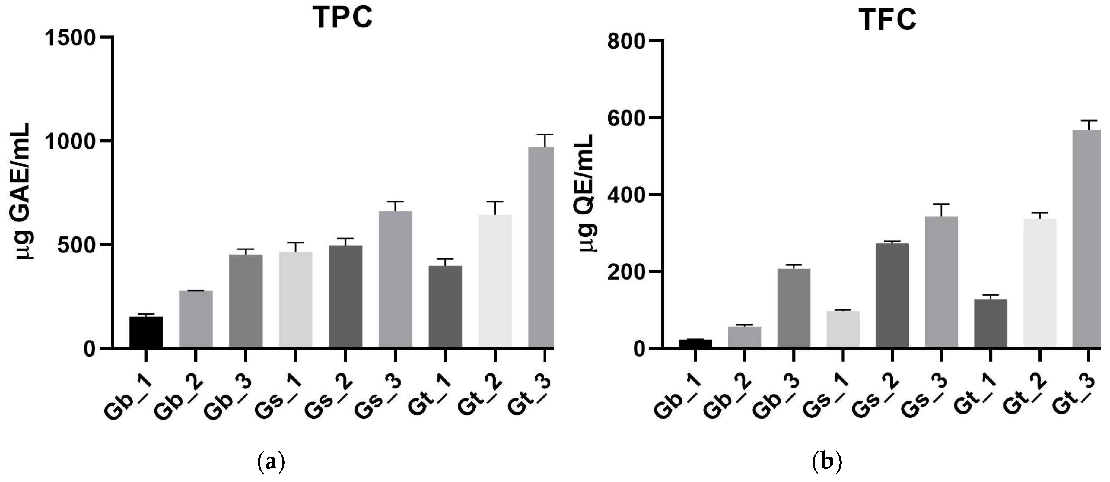
Figure 2.
Antioxidant activity of the Galeopsis samples: DPPH (a) ABTS (b) and FRAP (c) assays. ABTS: 2,2’-Azino-bis(3-ethylbenzothiazoline-6-sulfonic acid); DPPH: 2,2-Diphenyl-1-picrylhydrazyl; FRAP: Ferric-reducing antioxidant power; IC50: Half-maximal inhibitory concentration.
Figure 2.
Antioxidant activity of the Galeopsis samples: DPPH (a) ABTS (b) and FRAP (c) assays. ABTS: 2,2’-Azino-bis(3-ethylbenzothiazoline-6-sulfonic acid); DPPH: 2,2-Diphenyl-1-picrylhydrazyl; FRAP: Ferric-reducing antioxidant power; IC50: Half-maximal inhibitory concentration.
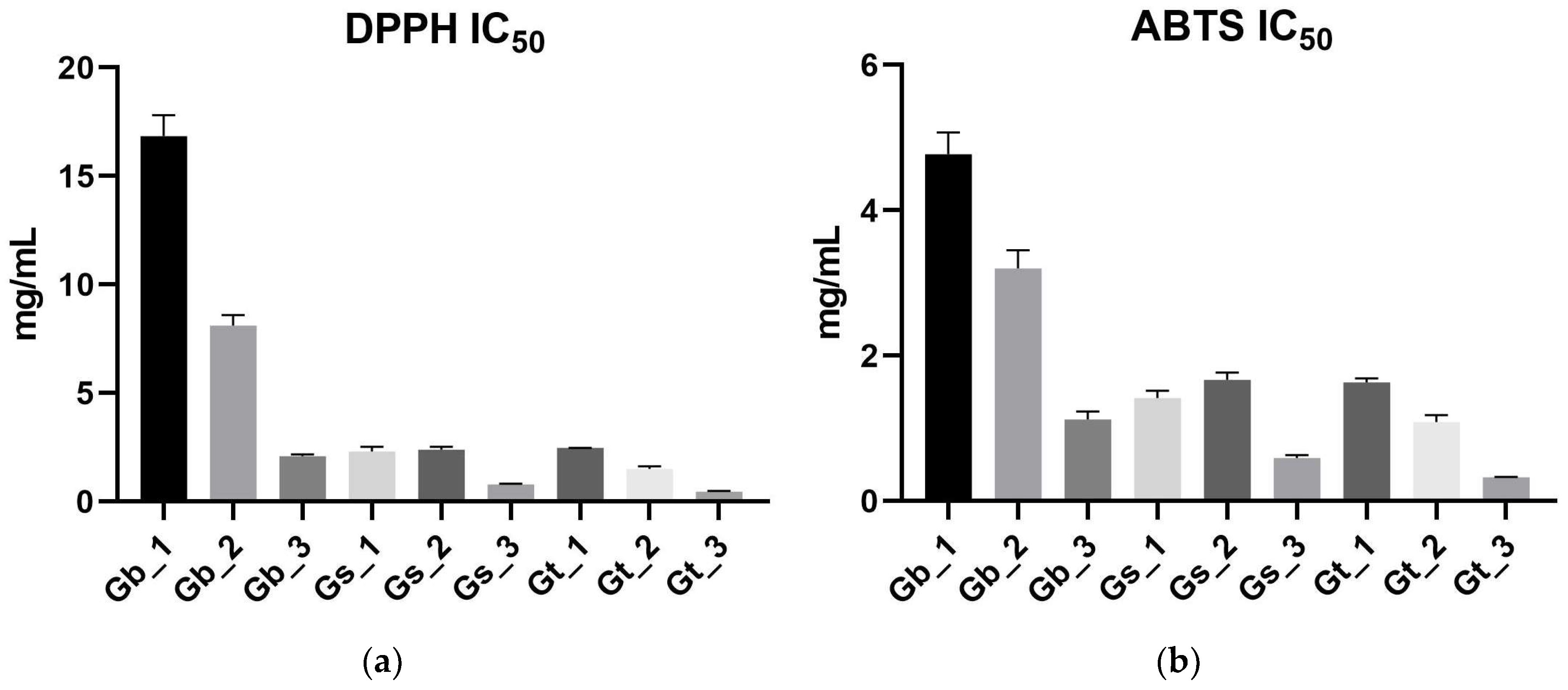
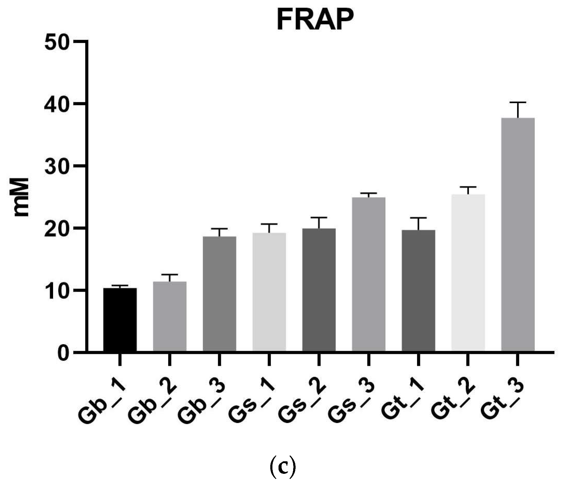
Figure 3.
AChE inhibitory activity IC50 values for Galeopsis samples. AChE: Acetylcholinesterase.
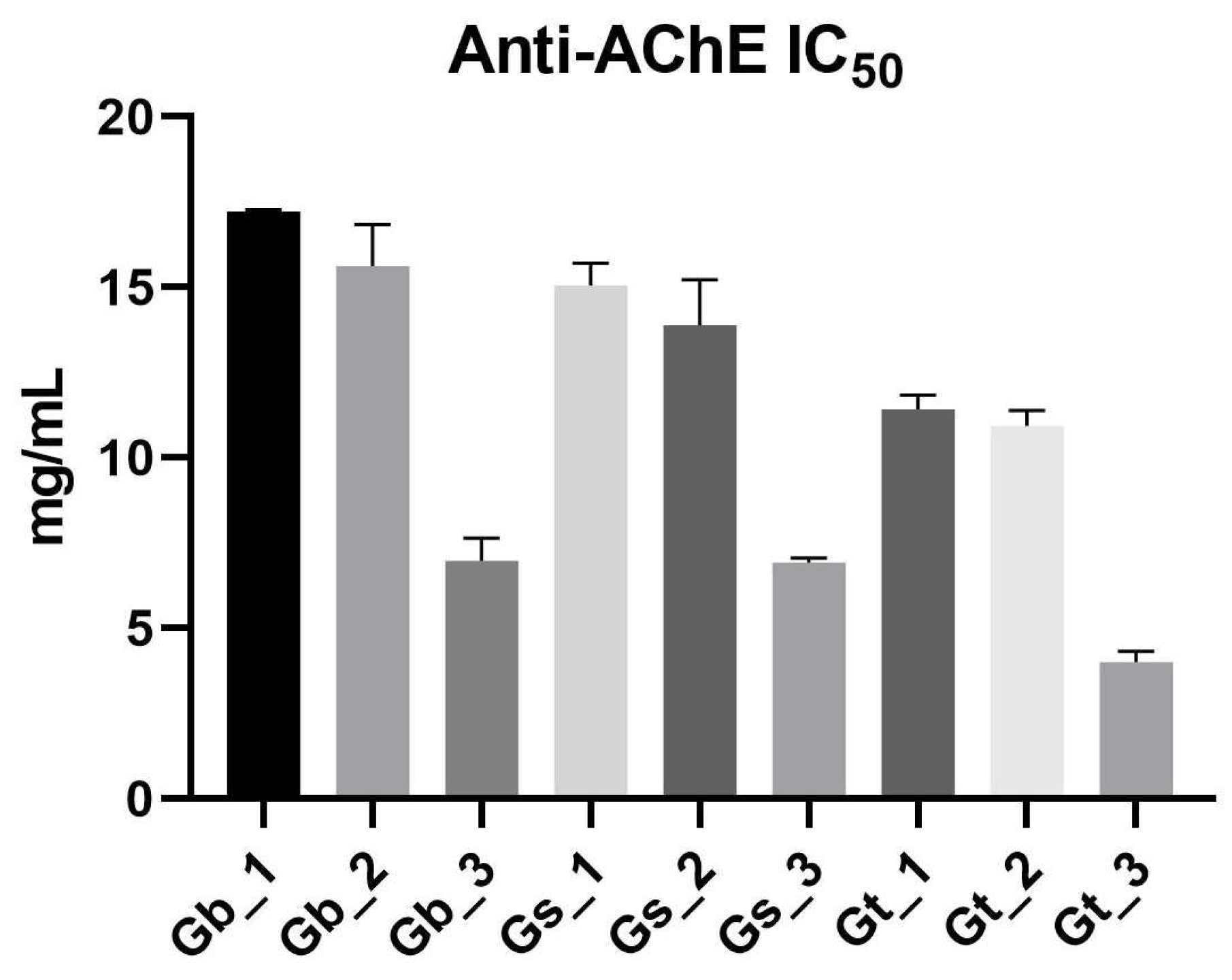
Figure 4.
HPTLC fingerprint of Galeopsis samples and reference standards: (a) 254 nm UV light, without derivatization; (b) 366 nm UV light, without derivatization; (c) 366 nm UV light, derivatization with NP–PEG reagent; (d) Antioxidant activity (DPPH assay, white light); (e) Neuroprotective activity (AChE inhibition assay, white light). Lanes: 1 – Caffeic acid; 2– Chlorogenic acid; 3 – Rutin; 4 – G. bifida roots; 5 – G. bifida aerial parts; 6 – G. bifida leaves; 7 – G. speciosa roots; 8 – G. speciosa aerial parts; 9 – G. speciosa leaves; 10 – G. tetrahit roots; 11 – G. tetrahit aerial parts; 12 – G. tetrahit leaves. AChE: Acetylcholinesterase; DPPH: 2,2-Diphenyl-1-picrylhydrazyl; HPTLC: High-performance thin-layer chromatography; NP–PEG: Natural products–polyethylene glycol; UV: Ultraviolet.
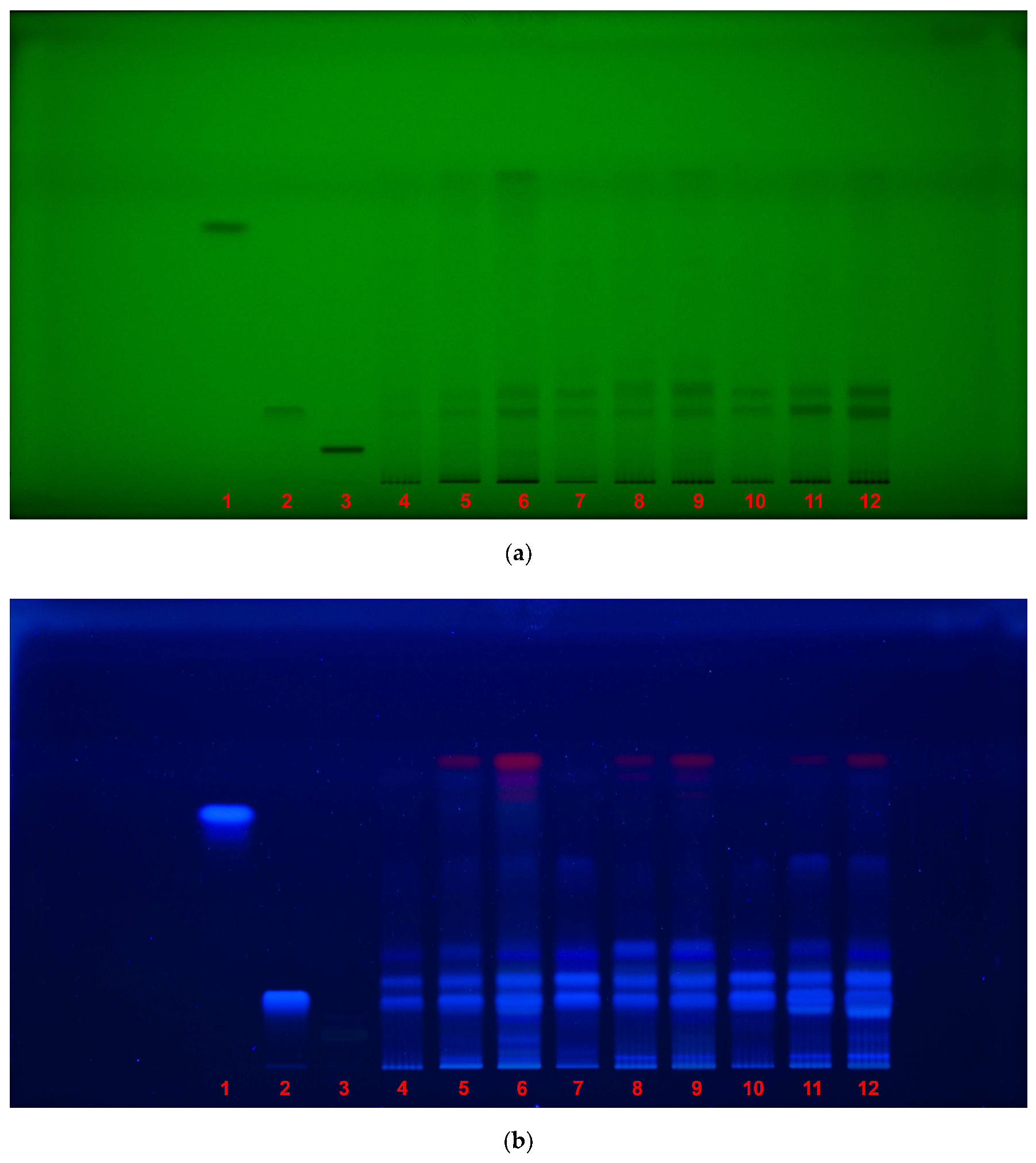

Table 1.
HPTLC fingerprint of Galeopsis samples and reference standards.
| HPTLC fingerprint | Description |
|---|---|
| 254 nm UV light, without derivatization |
• under shortwave UV light, dark bands indicate the presence of UV-absorbing compounds, such as phenolic acids and flavonoids; • caffeic acid (Rf 0.79) was not detected in any of the samples, confirming its absence or presence at undetectable concentrations (low amount demonstrated by UHPLC assay); • chlorogenic acid (Rf 0.22) was visible in all samples, confirming it as a major component of Galeopsis spp.; • rutin (Rf 0.085) was detected as dark band only as reference. |
| 366 nm UV light, without derivatization |
• under longwave UV light, compounds such as phenolic acids emit fluorescence, revealing their presence; • chlorogenic acid (Rf 0.22) was again observed in all samples, confirming its stability and prevalence across species; • rutin (Rf 0.085) was not visible under this condition, indicating that it does not fluoresce strongly without derivatization. |
| 366 nm UV light, derivatization with NP–PEG reagent |
• NP–PEG derivatization enhances flavonoid fluorescence (orange/yellow), allowing for their clearer visualization; • rutin (Rf 0.085) became visible after derivatization, confirming that its detection requires NP-PEG treatment; • strong flavonoid fluorescence was observed in G. tetrahit leaves, with a unique, orange-colored band that was absent in other species and plant parts, but not at the same Rf as rutin. |
| antioxidant activity (DPPH assay, white light) |
• DPPH assay was used to detect antioxidant activity, where active compounds appear as yellow bands against a purple background, indicating free radical scavenging activity; • chlorogenic acid (Rf 0.22) correlated strongly with antioxidant activity, as yellow bands were observed at this Rf across all samples; • for the DPPH HPTLC assay, extracts from aerial parts and leaves were diluted fivefold to prevent oversaturation of the plate and ensure accurate visualization of antioxidant activity. |
| neuroprotective activity (AChE inhibition assay, white light) |
• AChE inhibition assay was used to detect neuroprotective compounds, where active inhibitors appeared as clear bands against a purple background; • chlorogenic acid (Rf 0.22) demonstrated visible AChE inhibition in all samples, suggesting that it may contribute to the neuroprotective effects observed in Galeopsis spp.; • a distinct inhibition zone appeared at Rf 0.79, the same Rf as caffeic acid; however, since caffeic acid was not detected in the chemical fingerprinting, this suggests the presence of another compound with neuroprotective properties that migrates similarly; • slightly stronger inhibition zones were observed in G. tetrahit leaves, further reinforcing that this plant part contains potent neuroprotective compounds. |
AChE: Acetylcholinesterase; DPPH: 2,2-Diphenyl-1-picrylhydrazyl; HPTLC: High-performance thin-layer chromatography; NP–PEG: Natural products–polyethylene glycol; UHPLC: Ultra-high-performance liquid chromatography; UV: Ultraviolet.
Table 2.
Sample coding of plant material (Galeopsis spp.).
| Sample | Species/Vegetal Product | Date/Site of Collection (Southwest Romania Flora) | Voucher Specimen |
|---|---|---|---|
| Gb_1 | G. bifida/radix | 19 August 2024/Tismana City, Gorj County | GAL-BIF-2024-0819-2 |
| Gb_2 | G. bifida/herba | 19 August 2024/Tismana City, Gorj County | GAL-BIF-2024-0819-2 |
| Gb_3 | G. bifida/folium | 19 August 2024/Tismana City, Gorj County | GAL-BIF-2024-0819-2 |
| Gs_1 | G. speciosa/radix | 19 August 2024/Tismana City, Gorj County | GAL-SPC-2024-0819-2 |
| Gs_2 | G. speciosa/herba | 19 August 2024/Tismana City, Gorj County | GAL-SPC-2024-0819-2 |
| Gs_3 | G. speciosa/folium | 19 August 2024/Tismana City, Gorj County | GAL-SPC-2024-0819-2 |
| Gt_1 | G. tetrahit/radix | 21 July 2024/Lăpuşnicel Village, Caraş Severin County | GAL-TTH-2024-0721-2 |
| Gt_2 | G. tetrahit/herba | 21 July 2024/Lăpuşnicel Village, Caraş Severin County | GAL-TTH-2024-0721-2 |
| Gt_3 | G. tetrahit/folium | 21 July 2024/Lăpuşnicel Village, Caraş Severin County | GAL-TTH-2024-0721-2 |
Disclaimer/Publisher’s Note: The statements, opinions and data contained in all publications are solely those of the individual author(s) and contributor(s) and not of MDPI and/or the editor(s). MDPI and/or the editor(s) disclaim responsibility for any injury to people or property resulting from any ideas, methods, instructions or products referred to in the content. |
© 2025 by the authors. Licensee MDPI, Basel, Switzerland. This article is an open access article distributed under the terms and conditions of the Creative Commons Attribution (CC BY) license (http://creativecommons.org/licenses/by/4.0/).
Copyright: This open access article is published under a Creative Commons CC BY 4.0 license, which permit the free download, distribution, and reuse, provided that the author and preprint are cited in any reuse.


