Submitted:
05 March 2025
Posted:
06 March 2025
You are already at the latest version
Abstract
Infections caused by the protozoans Entamoeba histolytica (E. histolytica), Giardia lamblia (G. lamblia), and Trichomonas vaginalis (T. vaginalis) represent a global public health concern. Albendazole and mebendazole are drugs used to treat parasitosis resulting from protozoa. However, the low aqueous solubility of these compounds has led to the exploration of new strategies to enhance their solubility, with the formation of salts being a commonly employed strategy. The sulfonates A1, A2, and A3 of albendazole, and M1, M2, and M3 of mebendazole were synthesized, and their antiparasitic activity was determined in vitro against trophozoites of E. histolytica, G. lamblia, and T. vaginalis. Consequently, IC50 values ranged from 24.17 to 138.02 µM for the albendazole and mebendazole salts. For comparison, the IC50 values for the positive controls—albendazole, mebendazole, and metronidazole—ranged from 16.08 to 270.66 µM. Additionally, it was determined that salts A1, A3, M2, and M3 do not exhibit cytotoxic effects at concentrations of 500 µM on the VERO cell line. Taken together, these findings indicate that the formation of these new solid saline phases enhances the antiparasitic effects in vitro, which is crucial in the current search for improved, safe, and effective antiparasitic agents.
Keywords:
Benzimidazole
; Antiparasitic activity
; Entamoeba histolytica
; Giardia lamblia
; Trichomonas vaginalis
; Toxicity
; Selective index
; Sulfonate salts
1. Introduction
Parasitic infections caused by microaerophilic protozoa are a major problem affecting around 3 billion people globally. Among the main protozoa are Entamoeba histolytica (E. histolytica) and Giardia lamblia (G. lamblia), which are human pathogens of the gastrointestinal tract, and Trichomonas vaginalis (T. vaginalis), a pathogen of the human genitourinary tract [1,2,3].
Metronidazole (α-hydroxyethyl-2-methyl-5-nitroimidazole, Mtz) is the first choice for treating amoebiasis, giardiasis, and trichomoniasis [4,5]. However, overuse, indiscriminate over the counter (OTC) sales, and inadequate treatment regimens have resulted in the emergence of nitroimidazole-resistant strains with increased inhibitory concentrations of the drug, leading to treatment failures [6,7].
Resistance to Mtz in clinical isolates has been reported in G. lamblia [8,9], E. histolytica [10,11], and T. vaginalis [12,13]. Therefore, when treatment with Mtz is ineffective, it is recommended to use albendazole (methyl [5-(propylthio)-1H-benzimidazole-2-yl] carbamate, Abz) and mebendazole (methyl 5-benzoyl-1H-benzimidazole-2-yl-carbamate, Mbz) alone or in combination with Mtz [14,15,16,17,18].
The main mechanism of action of Abz and Mbz in protozoa is through their selective binding to β-tubulin dimers in the parasite, which prevents the polymerization of microtubules. Consequently, essential biological processes such as adhesion, motility, and cell division are directly affected [19,20,21]. In addition, benzimidazoles can induce oxidative stress by forming adducts in the parasite’s deoxyribonucleic acid (DNA) and causing DNA double-strand breaks, leading to cell cycle dysregulation and ultimately the death of the parasite [22,23].
Abz and Mbz have low water solubility, which implies low intestinal absorption [24,25,26]. To improve water solubility, various methodologies have been described, such as the formation of nanocapsules [27], solid dispersions [28], nanocrystals [29], liposomes [30], cyclodextrins [31], inclusion complexes [32], self-emulsifying drug delivery systems [33], and pharmaceutical salts [34]. These methodologies do not involve chemical structure modification but rather the combination with other molecules through mixtures or acid-base reactions.
The formation of salts with pharmaceutically acceptable counterions is useful for modifying physicochemical, biopharmaceutical, or therapeutic properties. These salts are considered chemical entities with improved pharmacokinetic, toxicological, and potency characteristics [35,36]. The use of sulfonic acids for salt formation has increased over time because they substantially improve the physicochemical properties of active pharmaceutical ingredients (APIs) and, in some cases, are safer than alternative salts [37,38].
The aim of the present study is to evaluate the in vitro amebicidal, trichomonicidal, and giardicidal activity of albendazole (A) and mebendazole (M) salts with benzenesulfonic acid (A1, M1), methanesulfonic acid (A2, M2), and p-toluenesulfonic acid (A3, M3). Additionally, their mean cytotoxicity concentration (CC50) on the VERO cell line and their selectivity index were assessed, aiming to select those with the best biological properties for subsequent in vivo pharmaceutical studies, which may serve as candidates for the treatment of parasitosis.
2. Materials and Methods
2.1. General Procedure to Prepare Abz and Mbz Sulfonic Salts
2.1.1. Reagents and Solvents
Albendazole (≥ 98%, CAS 54965-21-8), Mebendazole (≥ 98%, CAS 31431-39-7), p-toluensulfonic acid monohydrate (≥ 98.5% CAS 6192-52-5), benzenesulfonic acid monohydrate (97%, CAS 26158-00-9) and methanesulfonic acid (≥ 99%, CAS 75-75-2) were purchased from Sigma-Aldrich® and used without prior purification. Methanol (≥ 99.9%, CAS 67-56-1) and acetone (99.8%, CAS 67-64-1) were reagent grade and purchased from J.T. Baker.
2.1.2. Equipment and Conditions for Chemical Characterization
Powder X-ray diffraction (PXRD) was performed on a Bruker D2-Phaser. The data were collected in the interval 5–50° of 2θ angle and a stepsize of 0.02°. FT-IR spectra were recording using Thermo Scientific Spectrophotometer Nicolet iS10 and measured in the range of 4000–500cm-1 with diamond ATR accessory. NMR spectra were obtained on a Bruker AVANCE III HD 500 MHz (1H) and 125 MHz (13C) spectrometer using room temperature, samples of 30 mg, and DMSO-d6 as solvent for all samples. The melting point was determined by using a Fisher-Johns Melting Point Apparatus 12-144 from Fisher Scientific® in the range 20 – 300 °C.
2.1.3. General Method for Salts Preparation
An equimolar mixture of the benzimidazole drug (Abz, 265 mg; or Mbz, 295 mg) and the sulfonic acid (methane sulfonic acid, 97 mg; benzenesulfonic acid, 159 mg; or p-toluenesulfonic acid, 158 mg) was placed into a 10 mL glass vial provided with magnetic stirrer. Then enough solvent (acetone or methanol, ca. 3 mL) is poured to form a solid suspension. The reaction mixture is stirred at room temperature for 24 h. At terminus the suspension is filtered, and the solid is rinsed with an aliquot of fresh solvent to remove the starting material. Finally, the solid is dried under vacuum for 2 h. The yield of solid salt was 60-75%.
2.1.4. Chemical Characterization of Salt A1 (Albendazole Besylate)
1H NMR spectrum (DMSO-d6): δ (ppm) 7.61(d, 2H), 7.45(d, 1H), 7.37(dd, 1H), 7.31(m, 2H) 7.15(d, 2H), 3.78(s, 3H), 2.87(t, 2H), 1.53(sextet, 2H), 0.95(t, 3H). 13C NMR spectrum (DMSO-d6): δ (ppm) 154.33, 146.97, 135.3, 133.8, 128.3, 128.0, 127.6, 125.5, 124.1, 115.2, 114.0, 52.8, 36.3, 22.0, 13.1. XRPD (2θ angle): 7.0°, 7.5°, 11.0°, 11.4°, 18.0°, 18.7°, 19.5°, 20.8°, 24.9°, 25.8°, 27.3°, 29.4°. FT-IR: ν (cm-1) 3321, 2958, 1711, 1615, 1585, 1441, 1263, 1092. M.p. 182-184°C. Yield 65%.
2.1.5. Chemical Characterization of Salt A2 (Albendazole Mesylate)
1H NMR spectrum (DMSO-d6): δ (ppm) 7.54(m, 2H) 7.36(dd, 1H), 3.88(s, 3H), 2.95(t, 2H), 2.43(s, 3H)1.57(sextet, 2H), 0.97(t, 3H). 13C NMR spectrum (DMSO-d6): δ (ppm) 152.6, 144.2, 132.3, 130.0, 127.8, 125.6, 113.7, 112.9, 52.8, 36.3, 22.0, 13.1. XRPD (2θ angle): 7.0°, 11.9°, 13.0°, 13.7°, 16.3°, 16.9°, 19.0°, 19.6°, 20.7°, 21.3°, 22.9°, 23.6°, 25.4°, 26.1°, 27.4°, 29.1°, 30.3°. FT-IR: ν (cm-1) 3213, 1755, 1638, 1435, 1231, 1151, 1089. M.p. 154-155°C. Yield 71%.
2.1.6. Chemical Characterization of Salt A3 (Albendazole Tosylate)
1H NMR spectrum (DMSO-d6): δ (ppm) 7.55-7.47(m, 4H), 7.36(dd, 1H), 7.12(d, 2H), 3.88(s, 3H), 2.95(t, 2H), 2.28(s, 3H), 1.57(sextet, 2H), 0.97(t, 3H). 13C NMR spectrum (DMSO-d6): δ (ppm) 157.3, 153.1, 144.6, 138.3, 132.7, 130.6, 126.1, 125, 114.2, 113.5, 54.2, 35.7, 22.3, 21.2, 13.6. XRPD (2θ angle): 6.7°, 18.6°, 18.9°, 19.7°, 21.6°, 22.7°, 25.3°, 29.7°. FT-IR: ν (cm-1) 3063, 2927, 1758, 1644, 1601, 1442, 1237, 1155, 1096. M.p. 188-189°C. Yield 68%.
2.1.7. Chemical Characterization of Salt M1 (Mebendazole Besylate)
1H NMR spectrum (DMSO-d6): δ (ppm) 7.88(s, 1H), 7.72(d, 2H), 7.68-7.55(m, 6H), 7.34-7.27(m, 1H), 3.81(s, 3H). 13C NMR spectrum (DMSO-d6): δ (ppm) 195.35, 153.95, 148.44, 138.11, 132.03, 130.60, 129.37, 128.41, 128.32, 127.59, 125.47, 124.37, 115.75, 113.81, 52.97. XRPD (2θ angle): 6.4°, 7.0°, 7.5°, 11.0°, 11.4°, 18.1°, 18.8°, 20.7°, 22.4°, 23.6°, 24.8°, 25.7°, 27.1°. FT-IR: ν (cm-1) 3368, 2947, 1755, 1731, 1635, 1593, 1456, 1257, 1227, 1179, 1090. M.p. decomposition occurs prior to the melting of sample. Yield 70%.
2.1.8. Chemical Characterization of Salt M2 (Mebendazole Mesylate)
1H NMR spectrum (DMSO-d6): δ (ppm) 7.96(d, 1H), 7.73(m, 5H), 7.59(t, 2H), 3.89(s, 3H), 2.44(s, 6H). 13C NMR spectrum (120 MHz, DMSO-d6): δ (ppm) 194.94, 152.75, 146.06, 137.36, 133.15, 132.95, 132.70, 129.63, 128.68, 126.32, 115.34, 113.37, 53.95. XRPD (2θ angle): 6.7°, 10.3°, 12.8°, 17.0°, 18.0°, 18.5°, 19.3°, 19.8°, 20.5°, 21.8°, 22.8°, 23.4°, 25.3°, 26.3°, 28.5°, 29.3°. FT-IR: ν (cm-1) 3570, 3055, 1769, 1662, 1639, 1598, 1439, 1235, 1009. M.p. 207-208°C. Yield 65%.
2.1.9. Chemical Characterization of Salt M3 (Mebendazole Tosylate)
1H NMR spectrum (DMSO-d6): δ 7.95(d, 1H), 7.78-7.68(m, 5H), 7.58(t, 2H), 7.50(d, 2H), 7.12(d, 2H), 3.88(s, 3H), 2.28(s, 3H), 3H). 13C NMR spectrum (DMSO-d6): δ (ppm) 194.91, 152.89, 146.30, 145.34, 137.91, 137.43, 133.97, 133.97, 132.53, 130.19, 129.54, 128.58, 128.16, 125.98, 125.53, 115.35, 113.40, 53.75, 20.88. XRPD (2θ angle): 6.9°, 11.3°, 12.4°, 14.0°, 16.2°, 18.6°, 19.8°, 21.6°, 22.7°, 23.7°, 25.4°, 26.2°, 29.7°. FT-IR: ν (cm-1) 3567, 3063, 2804, 1764, 1654, 1641, 1602, 1441, 1231, 1200, 1009. M.p. 180-181°C. Yield 60%.
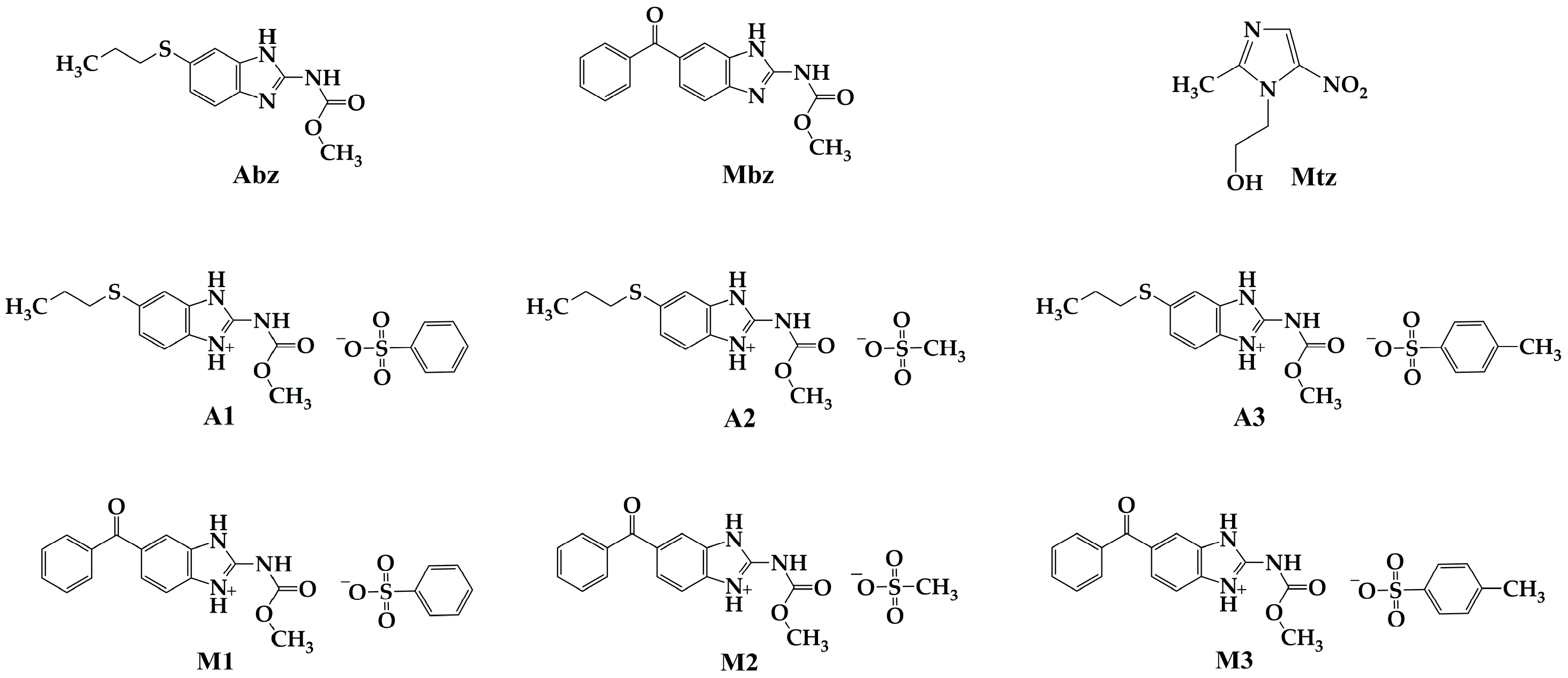
Figure 1.
Chemical structure of Abz and Mbz salts, as well as reference controls. A1: albendazole with benzenesulfonic acid; A2: albendazole with methanesulfonic acid; A3: albendazole with p-toluenesulfonic acid; M1: mebendazole with benzenesulfonic acid; M2: mebendazole with methanesulfonic acid; M3: mebendazole with p-toluenesulfonic acid; Abz: albendazole; Mbz: mebendazole and Mtz: metronidazole.
2.2. In Vitro Antiprotozoal Activity Assays
2.2.1. Stock Solutions
Stock solutions of Abz salts, Mbz salts, Abz, Mbz and Mtz where prepared by dissolving in 0.1 % (v / v) dimethyl sulfoxide (DMSO, AppliChem, Ottoweg, Darmstadt, Germany) at level at which no inhibition of trophozoites occurs[39]. The solutions were further diluted to 1 mL by adding freshly prepared culture medium to reach a concentration of 2 mg/mL. Two-fold serial dilutions were made in eppendorf tubes (Eppendorf) in 100 µL of culture medium. Each test included Mtz, Abz and Mbz as reference antiparasitic drugs, grow culture tubes (culture medium with trofozoites only) and blanck tubes (culture medium only).
2.2.2. Trophozoite Culture Conditions
Antiprotozoal activity was evaluated against trophozoites of E. histolytica (strain HM1: IMSS), G. lamblia (isolate J10) and T. vaginalis (isolate MB: FF09). E. histolytica trophozoites maintained axenically in BI-S-33 pH 6.5 medium supplemented with 10% calf serum (previously inactivated at 56 °C for 30 min (Microlab Laboratories, Mexico City, Mexico). G. lamblia trophozoites were maintained axenically in TYI-S-33 medium pH 6.8 supplemented with bovine bile and 10% fetal bovine serum previously inactivated (Microlab Laboratories, Mexico City, Mexico). T. vaginalis trophozoites were maintained axenically in modified Diamonds medium pH 7.0 supplemented with 10% calf serum previously inactivated (Microlab laboratories, Mexico City, Mexico). To determine the viability of the cultures and the number of trophozoites per milliliter, 0.4% trypan blue dye (In Vitro, Mexico City, Mexico) was used in a Neubauer chamber to guarantee viable cultures.
2.2.3. In Vitro Antiparasitic Activity Tests
To evaluate the antiparasitic effect of Abz and Mbz salts, the tube microdilution technique modified from Hernández-Ochoa [40] was used. 2 x 10⁴ trophozoites of G. lamblia and 1 x 10⁴ trophozoites of E. histolytica or T. vaginalis were used. The trophozoites were exposed for 48 h at 37 °C, with different concentrations of Abz and Mbz salts (2922- 2.08 µM). As positive inhibition controls, the drugs Abz, Mbz and Mtz were included under the same evaluation concentrations, and as negative controls, trophozoite cultures without treatment and trophozoites with 0.1% DMSO (AppliChem, Ottoweg, Darmstadt, Germany) were used. After exposure time, the percentage of cell viability and the percentage (%) of inhibition of trophozoite growth (% inhibition = 100 - % viability) were determined by microscopic counting, using 0.4% trypan blue staining (1:1, v / v) (In Vitro, Mexico City, Mexico) in a Neubauer chamber. Finally, the mean inhibitory concentration (IC50) was determined by linear regression using the concentration of compounds and the % inhibition. Three independent assays were performed in triplicate.
2.3. In Vitro Cytotoxicity Assays
2.3.1. Cell Line Culture Conditions
The cytotoxic activity was carried out on the VERO cell line (green monkey kidney cells: Cercopithecus aethiops) [39,41]). The cells were cultured in RPMI – 1640 medium (In Vitro, Mexico City, Mexico), supplemented with 10% fetal bovine serum previously inactivated (Microlab Laboratories, Mexico City, Mexico), L- glutamine 2Mm and 1% penicillin/streptomycin/amphotericin B (In Vitro, Mexico City, Mexico) at 37 °C with a 5% CO2 atmosphere. Cells were allowed to grow to a density of 85% and were subsequently harvested using a 0.05% trypsin-versene solution (In Vitro, Mexico City, Mexico) prior to each experiment. The viability and number of cells per milliliter were determined using 0.4% trypan blue dye (1:1, v / v) (In Vitro, Mexico City, Mexico) and a Neubauer chamber.
2.3.2. Cytotoxicity Assays
For the assay, cells were seeded in 96-well plates at concentrations of 1x10⁴ cells/ well and incubated for 24 h at 37 °C. After incubation, cells were treated with different concentrations (500 µM - 0.97 µM) of Abz and Mbz salts, in each assay the following were included as controls: cells with culture medium, cells with 0.1% DMSO (AppliChem, Ottoweg, Darmstadt, Germany), as well as the positive controls with the drugs Abz, Mbz and Mtz. Cells were incubated for 48 h at 37 °C. The cytotoxic effect of the salts evaluated on Vero cells was determined using the colorimetric technique with the WST–1 reagent (Roche Diagnostics GmbH, Mannheim, Germany), which allows measuring cell proliferation and viability by reducing the tetrazolium salt WST-1 to formazan by cellular dehydrogenases, the amount of formazan formed is directly correlated with the number of metabolically active cells in the culture, in this way the viability of the cells was measured with the optical density of the formazan products. With the results obtained, the Average Cytotoxic Concentration (CC50) was determined by linear regression with the percentage of inhibition and logarithm of the concentration. Three independent assays were performed in duplicate.
2.3.3. Selectivity Index (SI)
To determine the selectivity of the antiprotozoal activity, the cytotoxicity profile of the evaluated compounds was used, by calculating the SI: which is the quantitative relationship that exists between the cytotoxic activity against a mammalian cell line and the antiprotozoal activity of a certain compound (CC50 of the compounds evaluated on the VERO cell line / IC50 of the compounds evaluated on trophozoites)[39,41,42]. Normally an index greater than 1 is considered good since the in vitro antiparasitic activity is more selective towards the parasite, while a lower index than 1 is considered more selective towards mammalian cells according to what was reported [42,43].
3. Results
3.1. In Vitro Antiparasitic Activity
The antiparasitic activity of Abz and Mbz salts was evaluated in vitro against E. histolytica, G. lamblia, and T. vaginalis. The results are shown in Table 1 as IC50 values. Abz, Mbz, and Mtz were used as reference drugs.
Salt A1 did not present antiparasitic effects on E. histolytica, while salts A2 and A3 were 1.9 and 1.8 times more active than the Abz reference control, with IC50 values of 37.95 and 39.93 µM, respectively. All Mbz salts inhibited the growth of E. histolytica, with salt M3 having the lowest IC50 value (44.34 µM), being 1.3 times more active than Mbz. All three Abz salts were active against G. lamblia, with IC50 values ranging between 51.31 and 38.02 µM. Salt A3 increased its potency 5.2 times compared to the reference control Abz. Regarding Mbz salts, M1 had no antiparasitic effect on G. lamblia, while M2 and M3 salts were 3.2 and 3.3 times more active compared to Mbz. As for T. vaginalis, salts A1 and A3 did not present antiparasitic effects; however, salt A2 presented better antiparasitic activity, increasing its potency 1.68 times compared to Abz. All Mbz salts exhibited antiparasitic activity against T. vaginalis, with salt M1 having the lowest IC50 value (24.17 µM). Finally, the Mtz control presented IC50 values of 16.08 and 16.16 µM for E. histolytica and T. vaginalis, respectively, values that were lower than those obtained for the Abz and Mbz salts (Table 1). However, the IC50 value on G. lamblia was 97.63 µM, while the IC50 value obtained for salts A2, A3, M2, and M3 ranged from 51.31 to 79.62 µM.
Even though Abz and Mbz salts have good antiprotozoal activity, A1 does not inhibit the growth of E. histolytica and T. vaginalis. Nonetheless, the rest of the salts evaluated behaved as potent antiprotozoal agents, in almost all cases performing even better than the reference controls Abz and Mbz.
3.2. In Vitro Cytotoxicity
Biological assays were performed on the VERO cell line to better understand the cytotoxic effects of these salts on human cells, compared to the effects observed in protozoans. Cell viability was evaluated by the colorimetric assay with the WST-1 reagent (2-(4-iodophenyl)-3-(4-nitrophenyl)-5-(2,4-disulphenyl)-2H-tetrazolium). The results are expressed in Figure 2 and Figure 3 as percentage of cell viability (%CV). It is observed that the viability of VERO cells was not affected after being exposed to salts A1, A2, M2, and M3 at the highest concentration evaluated (500 µM) since a viability percentage greater than 98% was maintained. However, with salt A2, a decrease in cell viability was observed from the concentration of 250 µM (91%), and at the highest concentration evaluated, the viability was reduced to 37%.
On the other hand, with salt M1, a behavior similar to that of A2 is observed, since as the concentration of the salt increases, cell viability decreases. Starting at 31.25 µM, a decrease (95%) in viability is observed, reaching 38% at the concentration of 125 µM. As expected, 0.1% of DMSO did not alter cell growth; similarly, this occurred with the controls Abz, Mbz, and Mtz, which presented viability percentages greater than 95%
The CC50 was calculated using linear regression, determining the values to be 435.46 µM for salt A2 and 104.04 µM for salt M1 (Table 2). For the rest of the Abz and Mbz salts, as well as the controls, it was not possible to calculate the specific CC50 because they were not cytotoxic to VERO cells at the concentrations tested, so it was assumed to be greater than 500 µM (Figure 1 and Figure 2)
The selectivity index (SI) results for the three evaluated protozoans are presented in Table 3. For the salts that exhibited no cytotoxic effect at the tested concentrations on the VERO cell line, a CC50 of 500 µM was used as the basis for the SI calculation. SI exceeded 1 [42,43], indicating that the Abz and Mbz salts preferentially target protozoans.
4. Discussion
Our findings indicate that the selection of the chosen counterion for forming Abz and Mbz salts influences the antiparasitic activity against the tested parasites. Although the literature states that forming pharmaceutical salts does not alter the biological activity of the active pharmaceutical ingredient (API) [44], our in vitro findings contradict this assertion. We observed that G. lamblia A3 and M3 displayed the strongest antiparasitic effects, with A2 and M2 following, when compared to our references Mtz, Mbz, and Abz, indicating that a lower dosage is needed to achieve the equivalent antiparasitic effect (Table 1). A comparable behavior was noted with E. histolytica, as A2, M2, A3, and M3 demonstrated the most effective antiparasitic action in relation to Abz and Mbz; nevertheless, Mtz is the compound that showed the highest antiparasitic effect (Table 1). In conclusion, for T. vaginalis, the salt that exhibited the most effective antiparasitic properties was M1, which enhanced its effect compared to Mbz, whereas the other salts showed reduced or no activity against T. vaginalis. These findings suggest that the mesylate and tosylate ions increase the antimicrobial activity, akin to previous reports involving bacteria (Mycobacterium tuberculosis [45], Staphylococcus aureus, Bacillus subtilis, and Klebsiella pneumoniae [46]. Mesylate salts improve the antibacterial effectiveness when compared to the antibiotic in its unbound state. Comparable research has indicated that the antibacterial effectiveness of salts featuring different counterions and antimicrobials [47,48,49,50] varies based on the counterion utilized and the microorganism assessed. Nonetheless, the exact mechanism through which this behavior occurs is not completely clarified, and these researchers propose that enhancements in the physicochemical characteristics of salts facilitate specific interactions with the cell membrane of microorganisms, engaging with lipids, carbohydrates, and membrane proteins [48], thereby permitting a larger quantity of drug to penetrate the microorganism’s interior and be accessible to perform its biological function. Based on these assumptions, E. histolytica, G. lamblia, and T. vaginalis have distinct plasma membrane compositions, fluidities, and permeabilities [51,52,53], indicating that the interactions between the sulfonates Abz and Mbz with the cell surface vary among the parasites, influencing the uptake or expulsion of the drug by the cell. Our findings back this up, as the influence of one salt benefits one parasite but diminishes or reduces its effect on another (Table 1). Nonetheless, sufficient permeability research is required to clarify the synergistic interaction between the counterion and the medication (Abz and Mbz). Moreover, it is crucial to consider the toxicity of counterions when formulating a pharmaceutical salt [54]. Our findings indicated that the salts do not exhibit cytotoxicity towards VERO cells, except for A2 and M1 (Figure 2 and Figure 3). This phenomenon has been noted before, indicating that antibiotic sulfonate salts do not influence cell viability [45,55]. Nonetheless, it is essential to compare these values according to their antiparasitic effects; hence, we calculated the selectivity index (SI) to reflect the preference between parasites and host cells, with an IS > 1 considered the minimum threshold to demonstrate a preference for the parasites [42,43,56].
5. Conclusions
In conclusion, our findings showed that tosylate salts effectively inhibit the growth of E. histolytica and G. lamblia, outperforming the control drugs Abz, Mbz, and Mtz, all while preserving selectivity for parasites. Mesylate salts also effectively hinder the growth of E. histolytica and G. lamblia; however, A2 exhibited cytotoxic effects on kidney cells. Moreover, besylate salts reduce their effectiveness against G. lamblia and E. histolytica and are not an appropriate substitute for curtailing the proliferation of T. vaginalis. Therefore, it is crucial to emphasize that A3 and M3 are potential drug salts effective against protozoan parasitic infections. Ultimately, our study is consistent with others that examine how the use of sulfonates as counterions influences the antimicrobial efficacy of the parent compound, either positively or negatively.
Author Contributions
Conceptualization, M.G.B.P. and O.T.A.; methodology, M.G.B.P and J.G.G.; software, J.R.I.; validation, B.E.D.M., O.T.A..; formal analysis, J.R.I; investigation, M.G.B.P. ; data curation, M.G.B.P. and B.E.D.M.; writing—original draft preparation, J.G.G., D.D.M., M.G.B.P.; writing—review and editing, M.G.B.P, D.D.M., J.R.I.; visualization, B.E.D.M.; supervision, O.T.A.; project administration; O.T.A. All authors have read and agreed to the published version of the manuscript.
Funding
This research was funded by the “Secretaría de Ciencia, Humanidades, Tecnología e Innovación (SECIHTI), México” with grant number 745283 (CVU: 741821).
Institutional Review Board Statement
Not applicable
Informed Consent Statement
Not applicable
Data Availability Statement
All relevant data can be found in the article. Taken in part from the experimental work of the Ph. D. student Miriam Guadalupe Barón Pichardo.
Acknowledgments
Authors are grateful to Dr. María Esther Ramírez Moreno from the National School of Medicine and Homeopathy of the National Polytechnic Institute; to Dr. Martha Ponce Macotela from the Laboratory of Experimental Parasitology at the National Institute of Pediatrics for the donation of clinical isolates of G. lamblia J10; to Dr. Moisés León Juárez of the National Institute of Perinatology Unit 1 for the donation of the VERO cell line; to the Secretaría de Ciencia, Humanidades, Tecnología e Innovación (Secihti, México) for the scholarship with grant number 745283 (CVU 741821); and to the Faculty of Pharmacy at the Autonomous University of the State of Morelos for the facilities provided to carry out the project.
Conflicts of Interest
The authors declare no conflicts of interest.
Abbreviations
The following abbreviations are used in this manuscript:
| ABZ | Albendazole |
| API | Active Pharmaceutical Ingredient |
| CC50 | Cytotoxic Concentration 50% |
| DMSO | Dimethyl sulfoxide |
| DNA | Deoxyribonucleic acid |
| IC50 | Inhibitory Concentration 50% |
| MBZ | Mebendazole |
| MTZ | Metronidazole |
| NMR | Nuclear Magnetic Resonance |
| OTC | Over the counter |
| PXRD | Powder X-ray Diffraction |
| SI | Selectivity Index |
| %CV | Cell Viability percentage |
References
- Flores-Carrillo, P.; Velázquez-López, J.M.; Aguayo-Ortiz, R.; Hernández-Campos, A.; Trejo-Soto, P.J.; Yépez-Mulia, L.; Castillo, R. Synthesis, antiprotozoal activity, and chemoinformatic analysis of 2-(methylthio)-1H-benzimidazole-5-carboxamide derivatives: Identification of new selective giardicidal and trichomonicidal compounds. Eur. J. Med. Chem. 2017, 137, 211-220. [CrossRef]
- Hemphill, A.; Müller, N.; Müller, J. Comparative Pathobiology of the Intestinal Protozoan Parasites Giardia lamblia, Entamoeba histolytica, and Cryptosporidium parvum. Pathogens 2019, 8, 116. [CrossRef]
- Leitsch, D. Drug susceptibility testing in microaerophilic parasites: Cysteine strongly affects the effectivities of metronidazole and auranofin, a novel and promising antimicrobial. Int J Parasitol Drugs Drug Resist. 2017, 7, 321-327.
- Daneman, N.; Cheng, Y.; Gomes, T.; Guan, J.; Mamdani, M. M.; Saxena, F. E.; Juurlink, D. N. Metronidazole-associated Neurologic Events: A Nested Case-control Study. Clin Infect Dis. 2021, 72, 2095–2100.
- Löfmark, S.; Edlund, C.; Nord, C. E. Metronidazole is still the drug of choice for treatment of anaerobic infections. Clin Infect Dis. 2010, 50, 16–23. [CrossRef]
- Gonzales ,M.L.M.; Dans, L.F.; Sio-Aguilar, J. Antiamoebic drugs for treating amoebic colitis. Cochrane Database Syst Rev. 2019;1, CD006085. [CrossRef]
- Wongstitwilairoong, B.; Anothaisintawee, T.; Ruamsap, N.; Lertsethtakarn, P.; Kietsiri, P.; Oransathid, W.; Oransathid, W.; Gonwong, S.; Silapong, S.; Suksawad, U.; et al. Prevalence of Intestinal Parasitic Infections, Genotypes, and Drug Susceptibility of Giardia lamblia among Preschool and School-Aged Children: A Cross-Sectional Study in Thailand. Trop. Med. Infect. Dis. 2023, 8, 394. [CrossRef]
- Farbey, M.D.; Reynoldson, J.A.; Thompson, R.C. In vitro drug susceptibility of 29 isolates of Giardia duodenalis from humans as assessed by an adhesion assay. Int J Parasitol. 1995, 25, 593–9.
- Lemée, V.; Zaharia, I.; Nevez, G.; Rabodonirina, M.; Brasseur, P.; Ballet, J. J.; Favennec, L. Metronidazole and albendazole susceptibility of 11 clinical isolates of Giardia duodenalis from France. J Antimicrob Chemother. 2000, 46, 819-821. [CrossRef]
- Bansal, D.; Sehgal, R.; Chawla, Y.; Mahajan, R.C.; Malla, N. In vitro activity of antiamoebic drugs against clinical isolates of Entamoeba histolytica and Entamoeba dispar. Ann Clin Microbiol Antimicrob. 2004, 3, 27. [CrossRef]
- Singh, A.; Banerjee, T.; Shukla, S. K.; Upadhyay, S.; Verma, A. Creep in nitroimidazole inhibitory concentration among the Entamoeba histolytic isolates causing amoebic liver abscess and screening of andrographolide as a repurposing drug. Sci Rep. 2023,13, 12192. [CrossRef]
- Alves, M. S. D.; das Neves, R. N.; Sena-Lopes, Â.; Domingues, M.; Casaril, A. M.; Segatto, N. V.; Nogueira, T. C. M.; de Souza, M. V. N.; Savegnago, L.; Seixas, F. K.; Collares, T.; Borsuk, S. Antiparasitic activity of furanyl N-acylhydrazone derivatives against Trichomonas vaginalis: in vitro and in silico analyses. Parasit Vectors. 2020, 13,1, 59. [CrossRef]
- Mabaso, N.; Abbai, N. Distribution of genotypes in relation to metronidazole susceptibility patterns in Trichomonas vaginalis isolated from South African pregnant women. Parasitol Res, 2021, 120, 2233–2241. [CrossRef]
- Mørch, K.; Hanevik, K.; Robertson, L. J.; Strand, E. A.; Langeland, N. Treatment-ladder and genetic characterisation of parasites in refractory giardiasis after an outbreak in Norway. J Infect. 2008, 56, 268–273. [CrossRef]
- Speich, B.; Marti, H.; Ame, S.M.; Ali, S.M.; Bogoch, I.I.; Utzinger, J.; Albonico, M.; Keiser, J. Prevalence of intestinal protozoa infection among school-aged children on Pemba Island, Tanzania, and effect of single-dose albendazole, nitazoxanide and albendazole-nitazoxanide. Parasit Vectors, 2013, 4, 6-3. [CrossRef]
- Leitsch, D. Drug Resistance in the Microaerophilic Parasite Giardia lamblia. Curr Trop Med Rep. 2015, 2, 128-135. [CrossRef]
- Yereli, K.; Balcioğlu, I. C.; Ertan, P.; Limoncu, E.; Onağ, A. Albendazole as an alternative therapeutic agent for childhood giardiasis in Turkey. Clin Microbiol Infect. 2004, 10, 527–529. [CrossRef]
- Anichina, K.; Mavrova, A.; Vuchev, D. Benzimidazoles Containing Piperazine Skeleton at C-2 Position as Promising Tubulin Modulators with Anthelmintic and Antineoplastic Activity. Pharmaceuticals 2023, 16, 1518. [CrossRef]
- Juliano, C.; Monaco, G.; Bandiera, P., Tedde, G., Cappuccinelli, P. Action of anticytoskeletal compounds on in vitro cytopathic effect, phagocytosis, and adhesiveness of Trichomonas vaginalis. Genitourin Med. 1987, 63, 256–263. [CrossRef]
- Katiyar, S.K.; Gordon, V.R.; McLaughlin, G.L.; Edlind, T.D. Antiprotozoal activities of benzimidazoles and correlations with beta-tubulin sequence. Antimicrob Agents Chemother. 1994, 38, 2086-2090. [CrossRef]
- Benchimol, M.; Gadelha, A. P.; De Souza, W. Ultrastructural Alterations of the Human Pathogen Giardia intestinalis after Drug Treatment. Pathogens, 2023,12, 810. [CrossRef]
- Bártíková, H.; Vokřál, I.; Skálová, L.; Lamka, J.; Szotáková, B. Metabolismo oxidativo in vitro de xenobióticos en la duela de lanceta (Dicrocoelium dendriticum) y los efectos del albendazol y el sulfóxido de albendazol ex vivo. Xenobiotica, 2010, 4, 593–601. [CrossRef]
- Martínez-Espinosa, R.; Argüello-García, R.; Saavedra, E.; Ortega-Pierres, G. Albendazole induces oxidative stress and DNA damage in the parasitic protozoan Giardia duodenalis. Front Microbiol. 2015, 6, 800. [CrossRef]
- Buchter, V.; Priotti, J.; Leonardi, D.; Lamas, M. C.; Keiser, J. Preparation, Physicochemical Characterization and In Vitro and In Vivo Activity Against Heligmosomoides polygyrus of Novel Oral Formulations of Albendazole and Mebendazole. J Pharm Sci. 2020, 109, 1819–1826. [CrossRef]
- Ding, Y.; Zhiyuan, Z.; Ding, C.; Shufeng, X.; Zhe, Xu. “The Use of Cyclodextrin Inclusion Complexes to Increase the Solubility and Pharmacokinetic Profile of Albendazole”. Molecules, 2023, 28, 21: 7295. [CrossRef]
- Liang, Z.; Chen, M.; Yan, Y.; Chen, D.; Xie, S. Nanocrystal Suspensions for Enhancing the Oral Absorption of Albendazole. Nanomaterials. 2022,12, 3032. [CrossRef]
- Soleymani, N.; Sadr, S.; Santucciu, C.; Rahdar, A.; Masala, G.; Borji, H. Evaluation of the In-Vitro Effects of Albendazole, Mebendazole, and Praziquantel Nanocapsules against Protoscolices of Hydatid Cyst. Pathogens, 2024, 13, 790. [CrossRef]
- Suzuki, K.; Kawakami, K.; Fukiage, M.; Oikawa, M.; Nishida, Y.; Matsuda, M.; Fujita, T. Relevance of Liquid-Liquid Phase Separation of Supersaturated Solution in Oral Absorption of Albendazole from Amorphous Solid Dispersions. Pharmaceutics, 2020,13, 220. [CrossRef]
- Fateh, R.; Norouzi, R.; Mirzaei, E.; Nissapatron, V.; Nawaz, M.; Khalifeh-Gholi, M.; Hamta, A.; Adnani Sadati, S. J.; Siyadatpanah, A.; Fattahi Bafghi, A. In vitro evaluation of albendazole nanocrystals against Echinococcus granulosus protoscolices. Ann Parasitol. 2021, 67, 203–212. [CrossRef]
- Castro Alpízar, J.A.; Vargas Monge, R.; Madrigal Redondo, G.; Pacheco Molina, J.A. Development of novel microstructured lipid carriers for dissolution rate enhancement of albendazole. Int. J. Appl. Pharm. 2020, 12, 173–178.
- Eriksen, J.B.; Christensen, S.B.; Bauer-Brandl, A.; Brandl, M. Dissolution/permeation of albendazole in the presence of cyclodextrin and bile salts: A mechanistic in-vitro study into factors governing oral bioavailability. J. Pharm. Sci. 2021, 111, 1667–1673.
- Pacheco, P.A.; Rodrigues, L.N.C.; Ferreira, J.F.S.; Gomes, A.C.P.; Veríssimo, C.J.; Louvandini, H.; Costa, R.L.D.; Katiki, L.M. Inclusion complex and nanoclusters of cyclodextrin to increase the solubility and efficacy of albendazole. Parasitol. Res. 2018,117, 705–712.
- Meena, A.K.; Sharma, K.; Kandaswamy, M.; Rajagopal, S.; Mullangi, R. Formulation development of an albendazole self-emulsifying drug delivery system (SEDDS) with enhanced systemic exposure. Acta Pharm. 2012, 62, 563–580.
- Bolla, G.; Nangia, A. Novel pharmaceutical salts of albendazole. Cryst Eng Comm. 2018, 20, 6394- 6405.
- Elder, D. P.; Delaney, E.; Teasdale, A.; Eyley, S.; Reif, V. D.; Jacq, K.; Facchine, K. L.; Oestrich, R. S.; Sandra, P.; David, F. The utility of sulfonate salts in drug development. J Pharm Sci., 2010, 99, 2948–2961. [CrossRef]
- Thackaberry, E. A. Non-clinical toxicological considerations for pharmaceutical salt selection. Expert Opin Drug Metab Toxicol. 2012, 8, 1419–1433. [CrossRef]
- Verbeek, R.K.; Kanfer, I.; Walker, R.B. Generic substitution: The use of medicinal products containing different salts and implications for safety and efficacy. Eur J Pharm Sci. 2006, 28, 1–6.
- Emmerson, J.L.; Gibson, W.R.; Anderson, R.C. Acute toxicity of propoxyphene salts. Toxicol Appl Pharmacol. 1971, 19, 445–451.
- Duque-Montaño, B. E.; Gómez-Caro, L. C.; Sanchez-Sanchez, M.; Monge, A.; Hernández-Baltazar, E.; Rivera, G.; Torres-Angeles, O. Synthesis and in vitro evaluation of new ethyl and methyl quinoxaline-7-carboxylate 1,4-di-N-oxide against Entamoeba histolytic. Bioorg Med Chem. 2013, 21, 4550-4558. [CrossRef]
- Hernández-Ochoa, B.; Martínez-Rosas, V.; Morales-Luna, L.; Calderón-Jaimes, E.; Rocha-Ramírez, L.M.; Ortega-Cuellar, D.; Rufino-González, Y.; González-Valdez, A.; Arreguin-Espinosa, R.; Enríquez-Flores, S.; et al. Pyridyl Methylsulfinyl Benzimidazole Derivatives as Promising Agents against Giardia lamblia and Trichomonas vaginalis. Molecules, 2022, 27, 8902. [CrossRef]
- Carapina da Silva, C.; Silveira P.R.; Nascimento das Neves, R.; Dié Alves, M.S.; Sena-Lopes, A.; Moura, S., Borsuk, S.; Pereira de Pereira, C.M. Antiparasitic activity of synthetic curcumin monocarbonyl analogues against Trichomonas vaginalis. Biomed Pharmacother. 2019, 111, 367- 377. [CrossRef]
- Domínguez, V. I. G. Actividad anti-Giardia in vitro de los compuestos de Foeniculum vulgare y Citrus aurantifolia. Master’s Thesis, Autonomous University of Nuevo León, 2015.
- Quispe, A.; Zavala, D.; Rojas, J.; Posso, M.; Vaisberg, A. Efecto citotóxico selectivo in vitro de Muricin H (Acetogenina de Annona muricata) en cultivos celulares de cáncer de pulmón. Rev Peru Med Exp Salud Publica. 2006, 23, 265-269.
- Ortiz, L.J.C.; Balderrabano, L. A. Importancia de las sales orgánicas en la industria farmacéutica. Rev Mex Cienc Farm. 2017, 48, 18- 42.
- Silva, D.; Lopes, M.V.C.; Petrovski, Ž.; Santos, M.M.; Santos, J.P.; Yamada-Ogatta, S.F.; Bispo, M.L.F.; de Souza, M.V.N.; Duarte, A.R.C.; Lourenço, M.C.S.; et al. Novel Organic Salts Based on Mefloquine: Synthesis, Solubility, Permeability, and In Vitro Activity against Mycobacterium tuberculosis. Molecules 2022, 27, 5167. [CrossRef]
- Madeira, D.; Alves, C.; Silva, J.; Florindo, C.; Costa, A.; Petrovski, Ž.; Marrucho, I.M.; Pedrosa, R.; Santos, M.M.; Branco, L.C. Fluoroquinolone-Based Organic Salts and Ionic Liquids as Highly Bioavailable Broad-Spectrum Antimicrobials. Proceedings 2021, 78, 3. [CrossRef]
- Ferraz, R.; Santarém, N.; Santos, A.F.M.; Jacinto, M.L.; Cordeiro-da-Silva, A.; Prudêncio, C.; Noronha, J.P.; Branco, L.C.; Petrovski, Ž. Synthesis and Biological Evaluation of Amphotericin B Formulations Based on Organic Salts and Ionic Liquids against Leishmania infantum. Antibiotics 2022, 11, 1841. [CrossRef]
- Ferraz, R.; Silva, D.; Dias, A.R.; Dias, V.; Santos, M.M.; Pinheiro, L.; Prudêncio, C.; Noronha, J.P.; Petrovski, Ž.; Branco, L.C. Synthesis and Antibacterial Activity of Ionic Liquids and Organic Salts Based on Penicillin G and Amoxicillin hydrolysate Derivatives against Resistant Bacteria. Pharmaceutics 2020, 12, 221. [CrossRef]
- Mesallati, H.; Umerska, A.; Paluch, K.J.; Tajber, L. Amorphous Polymeric Drug Salts as Ionic Solid Dispersion Forms of Ciprofloxacin. Mol. Pharm. 2017, 14, 2209–2223.
- Mesallati, H.; Umerska, A.; Tajber, L. Fluoroquinolone Amorphous Polymeric Salts and Dispersions for Veterinary Uses. Pharmaceutics. 2019, 11, 268. [CrossRef]
- Angeles, R.G.M. Evaluación del efecto de los compuestos benzimidazolicos, CMC- 12, CMC- 19 y CMC- 20, sobre los niveles de transcripción de proteínas del metabolismo energético y del citoesqueleto de Giardia duodenalis. PhD. Thesis. Universidad Nacional Autónoma de México, Mexico city, 2014. https://ru.dgb.unam.mx/bitstream/20.500.14330/TES01000708272/3/0708272.pdf.
- Espinosa, C. M.; Martínez P. A. The plasma membrane of Entamoeba histolytica: structure and dynamics. Biology of the cell. 1991, 72, 189-200.
- Ibáñez, E.A.; Gómez, B.A. Trichomonas vaginalis: The versatility of a tenacious parasite. An Real Acad Farm. 2017, 83, 1, 10-47.
- Saal, C. and Becker, A. Pharmaceutical salts: A summary on doses of salt formers from the Orange Book. Eur J Pharm Sci. 2017, 49, 4, 614-623. [CrossRef]
- Florindo, C.; Costa, A.; Matos, C.; Nunes, S.L.; Matias, A.N.; Duarte, C.M.M.; Rebelo, L.P.N.; Branco, L.C.; Marrucho, I.M. Novel organic salts based on fluoroquinolone drugs: Synthesis, bioavailability and toxicological profiles. Int. J. Pharm. 2014, 469, 179–189.
- Camacho, M. D. R., Phillipson, J. D., Croft, S. L., Solis, P. N., Marshall, S. J., Ghazanfar, S. Screening of plant extracts for antiprotozoal and cytotoxic activities. J. Ethnopharmacol. 2003, 89, 185–191.
Figure 2.
Effects on cell viability of Abz salts on VERO cell line, after 48 h of exposure. Cells without treatment, 0.1% DMSO solvent control. Results are presented as the mean ± standard deviation of three independent assays in duplicate.
Figure 2.
Effects on cell viability of Abz salts on VERO cell line, after 48 h of exposure. Cells without treatment, 0.1% DMSO solvent control. Results are presented as the mean ± standard deviation of three independent assays in duplicate.
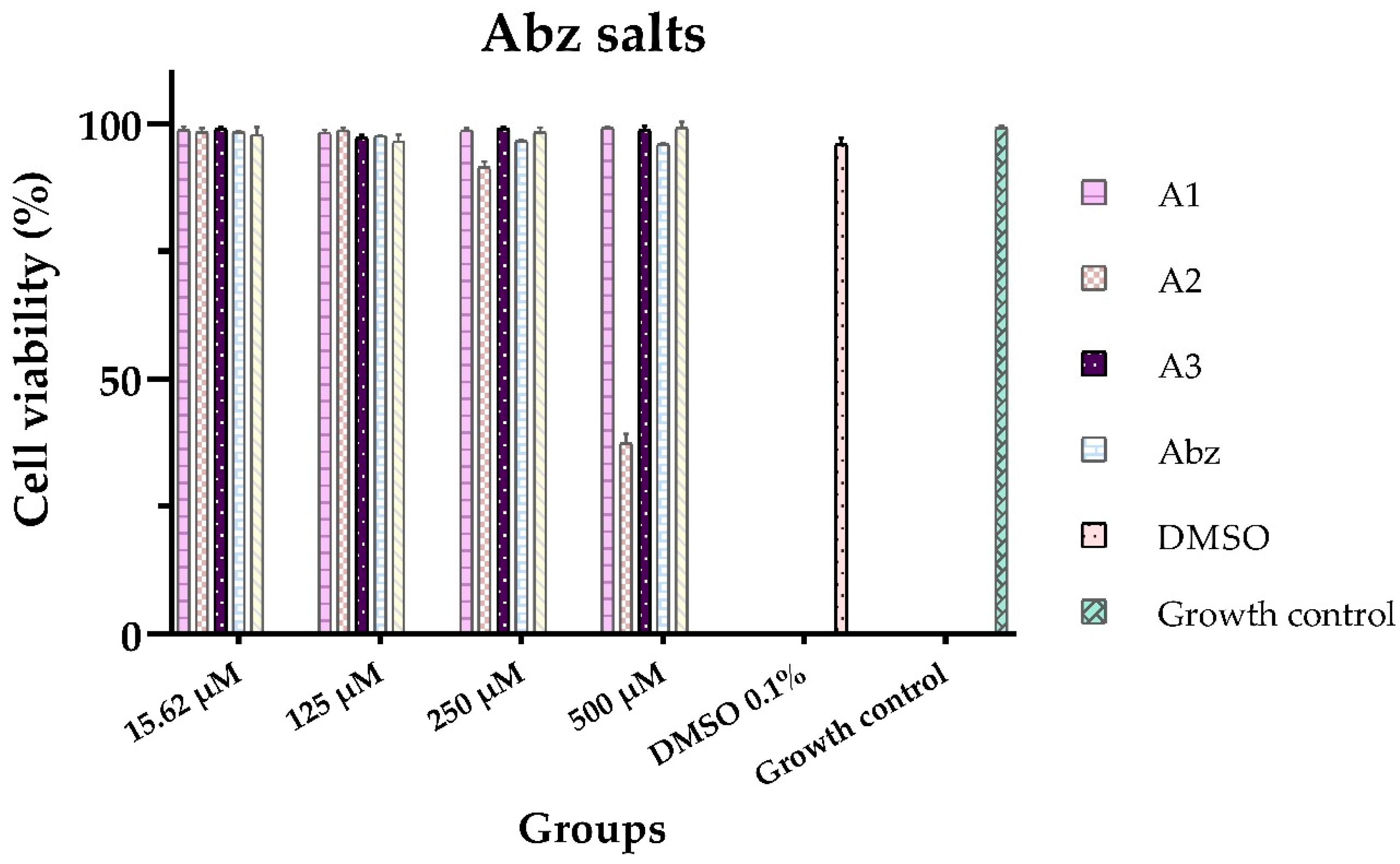
Figure 3.
Effect on cell viability of Mbz salts on VERO cell line, after 48 h of exposure. Cells without treatment, 0.1% DMSO solvent control. Results are presented as the mean ± standard deviation of three independent assays in duplicate.
Figure 3.
Effect on cell viability of Mbz salts on VERO cell line, after 48 h of exposure. Cells without treatment, 0.1% DMSO solvent control. Results are presented as the mean ± standard deviation of three independent assays in duplicate.
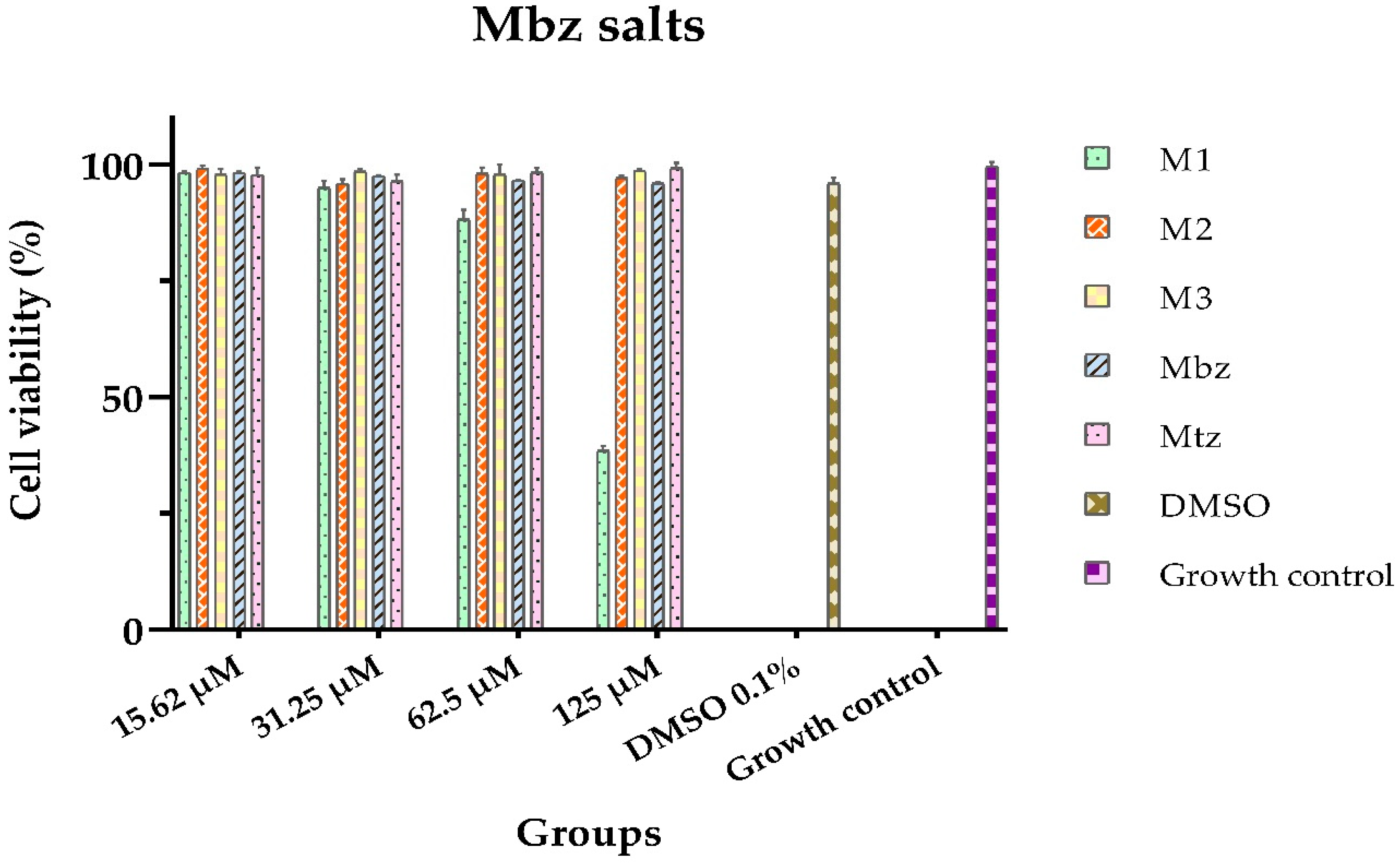

Table 1.
Antiparasitic activity of Abz and Mbz salts.
| Antiparasitic activity | |||
| Compound |
E. histolytica IC50 [µM] |
G. lamblia IC50 [µM] |
T. vaginalis IC50 [µM] |
| A1 | N. E.4 | 138.02 ± 1.41 | N. E.4 |
| A2 | 37.95 ± 0.72 | 78.05 ± 0.43 | 125.53 ± 1.80 |
| A3 | 39.93 ± 0.81 | 51.31 ± 1.23 | N. E.4 |
| M1 | 128.05 ± 2.69 | N. E.4 | 24.17 ± 0.15 |
| M2 | 57.72 ± 0.70 | 79.62 ± 0.99 | 49.86 ± 0.80 |
| M3 | 44.34 ± 0.45 | 77.98 ± 0.70 | 62.59 ± 1.44 |
| Abz1 | 73.51 ± 1.09 | 270.66 ± 1.89 | 211.71 ± 2.18 |
| Mbz2 | 59.81 ± 1.18 | 262.74 ± 2.49 | 36.59 ± 0.69 |
| Mtz3 | 16.08 ± 0.69 | 97.63 ± 1.43 | 16.16 ± 0.66 |
1Albendazole; 2mebendazole; 3metronidazole; 4No effect at the evaluated concentrations. Results are represented as the mean ± standard deviation of three independent assays in duplicate.
Table 2.
In vitro cytotoxicity values of Abz and Mbz salts in VERO cell line.
| Cytotoxic activity | |
| Salt | CC50 (µM) |
| A1 | > 500 |
| A2 | 435.46 ± 2.17 |
| A3 | > 500 |
| M1 | 104.04 ± 3.85 |
| M2 | > 500 |
| M3 | > 500 |
| Abz | > 500 |
| Mbz | > 500 |
| Mtz | > 500 |
The results are represented as the means of three independent tests in duplicate, ± SD: standard deviation.
Table 3.
Selectivity index of Abz and Mbz salts.
| Selectivity Index | |||
| Salt | E. histolytica | G. lamblia | T. vaginalis |
| A1 | N. E.1 | 3.62 | N. E.1 |
| A2 | 11.47 | 5.57 | 3.46 |
| A3 | 12.52 | 9.74 | N. E.1 |
| M1 | 0.81 | N. E.1 | 4.30 |
| M2 | 8.66 | 6.27 | 10.02 |
| M3 | 11.27 | 6.41 | 7.98 |
| Abz | 6.80 | 1.84 | 2.36 |
| Mbz | 8.35 | 1.90 | 13.66 |
| Mtz | 31.09 | 5.12 | 30.94 |
1No effect on the evaluated concentrations.
Disclaimer/Publisher’s Note: The statements, opinions and data contained in all publications are solely those of the individual author(s) and contributor(s) and not of MDPI and/or the editor(s). MDPI and/or the editor(s) disclaim responsibility for any injury to people or property resulting from any ideas, methods, instructions or products referred to in the content. |
© 2025 by the authors. Licensee MDPI, Basel, Switzerland. This article is an open access article distributed under the terms and conditions of the Creative Commons Attribution (CC BY) license (http://creativecommons.org/licenses/by/4.0/).
Copyright: This open access article is published under a Creative Commons CC BY 4.0 license, which permit the free download, distribution, and reuse, provided that the author and preprint are cited in any reuse.


