Submitted:
09 February 2025
Posted:
10 February 2025
You are already at the latest version
Abstract
Background and Objectives: Uveitis-masked syndromes or masquerade syndromes (UMSs) are a group of ocular conditions with several systemic underlying causes, malignant or nonmalignant, that mimic the inflammatory status of the uvea. They are often difficult to detect and diagnose with traditional techniques, such as ophthalmic exams. Ocular B (bidimensional)-ultrasound (OBU) is a non-invasive, repeatable, rapid ultrasound method effective in indirect signs that lead back to systemic diseases. It is comparable in effectiveness with other imaging tools. The cause of UMSs can often be serious, and therefore early diagnosis and prompt treatment are very critical. This study aims to identify the sonographic signs of these forms that can help physicians discover the cause underlying UMS. Materials and Methods: This is a consecutive, retrospective, nonrandomized study. This study is conducted at University Hospital Polyclinic of Bari, Italy, from January 2022 to December 2024. A total of 186 patients were included, from 10 to 85 years old. They all were undergone to B-scan ultrasonography (Quantel Medical ABSolu Ocular Ultrasound). Results: All patients reported blurred vision, that it can be accompanied by visual reduction (<20/40, Snellen charts), photophobia, floaters, flashes, proptosis, redness. In all cases we have noticed peculiar ultrasonographic signs which discriminate underlying systemic diagnosis, such as vitreous corpuscles, choroid thickening, solid tumors primitive or metastatic. Finally, they were identified different diseases, such as primary intraocular lymphoma (PIOL), and other lymphoproliferative conditions, orbital plasmacytoma, uveal melanoma, metastasis, endogenous endophthalmitis, retinal detachment, central serous retinopathy, metallic foreign bodies, ocular amyloidosis, toxoplasmosis, sarcoidosis, tuberculosis, syphilis, birdshot chorioretinitis, and drug-induced UMSs. lymphoma, other lymphoproliferative conditions, toxoplasmosis, sarcoidosis, tuberculosis, syphilis, UMSs caused by drugs, and others. Conclusion: Making a firm diagnosis of UMS is not easy, and the various forms are often underdiagnosed. Early detection of these forms is imperative. The use of OBU can help diagnose indirect signs of these forms early and treat them promptly. It compares well with other diagnostic imaging techniques, such as MRI, but this does not mean that it replaces them; it can be an added value in multimodal imaging.
Keywords:
1. Introduction
2. Systemic Diseases Related Masquerade Syndrome
3. Symptoms
4. Therapeutic Strategy
5. Multimodal Imaging and Other Tests
6. Role of B-Scan Ocular Ultrasound
7. Benefits and Limits of B-Scan Ocular Ultrasound
8. Methods
9. Results
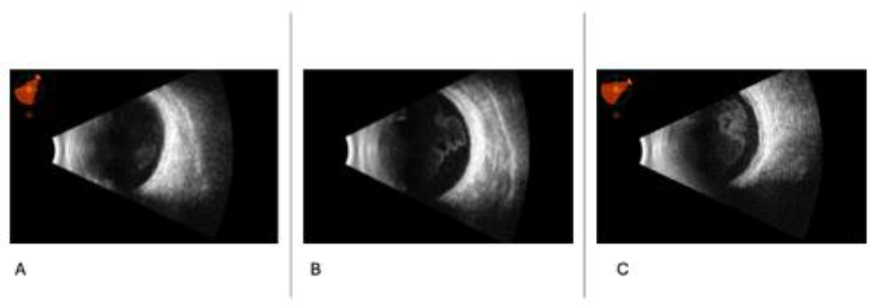
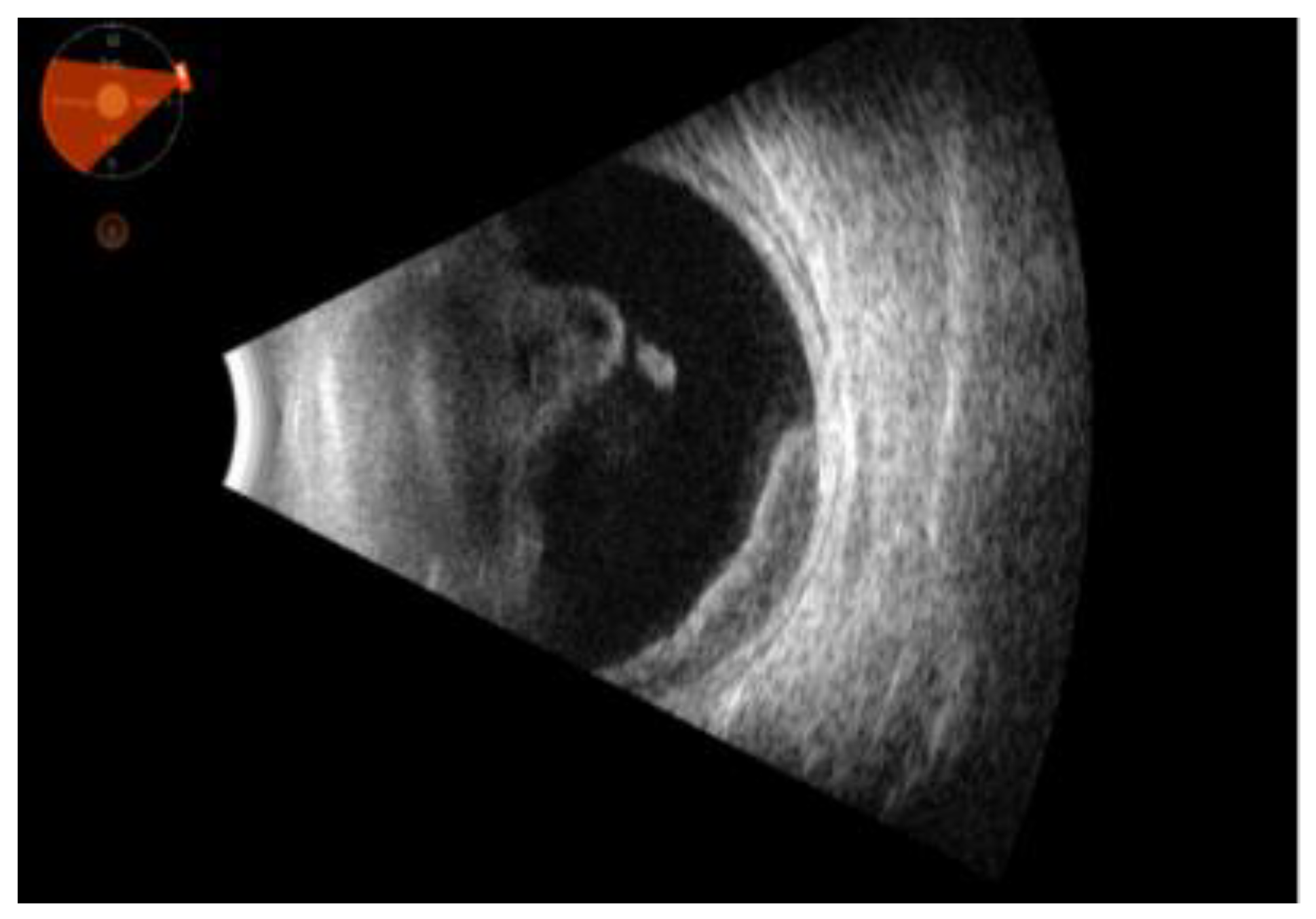
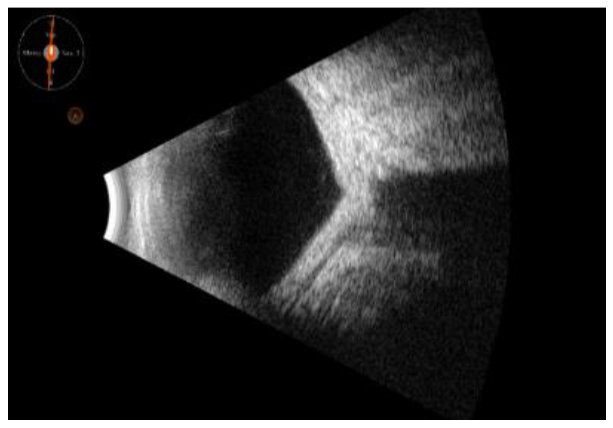
10. B-Scan Sonography Patterns
11. Discussion and Conclusions
Author Contributions
Funding
Conflicts of Interest
References
- Rothova, A. Uveitis masquerade syndromes. Ophthalmology 2001, 108, 386–399. [Google Scholar] [CrossRef] [PubMed]
- Rothova, A.; Groen, F.; Ten Berge, J.C.E.M.; Lubbers, S.M.; Vingerling, J.R.; Thiadens, A.A.H.J. CAUSES AND CLINICAL MANIFESTATIONS OF MASQUERADE SYNDROMES IN INTRAOCULAR INFLAMMATORY DISEASES. Retina 2021, 41, 2318–2324. [Google Scholar] [CrossRef] [PubMed]
- Grange, L.K.; Kouchouk, A.; Dalal, M.D.; Vitale, S.; Nussenblatt, R.B.; Chan, C.-C.; Sen, H.N. Neoplastic Masquerade Syndromes in Patients With Uveitis. American Journal of Ophthalmology 2014, 157, 526–531. [Google Scholar] [CrossRef]
- Coupland, S.E.; Chan, C.C.; Smith, J. Pathophysiology of Retinal Lymphoma. Ocular Immunology and Inflammation 2009, 17, 227–237. [Google Scholar] [CrossRef] [PubMed]
- Dini, G.; Capolsini, I.; Cerri, C.; Massei, M.S.; Mastrodicasa, E.; Perruccio, K.; Gorello, P.; Caniglia, M.; Verrotti, A.; Arcioni, F. Acute lymphoblastic leukemia relapse presenting with optic nerve infiltration. SAGE Open Medical Case Reports 2023, 11, 2050313X231175020. [Google Scholar] [CrossRef]
- Zloto, O.; Vahdani, K.; Stack, R.; Verity, D.H.; Rose, G.E. Periocular Presentation of Solitary Plasmacytomas and Multiple Myeloma. Ophthalmic Plastic & Reconstructive Surgery 2022, 38, 180–184. [Google Scholar] [CrossRef]
- Ashkenazy, N.; Harbour, J.W.; Dubovy, S.R.; Albini, T.A.; Sridhar, J.; Patel, N.; Hansen, E.D.; Uchiyama, E.; Rubsamen, P.E.; Correa, Z.M. Vitreous metastasis from cutaneous melanoma: diagnosis and management. ABO 2023, 87. [Google Scholar] [CrossRef] [PubMed]
- Feng, L.; Zhu, J.; Gao, T.; Li, B.; Yang, Y. Uveal Melanoma in the Peripheral Choroid Masquerading as Chronic Uveitis. Optometry and Vision Science 2014, 91, e222–e225. [Google Scholar] [CrossRef] [PubMed]
- Kafkala, C.; Daoud, Y.J.; Paredes, I.; Foster, C.S. Masquerade Scleritis. Ocular Immunology and Inflammation 2005, 13, 479–482. [Google Scholar] [CrossRef] [PubMed]
- Blitzer, A.L.; Schechet, S.A.; Shah, H.A.; Blair, M.P. Retinoblastoma presenting as pseudohypopyon and preserved visual acuity. American Journal of Ophthalmology Case Reports 2021, 23, 101141. [Google Scholar] [CrossRef]
- Maalouf, F.; Abdulaal, M.; Hamam, R.N. Chronic Postoperative Endophthalmitis: A Review of Clinical Characteristics, Microbiology, Treatment Strategies, and Outcomes. International Journal of Inflammation 2012, 2012, 1–6. [Google Scholar] [CrossRef] [PubMed]
- Joye, A.S.; Bhisitkul, R.B.; Pereira, D.D.S.; Gonzales, J.A. Rhegmatogenous retinal detachment masquerading as exudative panuveitis with intense anterior chamber inflammatory reaction. American Journal of Ophthalmology Case Reports 2020, 18, 100618. [Google Scholar] [CrossRef] [PubMed]
- Papadia, M.; Jeannin, B.; Herbort, C. Central serous chorioretinopathy misdiagnosed as posterior uveitis and the vicious circle of corticosteroid therapy. J Ophthalmic Vis Res 2015, 10, 303. [Google Scholar] [CrossRef]
- Yeh, S.; Ralle, M.; Phan, I.T.; Francis, P.J.; Rosenbaum, J.T.; Flaxel, C.J. Occult intraocular foreign body masquerading as panuveitis: inductively coupled mass spectrometry and electrophysiologic analysis. J Ophthal Inflamm Infect 2012, 2, 99–103. [Google Scholar] [CrossRef]
- Dammacco, R.; Merlini, G.; Lisch, W.; Kivelä, T.T.; Giancipoli, E.; Vacca, A.; Dammacco, F. Amyloidosis and Ocular Involvement: an Overview. Seminars in Ophthalmology 2020, 35, 7–26. [Google Scholar] [CrossRef]
- Kalogeropoulos, D.; Afshar, F.; Kalogeropoulos, C.; Vartholomatos, G.; Lotery, A.J. Diagnostic and therapeutic challenges in acute retinal necrosis; an update. Eye 2024, 38, 1816–1826. [Google Scholar] [CrossRef] [PubMed]
- Dutta Majumder, P.; Khetan, V.; Biswas, J. Masquerade syndrome: A review of uveitic imposters. Asia-Pacific Journal of Ophthalmology 2024, 13, 100054. [Google Scholar] [CrossRef] [PubMed]
- Agarwal, M.; Dutta Majumder, P.; Babu, K.; Konana, V.; Goyal, M.; Touhami, S.; Stanescu-Segall, D.; Bodaghi, B. Drug-induced uveitis: A review. Indian J Ophthalmol 2020, 68, 1799. [Google Scholar] [CrossRef]
- Witkin, A.J.; Hahn, P.; Murray, T.G.; Arevalo, J.F.; Blinder, K.J.; Choudhry, N.; Emerson, G.G.; Goldberg, R.A.; Kim, S.J.; Pearlman, J.; et al. Brolucizumab-Associated Intraocular Inflammation in Eyes Without Retinal Vasculitis. Journal of VitreoRetinal Diseases 2021, 5, 326–332. [Google Scholar] [CrossRef]
- Deitch-Harel, I.; Raskin, E.; Habot-Wilner, Z.; Friling, R.; Amer, R.; Kramer, M. Uveitis Induced by Biological Agents Used in Cancer Therapy. Ocular Immunology and Inflammation 2021, 29, 1370–1374. [Google Scholar] [CrossRef]
- Fardeau, C.; Bencheqroun, M.; Levy, A.; Bonnin, S.; Ferchaud, M.-A.; Fardeau, L.; Coscas, F.; Bodaghi, B.; Lebrun-Vignes, B. Uveitis Associated with Cancer Immunotherapy: Long-Term Outcomes. Immunotherapy 2021, 13, 1465–1481. [Google Scholar] [CrossRef] [PubMed]
- Aironi, V.; Gandage, S. Pictorial essay: B-scan ultrasonography in ocular abnormalities. Indian J Radiol Imaging 2009, 19, 109–115. [Google Scholar] [CrossRef]
- Nagaraju, R.M. Efficacy of High Frequency Ultrasound in Localization and Characterization of Orbital Lesions. JCDR 2015. [Google Scholar] [CrossRef] [PubMed]
- Lanni, V.; Iuliano, A.; Fossataro, F.; Russo, C.; Uccello, G.; Tranfa, F.; et al. The role of ultrasonography in differential diagnosis of orbital lesions. J Ultrasound. marzo 2021, 24, 35–40. [Google Scholar] [CrossRef] [PubMed]
- Shinar, Z.; Chan, L.; Orlinsky, M. Use of Ocular Ultrasound for the Evaluation of Retinal Detachment. The Journal of Emergency Medicine 2011, 40, 53–57. [Google Scholar] [CrossRef] [PubMed]
- Malgotra, S.; Sharma, M.; Sharma, N. Visualizing the Spectrum: B-scan Ultrasonography Across Diverse Ocular Abnormalities: A Pictorial Review. TNOA Journal of Ophthalmic Science and Research 2024, 62, 20–26. [Google Scholar] [CrossRef]
- Boricean, N.G.; Scripcă, O.R. Multifocal Choroiditis and Panuveitis - difficulties in diagnosis and treatment. rjo 2017, 61, 293–298. [Google Scholar] [CrossRef]
- Seguin, J.; Le, C.-K.; Fischer, J.W.; Tessaro, M.O.; Berant, R. Ocular Point-of-Care Ultrasound in the Pediatric Emergency Department. Pediatr Emer Care 2019, 35, e53–e58. [Google Scholar] [CrossRef] [PubMed]
- Blaivas, M.; Theodoro, D.; Sierzenski, P.R. A Study of Bedside Ocular Ultrasonography in the Emergency Department. Academic Emergency Medicine 2002, 9, 791–799. [Google Scholar] [CrossRef] [PubMed]
- Bergmann, K.R.; Reardon, R.F.; Flores, G.; Whitcomb, V.; Christensen, E.W.; Watson, D.; Kharbanda, A. Trends in Medical Claims and Utilization of Limited Ultrasonography Among Emergency Physicians and Radiologists Within a Large Health Plan Provider. J of Ultrasound Medicine 2019, 38, 1279–1286. [Google Scholar] [CrossRef]
- Wu, N.; Zhang, H.; Chen, B.; Ding, W. A novel application of B-ultrasonography at various head positions in the diagnosis of untypical uveitis-glaucoma-hyphema (UGH) syndrome: A case report. Medicine 2019, 98, e13891. [Google Scholar] [CrossRef]
- Song, J.L.; Elkhunovich, M.; Rankin, J.H. Child With Red Eye and Blurry Vision. Pediatr Emer Care 2017, 33, 703–705. [Google Scholar] [CrossRef]
Disclaimer/Publisher’s Note: The statements, opinions and data contained in all publications are solely those of the individual author(s) and contributor(s) and not of MDPI and/or the editor(s). MDPI and/or the editor(s) disclaim responsibility for any injury to people or property resulting from any ideas, methods, instructions or products referred to in the content. |
© 2025 by the authors. Licensee MDPI, Basel, Switzerland. This article is an open access article distributed under the terms and conditions of the Creative Commons Attribution (CC BY) license (http://creativecommons.org/licenses/by/4.0/).


