Preprint
Article

This version is not peer-reviewed.

Malignant Transformation of Proliferative Verrucous Leukoplakia: A Description of the Clinical Characteristics of These Oral Cancers

A peer-reviewed article of this preprint also exists.

Submitted:

06 February 2025

Posted:

07 February 2025

You are already at the latest version

Abstract

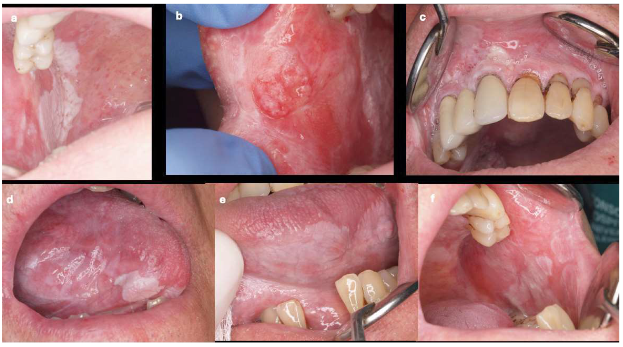
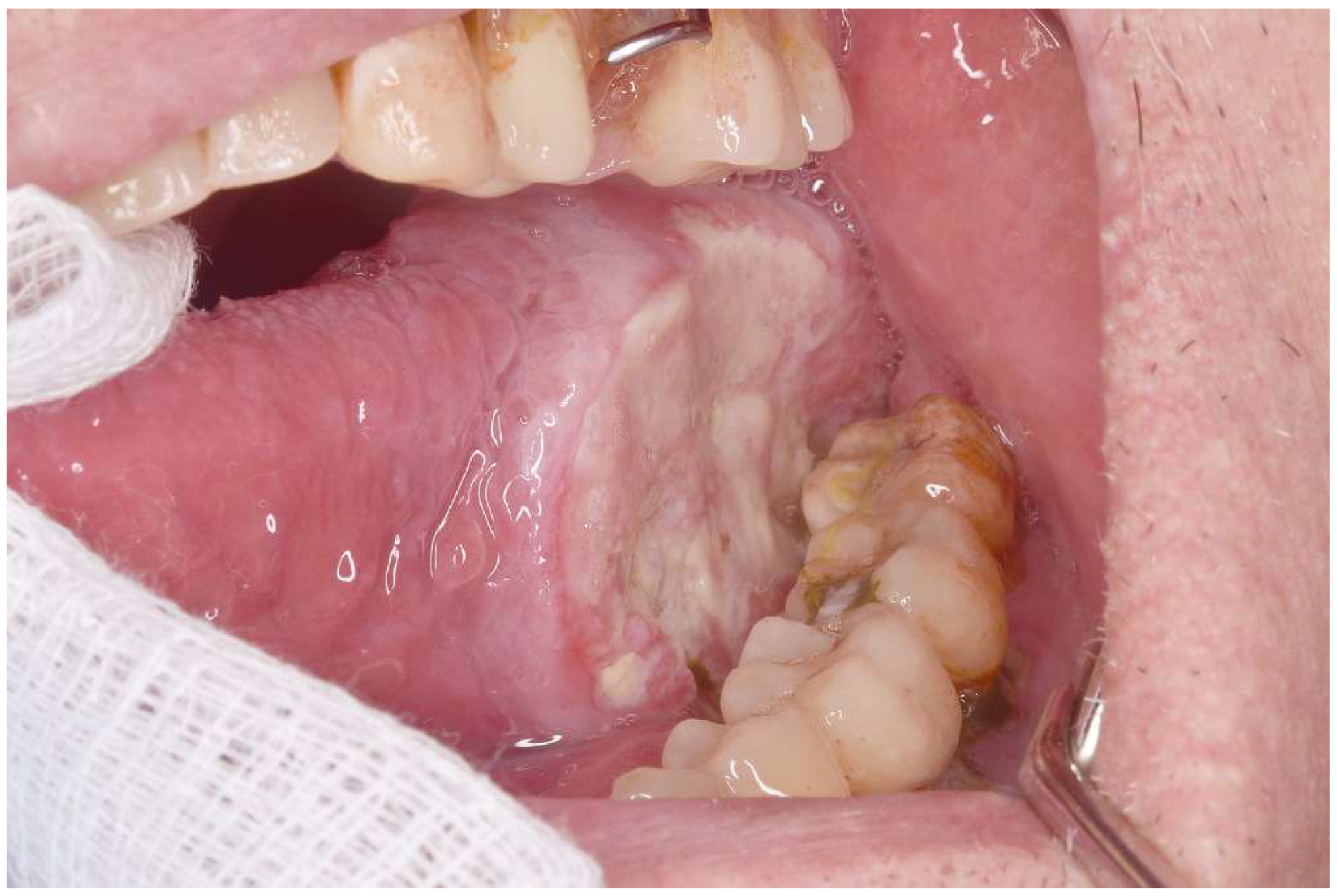
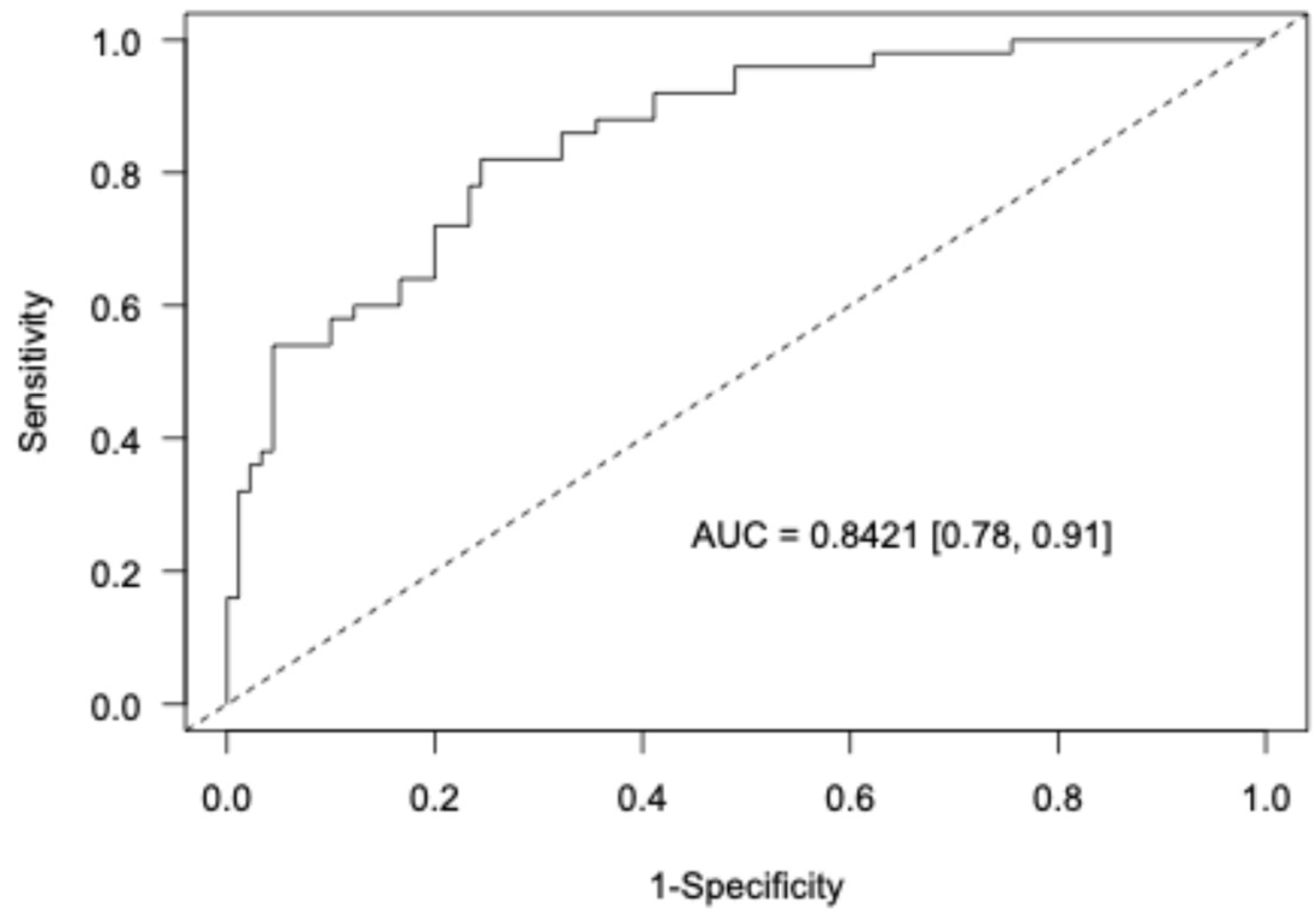
Background/Objectives: Proliferative verrucous leukoplakia (PVL) is the oral disorder with the greatest degree of malignant transformation. However, it is relatively rare. This study compared the clinical characteristics of patients with oral squamous cell carcinoma (OSCC) who had and had not been previously diagnosed with PVL. Methods: This case–control study compared the clinical characteristics of patients classified as early (T1 and T2) or advanced (T3 and T4) OSCC according to the TNM classification, including age, gender, location, and clinical type of cancer. The analysis involved 140 patients. Group 1: 50 OSCC patients with PVL (OSCC-PVL) and Group 2: 90 OSCC patients without PVL (OSCC-noPVL). Results: The patients with OSCC-PVL were younger than those with OSCC-noPVL, but this did not reach statistical significance. Regarding patient gender, those with OSCC-PVL were much more frequently female (70%), while OSCC-noPVL was more prevalent in men (65.5%) (p < 0.01). There were also significant differences in the oral locations between the two groups: the gingiva was most prevalent in OSCC-PVL and the tongue in OSCC-noPVL. Erythroleukoplastic forms were significantly more common in OSCC-PVL (30% vs. 7.7%), while ulcerated forms were more frequent in OSCC-noPVL (63.3% vs. 42%). Finally, early T stages were much more prevalent in our patients with OSCC-PVL. Conclusions: We found that OSCC preceded by PVL was much more frequent in women, had less aggressive clinical forms, and had significantly more frequent early T stages than in OSCC-noPVL.

Keywords: 
;  ;  

1. Introduction

Proliferative verrucous leukoplakia (PVL) is a rare disease, described by Hansen et al. in 1978 [1]. It manifests as plaques of leukoplakia that are initially homogeneous, but become multifocal, sometimes affecting a large part of the oral mucosa, frequently acquiring a verrucous character [1,2]. According to Warnakulasuriya et al., PVL is a potentially malignant disorder with a high tendency to develop cancer [3]. Other authors agree that PVL is most at risk for oral squamous cell carcinoma (OSCC) [4,5].
PVL has a high likelihood of recurrence after treatment with CO2 lasers or photodynamic therapy, its most common treatment modalities [6,7,8,9,10,11,12]. A meta-analysis yielded a recurrence rate of 67.2% (95% CI 48.3–81.8), without publication bias [13]. Novel drugs such as nivolumab have not had particularly beneficial outcomes. In a study of 33 patients (median age 63 [range 32–80] years; 18 [55%] female), including 8 (24%) with previously resected early OSCC by Hanna et al., 12 (36%) achieved a response based on the composite score, while 4 experienced progressive disease and 9 (27%) developed OSCC during the study [14].
The most significant issue with PVL is its high rate of transformation into oral cancer (43.87–65.8%), making PVL the oral disorder with the highest propensity for malignancy [15]. Other authors reported similar rates of malignant transformation [16].
The OSCC occurring in patients with PVL (OSCC-PVL) differs clinically from typical OSCC not preceded by PVL (OSCC-noPVL). Faustino et al. evaluated the prognostic outcomes of OSCC-PVL in terms of recurrence, new primary tumors, metastasis, and survival outcomes and reported that it has better clinical outcomes than OSCC-noPVL [17]. Similarly, Gonzalez-Moles et al. reported that OSCC-PVL has more favorable prognoses than OSCC-noPVL, particularly in the mortality rate [18]. However, Faustino et al. found little information on the key prognostic outcomes of OSCC-PVL [17]; therefore, studies need to compare OSCC-PVL with OSCC-noPVL.
In a recent preliminary study involving eight cases of PVL that progressed to cancer (OSCC-PVL) and 10 classical OSCC cases (OSCC-noPVL), we found that the clinical and evolutionary characteristics of these cancers differed. We also discovered that OSCC-PVL patients had lower expression of cancer-related genes. Hypermethylation in the promoter region of many genes was noted, suggesting that DNA methylation serves as a regulatory mechanism [19].
The current study compares the clinical characteristics of large groups with OSCC-PVL and OSCC-noPVL, to validate our preliminary results and demonstrate that OSCC-PVL has different clinical behavior to that of OSCC-noPVL.

2. Materials and Methods

This retrospective case–control study enrolled patients diagnosed with OSCC, seen between 2005 and August 2024, who visited the University General Hospital of Valencia, Spain, and or a private clinic nearby.
The subjects comprised 50 patients with OSCC preceded by proliferative verrucous leukoplakia lesions (OSCC-PVL) and 90 with oral squamous cell carcinoma who had never experienced PVL lesions (OSCC-noPVL). The study was approved by the clinical trials and drug committee of the University General Hospital on February 24, 2023 (reference 10-2023).
We recorded the patients’ ages and genders, the locations of the cancers in the oral cavity, and the clinical types, distinguishing between erythroleukoplakia, ulcerated, exophytic, and mixed. The inclusion criteria for the patients in the OSCC-PVL group were those published by Cerero et al. [20]. OSCC-noPVL consisted of OSCC patients who had not had PVL lesions. All patients with OSCC were diagnosed by biopsying the oral lesion. Finally, according to the TNM classification, we distinguished between early (T1 and T2) and advanced (T3 and T4) T stages [21,22,23].
The data were summarized using the mean (standard deviation) and median (1st and 3rd quartiles) for numerical variables and absolute (relative) frequencies for categorical variables. A Bayesian multivariable logistic regression model [24] was adjusted to assess whether the different clinical variables discriminated between the two groups. The uncertainty of the estimates was assessed by estimating the 95% credible intervals and the probability of direction (pd) was used as an index of effect existence [25].
We compared quantitative and qualitative variables between both groups to identify significant differences We used the Student’s test to compare quantitative variables (after verifying that the samples were normally distributed). For qualitative variables, we performed a contingency analysis using the χ2 test. Finally, we used logistic regression to assess which variables were more important for determining whether patients had OSCC-PVL or OSCC-noPVL. We set the level of significance at p < 0.05.
The performance of the model was assessed by estimating the area under the receiver operating characteristic curve (AUROC). Internal validation of the AUROC was performed using 10-fold cross-validation. All statistical analyses were performed using R (ver. 4.4.1) and the R packages brms (ver. 2.22.0) and bayestestR (ver. 0.15.0).

3. Results

The mean age of the 140 patients with OSCC was 68.48 ±11.42 (range 40–97) years. Of the 140 patients, 74 (52.9%) were male and 66 (47.1%) female. Table 1 shows the values of the different clinical variables analyzed. Comparing the two groups, the patients with OSCC-PVL were younger than those with OSCC-noPVL, but the difference was not significant (Mann–Whitney U = 1940, p > 0.05). There was a significant difference in gender; 70% of OSCC-PVL were female versus only 34.4% in the OSCC-noPVL group (p < 0.001).
Considering the location of the cancer in the mouth, 50% of the cancers in OSCC-PVL were located in the gingiva, with a lower percentage in OSCC-noPVL (p < 0.001). The erythroleukoplakia type (Figure 1) was much more frequent (30%) in OSCC-PVL, while the more clinically aggressive ulcerated form (Figure 2) predominated in OSCC-noPVL, comprising 63.3% of the cases. The differences were significant (p < 0.01). Finally, on comparing the differences between the initial (T1 and T2) and advanced (T3 and T4) stages of oral cancer in the two groups, 80% of the cases in the OSCC-PVL group were in the initial stages versus 57.8% in OSCC-noPVL; the difference was significant (p < 0.05).
The Bayesian logistic regression model showed that all the variables assessed differed between the OSCC-PVL and OSCC-noPVL groups. Specifically, being female was associated with OSCC-PVL (OR = 5.1, 95% CrI [1.9, 14.0], probability of effect > 99.99%). Advanced stage was associated with the cancer group (OR for OSCC-PVL = 0.34, 95% CrI [0.11, 0.91], 98.5% probability of effect). All clinical types except erythroleukoplakia were associated with the cancer group (ORs for OSCC-PVL = 0.21, 0.35, and 0.15, for ulcerative, exophytic, and mixed, respectively, probabilities of effect of 99.3, 92.4, and 98.0%). Finally, locations at the floor of the mouth, tongue and palate were also associated with the cancer group (ORs for OSCC-PVL = 0.03, 0.31, and 0.13, respectively, probabilities of effect 99.3, 98.8, and 97.4%) (Table 2).
Figure 4 assesses the performance of the model by estimating the ROC curve for discriminating between OSCC-noPVL and OSCC-PVL patients. The model performed well with AUC = 0.84. Interval validation using 10-fold cross-validation yielded a validated AUC = 0.81, indicating good out-of-sample performance of the model.

4. Discussion

In GLOBOCAN [26], published in August 2024, the overall incidence of all new cancers in both sexes in 2022 was 19,976,499, with 9,826,539 (49.2%) cases in Asia, 4,471,422 (22.4%) in Europe, 2,673,174 (13.4%) in Northern America,1,551,060 (7.8%) in Latin America, and 269,088 (1.3%) in Oceania. Of these new cancer cases, 10,311,610 affected men and 9,664,889 women. The most frequent cancers in men were lung, prostate and colorectal and in women, breast, lung, and colorectal. There were 9,743,832 deaths from cancer and there were 53,504,187 5-year prevalent cases, with 25,747,272 in men and 27,756,915 in women.
The 8th edition of the American Joint Committee on Cancer (AJCC) classification introduced two new parameters: depth of invasion (DOI) and extranodal extension (ENE). In this edition, patients with OSCC can be better classified in terms of overall survival (OS) and disease-specific survival (DSS), allowing more appropriate treatment protocols and better estimates of each patient’s prognosis [27]. Unfortunately, the prognosis of OSCC remains poor, with no improvement over recent decades.
A recent retrospective study analyzed the epidemiological, clinical, and prognostic characteristics of OSCC in 243 patients from Galicia, Spain. The average patient age was 67 years, and the majority were male (69.5%) [28]. However, the incidence and survival rates of patients with OSCC have risen in recent years. Currently, nearly one in five cancers are found in individuals with a prior cancer diagnosis. Among 6602 patients with a first primary OSCC, 640 (10%) second primary cancers were present [29].
The incidence of kidney cancer continues to increase by 1.5% annually, those of cancers of the pancreas, oral cavity, and pharynx by 1% annually (these are associated with human papillomavirus [HPV]), and those of the salivary gland, gingiva, and other mouth cancers by ≤ 0.5% annually [30].
In 2024, 2,001,140 new cancer cases and 611,720 cancer deaths are projected to occur in the United States. Cancer mortality continued to decline through 2021, averting over 4 million deaths since 1991 because of reductions in smoking, earlier detection of some cancers, and improved treatment options in both the adjuvant and metastatic settings [30].
In 2024, there were an estimated 58,450 new cases of oral cavity and pharynx cancers in the United States, with 41,510 in males and 16,940 in females. These cancers caused an estimated 12,230 deaths in United States: 8700 in males and 3530 in females. Oral cavity and pharynx cancers comprise 4% of new cases and were the 8th most frequent cancers in 2024 [30].
In a study of classical OSCC (OSCC-noPVL), Saldivia-Siracusa et al. reported that 58 (54.2%) of the patients were men, with a mean age of 60.69 years; 49 (45.8%) and 39 (36.5%) patients had histories of tobacco and alcohol use, respectively [31].
In a retrospective review of histopathological records from 1953 to 2019, conducted across three oral pathology laboratories in Southern Brazil, focusing on age, sex, anatomical site, clinical features, and histopathological diagnosis, females had a lower chance of developing classical OSCC than males, regardless of the decade (odds ratio = 0.30, p < 0.001). This trend was also observed in older individuals compared to those younger than 40 years [32].
PVL is a potentially malignant disorder that is enigmatic due to our ignorance of its etiopathogenesis and therapeutic management [1]. According Palefsky et al., there may be an association between PVL and HPV 16 infection, suggesting that this virus plays a role in the pathogenesis of PVL [33]. However, subsequent research has not established a link between HPV and PVL. Upadhyaya et al. tested the PVL lesions in 20 patients with clinically and biopsy-proven diagnoses of PVL for high-risk HPV using DNA in-situ hybridization. No expression of high-risk HPV was detected, while p53 staining was positive in less than 25% of the cells. A definite association between PVL and high-risk HPV infection could not be established [34]. Years before the Upsalad et al. publication, we had already reported that we had not detected HPV infection in PVL tissue or in the oral rinses of any of 13 patients studied. We concluded that there was no association between PVL and HPV infection in our patients [35].
In a study of 40 patients divided into four groups (10 patients each with PVL, Oral leukoplakia (OL), OSCC, and good health), we analyzed the presence or absence of oncovirus DNA through PCR amplification of viral genetic markers, followed by confirmation using gel electrophoresis. The amplified fragments were sequenced and identified bioinformatically. However, no association was found between PVL and the target viruses [36].
Recent research on the origins of PVL has concentrated on genetic analyses involving methylation, transcriptomics, and the microbiota. Morandi et al. evaluated the diagnostic value of methylation levels in a set of 18 genes using bisulfite next-generation sequencing, focusing on OSCC and other potentially malignant disorders, such as lichen planus and PVL. Their data highlight the importance of CpG islands location and accurate estimation of DNA methylation levels for an exact early diagnosis of OSCC [37].
To explore the pathophysiology of PVL using methylated DNA immunoprecipitation and high-throughput sequencing, we analyzed tissue samples from oral biopsies of 10 patients with PVL and five healthy individuals. We compared their epigenetic patterns. The integrative analysis revealed eight significantly upregulated (ARTN, CD8A, GATA3, HOXD10, MYO7A, OSR2, PLCB1, and SPOCK2) and five significantly downregulated (ANKRD6, DLG2, GPX3, PITX2, and ZNF736) genes in PVL compared to the controls. Our findings highlight the potential of methylation markers in PVL [38].
Okoturo et al. performed whole exome sequencing of five cases of OSCC-PVL, using paired blood samples to identify somatic mutations prevalent in the tumors. They discovered that, unlike classical OSCC, OSCC-PVL had rare TP53 mutations and altered patterns of PIK3CA and NOTCH1 mutations. They concluded that the two groups have differences in mutation and methylation profiles [39].
Some authors report that the risk of malignancy associated with PVL is approximately 50%. However, Villa et al. reported malignant transformation (MT) in 71.4% of patients with PVL after a median of 37 months from the initial visit; erythroleukoplakia underwent MT in 100% of cases [40]. Ramos-Garcia et al. stated that the pooled proportion of MT in PVL was 43.87%: females (64.02%) and males (35.98%). The most frequent PVL sites were the gingiva (39.6%) and buccal mucosa (21.6%). There were no conclusions regarding MT and sex or age distribution or tobacco or alcohol consumption. The gingiva was the most common site of MT (39.9%) [41].
In a recent preliminary study, we examined eight cases of PVL that progressed to cancer (OSCC-PVL) and 10 classical OSCC cases (OSCC-noPVL) and found that the clinical and evolutionary characteristics of these cancers differed. OSCC-PVL patients had lower expression of cancer-related genes. We also observed hypermethylation of the promoter regions of several genes, suggesting that DNA methylation has a regulatory mechanism. We found that patients with cancer and a history of PVL (OSCC-PVL) were more often women compared to OSCC-noPVL patients. None of our OSCC-PVL patients smoked, while it was noted in OSCC-noPVL. Gingival localization was more frequent in OSCC-PVL, while tongue localization was significantly more common in OSCC-noPVL Erythroplastic clinical forms were more prevalent in OSCC-PVL, whereas ulcerated forms were more common in OSCC-noPVL. We also observed increased lymph node involvement in OSCC-noPVL, and the most advanced TNM stages were found in OSCC-noPVL [19].
Kovalski et al. reported that in OSCC-noPVL, tobacco consumption (p = 0.003) and alcohol intake (p = 0.02) were significantly greater in males than in females [42]. This was confirmed by Amezaga-Fernandez et al., who found that 45.9% of patients with OSCC were smokers and 58.6% consumed alcohol [28].
In our recent preliminary study of eight patients with OSCC-PVL and 10 with OSCC-noPVL [43], we observed distinct tendencies in the clinical characteristics of these two groups, although some of the variables did not reach statistical significance due to the small number of cases. Consequently, in this study, we examined more cases in both groups: 50 with OSCC-PVL and 90 with OSCC-noPVL. With this increase, the previously detected differences became significant. For instance, 70% of the OSCC-PVL cases occurred in women versus 34.4% in the OSCC non-PVL group (χ2 = 116,307, p < 0.01), confirming the higher tendency for cancers to develop in females with OSCC-PVL. This finding is similar to the 64.02% Ramos-Garcia et al. reported for women with OSCC-PVL [41]. In comparison, the frequency of tobacco consumption was not a significant factor in OSCC-PVL compared to OSCC-noPVL. Furthermore, the mean ages of the patients with OSCC-PVL and OSCC-noPVL were not significantly different. Smoking was not a significant etiological factor in OSCC-PVL compared to OSCC-noPVL, where 40% were smokers and 60% were non-smokers (Table 1).
The most common sites for classical OSCC are the tongue (40%) and floor of the mouth (33%) [44,45]. Oliver et al. found that the most frequent location of OSCC was on the tongue (28.26% of cases), followed by the floor of the mouth (26.09%) [46]. Mashberg et al. studied 102 symptomatic cases of OSCC and the floor of the mouth, oral tongue, and soft palate complex accounted for 75% of all locations and 84% when the posterior pillar was excluded. The soft palate alone accounted for 75% of all locations, and 84% of locations when the posterior abutment was excluded [47]. In contrast to the typical location of OSCC-noPVL, in our OSCC-PVL cases, the gingiva was the most frequently affected area, accounting for 50% of the cases (Table 1), while the most common locations in OSCC-noPVL were the tongue (41.4%) and gingiva (28.9%). This concurs with reports that the tongue was the most frequent location in OSCC-noPVL [44]. We previously also described the typical involvement of the gingiva in OSCC-PVL [48].
According to Gonzalez-Ruiz et al., the primary cause of the high mortality rate in oral cancer is its diagnosis at advanced stages (T3 and T4), where treatment often has poor efficacy, leading to challenges, mutilations, or disabilities [49]. Amezaga-Fernandez et al. reported that cases diagnosed at advanced stages accounted for 48.1% of their sample, and that 38.7% relapsed. The 5-year OS and DSS rates were 39.9% and 46.1%, respectively [28]. In a meta-analysis of 23 studies encompassing 505 patients with PVL, of whom 288 developed a total of 504 carcinomas, Gonzalez-Moles et al. found that OSCC-PVL has favorable prognostic parameters, particularly regarding the mortality rate [50]. The same group found that cancers accompanied by PVL had a better prognosis than OSCC-noPVL [49]. Although OSCC survival has not changed substantially in recent years, an early diagnosis is important [51], particularly when comparing our two OSCC groups.

5. Conclusions

In conclusion, for OSCC patients with PVL, the Bayesian logistic regression model revealed differences among the variables, stage, location, gender, and clinical type between groups that discriminated between OSCC-noPVL and OSCC-PVL patients. To our knowledge, this is the largest comparative clinical study between groups of patients with OSCC-PVL and OSCC-noPVL.

Author Contributions

Conceptualization, J.B. and L.B.; methodology, J.V. and L.B.; software, D.H.;validation, D.H. and J.B.;investigation,J.B., L.B.,J.M. and J.M.S.; resources, J.B., L.B.,J.M. and J.M.S.; writing—original draft preparationwriting—original draft preparation, J.B. and D.H.; writing—review and editing, J.B., L.B.and D.H.; supervision, J.B., L.B.; funding acquisition, J.B. and L.B.All authors have read and agreed to the published version of the manuscript.”

Funding

This research was funded by the Ministerio de Ciencia e Innovación (Spain) through the project “PID2022-138398OB-I00”, Principal investigator: Jose Bagan.

Institutional Review Board Statement

The study was approved by the clinical trials and drug committee of the University General Hospital of Valencia, Spain, on February 24, 2023 (reference 10-2023).

Data Availability Statement

No new data were created; the clinical data from this study are described in the tables provided in the document (Table 1 and Table 2).

Conflicts of Interest

“The authors declare no conflicts of interest.” The authors declare that they have no known competing financial interests or personal relationships that could have appeared to influence the work reported in this paper.

References

  1. Hansen LS, Olson JA, Silverman S Jr. Proliferative verrucous leukoplakia. A long-term study of thirty patients. Oral Surg Oral Med Oral Pathol. 1985 Sep;60(3):285-98. PMID: 3862042. [CrossRef]
  2. Torrejon-Moya A, Jané-Salas E, López-López J. Clinical manifestations of oral proliferative verrucous leukoplakia: A systematic review. J Oral Pathol Med. 2020 May;49(5):404-408. Epub 2020 Feb 9. PMID: 31990082. [CrossRef]
  3. Warnakulasuriya S, Kujan O, Aguirre-Urizar JM, Bagan JV, González-Moles MÁ, Kerr AR, Lodi G, Mello FW, Monteiro L, Ogden GR, Sloan P, Johnson NW. Oral potentially malignant disorders: A consensus report from an international seminar on nomenclature and classification, convened by the WHO Collaborating Centre for Oral Cancer. Oral Dis. 2021 Nov;27(8):1862-1880. Epub 2020 Nov 26. PMID: 33128420. [CrossRef]
  4. Brignardello-Petersen R. Proliferative verrucous leukoplakia and erythroplakia are probably the disorders with the highest rate of malignant transformation. J Am Dent Assoc. 2020 Aug;151(8):e62. Epub 2020 May 26. PMID: 32471555. [CrossRef]
  5. Zhu L, Ou L, Yang Y, Zhao D, Liu B, Liu R, Liu O, Feng H. A Bibliometric and Visualised Analysis of Proliferative Verrucous Leucoplakia From 2003 to 2023. Int Dent J. 2025 Feb;75(1):333-344. Epub 2024 Jul 22. PMID: 39043528. [CrossRef]
  6. Chainani-Wu N, Purnell DM, Silverman S Jr. A case report of conservative management of extensive proliferative verrucous leukoplakia using a carbon dioxide laser. Photomed Laser Surg. 2013 Apr;31(4):183-7. Epub 2013 Mar 8. PMID: 23473346. [CrossRef]
  7. Bombeccari GP, Garagiola U, Candotto V, Pallotti F, Carinci F, Giannì AB, Spadari F. Diode laser surgery in the treatment of oral proliferative verrucous leukoplakia associated with HPV-16 infection. Maxillofac Plast Reconstr Surg. 2018 Jul 30;40(1):16. PMID: 30105220; PMCID: PMC6064714. [CrossRef]
  8. Yan Y, Li Z, Tian X, Zeng X, Chen Q, Wang J. Laser-assisted photodynamic therapy in proliferative verrucous oral leukoplakia. Photodiagnosis Photodyn Ther. 2022 Sep;39:103002. Epub 2022 Jul 6. PMID: 35809828. [CrossRef]
  9. Wang F, Shi Y, Dong Y, Liu T, Dan H, Wang J, Zeng X. Photodynamic therapy combined with laser drilling successfully prevents the recurrence of refractory oral proliferative verrucous leukoplakia. Photodiagnosis Photodyn Ther. 2021 Dec;36:102564. Epub 2021 Oct 3. PMID: 34610431. [CrossRef]
  10. Cobb CM, Beaini NE, Scully J, Gibson TM. Proliferative verrucous leukoplakia: Case study of 24 years and outcome of treatment with CO2 laser. Clin Adv Periodontics. 2024 Oct 24. Epub ahead of print. PMID: 39447057. [CrossRef]
  11. Vergier V, Porporatti AL, Babajko S, Taihi I. Gingival Proliferative Verrucous Leukoplakia and Cancer: A Systematic Review With Meta-Analysis. Oral Dis. 2024 Oct 9. Epub ahead of print. PMID: 39385511. [CrossRef]
  12. Pedroso CM, do Santos ES, Alves FA, Martins MD, Kowalski LP, Lopes MA, Warnakulasuriya S, Villa A, Santos-Silva AR. Surgical protocols for oral leukoplakia and precancerous lesions across three different anatomic sites. Oral Dis. 2024 Aug 18. Epub ahead of print. PMID: 39155483. [CrossRef]
  13. Proaño-Haro A, Bagan L, Bagan JV. Recurrences following treatment of proliferative verrucous leukoplakia: A systematic review and meta-analysis. J Oral Pathol Med. 2021 Sep;50(8):820-828. Epub 2021 Apr 13. PMID: 33765364. [CrossRef]
  14. Hanna GJ, Villa A, Nandi SP, Shi R, ONeill A, Liu M, Quinn CT, Treister NS, Sroussi HY, Vacharotayangul P, Goguen LA, Annino DJ Jr, Rettig EM, Jo VY, Wong KS, Lizotte P, Paweletz CP, Uppaluri R, Haddad RI, Cohen EEW, Alexandrov LB, William WN Jr, Lippman SM, Woo SB. Nivolumab for Patients With High-Risk Oral Leukoplakia: A Nonrandomized Controlled Trial. JAMA Oncol. 2024 Jan 1;10(1):32-41. PMID: 37971722; PMCID: PMC10654930. [CrossRef]
  15. Zhang C, Lan Q, Wei P, Gao Y, Zhang J, Hua H. Clinical, histopathological characteristics and malignant transformation of proliferative verrucous leukoplakia with 36 patients: a retrospective longitudinal study. BMC Oral Health. 2024 May 30;24(1):639. PMID: 38816724; PMCID: PMC11138006. [CrossRef]
  16. Ramos-García P, González-Moles MÁ, Mello FW, Bagan JV, Warnakulasuriya S. Malignant transformation of oral proliferative verrucous leukoplakia: A systematic review and meta-analysis. Oral Dis. 2021 Nov;27(8):1896-1907. Epub 2021 May 19. PMID: 34009718. [CrossRef]
  17. Faustino ISP, de Pauli Paglioni M, de Almeida Mariz BAL, Normando AGC, Pérez-de-Oliveira ME, Georgaki M, Nikitakis NG, Vargas PA, Santos-Silva AR, Lopes MA. Prognostic outcomes of oral squamous cell carcinoma derived from proliferative verrucous leukoplakia: A systematic review. Oral Dis. 2023 May;29(4):1416-1431. Epub 2022 Mar 2. PMID: 35199416. [CrossRef]
  18. González-Moles MÁ, Warnakulasuriya S, Ramos-García P. Prognosis Parameters of Oral Carcinomas Developed in Proliferative Verrucous Leukoplakia: A Systematic Review and Meta-Analysis. Cancers (Basel). 2021 Sep 28;13(19):4843. PMID: 34638327; PMCID: PMC8507842. [CrossRef]
  19. Herreros-Pomares A, Hervás D, Bagan-Debon L, Proaño A, Garcia D, Sandoval J, Bagan J. Oral cancers preceded by proliferative verrucous leukoplakia exhibit distinctive molecular features. Oral Dis. 2024 Apr;30(3):1072-1083. Epub 2023 Mar 21. PMID: 36892444. [CrossRef]
  20. Cerero-Lapiedra R, Baladé-Martínez D, Moreno-López LA, Esparza-Gómez G, Bagán JV. Proliferative verrucous leukoplakia: a proposal for diagnostic criteria. Med Oral Patol Oral Cir Bucal. 2010 Nov 1;15(6):e839-45. PMID: 20173704.
  21. Nagesh M, Gowtham S, Bharadwaj B, Ali M, Goud AK, Siddiqua S. Evolution of TNM Classification for Clinical Staging of Oral Cancer: The Past, Present and the Future. J Maxillofac Oral Surg. 2023 Sep;22(3):710-719. Epub 2023 Apr 11. PMID: 37534341; PMCID: PMC10390384. [CrossRef]
  22. Erazo-Puentes MC, Sánchez-Torres A, Aguirre-Urizar JM, Bara-Casaus J, Gay-Escoda C. Has the 8th American joint committee on cancer TNM staging improved prognostic performance in oral cancer? A systematic review. Med Oral Patol Oral Cir Bucal. 2024 Mar 1;29(2):e163-e171. PMID: 38368527; PMCID: PMC10945871. [CrossRef]
  23. Sridharan S, Thompson LDR, Purgina B, Sturgis CD, Shah AA, Burkey B, Tuluc M, Cognetti D, Xu B, Higgins K, Hernandez-Prera JC, Guerrero D, Bundele MM, Kim S, Duvvuri U, Ferris RL, Gooding WE, Chiosea SI. Early squamous cell carcinoma of the oral tongue with histologically benign lymph nodes: A model predicting local control and vetting of the eighth edition of the American Joint Committee on Cancer pathologic T stage. Cancer. 2019 Sep 15;125(18):3198-3207. Epub 2019 Jun 7. PMID: 31174238; PMCID: PMC7723468. [CrossRef]
  24. Bürkner PC. brms An R package for Bayesian multilevel models using Stan Journal of statistical software. 2017 Aug 29;80:1-28.
  25. Makowski D, Ben-Shachar MS, Chen SHA, Lüdecke D. Indices of Effect Existence and Significance in the Bayesian Framework. Front Psychol. 2019 Dec 10;10:2767. PMID: 31920819; PMCID: PMC6914840. [CrossRef]
  26. Ferlay J, Ervik M, Lam F, Laversanne M, Colombet M, Mery L, Piñeros M, Znaor A, Soerjomataram I, Bray F (2024). Global Cancer Observatory: Cancer Today. https://gco.iarc.who.int/media/globocan/factsheets/populations/900-world-fact-sheet.pdf.
  27. Erazo-Puentes MC, Sánchez-Torres A, Aguirre-Urizar JM, Bara-Casaus J, Gay-Escoda C. Has the 8th American joint committee on cancer TNM staging improved prognostic performance in oral cancer? A systematic review. Med Oral Patol Oral Cir Bucal. 2024 Mar 1;29(2):e163-e171. PMID: 38368527; PMCID: PMC10945871. [CrossRef]
  28. Amezaga-Fernandez I, Aguirre-Urizar JM, Suárez-Peñaranda JM, Chamorro-Petronacci C, Lafuente-Ibáñez de Mendoza I, Marichalar-Mendia X, Blanco-Carrión A, Antúnez-López J, García-García A. Epidemiological, clinical, and prognostic analysis of oral squamous cell carcinoma diagnosed and treated in a single hospital in Galicia (Spain): a retrospective study with 5-year follow-up. Med Oral Patol Oral Cir Bucal. 2024 Jan 1;29(1):e36-e43. PMID: 37330964; PMCID: PMC10765332. [CrossRef]
  29. Mroueh R, Nevala A, Haapaniemi A, Pitkäniemi J, Salo T, Mäkitie AA. Risk of second primary cancer in oral squamous cell carcinoma. Head Neck. 2020 Aug;42(8):1848-1858. Epub 2020 Feb 14. PMID: 32057158. [CrossRef]
  30. Siegel RL, Giaquinto AN, Jemal A. Cancer statistics, 2024. CA Cancer J Clin. 2024 Jan-Feb;74(1):12-49. Epub 2024 Jan 17. Erratum in: CA Cancer J Clin. 2024 Mar-Apr;74(2):203. doi: 10.3322/caac.21830. PMID: 38230766. [CrossRef]
  31. Saldivia-Siracusa C, Araújo AL, Arboleda LP, Abrantes T, Pinto MB, Mendonça N, Cordero-Torres K, Gilligan G, Piemonte E, Panico R, De-Abreu-Álves F, Villaroel-Dorrego M. Insights into incipient oral squamous cell carcinoma: a comprehensive south-american study. Med Oral Patol Oral Cir Bucal. 2024 Jul 1;29(4):e575-e583. PMID: 38794942; PMCID: PMC11249374. [CrossRef]
  32. Daroit NB, Martins LN, Garcia AB, Haas AN, Maito FLDM, Rados PV. Oral cancer over six decades: a multivariable analysis of a clinicopathologic retrospective study. Braz Dent J. 2023 Dec 22;34(5):115-124. PMID: 38133466; PMCID: PMC10759960. [CrossRef]
  33. Palefsky JM, Silverman S Jr, Abdel-Salaam M, Daniels TE, Greenspan JS. Association between proliferative verrucous leukoplakia and infection with human papillomavirus type 16. J Oral Pathol Med. 1995 May;24(5):193-7. PMID: 7616456. [CrossRef]
  34. Upadhyaya JD, Fitzpatrick SG, Islam MN, Bhattacharyya I, Cohen DM. A Retrospective 20-Year Analysis of Proliferative Verrucous Leukoplakia and Its Progression to Malignancy and Association with High-risk Human Papillomavirus. Head Neck Pathol. 2018 Dec;12(4):500-510. Epub 2018 Feb 9. PMID: 29427033; PMCID: PMC6232220. [CrossRef]
  35. Bagan JV, Jimenez Y, Murillo J, Gavaldá C, Poveda R, Scully C, Alberola TM, Torres-Puente M, Pérez-Alonso M. Lack of association between proliferative verrucous leukoplakia and human papillomavirus infection. J Oral Maxillofac Surg. 2007 Jan;65(1):46-9. PMID: 17174763. [CrossRef]
  36. García-López R, Moya A, Bagan JV, Pérez-Brocal V. Retrospective case-control study of viral pathogen screening in proliferative verrucous leukoplakia lesions. Clin Otolaryngol. 2014 Oct;39(5):272-80. PMID: 25099922. [CrossRef]
  37. Morandi L, Gissi D, Tarsitano A, Asioli S, Gabusi A, Marchetti C, Montebugnoli L, Foschini MP. CpG location and methylation level are crucial factors for the early detection of oral squamous cell carcinoma in brushing samples using bisulfite sequencing of a 13-gene panel. Clin Epigenetics. 2017 Aug 15;9:85. PMID: 28814981; PMCID: PMC5558660. [CrossRef]
  38. Herreros-Pomares A, Llorens C, Soriano B, Bagan L, Moreno A, Calabuig-Fariñas S, Jantus-Lewintre E, Bagan J. Differentially methylated genes in proliferative verrucous leukoplakia reveal potential malignant biomarkers for oral squamous cell carcinoma. Oral Oncol. 2021 May;116:105191. Epub 2021 Feb 28. PMID: 33657465. [CrossRef]
  39. Okoturo E, Green D, Clarke K, Liloglou T, Boyd MT, Shaw RJ, Risk JM. Whole genome DNA methylation and mutational profiles identify novel changes in proliferative verrucous leukoplakia. Oral Surg Oral Med Oral Pathol Oral Radiol. 2023 Jun;135(6):893-903. Epub 2023 Mar 17. PMID: 37088660. [CrossRef]
  40. Villa A, Menon RS, Kerr AR, De Abreu Alves F, Guollo A, Ojeda D, Woo SB. Proliferative leukoplakia: Proposed new clinical diagnostic criteria. Oral Dis. 2018 Jul;24(5):749-760. Epub 2018 May 2. PMID: 29337414. [CrossRef]
  41. Ramos-García P, González-Moles MÁ, Mello FW, Bagan JV, Warnakulasuriya S. Malignant transformation of oral proliferative verrucous leukoplakia: A systematic review and meta-analysis. Oral Dis. 2021 Nov;27(8):1896-1907. Epub 2021 May 19. PMID: 34009718. [CrossRef]
  42. Kovalski LNS, Zanella VG, Jardim LC, Só BB, Girardi FM, Kroef RG, Barra MB, Carrard VC, Martins MD, Martins MAT. Prognostic factors from squamous cell carcinoma of the hard palate, gingiva and upper alveolar ridge. Braz Oral Res. 2022 May 2;36:e058. PMID: 36507745. [CrossRef]
  43. Herreros-Pomares A, Hervás D, Bagan-Debon L, Proaño A, Garcia D, Sandoval J, Bagan J. Oral cancers preceded by proliferative verrucous leukoplakia exhibit distinctive molecular features. Oral Dis. 2024 Apr;30(3):1072-1083. Epub 2023 Mar 21. PMID: 36892444. [CrossRef]
  44. Bagan J, Sarrion G, Jimenez Y. Oral cancer: clinical features. Oral Oncol. 2010 Jun;46(6):414-7. Epub 2010 Apr 18. PMID: 20400366. [CrossRef]
  45. Hirata RM, Jaques DA, Chambers RG, Tuttle JR, Mahoney WD. Carcinoma of the oral cavity. An analysis of 478 cases. Ann Surg. 1975 Aug;182(2):98-103. PMID: 813586; PMCID: PMC1343824. [CrossRef]
  46. Oliver AJ, Helfrick JF, Gard D. Primary oral squamous cell carcinoma: a review of 92 cases. J Oral Maxillofac Surg. 1996 Aug;54(8):949-54; discussion 955. PMID: 8765383. [CrossRef]
  47. Mashberg A, Merletti F, Boffetta P, Gandolfo S, Ozzello F, Fracchia F, Terracini B. Appearance, site of occurrence, and physical and clinical characteristics of oral carcinoma in Torino, Italy. Cancer. 1989 Jun 15;63(12):2522-7. PMID: 2720601. [CrossRef]
  48. Bagan JV, Jimenez Y, Sanchis JM, Poveda R, Milian MA, Murillo J, Scully C. Proliferative verrucous leukoplakia: high incidence of gingival squamous cell carcinoma. J Oral Pathol Med. 2003 Aug;32(7):379-82. PMID: 12846783. [CrossRef]
  49. González-Ruiz I, Ramos-García P, Ruiz-Ávila I, González-Moles MÁ. Early Diagnosis of Oral Cancer: A Complex Polyhedral Problem with a Difficult Solution. Cancers (Basel). 2023 Jun 21;15(13):3270. PMID: 37444379; PMCID: PMC10340032. [CrossRef]
  50. González-Moles MÁ, Warnakulasuriya S, Ramos-García P. Prognosis Parameters of Oral Carcinomas Developed in Proliferative Verrucous Leukoplakia: A Systematic Review and Meta-Analysis. Cancers (Basel). 2021 Sep 28;13(19):4843. PMID: 34638327; PMCID: PMC8507842. [CrossRef]
  51. Tan Y, Wang Z, Xu M, Li B, Huang Z, Qin S, Nice EC, Tang J, Huang C. Oral squamous cell carcinomas: state of the field and emerging directions. Int J Oral Sci. 2023 Sep 22;15(1):44. PMID: 37736748; PMCID: PMC10517027. [CrossRef]
Figure 2. Case of proliferative verrucous leukoplakia. (a) leukoplakia lesions in the posterior third of the right buccal mucosa; (b) oral squamous cell carcinoma in initial erythroplastic form in the anterior third of the right buccal mucosa; (c) large leukoplakia lesions in the vestibular area of the upper gingiva; (d) leukoplakia lesions on the right lateral border of the tongue; (e) Leukoplakia on the left lateral border of the tongue; (f) Leukoplakia on the left buccal mucosa. All these images (a-f) correspond to the same patient with OSCC-PVL.
Preprints 148550 g001
Figure 3. Advanced case of ulceration on the left lateral edge of the tongue with extensive infiltration. It is almost OSCC-non-PVL.
Preprints 148550 g002
Figure 4. ROC curve depicting the discrimination power of the Bayesian logistic regression model.
Preprints 148550 g003
Table 1. Clinical characteristics of the cancer and the PVL-cancer patients.
Variable Cancer (n=90) PVL-Cancer (n=50)
Age
mean (sd) 69.51 (11.27) 66.62 (11.59)
median (q1, q3) 70 (60.25, 77) 66 (59, 73.75)
Gender
M 59 (65.56 %) 15 (30 %)
F 31 (34.44 %) 35 (70 %)
Location.cancer
Gingiva 26 (28.89 %) 25 (50 %)
Buccal mucosa 8 (8.89 %) 8 (16 %)
Tongue 37 (41.11 %) 9 (18 %)
Lips 2 (2.22 %) 7 (14 %)
Floor of the mouth 6 (6.67 %) 0 (0 %)
Palate 11 (12.22 %) 1 (2 %)
Clinical.type
Erytrholeukoplakia 7 (7.78 %) 15 (30 %)
Ulcerative 57 (63.33 %) 21 (42 %)
Exophitic 16 (17.78 %) 11 (22 %)
Mixed 10 (11.11 %) 3 (6 %)
Stage
Early 52 (57.78 %) 40 (80 %)
Advanced 38 (42.22 %) 10 (20 %)
Table 2. Results of the Bayesian logistic regression model.
Variables Estimate Std. Error OR 95% CrI Prob. effect
Intercept 0.432 0.757 - [-1.1, 1.9] 0.711
Location buccal mucosa 0.865 0.712 2.376 [0.58, 10.0] 0.891
Location tongue -1.16 0.546 0.313 [0.10, 10.0] 0.988
Location lips 1.028 0.951 2.797 [0.49, 21.7] 0.867
Location floor of the mouth -3.496 1.799 0.03 [0.001, 0.64] 0.993
Location palate -2.017 1.128 0.133 [0.01, 1.0] 0.974
Stage advanced -1.095 0.524 0.335 [0.11, 0.91] 0.985
Gender F 1.621 0.507 5.058 [1.9, 14.0] 1
Clinical type Ulcerative -1.578 0.656 0.206 [0.06, 0.73] 0.993
Clinicaltype Exophitic -1.047 0.733 0.351 [0.08, 1.46] 0.924
Clinical type Mixed -1.916 0.961 0.147 [0.02, 0.90] 0.98
Disclaimer/Publisher’s Note: The statements, opinions and data contained in all publications are solely those of the individual author(s) and contributor(s) and not of MDPI and/or the editor(s). MDPI and/or the editor(s) disclaim responsibility for any injury to people or property resulting from any ideas, methods, instructions or products referred to in the content.
Copyright: This open access article is published under a Creative Commons CC BY 4.0 license, which permit the free download, distribution, and reuse, provided that the author and preprint are cited in any reuse.
Prerpints.org logo

Preprints.org is a free preprint server supported by MDPI in Basel, Switzerland.

Subscribe

Disclaimer

Terms of Use

Privacy Policy

Privacy Settings

© 2025 MDPI (Basel, Switzerland) unless otherwise stated