Submitted:
30 January 2025
Posted:
31 January 2025
You are already at the latest version
Abstract
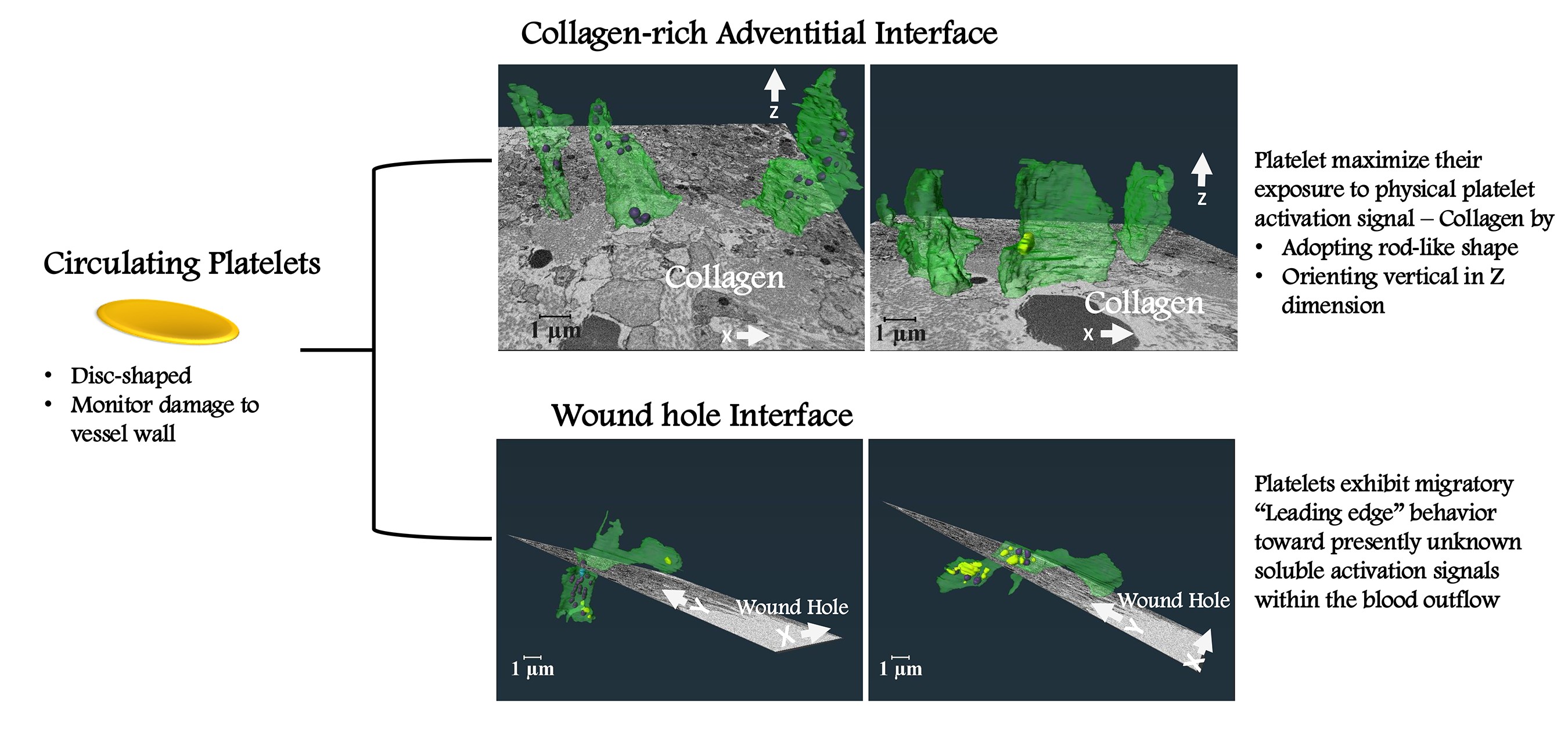
We focused on two microenvironments within a 5-min jugular vein puncture wound thrombus. The goal was to infer from 3D platelet morphodynamics how microenvironments determine platelet activation state. Platelets at the collagen-rich adventitial interface were rod shaped and oriented vertical to flow vectors. The net effect was to maximize their interaction with collagen. At the wound hole interface, soluble signaling should predominant. Here, the typical platelet exhibited what appeared to be a “migratory”, granule-free, leading edge extending toward the wound hole from an organelle rich cell body. Morphologically, this structure resembled the leading edge of a wound associated fibroblast. Platelets were aligned parallel with one another and at ~45 degrees to blood flow. We submit the morphodynamics observed here indicate novel similarities between platelets and wound associated nucleated cells.
Keywords:
1. Introduction
2. Results
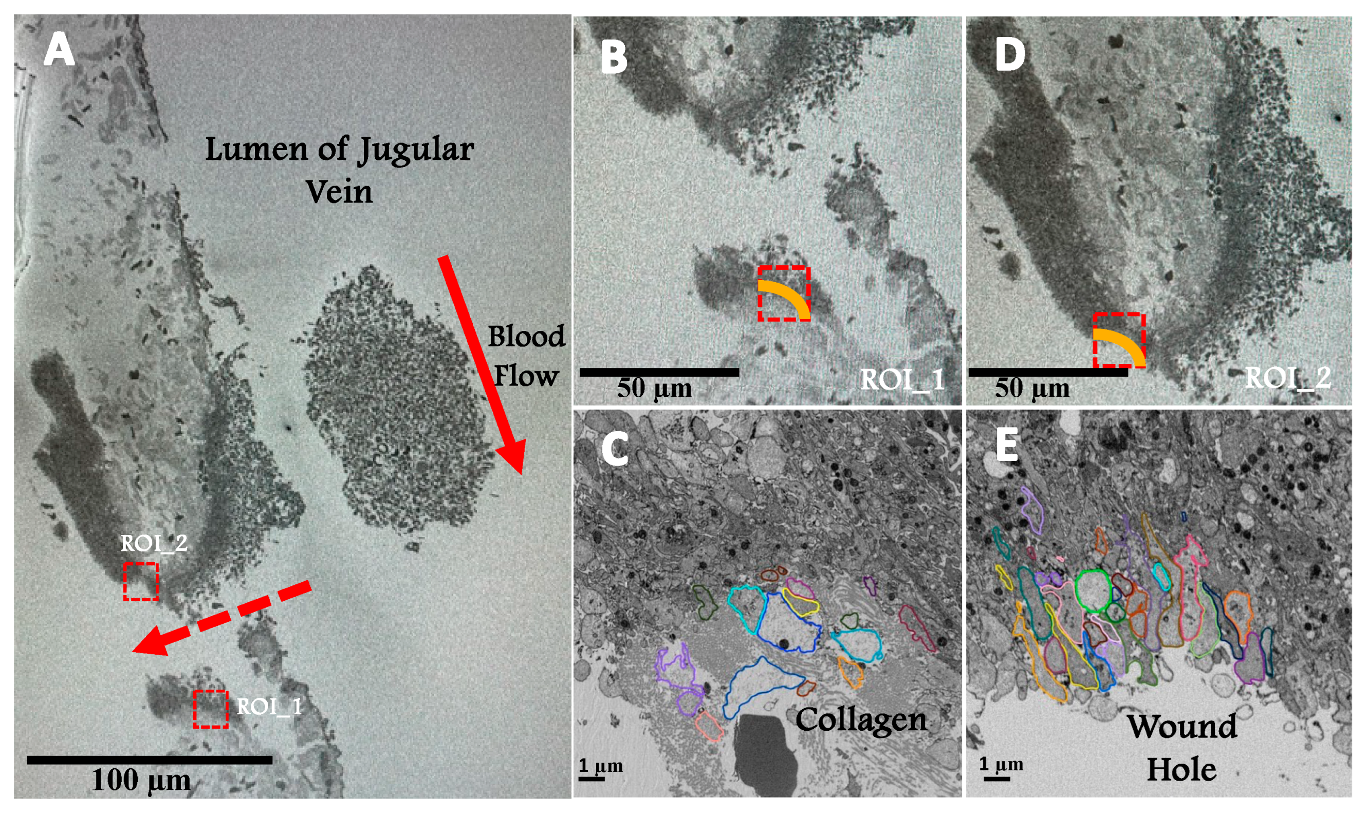
2.1. Experimental System
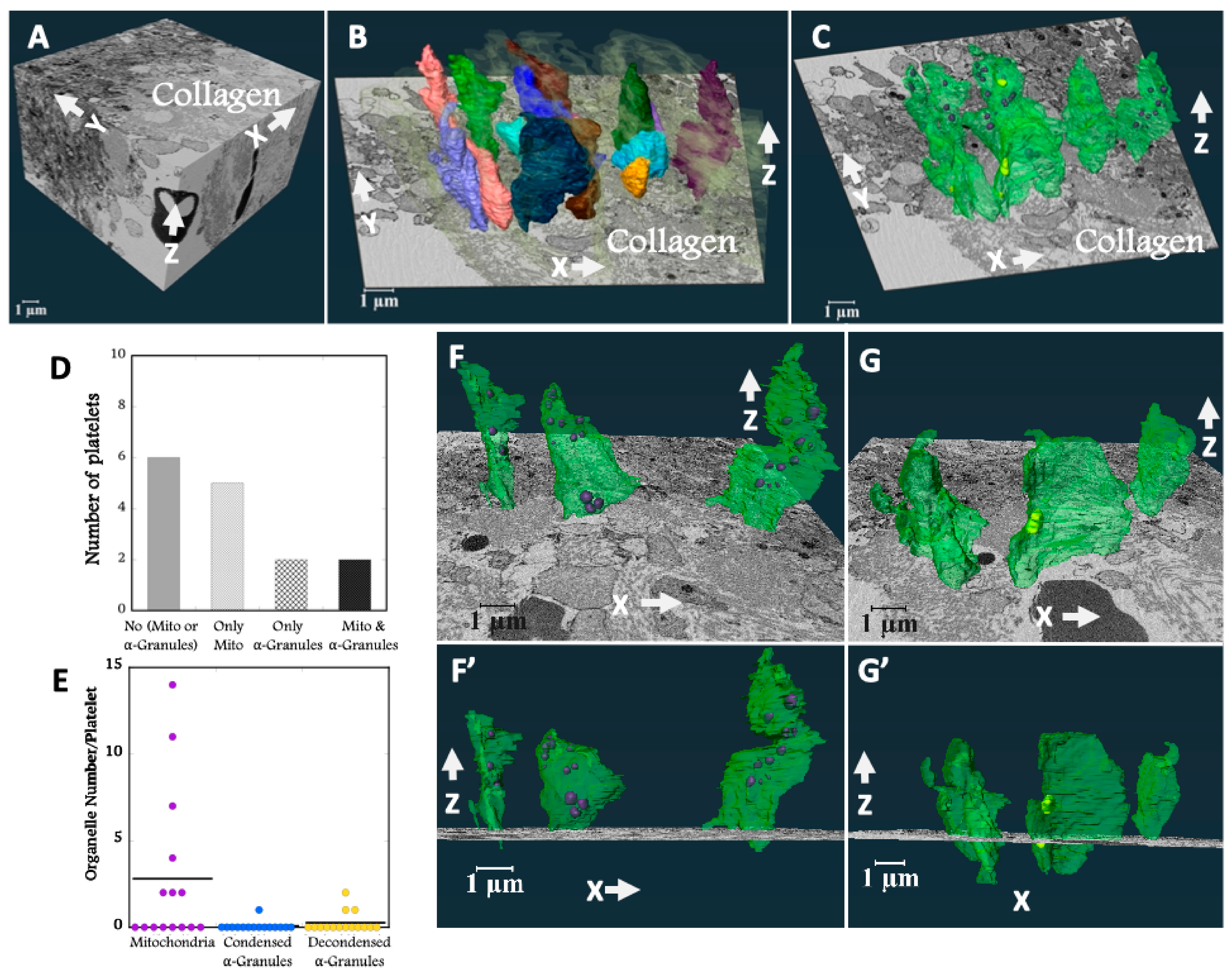
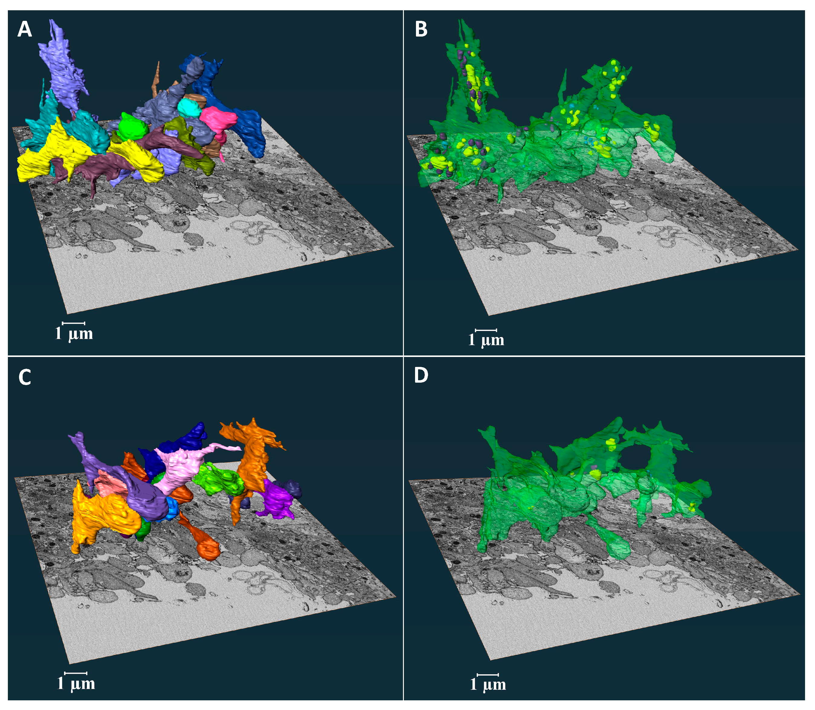
2.2. Platelet Morphodynamics at the Collagen-Rich Adventitial Interface
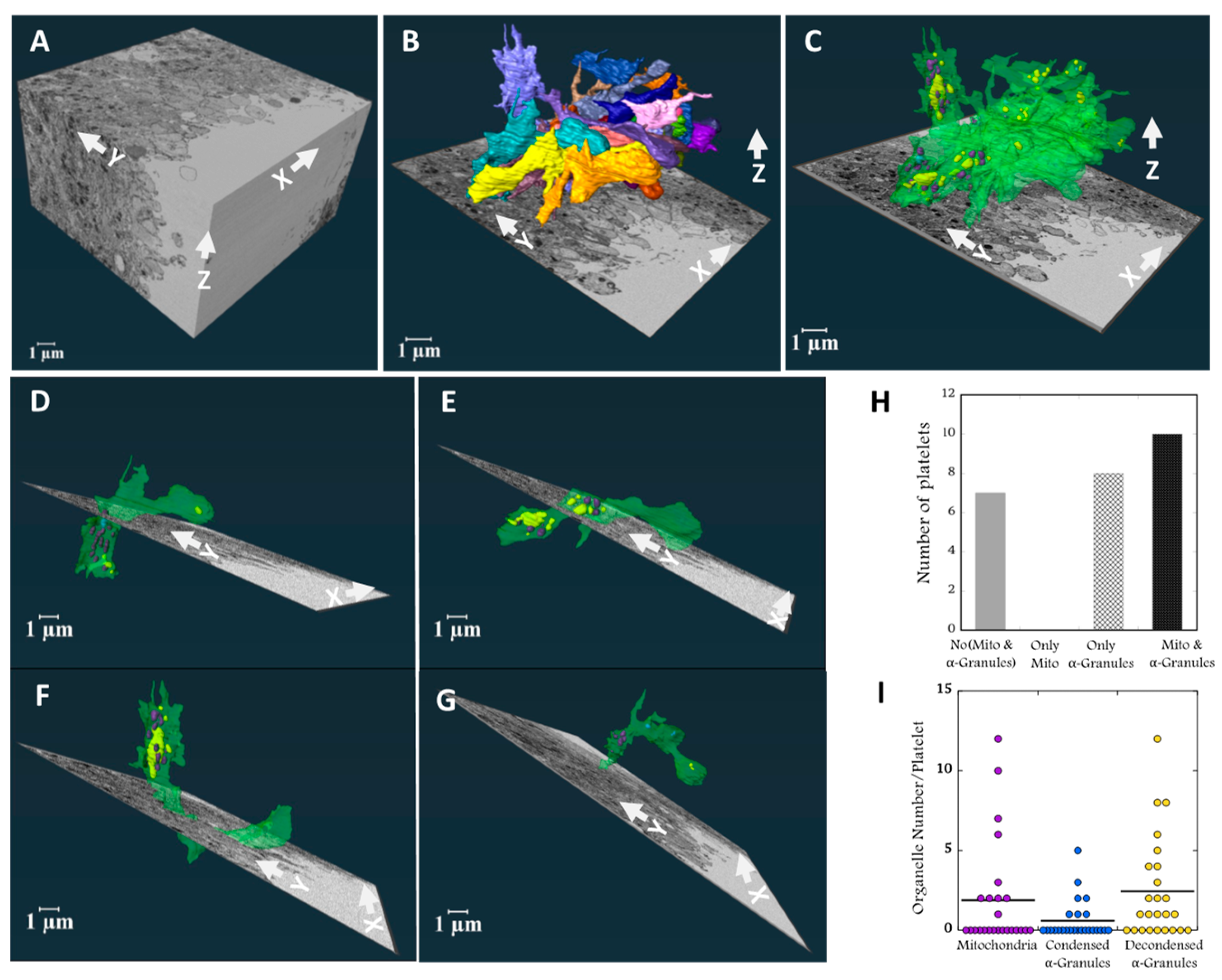
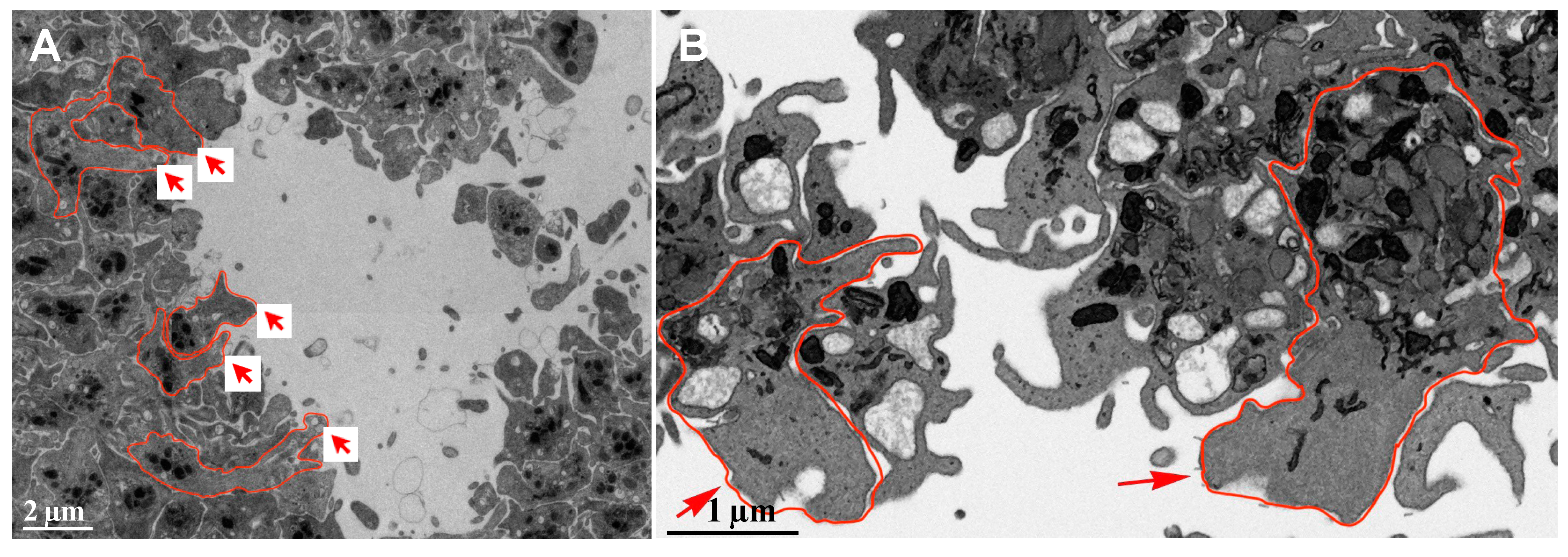
2.3. Platelet Morphodynamics at the Wound Hole Interface
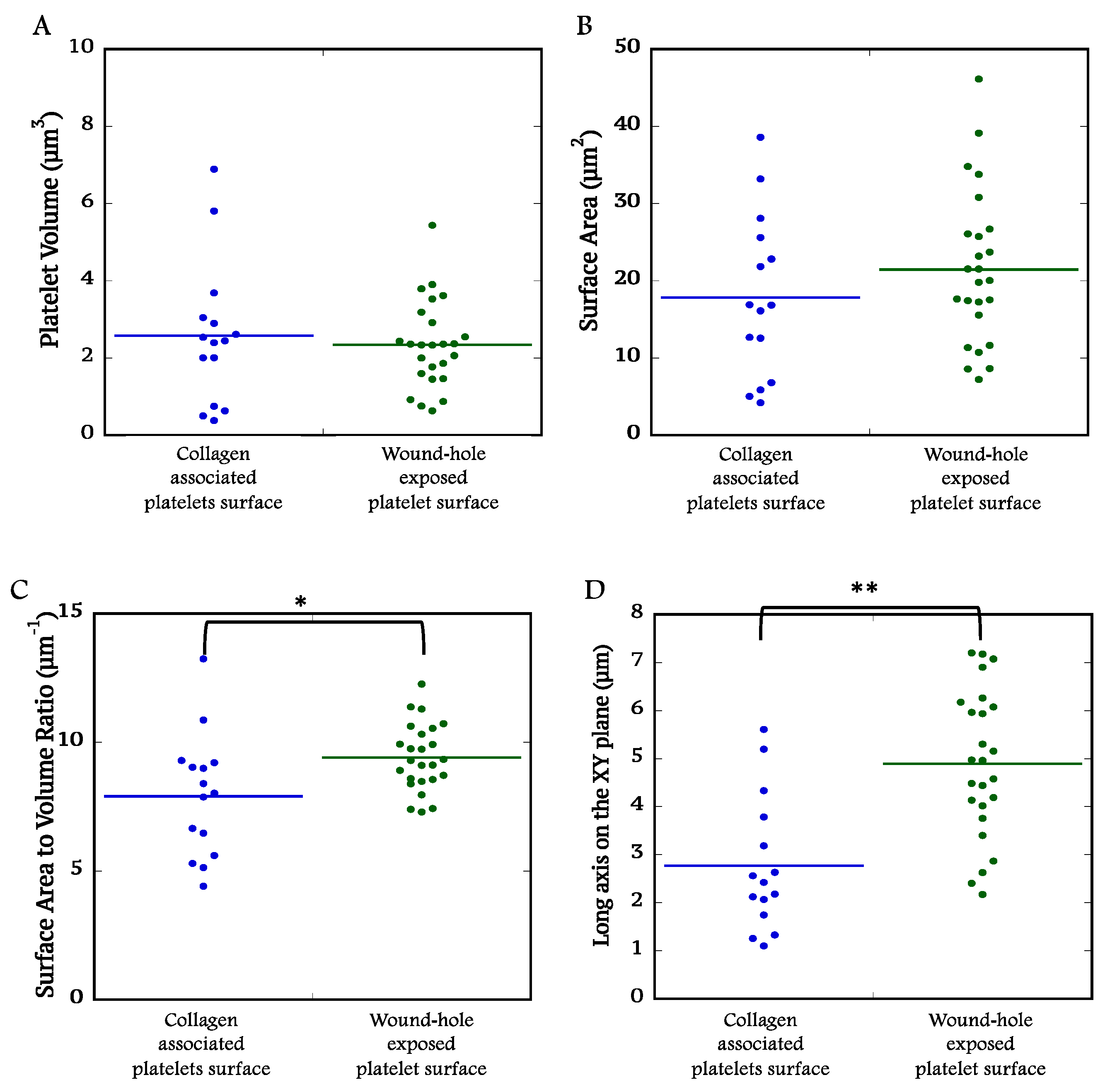
2.4. Quantitative Comparisons of Platelet at the Collagen-Rich Adventitial Interface (ROI 1) and Wound Hole Interface (ROI 2)
2.5. Platelets Exhibited Leading Edge Behavior in Vaulted Regions of Femoral Artery Puncture Wounds and in Ex-Vivo Experiments
3. Discussion
4. Materials and Methods
4.1. Mice and Reagents
4.2. Jugular Vein and Femoral Artery Puncture Wounds and Sample Preparation for Serial Block Face-Scanning Electron Microscopy (SBF-SEM)
4.3. Election Microscopy - Region of Interest (ROI) Imaging
4.4. ROI Analysis
4.5. Statistical Analysis
4.6. Data Availability
Supplementary Materials
Author Contributions
Funding
Animal Use Statement
Data Availability Statement
Acknowledgments
Conflicts of Interest
References
- McBride, E.; Rao, A.; Zhang, G.; Hoyne, J.; Calco, G.; Kuo, B.; He, Q.; Prince, A.; Pokrovskaya, I.; Storrie, B.; et al. Comparison of 3D cellular imaging techniques based on scanned electron probes: Serial block face SEM vs. Axial bright-field STEM tomography. J. Struct. Biol. 2018, 202, 216–228. [Google Scholar] [CrossRef] [PubMed]
- Pokrovskaya, I.; Tobin, M.; Desai, R.; Aronova, M.A.; Kamykowski, J.A.; Zhang, G.; Joshi, S.; Whiteheart, S.W.; Leapman, R.D.; Storrie, B. Structural analysis of resting mouse platelets by 3D-EM reveals an unexpected variation in α-granule shape. Platelets 2020, 32, 608–617. [Google Scholar] [CrossRef] [PubMed]
- Pokrovskaya, I.D.; Joshi, S.; Tobin, M.; Desai, R.; Aronova, M.A.; Kamykowski, J.A.; Zhang, G.; Whiteheart, S.W.; Leapman, R.D.; Storrie, B. SNARE-dependent membrane fusion initiates α-granule matrix decondensation in mouse platelets. Blood Adv. 2018, 2, 2947–2958. [Google Scholar] [CrossRef] [PubMed]
- Pokrovskaya, I.D.; Rhee, S.W.; Ball, K.K.; Kamykowski, J.A.; Zhao, O.S.; Cruz, D.R.; Cohen, J.; Aronova, M.A.; Leapman, R.D.; Storrie, B. Tethered platelet capture provides a mechanism for restricting circulating platelet activation to the wound site. Res. Pr. Thromb. Haemost. 2023, 7, 100058. [Google Scholar] [CrossRef] [PubMed]
- Falati, S.; Gross, P.; Merrill-Skoloff, G.; Furie, B.C.; Furie, B. Real-time in vivo imaging of platelets, tissue factor and fibrin during arterial thrombus formation in the mouse. Nat. Med. 2002, 8, 1175–1180. [Google Scholar] [CrossRef] [PubMed]
- Furie, B.; Furie, B.C. Thrombus formation in vivo. J. Clin. Investig. 2005, 115, 3355–3362. [Google Scholar] [CrossRef] [PubMed]
- Abercrombie, M.; Heaysman, J.E.; Pegrum, S.M. The locomotion of fibroblasts in culture I. Movements of the leading edge. Exp. Cell Res. 1970, 59, 393–398. [Google Scholar] [CrossRef] [PubMed]
- Nechipurenko, D.Y.; Receveur, N.; Yakimenko, A.O.; Shepelyuk, T.O.; Yakusheva, A.A.; Kerimov, R.R.; Obydennyy, S.I.; Eckly, A.; Leon, C.; Gachet, C.; et al. Clot Contraction Drives the Translocation of Procoagulant Platelets to Thrombus Surface. Arterioscler. Thromb. Vasc. Biol. 2019, 39, 37–47. [Google Scholar] [CrossRef] [PubMed]
- Kholmukhamedov, A.; Jobe, S. Procoagulant Platelets Get Squeezed to Define the Boundaries of the Hemostatic Plug. Arter. Thromb. Vasc. Biol. 2019, 39, 5–6. [Google Scholar] [CrossRef] [PubMed]
- Ren, Q.; Barber, H.K.; Crawford, G.L.; Karim, Z.A.; Zhao, C.; Choi, W.; Wang, C.-C.; Hong, W.; Whiteheart, S.W.; Woo, S.S.; et al. Endobrevin/VAMP-8 Is the Primary v-SNARE for the Platelet Release Reaction. Mol. Biol. Cell 2007, 18, 24–33. [Google Scholar] [CrossRef] [PubMed]
- Humbert, M.; Nurden, P.; Bihour, C.; Pasquet, J.-M.; Winckler, J.; Heilmann, E.; Savi, P.; Herbert, J.-M.; Kunicki, T.J.; Nurden, A.T. Ultrastructural Studies of Platelet Aggregates From Human Subjects Receiving Clopidogrel and From a Patient With an Inherited Defect of an ADP-Dependent Pathway of Platelet Activation. Arter. Thromb. Vasc. Biol. 1996, 16, 1532–1543. [Google Scholar] [CrossRef] [PubMed]
- Ball, K.; Pokrovskaya, I.; Storrie, B. Puncture Wound Hemostasis and Preparation of Samples for Montaged Wide-Area Electron Microscopy Analysis. J. Vis. Exp. 2024, e66479. [Google Scholar] [CrossRef] [PubMed]
- Rhee, S.W.; Pokrovskaya, I.D.; Ball, K.K.; Ling, K.; Vedanaparti, Y.; Cohen, J.; Cruz, D.R.D.; Zhao, O.S.; Aronova, M.A.; Zhang, G.; et al. Venous puncture wound hemostasis results in a vaulted thrombus structured by locally nucleated platelet aggregates. Commun. Biol. 2021, 4, 1–13. [Google Scholar] [CrossRef]
- Joshi, S.; Smith, A.N.; Prakhya, K.S.; Alfar, H.R.; Lykins, J.; Zhang, M.; Pokrovskaya, I.; Aronova, M.; Leapman, R.D.; Storrie, B.; et al. Ferric Chloride-Induced Arterial Thrombosis and Sample Collection for 3D Electron Microscopy Analysis. J. Vis. Exp. 2023, e64985. [Google Scholar] [CrossRef]
Disclaimer/Publisher’s Note: The statements, opinions and data contained in all publications are solely those of the individual author(s) and contributor(s) and not of MDPI and/or the editor(s). MDPI and/or the editor(s) disclaim responsibility for any injury to people or property resulting from any ideas, methods, instructions or products referred to in the content. |
© 2025 by the authors. Licensee MDPI, Basel, Switzerland. This article is an open access article distributed under the terms and conditions of the Creative Commons Attribution (CC BY) license (http://creativecommons.org/licenses/by/4.0/).


