Preprint
Article

This version is not peer-reviewed.

Circulating Microvesicles Enriched in miR–126–5p and miR–223–3p: Potential Biomarkers in Acute Coronary Syndrome

A peer-reviewed article of this preprint also exists.

Submitted:

15 January 2025

Posted:

16 January 2025

You are already at the latest version

Abstract

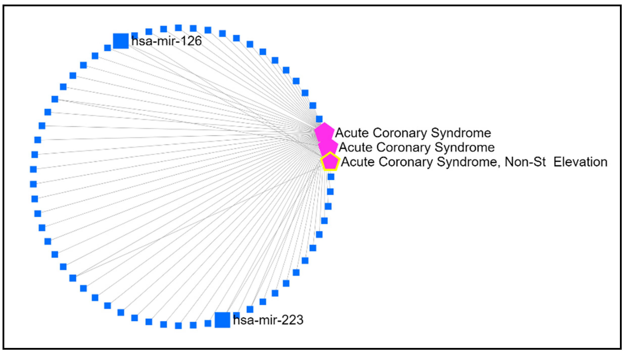
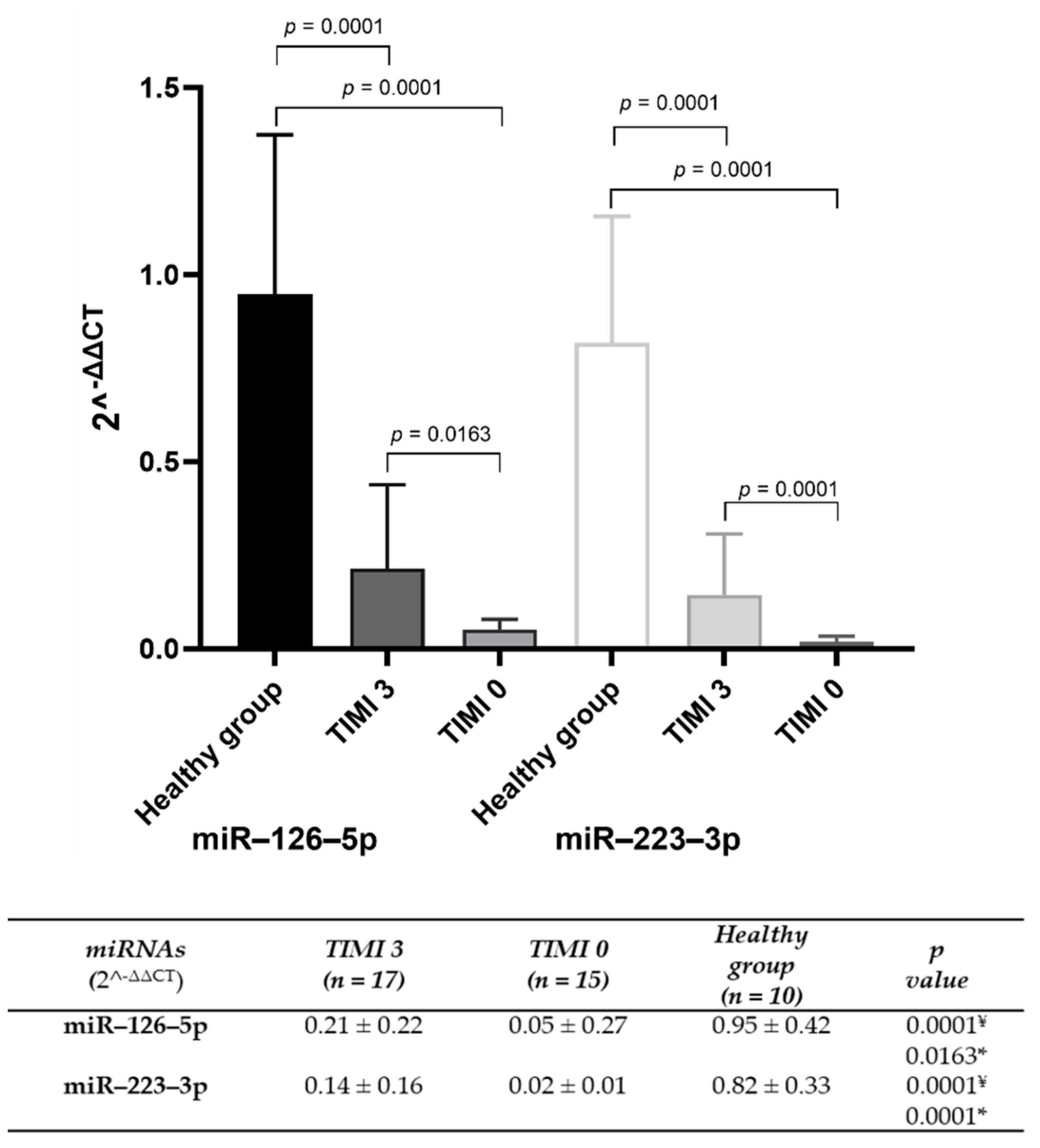
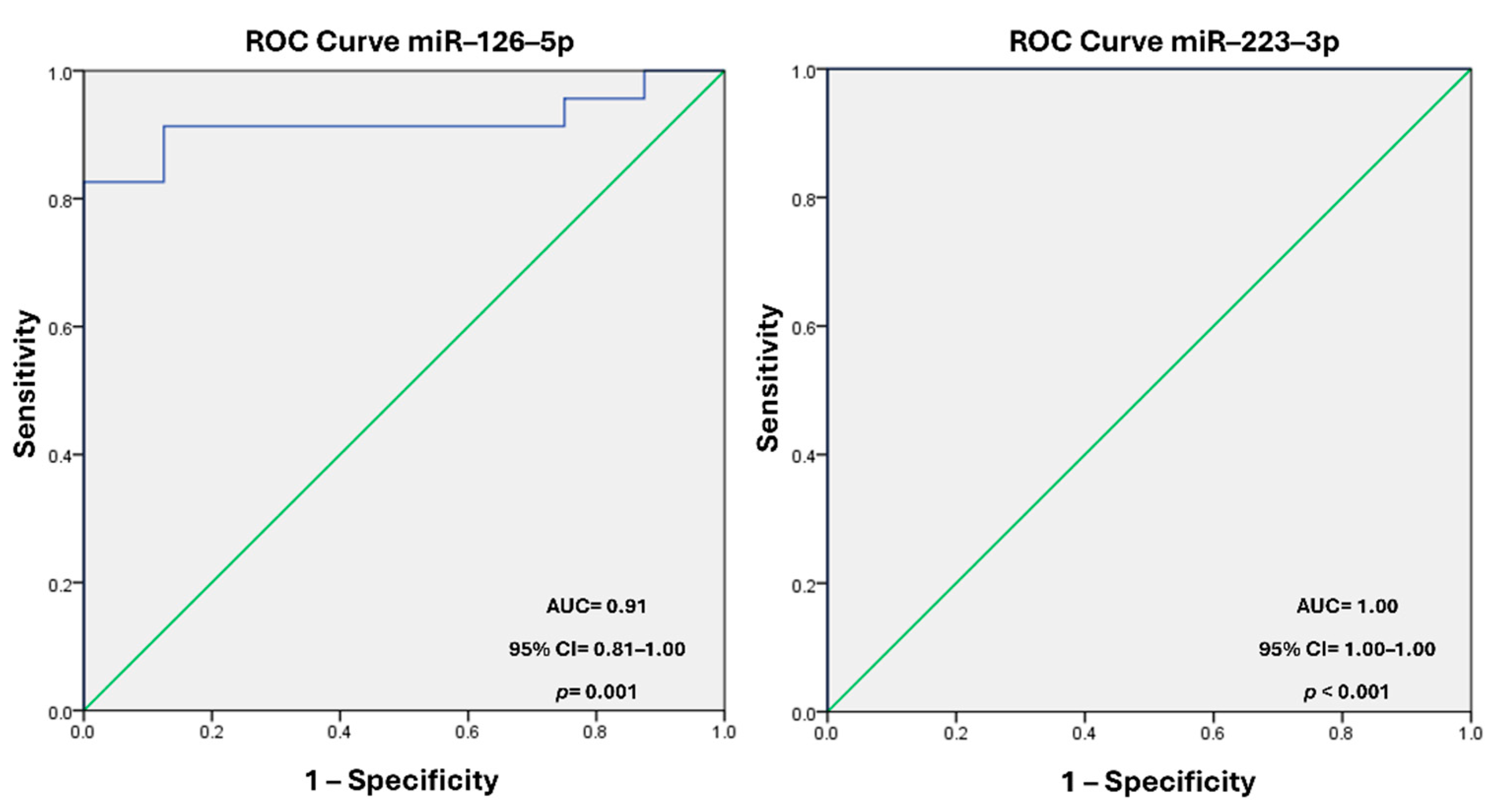
Background: The molecular mechanisms underlying acute coronary syndrome (ACS) have been extensively investigated, with a particular focus on the role of circulating microvesicles (MVs) as carriers of regulatory elements that influence hemodynamic changes and coronary flow. Endo-thelial and platelet dysfunction during ACS alters MVs composition, impacting clinical outcomes. This study explores the levels of miR–126–5p and miR–223–3p in circulating MVs and their asso-ciation with the Thrombolysis in Myocardial Infarction (TIMI) coronary flow classification scale, proposing their potential as biomarkers. Methods. Bioinformatic tools identified miRNAs linked to ACS. Plasma MVs were isolated from ACS patients and healthy controls through high-speed centrifugation. miRNA levels were quantified using quantitative reverse transcription polymerase chain reaction (qRT-PCR) and compared across TIMI 0 and TIMI 3 groups. Diagnostic efficacy was assessed via receiver operating characteristic (ROC) curve analysis. Results. The bioinformatic analysis identified miR–126 and miR–223 present in ACS. miR–126–5p and miR–223–3p were sig-nificantly reduced in MVs from TIMI 0 patients compared to TIMI 3. ROC analysis showed high diagnostic accuracy for miR–126–5p (AUC = 0.918; 95% CI: 0.818–1.00; p = 0.001) and miR–223–3p (AUC = 1.00; 95% CI: 1.00–1.00; p < 0.001). Conclusion. Reduced levels of miR–126–5p and miR–223–3p in circulating MVs are strongly associated with impaired coronary flow, positioning these miRNAs as potential biomarkers for ACS risk stratification and therapeutic targeting.

Keywords: 
;  ;  ;  ;  

1. Introduction

Acute coronary syndrome (ACS) encompasses a spectrum of clinical conditions caused by insufficient coronary blood flow, including acute myocardial infarction (AMI) and unstable angina (UA) [1]. ACS is a leading cause of morbidity and mortality worldwide, with cardiovascular diseases (CVDs) responsible for over 17.9 million deaths annually, according to the World Health Organization (2023) [2]. This high prevalence underscores the urgent need for improved diagnostic and prognostic tools to better understand ACS pathophysiology and enhance patient care. A key clinical tool for assessing ACS severity is the Thrombolysis in Myocardial Infarction (TIMI) coronary flow classification scale, which uses coronary angiography to categorize epicardial blood flow into four grades: TIMI 0 (no perfusion), TIMI 1 (minimal perfusion), TIMI 2 (reduced-speed perfusion), and TIMI 3 (normal perfusion) [3]. Stratification using the TIMI scale has been linked to clinical outcomes such as reinfarction [4], mortality [5,6], ventricular aneurysm formation [7], and arrhythmias [8]. Achieving TIMI 3 flow is a critical benchmark for therapeutic success [9,10]. Despite advances in reperfusion therapies and pharmacological treatments, ACS remains a clinical challenge due to its complex and multifactorial nature. The lack of reliable biomarkers to identify molecular and hemodynamic changes in ACS limits clinicians' ability to accurately predict outcomes [11]. Circulating microvesicles (MVs) have emerged as promising biomarkers and mediators in cardiovascular disease. MVs are extracellular vesicles released by platelets [12], endothelial cells [13], and leukocytes [14] in response to stress or activation. They carry bioactive molecules, including proteins, lipids, and microRNAs (miRNAs) [15]. miRNAs, small non-coding RNAs (21–25 nucleotides), regulate gene expression post-transcriptionally by targeting messenger RNA for degradation or translational repression, influencing up to 30% of human genes [16]. In ACS, miRNAs such as miR–26, miR–126, miR–133, miR–144, miR–208, miR–223, and miR–483 modulate platelet activity, oxidative stress, cardiac remodeling, and inflammation [17,18]. MVs have been implicated in endothelial dysfunction, platelet activation, and disrupted coronary blood flow [19,20,21]. These vesicles facilitate platelet–platelet and platelet–endothelial communication, regulating thrombotic processes [22,23,24]. Alterations in MV cargo, including miRNAs, during ACS reflect underlying pathophysiological processes. This study focuses on miR–126–5p and miR–223–3p levels in circulating MVs and examines their potential as biomarkers for ACS, particularly in relation to the TIMI flow scale.

2. Materials and Methods

2.1. Study Population

This cross-sectional study included, by convenience, 42 participants: 32 ACS patients and 10 healthy controls. Patients were recruited from the Hemodynamics Department of the National Institute of Cardiology (INC) Ignacio Chávez, Mexico City. The inclusion criteria were age from 40 to 75 years, diagnosis of UA, ST–segment elevation (STEMI) and non–ST–segment elevation (NSTEMI) AMI made by angiographic according to the criteria of the American College of Cardiology [25]. Patients were stratified according to the TIMI flow scale by an expert cardiologist from the Hemodynamics Department. This stratification considered the impact of biological and mechanistic factors associated with total reperfusion or lack of reperfusion. The healthy group comprised clinically healthy subjects 40 to 75 years old from the blood bank of the INC. Exclusion criteria included autoimmune, hepatic, renal, or oncological diseases. All participants provided informed consent, and the study was approved by the Research and Ethics Committees of INC with register number FIMICOR 18-1043.

2.2. Microvesicles Extraction

Blood samples were collected in sodium citrate (0.109 M, relation 1:9) tubes and centrifuged at 1500 × g for 15 min to obtain plasma. Plasma was further centrifuged at 13,000 × g for 2 min and stored at −80°C. MVs were isolated through high-speed centrifugation (20,000 × g for 90 min, 4°C) and resuspended in 100 µL of phosphate buffer, was added 400 µL of QIAzol Lysis Reagent and stored in aliquots at –20 °C, before RNA extraction.

2.3. RNA Extraction and Quantitative Reverse Transcription Polymerase Chain Reaction (RT-qPCR)

RNA was extracted from MVs using the Direct–zolTM RNA MiniPrep Kit (Zymo Research, CA, USA), according to the manufacturer’s instructions, and was eluted in 25 µL of RNase–free water. RNA samples were stored at –80 °C until their processing. For the miRNA complementary DNA (cDNA), we employed the TaqMan© MicroRNA Reverse Transcription Kit (Applied Biosystems, Foster City, CA, USA), following the manufacturer’s instructions. Retrotranscription was developed according to the manufacturer’s instructions. The results were analyzed and the samples with amplification after 35 cycles were discarded. miRNA levels were quantified using TaqMan© miRNA Assays (Applied Biosystems, Foster City, CA, USA) for miR–126–5p, miR–223–3p, and miR–39–3p as control were employed in a Roche LightCycler® 480–II instrument (Roche Applied Sciences, Beijing, China), under standard amplification conditions. Relative expression was calculated using the delta-delta Ct method (2-ΔΔCt).

2.4. Bioinformatic Analysis

Potential ACS-associated miRNAs were identified using miRbase (https://www.mirbase.org/), miRNet (https://www.mirnet.ca/), the Human microRNA Disease Database (HMDD, http://www.cuilab.cn/hmdd#fragment–1) and miRandola (http://mirandola.iit.cnr.it/index.php). miRNAs present in at least three databases with validated roles in cardiovascular diseases were selected. The analysis focused on identifying miRNAs associated with ACS in the bloodstream, particularly those with previously reported controversial level patterns. This approach aimed to investigate whether these miRNAs could indicate potential transport mechanism via MVs. The selection included miRNAs that had functional evidence and were validated in the literature for their roles in cardiovascular disease.

2.5. Statistical Analysis

Quantitative data are expressed as mean ± standard deviation (SD) or median (interquartile range), and the qualitative variables are presented as frequencies (n) and percentages (%). We analyzed the normal distribution of data by Shapiro-Wilk; group differences were analyzed using ANOVA with Dunnett's post hoc test. A receiver operator characteristic (ROC) curve analysis assessed diagnostic efficacy, and the area under the curve (AUC) with their 95% confidence intervals (CI) was calculated. Statistical significance was set at p < 0.05. Analyses were conducted using SPSS v23.0 Software (SPSS Inc., Chicago, IL, USA) and GraphPad Prism Software 8.0.1 (GraphPad Software, La Jolla, CA, USA).

3. Results

3.1. MVs miRNAs Associated with ACS

Considering the association with ACS, the analysis revealed that the MVs content of hsa-miR–126–5p and hsa-miR–223–3p were identified in four of the evaluated databases. Figure 1 is a schematic representation of the network obtained from the miRNet database, which highlights the miRNAs selected for this study.

3.2. Sociodemographic Data and Clinical Variables of Study Population

The clinical characteristics of the study population were obtained from clinical records and are presented in Table 1.
3.3. miR–126–5p and miR–223–3p Levels
The 2^-ΔΔCT (Figure 2) of each analyzed sample represents the relative level of each of the miRNAs evaluated. miR–126–5p and miR–223–3p levels were significantly lower in TIMI 0 and TIMI 3 patients compared with healthy control. In TIMI 0 patients, miRNA–126–5p and miRNA–223–3p levels were 4.2 and 7.0 times lower, respectively, when compared with TIMI 3 patients.

3.4. Diagnostic Value of miR–126–5p and miR–223–3p Levels

ROC analysis demonstrated high diagnostic potential, with AUC values of 0.918 for miR–126–5p 95% CI = 0.818–1.00, p = 0.001, while for miR–223–3p, the AUC was 1.00 (95% CI = 1.00–1.00, p < 0.001), Figure 3.

4. Discussion

The development of innovative strategies for diagnosing and stratifying the risk of ACS remains a critical area for both basic and clinical research. In this study, we analyzed a cohort of ACS patients and investigated the levels of miRNAs (miR–126–5p and miR–223–3p) present in circulating plasma MVs as potential mechanisms associated with ACS pathophysiology. Notably, both miRNAs are known to be present in circulating MVs [26,27].
miRNAs play a role in phenotypic changes influenced by environmental factors, including DNA methylation, histone modification, and chromatin remodeling [28]. The epigenetic involvement of miRNAs and their distribution to critical sites for coronary ischemic diseases underscore their role in regulating platelet activity and individual susceptibility to coronary conditions. This makes them valuable biomarkers for predicting coronary disease risk [29].
By studying miRNAs within MVs, we focused on two key mechanisms directly impacting the physiological environment. Recent reviews highlight the significance of MVs in various biological processes [30], reinforcing the importance of analyzing miRNAs within these vesicles. Bioinformatics tools offer a powerful methodology within omics sciences, facilitating the interpretation of clinical and molecular data [31,32,33]. By integrating bioinformatic analyses with molecular studies, we identified miRNAs consistently associated with ACS, providing robust evidence of their biomarker potential.
miR–223, known for its anti-thrombotic properties, is highly prevalent in platelets and their derived MVs. It regulates platelet activity in atherosclerotic progression by inhibiting platelet activation, aggregation, and granule secretion [34,35,36,37]. miR–223 is thus a promising marker of platelet activation in CVDs [38]. Similarly, miR–126 plays a vital role in maintaining endothelial integrity, vascular repair, and platelet activation/aggregation [34,39]. It serves as a potential biomarker for endothelial homeostasis in CVDs and ACS risk stratification [34,40].
Our study underscores the diagnostic and prognostic potential of miR–126–5p and miR–223–3p in ACS. Both miRNAs were significantly reduced in TIMI 0 and TIMI 3 patients. Their levels reflect the physiological and regulatory mechanisms underlying atherosclerosis progression and platelet activity, with their absence strongly correlating with impaired blood flow. In TIMI 0 patients, the reduction of these miRNAs is associated with coronary flow impairment and endothelial dysfunction, hallmarks of ACS pathophysiology. Additionally, we observed statistically significant differences in miR–126–5p and miR–223–3p levels in ACS patients compared to healthy individuals. ACS patients exhibited lower miRNA levels, with a more pronounced decrease in miR–223–3p expression relative to miR–126–5p.
In the literature, only a few studies have explored the levels of these miRNAs in ACS, with contradictory findings. For instance, Massodi et al. analyzed the expression of several platelet-derived miRNAs, including miR–223 and miR–126, in ACS patients but found no statistically significant differences compared to control subjects [41]. In contrast, Gager et al., in a prospective observational study, reported a marginal increase in miR–223 levels in plasma from ACS patients, particularly when comparing those with non-ST-segment elevation ACS (NSTE-ACS) to STEMI patients [42].
Another study observed elevated miR–126 levels in plasma during the acute phase of AMI patients compared to controls, followed by a significant decrease after a follow-up period [43]. miR–126 has been linked to modifications in platelet reactivity [44] and monocyte–platelet aggregates (MPAs) [45]. Ling et al. further reported that exosomal and serum miR–126 levels were significantly higher in patients with AMI and UA compared to controls [46].
Becker et al. evaluated miR–126 in relation to platelet reactivity in blood platelet in NSTE-ACS patients and suggested that miR–126 could serve as a marker for changes in platelet activity [44]. Stojkovic et al. found an association between platelet miR–126 and MPAs in ACS patients undergoing antiplatelet therapy [45]. Lastly, Szelenberger et al. identified significantly elevated levels of miR–223–3p in blood platelet in ACS patients compared to controls, but no notable differences were observed in miR–126–3p levels between the groups [47]. Currently, no studies have specifically examined the relationship between miRNAs in MVs and the TIMI flow scale. However, Jasen et al. analyzed miRNA expression (miR–126) in circulating MVs but in patients with coronary artery disease (CAD), they found lower levels of miR–126 in CAD patients vs no CAD group [26]. On the other hand, Zhang et al. investigated the association between circulating miR–660–5p and the no-reflow phenomenon in STEMI patients. In their study, NRP was defined as a TIMI flow grade lower than 2 or 3 [48].
The originality of our study lies in two key aspects: (1) it evaluates the miRNA profiles derived from circulating MVs in patients with ACS, in contrast to prior studies that focused on platelets, and (2) it includes a stratified analysis based on the TIMI flow scale.
This approach reflects a specific pathophysiological condition and minimizes bias by avoiding comparisons with a control group of healthy individuals. Instead, both groups in our study were under the same treatment and pathological conditions. Our findings have important clinical implications. First, the association of miR–126–5p and miR–223–3p with TIMI flow grades highlights their potential utility in stratifying ACS severity and guiding therapeutic decisions. Second, the high diagnostic accuracy of these miRNAs, demonstrated through ROC curve analyses, underscores their potential as biomarkers for distinguishing ACS patients from healthy individuals. These findings suggest that miR–126–5p and miR–223–3p could complement existing diagnostic tools, providing a non-invasive method to assess disease progression and optimize patient management. Similarly, studies by Masoodi et al. [41], Ling et al. [46], and Gager et al. [42] have proposed miRNAs as innovative biomarkers with significant diagnostic value.
We propose that miRNA levels reflect the dynamic processes occurring during platelet activation, a hallmark of ACS [49]. Platelet activation plays a pivotal role in thrombus formation following plaque rupture [50], and these miRNAs may offer valuable insights into cardiac blood flow dynamics after an ischemic event. Their study could pave the way for improved biomarker-based strategies to monitor and manage ACS patients effectively.
To the best of our knowledge, this is the first study to evaluate circulating MVs as carriers of miRNAs in ACS within the Mexican population. Previous studies have focused on European, Asian, or American populations, where genetic backgrounds may contribute to distinct clinical phenotypes and varying susceptibilities to diseases [51,52,53].
Our study highlights several key findings. Despite the small sample size, we detected statistically significant differences in MVs miRNA content, which suggests that our experimental approach could serve as a potential method for identifying ACS patients. Despite these promising results, the study has limitations. A future longitudinal study should explore changes in miRNA levels and their response to therapeutic interventions. Integrating our findings with additional mechanisms, such as genetic variants and epigenetic factors (e.g., DNA methylation), would provide a more comprehensive understanding. Furthermore, exploring other miRNAs involved in ACS pathophysiology would strengthen our conclusions.

5. Conclusion

In conclusion, miR–126–5p and miR–223–3p levels in circulating MVs provide valuable insights into the pathophysiology of ACS. Their diagnostic and prognostic utility warrants further investigation, with potential applications in precision medicine to improve risk stratification and therapeutic monitoring in ACS patients.

Author Contributions

Conceptualization, A.P.D. and E.A.C.; methodology, J.R.H.L., F.S.M., H.D.R., M.A.M.R.; software, E.G.F. and B.G.C.S.; validation, A.P.D. and M.F.G.; formal analysis, E.G.F., M.F.G., B.G.C.S. and J.R.H.L.; investigation, A.P.D. and E.A.C.; resources, A.P.D.; data curation, M.A.P.D., F.S.M., M.A.B.V. and M.F.G.; writing—original draft preparation, J.R.H.L., E.G.F., M.F.G., B.G.C.S.; writing—review and editing, A.P.D. and E.A.C.; visualization, A.P.D.; supervision, A.P.D.; project administration, A.P.D. and E.A.C.; funding acquisition, A.P.D. All authors have read and agreed to the published version of the manuscript.

Funding

This research was funded by Grants IN219117 from Dirección General de apoyo al Personal Académico (DGAPA) and Programa de Apoyo a Proyectos de Investigación e Innovación Tecnológica (PAPIIT) from Fibrinolytic Microparticles: protein, functional and mRNA evaluation in Acute Coronary Syndrome (FIMICOR) 047/2014.

Institutional Review Board Statement

This study was approved by the Ethical and Research Committees of INC with the number registered FIMICOR 18-1043 (03/22/2018). All participants provided informed consent for the collection of blood samples and participation in the study. Moreover, the study was performed according to the Helsinki Declaration.

Informed Consent Statement

Informed consent was obtained from all subjects involved in the study.

Data Availability Statement

The data underlying this article will be shared on reasonable request to the corresponding author.

Acknowledgments

Dr. Rubicel Hernández has a Postdoctoral Scholarship provided by the Dirección General de Apoyo al Personal Académico (DGAPA), UNAM. Likewise, we acknowledge Oscar Zapeda García for their valuable participation as social service student.

Conflicts of Interest

The authors declare no conflicts of interest.

References

  1. Byrne, R.A.; Rossello, X.; Coughlan, J.J.; Barbato, E.; Berry, C.; Chieffo, A.; Claeys, M.J.; Dan, G.A.; Dweck, M.R.; Galbraith, M.; et al.; ESC Scientific Document Group. 2023 ESC Guidelines for the management of acute coronary syndromes. Eur Heart J Acute Cardiovasc Care. 2024, 13, 55-161. Erratum in: Eur Heart J Acute Cardiovasc Care 2024, 13, 455. [CrossRef]
  2. World health statistics 2023: monitoring health for the SDGs, Sustainable Development Goals. Geneva: World Health Organization; 2023. Licence: CC BY-NC-SA 3.0 IGO.
  3. Abd, M.E.; Bahgat, A.M.H.; Khaled, H.R; Ibrahim, S.F. Prediction of angiographic (TIMI GRADE) blood flow using the novel CHA2DS2-VASC-HSF score in patients with STEMI. Int. J. Cardiol. Sci. 2024, 6, 114-123. [CrossRef]
  4. Kievit, P.C; Brouwer, M.A.; Veen, G.; Karreman, A.J.; Verheugt, F.W.A. High-grade infarct-related stenosis after successful thrombolysis: strong predictor of reocclusion, but not of clinical reinfarction. Am Heart J 2004, 148, 826-833. [CrossRef]
  5. Gibson, C.M.; Cannon, C.P.; Murphy, S.A.; Ryan, K.A.; Mesley, R.; Marble, S.J.; McCabe, C.H.; van de Werf, F.; Braunwald, E. Relationship of TIMI myocardial perfusion grade to mortality after administration of thrombolytic drugs. Circulation 2000, 101, 125-30. [CrossRef]
  6. Stringer, K.A. TIMI grade flow, mortality, and the GUSTO-III trial. Pharmacotherapy 1998, 18, 699-705.
  7. Suzuki, M.; Enomoto, D.; Seike, F.; Fujita, S.; Honda, K. Clinical features of early myocardial rupture of acute myocardial infarction. Angiology 2012, 63, 453-456. [CrossRef]
  8. Tikiz, H.; Atak, R.; Balbay, Y.; Genç, Y.; Kütük, E. Left ventricular aneurysm formation after anterior myocardial infarction: clinical and angiographic determinants in 809 patients. Int J Cardiol 2002, 82, 7-14. [CrossRef]
  9. Gibson, C.M.; Pride, Y.B.; Buros, J.L.; Lord, E.; Shui, A.; Murphy, S.A.; Pinto, D.S.; Zimetbaum, P.J.; Sabatine, M.S.; Cannon, C.P.; et al; TIMI Study Group. Association of impaired thrombolysis in myocardial infarction myocardial perfusion grade with ventricular tachycardia and ventricular fibrillation following fibrinolytic therapy for ST-segment elevation myocardial infarction. J Am Coll Cardiol 2008, 51, 546-551. [CrossRef]
  10. Bates, E.R. Reperfusion therapy reduces the risk of myocardial rupture complicating ST-elevation myocardial infarction. J Am Heart Assoc. 2014, 3, e001368. [CrossRef]
  11. Cannon, C.P. Importance of TIMI 3 flow. Circulation 2001, 104, 624-626. [CrossRef]
  12. Gersh, B.J. Current issues in reperfusion therapy. Am J Cardiol 1998, 82, 3-11. [CrossRef]
  13. Caffè, A.; Animati, F.M.; Iannaccone, G.; Rinaldi, R.; Montone, R.A. Precision Medicine in Acute Coronary Syndromes. J Clin Med 2024, 13, 4569. [CrossRef]
  14. Balakrishnan, R.; Subbarayan, R.; Shrestha, R.; Chauhan, A.; Krishnamoorthy, L. Exploring platelet-derived microvesicles in vascular regeneration: unraveling the intricate mechanisms and molecular mediators. Mol Biol Rep 2024, 51, 393. [CrossRef]
  15. Taus, F.; Meneguzzi, A.; Castelli, M.; Minuz, P. Platelet-Derived Extracellular Vesicles as Target of Antiplatelet Agents. What Is the Evidence? Front Pharmacol 2019, 10, 1256. [CrossRef]
  16. Bartel, D.P. MicroRNAs: target recognition and regulatory functions. Cell 2009, 136, 215-233. [CrossRef]
  17. Katsioupa, M.; Kourampi, I.; Oikonomou, E.; Tsigkou, V.; Theofilis, P.; Charalambous, G.; Marinos, G.; Gialamas, I.; Zisimos, K.; Anastasiou, A.; et al. Novel Biomarkers and Their Role in the Diagnosis and Prognosis of Acute Coronary Syndrome. Life (Basel) 2023, 13, 1992. [CrossRef]
  18. Tanase, D.M.; Gosav, E.M.; Ouatu, A.; Badescu, M.C.; Dima, N.; Ganceanu-Rusu, A.R.; Popescu, D.; Floria, M.; Rezus, E.; Rezus, C. Current Knowledge of MicroRNAs (miRNAs) in Acute Coronary Syndrome (ACS): ST-Elevation Myocardial Infarction (STEMI). Life (Basel) 2021, 11, 1057. [CrossRef]
  19. Federici, C.; Shahaj, E.; Cecchetti, S.; Camerini, S.; Casella, M.; Iessi, E.; Camisaschi, C.; Paolino, G.; Calvieri, S.; Ferro, S.; et al. Natural-Killer-Derived Extracellular Vesicles: Immune Sensors and Interactors. Front Immunol 2020, 11, 262. [CrossRef]
  20. Melki, I.; Tessandier, N.; Zufferey, A.; Boilard, E. Platelet microvesicles in health and disease. Platelets 2017, 28, 214-221. [CrossRef]
  21. Lv, Y.; Tan, J.; Miao, Y.; Zhang, Q. The role of microvesicles and its active molecules in regulating cellular biology. J Cell Mol Med 2019, 23, 7894-7904. [CrossRef]
  22. Wang, X.L.; Zhang, W.; Li, Z.; Han, W.Q.; Wu, H.Y.; Wang, Q.R.; Liu, X.H.; Xing, K.; Cheng, G.; Chang, F.J. Vascular damage effect of circulating microparticles in patients with ACS is aggravated by type 2 diabetes. Mol Med Rep 2021, 23, 474. [CrossRef]
  23. Zaldivia, M.T.K.; McFadyen, J.D.; Lim, B.; Wang, X.; Peter, K. Platelet-Derived Microvesicles in Cardiovascular Diseases. Front Cardiovasc Med 2017, 4, 74. [CrossRef]
  24. Edelstein, L.C. The role of platelet microvesicles in intercellular communication. Platelets 2017, 28, 222-227. [CrossRef]
  25. Cannon, C.P.; Brindis, R.G.; Chaitman, B.R.; Cohen, D.J.; Cross, J.T.; Drozda, J.P.; Fesmire, F.M.; Fintel, D.J.; Fonarow, G.C.; Fox, K.A.; et al; American College of Cardiology Foundation/American Heart Association Task Force on Clinical Data Standards; American College of Emergency Physicians; Emergency Nurses Association; National Association of Emergency Medical Technicians; National Association of EMS Physicians; Preventive Cardiovascular Nurses Association; Society for Cardiovascular Angiography and Interventions; Society of Cardiovascular Patient Care; Society of Thoracic Surgeons. 2013 ACCF/AHA key data elements and definitions for measuring the clinical management and outcomes of patients with acute coronary syndromes and coronary artery disease: a report of the American College of Cardiology Foundation/American Heart Association Task Force on Clinical Data Standards (Writing Committee to Develop Acute Coronary Syndromes and Coronary Artery Disease Clinical Data Standards). Circulation 2013, 127, 1052-1089. [CrossRef]
  26. Jansen, F.; Yang, X.; Proebsting, S.; Hoelscher, M.; Przybilla, D.; Baumann, K.; Schmitz, T.; Dolf, A.; Endl, E.; Franklin, B.S.; et al. MicroRNA expression in circulating microvesicles predicts cardiovascular events in patients with coronary artery disease. J Am Heart Assoc 2014, 3: e001249. [CrossRef]
  27. Hunter, M.P.; Ismail, N.; Zhang, X.; Aguda, B.D.; Lee, E.J.; Yu, L.; Xiao, T.; Schafer, J.; Lee, M.L.; Schmittgen, T.D.; et al. Detection of microRNA expression in human peripheral blood microvesicles. PLoS One 2008, 3: e3694. [CrossRef]
  28. Colpaert, R.M.W.; Calore, M. Epigenetics and microRNAs in cardiovascular diseases. Genomics 2021; 113: 540-551. [CrossRef]
  29. Kong, A.S.; Lai, K.S.; Lim, S.E.; Sivalingam, S.; Loh, J.Y.; Maran, S. miRNA in Ischemic Heart Disease and Its Potential as Biomarkers: A Comprehensive Review. Int J Mol Sci 2022, 23: 9001. [CrossRef]
  30. Plawinski, L.; Cras, A.; Hernández, L.J.R.; de la Peña, A.; van der Heyden, A.; Belle, C.; Toti, F.; Anglés-Cano, E. Plasmin-Generating Microvesicles: Tiny Messengers Involved in Fibrinolysis and Proteolysis. Int J Mol Sci 2023, 24, 1571. [CrossRef]
  31. Luna, B.D.; Lovering, R.C.; Caporali, A. Insights into Online microRNA Bioinformatics Tools. Noncoding RNA 2023, 9, 18. [CrossRef]
  32. Vitorino, R. Transforming Clinical Research: The Power of High-Throughput Omics Integration. Proteomes 2024, 12, 25. [CrossRef]
  33. Shaker, F.; Nikravesh, A.; Arezumand, R.; Aghaee-Bakhtiari, S.H. Web-based tools for miRNA studies analysis. Comput Biol Med 2020, 127, 104060. [CrossRef]
  34. Gatsiou, A.; Boeckel, J.N.; Randriamboavonjy, V.; Stellos, K. MicroRNAs in platelet biogenesis and function: implications in vascular homeostasis and inflammation. Curr Vasc Pharmacol 2012, 10, 524-531. [CrossRef]
  35. Elgheznawy, A.; Fleming, I. Platelet-Enriched MicroRNAs and Cardiovascular Homeostasis. Antioxid Redox Signal 2018, 29, 902-921. [CrossRef]
  36. Zhang, M.W.; Shen, Y.J.; Shi, J.; Yu, J.G. MiR-223-3p in Cardiovascular Diseases: A Biomarker and Potential Therapeutic Target. Front Cardiovasc Med 2021, 7, 610561. [CrossRef]
  37. Provost, P. Existence of a microRNA pathway in anucleate platelets. Nat Struct Mol Biol 2009, 16, 961-966. [CrossRef]
  38. Pordzik, J.; Pisarz, K.; de Rosa, S.; Jones, A.D.; Eyileten, C.; Indolfi, C.; Malek, L.; Postula, M. The Potential Role of Platelet-Related microRNAs in the Development of Cardiovascular Events in High-Risk Populations, Including Diabetic Patients: A Review. Front Endocrinol (Lausanne) 2018, 9, 74. [CrossRef]
  39. Czajka, P.; Fitas, A.; Jakubik, D.; Eyileten, C.; Gasecka, A.; Wicik, Z.; Siller-Matula, J.M.; Filipiak, K.J.; Postula, M. MicroRNA as Potential Biomarkers of Platelet Function on Antiplatelet Therapy: A Review. Front. Physiol 2021, 12, 652579. [CrossRef]
  40. Leng, Q.; Ding, J.; Dai, M.; Liu, L.; Fang, Q.; Wang, D.W.; Wu, L.; Wang, Y. Insights Into Platelet-Derived MicroRNAs in Cardiovascular and Oncologic Diseases: Potential Predictor and Therapeutic Target. Front Cardiovasc Med 2022, 9, 879351. [CrossRef]
  41. Masoodi, K.P.; Ghydari, M.E.; Vazifeh, S.N.; Shirazy, M.; Hamidpour, M. Platelet MicroRNA-484 as a Novel Diagnostic Biomarker for Acute Coronary Syndrome. Lab Med 2023, 54, 256-261. [CrossRef]
  42. Gager, G.M.; Eyileten, C.; Postula, M.; Gasecka, A.; Jarosz-Popek, J.; Gelbenegger, G.; Jilma, B.; Lang, I.; Siller-Matula, J. Association Between the Expression of MicroRNA-125b and Survival in Patients With Acute Coronary Syndrome and Coronary Multivessel Disease. Front Cardiovasc Med 2022, 9, 948006. [CrossRef]
  43. Ewelina, K.; Eljaszewicz, A.; Kazimierczyk, R.; Tynecka, M.; Zembko, P.; Tarasiuk, E.; Kaminski, K.; Sobkowicz, B.; Moniuszko, M.; Tycinska, A. Altered microRNA dynamics in acute coronary syndrome. Postepy Kardiol Interwencyjnej 2020, 16, 287-293. [CrossRef]
  44. Becker, K.C.; Kwee, L.C.; Neely, M.L.; Grass, E.; Jakubowski, J.A.; Fox, A.A.; White, H.D.; Gregory, S.G.; Gurbel, P.A.; Carvalho, L.P.; et al. Circulating MicroRNA Profiling in Non-ST Elevated Coronary Artery Syndrome Highlights Genomic Associations with Serial Platelet Reactivity Measurements. Sci Rep 2020, 10, 6169. [CrossRef]
  45. Stojkovic, S.; Wadowski, P.P.; Haider, P.; Weikert, C.; Pultar, J.; Lee, S.; Eichelberger, B.; Hengstenberg, C.; Wojta, J.; Panzer, S.; et al. Circulating MicroRNAs and Monocyte-Platelet Aggregate Formation in Acute Coronary Syndrome. Thromb Haemost 2021, 121, 913-922. [CrossRef]
  46. Ling, H.; Guo, Z.; Shi, Y.; Zhang, L.; Song, C. Serum Exosomal MicroRNA-21, MicroRNA-126, and PTEN Are Novel Biomarkers for Diagnosis of Acute Coronary Syndrome. Front Physiol 2020, 11, 654. [CrossRef]
  47. Szelenberger, R.; Karbownik, M.S.; Kacprzak, M.; Synowiec, E.; Michlewska, S.; Bijak, M.; Zielińska, M.; Olender, A.; Saluk-Bijak, J. Dysregulation in the Expression of Platelet Surface Receptors in Acute Coronary Syndrome Patients-Emphasis on P2Y12. Biology (Basel) 2022, 11(5):644. [CrossRef]
  48. Zhang, J.; He, L. Circulating miR-660-5p is associated with no-reflow phenomenon in patients with ST segment elevation myocardial infarction undergoing primary percutaneous coronary intervention. Anatol J Cardiol 2021, 25(5): 323-329. [CrossRef]
  49. Fuchs, I.; Frossard, M.; Spiel, A.; Riedmüller, E.; Laggner, A.N.; Jilma, B. Platelet function in patients with acute coronary syndrome (ACS) predicts recurrent ACS. J Thromb Haemost 2006, 4, 2547–2552. [CrossRef]
  50. van der Meijden, P.E.J.; Heemskerk, J.W.M. Platelet biology and functions: new concepts and clinical perspectives. Nat Rev Cardiol 2019, 16: 166-179. [CrossRef]
  51. Jiang, Y.; Zhang, R.; Lv, H.; Li, J.; Wang, M.; Chang, Y.; Lv, W.; Sheng, X.; Zhang, J.; Liu, P.; et al. HGPGD: the human gene population genetic difference database. PLoS One 2013, 8, e64150. [CrossRef]
  52. Wu, D.D.; Zhang, Y.P. Different level of population differentiation among human genes. BMC Evol Biol 2011, 11, 16. [CrossRef]
  53. 1000 Genomes Project Consortium; Auton, A.; Brooks, L.D.; Durbin, R.M.; Garrison, E.P.; Kang, H.M.; Korbel, J.O.; Marchini, J.L.; McCarthy, S.; McVean, G.A; Abecasis, G.R. A global reference for human genetic variation. Nature 2015, 526, 68-74. [CrossRef]
Figure 1. A schematic representation of the network from miRNAs associated with ACS, was developed using the miRNet database (https://www.mirnet.ca/miRNet/home.xhtml). The pink pentagon represents the disease (ACS); the square represents the miRNA of interest; the line represents the interaction between miRNAs and the disease.
Preprints 146304 g001
Figure 2. Levels of miR–126–5p and miR–223–3p in the study population. Data represent the mean ± SD. Differences in each miRNA between groups were compared by one-way ANOVA with Dunnet as a post hoc test with the SPSS v23.0 program. A p value < 0.05 was considered statistically significant. ¥ healthy group vs TIMI 3 and TIMI 0; * TIMI 3 vs TIMI 0;. miRNA, Small noncoding RNAs; 2^-ΔΔCT, delta-delta Ct method.
Preprints 146304 g002
Figure 3. ROC Curve of miR–126–5p and miR–223–3p levels in the study population. SPSS v23.0 program. A p value < 0.05 was considered statistically significant. AUC, area under the curve; CI, confidence interval; ROC, receiver operator characteristic.
Preprints 146304 g003
Table 1. Sociodemographic data and clinical variables of the patients according to their TIMI classification.
Clinical variable TIMI 3
(n = 17)
TIMI 0
(n = 15)
p value
Age [years] 62.0 [57.0–66.0] 62.5 [55.0–65.7] 0.85
Male n (%) 9 (52.9) 9 (60.0) 0.24
BMI [kg/m2] 24.9 [22.2–30.1] 25.8 [23.7–31.0] 0.48
Laboratory data
Glucose [mg/dL] 136.0 ± 47.0 112.9 ± 26.6 0.78
Triglyceride [mg/dL] 125.0 [96.1–183.1] 162.2 [130.0–257.2] 0.17
Total cholesterol [mg/dL] 150.5 ± 47.3 156.4 ± 43.3 1.00
LDL [mg/dL] 84.3 ± 44.9 90.5 ± 32.5 0.72
HDL [mg/dL] 41.0 ± 12.5 34.1 ± 9.5 0.18
Platelets [x103/µL] 214.1 ± 69.9 210.5 ± 86.3 0.91
MPV [fL] 9.2 ± 1.3 9.4 ± 1.4 0.74
Hemoglobin [g/dL] 14.9 [13.3–17.0] 15.0 [13.9–16.4] 0.87
Hematocrit [%] 41.9 ± 8.6 45.2 ± 6.3 0.31
McV [fL] 91.5 ± 7.1 91.3 ± 3.9 0.91
McH [pg] 32.1 ± 1.3 30.2 ± 1.7 0.08
CPK-MB [U/mL] 5.9 [3.2–54.1] 3.1 [2.6–103.3] 0.77
Troponin [ng/mL] 1.1 [0.3–37.4] 5.1 [0.6–16.3] 1.00
C-reactive protein [mg/dL] 8.3 [1.4–63.9] 4.0 [0.6–21.5] 0.39
Diabetes n (%) 6 (35.3) 2 (13.3) 0.20
Data are represented by mean ± SD or median (interquartile range), or n and (%) according to data type and its distribution (Shapiro-Wilk test). Mann‒Whitney U test or Chi-square test with the SPSS v23.0 program. *p < 0.05 was considered statistically significant. ACS, acute coronary syndrome; BMI, body mass index; LDL, low- density lipoprotein; HDL, high-density lipoprotein; MPV, mean platelet volume; McV, mean corpuscular volume; McH, mean corpuscular hemoglobin; CPK–MB, creatine phosphokinase–mb; TIMI, Thrombolysis in Myocardial Infarction; %, percentage; µL, microliter; fl, phentoliter; g/dL, gram/deciliter; kg/m2, kilogram/square metre; mg/dL, milli-gram/deciliter; ng/mL, nano-gram/mililiter; pg, pico-gram; U/mL, units/mililiter.
Disclaimer/Publisher’s Note: The statements, opinions and data contained in all publications are solely those of the individual author(s) and contributor(s) and not of MDPI and/or the editor(s). MDPI and/or the editor(s) disclaim responsibility for any injury to people or property resulting from any ideas, methods, instructions or products referred to in the content.
Copyright: This open access article is published under a Creative Commons CC BY 4.0 license, which permit the free download, distribution, and reuse, provided that the author and preprint are cited in any reuse.
Prerpints.org logo

Preprints.org is a free preprint server supported by MDPI in Basel, Switzerland.

Subscribe

Disclaimer

Terms of Use

Privacy Policy

Privacy Settings

© 2025 MDPI (Basel, Switzerland) unless otherwise stated