Preprint
Brief Report

This version is not peer-reviewed.

Effective Intramuscular Vitamin D2 in Patients with Systemic Sclerosis Non-Responding to Oral Supplementation; A Retrospective Observational Case Series

Submitted:

14 January 2025

Posted:

14 January 2025

You are already at the latest version

Abstract

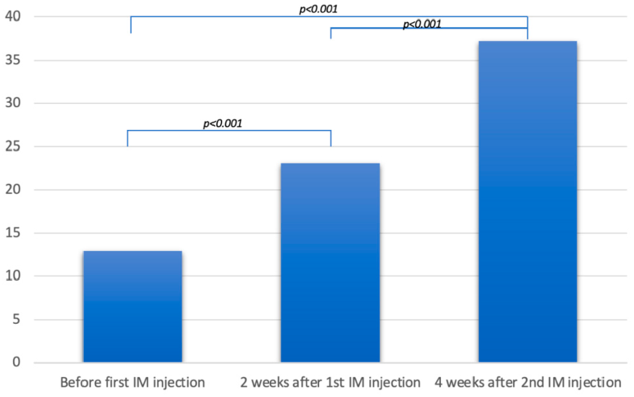
Objective: To assess the response to intramuscular vitamin D in patients not respond-ing to oral supplementation. Methods: A retrospective series included patients, with systemic sclerosis and a history of subclinical poor vitamin D status that was resistant to at least 6 months of oral supplementation, to whom intramuscular vitamin D2 was administered. Results: Twelve patients were identified, with a mean age of 47.8 years. All were women. Five had diffuse systemic sclerosis and seven had localized systemic sclerosis. The mean duration of the disease was 17.9 years, with a mean modified Rod-nan skin score of 14.9. All patients were twice injected, at a 15-day interval, 300,000 IU of ergocalciferol into the anterior gluteus muscle. The mean serum level of 25(OH)D increased from 12.9 ng/mL before the first injection, to 23 ng/mL two weeks after the first injection, and 37.1 ng/mL four weeks after the second injection (p<0.001). No side effects were observed. Conclusion: It is the first report of safely normalizing vitamin D levels with intramuscular ergocalciferol in patients with systemic sclerosis.

Keywords: 
;  ;  ;  ;  

1. Introduction

Systemic sclerosis (SSc) is a connective tissue disease characterized by a progressive loss of the microvascular bed leading to progressive fibrotic modifications in the affected organs and tissues. Depending on skin involvement, it can be either limited cutaneous SSc (lcSSc), restricted to the face and to below elbows and knees, or diffuse cutaneous SSc (dcSSc) [1].
A remarkably high prevalence of vitamin D deficiency has been reported in patients with connective tissue disorders, such as systemic lupus erythematosus, granulomatosis with polyangiitis, and SSc [2,3]. Such deficiency could be multifactorial in SSc where disease can affect the different organs involved in normal vitamin D metabolism. The two primary sources of vitamin D consist of endogenous synthesis through skin sun exposure – which is the main source – and exogeneous dietary consumption [4]. In the epidermis, 7- dehydrocholesterol (or provitamin D3) transforms into previtamin D3 after exposure to UV rays, especially UVB [5,6]. Previtamin D3 is then converted to vitamin D3 (cholecalciferol) through a heat dependent reaction. Besides, ergocalciferol is absorbed from food in the digestive tract, [4] especially in the small intestine [7]. Dietary sources include supplements and food such as milk, dairy products, fish, meat, poultry, eggs, and sweets. [8] Both cholecalciferol and ergocalciferol enter the bloodstream and are stored in the liver as 25-hydroxyvitamin D (25(OH)D) after being hydroxylated by the liver 25-alpha-hydroxylase. In order to produce the biologically active form of vitamin D, 25(OH)D undergoes a second hydroxylation in various tissues by the 1-alpha-hydroxylase, resulting in the formation of 1,25-dihydroxyvitamin D (calcitriol or 1,25(OH)2D) [9,10]. Many factors can contribute to vitamin D deficiency in SSc patients. First, dermal fibrous thickening and capillary damage may impact 7- dehydrocholesterol conversion to pre-vitamin D3. Serum levels of 25(OH)D were inversely correlated to skin thickness, being significantly lower in dcSSc compared to lcSSc patients [11]. Second, vascular damage, nerve dysfunction, smooth muscle atrophy, and intestinal wall fibrosis cause hypomotility and small intestinal bacterial growth, leading to subclinical malabsorption of nutrients and vitamins [12,13]. Third, physical impairment with a sedentary lifestyle reduces exposure to sunlight. Furthermore, it was suggested, though not confirmed, that lower vitamin D serum levels correlate with SSc severity or activity [2,14,15,16]. Conversely, in recent experiments on mouse cells, vitamin D has been proven to have a direct antifibrotic action. It inhibits the production of TGFβ1, a pro-fibrotic cytokine, as well as the synthesis of collagen I and collagen III in mesenchymal multipotent cells while increasing the expression of anti-fibrotic factors such as matrix metalloproteinase 8 [17]. Hence, it is still unsure whether hypovitaminosis D is a risk factor or a consequence of SSc [11].
The oral route is considered the most common way of vitamin D supplementation [18]. However, it did not show significant improvement in vitamin D levels in SSc patients [2]. To our knowledge, many studies have analyzed the frequent vitamin D deficiency in systemic sclerosis. Still, none has yet reported an efficient therapy or evaluated the effect of intramuscular (IM) vitamin D supplementation [2,11,14,15,19]. Herein, we report a retrospective series of patients, with SSc and low vitamin D not responding to oral supplementation, to whom IM vitamin D was given.

2. Methods

2.1. Patients

The study was retrospective. The population consisted of patients examined at our dermatology department between 2018 and 2022. We included those with the following criteria: (i) SSc diagnosis according to the ACR/EULAR 2013 classification [1], (ii) age between 30 and 65 years, (iii) subclinical 25(OH) D serum level < 30 ng/ml after at least 6 months of oral supplementation, (iv) administration of IM vitamin D during the study period.

2.2. Data Collection

The following data were retrieved from the patient’s file: age, sex, SSc type, disease duration, dose, and duration of oral vitamin D3 cure, serum levels of 25(OH) D, calcium, phosphorus, and parathormone whenever measured before, during, or after IM injection of vitamin D2, the posology of IM vitamin D2, and the report of any systemic side effects or reactions at the point of injection.

2.3. Protocol

All patients were injected 300,000 IU of ergocalciferol or vitamin D2 (Stérogyl 15 “H”; 600,000 IU/ 1.5 ml; Desma Pharma, France) into the anterior gluteus muscle. A similar injection was administered 15 days later. Blood samples were taken 3 times: first, to check 25(OH)D serum level after at least 6 months of oral supplementation; second, to check 25(OH)D serum level, 2 weeks after the first injection, and third, to check 25(OH)D, calcium, phosphorus, and parathormone serum level, 4 weeks after the second injection.

2.4. Measurement of Serum 25(OH)D

According to the manufacturer's protocol, the 25(OH)D serum level was invariably measured on peripheral blood samples by the Liaison® chemiluminescence immunoassay, DiaSorin Inc., Italy. We have considered all patients with levels inferior to 30ng/ml as having a poor status, without subdividing them into deficiency and insufficiency ranges.

2.5. Statistical Analysis

Statistical analysis was performed using SPSS (version 29.0.2). Repeated measures ANOVA with Bonferroni post hoc test were used to find significant difference between serum levels of 25(OH)D before the 1st IM injection, 2 weeks after the 1st IM injection and 4 weeks after the 2nd IM injection of ergocalciferol. P values < 0.05 were accepted as significant.

2.6. Patient Consent

The observational retrospective design to share anonymously our experience over five years with patients who have received intramuscular ergocalciferol in the department did not need the ethics committee's approval. Treatment delivery and data collection were conducted in compliance with the code of ethics of the World Medical Association (Declaration of Helsinki). Patients first signed a written consent to treatment and later a second written consent for eventual publication when we started to retrospectively collect the data.

3. Results

Of 36 patients screened with SSc, thirty were between 30 and 65 years old. Twenty patients had 25(OH) D serum levels < 30 ng/ml, of whom 16 were still low after at least 6 months of oral supplementation, which invariably consisted of weekly 10,000 UI of cholecalciferol capsules. Finally, we identified 12 of these patients who were administered IM vitamin D. All were women with a mean age of 47.8 years. Five had dcSSc and seven had lcSSc. The mean duration of the disease was 17.9 years.
The repeated measures ANOVA showed differences between serum levels of 25(OH)D before the 1st injection, 2 weeks after the 1st injection and 4 weeks after the 2nd injection (F (1.2, 13.15) = 87.5, p < 0.001, η2 = 0.81). Differences occurred between serum 25(OH)D levels before and 2 weeks after the 1st injection (p < 0.001, d = 2.6), before and 4 weeks after the 2nd injection (p <0.001, d = 2.9) and 2 weeks and 4 weeks after the 2nd injection of ergocalciferol (p < 0.001, d = 2.4) (Fig. 1). All observed effect sizes are large according to Cohen 1992 [20].
Table 1 displays the patients’ characteristics and the different serum levels of 25(OH)D during the treatment. All calcium, phosphorus, and parathormone serum levels were normal. No clinical side effect was reported in patients’ follow-ups.

4. Discussion

We report the efficacy and safety of IM ergocalciferol in a small series of patients with SSc and a low vitamin D level refractory to oral supplementation.
To our knowledge, it is the first shared experience of normalizing vitamin D levels with IM injection in patients having a connective tissue disease and a poor vitamin D status refractory to oral supplementation. Such a route of administration was reported in vitamin D-deficient diabetic patients [21]. We thought to use it on our first patient in 2018; the good response and safety led us to try the same treatment on other patients.
The limitations of this study include a retrospective observation with a small sample size, a short follow-up, and restricted clinical data. Although the poor vitamin D status was subclinical, we thought, based on literature data on the possible interaction between low vitamin D and disease status, that rapidly raising the level would be theoretically beneficial. We have investigated the improvement of vitamin D levels only, without other scleroderma-related manifestations which would have carried more weight to this study. Furthermore, the patients were not thoroughly investigated for the precise cause of low vitamin D like abnormal gastrointestinal absorption, renal disease, menopausal status, concomitant medication, or poor drug compliance. Finally, higher doses of oral vitamin D are recommended in patients with malabsorption, like after bariatric surgery; up to 6000IU of daily vitamin D3 are preferred to up to 50.000IU of 3 times weekly vitamin D2 [22]. We could have tested this recommendation on our patients, but we considered the IM route safer and of lower cost.
The most efficient replacement therapy has been the subject of years of debate [23]. If a choice can be made, recent guidance prefers vitamin D3 to vitamin D2 in oral supplementation [24]. Equal efficacy and safety were shown in daily, weekly, or monthly dosing regimens [25]. Compared to the IM route, higher and earlier peaks are observed with the oral route [26,27]. Compared to IM cholecalciferol, lower peaks were seen with IM ergocalciferol. For our patients, ergocalciferol was the only IM vitamin D available. The vial dose was divided into two halves at a 15-day interval to reduce the risk of side effects.
IM vitamin D injections have shown promising results. In healthy Korean adults with vitamin D deficiency, a single injection of 200,000 IU of vitamin D3 raised, within 12 weeks, the 25(OH)D levels above 20 ng/mL and 30 ng/mL in around 90% and 50% of individuals, respectively [28]. Moreover,10 adults with a variety of past and present medical conditions received 600 000 IU of IM vitamin D3; blood samples showed a peak of 25(OH)D at 4 weeks for most participants and the levels were still remarkably higher than baseline at 24 weeks [18]. Similarly, Wylon et al. compared the pharmacokinetic evolution of a single 100 000 IU IM dose of cholecalciferol to that of an 84-day oral supplementation; serum 25(OH)D level peaked one week after the first dose of oral cholecalciferol and 4 weeks after the IM injection. Unlike the IM route, the fast rise with the oral route was transitory. The sustained levels following IM injection were explained by a delayed release along with the storage capacity of vitamin D in the adipose tissue [26]. Likewise, Gupta et al. compared the mean serum 25(OH)D levels in 2 healthy groups with vitamin D deficiency. Twenty weekly received 60,000 IU of cholecalciferol for 5 weeks and twenty others received a single IM injection of 300,000 IU of cholecalciferol. At 12 weeks, levels were significantly higher in the second group (25.46 ± 1.37 vs. 16.66 ± 1.36 ng/mL; p<0.001), hence showing a sustained increase from baseline [12].
In conclusion, the IM administration of vitamin D2 showed promising results in correcting a refractory low level, though subclinical, in patients with systemic sclerosis. Based on the response of our patients and the results of other studies of IM vitamin D in healthy subjects, close monitoring of 25(OH)D in the blood can help define the frequency of supplementation. Moreover, extrapolating this delivery route to other diseases with subclinical malabsorption is expected to be promising. This case series is not the strongest source of evidence; however, it provides descriptive data that contributes to generating more precise hypotheses to test.

Author Contributions

All authors were involved in drafting or revising the article critically for important intellectual content, and all authors approved the final version to be published. JH and BS had full access to all the data in the study and took responsibility for the integrity of the data and the accuracy of the data analysis. Study conception and design: JH, BS. Acquisition of data FM, MA, RJ, SH, BS, JH. Analysis and interpretation of data FM, MA, RJ, SH, BS, JH.

Consent to participate

Patients have signed an informed consent form before every injection.

Written Consent for publication

Patients have signed a consent form for eventual publication.

Data Availability Statement

data are available upon request.

Acknowledgments

A preprint of this manuscript has previously been published: Farid Mallat, Maroun Aoun, Rita Jabbour et al. Subclinical low vitamin D related to systemic sclerosis and resistant to long-term oral supplementation rapidly improves after two intramuscular injections of ergocalciferol: a retrospective observational case series., 01 August 2023, PREPRINT (Version 1) available at Research Square [https://doi.org/10.21203/rs.3.rs-3093126/v1].

Conflicts of Interest

The authors have stated explicitly that there are no conflicts of interest in connection with this article.

Abbreviations

IM intramuscular; SSc Systemic sclerosis; lcSSc limited cutaneous systemic sclerosis; dsSSc diffuse cutaneous systemic sclerosis.

References

  1. van den Hoogen F, Khanna D, Fransen J, Johnson SR, Baron M, Tyndall A, et al. 2013 classification criteria for systemic sclerosis: an American College of Rheumatology/European League against Rheumatism collaborative initiative. Arthritis Rheum 2013;65:2737–47. [CrossRef]
  2. Giuggioli D, Colaci M, Cassone G, Fallahi P, Lumetti F, Spinella A, et al. Serum 25-OH vitamin D levels in systemic sclerosis: analysis of 140 patients and review of the literature. Clin Rheumatol 2017;36:583–90. [CrossRef]
  3. Diaconu A-D, Ostafie I, Ceasovschih A, Șorodoc V, Lionte C, Ancuța C, et al. Role of Vitamin D in Systemic Sclerosis: A Systematic Literature Review. J Immunol Res 2021;2021:9782994. [CrossRef]
  4. Kechichian E, Ezzedine K. Vitamin D and the Skin: An Update for Dermatologists. Am J Clin Dermatol 2018;19:223–35. [CrossRef]
  5. Chen TC, Chimeh F, Lu Z, Mathieu J, Person KS, Zhang A, et al. Factors that influence the cutaneous synthesis and dietary sources of vitamin D. Arch Biochem Biophys 2007;460:213–7. [CrossRef]
  6. Tripkovic L, Lambert H, Hart K, Smith CP, Bucca G, Penson S, et al. Comparison of vitamin D2 and vitamin D3 supplementation in raising serum 25-hydroxyvitamin D status: a systematic review and meta-analysis. Am J Clin Nutr 2012;95:1357–64. [CrossRef]
  7. Bikle DD. Vitamin D Insufficiency/Deficiency in Gastrointestinal Disorders. Journal of Bone and Mineral Research 2007;22:V50–4. [CrossRef]
  8. Gannagé-Yared M-H, Chemali R, Sfeir C, Maalouf G, Halaby G. Dietary calcium and vitamin D intake in an adult Middle Eastern population: food sources and relation to lifestyle and PTH. Int J Vitam Nutr Res 2005;75:281–9. [CrossRef]
  9. Lucas RM, Gorman S, Geldenhuys S, Hart PH. Vitamin D and immunity. F1000Prime Rep 2014;6:118. [CrossRef]
  10. Wharton B, Bishop N. Rickets. Lancet 2003;362:1389–400. [CrossRef]
  11. Corrado A, Colia R, Mele A, Di Bello V, Trotta A, Neve A, et al. Relationship between Body Mass Composition, Bone Mineral Density, Skin Fibrosis and 25(OH) Vitamin D Serum Levels in Systemic Sclerosis. PLoS One 2015;10:e0137912. [CrossRef]
  12. Gupta N, Farooqui KJ, Batra CM, Marwaha RK, Mithal A. Effect of oral versus intramuscular Vitamin D replacement in apparently healthy adults with Vitamin D deficiency. Indian J Endocrinol Metab 2017;21:131–6. [CrossRef]
  13. Marie I, Ducrotté P, Denis P, Menard J-F, Levesque H. Small intestinal bacterial overgrowth in systemic sclerosis. Rheumatology (Oxford) 2009;48:1314–9. [CrossRef]
  14. Arnson Y, Amital H, Agmon-Levin N, Alon D, Sánchez-Castañón M, López-Hoyos M, et al. Serum 25-OH vitamin D concentrations are linked with various clinical aspects in patients with systemic sclerosis: a retrospective cohort study and review of the literature. Autoimmun Rev 2011;10:490–4. [CrossRef]
  15. Trombetta AC, Smith V, Gotelli E, Ghio M, Paolino S, Pizzorni C, et al. Vitamin D deficiency and clinical correlations in systemic sclerosis patients: A retrospective analysis for possible future developments. PLoS One 2017;12:e0179062. [CrossRef]
  16. An L, Sun M-H, Chen F, Li J-R. Vitamin D levels in systemic sclerosis patients: a meta-analysis. Drug Des Devel Ther 2017;11:3119–25. [CrossRef]
  17. Artaza JN, Norris KC. Vitamin D reduces the expression of collagen and key profibrotic factors by inducing an antifibrotic phenotype in mesenchymal multipotent cells. J Endocrinol 2009;200:207–21. [CrossRef]
  18. Gorman S, Zafirau MZ, Lim EM, Clarke MW, Dhamrait G, Fleury N, et al. High-Dose Intramuscular Vitamin D Provides Long-Lasting Moderate Increases in Serum 25-Hydroxvitamin D Levels and Shorter-Term Changes in Plasma Calcium. J AOAC Int 2017;100:1337–44. [CrossRef]
  19. Zhang L, Duan Y, Zhang T-P, Huang X-L, Li B-Z, Ye D-Q, et al. Association between the serum level of vitamin D and systemic sclerosis in a Chinese population: a case control study. Int J Rheum Dis 2017;20:1002–8. [CrossRef]
  20. Cohen J. Statistical Power Analysis. Curr Dir Psychol Sci 1992;1:98–101. [CrossRef]
  21. Dwivedi A, Gupta B, Tiwari S, Pratyush DD, Singh S, Singh SK. Parenteral vitamin D supplementation is superior to oral in vitamin D insufficient patients with type 2 diabetes mellitus. Diabetes Metab Syndr 2017;11 Suppl 1:S373–5. [CrossRef]
  22. Mechanick JI, Apovian C, Brethauer S, Garvey WT, Joffe AM, Kim J, et al. CLINICAL PRACTICE GUIDELINES FOR THE PERIOPERATIVE NUTRITION, METABOLIC, AND NONSURGICAL SUPPORT OF PATIENTS UNDERGOING BARIATRIC PROCEDURES - 2019 UPDATE: COSPONSORED BY AMERICAN ASSOCIATION OF CLINICAL ENDOCRINOLOGISTS/AMERICAN COLLEGE OF ENDOCRINOLOGY, THE OBESITY SOCIETY, AMERICAN SOCIETY FOR METABOLIC & BARIATRIC SURGERY, OBESITY MEDICINE ASSOCIATION, AND AMERICAN SOCIETY OF ANESTHESIOLOGISTS - EXECUTIVE SUMMARY. Endocr Pract 2019;25:1346–59. [CrossRef]
  23. Sanders KM, Seibel MJ. Therapy: New findings on vitamin D3 supplementation and falls - when more is perhaps not better. Nat Rev Endocrinol 2016;12:190–1. [CrossRef]
  24. Camacho PM, Petak SM, Binkley N, Diab DL, Eldeiry LS, Farooki A, et al. AMERICAN ASSOCIATION OF CLINICAL ENDOCRINOLOGISTS/AMERICAN COLLEGE OF ENDOCRINOLOGY CLINICAL PRACTICE GUIDELINES FOR THE DIAGNOSIS AND TREATMENT OF POSTMENOPAUSAL OSTEOPOROSIS-2020 UPDATE. Endocr Pract 2020;26:1–46. [CrossRef]
  25. Fassio A, Adami G, Rossini M, Giollo A, Caimmi C, Bixio R, et al. Pharmacokinetics of Oral Cholecalciferol in Healthy Subjects with Vitamin D Deficiency: A Randomized Open-Label Study. Nutrients 2020;12:E1553. [CrossRef]
  26. Wylon K, Drozdenko G, Krannich A, Heine G, Dölle S, Worm M. Pharmacokinetic Evaluation of a Single Intramuscular High Dose versus an Oral Long-Term Supplementation of Cholecalciferol. PLoS One 2017;12:e0169620. [CrossRef]
  27. Yu SB, Lee Y, Oh A, Yoo H-W, Choi J-H. Efficacy and safety of parenteral vitamin D therapy in infants and children with vitamin D deficiency caused by intestinal malabsorption. Ann Pediatr Endocrinol Metab 2020;25:112–7. [CrossRef]
  28. Choi HS, Chung Y-S, Choi YJ, Seo DH, Lim S-K. Efficacy and safety of vitamin D3 B.O.N intramuscular injection in Korean adults with vitamin D deficiency. Osteoporos Sarcopenia 2016;2:228–37. [CrossRef]
Figure 1. Mean serum 25(OH)D levels before ergocalciferol injection, 2 weeks after the first IM injection, and 4 weeks after the second IM injection.
Preprints 146158 g001
Table 1. Patients’ characteristics and serum levels of 25(OH)D before the first IM injection, 2 weeks after the first IM injection, and 4 weeks after the second IM injection of ergocalciferol; SSc systemic sclerosis, IM: intramuscular, F: female.
Patient Age (years) Sex SSc type Disease duration
(years)
Modified Rodnan skin score Previous cure with oral vitamin D3 (cholecalciferol) Serum level of 25(OH)D (ng/mL)
Dose (IU/week) Duration (months) Before the first IM injection 2 weeks after the first IM injection 4 weeks after the second IM injection
1 50 F systemic 17 22 10.000 6 12 27 41
2 54 F localized 21 12 10.000 6 09 21 47
3 62 F systemic 30 28 10.000 6 14 23 34
4 41 F localized 10 8 10.000 6 12 30 47
5 49 F localized 18 10 10.000 6 21 29 36
6 60 F localized 25 13 10.000 6 10 21 45
7 40 F systemic 15 18 10.000 6 12 25 39
8 43 F systemic 17 20 10.000 6 11 21 31
9 51 F localized 28 15 10.000 6 6 15 29
10 35 F systemic 12 12 10.000 8 18 22 32
11 49 F localized 13 8 10.000 6 15 21 29
12 39 F localized 9 13 10.000 7 15 22 36
Mean 47.8 17.9 14.9 6 12.9 23 37.1
Disclaimer/Publisher’s Note: The statements, opinions and data contained in all publications are solely those of the individual author(s) and contributor(s) and not of MDPI and/or the editor(s). MDPI and/or the editor(s) disclaim responsibility for any injury to people or property resulting from any ideas, methods, instructions or products referred to in the content.
Copyright: This open access article is published under a Creative Commons CC BY 4.0 license, which permit the free download, distribution, and reuse, provided that the author and preprint are cited in any reuse.
Prerpints.org logo

Preprints.org is a free preprint server supported by MDPI in Basel, Switzerland.

Subscribe

Disclaimer

Terms of Use

Privacy Policy

Privacy Settings

© 2025 MDPI (Basel, Switzerland) unless otherwise stated