Preprint
Article

This version is not peer-reviewed.

GEF1 Is a Novel Factor Regulating Nickel Ion Metabolism in Saccharomyces cerevisiae

Submitted:

14 January 2025

Posted:

14 January 2025

You are already at the latest version

Abstract

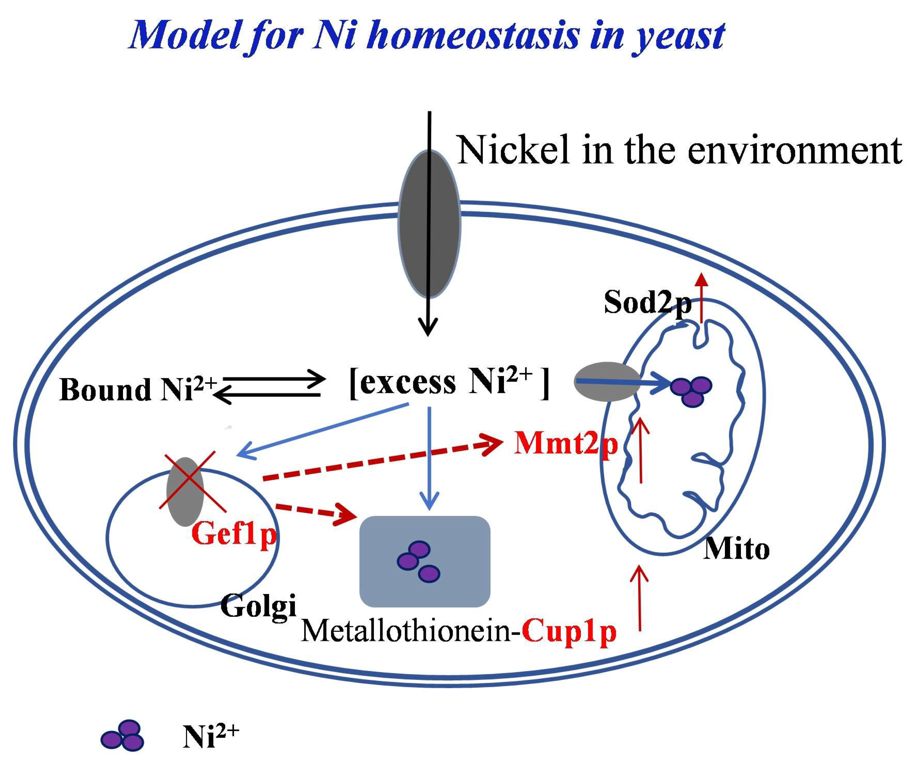
Nickel is a trace element essential for human life. Currently, studies on the metabolism and function of nickel ions mainly focus on prokaryotes, while the role of nickel ions in eukaryotes is still being explored. In this study, we found for the first time that GEF1 which encodes Gef1p, a chloride transport protein located in the Golgi membrane, is involved in nickel ion metabolism in yeast. The GEF1 knockout strain (gef1∆) showed strong resistance to excess nickel ions, and the content of nickel ions in gef1∆ cells was significantly elevated. The results of transcriptomics analysis showed significant upregulation of MMT2 and CUP1 in gef1∆ cells supplemented with nickel. Both MMT2 and CUP1 overexpressed in the gef1∆ strain showed a growth advantage on nickel media. Nickel ion content in the mitochondria of cells overexpressing MMT2 was significantly elevated, and the levels of reactive oxygen species were significantly decreased in strains overexpressing either the MMT2 or CUP1 genes. This study reveals that the GEF1 gene plays an important role in nickel homeostasis, and that upregulation of MMT2 and CUP1 is critical for gef1∆ strains to counteract ROS formation and growth defect by nickel ions.

Keywords: 
;  ;  ;  ;  

1. Introduction

Nickel is a necessary metal for plants, animals and microorganisms to participate in various physiological activities, and is also a necessary micronutrient for the human body[1]. Adults consume approximately 79 to 105 μg/day of nickel from diet and supplements[2]. After being ingested into human body, nickel resides in various organs such as the brain[3], liver and heart[4], where it is believed to play a role in the normal physiological activities of the human body. Nickel is redox active metal which is normally found in the +2 oxidation state. Excess nickel has detrimental effects through its action as a ROS generator. For example, nickel inhibits differentiation of neuronal cells[5], causes premature ovarian failure by inhibiting cell proliferation[6], and causes oxidative damage to the testis which inhibits sperm formation[7]. Nickel was shown to inhibit the proliferation of cultured immune cells[8], and reversal of this inhibition is of great interest for the prevention and treatment of nickel-induced cancer. On the other hand, many microbial enzymes depend on the presence of nickel for their functions, such as urease, methyl coenzyme M reductase, CO-dehydrogenase, nickel superoxide dismutase, glyoxalase, acireductase dioxygenase, lactate racemase, prolyl cis-trans isomerase and [NiFe] hydrogenase[9,10]. Therefore, organisms must maintain nickel homeostasis to prevent toxicity but allow for its essential functions.
At present, two metabolic pathways of nickel have been found in microorganisms. One of the ways for microorganisms to absorb nickel is the nickel-specific ABC transporter found in Escherichia coli, which is composed of five proteins NikA, B, C, D and E and relies on ATP hydrolysis to provide energy. Other microorganisms have similar nickel transporters such as NixA, a high-affinity nickel transporter responsible Ni2+ uptake in Helicobacter pylori[11]. The second way for microorganisms to absorb nickel is by facilitated diffusion through nickel-specific permeases. There are few studies on nickel metabolism in higher eukaryotes such as humans or animals and no specific nickel-containing enzymes have been identified, despite nickel being recognized as an essential trace mineral. Therefore, it is particularly necessary to study the role of nickel in eukaryotes. Many genes are involved in the metabolism of nickel ions, and more related genes need to be discovered. Our preliminary data found that GEF1 showed significant resistance to nickel ions.
The GEF1 gene belongs to the Guanine nuclear exchange factor (GEF) family and encodes Gef1p, a multifunctional membrane protein located on the Golgi membrane of Saccharomyces cerevisiae cells. Previous studies in unicellular eukaryotes have shown that GEF1 is located on the cilia or matrix and may activate G proteins and find nuclear targeting sequences[12,13]. Greene JR et al. found that the 13 transmembrane domains encoded by GEF1 have high homology with chloride ion channel proteins of humans and mammals, suggest that GEF1 may encode a protein of intracellular iron metabolism[14]. Flis K et al. also found that Gef1p has high amino acid homology with the CLC chloride ion channel family[15]. Gaxiola et al. found that the deletion of GEF1 leads to increased iron demand and sensitivity to cations in cells[16]. López-Rodríguez A et al. found that the expression of GEF1 in HEK293 cells keeps chloride ion channels open, and are essential to the protein’s function[17]. Sasvari et al. found that Gef1p played a key role in the RNA replication of TBSV (Tomato bushy stunt virus), and verified in Saccharomyces cerevisiae that Gef1p knockout inhibited RNA replication of the virus by regulating intracellular copper[18]. At present, many functions of GEF1 have not been explored, and the metabolic regulation of nickel ions by this gene has not been reported in the literature. Therefore, it is of great significance to investigate the regulatory mechanism between GEF1 and nickel ions. This work found for the first time that GEF1 is related to nickel ion metabolism, which provides a new idea for the study of nickel metabolism in eukaryotes.
To better understand the function and metabolism of nickel in eukaryotes, we used a yeast knockout library to screen nickel-related genes in Saccharomyces cerevisiae with excessive nickel supplementation. In this paper, we found that GEF1 participated in the metabolic balance of nickel ions, a GEF1 knockout (gef1∆) strain showed resistance to nickel ions, and gef1∆ in the presence of nickel caused changes to the expression of several genes related to metal homeostasis. We identified MMT2 and CUP1 as the key factors for gef1∆-mediated resistance to nickel ions. MMT2 is a gene located in mitochondrial membrane whose function is related to export of iron ions out of the mitochondria[19], while CUP1 is a metallothionein, which is a metal-binding gene[20]. We showed that these genes reduce the content of bioactive nickel ions in the cytoplasm resulting in lower ROS and recovered cell growth, which provides a new perspective for elucidating the metabolism of nickel ions in eukaryotes.

2. Materials and Methods

2.1. Yeast Strains, Culture Media, and Growth Assays

A haploid control yeast S. cerevisiae strain, BY4741, and other knock out mutants (gef1Δ, mtm2Δ)[21]were purchased from the Open Biosystems. Yeast strains were grown in synthetic complete media lacking uracil (SC-ura) for plasmid selection as indicated in the figure legends. 1.5% agar was supplemented into the liquid media for solid medium plates. Yeast strains were cultured at 30 °C.
For yeast cell growth assays[22], wild-type (WT) or knock out cells transformed with either empty vector, MMT2 or CUP1 plasmids were grown overnight in SC-ura media, re-inoculated (OD600= 0.2) into fresh media, and grown to the mid-log phase (OD600= 0.8–1.0). After dilution to OD600= 0.1 and 3× serial dilutions in sterilized water, ~5 µl of cells were spotted on SC plates supplemented with metal at specified concentrations. Cells were incubated at 30 °C for 2-3 days prior to photography. Each assay was repeated at least three times using three different colonies to confirm results.

2.2. Plasmid Manipulation

GEF1 gene cloned by PCR was inserted into Bam HI and XhoI sites in the p416-TEF vector for TEF2 gene promoter-mediated constitutive expression in yeast[23]. The GFP epitope-tag was inserted at the C-terminus of Gef1p at a NotI restriction enzyme site which was generated in the PCR primer before the stop codon. Common molecular biology techniques, including plasmid amplification, purification using Escherichia coli and expression in S. cerevisiae, followed previously established methods[24]. All plasmids were sequenced to confirm inserted gene sequences. Yeast transformations were performed via the lithium acetate method[25].

2.3. Western Blot Nalysis

WT and gef1Δ cells expressing empty vector (p416-TEF) or GEF1 tagged with GFP were cultured in SC-ura media beginning at OD600 0.2 for 4 h. Total protein was prepared via glass bead disruption in phosphate-buffered saline (PBS) containing 1% Triton X-100 and protease inhibitor complex (Roche Applied Science). The BCA kit (Pierce) was used for protein concentration measurements following the manufacturer’s instructions. Total protein was denatured in SDS buffer containing 100 mM DTT (dithiothreitol) for 15 min at 60 °C and subjected to SDS-PAGE. Green fluorescent protein (GFP) epitope-tagged Gef1p was detected by anti-GFP antibody (Rockland) and the secondary antibody was anti-rabbit IgG antibody (Santa Cruz). The stained gel was used as a loading control.

2.4. Metal Measurement

Yeast cells were cultured to mid-log phase in SC media. 5 OD.600 cells (OD.600 = 5 cell amount) were collected and washed four times in PBS buffer with 10 mM EDTA to remove cell surface Ni. The washed cell pellets were dissolved in 70% nitric acid at 70 °C for 3 h. After overnight at room temperature, these samples were diluted in 10% nitric acid. ICP-MS (inductively coupled plasma mass spectrometry, Agilent Model 7500cs, Santa Clara, CA) was used to measure metal ion levels. Metal ion contents were normalized to protein concentrations[26].

2.5. Quantitative PCR

WT and gef1Δ cells were cultured in the SC media overnight. Cells were then re-inoculated (OD.600 0.2) into fresh media for 4h culture containing NiSO4 (0, 0.5, 1 mM). Total RNA was subjected to cDNA synthesis followed by quantitative PCR (qTOWER, 3.0 G) using primer sets that are specific for MMT2 (F:5’-AGC AGCGGGTTCTATCTTGG; R:5’-ACCAACGCTACCAACGATGT), CUP1 (F:5’-GCCAATGTGGTAGCTGCAAA; R:5’-TCAGACTTGTTACCGCAGGG) and ß-actin (F:5’-AAATGCAAACCGCTGCTCA-3’; R:5’-TACCGGCAGATTCCAA ACCC -3’).

2.6. Measurement of Intracellular ROS Level

Intracellular ROS levels were determined by nitroblue tetrazolium (NBT) assay with some modifications[27]. Briefly, WT and gef1Δ cells were cultured to mid-log phase with indicated NiSO4 treatments. Cells were incubated for 90 minutes in PBS containing 0.2% NBT before being collected. ROS reduced NBT to a dark blue insoluble form of NBT named formazan. The mixtures were centrifuged at 5,000 rpm for 2 min at 4°C. The supernatant was removed and the formazan was dissolved in 300 µl 50% acetic acid by sonication (three pulses of 6 s). The samples were shortly spun down and the absorbance of the supernatant was determined at 560 nm by a spectrometer.

2.7. Fluorescence Microscopy

Mid-log phase cells (p416TEF-GEF1-GFP) were cultured with and without NiSO4 in SC-ura media. Cells were collected by centrifugation and washed once with fresh media and then were washed once with phosphate-buffered saline by centrifugation. GFP-fused Gef1p signals were photographed on a confocal microscope (Olympus FV500). Differential interference contrast (DIC) images were also captured to present cell morphology. GFP signals and cell images were overlaid to determine GEF1-GFP subcellular distribution[26].

2.8. Statistical Analysis

The image quantification was determined by ImageJ software (http://rsbweb.nih.gov/ij/). Descriptive analyses were presented as the means ±S.D. and statistical comparisons of control and experimental groups were performed using Student’s t-test. p<0.05 was considered to be significant.

3. Results

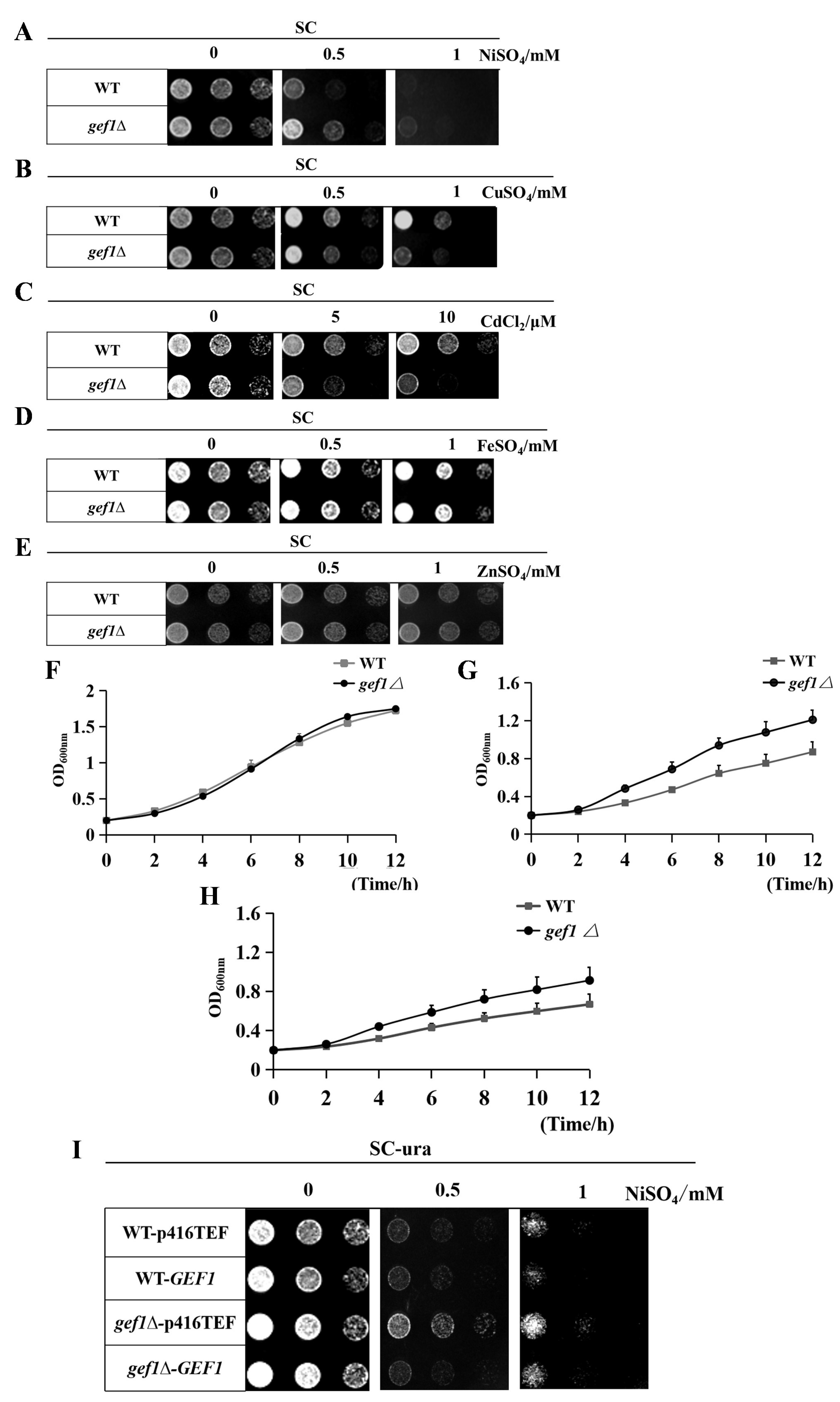
3.1. Gef1p Is a New Molecular Factor Involved in Nickel Ion Metabolism in Saccharomyces cerevisiae

In order to study the metabolic mechanism of nickel ion in S. cerevisiae and screen genes related to nickel ion metabolism, we prepared SC solid media containing 0 mM, 0.5 mM and 1 mM NiSO4 respectively, then observed the growth of gene-deficient strains on the media over 3 days. GEF1 gene knockout cells (gef1Δ) grew better than WT cells in SC solid medium containing 0.5 mM and 1 mM nickel ions, which demonstrates gef1Δ cells had resistance to nickel ion toxicity (Figure 1A). Next, we confirmed the specificity of the phenotype by repeating the experiment using a dose curve of several other heavy metal ions. Gef1Δ cells were sensitive to both copper ions and cadmium ions, and their growth trend was noticeably worse than that of WT cells when 0.5 mM copper (Figure 1B) or 5 µM cadmium (Figure 1C) was added. However, iron ions (Figure 1D) and zinc ions (Figure 1E) had little effect on gef1∆ growth. Therefore, we showed the ion resistance phenotype of the gef1Δ strain was specific to nickel ions. To further verify the experimental results, we prepared SC liquid culture medium with nickel ion concentrations of 0 mM, 0.5 mM and 1 mM respectively, then observed the growth of WT and gef1Δ cells. In SC liquid medium without nickel ions, the growth of WT and gef1Δ cells were almost the same (Figure 1F). After adding 0.5 mM nickel ion, the growth of gef1Δ cells in SC liquid medium was much higher than that of WT cells (Figure 1G). The growth trend of gef1Δ cells was still better than that of WT cells in SC liquid medium containing 1 mM nickel ions (Figure 1H). Adding excessive nickel ions promoted the growth of gef1Δ cells, which was consistent with the results of the solid growth experiment and further confirmed that gef1Δ cells are resistant to nickel ions. Next, we cloned the GEF1 gene, transformed it into WT and gef1Δ cells, and observed their growth trends under the condition of excessive nickel. Gef1∆ cells still showed resistance to nickel ions after being transformed with the empty plasmid. However, after expressing the GEF1 gene in gef1Δ cells, the cells became sensitive to nickel (Figure 1I). These results collectively indicate that there is a regulatory mechanism between the GEF1 gene and nickel ion homeostasis.

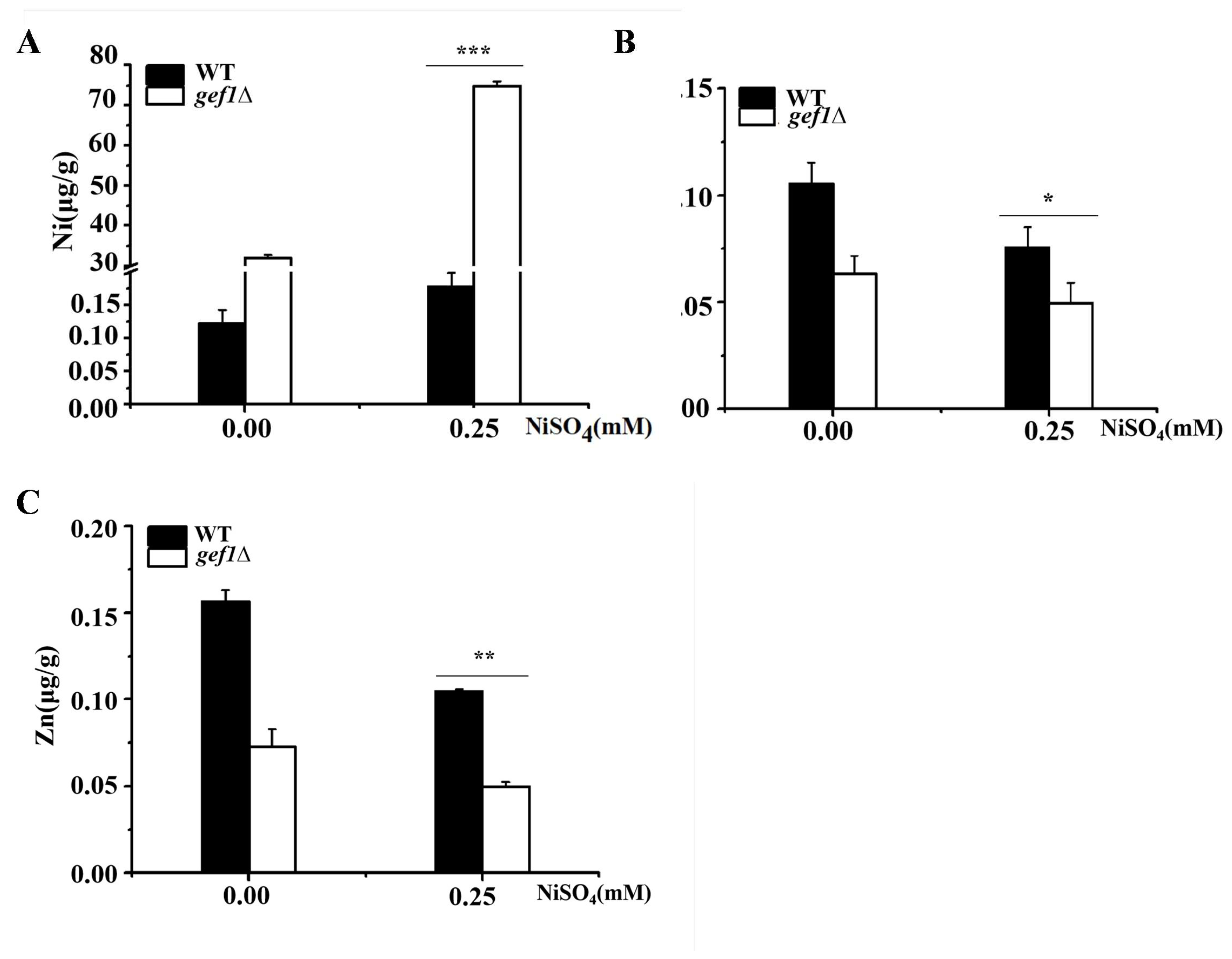
3.2. GEF1 Deletion Significantly Increases the Nickel Content in Cells Under Condition of Nickel Excess

We suspected that the reason for the resistance of gef1Δ strain was that nickel ions in the culture medium would supplement the deficiency of nickel ions in gef1Δ cells. Therefore, we used ICP-MS to detect the changes in nickel and other divalent metal ions in cells cultured with and without supplemental nickel. The content of nickel in gef1Δ cells supplemented with nickel increased relative to WT controls (Figure 2A). However, the contents of iron (Figure 2B) and zinc (Figure 2C) decreased due to competitive absorption with nickel. Taken together, these data indicate that GEF1 has a regulatory effect on nickel ions in yeast cells.

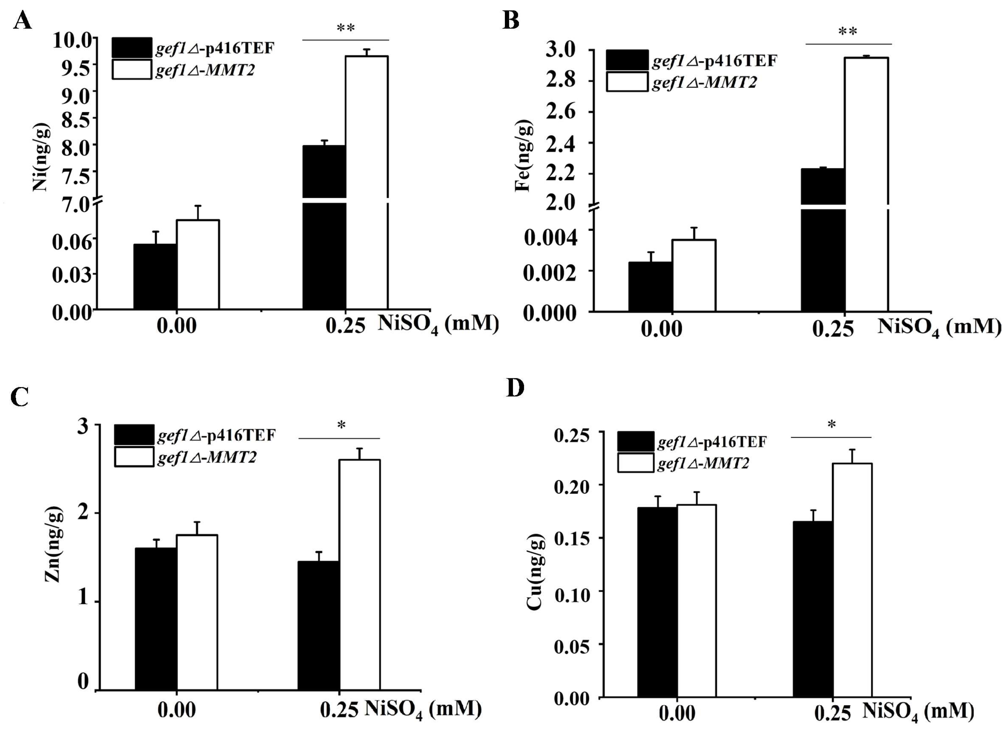
3.3. MMT2 Transports Excess Nickel to Mitochondria and Changes the Distribution of Nickel Ions

Given GEF1 is not known to directly affect nickel, we supposed that changes to gene expression in gef1Δ cells exposed to nickel was responsible for mediating nickel resistance. Therefore, we performed transcriptome sequencing to determine changes in gene expression resulting from supplemented nickel in gef1Δ cells. We assessed the related functions of differentially expressed genes and selected nine Ni-related genes which are all related to metal ion metabolism to test mRNA expression levels in WT and gef1Δ cells under supplemental nickel ions. Compared with the WT cells, the expression levels of FRE2, FIT2, ARN2, FET3, FRE1, MMT2, ARN1, FTR1 and CUP1 were all significantly increased (Figure 3). Of these 9, MMT2 and CUP1 had exceptionally large increase in expression relative to WT controls and therefore were prioritized for further study. The MMT2 gene is located on the mitochondrial membrane where it functions to export iron ions from the mitochondria into the cytosol[28]. According to previous research in our laboratory, most of the genes that transport iron ions can also transport nickel ions[29], so we speculated that MMT2 can transport nickel ions from the outside of mitochondria to the inside. To verify this conjecture, we used ICP-MS to detect the content of nickel ions in mitochondria when MMT2 gene was overexpressed in WT and gef1∆ cells, respectively. Compared with the overexpression of empty plasmid in gef1Δ cells treated with nickel ion, the content of nickel ion in the mitochondria of the gef1Δ cells with MMT2 gene overexpressed increased significantly (Figure 4A). Moreover, the contents of iron, zinc and copper also increased (Figure 4B,C,D). It can be concluded that Mmt2p can transport nickel ions from cytoplasm to mitochondria, thus reducing the toxic effect of nickel ions on cells, which is one of the reasons gef1Δ cells are resistant to nickel ions.

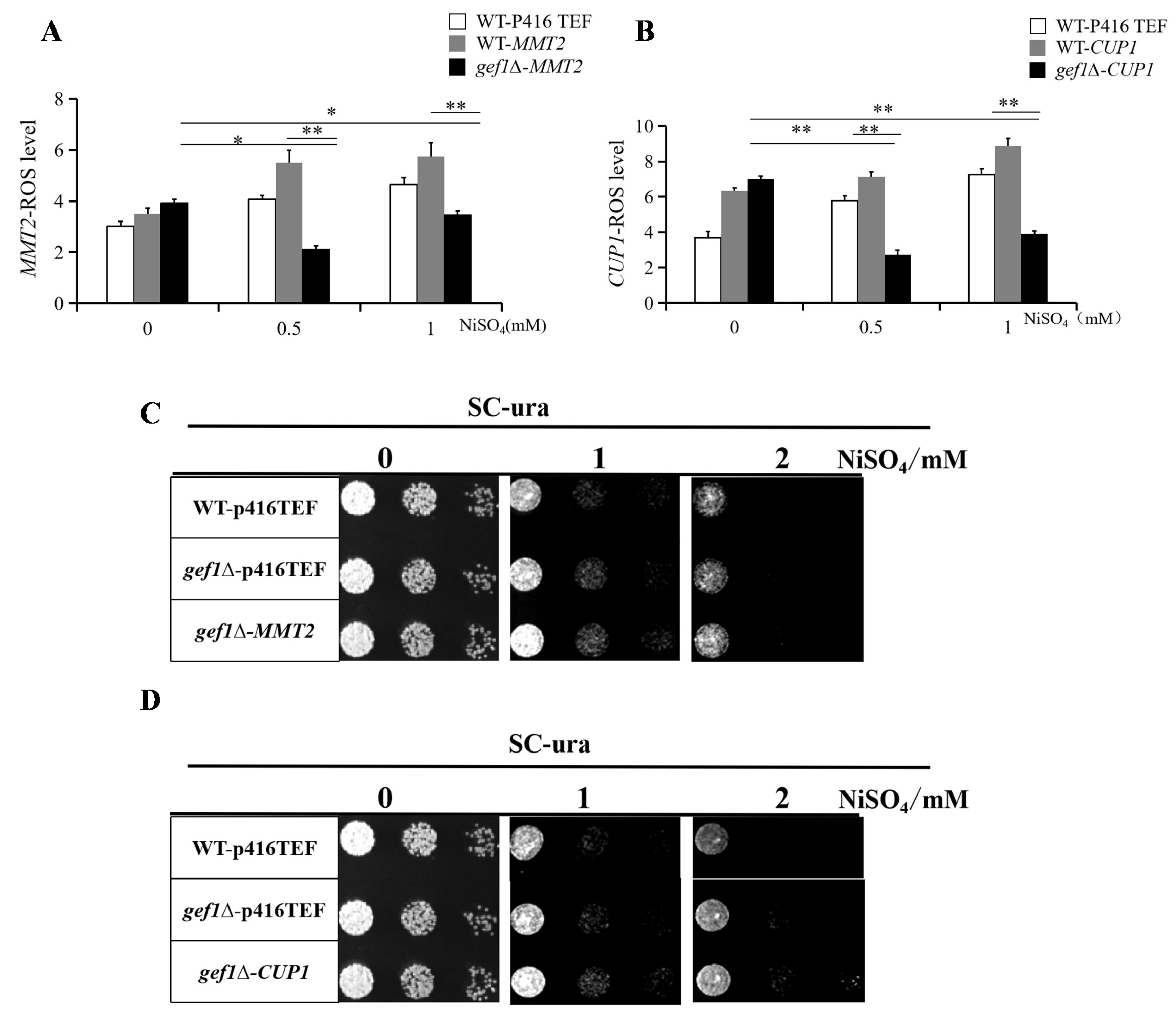
3.4. Over-Expression of MMT2/CUP1 Reduces Intracellular ROS Level

CUP1 encodes a metallothionein which binds copper and other intracellular metals. We suspected that the upregulation of GEF1 gene expression in gef1∆ cells led to an increase in bound nickel ions, thus reducing the content of ROS-producing free nickel ions. Furthermore, by transporting nickel ions into the mitochondria, MMT2 was predicted to mitigate ROS production, potentially through increase in nickel-requiring Sod2p antioxidant enzyme activity [26]. Therefore, we overexpressed MMT2 and CUP1 in gef1Δ cells treated with excessive nickel ions and analyzed intracellular ROS levels. The ROS levels in both knockouts decreased (Figure 5A,B) compared to WT controls which demonstrated the ability of genes upregulated by gef1Δ+nickel to protect cells from nickel-mediated ROS. As further evidence, we overexpressed MMT2 and CUP1 in gef1Δ grown on nickel-supplemented plates to assess cell growth. The MMT2 overexpressing gef1Δ cells grew better than control strains (Figure 5C). The growth of CUP1 overexpressing gef1Δ cells was also better than that of control cells (Figure 5D). The superior growth of both overexpression cells supports the finding of decreased ROS relative to controls.

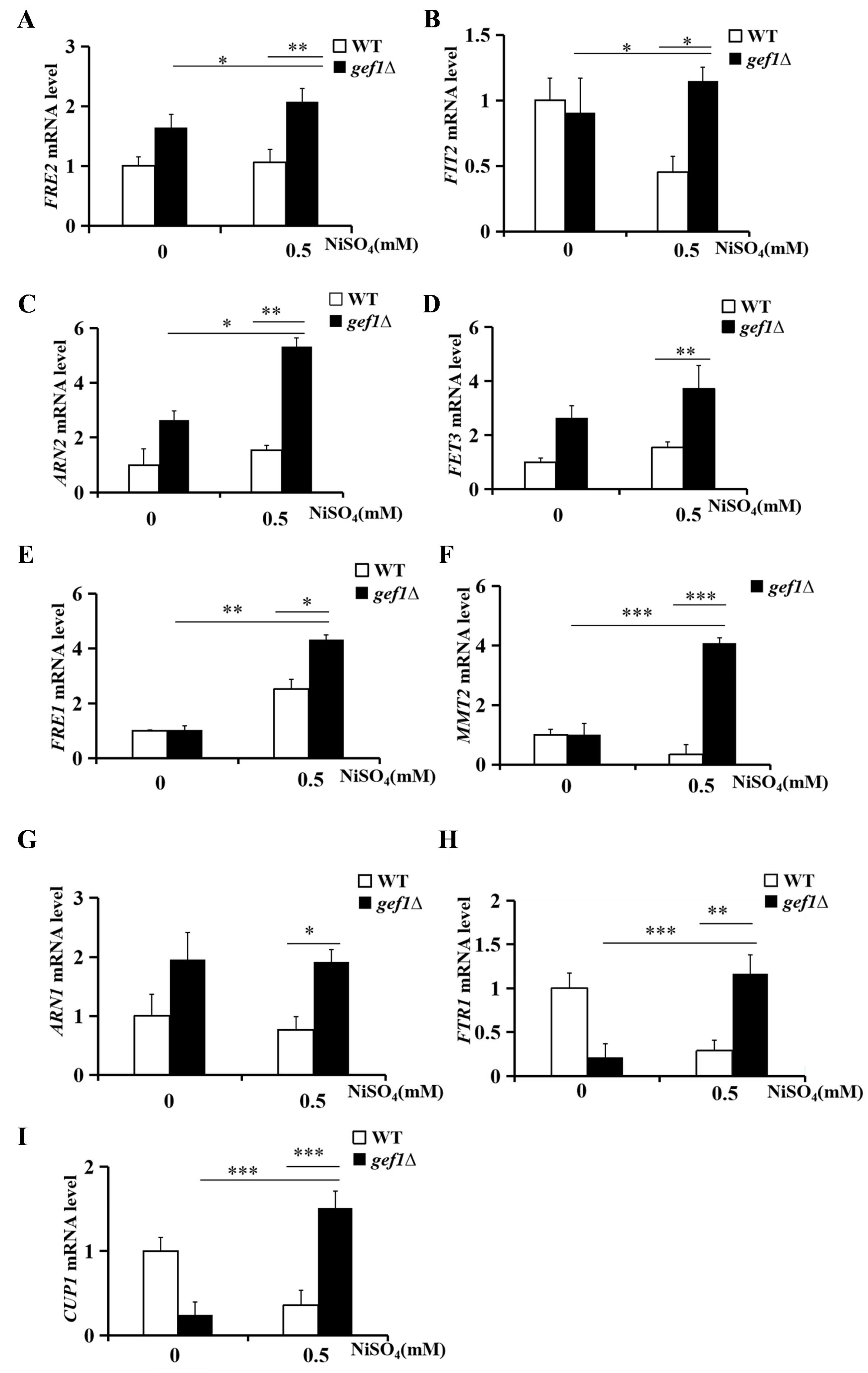
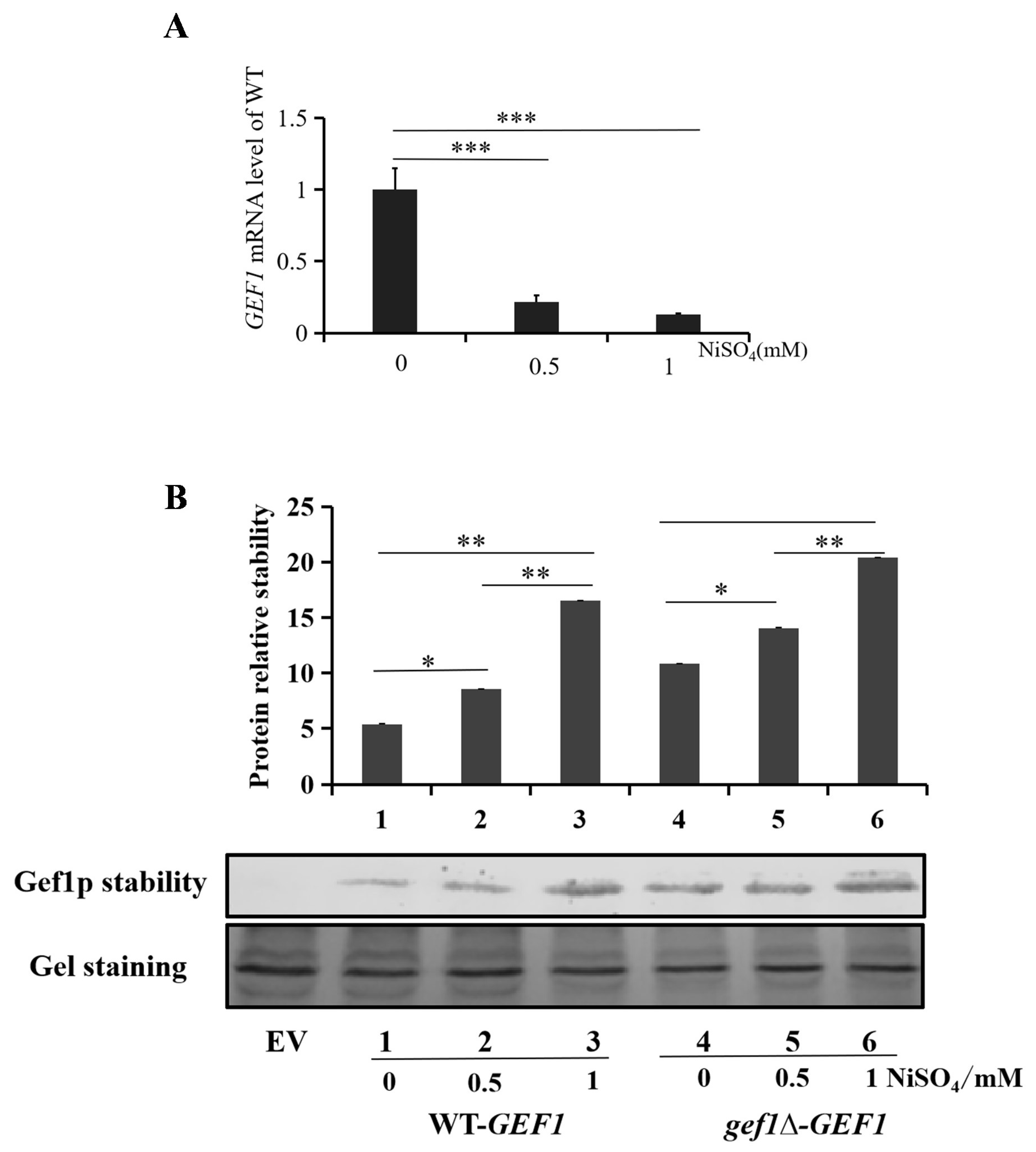
3.5. Nickel Ions Play an Important Role in Regulating mRNA and Protein Expression of GEF1

GEF1 regulates the expression of MMT2 and CUP1 and reduces the intracellular ROS level, yet it is not clear whether the increase of nickel ion content in the gef1Δ cells affects the expression of GEF1. We cultured WT cells with three densities of nickel ions (0 mM, 0.5 mM and 1 mM) in the medium, then extracted the RNA of the cells and detected the mRNA expression levels. The mRNA expression level of GEF1 decreased by more than ten times under the interference of nickel ions (Figure 6A). Next, we added the three densities of nickel ions to SC-Ura medium and using WT-p416TEF as a blank control, assessed the protein stability of WT-GEF1 and gef1∆-GEF1 by western blot. Both GEF1-overexpressing cell lines showed increased Gef1p expression proportional to the concentration of nickel in the medium (Figure 6B). Overall, these results revealed that the addition of exogenous nickel ions to the culture medium enhances the protein stability of gef1p, which indirectly indicates that not only can GEF1 regulate nickel ions, but also nickel ions can regulate GEF1.

4. Discussion

In this study, we studied the function of GEF1 in nickel metabolism of S. cerevisiae. Previously, we found that the GEF1-deficient strain showed resistance to toxicity from medium containing 0.5 mM and 1 mM nickel ions. From there we assessed the regulatory mechanism of GEF1 on nickel ions, thus discovering new functions of GEF1 and laying a foundation for studying diseases related to nickel ion metabolism.
We proposed a model for the regulation of Ni distribution by Mmt2p and reduction of ROS levels by MMT2/CUP1 overexpression (Figure 7). According to transcriptomics, GEF1 can maintain intracellular nickel ion levels in a stable range by regulating gene expression. We verified the regulation of nine genes by qPCR and focused on two highly upregulated genes with potential roles in nickel metabolism, MMT2 and CUP1. MMT2 is located on the mitochondrial membrane and functions to transport iron from the mitochondria to the cytosol. According to previous results in our laboratory, we judged that most of the genes that transport iron ions can also transport nickel ions, so we speculated that MMT2 gene could transport nickel ions from the cytosol to the mitochondria. CUP1 is a metallothionein, which is a metal-binding gene whose expression is upregulated in gef1∆ cells. We speculated this could lead to an increase in bound nickel ions, allowing for the increase in total nickel ions which we observed in the gef1∆ strain. We used inductively coupled plasma mass spectrometry (ICP-MS) and solid-state growth experiments in cells overexpressing MMT2 and CUP1 to show that MMT2 transported nickel from cytosol to mitochondria, and both MMT2 and CUP1 restored growth in nickel-supplemented media. Furthermore, given that nickel ions can induce the production of ROS, we assessed redox status of MMT2 and CUP1-overexpressing cells in the presence of excess nickel in the media. For both cell lines ROS were greatly decreased by over 50% compared to controls. These data suggest the gef1∆ strain was resistant to nickel ions through upregulation of MMT2 and CUP1 which act to reduce intracellular redox active nickel ions via their sequestration in the mitochondria and cytosol, respectively.
Finally, we identified a reciprocal relationship between GEF1 and nickel whereby just as GEF1 can regulate the metabolism of nickel ions, the nickel ions can also affect the expression of GEF1. Real-time fluorescence quantitative PCR experiments showed that nickel ions reduced the mRNA expression level of GEF1. The results of western blot showed that nickel ions could stabilize Gef1p. A possible explanation for this regulation is that excess nickel in the media impairs the uptake of iron and other metals through competitive inhibition (supported by Figure 2). Therefore, stabilization of gef1p, a chloride channel implicated in iron and copper homeostasis [30], may be a compensatory mechanism to maintain proper micronutrient balance. To summarize, this work demonstrated new roles for GEF1 in the absorption, utilization and storage of nickel ions in cells, which provides new insight for studying diseases related to nickel ion metabolism.
To further explore the relationship between GEF1 and nickel ions, it is necessary to identify the mechanism of how nickel ions reduce the mRNA level of GEF1 and enhance stability of the gef1p protein. A possibility is that nickel ions may directly interact with transcription factors to affect GEF1 expression in a manner similar to NikR in microbes [31]. Nickel ions may also directly interact with gef1p protein to affect its stability. For example, nickel binding to HpNikR was shown to affect its conformation and stability [32] and nickel dramatically increased the thermostability of UreE [33]. Additionally, it is necessary to explore how GEF1 regulates the transcription level of MMT2 and CUP1 including the extent to which nickel and other metal ions are implicated. Furthermore, it is unknown whether higher eukaryotes or humans have processes analogous to the relationship between GEF1 and nickel shown in S. cerevisiae. Our work hopes to provide a basis for further study of nickel metabolism in eukaryotes which is greatly lacking despite being considered an essential trace mineral. It is hoped that industrial yeast strains with high nickel tolerance can be constructed by using the function of GEF1 gene in the future, thus making certain contributions to industrial development.
Finally, we identified a reciprocal relationship between GEF1 and nickel whereby just as GEF1 can regulate the metabolism of nickel ions, the nickel ions can also affect the expression of GEF1. Real-time fluorescence quantitative PCR experiments showed that nickel ions reduced the mRNA expression level of GEF1. The results of western blot showed that nickel ions could stabilize Gef1p. A possible explanation for this regulation is that excess nickel in the media impairs the uptake of iron and other metals through competitive inhibition (supported by Figure 2). Therefore, stabilization of gef1p, a chloride channel implicated in iron and copper homeostasis [32], may be a compensatory mechanism to maintain proper micronutrient balance. To summarize, this work demonstrated new roles for GEF1 in the absorption, utilization and storage of nickel ions in cells, which provides new insight for studying diseases related to nickel ion metabolism.
To further explore the relationship between GEF1 and nickel ions, it is necessary to identify the mechanism of how nickel ions reduce the mRNA level of GEF1 and enhance stability of the gef1p protein. A possibility is that nickel ions may directly interact with transcription factors to affect GEF1 expression in a manner similar to NikR in microbes [33]. Nickel ions may also directly interact with gef1p protein to affect its stability. For example, nickel binding to HpNikR was shown to affect its conformation and stability [34] and nickel dramatically increased the thermostability of UreE [35]. Additionally, it is necessary to explore how GEF1 regulates the transcription level of MMT2 and CUP1 including the extent to which nickel and other metal ions are implicated. Furthermore, it is unknown whether higher eukaryotes or humans have processes analogous to the relationship between GEF1 and nickel shown in S. cerevisiae. Our work hopes to provide a basis for further study of nickel metabolism in eukaryotes which is greatly lacking despite being considered an essential trace mineral. It is hoped that industrial yeast strains with high nickel tolerance can be constructed by using the function of GEF1 gene in the future, thus making certain contributions to industrial development.

Supplementary Materials

The following supporting information can be downloaded at: www.mdpi.com/xxx/s1, Figure S1: title; Table S1: title; Video S1: title.

Author Contributions

Formal analysis, L.Z., Q. L.; Investigation, Q.L., L.Z., R.H., C.L., J.B. and X.W.; Writing—review & editing, J.B., J.B. and X.W.; Project administration, X.W.; Funding acquisition, X.W. All authors have read and agreed to the published version of the manuscript.

Funding

This work was supported by Shanghai Agriculture Applied Technology Development Program, China (No. 2021-02-08-00-12-F00779), National Key R&D Program of China (No. 2018YFC1604403) and the Fund of Shanghai Engineering Research Center of Plant Germplasm Resources (Grant No. 17DZ2252700).

Institutional Review Board Statement

Not applicable.

Informed Consent Statement

Not applicable.

Data Availability Statement

Data are contained within the article.

Conflicts of Interest

The authors declare no conflict of interest.

References

  1. Anke, M.; Groppel, B.; Kronemann, H.; Grün, M. Nickel--an Essential Element. IARC Sci Publ. 1984, 339–365. [Google Scholar]
  2. Trumbo, P.; Yates, A.A.; Schlicker, S.; Poos, M. Dietary Reference Intakes: Vitamin A, Vitamin K, Arsenic, Boron, Chromium, Copper, Iodine, Iron, Manganese, Molybdenum, Nickel, Silicon, Vanadium, and Zinc. J Am Diet Assoc. 2001, 101, 294–301. [Google Scholar] [CrossRef] [PubMed]
  3. Das, K.K.; Honnutagi, R.; Mullur, L.; Reddy, R.C.; Das, S.; Majid, D.S.A.; Biradar, M.S. Chapter 26 - Heavy Metals and Low-Oxygen Microenvironment—Its Impact on Liver Metabolism and Dietary Supplementation In Dietary Interventions in Liver Disease; Watson, R.R., Preedy, V.R., Eds.; Academic Press, 2019; pp. 315–332 ISBN 978-0-12-814466-4.
  4. Genchi, G.; Carocci, A.; Lauria, G.; Sinicropi, M.S.; Catalano, A. Nickel: Human Health and Environmental Toxicology. Int J Environ Res Public Health. 2020, 17, 679. [Google Scholar] [CrossRef] [PubMed]
  5. Slotkin, T.A.; Skavicus, S.; Card, J.; Levin, E.D.; Seidler, F.J. Diverse Neurotoxicants Target the Differentiation of Embryonic Neural Stem Cells into Neuronal and Glial Phenotypes. Toxicology. 2016, 372, 42–51. [Google Scholar] [CrossRef]
  6. Bian, J.; Shi, X.; Li, Q.; Zhao, M.; Wang, L.; Lee, J.; Tao, M.; Wu, X. A Novel Functional Role of Nickel in Sperm Motility and Eukaryotic Cell Growth. J Trace Elem Med Biol. 2019, 54, 142–149. [Google Scholar] [CrossRef]
  7. Rizvi, A.; Parveen, S.; Khan, S.; Naseem, I. Nickel Toxicology with Reference to Male Molecular Reproductive Physiology. Reprod Biol. 2020, 20, 3–8. [Google Scholar] [CrossRef]
  8. Noguromi, M.; Yamaguchi, Y.; Sato, K.; Oyakawa, S.; Okamoto, K.; Murata, H.; Tsukuba, T.; Kadowaki, T. Rab44 Deficiency Induces Impaired Immune Responses to Nickel Allergy. Int J Mol Sci. 2023, 24, 994. [Google Scholar] [CrossRef]
  9. Zambelli, B.; Ciurli, S. Nickel and Human Health. Met Ions Life Sci. 2013, 13, 321–357. [Google Scholar]
  10. Can, M.; Armstrong, F.A.; Ragsdale, S.W. Structure, Function, and Mechanism of the Nickel Metalloenzymes, CO Dehydrogenase, and Acetyl-CoA Synthase. Chem Rev. 2014, 114, 4149–4174. [Google Scholar] [CrossRef]
  11. Mobley, H.L.; Garner, R.M.; Bauerfeind, P. Helicobacter Pylori Nickel-Transport Gene nixA: Synthesis of Catalytically Active Urease in Escherichia Coli Independent of Growth Conditions. Mol Microbiol. 1995, 16, 97–109. [Google Scholar] [CrossRef]
  12. Bell, A.J.; Guerra, C.; Phung, V.; Nair, S.; Seetharam, R.; Satir, P. GEF1 Is a Ciliary Sec7 GEF of Tetrahymena Thermophila. Cell Motil Cytoskeleton. 2009, 66, 483–499. [Google Scholar] [CrossRef] [PubMed]
  13. Nair, S.; Guerra, C.; Satir, P. A Sec7-Related Protein in Paramecium. FASEB J. 1999, 13, 1249–1257. [Google Scholar] [CrossRef] [PubMed]
  14. Greene, J.R.; Brown, N.H.; DiDomenico, B.J.; Kaplan, J.; Eide, D.J. The GEF1 Gene of Saccharomyces Cerevisiae Encodes an Integral Membrane Protein; Mutations in Which Have Effects on Respiration and Iron-Limited Growth. Mol Gen Genet. 1993, 241, 542–553. [Google Scholar] [CrossRef] [PubMed]
  15. Flis, K.; Bednarczyk, P.; Hordejuk, R.; Szewczyk, A.; Berest, V.; Dolowy, K.; Edelman, A.; Kurlandzka, A. The Gef1 Protein of Saccharomyces Cerevisiae Is Associated with Chloride Channel Activity. Biochem Biophys Res Commun. 2002, 294, 1144–1150. [Google Scholar] [CrossRef] [PubMed]
  16. Gaxiola, R.A.; Yuan, D.S.; Klausner, R.D.; Fink, G.R. The Yeast CLC Chloride Channel Functions in Cation Homeostasis. Proc Natl Acad Sci U S A. 1998, 95, 4046–4050. [Google Scholar] [CrossRef]
  17. López-Rodríguez, A.; Trejo, A.C.; Coyne, L.; Halliwell, R.F.; Miledi, R.; Martínez-Torres, A. The Product of the Gene GEF1 of Saccharomyces Cerevisiae Transports Cl- across the Plasma Membrane. FEMS Yeast Res. 2007, 7, 1218–1229. [Google Scholar] [CrossRef]
  18. Sasvari, Z.; Kovalev, N.; Nagy, P.D. The GEF1 Proton-Chloride Exchanger Affects Tombusvirus Replication via Regulation of Copper Metabolism in Yeast. J Virol. 2013, 87, 1800–1810. [Google Scholar] [CrossRef]
  19. Ramos-Alonso, L.; Romero, A.M.; Martínez-Pastor, M.T.; Puig, S. Iron Regulatory Mechanisms in Saccharomyces Cerevisiae. Front Microbiol. 2020, 11, 582830. [Google Scholar] [CrossRef]
  20. Kim, J.E.; Lindahl, P.A. CUP1 Metallothionein from Healthy Saccharomyces Cerevisiae Colocalizes to the Cytosol and Mitochondrial Intermembrane Space. Biochemistry. 2023, 62, 62–74. [Google Scholar] [CrossRef]
  21. Winzeler, E.A.; Shoemaker, D.D.; Astromoff, A.; Liang, H.; Anderson, K.; Andre, B.; Bangham, R.; Benito, R.; Boeke, J.D.; Bussey, H.; et al. Functional Characterization of the S. Cerevisiae Genome by Gene Deletion and Parallel Analysis. Science. 1999, 285, 901–906. [Google Scholar] [CrossRef]
  22. Chen, X.; Zhang, R.; Sun, J.; Simth, N.; Zhao, M.; Lee, J.; Ke, Q.; Wu, X. A Novel Assessment System of Toxicity and Stability of CuO Nanoparticles via Copper Super Sensitive Saccharomyces Cerevisiae Mutants. Toxicol In Vitro. 2020, 69, 104969. [Google Scholar] [CrossRef] [PubMed]
  23. Mumberg, D.; Müller, R.; Funk, M. Yeast Vectors for the Controlled Expression of Heterologous Proteins in Different Genetic Backgrounds. Gene. 1995, 156, 119–122. [Google Scholar] [CrossRef] [PubMed]
  24. Bian, J.; Wang, L.; Wu, J.; Simth, N.; Zhang, L.; Wang, Y.; Wu, X. MTM1 Plays an Important Role in the Regulation of Zinc Tolerance in Saccharomyces Cerevisiae. J Trace Elem Med Biol. 2021, 66, 126759. [Google Scholar] [CrossRef] [PubMed]
  25. Gietz, R.D.; Schiestl, R.H.; Willems, A.R.; Woods, R.A. Studies on the Transformation of Intact Yeast Cells by the LiAc/SS-DNA/PEG Procedure. Yeast. 1995, 11, 355–360. [Google Scholar] [CrossRef] [PubMed]
  26. Xu, N.; Xu, Y.; Smith, N.; Chen, H.; Guo, Z.; Lee, J.; Wu, X. MTM1 Displays a New Function in the Regulation of Nickel Resistance in Saccharomyces Cerevisiae. Metallomics. 2022, 14, mfac074. [Google Scholar] [CrossRef]
  27. Furukawa, S.; Fujita, T.; Shimabukuro, M.; Iwaki, M.; Yamada, Y.; Nakajima, Y.; Nakayama, O.; Makishima, M.; Matsuda, M.; Shimomura, I. Increased Oxidative Stress in Obesity and Its Impact on Metabolic Syndrome. J Clin Invest. 2004, 114, 1752–1761. [Google Scholar] [CrossRef]
  28. Li, L.; Bertram, S.; Kaplan, J.; Jia, X.; Ward, D.M. The Mitochondrial Iron Exporter Genes MMT1 and MMT2 in Yeast Are Transcriptionally Regulated by Aft1 and Yap1. J Biol Chem. 2020, 295, 1716–1726. [Google Scholar] [CrossRef]
  29. Soboh, B.; Adrian, L.; Stripp, S.T. An in Vitro Reconstitution System to Monitor Iron Transfer to the Active Site during the Maturation of [NiFe]-Hydrogenase. J Biol Chem. 2022, 298, 102291. [Google Scholar] [CrossRef]
  30. Davis-Kaplan, S.R.; Askwith, C.C.; Bengtzen, A.C.; Radisky, D.; Kaplan, J. Chloride Is an Allosteric Effector of Copper Assembly for the Yeast Multicopper Oxidase Fet3p: An Unexpected Role for Intracellular Chloride Channels. Proc Natl Acad Sci U S A. 1998, 95, 13641–13645. [Google Scholar] [CrossRef]
  31. Phillips, C.M.; Schreiter, E.R.; Stultz, C.M.; Drennan, C.L. Structural Basis of Low-Affinity Nickel Binding to the Nickel-Responsive Transcription Factor NikR from Escherichia Coli. Biochemistry. 2010, 49, 7830–7838. [Google Scholar] [CrossRef]
  32. Baksh, K.A.; Pichugin, D.; Prosser, R.S.; Zamble, D.B. Allosteric Regulation of the Nickel-Responsive NikR Transcription Factor from Helicobacter Pylori. J Biol Chem. 2021, 296, 100069. [Google Scholar] [CrossRef] [PubMed]
  33. Lee, Y.-H.; Won, H.-S.; Lee, M.-H.; Lee, B.-J. Effects of Salt and Nickel Ion on the Conformational Stability of Bacillus Pasteurii UreE. FEBS Lett. 2002, 522, 135Thank you for your reply. We have updated the manuscript and system based on your email. We will proceed the manuscript as soon as possible–140. [Google Scholar] [CrossRef]
Figure 1. Gef1p is a new molecular factor involved in nickel ion metabolism in Saccharomyces cerevisiae. WT control and gef1Δ strains were cultured with NiSO4 (A) or CuSO4 (B) or CdCl2 (C) or FeSO4 (D) or ZnSO4 (E) on SC plates containing glucose to assess growth. WT control and gef1Δ strains in were cultured in SC liquid medium with 0 mM (F), 0.5 mM (G) and 1 mM (H) NiSO4 and growth curves are shown. (I) WT or gef1Δ cells were transformed with empty vector (p416TEF) or GEF1 and grown on SC-Ura medium with or without nickel supplementation. All growth tests were carried out with at least four different clones.
Preprints 146108 g001
Figure 2. Metal content in cells of WT control and gef1Δ strains. Nickel (A), iron (B) and zinc (C) in cells were determined by ICP-MS from cells cultured in SC medium with or without NiSO4, and then normalized to protein concentration. All metal level analyses were performed with at least four different clones. Average and standard deviation (n=4) are indicated. The asterisk (*) indicates that there are significant differences among different strains with the same treatment (*p < 0.05, **p < 0.01, *** p < 0.001).
Preprints 146108 g002
Figure 3. The mRNA expression levels of different genes in WT control and gef1Δ strains treated with or without NiSO4. The mRNA expression levels of FRE2 (A), FIT2 (B), ARN2 (C), FET3 (D), FRE1 (E), MMT2 (F), ARN (G), FTR1 (H) and CUP1 (I). All treatments were carried out on at least four different clones. The mean and standard deviation is given. Significant differences are indicated by asterisks (*),.(* p < 0.05, ** p < 0.01, *** p < 0.001).
Preprints 146108 g003
Figure 4. Heavy metal content in mitochondria during MMT2 overexpression in gef1Δ strain. Nickel (A), iron (B), zinc (C), and copper (D) levels in mitochondria during MMT2 overexpression were determined by ICP-MS and normalized to protein concentrations. All metal level analyses were performed with at least four different clones. Mean ± standard deviation (n=4) is indicated. Asterisks (*) indicate significant differences between lines with the same treatment.(*p < 0.05, **p < 0.01, ***p < 0.001).
Preprints 146108 g004
Figure 5. MMT2/CUP1 overexpression reduces intracellular ROS levels. (A) Levels of reactive oxygen species produced by MMT2 when overexpressed in WT and gef1∆ strains. (B) Levels of reactive oxygen species produced by CUP1 when overexpressed in WT and gef1∆ strains. (C) Spot assay of MMT2 overexpressed in WT and gef1∆ strains. (D) Spot assay of CUP1 overexpressed in WT and gef1∆ strains. All treatments were performed on at least four different cell lines. Means ± standard deviation are given. Significant differences are indicated by an asterisk (*). (*p < 0.05, **p < 0.01, ***p < 0.001).
Preprints 146108 g005
Figure 6. Nickel ions regulate the mRNA expression and protein expression level of GEF1. (A) The mRNA expression levels of GEF1 in WT strains treated with different concentrations of nickel ions. (B) Protein expression levels of GEF1-GFP in WT cells and gef1Δ cells. EV represents empty vector. All treatments were performed on at least four different cell lines. Mean values ± standard deviation are given. Significant differences are indicated by an asterisk (*).(*p < 0.05, **p < 0.01, and ***p < 0.001).
Preprints 146108 g006
Figure 7. Proposed model for the regulation of Ni distribution by Mmt2p and reduction of ROS levels by MMT2/CUP1 overexpression.
Preprints 146108 g007
Disclaimer/Publisher’s Note: The statements, opinions and data contained in all publications are solely those of the individual author(s) and contributor(s) and not of MDPI and/or the editor(s). MDPI and/or the editor(s) disclaim responsibility for any injury to people or property resulting from any ideas, methods, instructions or products referred to in the content.
Copyright: This open access article is published under a Creative Commons CC BY 4.0 license, which permit the free download, distribution, and reuse, provided that the author and preprint are cited in any reuse.
Prerpints.org logo

Preprints.org is a free preprint server supported by MDPI in Basel, Switzerland.

Subscribe

Disclaimer

Terms of Use

Privacy Policy

Privacy Settings

© 2025 MDPI (Basel, Switzerland) unless otherwise stated