Preprint
Review

This version is not peer-reviewed.

Thyroid Hormones and Brain Development: A Focus on the Role of Mitochondria as Regulators of Developmental Time

A peer-reviewed article of this preprint also exists.

Submitted:

09 January 2025

Posted:

09 January 2025

You are already at the latest version

Abstract

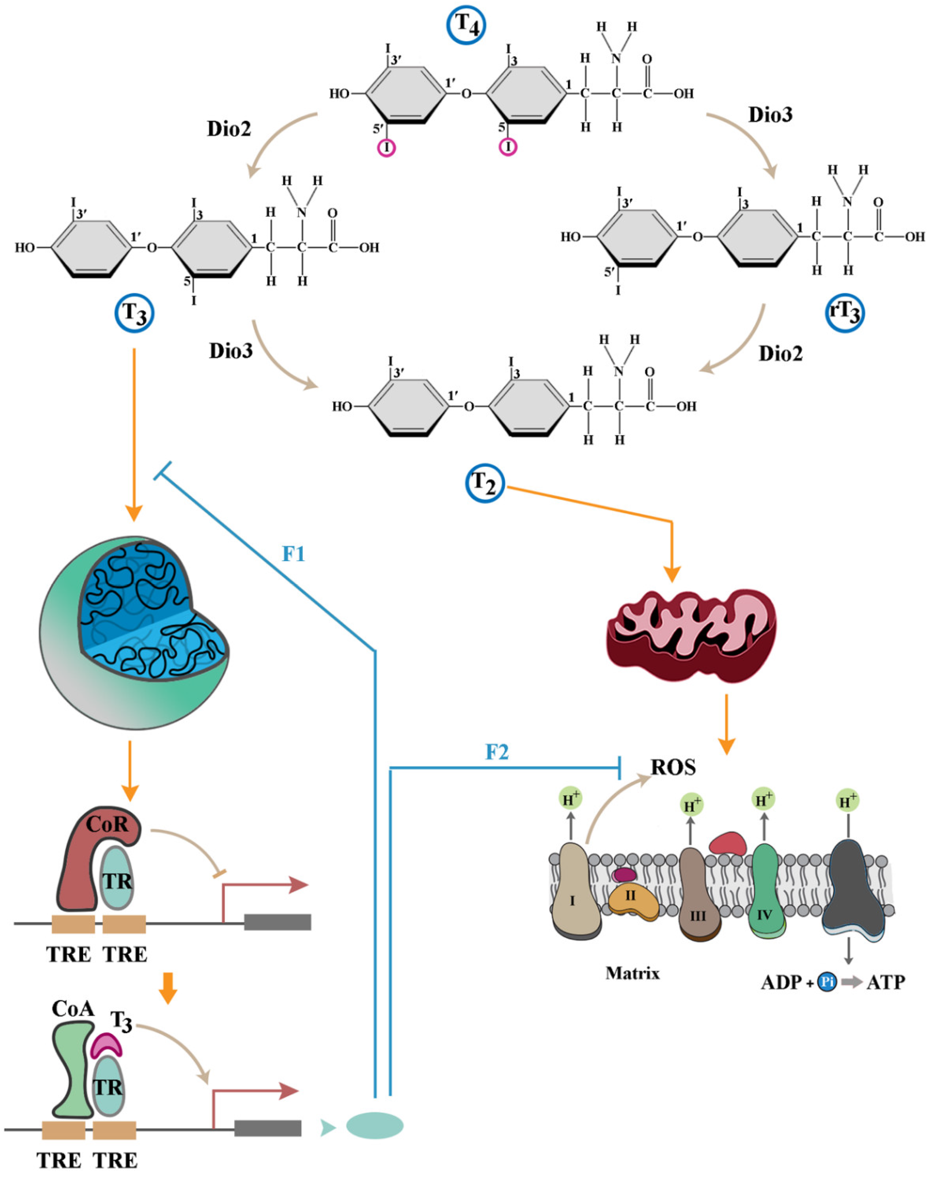
Thyroid hormones (TH) regulate metabolism in a homeostatic state in an adult organism. During the prenatal period, prior to establishment of homeostatic mechanisms, TH assume additional functions as key regulators of brain development. Here, we focus on reviewing the role of TH in orchestrating cellular dynamics in a developing brain. We provide evidence that developmental roles of the hormones are predominantly mediated by non-genomic mitochondrial effects of TH due to attenuation of genomic effects of TH that antagonise non-genomic impacts. We argue that the key function of TH signalling during brain development is to orchestrate the tempo of self-organisation of neural progenitor cells. Further, evidence is provided that major neurodevelopmental consequences of hypothyroidism stem from an altered tempo of cellular self-organisation.

Keywords: 
;  ;  ;  

Introduction

The role of thyroid hormones in brain development has been extensively reviewed and readers are referred to scholarly articles covering this topic [1,2,3,4,5,6,7]. The research in this field has typically focused on genomic activities of thyroid hormones during neurodevelopment [8]. This genome-centric view of TH activity is gradually changing as accumulating evidence suggest that non-genomic effects of the hormones in regulating brain development are as important as the genomic influences [9,10]. A pathological condition which clearly illustrate this notion is brain development in congenital hypothyroidism manifest as cretinism [11]. With an incidence of 1 in 3500 live births [12], congenital hypothyroidism significantly affects brain development as evidenced by a mean intelligence quotient (IQ) of 76 in affected individuals [13]. Interestingly, while induced hypothyroidism negatively influences cerebellar development in animal models, no such negative effect is observed in mice lacking thyroid receptor-α1 [14]. This observation suggests that impairment of non-genomic effects of TH underpin certain neurodevelopmental consequences of hypothyroidism. A further corollary of the findings of the latter study is that non-genomic effects of TH counterbalance the genomic impacts of the hormone on brain development. Aside from a divergence of outcomes, genomic and non-genomic effects are driven by different forms of TH. While L-triiodothyronine (T3) is responsible for receptor-mediated functions of TH, non-genomic effects are mainly driven by 3,5-Diodothyronine (3,5-T2), a degradation by-product of T3 [10]. Given a divergence of mediators and outcomes, an objective of this review is to dissect the role of genomic and non-genomic outcomes communicated by TH signalling in neurodevelopment from a mechanistic perspective.
Focusing on the non-genomic impact of TH, evidence suggests that mitochondria mediate some of the reported effects [10,15,16]. The interactions of TH with mitochondria are typically studied in the context of regulation of metabolism in a homeostatic state of an adult organism. However, just as the notion of the role of mitochondria as primarily contributing to homeostasis by provision of ATP has started to change, so has understanding of the interactions of TH with mitochondria in an emerging paradigm. The new paradigm concerns the role of mitochondria in orchestrating developmental landscape during the prenatal period when most homeostatic mechanisms are absent. The paradigm is an extension of a growing body of research suggesting that mitochondrial dynamics and neuronal differentiation are intimately linked [17,18,19,20,21,22]. Exploration of the mechanism underlying mitochondrial facilitation of neuronal differentiation revealed that the so-called mitochondrial metabolic by-products (e.g., thermal flux [23,24,25] and reactive oxygen species [18,26]), that are typically removed by homeostatic mechanisms, propel neuronal differentiation. This is hardly surprising as most homeostatic mechanisms are either absent or are ineffective in early stages of development. For example, a functional circulation to dissipate heat is not established until embryonic day 10 in developing mouse embryos [27] and yet heat is generated at a high rate of 30 nW/cell at a much earlier, two-cell stage, of embryogenesis [28]. This brings about a window of opportunity, characterised by relative absence of homeostatic mechanisms, in which mitochondrial metabolic by-products (i.e., abiotic signals) could exceed a threshold level to drive reprogramming of other signalling pathways [23]. Given the documented role of TH in amplifying mitochondrial generation of these abiotic signals [29,30], a second goal of the review is to explore whether non-genomic effects of TH in early neurodevelopment are mediated by mitochondrial abiotic signals. We propose that TH orchestrate brain development in a bistable manner. Non-genomic effects emerge first, require amplification of mitochondrial metabolic by-products that regulate multicellular self-organisation during brain development. Genomic receptor-mediated effects are delayed compared to non-genomic effects and counterbalance the impact of mitochondrial activities. Finally, we provide evidence that the genomic and non-genomic signalling activities of TH can be transiently uncoupled to enhance the impact of non-genomic effects during brain development. This proposal is validated in the light of existing evidence regarding the impact of altered thyroid activity on brain development.

An Overview of the Signalling Landscape of Thyroid Hormones

A detailed account of regulation of thyroid hormones can be found in a recent review [31] while the overview of TH signalling provided here is a prelude to the discussion that follows. Thyroid gland releases 3,5,3′,5′-tetraiodothyronine (alias: T4 or thyroxine) into the blood stream. The enzymatic activity of 5′-deiodinase (5′-D, dio2) then converts T4 to T3, the active form that binds to thyroid hormone receptors (TR). A subsequent deiodination event catalysed by 5-deiodinase (5-D, dio3) converts T4 and T3 to rT3 (reverse T3) and T2, respectively. Although the impacts of different forms of TH are diverse and somewhat context-dependent, the elemental outline of TH signalling is shaped by two general principles. Outcomes induced by TH are either transcription-dependent (genomic effects), or occur independently of nuclear dynamics (non-genomic effects) [30] (Figure 1). Further, the genomic and non-genomic interactions appear to invoke antagonistic outcomes as detailed below.
Genomic effects of TH are mediated via binding of T3 to different isoforms of nuclear thyroid receptor-α (TRα) and TRβ [30]. Transcriptional outcomes invoked by this complex depend on the target cell. In pituitary and thyroid glands, T3/TR complex inhibits expression of the genomic loci encoding the thyrotropin-α and -β subunits and thyrotropin-releasing hormone [32], thereby exerting a negative feedback input on production of TH by the thyroid gland. In other peripheral target tissues, the genomic response to TH is context dependent. The consensus view is that the complex of T3/TR binds to thyroid hormone response elements (TREs) in a homo- or heterodimeric form in the regulatory regions of specific genes to activate transcription of these loci. While the receptor-mediated activity of TH drives a plethora of adaptive changes [32,33], closer examination reveals a generic pattern of regulation of peripheral gene expression to attenuate the consequences of non-genomic functions of TH. An example of the latter activity is observed during T3-mediated upregulation of genes that contribute to glutathione synthesis [34]. Reduced glutathione is the key mammalian non-enzymatic antioxidant [35] essential for elimination of reactive oxygen species (ROS) of mitochondrial origin that are amplified by non-genomic impacts of TH [36,37]. In the absence of TH, the hormone-free TR recruits a corepressor complex with histone deacetylase activity which inhibits the latter genomic loci with a binding motif for TR [32] (Figure 1). Upon exposure to TH, just as T2 amplifies mitochondrial production of ROS, formation of T3/TR complex reinitiates transcription from these TR-inhibited loci [32] leading to enhanced synthesis of glutathione to eliminate the generated ROS. As expected from the antagonistic interaction of non-genomic and TR-mediated genomic impacts of TH, experimental knockdown of TRs not only does not abolish signalling by TH, but also leads to an accelerated progression of developmental events mediated by these hormones [38,39]. Further, compared to severe consequences of TH deficiency, mice with a deletion of both TRα1 and TRβ exhibit milder phenotypes including a hyperactive pituitary-thyroid axis [40]. A hyperactive pituitary-thyroid axis is consistent with a lack of negative feedback communicated via T3/TR complex to genomic loci encoding proteins that synthesize TH and thyrotropin. Acceleration of developmental dynamics in TR-null animal models, on the other hand, suggests a dominance of TR-independent non-genomic functions of TH that are otherwise dampened by TR-mediated negative feedback. An interpretation of the accelerated development in TR-null animals is that dynamics of organogenesis are in part orchestrated by non-genomic effects of TH.
The non-genomic functions of TH are mediated by interaction of the hormones with mitochondria or other cytoplasmic entities (Figure 1). Non-genomic mitochondria-independent functions of TH have been reviewed in detail elsewhere [30]. A characteristic feature of the non-genomic cytoplasmic effects of TH is the short timeframe of occurrence. A major cluster of non-genomic activities of TH develop within seconds of exposure to the hormones and prime the cell for activation of citric acid cycle and electron transport chain. T3 induces a rapid increase in cytoplasmic [Ca2+] [41] and glucose uptake [42,43]. A subsequent activation of Ca2+-ATPase activity by TH [44] triggers expulsion of cytoplasmic Ca2+, thus restricting the temporal window of Ca2+ uptake and hence the concentration of this ion within the cytoplasm. Calcium facilitates dephosphorylation of phosphorylated pyruvate dehydrogenase thereby activating the enzyme [45]. Calcium also stimulates the entire oxidative phosphorylation cascade [46] within a specific range of ionic concentration [47]. Therefore, it can be argued that the pro-metabolic non-genomic activities of TH, that occur within seconds, prime mitochondria for amplified activity of the electron transport chain. Another rapid, TH-driven, non-genomic phenomenon is stimulation of actin polymerisation [48,49,50]. While the exact mechanism of TH-induced F-actin formation remains largely unknown, it seems plausible that GTP supply to small GTPase organisers of actin polymerisation (e.g., Rho GTPases) could underpin this effect [51,52,53,54]. Given that GTP is produced in citric acid cycle, F-actin formation could be potentially linked to Ca2+-mediated activation of pyruvate dehydrogenase with consequential activation of the downstream citric acid cycle. Further, TH amplifies production of reactive oxygen species (ROS) by mitochondria [36,37]. ROS-mediated oxidisation boosts the activity of small GTPases (in particular Cdc42) by approximately three orders of magnitude by stimulating dissociation of GDP from inactive enzyme [55].
Complementing the cytoplasmic effects that energise citric acid cycle, TH directly activate the mitochondrial respiratory chain (Figure 1). Exposure of isolated mitochondria from hypothyroid animals to T3 stimulates oxidative phosphorylation within minutes [56,57]. Interestingly, this stimulatory effect is not confined to T3. The activity of the respiratory chain is rapidly enhanced upon exposure of mitochondria to T2, a degradation metabolite of T3 [58,59,60]. Activation of oxidative phosphorylation is, in part, due to binding of T2 to subunit Va of cytochrome c oxidase which abolishes the allosteric inhibition of the complex by ATP [61]. An insight into the significance of dis-inhibition of cytochrome c oxidase by T2 is afforded by dissecting the molecular basis for allosteric inhibition of the complex by ATP. Capacity of the electron transport chain is mainly determined by the rate-limited activity of cytochrome c oxidase [62]. The complex operates in an excited or a relaxed mode by integrating negative input from ATP [63]. The inhibition of cytochrome c oxidase by ATP is switched on by cAMP-dependent phosphorylation of the complex and switched off by Ca2+-activated dephosphorylation [64]. In a dephosphorylated excited state, the activity of the complex increases by five- to ten-fold [62]. In this excited mode, ROS generation by the electron transport chain occurs at a higher rate [63]. Likewise, a major outcome of T2-mediated dis-inhibition of cytochrome c oxidase is amplified generation ROS by the mitochondrial electron transport chain [61]. Following this line of reasoning, one may question the relevance of ROS to TH signalling.
It appears that critical nodes in eukaryotic signalling pathways have been populated by redox-sensitive proteins that abort downstream communication of signals [65]. Reprogramming of these proteins occurs by interaction with ROS which transiently rewire the network topology of signalling pathways and facilitate downstream transmission of signals, as discussed elsewhere [65]. A focus on the role of ROS in facilitating PI3K signalling pathway illustrates this point. Upon activation, PI3K catalyses the conversion of PIP2 to PIP3 which prompts Akt signalling [66]. Concurrent activation of PTEN by catalytic activity of protein phosphatase 2A [67] or by auto-dephosphorylation [68] antagonises the function of PI3K by converting PIP3 to PIP2. In an oxidising milieu (e.g., high [ROS]), PTEN becomes reversibly inactivated due to the formation of an intramolecular disulfide between the essential active Cys-124 residue and Cys-71 [69]. This transient inactivation of PTEN facilitates downstream communication of PI3K/Akt signals. The redox-mediated remodelling of signalling cascades enables control of the rate of biochemical events at a cellular level thus giving rise to an adjustable cellular clock [65]. This insight could be utilised in revisiting the role of T2 binding to cytochrome c oxidase and relieving the ATP-mediated inhibition of the complex. Upon binding of T2, transition to an excited state of cytochrome c oxidase induces a transient shift to an oxidising milieu owing to enhanced production of ROS [36,37]. While a shift to a pro-oxidizing state will be short-lived as TH trigger production of antioxidants via the TR-mediated genomic pathway [34], recent findings suggest that transient amplification of ROS is sufficient to accelerate the rate of neuronal differentiation and that of brain development [18]. Hence, two parallel non-genomic arms of TH signalling, cytoplasmic and mitochondrial effectors, combine to amplify production of ROS by mitochondria. These non-genomic effects are counterbalanced by a delayed adaptive genomic response invoked via TRs. The notion of biphasic activity of TH whereby TR-mediated effects antagonises the non-genomic impact of TH is bolstered by the finding that while hypothyroidism brings about a host of developmental anomalies, elimination of TRα1 prevents hypothyroidism-related anomalies in developing cerebellum [14]. A plausible explanation for this observation is that elimination of TR-mediated antagonistic effects augments non-genomic effects of TH in a hypothyroid state, thus restoring normal developmental dynamics. The proposal that TH operate in a biphasic manner is further supported by dissection of the evolutionary interface of genomic and non-genomic functions of TH.

Thyroid Hormones: A Broad Evolutionary Perspective

Emergence of TH predates evolution of the thyroid gland [70,71]. TH and associated metabolites are utilised by various species in the animal and plant kingdoms which lack a thyroid gland [70,72]. In the absence of an endogenous capacity to synthesize the entities sea urchins utilise exogenous TH of plankton origin as an ecological cue to pace the tempo of development to availability of food [73]. In these animals, acquisition of exogenous TH accelerates metamorphosis [73]; the hormones are therefore considered to be ecological programmers of development [74]. Interestingly, the impacts of TH on larval development (i.e., inhibition of larval development and accelerated development of juvenile structures) are replicated by rearing larvae in a nutrient-rich condition [73]. The crosstalk between nutrient availability and TH production extends beyond the provided example of larval development. In mammals, the level of TH positively correlates with food availability, decreasing during periods of energy restriction and increasing upon access to energy substrates [75]. Another distinguishing feature of primitive TH signalling in invertebrates is the absence of a hormone receptor [71]. Therefore, it can be concluded that non-genomic effects of TH are more ancient that genomic effects that are mediated via receptors. The ancestral state of non-genomic effects compared to TR-mediated genomic effects is carried over to a functional level where it manifests as dominance of non-genomic effects over genomic effects in driving development in the species with functional TR. While initial reports suggested that the impact of TH on metamorphosis is mediated via TR [76,77], subsequent gene knockout studies revealed that TR are not essential for induction of metamorphosis by TH [76,77]. On the contrary, metamorphic transition is accelerated in the absence of TR, a phenomenon that has been partially attributed to the removal of TR-mediated gene repression [76,77]. Evidence suggests that not only the evolutionary emergence and deployment of TH predates TRs [78], but also that developmental phenomena are predominantly regulated by more ancient TR-independent non-genomic activities of TH. The alternative interpretation that TRs were initially acquired and then lost in unicellular organisms and some basal metazoans suggests that non-genomic functions of TH are key to driving basic developmental events. Revisiting metamorphosis reveals another facet of primal TH signalling. It is noteworthy that mitochondrial dysfunction perturbs aspects of insect wing development during metamorphosis [79]. This effect is unlikely to be primarily related to an energy crisis for two reasons. It is known that the metabolic rate declines sharply at the beginning of metamorphosis and remains low until the completion of morphogenesis [80,81]. Further, consequences of perturbation of metamorphosis as a result of mitochondrial dysfunction are localised to the wings as opposed to a more generalised impact on metamorphosis as is expected to occur in an energy crisis scenario [79]. A deeper insight into the role of mitochondria in TH signalling is provided by exploring activity of the electron transport chain components in insect metamorphosis. Investigation revealed an unexpected finding that succinate rather than pyruvate is used as a metabolite of the respiratory chain [82]. In the same study, cytochrome c oxidase showed a higher turnover rate concurrent with initiation of metamorphosis [82]. Preferential utilisation of succinate by complex II of the electron transport chain is known to triggers a reverse electron flow to complex I leading to a significant production of ROS [83]. Likewise, impaired activity of cytochrome c oxidase (evidenced by a high turn-over rate during metamorphosis [82]) contributes to reverse electron flow and ROS production [84]. Given the suggested role of ROS in driving metamorphosis [29,85,86], it seems plausible that succinate-mediated reversal of the electron transport chain and a high turn-over of cytochrome c oxidase are deployed to increase ROS production and to tune the tempo of metamorphosis. By the same line of reasoning, contribution of TH to generation of ROS in metamorphosis [29] could be attributed to the impact of T2 on cytochrome c oxidase discussed in a previous section [63], or a recently shown inhibitory impact of T3 and T4 on cytochrome c oxidase [87] which activates reverse electron flow [84].
Revisiting the conserved evolutionary functions of TH, it becomes apparent that TH in marine species functions in the absence of TR as an ecological currency for the availability of food. Accumulation of TH in these species boost the energetic capacity of mitochondria in anticipation of a nutrient-rich environment. Production of ROS by mitochondria, on the other hand, primes that eukaryotic host for utilisation of the energy extracted from nutrients. As discussed, this occurs by ROS-mediated reprogramming of eukaryotic signalling cascades. It can be said that TH unlocks a mitochondrial biochemical potential and the resultant mitochondrial ROS then unlock the host’s biochemical potential. From this perspective, it can be envisaged that emergence of TRs was a subsequent evolutionary adaptation of metazoans that occurred in a stepwise manner to put an upper limit to the energetic capacity of a cell. This initially occurred by providing a delayed antagonistic input to counterbalance the non-genomic functions of TH on peripheral cells (e.g., by upregulation of antioxidants [34]). In the final stage of evolution and concurrent with emergence of a functional thyroid gland, the scope of activity of TR expanded to provide a negative feedback input to dampen the endocrine synthesis of TH [88]. Considering the evolutionary history of TH and TR, it is not surprising that abolishing TR-mediated genomic reprogramming boosts non-genomic effects of TH [38,39]. While the latter is an insight afforded by an experimental model, one may ask whether there is a biological mechanism to delay or transiently abolish TR-mediated transcriptional remodelling and to amplify non-genomic mitochondrial effects of TH.

Uncoupling of the Genomic and non-Genomic Impacts of TH During Development

Nucleophagy is digestion of nuclear components in a manner that is similar to, but independent of, autophagy [89]. Nucleophagy is triggered within seconds [90] and facilitates reprograming of nuclear function during differentiation [91] and trans-differentiation [90] by erasing aspects of the (epi)genomic memory of a cell. During nucleophagy occurring at an early stage of differentiation, the nucleus transiently resides in an uncoupled state from the cytoplasmic milieu, characterised by unresponsiveness to cytoplasmic cues. Notably, recent evidence suggests that nuclear uncoupling is triggered by mitochondria upon induction of neural differentiation [22]. To this end, mitochondrial outer membrane transiently fuses with the nuclear membrane followed by acquisition and degradation of nuclear-encoded RNAs in the mitochondrial intermembrane space [22]. Additional consequences of the inter-organellar communication are transient inhibition of mitochondrial metabolic activity, suppression of ATP synthesis and switching to ATP hydrolysis by F1F0 ATP synthase. Depletion of nuclear mRNAs and a reduced energetic budget for protein synthesis combined with enhanced autophagic flux brings about an effective nuclear uncoupling. Therefore, it can be argued that within a refractory window characterised by nuclear unresponsiveness during early differentiation, the TR-mediated genomic impact of TH will be attenuated, whereas mitochondrial activities will remain largely unaffected. During this refractory window, dominance of non-genomic effects reprograms the biochemical landscape of recipient cells by an unbalanced overproduction of ROS. In this transient ROShigh state, the tempo of biochemical reactions are expected to increase, winding up the “cellular clock” of development [65]. The discussion so far concerns the impact of TH on individual cells. For TH to program brain development, the cell level impacts of TH need to be translated to population level dynamics. We therefore address how modulation of the behaviour of an individual cell could influence the collective behaviour of a population of cells during organisation of a developing brain. Self-organisation is the principle by which signals arising from the behaviour of individual cells are collected and integrated to shape the collective dynamics of a population of cells during organogenesis.

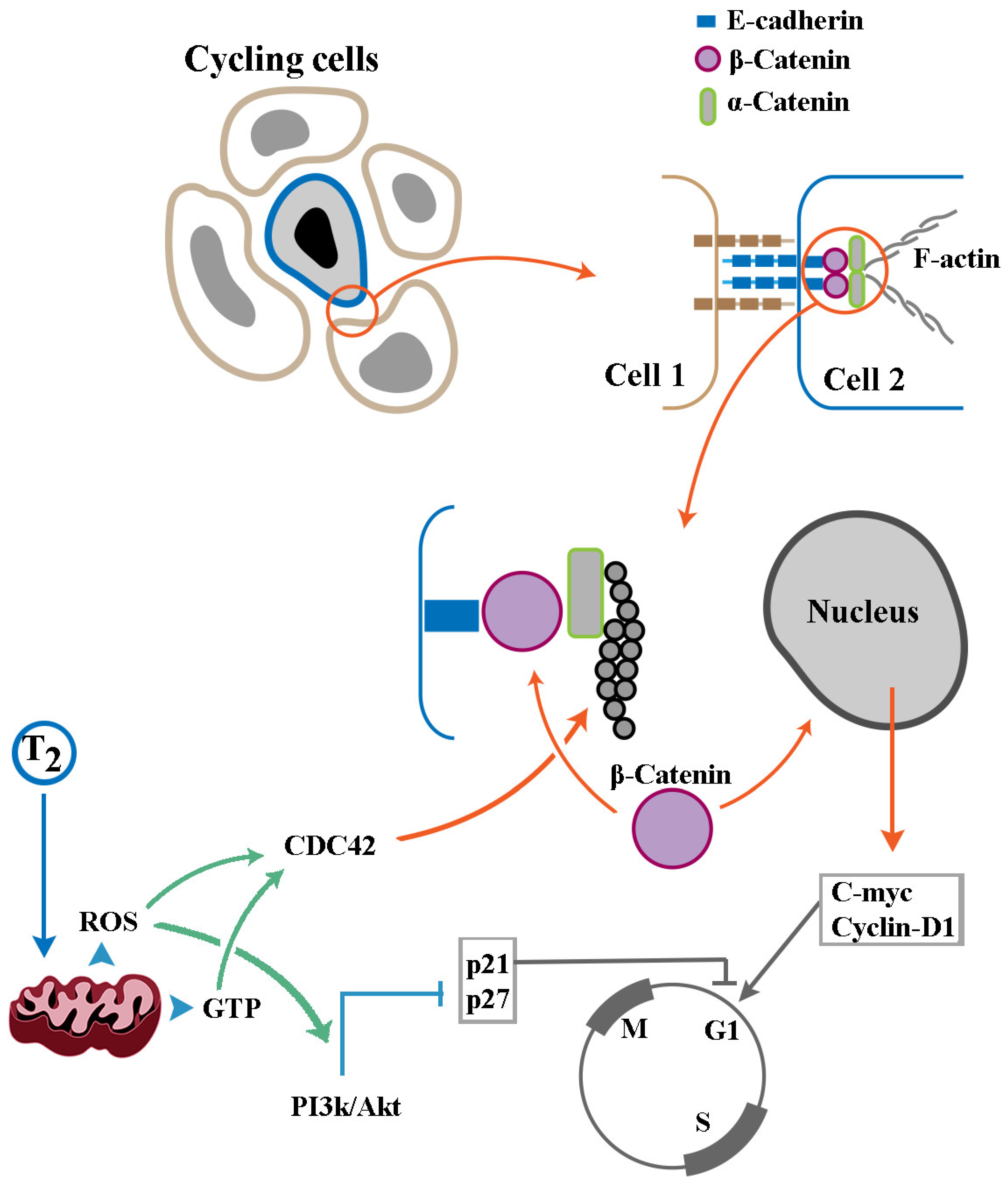
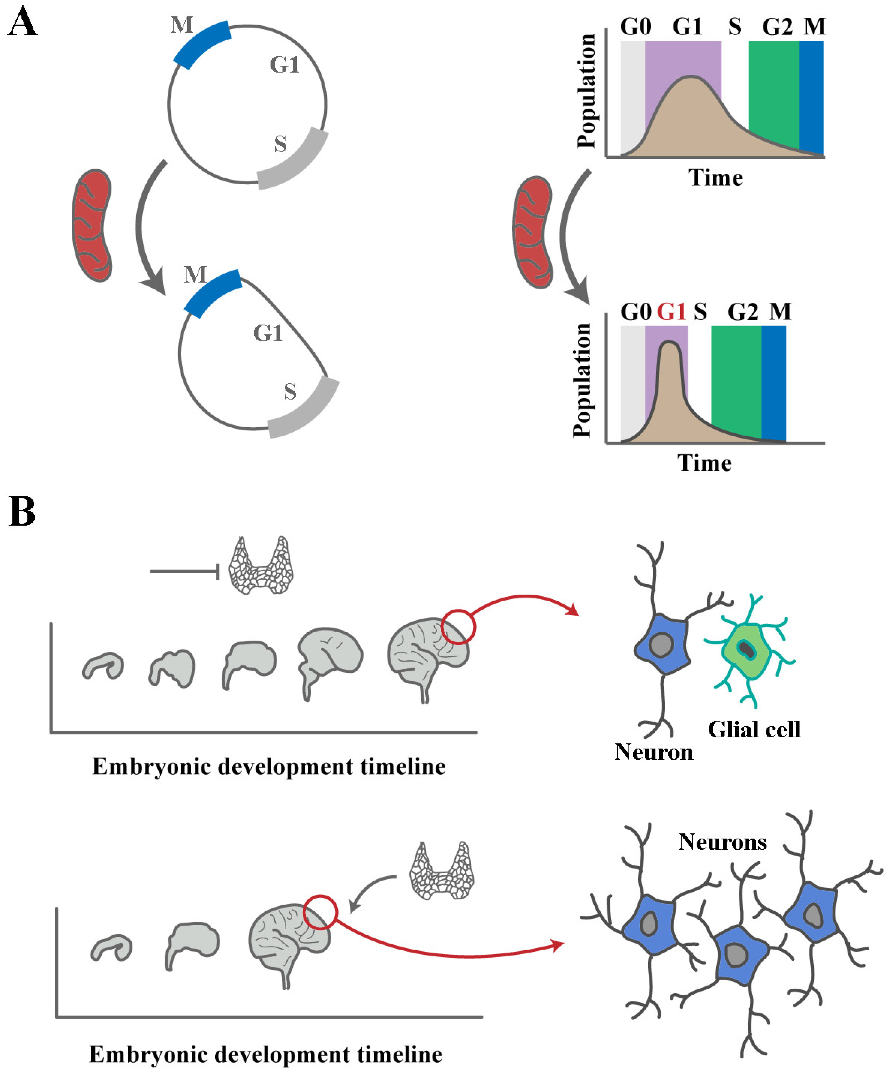
Cellular Self-Organisation and Brain Development: Tempo Informs Function and Spatial Organisation During Organogenesis

In the broadest sense, self-organization refers to emergence of order at a global level by simple local interactions between components of a system [92]. In the context of organogenesis, recursive self-organising interactions between progenitor cells during organ development determine whether cycling cells remain in a proliferative pool or embark upon differentiation [93]. These interactions also determine where along a migratory path within a developing embryo, cells assume a differentiated fate [94]. To this end, cadherin-mediated intercellular interactions appear to be central to spatial organisation as well as resolution of fate dichotomies and the resultant emergence of form and function by self-organisation [94,95]. To regulate spatial organisation, cadherin-based homo-polymeric interactions determine the directionality of collective cell migration by organising intracellular actin bundles [96] (Figure 2). Regulation of cell cycle by cadherin-based junctions is underpinned by dual functionality of β-catenin [97]. This protein not only serves as a structural component of cadherin-based junctions [97], but also trans-activates two major drivers of G1 phase of cell cycle, cyclin-D1 [98] and c-Myc [99] upon migration to the nucleus. Junctional storage of β-catenin generates a reserve protected pool of the protein as the unbound free cytoplasmic protein is unstable and rapidly degraded by a destruction complex subsequent to phosphorylation by Gsk-3β [100]. Upon release from the junctional complexes, β-catenin faces two opposing fates. If Gsk-3β resides in a repressed state, β-catenin will migrate to nucleus to trans-activate cyclin-D1 [98] and c-Myc [99], otherwise the protein will be phosphorylated and degraded leading to a prolonged G1 phase of cell cycle [101]. Therefore, factors that strengthen cadherin-based junctions (e.g., by promoting actin polymerisation [102]) and simultaneously inhibit Gsk-3β not only modulate spatial organisation of migrating cells, but also reprogram the cell cycle properties (e.g., the length of G1 phase) of these cells. One such factor is the small GTPase Cdc42 [103] that operates as a key organiser of actin polymerisation and an inhibitor of Gsk-3β [104]. A second pathway that regulates actin dynamics [105] and simultaneously contributes to stabilisation of free β-catenin is the PI3k/Akt signalling pathway [106,107]. To understand how non-genomic impacts of TH modulate self-organisation, it is necessary to address how the hormones regulate the dynamics of cadherin-based junctions by influencing the activity of key players such as PI3k/Ask and Cdc42.
We first focus on the capacity for junctional storage of β-catenin. Recruitment of β-catenin to cadherins is regulated at multiple levels [108]. In general, two antagonistic inputs regulate the stability of cadherin-based junctions. IQGAP1 dissociates α-catenin from the E-cadherin-β-catenin complex destabilising it whereas activated Cdc42 and Rac GTPases offset the effect of IQGAP1. A third player is calmodulin which attenuates the binding of IQGAP1 to E-cadherin [109] in response to increased intracellular concentration of Ca2+ [110] thus weakening E-cadherin junctional complexes [111]. Finally, enhanced actin polymerisation increases the stability of cadherin-based junctions [102]. TH interface with the described regulatory dynamics of cadherin-based junctions by stimulating mitochondrial production of ROS [36,37] (Figure 2). ROS-mediated oxidisation enhances the intrinsic rate of GDP dissociation from small GTPases, in particular Cdc42, amplifying the activity of the enzyme by approximately three orders of magnitude [55]. This impact of ROS is expected to stabilise cadherin-based junctions and enrich β-catenin at these junctions [18]. TH also stimulate actin polymerisation [48,49,50] by supplying GTP to small GTPase organisers of actin polymerisation (e.g., Rho GTPases) [51,52,53,54] along with ROS-mediated stimulation of GDP release to activate the enzymes [55]. By these activities, β-catenin will be enriched in cadherin-based junctions. TH-induced elevation of cytoplasmic [Ca2+] [41] could then associate with calmodulin to attenuate binding of IQGAP1 to E-cadherin [109] thus weakening E-cadherin junctional complexes [111] and triggering the release of β-catenin. The released β-catenin is expected to be stabilised by TH [112] via multiple mechanisms. First, mitochondrial ROS amplify the activity of Dishevelled (Dvl), a scaffolding protein which disrupts the GSK3β-mediated phosphorylation of β-catenin leading to accumulation of the stabilised cytoplasmic protein [113]. The role of TH-amplified mitochondrial ROS in activating Cdc42 and PI3k/Akt, both of which stabilise β-catenin by regulating the activity of Gsk-3β [103,106,107], complements junctional enrichment of this protein, thus facilitating nuclear translocation of the protein to induce transcription of positive regulators of cell cycle [107]. However, positive input into cell cycle is only effective when inhibitors of cell cycle dynamics are arrested. Accordingly, ROS-mediated rewiring of PI3k/Akt [65] by TH [114] prompts a series of phosphorylation events that inactivate inhibitors of cell cycle progression [115].
Again TH operate in a biphasic manner in regulating the cell cycle. Non-genomic mitochondrial effects of the hormones function as accelerators of cell cycle [116], whereas TR-mediated transcriptional regulation induces cell cycle arrest [117]. This TR-mediated negative feedback is aligned to the antagonistic interaction between genomic and non-genomic consequences of TH signalling. In summary, mechanisms that regulate the tempo of biochemical events within an individual cell, orchestrate self-organisation dynamics at a higher level by regulating the level and fate of β-catenin and the tempo of cell cycle. Following this line of reasoning, it is relevant to ask how accelerated cycling occurring as a consequence of the non-genomic impacts of TH signalling would alter dynamics of self-organisation during brain development.

Heterochronic Signatures of TH in Brain Development

Heterochrony describes a reprograming of ontogeny by changing the timing or the rate of developmental events [118,119]. From this perspective, TH can be classified as bistable heterochronic programmers of brain development. The bistability [120] of outcomes driven by TH stems from a competition between genomic and non-genomic functions of the hormone. Focusing on the impact of TH on cell cycle clearly illustrates this notion. Accelerated progression through G1 phase of cell cycle leads to an enhanced proliferative capacity at an individual cell level and an amplified synchronicity of cycling neural progenitors at a population level [121] (Figure 3). The accelerated synchronised cycling tends to reduce the differentiation propensity of cells by two mechanisms. G1 phase dynamics are pro-differentiation and rapid progression through this phase of cycle renders individual cells more resistant to differentiation cues [122,123]. At a population level, synchronized cycling restricts differentiation-sensitive G1 phases of cycling cells to a narrow temporal window compared to dispersion of G1 phases in an asynchronous population [121]. Hence, differentiation cues that arise during organogenesis are less likely to interact with synchronised rapidly cycling cells to trigger differentiation. The anti-differentiation non-genomic impacts of TH exerted via ROS are counterbalanced by TR-mediated genomic impacts of the hormone that induce cell cycle arrest [117]. Aside from modulation of cell cycle, there is some evidence that receptor-mediated activity of TH primes cycling cells for differentiation by upregulating pro-neural transcription factor NeuroD in developing cerebellum [124]. Owing to acceleration of cell cycle dynamics by TH [116], a deficiency of the hormone is expected to slow brain development (Figure 3). In an MRI-based study of the impact of thyroid disorders on brain development, hypothyroidism was found to be associated with reductions in bilateral total cerebellar and pallidum volumes [125]. In another study, both low and high maternal thyroid functions were found to be associated with smaller child total grey matter and cortical volumes [126]. One plausible explanation for the reduced size of brain in hyperthyroidism is that increased level of TH tips the balance of competition between genomic and non-genomic effects in favour of TR-mediated genomic effects, thus accelerating differentiation of neural progenitor cells and reducing the pool of proliferating cells. Given the absence of histological evidence in the latter study [126], the validity of this hypothesis remains to be assessed. In animal models, reduction of the number of neurons is more evident in regions with significant neurogenic capacity including the olfactory bulb and the granular layers of the hippocampus and cerebellum [127,128,129]. Corroborating the concept of heterochronic reprogramming in hypothyroidism (i.e., reduced tempo of development) is the finding that in hypothyroidism several developmental milestones of brain are delayed. Reported phenotypes include disappearance of the subplate in the cortex [130], delayed regression of the external granular layer of the cerebellum [14], and delayed emergence of Cajal-Retzius cells of cerebellum [131]. In the latter example, cerebellar development was surprisingly normal in mutant mice lacking TRα1 [14]. This observation is aligned to the suggestion that non-genomic effects of TH are responsible for programming brain development and that TR-mediated impacts counterbalance these non-genomic effects. Additional supportive evidence for bistable TH-mediated programming of ontogeny is afforded by the study of myelination during brain development. In agreement with the proposal for a bistable impact on neurogenesis of TH, while hypothyroidism causes delayed deposition of myelin [132,133,134,135], the inverse phenotype of accelerated myelination is observed in hyperthyroidism [136]. Therefore, it can be said that structural and functional modifications of a developing brain in hypothyroidism [137] are inevitable consequences of the altered tempo of development that requires heterochronic reprogramming of cell cycle (Figure 3). A reprogrammed cell cycle not only alters the tempo of development but also modifies differentiation outcome and spatial organisation of neural progenitor cells, some of which will persist as irreversible signature of TH deficiency during brain development [138].

Conclusion

Here we provide evidence for a proposal that thyroid hormones orchestrate development of brain by regulating the tempo of cellular self-organisation. Basic tenets of the proposal are as follows:
  • TH orchestrate brain development by controlling the balance of competition between receptor-mediated genomic and non-genomic mitochondrial effects.
  • A transient suppression of nuclear dynamics facilitates predominance of non-genomic impacts of TH over TR-mediated genomic effects.
  • To assume a morphogenic role, TH reprogram mitochondria to produce reactive oxygen species at an amplified rate.
  • Transient shift to an oxidising milieu as a result of TH signalling leads to rewiring of certain signalling pathways, an accelerated cell cycle, and enhanced tempo of cellular self-organisation.
  • Enhanced tempo of self-organisation in TH signalling is a major determinant of emergence of spatial and functional signatures of cellular self-organisation.
  • In hypothyroidism, the reduced tempo of cellular self-organisation underpins key anatomical and functional alterations of a developing brain.
We suggest that TH signalling during development activates two antagonistic outcomes, direct mitochondrial reprogramming and an adaptive remodelling of transcriptional profile to offset the mitochondrial activities. It is the balance of competition between these two outcomes that determines the tempo of self-organisation and emergence of form and function in a developing brain.

References

  1. Ahmed, O. M.; El-Gareib, A. W.; El-Bakry, A. M.; Abd El-Tawab, S. M.; Ahmed, R. G. Thyroid hormones states and brain development interactions. Int J Dev Neurosci 2008, 26, 147–209. [Google Scholar] [CrossRef]
  2. Bernal, J.; Nunez, J. Thyroid hormones and brain development. Eur J Endocrinol 1995, 133, 390–8. [Google Scholar] [CrossRef] [PubMed]
  3. De Angelis, M.; Maity-Kumar, G.; Schriever, S. C.; Kozlova, E. V.; Muller, T. D.; Pfluger, P. T.; Curras-Collazo, M. C.; Schramm, K. W. Development and validation of an LC-MS/MS methodology for the quantification of thyroid hormones in dko MCT8/OATP1C1 mouse brain. J Pharm Biomed Anal 2022, 221, 115038. [Google Scholar] [CrossRef]
  4. Dussault, J. H.; Ruel, J. Thyroid hormones and brain development. Annu Rev Physiol 1987, 49, 321–34. [Google Scholar] [CrossRef] [PubMed]
  5. Kilby, M. D. Thyroid hormones and fetal brain development. Clin Endocrinol (Oxf) 2003, 59, 280–1. [Google Scholar] [CrossRef]
  6. Pemberton, H. N.; Franklyn, J. A.; Kilby, M. D. Thyroid hormones and fetal brain development. Minerva Ginecol 2005, 57, 367–78. [Google Scholar]
  7. Rovet, J. F. The role of thyroid hormones for brain development and cognitive function. Endocr Dev 2014, 26, 26–43. [Google Scholar] [PubMed]
  8. Gil-Ibanez, P.; Garcia-Garcia, F.; Dopazo, J.; Bernal, J.; Morte, B. Global Transcriptome Analysis of Primary Cerebrocortical Cells: Identification of Genes Regulated by Triiodothyronine in Specific Cell Types. Cereb Cortex 2017, 27, 706–717. [Google Scholar] [CrossRef] [PubMed]
  9. Leonard, J. L. Non-genomic actions of thyroid hormone in brain development. Steroids 2008, 73, 1008–12. [Google Scholar] [CrossRef] [PubMed]
  10. Giammanco, M.; Di Liegro, C. M.; Schiera, G.; Di Liegro, I. Genomic and Non-Genomic Mechanisms of Action of Thyroid Hormones and Their Catabolite 3,5-Diiodo-L-Thyronine in Mammals. Int J Mol Sci 2020, 21. [Google Scholar] [CrossRef] [PubMed]
  11. Zoeller, T. R.; Dowling, A. L.; Herzig, C. T.; Iannacone, E. A.; Gauger, K. J.; Bansal, R. Thyroid hormone, brain development, and the environment. Environ Health Perspect 2002, 110 Suppl 3, 355–61. [Google Scholar] [CrossRef]
  12. Delange, F. Neonatal screening for congenital hypothyroidism: results and perspectives. Horm Res 1997, 48, 51–61. [Google Scholar] [CrossRef]
  13. Klein, R. , History of congenital hypothyroidism. In Neonatal Thyroid Screening, 1980; pp 51-59.
  14. Morte, B.; Manzano, J.; Scanlan, T.; Vennstrom, B.; Bernal, J. Deletion of the thyroid hormone receptor alpha 1 prevents the structural alterations of the cerebellum induced by hypothyroidism. Proc Natl Acad Sci U S A 2002, 99, 3985–9. [Google Scholar] [CrossRef]
  15. Harper, M. E.; Seifert, E. L. Thyroid hormone effects on mitochondrial energetics. Thyroid 2008, 18, 145–56. [Google Scholar] [CrossRef]
  16. Soboll, S. Thyroid hormone action on mitochondrial energy transfer. Biochim Biophys Acta 1993, 1144, 1–16. [Google Scholar] [CrossRef] [PubMed]
  17. Iwata, R.; Casimir, P.; Vanderhaeghen, P. Mitochondrial dynamics in postmitotic cells regulate neurogenesis. Science 2020, 369, 858–862. [Google Scholar] [CrossRef]
  18. Ozsoy, S.; Vujovic, F.; Simonian, M.; Valova, V.; Hunter, N.; Farahani, R. M. Cannibalized erythroblasts accelerate developmental neurogenesis by regulating mitochondrial dynamics. Cell Rep 2021, 35, 108942. [Google Scholar] [CrossRef]
  19. Beckervordersandforth, R.; Ebert, B.; Schaffner, I.; Moss, J.; Fiebig, C.; Shin, J.; Moore, D. L.; Ghosh, L.; Trinchero, M. F.; Stockburger, C.; Friedland, K.; Steib, K.; von Wittgenstein, J.; Keiner, S.; Redecker, C.; Holter, S. M.; Xiang, W.; Wurst, W.; Jagasia, R.; Schinder, A. F.; Ming, G. L.; Toni, N.; Jessberger, S.; Song, H.; Lie, D. C. Role of Mitochondrial Metabolism in the Control of Early Lineage Progression and Aging Phenotypes in Adult Hippocampal Neurogenesis. Neuron 2017, 93, 1518. [Google Scholar] [CrossRef] [PubMed]
  20. Khacho, M.; Clark, A.; Svoboda, D. S.; Azzi, J.; MacLaurin, J. G.; Meghaizel, C.; Sesaki, H.; Lagace, D. C.; Germain, M.; Harper, M. E.; Park, D. S.; Slack, R. S. Mitochondrial Dynamics Impacts Stem Cell Identity and Fate Decisions by Regulating a Nuclear Transcriptional Program. Cell Stem Cell 2016, 19, 232–247. [Google Scholar] [CrossRef] [PubMed]
  21. Khacho, M.; Slack, R. S. Mitochondrial dynamics in the regulation of neurogenesis: From development to the adult brain. Dev Dyn 2018, 247, 47–53. [Google Scholar] [CrossRef]
  22. Vujovic, F.; Simonian, M.; Hughes, W. E.; Shepherd, C. E.; Hunter, N.; Farahani, R. M. Mitochondria facilitate neuronal differentiation by metabolising nuclear-encoded RNA. Cell Commun Signal 2024, 22, 450. [Google Scholar] [CrossRef] [PubMed]
  23. Vujovic, F.; Hunter, N.; Farahani, R. M. Notch ankyrin domain: evolutionary rise of a thermodynamic sensor. Cell Commun Signal 2022, 20, 66. [Google Scholar] [CrossRef] [PubMed]
  24. Chuma, S.; Kiyosue, K.; Akiyama, T.; Kinoshita, M.; Shimazaki, Y.; Uchiyama, S.; Sotoma, S.; Okabe, K.; Harada, Y. Implication of thermal signaling in neuronal differentiation revealed by manipulation and measurement of intracellular temperature. Nat Commun 2024, 15, 3473. [Google Scholar] [CrossRef]
  25. Belinsky, G. S.; Antic, S. D. Mild hypothermia inhibits differentiation of human embryonic and induced pluripotent stem cells. Biotechniques 2013, 55, 79–82. [Google Scholar] [CrossRef] [PubMed]
  26. Kennedy, K. A.; Sandiford, S. D.; Skerjanc, I. S.; Li, S. S. Reactive oxygen species and the neuronal fate. Cell Mol Life Sci 2012, 69, 215–21. [Google Scholar] [CrossRef]
  27. McGrath, K. E.; Koniski, A. D.; Malik, J.; Palis, J. Circulation is established in a stepwise pattern in the mammalian embryo. Blood 2003, 101, 1669–76. [Google Scholar] [CrossRef]
  28. Rodenfels, J.; Neugebauer, K. M.; Howard, J. Heat Oscillations Driven by the Embryonic Cell Cycle Reveal the Energetic Costs of Signaling. Dev Cell, 2019; 48, 646–658.e6. [Google Scholar]
  29. Kashiwagi, A.; Hanada, H.; Yabuki, M.; Kanno, T.; Ishisaka, R.; Sasaki, J.; Inoue, M.; Utsumi, K. Thyroxine enhancement and the role of reactive oxygen species in tadpole tail apoptosis. Free Radic Biol Med 1999, 26, 1001–9. [Google Scholar] [CrossRef]
  30. Cheng, S. Y.; Leonard, J. L.; Davis, P. J. Molecular aspects of thyroid hormone actions. Endocr Rev 2010, 31, 139–70. [Google Scholar] [CrossRef]
  31. van der Spek, A. H.; Fliers, E.; Boelen, A. The classic pathways of thyroid hormone metabolism. Mol Cell Endocrinol 2017, 458, 29–38. [Google Scholar] [CrossRef] [PubMed]
  32. Wu, Y. F.; Koenig, R. J. Gene regulation by thyroid hormone. Trends Endocrin Met 2000, 11, 207–211. [Google Scholar] [CrossRef]
  33. Giannocco, G.; Kizys, M. M. L.; Maciel, R. M.; de Souza, J. S. Thyroid hormone, gene expression, and Central Nervous System: Where we are. Semin Cell Dev Biol 2021, 114, 47–56. [Google Scholar] [CrossRef]
  34. Dasgupta, A.; Das, S.; Sarkar, P. K. Thyroid hormone promotes glutathione synthesis in astrocytes by up regulation of glutamate cysteine ligase through differential stimulation of its catalytic and modulator subunit mRNAs. Free Radic Biol Med 2007, 42, 617–26. [Google Scholar] [CrossRef]
  35. Averill-Bates, D. A. The antioxidant glutathione. Vitam Horm 2023, 121, 109–141. [Google Scholar]
  36. Venditti, P.; Di Meo, S. Thyroid hormone-induced oxidative stress. Cell Mol Life Sci 2006, 63, 414–34. [Google Scholar] [CrossRef]
  37. Chattopadhyay, S.; Sahoo, D. K.; Roy, A.; Samanta, L.; Chainy, G. B. Thiol redox status critically influences mitochondrial response to thyroid hormone-induced hepatic oxidative injury: A temporal analysis. Cell Biochem Funct 2010, 28, 126–34. [Google Scholar] [CrossRef]
  38. Shi, Y. B. Life Without Thyroid Hormone Receptor. Endocrinology 2021, 162. [Google Scholar] [CrossRef] [PubMed]
  39. Buchholz, D. R.; Shi, Y. B. Dual function model revised by thyroid hormone receptor alpha knockout frogs. Gen Comp Endocrinol 2018, 265, 214–218. [Google Scholar] [CrossRef] [PubMed]
  40. Gothe, S.; Wang, Z.; Ng, L.; Kindblom, J. M.; Barros, A. C.; Ohlsson, C.; Vennstrom, B.; Forrest, D. Mice devoid of all known thyroid hormone receptors are viable but exhibit disorders of the pituitary-thyroid axis, growth, and bone maturation. Genes Dev 1999, 13, 1329–41. [Google Scholar] [CrossRef] [PubMed]
  41. Segal, J.; Ingbar, S. H. An immediate increase in calcium accumulation by rat thymocytes induced by triiodothyronine: its role in the subsequent metabolic responses. Endocrinology 1984, 115, 160–6. [Google Scholar] [CrossRef] [PubMed]
  42. Segal, J.; Ingbar, S. H. Stimulation by triiodothyronine of the in vitro uptake of sugars by rat thymocytes. J Clin Invest 1979, 63, 507–15. [Google Scholar] [CrossRef] [PubMed]
  43. Segal, J.; Ingbar, S. H. Stimulation of 2-deoxy-D-glucose uptake in rat thymocytes in vitro by physiological concentrations of triiodothyronine, insulin, or epinephrine. Endocrinology 1980, 107, 1354–8. [Google Scholar] [CrossRef]
  44. Segal, J.; Hardiman, J.; Ingbar, S. H. Stimulation of calcium-ATPase activity by 3,5,3′-tri-iodothyronine in rat thymocyte plasma membranes. A possible role in the modulation of cellular calcium concentration. Biochem J 1989, 261, 749–54. [Google Scholar] [CrossRef]
  45. Pettit, F. H.; Roche, T. E.; Reed, L. J. Function of calcium ions in pyruvate dehydrogenase phosphatase activity. Biochem Biophys Res Commun 1972, 49, 563–71. [Google Scholar] [CrossRef] [PubMed]
  46. Glancy, B.; Willis, W. T.; Chess, D. J.; Balaban, R. S. Effect of calcium on the oxidative phosphorylation cascade in skeletal muscle mitochondria. Biochemistry 2013, 52, 2793–809. [Google Scholar] [CrossRef] [PubMed]
  47. Vilas-Boas, E. A.; Cabral-Costa, J. V.; Ramos, V. M.; Caldeira da Silva, C. C.; Kowaltowski, A. J. Goldilocks calcium concentrations and the regulation of oxidative phosphorylation: Too much, too little, or just right. J Biol Chem 2023, 299, 102904. [Google Scholar] [CrossRef] [PubMed]
  48. Farwell, A. P.; Dubord-Tomasetti, S. A.; Pietrzykowski, A. Z.; Stachelek, S. J.; Leonard, J. L. Regulation of cerebellar neuronal migration and neurite outgrowth by thyroxine and 3,3′,5′-triiodothyronine. Brain Res Dev Brain Res 2005, 154, 121–35. [Google Scholar] [CrossRef]
  49. Siegrist-Kaiser, C. A.; Juge-Aubry, C.; Tranter, M. P.; Ekenbarger, D. M.; Leonard, J. L. Thyroxine-dependent modulation of actin polymerization in cultured astrocytes. A novel, extranuclear action of thyroid hormone. J Biol Chem, 1990; 265, 5296–302. [Google Scholar]
  50. Farwell, A. P.; Dubord-Tomasetti, S. A.; Pietrzykowski, A. Z.; Leonard, J. L. Dynamic nongenomic actions of thyroid hormone in the developing rat brain. Endocrinology 2006, 147, 2567–74. [Google Scholar] [CrossRef]
  51. Aspenstrom, P. The Rho GTPases have multiple effects on the actin cytoskeleton. Exp Cell Res 1999, 246, 20–5. [Google Scholar] [CrossRef] [PubMed]
  52. Begum, R.; Nur, E. K. M. S.; Zaman, M. A. The role of Rho GTPases in the regulation of the rearrangement of actin cytoskeleton and cell movement. Exp Mol Med 2004, 36, 358–66. [Google Scholar] [CrossRef]
  53. Sit, S. T.; Manser, E. Rho GTPases and their role in organizing the actin cytoskeleton. J Cell Sci 2011, 124, 679–83. [Google Scholar] [CrossRef] [PubMed]
  54. Ma, L.; Rohatgi, R.; Kirschner, M. W. The Arp2/3 complex mediates actin polymerization induced by the small GTP-binding protein Cdc42. Proc Natl Acad Sci U S A 1998, 95, 15362–7. [Google Scholar] [CrossRef] [PubMed]
  55. Heo, J.; Campbell, S. L. Mechanism of redox-mediated guanine nucleotide exchange on redox-active Rho GTPases. J Biol Chem 2005, 280, 31003–10. [Google Scholar] [CrossRef]
  56. Thomas, W. E.; Crespo-Armas, A.; Mowbray, J. The influence of nanomolar calcium ions and physiological levels of thyroid hormone on oxidative phosphorylation in rat liver mitochondria. A possible signal amplification control mechanism. Biochem J 1987, 247, 315–20. [Google Scholar] [CrossRef] [PubMed]
  57. Sterling, K.; Milch, P. O.; Brenner, M. A.; Lazarus, J. H. Thyroid hormone action: the mitochondrial pathway. Science 1977, 197, 996–9. [Google Scholar] [CrossRef]
  58. Goglia, F.; Lanni, A.; Barth, J.; Kadenbach, B. Interaction of diiodothyronines with isolated cytochrome c oxidase. FEBS Lett 1994, 346, 295–8. [Google Scholar]
  59. Lanni, A.; Moreno, M.; Horst, C.; Lombardi, A.; Goglia, F. Specific binding sites for 3,3′-diiodo-L-thyronine (3,3′-T2) in rat liver mitochondria. FEBS Lett 1994, 351, 237–40. [Google Scholar] [CrossRef]
  60. Leary, S. C.; Barton, K. N.; Ballantyne, J. S. Direct effects of 3,5,3′-triiodothyronine and 3,5-diiodothyronine on mitochondrial metabolism in the goldfish Carassius auratus. Gen Comp Endocrinol 1996, 104, 61–6. [Google Scholar] [CrossRef] [PubMed]
  61. Arnold, S.; Goglia, F.; Kadenbach, B. 3,5-Diiodothyronine binds to subunit Va of cytochrome-c oxidase and abolishes the allosteric inhibition of respiration by ATP. Eur J Biochem 1998, 252, 325–30. [Google Scholar] [CrossRef]
  62. Groen, A. K.; Wanders, R. J. A.; Westerhoff, H. V.; Vandermeer, R.; Tager, J. M. Quantification of the Contribution of Various Steps to the Control of Mitochondrial Respiration. Journal of Biological Chemistry 1982, 257, 2754–2757. [Google Scholar] [CrossRef] [PubMed]
  63. Ludwig, B.; Bender, E.; Arnold, S.; Huttemann, M.; Lee, I.; Kadenbach, B. Cytochrome C oxidase and the regulation of oxidative phosphorylation. Chembiochem 2001, 2, 392–403. [Google Scholar] [CrossRef] [PubMed]
  64. Bender, E.; Kadenbach, B. The allosteric ATP-inhibition of cytochrome c oxidase activity is reversibly switched on by cAMP-dependent phosphorylation. FEBS Lett 2000, 466, 130–4. [Google Scholar] [CrossRef] [PubMed]
  65. Vujovic, F.; Shepherd, C. E.; Witting, P. K.; Hunter, N.; Farahani, R. M. Redox-Mediated Rewiring of Signalling Pathways: The Role of a Cellular Clock in Brain Health and Disease. Antioxidants (Basel) 2023, 12. [Google Scholar] [CrossRef]
  66. Hemmings, B. A.; Restuccia, D. F. The PI3K-PKB/Akt pathway. Cold Spring Harb Perspect Biol 2015, 7. [Google Scholar] [CrossRef] [PubMed]
  67. Nakahata, S.; Ichikawa, T.; Maneesaay, P.; Saito, Y.; Nagai, K.; Tamura, T.; Manachai, N.; Yamakawa, N.; Hamasaki, M.; Kitabayashi, I.; Arai, Y.; Kanai, Y.; Taki, T.; Abe, T.; Kiyonari, H.; Shimoda, K.; Ohshima, K.; Horii, A.; Shima, H.; Taniwaki, M.; Yamaguchi, R.; Morishita, K. Loss of NDRG2 expression activates PI3K-AKT signalling via PTEN phosphorylation in ATLL and other cancers. Nat Commun 2014, 5, 3393. [Google Scholar] [CrossRef]
  68. Zhang, X. C.; Piccini, A.; Myers, M. P.; Van Aelst, L.; Tonks, N. K. Functional analysis of the protein phosphatase activity of PTEN. Biochem J 2012, 444, 457–64. [Google Scholar] [CrossRef]
  69. Lee, S. R.; Yang, K. S.; Kwon, J.; Lee, C.; Jeong, W.; Rhee, S. G. Reversible inactivation of the tumor suppressor PTEN by H2O2. J Biol Chem 2002, 277, 20336–42. [Google Scholar] [CrossRef] [PubMed]
  70. Eales, J. G. Iodine metabolism and thyroid-related functions in organisms lacking thyroid follicles: Are thyroid hormones also vitamins? P Soc Exp Biol Med 1997, 214, 302–317. [Google Scholar] [CrossRef]
  71. Heyland, A.; Moroz, L. L. Cross-kingdom hormonal signaling: an insight from thyroid hormone functions in marine larvae. J Exp Biol 2005, 208, 4355–61. [Google Scholar] [CrossRef]
  72. Heyland, A.; Hodin, J.; Reitzel, A. M. Hormone signaling in evolution and development: a non-model system approach. Bioessays 2005, 27, 64–75. [Google Scholar] [CrossRef]
  73. Heyland, A.; Hodin, J. Heterochronic developmental shift caused by thyroid hormone in larval sand dollars and its implications for phenotypic plasticity and the evolution of nonfeeding development. Evolution 2004, 58, 524–38. [Google Scholar]
  74. Zwahlen, J.; Gairin, E.; Vianello, S.; Mercader, M.; Roux, N.; Laudet, V. The ecological function of thyroid hormones. Philos Trans R Soc Lond B Biol Sci 2024, 379, 20220511. [Google Scholar] [CrossRef] [PubMed]
  75. Behringer, V.; Deimel, C.; Hohmann, G.; Negrey, J.; Schaebs, F. S.; Deschner, T. Applications for non-invasive thyroid hormone measurements in mammalian ecology, growth, and maintenance. Horm Behav 2018, 105, 66–85. [Google Scholar] [CrossRef] [PubMed]
  76. Buchholz, D. R.; Hsia, S. C. V.; Fu, L. Z.; Shi, Y. B. A dominant-negative thyroid hormone receptor blocks amphibian metamorphosis by retaining corepressors at target genes. Mol Cell Biol 2003, 23, 6750–6758. [Google Scholar] [CrossRef] [PubMed]
  77. Buchholz, D. R.; Tomita, A.; Fu, L. Z.; Paul, B. D.; Shi, Y. B. Transgenic analysis reveals that thyroid hormone receptor is sufficient to mediate the thyroid hormone signal in frog metamorphosis. Mol Cell Biol 2004, 24, 9026–9037. [Google Scholar] [CrossRef] [PubMed]
  78. Taylor, E.; Heyland, A. Evolution of thyroid hormone signaling in animals: Non-genomic and genomic modes of action. Mol Cell Endocrinol 2017, 459, 14–20. [Google Scholar] [CrossRef]
  79. Jiao, Y.; Palli, S. R. Mitochondria dysfunction impairs Tribolium castaneum wing development during metamorphosis. Commun Biol 2022, 5, 1252. [Google Scholar] [CrossRef]
  80. Merkey, A. B.; Wong, C. K.; Hoshizaki, D. K.; Gibbs, A. G. Energetics of metamorphosis in Drosophila melanogaster. J Insect Physiol 2011, 57, 1437–45. [Google Scholar] [CrossRef] [PubMed]
  81. Odell, J. P. Energetics of metamorphosis in two holometabolous insect species:: (Lepidoptera: Sphingidae) and (Coleoptera: Tenebrionidae). J Exp Zool 1998, 280, 344–353. [Google Scholar] [CrossRef]
  82. Chamberlin, M. E. Changes in mitochondrial electron transport chain activity during insect metamorphosis. Am J Physiol Regul Integr Comp Physiol 2007, 292, R1016–22. [Google Scholar] [CrossRef] [PubMed]
  83. Chouchani, E. E.; Pell, V.; Gaude, E.; Aksentijevic, D.; Sundier, S.; Duchen, M.; Shattock, M.; Frezza, C.; Krieg, T.; Murphy, M.; Uk, M. R. C.; Res, C. I. H.; Trust, G. C.; Fdn, B. H. Ischaemic accumulation of succinate controls reperfusion injury through mitochondrial ROS. Eur J Heart Fail 2015, 17, 29–30. [Google Scholar] [CrossRef] [PubMed]
  84. Taylor, C. T.; Moncada, S. Nitric oxide, cytochrome C oxidase, and the cellular response to hypoxia. Arterioscler Thromb Vasc Biol 2010, 30, 643–7. [Google Scholar] [CrossRef] [PubMed]
  85. Inoue, M.; Sato, E. F.; Nishikawa, M.; Hiramoto, K.; Kashiwagi, A.; Utsumi, K. Free radical theory of apoptosis and metamorphosis. Redox Rep 2004, 9, 237–47. [Google Scholar] [CrossRef]
  86. Hanada, H.; Kashiwagi, A.; Takehara, Y.; Kanno, T.; Yabuki, M.; Sasaki, J.; Inoue, M.; Utsumi, K. Do reactive oxygen species underlie the mechanism of apoptosis in the tadpole tail? Free Radical Bio Med 1997, 23, 294–301. [Google Scholar] [CrossRef] [PubMed]
  87. Oleynikov, I. P.; Sudakov, R. V.; Azarkina, N. V.; Vygodina, T. V. Direct Interaction of Mitochondrial Cytochrome c Oxidase with Thyroid Hormones: Evidence for Two Binding Sites. Cells 2022, 11. [Google Scholar] [CrossRef]
  88. Tagami, T.; Madison, L. D.; Nagaya, T.; Jameson, J. L. Nuclear receptor corepressors activate rather than suppress basal transcription of genes that are negatively regulated by thyroid hormone. Mol Cell Biol 1997, 17, 2642–2648. [Google Scholar] [CrossRef]
  89. Papandreou, M. E.; Tavernarakis, N. Nucleophagy: from homeostasis to disease. Cell Death Differ 2019, 26, 630–639. [Google Scholar] [CrossRef]
  90. Rezaei-Lotfi, S.; Vujovic, F.; Simonian, M.; Hunter, N.; Farahani, R. M. Programmed genomic instability regulates neural transdifferentiation of human brain microvascular pericytes. Genome Biol 2021, 22, 334. [Google Scholar] [CrossRef]
  91. Akinduro, O.; Sully, K.; Patel, A.; Robinson, D. J.; Chikh, A.; McPhail, G.; Braun, K. M.; Philpott, M. P.; Harwood, C. A.; Byrne, C.; O’Shaughnessy, R. F. L.; Bergamaschi, D. Constitutive Autophagy and Nucleophagy during Epidermal Differentiation. J Invest Dermatol 2016, 136, 1460–1470. [Google Scholar] [CrossRef]
  92. Wedlich-Soldner, R.; Betz, T. Self-organization: the fundament of cell biology. Philos Trans R Soc Lond B Biol Sci 2018, 373. [Google Scholar] [CrossRef]
  93. Petridis, A. K.; El-Maarouf, A.; Rutishauser, U. Polysialic acid regulates cell contact-dependent neuronal differentiation of progenitor cells from the subventricular zone. Dev Dyn 2004, 230, 675–84. [Google Scholar] [CrossRef]
  94. Rezaei-Lotfi, S.; Hunter, N.; Farahani, R. M. Coupled cycling programs multicellular self-organization of neural progenitors. Cell Cycle 2019, 18, 2040–2054. [Google Scholar] [CrossRef]
  95. Toda, S.; Blauch, L. R.; Tang, S. K. Y.; Morsut, L.; Lim, W. A. Programming self-organizing multicellular structures with synthetic cell-cell signaling. Science 2018, 361, 156–162. [Google Scholar] [CrossRef] [PubMed]
  96. Cai, D.; Chen, S. C.; Prasad, M.; He, L.; Wang, X.; Choesmel-Cadamuro, V.; Sawyer, J. K.; Danuser, G.; Montell, D. J. Mechanical feedback through E-cadherin promotes direction sensing during collective cell migration. Cell 2014, 157, 1146–59. [Google Scholar] [CrossRef]
  97. Aberle, H.; Schwartz, H.; Kemler, R. Cadherin-catenin complex: protein interactions and their implications for cadherin function. J Cell Biochem 1996, 61, 514–23. [Google Scholar] [CrossRef]
  98. Shtutman, M.; Zhurinsky, J.; Simcha, I.; Albanese, C.; D’Amico, M.; Pestell, R.; Ben-Ze’ev, A. The cyclin D1 gene is a target of the beta-catenin/LEF-1 pathway. Proc Natl Acad Sci U S A 1999, 96, 5522–7. [Google Scholar] [CrossRef]
  99. He, T. C.; Sparks, A. B.; Rago, C.; Hermeking, H.; Zawel, L.; da Costa, L. T.; Morin, P. J.; Vogelstein, B.; Kinzler, K. W. Identification of c-MYC as a target of the APC pathway. Science 1998, 281, 1509–12. [Google Scholar] [CrossRef]
  100. Wu, D.; Pan, W. GSK3: a multifaceted kinase in Wnt signaling. Trends Biochem Sci 2010, 35, 161–8. [Google Scholar] [CrossRef]
  101. Suh, K.; Cho, Y. K.; Breinyn, I. B.; Cohen, D. J. E-cadherin biomaterials reprogram collective cell migration and cell cycling by forcing homeostatic conditions. Cell Rep 2024, 43, 113743. [Google Scholar] [CrossRef] [PubMed]
  102. Rao, M. V.; Zaidel-Bar, R. Formin-mediated actin polymerization at cell-cell junctions stabilizes E-cadherin and maintains monolayer integrity during wound repair. Mol Biol Cell 2016, 27, 2844–56. [Google Scholar] [CrossRef]
  103. Wu, X.; Quondamatteo, F.; Lefever, T.; Czuchra, A.; Meyer, H.; Chrostek, A.; Paus, R.; Langbein, L.; Brakebusch, C. Cdc42 controls progenitor cell differentiation and beta-catenin turnover in skin. Genes Dev 2006, 20, 571–85. [Google Scholar] [CrossRef] [PubMed]
  104. Etienne-Manneville, S.; Hall, A. Cdc42 regulates GSK-3beta and adenomatous polyposis coli to control cell polarity. Nature 2003, 421, 753–6. [Google Scholar] [CrossRef]
  105. Malin, J.; Rosa-Birriel, C.; Hatini, V. Pten, PI3K, and PtdIns(3,4,5)P(3) dynamics control pulsatile actin branching in Drosophila retina morphogenesis. Dev Cell, 2024; 59, 1593–1608 e6. [Google Scholar]
  106. Fukumoto, S.; Hsieh, C. M.; Maemura, K.; Layne, M. D.; Yet, S. F.; Lee, K. H.; Matsui, T.; Rosenzweig, A.; Taylor, W. G.; Rubin, J. S.; Perrella, M. A.; Lee, M. E. Akt participation in the Wnt signaling pathway through Dishevelled. J Biol Chem 2001, 276, 17479–83. [Google Scholar] [CrossRef] [PubMed]
  107. Perry, J. M.; He, X. C.; Sugimura, R.; Grindley, J. C.; Haug, J. S.; Ding, S.; Li, L. Cooperation between both Wnt/beta-catenin and PTEN/PI3K/Akt signaling promotes primitive hematopoietic stem cell self-renewal and expansion. Genes Dev 2011, 25, 1928–42. [Google Scholar] [CrossRef]
  108. Briggs, M. W.; Sacks, D. B. IQGAP1 as signal integrator: Ca2+, calmodulin, Cdc42 and the cytoskeleton. FEBS Lett 2003, 542, 7–11. [Google Scholar] [CrossRef]
  109. Li, Z.; Kim, S. H.; Higgins, J. M.; Brenner, M. B.; Sacks, D. B. IQGAP1 and calmodulin modulate E-cadherin function. J Biol Chem 1999, 274, 37885–92. [Google Scholar] [CrossRef] [PubMed]
  110. Joyal, J. L.; Annan, R. S.; Ho, Y. D.; Huddleston, M. E.; Carr, S. A.; Hart, M. J.; Sacks, D. B. Calmodulin modulates the interaction between IQGAP1 and Cdc42. Identification of IQGAP1 by nanoelectrospray tandem mass spectrometry. J Biol Chem 1997, 272, 15419–25. [Google Scholar] [CrossRef] [PubMed]
  111. Angst, B. D.; Marcozzi, C.; Magee, A. I. The cadherin superfamily: diversity in form and function. J Cell Sci 2001, 114, 629–41. [Google Scholar] [CrossRef]
  112. Wang, L.; Shao, Y. Y.; Ballock, R. T. Thyroid hormone interacts with the Wnt/beta-catenin signaling pathway in the terminal differentiation of growth plate chondrocytes. J Bone Miner Res 2007, 22, 1988–95. [Google Scholar] [CrossRef]
  113. Funato, Y.; Michiue, T.; Asashima, M.; Miki, H. The thioredoxin-related redox-regulating protein nucleoredoxin inhibits Wnt-beta-catenin signalling through dishevelled. Nat Cell Biol 2006, 8, 501–8. [Google Scholar] [CrossRef]
  114. Cao, X.; Kambe, F.; Moeller, L. C.; Refetoff, S.; Seo, H. Thyroid hormone induces rapid activation of Akt/protein kinase B-mammalian target of rapamycin-p70S6K cascade through phosphatidylinositol 3-kinase in human fibroblasts. Mol Endocrinol 2005, 19, 102–12. [Google Scholar] [CrossRef] [PubMed]
  115. Fujita, N.; Sato, S.; Katayama, K.; Tsuruo, T. Akt-dependent phosphorylation of p27Kip1 promotes binding to 14-3-3 and cytoplasmic localization. J Biol Chem 2002, 277, 28706–13. [Google Scholar] [CrossRef]
  116. Alisi, A.; Spagnuolo, S.; Napoletano, S.; Spaziani, A.; Leoni, S. Thyroid hormones regulate DNA-synthesis and cell-cycle proteins by activation of PKCalpha and p42/44 MAPK in chick embryo hepatocytes. J Cell Physiol 2004, 201, 259–65. [Google Scholar] [CrossRef]
  117. Lin, Y. H.; Huang, Y. H.; Wu, M. H.; Wu, S. M.; Chi, H. C.; Liao, C. J.; Chen, C. Y.; Tseng, Y. H.; Tsai, C. Y.; Tsai, M. M.; Lin, K. H. Thyroid hormone suppresses cell proliferation through endoglin-mediated promotion of p21 stability. Oncogene 2013, 32, 3904–14. [Google Scholar] [CrossRef] [PubMed]
  118. Ambros, V. Control of developmental timing in Caenorhabditis elegans. Curr Opin Genet Dev 2000, 10, 428–33. [Google Scholar] [CrossRef]
  119. Ambros, V.; Moss, E. G. Heterochronic genes and the temporal control of C. elegans development. Trends Genet 1994, 10, 123–7. [Google Scholar] [CrossRef] [PubMed]
  120. Rombouts, J.; Gelens, L. Dynamic bistable switches enhance robustness and accuracy of cell cycle transitions. PLoS Comput Biol 2021, 17, e1008231. [Google Scholar] [CrossRef] [PubMed]
  121. Farahani, R.; Rezaei-Lotfi, S.; Simonian, M.; Hunter, N. Bi-modal reprogramming of cell cycle by MiRNA-4673 amplifies human neurogenic capacity. Cell Cycle 2019, 18, 848–868. [Google Scholar] [CrossRef] [PubMed]
  122. Calder, A.; Roth-Albin, I.; Bhatia, S.; Pilquil, C.; Lee, J. H.; Bhatia, M.; Levadoux-Martin, M.; McNicol, J.; Russell, J.; Collins, T.; Draper, J. S. Lengthened G1 phase indicates differentiation status in human embryonic stem cells. Stem Cells Dev 2013, 22, 279–95. [Google Scholar] [CrossRef] [PubMed]
  123. Soufi, A.; Dalton, S. Cycling through developmental decisions: how cell cycle dynamics control pluripotency, differentiation and reprogramming. Development 2016, 143, 4301–4311. [Google Scholar] [CrossRef] [PubMed]
  124. Chantoux, F.; Francon, J. Thyroid hormone regulates the expression of NeuroD/BHF1 during the development of rat cerebellum. Mol Cell Endocrinol 2002, 194, 157–63. [Google Scholar] [CrossRef]
  125. Chambers, T.; Anney, R.; Taylor, P. N.; Teumer, A.; Peeters, R. P.; Medici, M.; Caseras, X.; Rees, D. A. Effects of Thyroid Status on Regional Brain Volumes: A Diagnostic and Genetic Imaging Study in UK Biobank. J Clin Endocrinol Metab 2021, 106, 688–696. [Google Scholar] [CrossRef] [PubMed]
  126. Jansen, T. A.; Korevaar, T. I. M.; Mulder, T. A.; White, T.; Muetzel, R. L.; Peeters, R. P.; Tiemeier, H. Maternal thyroid function during pregnancy and child brain morphology: a time window-specific analysis of a prospective cohort. Lancet Diabetes Endocrinol 2019, 7, 629–637. [Google Scholar] [CrossRef]
  127. Madeira, M. D.; Paula-Barbosa, M.; Cadete-Leite, A.; Tavares, M. A. Unbiased estimate of hippocampal granule cell numbers in hypothyroid and in sex-age-matched control rats. J Hirnforsch 1988, 29, 643–50. [Google Scholar]
  128. Madeira, M. D.; Cadete-Leite, A.; Andrade, J. P.; Paula-Barbosa, M. M. Effects of hypothyroidism upon the granular layer of the dentate gyrus in male and female adult rats: a morphometric study. J Comp Neurol 1991, 314, 171–86. [Google Scholar] [CrossRef] [PubMed]
  129. Madeira, M. D.; Pereira, A.; Cadete-Leite, A.; Paula-Barbosa, M. M. Estimates of volumes and pyramidal cell numbers in the prelimbic subarea of the prefrontal cortex in experimental hypothyroid rats. J Anat 1990, 171, 41–56. [Google Scholar]
  130. Berbel, P.; Navarro, D.; Auso, E.; Varea, E.; Rodriguez, A. E.; Ballesta, J. J.; Salinas, M.; Flores, E.; Faura, C. C.; de Escobar, G. M. Role of late maternal thyroid hormones in cerebral cortex development: an experimental model for human prematurity. Cereb Cortex 2010, 20, 1462–75. [Google Scholar] [CrossRef]
  131. Alvarez-Dolado, M.; Ruiz, M.; Del Rio, J. A.; Alcantara, S.; Burgaya, F.; Sheldon, M.; Nakajima, K.; Bernal, J.; Howell, B. W.; Curran, T.; Soriano, E.; Munoz, A. Thyroid hormone regulates reelin and dab1 expression during brain development. J Neurosci 1999, 19, 6979–93. [Google Scholar] [CrossRef]
  132. Balazs, R.; Brooksbank, B. W.; Davison, A. N.; Eayrs, J. T.; Wilson, D. A. The effect of neonatal thyroidectomy on myelination in the rat brain. Brain Res 1969, 15, 219–32. [Google Scholar] [CrossRef] [PubMed]
  133. Malone, M. J.; Rosman, N. P.; Szoke, M.; Davis, D. Myelination of brain in experimental hypothyroidism. An electron-microscopic and biochemical study of purified myelin isolates. J Neurol Sci, 1975; 26, 1–11. [Google Scholar]
  134. Noguchi, T.; Sugisaki, T. Hypomyelination in the cerebrum of the congenitally hypothyroid mouse (hyt). J Neurochem 1984, 42, 891–3. [Google Scholar] [CrossRef]
  135. Rodriguez-Pena, A.; Ibarrola, N.; Iniguez, M. A.; Munoz, A.; Bernal, J. Neonatal hypothyroidism affects the timely expression of myelin-associated glycoprotein in the rat brain. J Clin Invest 1993, 91, 812–8. [Google Scholar] [CrossRef]
  136. Adamo, A. M.; Aloise, P. A.; Soto, E. F.; Pasquini, J. M. Neonatal hyperthyroidism in the rat produces an increase in the activity of microperoxisomal marker enzymes coincident with biochemical signs of accelerated myelination. J Neurosci Res 1990, 25, 353–9. [Google Scholar] [CrossRef]
  137. O’Shaughnessy, K. L.; Thomas, S. E.; Spring, S. R.; Ford, J. L.; Ford, R. L.; Gilbert, M. E. A transient window of hypothyroidism alters neural progenitor cells and results in abnormal brain development. Sci Rep 2019, 9, 4662. [Google Scholar] [CrossRef] [PubMed]
  138. Kooistra, L.; van der Meere, J. J.; Vulsma, T.; Kalverboer, A. F. Sustained attention problems in children with early treated congenital hypothyroidism. Acta Paediatr 1996, 85, 425–9. [Google Scholar] [CrossRef] [PubMed]
  139. Mitra, K.; Wunder, C.; Roysam, B.; Lin, G.; Lippincott-Schwartz, J. A hyperfused mitochondrial state achieved at G-S regulates cyclin E buildup and entry into S phase. P Natl Acad Sci USA 2009, 106, 11960–11965. [Google Scholar] [CrossRef] [PubMed]
Figure 1. An overview of TH signalling. Genomic effects of thyroid hormones are triggered by association T3 with TRs, an event that relieves the inhibitory activity of TR-bound co-repressor complex (CoR) by inducing conformational change of TRs and the resultant recruitment of a co-activator complex (CoA). Genes activated by this mechanism provide negative feedback to T3-mediated signalling (F1) or counteract non-genomic impact of T2 (F2). Downstream signalling by T2 is mainly mediated by direct reprogramming of the mitochondrial electron transport chain leading to an overproduction of ROS.
Preprints 145657 g001
Figure 2. Mitochondrial regulation of cellular self-organisation dynamics. In a population of cycling cells, cadherin-based junctional complexes inform spatial organisation and the length of G1 phase of cell cycle. Recruitment of β-catenin stabilises junctional complexes, while nuclear localisation of the protein trans-activates genes required for progression of cell cycle. ROS also rewire and activate the PI3k/Akt pathway, a signalling cascade which represses the main inhibitors of cell cycle. Mitochondrial supply of ROS and GTP activate small GTPases (e.g., Cdc42) to stabilize association of F-actin with cadherin-based junctions.
Preprints 145657 g002
Figure 3. Heterochronic programming of brain development by TH. A. Mitochondrial activity driven by TH accelerates progression through G1 phase of cell cycle [139] (left), winding up developmental time at an individual cell level. At a population level, a shortened G1 phase leads to increased synchronicity of cycling cells (right). B. Given that cell cycle regulates differentiation tendency and spatial organisation of cycling cells, a requirement for reprogramming cell cycle to accelerate developmental time means that an altered tempo of organogenesis will be inevitably linked to a modified form and function of a developing brain.
Preprints 145657 g003
Disclaimer/Publisher’s Note: The statements, opinions and data contained in all publications are solely those of the individual author(s) and contributor(s) and not of MDPI and/or the editor(s). MDPI and/or the editor(s) disclaim responsibility for any injury to people or property resulting from any ideas, methods, instructions or products referred to in the content.
Copyright: This open access article is published under a Creative Commons CC BY 4.0 license, which permit the free download, distribution, and reuse, provided that the author and preprint are cited in any reuse.
Prerpints.org logo

Preprints.org is a free preprint server supported by MDPI in Basel, Switzerland.

Subscribe

Disclaimer

Terms of Use

Privacy Policy

Privacy Settings

© 2025 MDPI (Basel, Switzerland) unless otherwise stated