Submitted:
06 January 2025
Posted:
07 January 2025
You are already at the latest version
Abstract
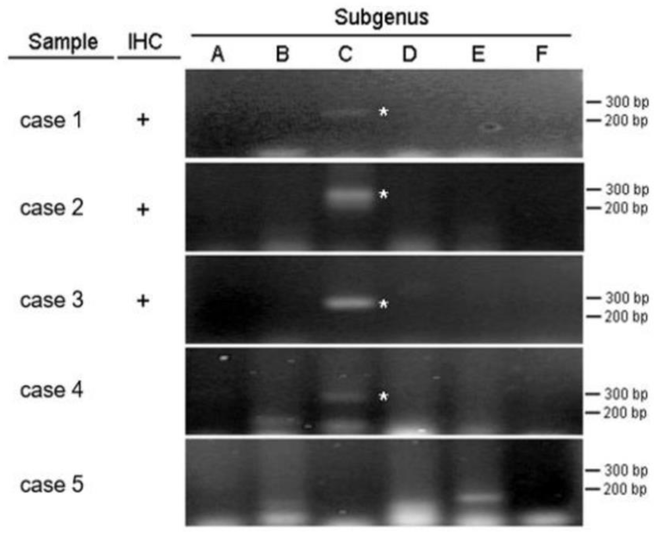
Background: Although some cases of intussusception in older children are associated with pathological changes such as lymphoma or polyps, the cause of most cases in infants is unknown. Objective: Several reports have identified an association between adenovirus infection and intussusception in children. However, much of the evidence is indirect, such as stool samples or throat swab data. Our study analyzed intestinal tissue, which may be more direct evidence of the relationship between adenovirus infection and intussusception. Methods: We retrospectively reviewed children <6 years of age with intussusception who underwent surgery for failed reduction. The pathological tissue was processed into formalin-fixed paraffin-embedded (FFPE) sections. Adenovirus immunohistochemistry (IHC) and polymerase chain reaction (PCR) testing were performed to obtain direct evidence of the relationship between adenovirus infection and intussusception. Results: Our study included 29 patients, 27 appendiceal and 8 intestinal tissues. Only 8 appendix specimens were successfully processed into FFPE tissue. IHC testing was positive in 3 cases (37.5%) and PCR testing was positive for adenovirus type C in 4 cases (50%). The control group consisted of 8 children < 6 years who underwent incidental appendectomies, and all control subjects had negative IHC and PCR analyses. Compared with the control group, the P value of IHC was >0.05, with no statistical difference, while the P value of PCR was <0.05, with statistical difference. Conclusions: We directly confirmed the relationship between adenovirus infection and intussusception through IHC analysis and PCR detection of pathological evidence. PCR is more sensitive than IHC for diagnosing adenovirus in intussusception.
Keywords:
1. Introduction
2. Materials and Methods
2.1. Study Participants
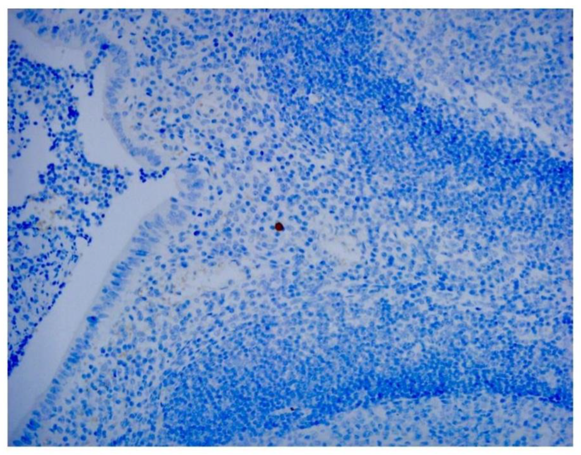
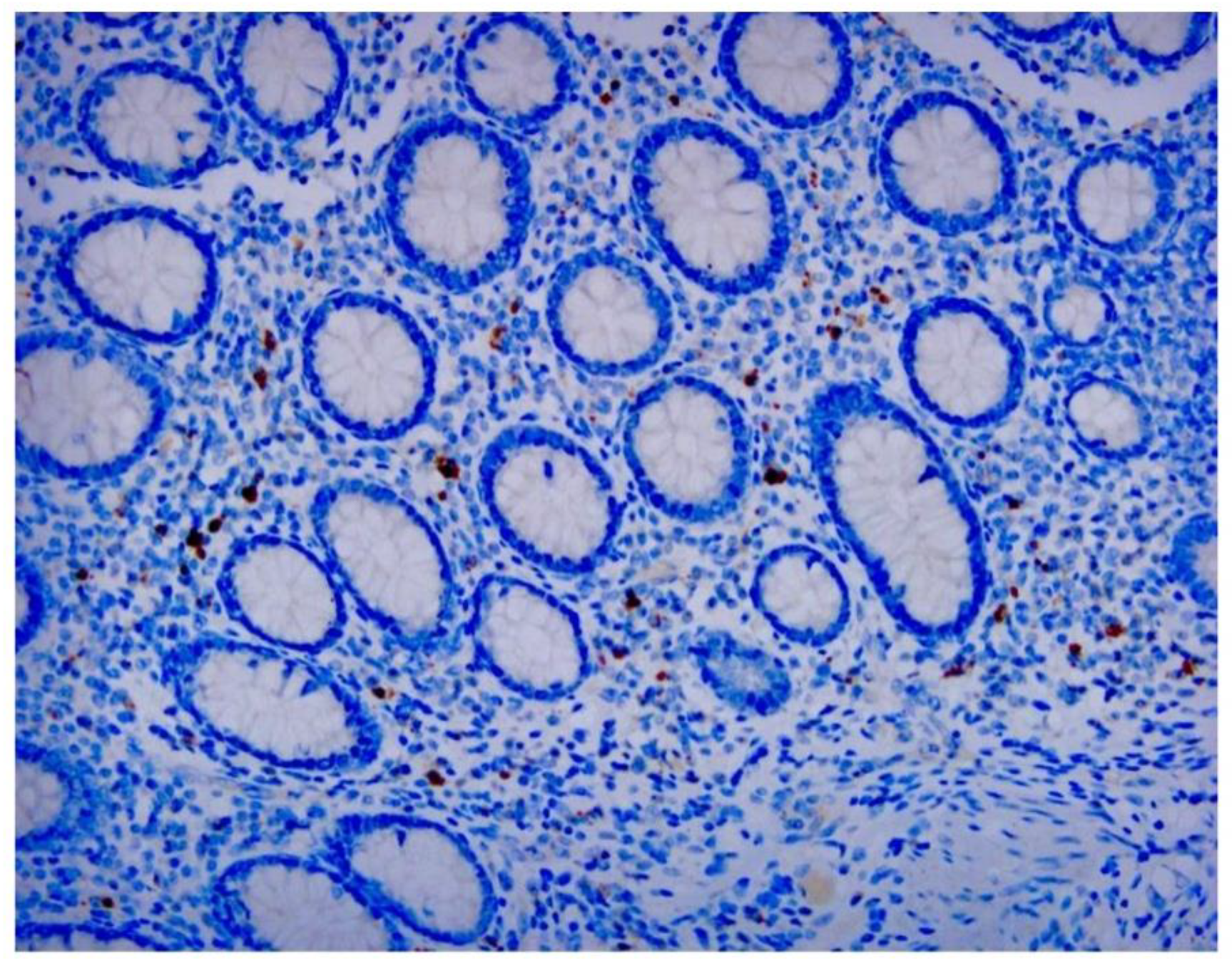
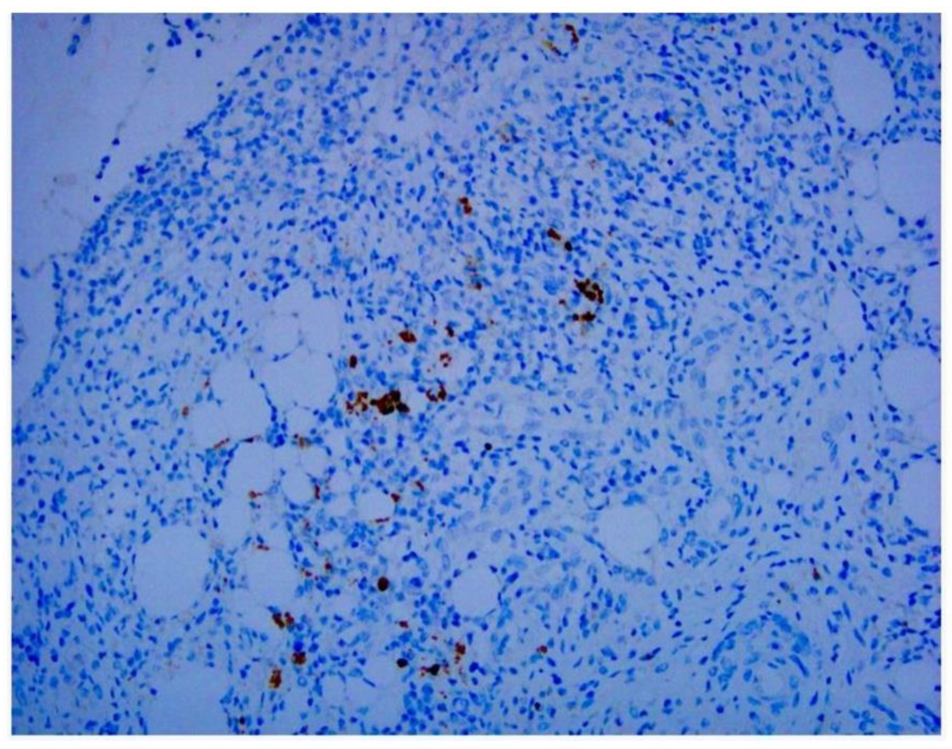
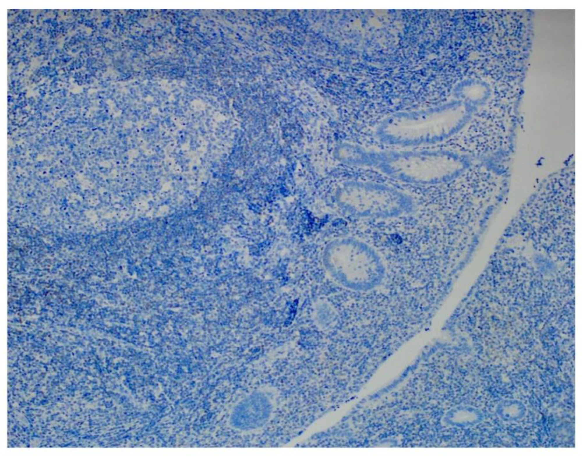
2.2. Immunohistochemical Analysis
2.3. Nested PCR for Adenovirus Species C Hexon DNA
2.4. Statistical Analysis
3. Results
4. Discussion
5. Conclusions
Author Contributions
Funding
Institutional Review Board Statement
Informed Consent Statement
Data Availability Statement
Acknowledgments
Conflicts of Interest
Abbreviations
References
- Jain, S.; Haydel, M.J. Child Intussusception. In: StatPearls. Treasure Island (FL): StatPearls Publishing; 2024, Jan–. pmid: 28613732.
- Shieh, W.J. Human adenovirus infections in pediatric population - an update on clinico-pathologic correlation. Biomed J. 2022, 45, 38–49. [Google Scholar] [CrossRef]
- Lopez-Rippe, J.; Davis, J.C.; Dennis, R.A.; et al. Impact of a 6-12-h delay between ileocolic intussusception diagnostic US and fluoroscopic reduction on patients’ outcomes. Pediatr Radiol. 2024, 54, 1294–1301. [Google Scholar] [CrossRef] [PubMed]
- Niittykoski, M.; von Und Zu Fraunberg, M.; Martikainen, M.; et al. Immunohistochemical characterization and sensitivity to human adenovirus serotypes 3, 5, and 11p of new cell lines derived from human diffuse grade II to IV gliomas. Transl Oncol. 2017, 10, 772–779. [Google Scholar] [CrossRef]
- Lin, L.H.; Huang, C.J.; Lo, C.Y.; et al. Proof of the association between adenovirus infection and appendicitis in children through pathological evidence. J Clin Pathol. 2024, 6, 1–6. [Google Scholar]
- Pring-Akerblom, P.; Trijssenaar, F.E.; Adrian, T.; et al. Multiplex polymerase chain reaction for subgenus-specific detection of human adenoviruses in clinical samples. J Med Virol. 1999, 58, 87–92. [Google Scholar] [CrossRef]
- Park, A.; Lee, C.; Lee, J.Y. Genomic evolution and recombination dynamics of human adenovirus D species: insights from comprehensive bioinformatic analysis. J Microbiol. 2024, 62, 393–407. [Google Scholar] [CrossRef]
- Lin, L.H. Perspectives on intussusception. Pediatr Neonatol. 2013, 54, 143–144. [Google Scholar] [CrossRef]
- Ho, W.L.; Yang, T.W.; Chi, W.C.; et al. Intussusception in Taiwanese children: analysis of incidence, length of hospitalization and hospital costs in different age groups. J Formos Med Assoc. 2005, 104, 398–401. [Google Scholar]
- Carter, M.; Afowork, J.; Pitt, J.B.; et al. Scoring system to evaluate risk of nonoperative management failure in children with intussusception. J Surg Res. 2024, 300, 503–513. [Google Scholar] [CrossRef]
- Clarke Jr, E.J.; Phillips, I.A.; Alexander, E.R. Adenovirus infection in intussusception in children in Taiwan. JAMA. 1969, 208, 1671–1674. [Google Scholar] [CrossRef] [PubMed]
- Bhisitkul, D.M.; Todd, K.M.; Listernick, R. Adenovirus infection and childhood intussusception. Am J Dis Child. 1992, 146, 1331–1333. [Google Scholar] [CrossRef] [PubMed]
- Guarner, J.; de Leon-Bojorge, B.; Lopez-Corella, E.; et al. Intestinal intussusception associated with adenovirus infection in Mexican children. Am J Clin Pathol. 2003, 120, 845–850. [Google Scholar] [CrossRef] [PubMed]
- Chen, S.C.; Wang, J.D.; Hsu, H.Y.; et al. Epidemiology of childhood intussusception and determinants of recurrence and operation: analysis of national health insurance data between 1998 and 2007 in Taiwan. Pediatr Neonatol. 2010, 51, 285–291. [Google Scholar] [CrossRef]
- Okimoto, S.; Hyodo, S.; Yamamoto, M.; et al. Association of viral isolates from stool samples with intussusception in children. Int J Infect Dis. 2011, 15, e641–e645. [Google Scholar] [CrossRef]
- Ukarapol, N.; Khamrin, P.; Khorana, J.; et al. Adenovirus infection: a potential risk for developing intussusception in pediatric patients. J Med Virol. 2016, 88, 1930–1935. [Google Scholar] [CrossRef] [PubMed]
- Jang, J.; Lee, Y.J.; Kim, J.S.; et al. Epidemiological correlation between fecal adenovirus subgroups and pediatric intussusception in Korea. J Korean Med Sci. 2017, 32, 1647–1656. [Google Scholar] [CrossRef] [PubMed]
- Tseng, W.Y.; Chao, H.C.; Chen, C.C.; et al. Adenovirus infection is a risk factor for recurrent intussusception in pediatric patients. Pediatr Neonatol. 2023, 64, 428–434. [Google Scholar] [CrossRef] [PubMed]
- Chen, C.Y.; Lin, L.H. The association of intussusception and adenovirus infection in children: a single center study in Taiwan. FJJM. 2013, 11, 297–303. [Google Scholar]
- Lee, Y.H.; Lin, L.H.; Hung, S.P. Simultaneous intussusception associated with adenovirus infection in monozygotic twins: a case report. Medicine (Baltimore). 2019, 98, e18294. [Google Scholar] [CrossRef] [PubMed]
- Zhou, Y.; Tao, L.; Qiu, J.; et al. Tumor biomarkers for diagnosis, prognosis and targeted therapy. Signal Transduct Target Ther. 2024, 9, 132. [Google Scholar]
- Oumarou Hama, H.; Aboudharam, G.; Barbieri, R.; et al. Immunohistochemical diagnosis of human infectious diseases: a review. Diagn Pathol. 2022, 17, 17. [Google Scholar] [CrossRef]
- Koryukov, M.A.; Oscorbin, I.P.; Novikova, L.M.; et al. A novel multiplex LAMP assay for the detection of respiratory human adenoviruses. Int J Mol Sci. 2024, 25, 7215. [Google Scholar] [CrossRef] [PubMed]
- Jasuja, J.K.; Bub, F.; Veit, J.; et al. Multiplex PCR for bacterial, viral and protozoal pathogens in persistent diarrhoea or persistent abdominal pain in Côte d'Ivoire, Mali and Nepal. Sci Rep. 2024, 14, 10926. [Google Scholar] [CrossRef]

| case number |
Positive IHC analysis |
Positive PCR analysis |
Control group IHC and PCR |
| 1 | + | + | - |
| 2 | + | + | - |
| 3 | + | + | - |
| 4 | - | + | - |
| 5 | - | - | - |
| 6 | - | - | - |
| 7 | - | - | - |
| 8 | - | - | - |
| IHC | |||
| P | |||
| Case | Control | 0.2 | |
| Positive | 3 | 0 | |
| Negative | 5 | 8 | |
| PCR | |||
| P | |||
| Case | Control | 0.047 | |
| Positive | 4 | 0 | |
| Negative | 4 | 8 |
Disclaimer/Publisher’s Note: The statements, opinions and data contained in all publications are solely those of the individual author(s) and contributor(s) and not of MDPI and/or the editor(s). MDPI and/or the editor(s) disclaim responsibility for any injury to people or property resulting from any ideas, methods, instructions or products referred to in the content. |
© 2025 by the authors. Licensee MDPI, Basel, Switzerland. This article is an open access article distributed under the terms and conditions of the Creative Commons Attribution (CC BY) license (http://creativecommons.org/licenses/by/4.0/).


