

Submitted:
01 January 2025
Posted:
03 January 2025
You are already at the latest version
Abstract
Background/Objectives: Spindle cell oncocytomas (SCO) of the pituitary gland are rare tumors often misdiagnosed for nonfunctioning pituitary macroadenomas. Although classified as grade 1, they are often challenging in terms of diagnosis and treatment. Pituitary SCOs harbor peculiar features such as hypervascularity and stronger adherence to surrounding structures, with increased risk of hemorrhage, partial resection, and significantly higher recurrence rate. Almost 100 cases have been reported so far. The role of surgery is still crucial for decompression of the optic chiasm as well as for achieving diagnosis. However, given the higher tendency of recurrence, the role of postoperative radiotherapy has been investigated over the last few years. Case presentation: Here we report a case of a 48-year-old female with a pituitary SCO treated at our Institution focusing on diagnosis, treatment and follow-up. Conclusions: This type of tumor presents a challenge related to its higher vascularity and strong adherence to the surrounding structures. Adjuvant radiotherapy is something that should be considered, especially when gross total resection is not achieved and finally SCOs require diligent follow-up to monitor for any disease recurrence or progression.
Keywords:
spindle cell oncocytoma
; pituitary tumors
; endoscopic endonasal approach
; neuro-oncology
1. Introduction
Spindle cell oncocytomas (SCO) are rare non-neuroendocrine tumors arising from the pituicytes of the posterior pituitary gland. They were included in the World Health Organization (WHO) classification of central nervous system tumors in 2007 after being first described by Roncaroli et al. in 2002 [1,2].
SCOs were previously thought to stem from the adenohypophysis, however, more recent research has identified that SCOs are positive for thyroid transcription factor 1 (TTF1) which is only found in pituicytes and not in folliculostellate cells of the adenohypophysis [3]. In our search from 2002 to 2024, there have only been 98 cases published in literature which highlights the rarity of this type of tumor.
SCOs are often misdiagnosed as pituitary adenomas; yet are important to distinguish as they usually have higher vascularity as well as an increased tendency to invade the surrounding structures. This results in higher recurrence rate, thus requiring closer follow-ups [4]. If gross total resection (GTR) is not achieved, 50% of tumors show significant progression requiring extra treatment by two years and recurrence occurs in 20% of people and even distant metastasis has been reported [4].
Despite these factors, the WHO considers pituitary SCOs a grade 1 tumor with benign behavior [5]. Moreover, although this tumor is considered grade 1, it is important to understand these critical features as it guides treatment and follow up and will improve patient outcomes.
Due to the low incidence of this neoplasm, there is no definitive understanding of the unique clinical signs and symptoms, diagnostic imaging (DI), immunohistochemistry (IHC), and treatment. With this report and literature review we aim to provide further knowledge and understanding of these factors to improve the identification and treatment of individuals with SCO.
2. Case Presentation
We present the case of a 48-year-old female with a two-month history of persistent headaches, mental and physical fatigue, and light sensitivity. Her past medical history includes chronic migraines, fibromyalgia, psoriasis, anxiety, obesity, and obstructive sleep apnea. When looking back, the patient realized that her headache symptoms had changed from her normal migraine symptoms to more frequent albeit less severe frontal pain and aching in the previous months.
Given the persistence of the above-mentioned symptoms, she was investigated with a head computed tomography (CT) scan that showed a pituitary lesion with sellar and suprasellar extension. For further investigation, she underwent a brain magnetic resonance imaging (MRI) scan that demonstrated a homogenous mass in the pituitary gland (Figure 1). The MRI also showed that the mass was compressing the optic chiasm and was encasing both internal carotid arteries. These findings were thought to be compatible with pituitary macroadenoma.
On physical examination the patient had no cranial nerves deficits, pupils were equal, round, and reactive to light and accommodation. She did not have any focal motor or sensory deficit. She also underwent an ophthalmological assessment that ruled-out papilledema and confirmed normal bilateral visual fields. Lastly, a thorough endocrinological assessment was completed including a full hormonal panel that demonstrated only mild hyperprolactinemia.
Given the clinical and radiological findings, an elective surgical treatment was recommended and carried out using an endoscopic endonasal transsphenoidal approach. Intraoperatively, the tumor had the usual soft consistency, but it presented stronger adherence with what was thought to be the normal pituitary gland and therefore required extra-work to separate it. Although no major bleeding was identified, an unusual constant tumor oozing made the procedure more challenging. An apparent GTR of the tumor was achieved and confirmed by direct visualization of the suprasellar cistern coming down into the sella turcica. For this reason, no adjuvant radiotherapy was performed.
The patient had an uneventful postoperative period. She spent two days in the intensive care unit and was then transferred to the floor, low levels of cortisol and mild diabetes insipidus were diagnosed and treated medically. Post-operative CT ruled out intraoperative complications and confirmed decompression of the optic chiasm (Figure 2). She was discharged home on postoperative day seven, neurologically intact.
She was reassessed in follow up four weeks after surgery and no obvious focal deficits were identified. Her headache and light sensitivity had notably improved. A repeat MRI was completed three months postoperatively and it confirmed GTR of the SCO.
Microscopy examination of the specimen demonstrated fascicles of spindle cell tumor cells with eosinophilic cytoplasm and elongated nuclei showing moderate pleomorphism (Figure 3). Other areas of tumor cells showed more eosinophilic cytoplasm with clear borders and contained round to oval nuclei with mild pleomorphism. Granular cytoplasm was not seen; however, mitotic figures were present. No tumor necrosis was noted. The tumor cells were diffusely positive for TTF-1, S100, CD56, and synaptophysin (Figure 4).
3. Discussion
SCOs are very rare tumors of the posterior pituitary gland that originate from the pituicytes located in the neurohypophysis. They are called SCOs because their cells are spindle shaped under microscopic examination and contain many mitochondria in their cytoplasm [6]. Oncocytomas can occur in many different areas of the body, including kidney, breast, prostate gland, thyroid and salivary glands [6]. Although, they are often only called oncocytomas in these locations as they lack the spindle shaped cells, which are characteristic of the pituitary SCO [6,7].
Unfortunately, pituitary SCOs appear very similar to non-functioning pituitary adenomas on clinical presentation and diagnostic imaging. Yet, they require unique care because of two specific features: increased vascularity and increased fibrotic adherence to surrounding structures.
Of note, SCOs higher vascularization can cause significant intraoperative bleeding. Borges et al. report a case of recurrent subclinical intratumoral bleeding occurring in a recurrent SCO [8]. In the same manuscript, Borges reports that 29% of 28 case reports reviewed noted excessive intraoperative bleeding and 46% of cases noted the tumor to be highly vascular intraoperatively [8]. There were also three cases of spontaneous tumor hemorrhage.
Careful tumor dissection and strategic use of cottonoids with hemostatic agents is crucial when dealing with any tumor of the posterior pituitary gland, especially SCO.
In addition to that, SCOs have a significant risk of progression or recurrence because of the adherent nature of the tumor. In a study by Hasegawa et al., it was found that GTR was only achieved in 24% of cases mainly because of those tumor features [5]. These characteristics make recurrence of SCOs very likely, with 50% of tumors showing significant progression if GTR is not achieved and radiotherapy is not added [5].
There are also case reports of SCOs managed trans-cranially. In fact, in cases with extensive suprasellar extension, this approach may provide more intraoperative maneuverability to deal with excessive bleeding and better access to adherent areas of tumor.
In cases of recurrent tumor or growth of residual tumor after subtotal resection, patients should seek multidisciplinary management, with consideration given to both reoperation and possible radiation therapy.
The role of preoperative radiotherapy is still being debated; Hasegawa et al. found in their meta-analysis that preoperative radiotherapy did not have an impact on those who achieved GTR [5]. There was also no statistical difference between the non-GTR group that received preoperative radiotherapy and the non-GTR group that did not receive preoperative radiotherapy. However, in patients who do not achieve GTR, postoperative radiotherapy should be considered as it has shown promising results in controlling tumor progression [9].
In our review of literature (Table 1), we analyzed every case that has been reported in literature which now totals 98. We found the mean age to be fifty-seven and forty-seven patients were female and fifty-one were male. Of particular importance, we found that out of 98 cases of SCO, there was recurrence or tumor growth in almost 25% of patients, highlighting the importance of continued surveillance with these tumors. Also, recurrence was even found in cases that appeared to achieve GTR. Many of the reports noted how partial resection was all that could be achieved due to the highly vascular nature of the tumor.
Regarding the use of adjuvant radiotherapy, we found that 11 cases utilized radiotherapy postoperatively and nine of those cases had follow up information that showed no recurrence or progression. The remaining two cases had recurrence/progression which required further treatment. Five of the cases that utilized adjuvant radiotherapy were in the report by Akyoldasa et al. which showed all the tumors to be stable at 51 months, the average follow-up [10]. This information on radiotherapy after surgery shows that it has the potential to be very useful in achieving tumor stability and decreases the chances of progression and need for further operations.
Our case specifically highlights the importance of being aware of the high vascularity of these tumors due to their increased risk of bleeding during surgical resection. If a SCO is suspected/identified, measures can be put into place to prepare for increased bleeding, such as careful tumor dissection and strategic use of cottonoids with hemostatic agents in addition to blood products typed and matched if they are needed. Special consideration is also needed regarding the follow-up plan for patients with SCO due to their increased rate of recurrence.
4. Conclusions
SCOs are rare tumors of the posterior pituitary gland that have many unique features that require specific treatment and follow up. Although this tumor presents similarly to a pituitary adenoma there are marked differences in physical appearance of the tumor, IHC, and follow-up required. From a surgical perspective, the challenge is related to higher vascularity and stronger adherence to the surrounding structures. This makes the surgery itself more difficult and a GTR less likely, with higher recurrence rate.
To date, the use of radiotherapy has not been well established. Although our review only provides a small sample size of patients that received postoperative radiotherapy it did show promising results in those patients.
In our opinion, radiotherapy is definitely something that should be considered, especially when GTR is not achieved. In our case, a GTR was achieved thus we decided to proceed without radiotherapy and with closer follow up appointments to identify any early sign of recurrence.
Author Contributions
Conceptualization, S.P. and J.H.; Data Curation, J.H.; Writing – Original Draft Preparation, J.H and Z.G.; Writing – Review & Editing, S.P and B.M.; Visualization, J.H. and B.M.; Supervision, S.P.; Project Administration, S.P. and J.H. All authors have read and agreed to the published version of the manuscript.
Funding
This research received no external funding.
Institutional Review Board Statement
This case report was conducted in accordance with the Declaration of Helsinki. Institutional Review Board (IRB) approval was not required for this case report, as it involves a single patient and does not include any experimental procedures.
Informed Consent Statement
The authors confirm that patient consent is not applicable to this article. This is a retrospective case report using de-identified data; therefore, the IRB did not require consent from the patient.
Data Availability Statement
The original contributions presented in the study are included in the article and further inquiries can be directed to the corresponding author.
Conflicts of Interest
The authors declare no conflicts of interest.
Abbreviations
The following abbreviations are used in this manuscript:
| IHC | Immunohistochemistry |
| SCO | Spindle cell oncocytoma |
| CT | Computed Tomography |
| MRI | Magnetic resonance imaging |
| TTF-1 | Thyroid Transcription Factor-1 |
| EMA | Epithelial Membrane Antigen |
| NVD | Nausea, vomiting, and diarrhea |
| F/U | Follow up |
| VS | Vision |
| VD | Visual defect |
| PIT-1 | Pituitary-specific positive transcription factor-1 |
| PTTG-1 | Pituitary Tumor Transforming Gene-1 |
| GFAP | Glial Fibrillary Acidic Protein |
| Gal3 | Galectin-3 |
| Bcl2 | B-cell lymphoma 2 |
| TSR | Transsphenoidal resection |
| GK | Gamma Knife |
| Gy | Gray |
| ND | Not described |
| AMA | Anti-mitochondrial Ab |
References
- Roncaroli, F., Scheithauer, B. W., Cenacchi, G., Horvath, E., Kovacs, K., Lloyd, R. V., Abell-Aleff, P., Santi, M., & Yates, A. J. (2002). Spindle cell oncocytoma of the adenohypophysis: A tumor of folliculostellate cells? The American Journal of Pathology, 26(8), 1048-1055.
- Joshi, S., Krishnamurthy, B., McKelvie, P., & Dhillon, R. (2024). Case report of pituitary spindle cell oncocytoma concurrent to growth-hormone secreting pituitary adenoma. Interdisciplinary Neurosurgery: Advanced Techniques and Case Management, 36, 101971. [CrossRef]
- Guerrero-Pérez, F., Vidal, N., Marengo, A. P., Del Pozo, C., Blanco, C., Rivero-Celada, D., Díez, J. J., Iglesias, P., & Picó, A. (2019). Posterior pituitary tumours: The spectrum of a unique entity. Endocrine, 63, 36-43. [CrossRef]
- Kunihiro, J., Inoue, A., Miyake, T., Ohno, T., Kitazawa, R., & Kunieda, T. (2023). Clinicopathological features and endoscopic findings of spindle cell oncocytoma: A case report and review of the literature. International Journal of Surgery Case Reports, 109, 108536. [CrossRef]
- Hasegawa, H., et al. (2021). A comprehensive study of spindle cell oncocytoma of the pituitary gland: Series of 6 cases and meta-analysis of 85 cases. World Neurosurgery, 149, e197–e216. [CrossRef]
- Hodzic, Z., Rowan, N. R., Kashiwazaki, R., Willson, T. J., Wang, E. W., & Lee, S. E. (2017). A systematic review of sinonasal oncocytomas and oncocytic carcinomas: Diagnosis, management, and technical considerations. International Forum of Allergy & Rhinology, 7(5), 514–524. [CrossRef]
- Hsieh, Y.-Y., Chien, S.-C., Tsai, H.-C., Wei, K.-C., Chuang, C.-C., & Jung, S.-M. (2024). Pituitary spindle cell oncocytoma: Two cases report and literature review. International Journal of Surgery Case Reports, 124, 110328. [CrossRef]
- Borges, M. T., Lillehei, K. O., & Kleinschmidt-DeMasters, B. K. (2011). Spindle cell oncocytoma with late recurrence and unique neuroimaging characteristics due to recurrent subclinical intratumoral bleeding. Journal of Neuro-Oncology, 101(2), 145–154. [CrossRef]
- Chang, C. N., & Shen, C. C. (2023). Spindle cell oncocytoma of the pituitary tumor: A rare case report and literature reviews. Frontiers in Surgery, 9, 1021680. [CrossRef]
- Akyoldas, G., Hergunsel, O. B., Ozdemir, I. E., Sengoz, M., & Peker, S. (2019). Gamma Knife radiosurgery for pituitary spindle cell oncocytomas. Clinical Neurology and Neurosurgery, 187, 105560. [CrossRef]
- Shimizu, A., Nonami, Y., Kanamuro, T., Masui, K., Yamamoto, T., Amano, K., Kawamata, T., Ichihara, A., & Nagashima, Y. (2022). Pituicytoma with pleomorphism: A case report with cytological findings. Diagnostic Cytopathology, 51(1), E1–E5. [CrossRef]
- Tena-Suck, M. L., Hernández-Pacheco, A. A., Rocandio-Hernández, D., Salinas-Lara, C., & Sánchez-Garibay, C. (2022). Spindle cells oncocytoma and Rathke’s cyst; A collision sellar tumor–Brief communication & case report. Medical and Clinical Case Reports, 2(2), 1-5.
- Abdulrazeq, H., Anderson, M., Poggi, J., Sampath, S., Kanach, C., Dellale, I., & Sampath, P. (2021). Management of pituitary spindle cell oncocytomas: A case report and review of the literature. Interdisciplinary Neurosurgery: Advanced Techniques and Case Management, 23, 100972. [CrossRef]
- Kim, D. J., Lee, S. H., Kim, M. S., Hwang, J. H., & Hahm, M. H. (2021). Spindle cell oncocytoma in sella turcica with anaplastic features and rapid progression in short-term follow-up: A case report with proposal of distinctive radiologic features. Journal of Pathology and Translational Medicine, 55(3), 225-229. [CrossRef]
- Kottangal, G. V., Madhavan, L., Kuruvilla, S., Parameswaran, K. K., & Kollathodi, S. B. (2021). Spindle cell oncocytoma, a misdiagnosed rare entity of the pituitary – A case report with review of literature and special emphasis on the morphological differentials. Indian Journal of Pathology and Oncology, 8(4), 533–537. [CrossRef]
- Taka, T. M., Yang, C. Y., Limbo, J. N., Chan, A. Y., Davies, J., Kuan, E. C., Turner, S. G., & Hsu, F. P. K. (2021). Pituitary spindle cell oncocytoma: Illustrative case. JNS Case Lessons, 2(14), CASE21356. [CrossRef]
- Tariciotti, L., Arrichiello, A., Fiore, G., Bertani, G., Conte, G., Paolucci, A., Ferrante, E., Mantovani, G., Locatelli, M. (2020). Arterial embolization and second-look in spindle cell oncocytoma of the pituitary gland: Case report and review of literature. World Neurosurgery, 142, 87-92. [CrossRef]
- Samadian, M., Mousavinejad, S. A., Khoshsirat, S., Dehghan, M., Sharifi, G., Ebrahimzadeh, K., & Rezaei, O. (2020). Trans-nasal trans-sphenoidal endoscopic resection of spindle cell oncocytoma of adenohypophysis: The first case report in a child and a review of literature. Asian Journal of Neurosurgery, 15(1), 210-213. [CrossRef]
- Borg, A., Jaunmuktane, Z., & Dorward, N. (2020). Tumors of the neurohypophysis: One unit’s experience and literature review. World Neurosurgery, 134, e968-e978. [CrossRef]
- Li, Y., Chen, S. H., Sheinberg, D., Buttrick, S., Levine, C. G., Bhatia, R. G., Lam, B. L., Pasol, J., Ayala, A. R., & Starke, R. M. (2020). Imaging characteristics of a hypervascular pituitary spindle cell oncocytoma on magnetic resonance imaging and digital subtraction angiography. World Neurosurgery, 133, 56-59. [CrossRef]
- Chainey, J., Chan, V. K.-Y., Au, K., & Das, S. (2020). Multiple recurrences of spindle cell oncocytoma: A case report and literature review. Clinical Neuropathology, 39(1), 32-39. [CrossRef]
- Sollfrank, L., Lettmaier, S., Erdmann, M., & Uslu, U. (2019). Panniculitis under successful targeted inhibition of the MAPK/ERK signaling pathway in a patient with BRAF V600E-mutated spindle cell oncocytoma of the pituitary gland. Anticancer Research, 39(8), 3955-3959. [CrossRef]
- Yip, C., Lee, H., & Hsieh, P. (2019). Pituitary spindle cell oncocytoma presented as pituitary apoplexy. Journal of Surgical Case Reports, 2019(6), 1–4. [CrossRef]
- Cole, T. S., Potla, S., Sarris, C. E., Przybylowski, C. J., Baranoski, J. F., Mooney, M. A., Barranco, F. D., White, W. L., Eschbacher, J. M., & Little, A. S. (2019). Rare thyroid transcription factor 1-positive tumors of the sellar region: Barrow Neurological Institute retrospective case series. World Neurosurgery, 129, e294-e302. [CrossRef]
- Witte, H. M., Riecke, A., Saeger, W., Hackenbroch, C., Mathieu, R., Mauer, U. M., & Schulz, C. (2018). Spindle cell oncocytoma of the neurohypophysis with metastasis to the sphenoparietal sinus and immunohistochemical negativity for S100 and epithelial membrane antigen (EMA). British Journal of Neurosurgery, 37(4), 641–646. [CrossRef]
- Larsen, A. M., Cote, D. J., Zaidi, H. A., Bi, W. L., Schmitt, P. J., Iorgulescu, J. B., Miller, M. B., Smith, T. R., Lopes, M. B., Jane, J. A. Jr., & Laws, E. R. Jr. (2018). Spindle cell oncocytoma of the pituitary gland. Journal of Neurosurgery, 131(2), 517–525. [CrossRef]
- Gupta, R. K., Batra, V. V., Singh, D., Sharma, M. C., & Kumar, V. (2018). Is spindle cell oncocytoma a true entity or a variant of pituicytoma? A case report with review of literature. Neurology India, 66(5), 1413-1418. [CrossRef]
- Yoshida, K., Toda, M., Akiyama, T., Takahashi, S., Nishimoto, M., Ozawa, H., & Ikari, Y. (2018). Combined endoscopic endonasal and video-microscopic transcranial approach with preoperative embolization for a posterior pituitary tumor. World Neurosurgery, 119, 201-208. [CrossRef]
- Nagata, Y., Inoshita, N., Fukuhara, N., Yamaguchi-Okada, M., Nishioka, H., & Yamada, S. (2018). Low-grade glioma of the neurohypophysis: Clinical characteristics and surgical outcomes. World Neurosurgery, 114, 1225–1231. [CrossRef]
- Sosa, S., Danilowicz, K., González Abbati, S., & Sevlever, G. (2018). Pituitary spindle cell oncocytoma: A case report with review of literature. Medicina (Buenos Aires), 78, 33–36.
- Xie, J., Silverman, J. F., Pu, C., Graner, S., Storto, P., Donangelo, I., & Jasnosz, K. M. (2017). Spindle cell oncocytoma of adenohypophysis: Cytogenetics and β-catenin findings with pathology differential diagnosis and review of the literature. Human Pathology: Case Reports, 9, 71–75. [CrossRef]
- Rafiq, N. M., Kuniak, M., Anichini, G., Togersen, A., & Kamel, M. H. (2017). Spindle cell oncocytoma of the adenohypophysis: 2 case reports of unusual radiological and intra-operative findings. Interdisciplinary Neurosurgery: Advanced Techniques and Case Management, 10, 81–85. [CrossRef]
- Osman, M., Wild, A., et al. (2017). Spindle cell oncocytoma of the anterior pituitary presenting with an acute clinical course due to intraventricular hemorrhage: A case report and review of literature. American Journal of Case Reports, 18, 894–901. [CrossRef]
- Manoranjan, B., Koziarz, A., Kameda-Smith, M. M., & Provias, J. P. (2017). Multiple recurrences require long-term follow-up in patients diagnosed with spindle cell oncocytoma of the sella turcica. Journal of Clinical Neuroscience, 43, 134–146. [CrossRef]
- Sali, A., Epari, S., Tampi, C., & Goel, A. (2017). Spindle cell oncocytoma of adenohypophysis: Review of literature and report of another recurrent case. Neuropathology, 37, 535–543. [CrossRef]
- Billeci, D., Marton, E., Giordan, E., Carraro, V., & Ronzon, M. (2017). Spindle cell oncocytoma: Report of two cases with massive bleeding and review of the literature. Journal of Clinical Neuroscience, 39, 39–44. [CrossRef]
- Kong, X., Li, D., Kong, Y., & Zhong, D. (2017). Malignant adenohypophysis spindle cell oncocytoma with repeating recurrences and a high Ki-67 index. Medicine, 96(4), e5657. [CrossRef]
- Hagel, C., Buslei, R., Buchfelder, M., Fahlbusch, R., Bergmann, M., Giese, A., Flitsch, J., Ludecke, D. K., Glatzel, M., & Saeger, W. (2017). Immunoprofiling of glial tumors of the neurohypophysis suggests a common pituicytic origin of neoplastic cells. Pituitary, 20(2), 211–217. [CrossRef]
- Custodio, P. J. P., Jho, D. H., Pu, C., Gordon, M. B., & Donangelo, I. (2016). Spindle cell oncocytoma of the pituitary presenting with severe hyponatremia. AACE Clinical Case Reports, 2(3), e237-e243. [CrossRef]
- Hasiloglu, Z. I., Ure, E., Comunoglu, N., Tanriover, N., Oz, B., & Gazioglu, N. (2016). New radiological clues in the diagnosis of spindle cell oncocytoma of the adenohypophysis. Clinical Radiology, 71, 937.e5-937.e11. [CrossRef]
- Guadagno, E., Cervasio, M., Di Somma, A., Califano, M., Solari, D., & Del Basso De Caro, M. (2016). Essential role of ultrastructural examination for spindle cell oncocytoma: Case report of a rare neoplasm and review of the literature. Ultrastructural Pathology, 40(2), 121-124. [CrossRef]
- Vuong, H. G., Kondo, T., Tran, T. M., Oishi, N., Nakazawa, T., Mochizuki, K., Inoue, T., Kasai, K., Tahara, I., & Katoh, R. (2016). Spindle cell oncocytoma of adenohypophysis: Report of a case and immunohistochemical review of literature. Pathology - Research and Practice, 212(3), 222-225. [CrossRef]
- Zygourakis, C. C., Rolston, J. D., Lee, H. S., Partow, C., Kunwar, S., & Aghi, M. K. (2015). Pituicytomas and spindle cell oncocytomas: Modern case series from the University of California, San Francisco. Pituitary, 18(2), 150-158. [CrossRef]
- Mu, Q., Yu, J., Qu, L., Hu, X., Gao, H., Liu, P., Zheng, X., Sun, Y., & Huang, H. (2015). Spindle cell oncocytoma of the adenohypophysis: Two case reports and a review of the literature. Molecular Medicine Reports, 12, 871-876. [CrossRef]
- Rotman, J. A., Kucharczyk, W., Zadeh, G., Kiehl, T.-R., & Al-Ahmadi, H. (2014). Spindle cell oncocytoma of the adenohypophysis: A case report illustrating its natural history with 8-year observation and a review of the literature. Clinical Imaging, 38(4), 499-504. [CrossRef]
- Fujisawa, H., Tohma, Y., Muramatsu, N., Kida, S., Kaizaki, Y., & Tamamura, H. (2012). Spindle cell oncocytoma of the adenohypophysis with marked hypervascularity. Neurologia Medico-Chirurgica, 52(8), 594-598.
- Alexandrescu, S., Brown, R. E., Tandon, N., & Bhattacharjee, M. B. (2012). Neuron precursor features of spindle cell oncocytoma of adenohypophysis. Annals of Clinical & Laboratory Science, 42(2), 123-128.
- Singh, M., Agarwal, S., Sharma, M. C., Suri, V., Sarkar, C., Garg, A., & Kale, S. S. (2012). Spindle cell oncocytoma of the adenohypophysis: Report of a rare case and review of literature. Clinical Neurology and Neurosurgery, 114(3), 267–271. [CrossRef]
- Ogiwara, H., Dubner, S., Shafizadeh, S., Raizer, J., & Chandler, J. P. (2011). Spindle cell oncocytoma of the pituitary and pituicytoma: Two tumors mimicking pituitary adenoma. Surgical Neurology International, 2, 116. [CrossRef]
- Romero-Rojas, A. E., Diaz-Perez, J. A., Amaro, D., & Lozano, J. M. (2011). Spindle cell oncocytoma of the adenohypophysis: A rare tumor mimicking pituitary adenoma. Journal of Clinical Pathology, 64(9), 823–827. [CrossRef]
- Vajtai, I., Beck, J., Kappeler, A., & Hewer, E. (2011). Spindle cell oncocytoma of the pituitary gland with follicle-like component: Organotypic differentiation to support its origin from folliculo-stellate cells. Acta Neuropathologica, 122(2), 253–258. [CrossRef]
- Mlika, M., Azouz, H., Chelly, I., Ben Saïd, I., Jemel, H., Haouet, S., Zitouna, M., & Kchir, N. (2011). Spindle cell oncocytoma of the adenohypophysis in a woman: A case report and review of the literature. Journal of Medical Case Reports, 5, 64. [CrossRef]
- Matyja, E., Maksymowicz, M., Grajkowska, W., Olszewski, W., & Zieliński, G. (2010). Spindle cell oncocytoma of the adenohypophysis – a clinicopathological and ultrastructural study of two cases. Folia Neuropathologica, 48(3), 175–184.
- Demssie, Y. N., Joseph, J., Dawson, T., Roberts, G., de Carpentier, J., & Howell, S. (2011). Recurrent spindle cell oncocytoma of the pituitary: A case report and review of the literature. Pituitary, 14(4), 367–370. [CrossRef]
- Borota, O. C., Scheithauer, B. W., Fougner, S. L., Hald, J. K., Ramm-Pettersen, J., & Bollerslev, J. (2009). Spindle cell oncocytoma of the adenohypophysis: Report of a case with marked cellular atypia and recurrence despite adjuvant treatment. Clinical Neuropathology, 28(2), 91-95.
- Coiré, C. I., Horvath, E., Smyth, H. S., & Kovacs, K. (2009). Rapidly recurring folliculostellate cell tumor of the adenohypophysis with the morphology of a spindle cell oncocytoma: Case report with electron microscopic studies. Clinical Neuropathology, 28(4), 303-308.
- Farooq, M. U., Bhatt, A., & Chang, H. T. (2008). Teaching NeuroImage: Spindle cell oncocytoma of the pituitary gland. Neurology, 71(17), e3. [CrossRef]
- Vajtai, I., Sahli, R., & Kappeler, A. (2006). Spindle cell oncocytoma of the adenohypophysis: Report of a case with a 16-year follow-up. Pathology - Research and Practice, 202(10), 745-750. [CrossRef]
- Dahiya, S., Sarkar, C., Hedley-Whyte, E. T., Sharma, M. C., Zervas, N. T., Sridhar, E., & Louis, D. N. (2005). Spindle cell oncocytoma of the adenohypophysis: Report of two cases. Acta Neuropathologica, 110(1), 97-99. [CrossRef]
- Kloub, O., Perry, A., Tu, P.-H., Lipper, M., & Lopes, M. B. (2005). Spindle cell oncocytoma of the adenohypophysis: Report of two recurrent cases. American Journal of Surgical Pathology, 29(2), 247-253. [CrossRef]
Figure 1.
Preoperative MRI scans demonstrating a homogenous mass in the pituitary gland. The MRI also showed that the mass was compressing the optic chiasm and was encasing the carotid arteries. (A) T1 MRI sagittal view (B) T1 MRI coronal view (C) T2 MRI coronal view.
Figure 1.
Preoperative MRI scans demonstrating a homogenous mass in the pituitary gland. The MRI also showed that the mass was compressing the optic chiasm and was encasing the carotid arteries. (A) T1 MRI sagittal view (B) T1 MRI coronal view (C) T2 MRI coronal view.
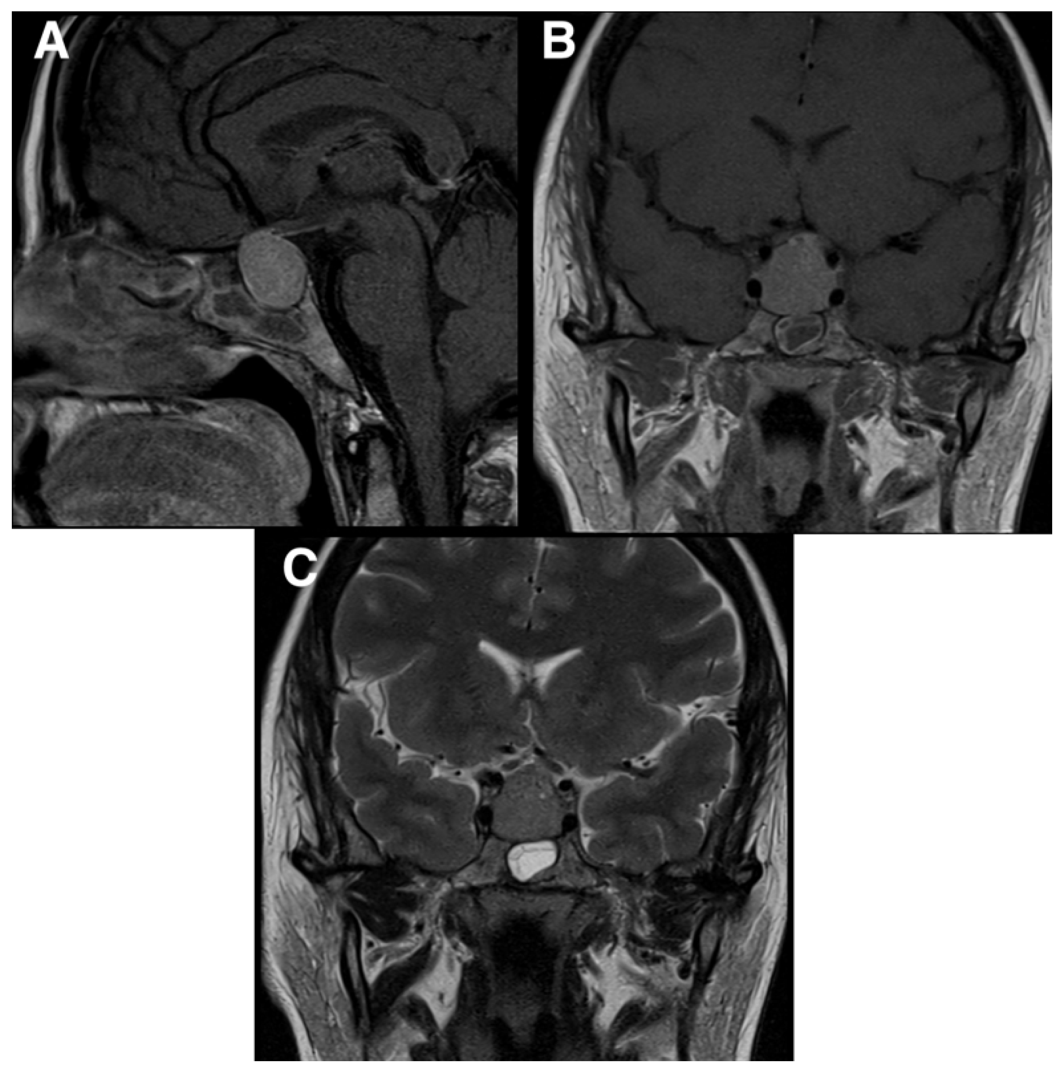
Figure 2.
Post-operative CT showing GTR of the tumor with decompression of the optic chiasm and no evidence of intracranial complications.
Figure 2.
Post-operative CT showing GTR of the tumor with decompression of the optic chiasm and no evidence of intracranial complications.
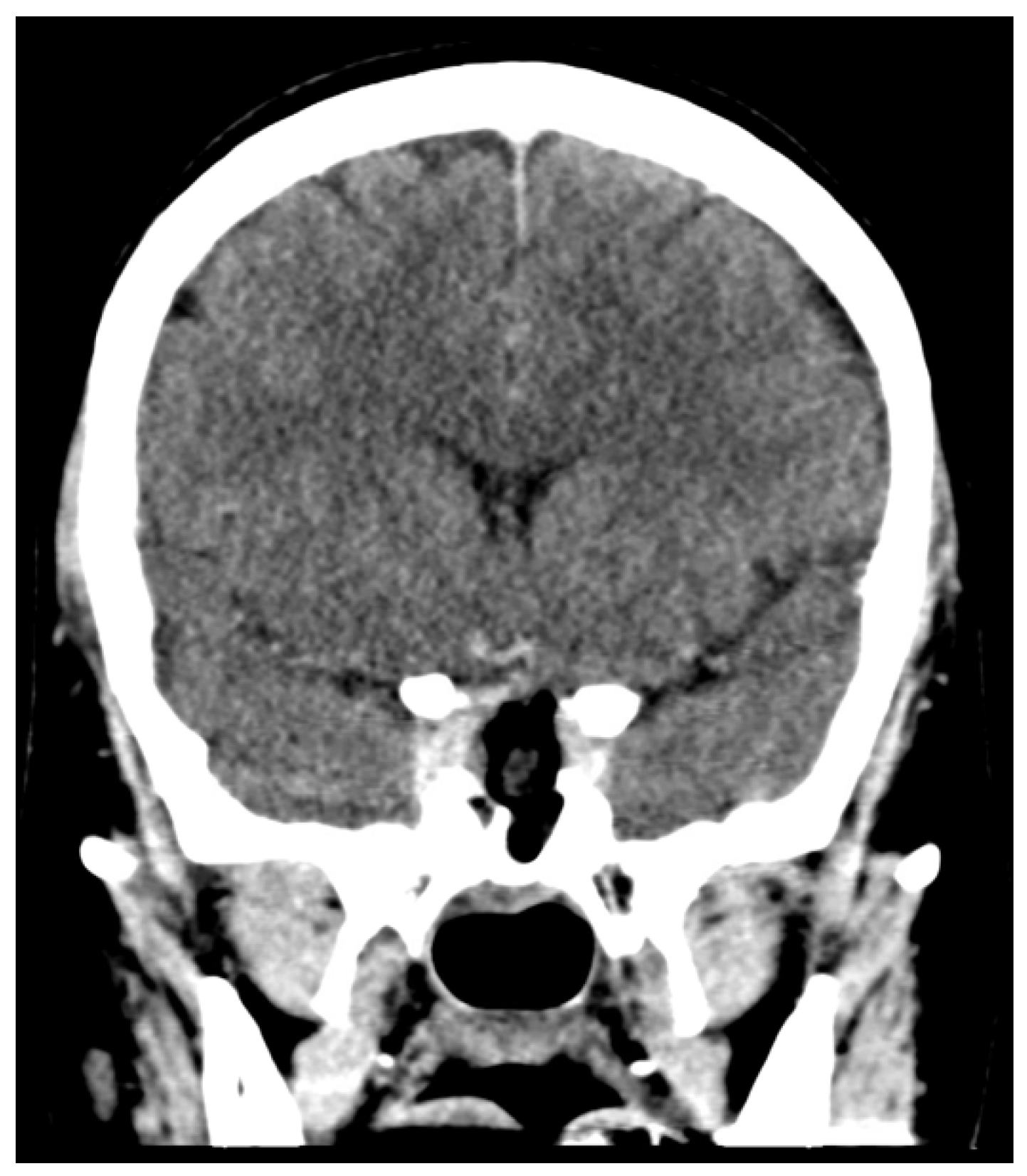
Figure 3.
Hematoxylin and eosin (H&E) 20× showing spindled cells with eosinophilic cytoplasm arranged in nests and short fascicles.
Figure 3.
Hematoxylin and eosin (H&E) 20× showing spindled cells with eosinophilic cytoplasm arranged in nests and short fascicles.
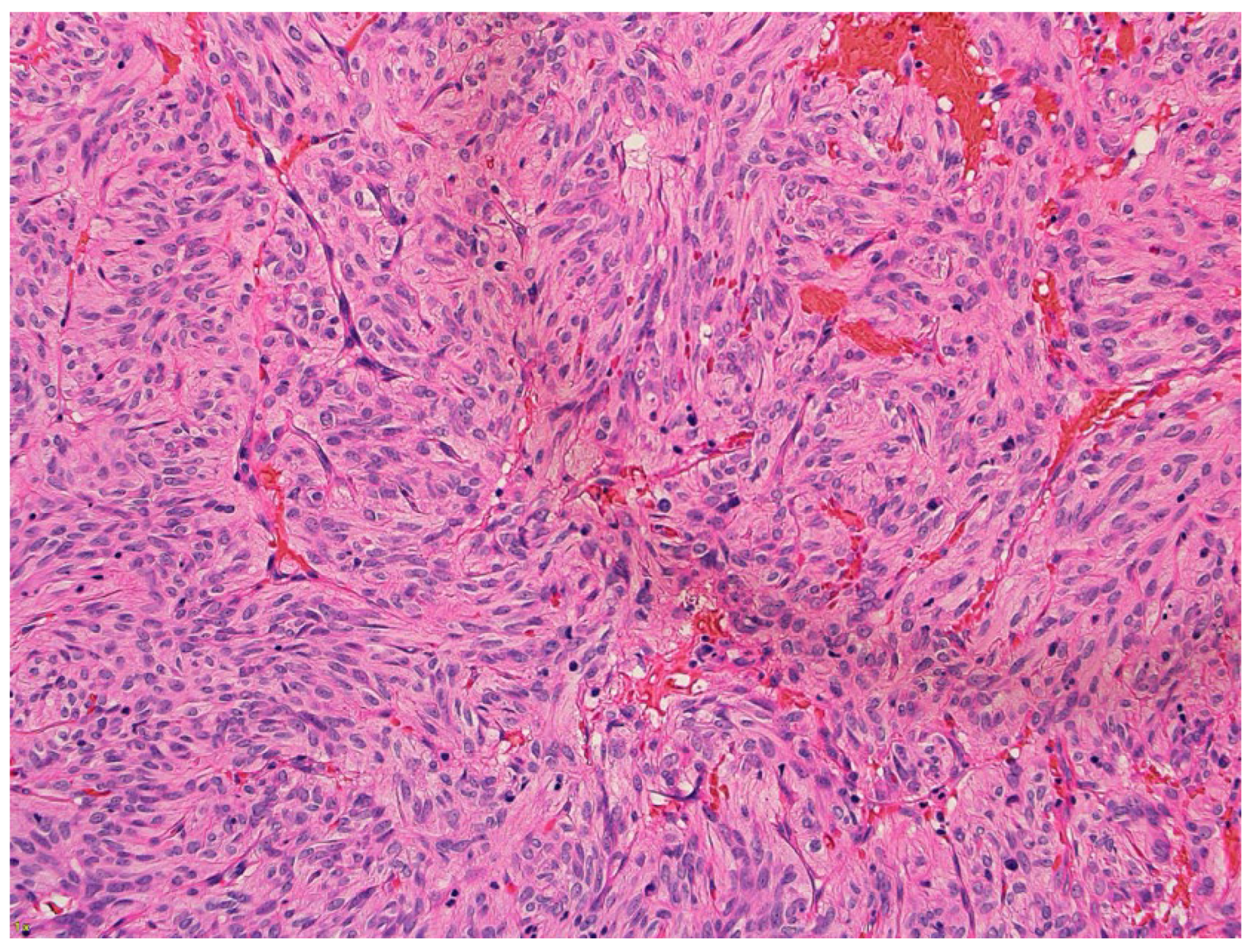
Figure 4.
IHC: The neoplastic cells are positive for (A) TTF1 (10x), (B) synaptophysin (10x), (C) CD56 (10x) and (D) S100 (10x).
Figure 4.
IHC: The neoplastic cells are positive for (A) TTF1 (10x), (B) synaptophysin (10x), (C) CD56 (10x) and (D) S100 (10x).
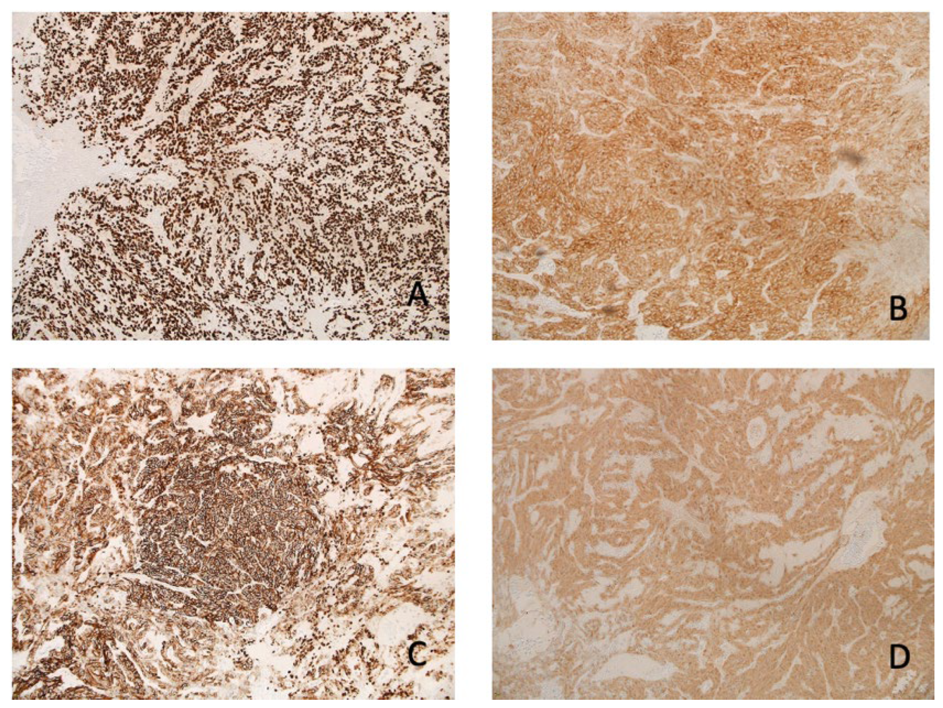

Table 1.
Review of literature (2002 - 2024).
| Age | Sex | Clinical presentation | Diagnostic Imaging | IHC | Surgery | Post-op Radiotherapy | Outcomes | |
|---|---|---|---|---|---|---|---|---|
| Hsieh et al., 2024 | 69 | Male | Facial pain and mild decrease in visual acuity | Sellar and suprasellar lesion | S-100 protein, EMA and TTF-1 | Transsphenoidal partial resection Tan-pink, soft, and well-circumscribed mass |
ND | Uneventful post-op period, follow up ND |
| 68 | Female | Bi-temporal visual defect | Large sellar mass with mass effect over brainstem and optic chiasm | S-100 protein, EMA(E29) and TTF-1 | Transsphenoidal partial resection | Yes 500 ×5 cGy |
2 years follow-up MRI showed partial size decreased of the tumor | |
| Joshi et al., 2024 | 71 | Male | ND | Sellar mass, 4 mm | ND | Transsphenoidal resection | ND | 8 week follow up no headache or visual disturbances |
| Chang et al., 2023 | 31 | Male | Bilateral vision loss – temporal defect | Sellar mass with suprasellar extension | EMA, S-100, and TTF-1 | Transsphenoidal total resection, mass was yellow and soft with easy bleeding | ND | ND |
| Kunihiro et al., 2023 | 53 | Male | Headache and diplopia | Sellar mass with suprasellar extension | S-100, TTF-1 and vimentin | Transsphenoidal total resection, mass was yellow and soft significant bleeding |
No | No enlargement of residual tumor at 1 year follow up |
| Shimizu et al., 2022 | 40s | Female | Headache | Lesion with cystic area in the intra-suprasellar region | TTF-1, S-100 protein, vimentin, GFAP, EMA | Transsphenoidal resection, tumor was fibrous and easy to bleed | ND | No recurrence at 3 years |
| Tena-Suck et al., 2022 | 66 | Male | Headache, chiasmatic syndrome, and bitemporal hemianopsia | Sellar lesion and a left frontal cystic lesion | Imentin, pit-1, PTTG-1, TTF-1, S100 | ND | ND | ND |
| Abdulrazeq et al., 2021 | 74 | Female | Persistent headaches and vertigo | Sellar mass with lateral extension | Vimentin, annexin A1, S-100, and TTF-1 | Transsphenoidal partial resection | No | No progression at 6 month follow up |
| Hasegawa et al., 2021 | 49 | Female | Galactorrhea, numbness, headache | Sellar mass with slight suprasellar extension | S-100, TTF-1, and GFAP | Transsphenoidal partial resection Grayish, firm, markedly adhesive |
No | Radiosurgery at 20 month for slight tumor progression, MRI at 39 months showed tumor stability |
| 55 | Male | Fatigue, muscle weakness, weight loss |
Sellar mass with suprasellar extension | S-100 protein, TTF-1 | Transsphenoidal partial resection Firm, hypervascular |
No | GK at 12 months for residual | |
| 78 | Male | Fatigue, visual deficit, hyponatremia | Sellar mass with suprasellar extension | S-100 protein, TTF-1 | Transsphenoidal partial resection | No | GK at 7 months for residual | |
| 59 | Female | Fatigue, hyponatremia | Sellar mass with suprasellar extension | S-100 protein, TTF-1 | Transsphenoidal partial resection | No | Gamma knife at 7 months for residual | |
| 56 | Male | hyponatremia | Sellar mass with suprasellar extension | S-100 protein, TTF-1 | Transsphenoidal partial resection | No | Transsphenoidal surgery at 77 months for recurrence | |
| 66 | Female | Nausea, vomiting, weight loss | Sellar mass with suprasellar extension | S-100 protein, TTF-1 | Transsphenoidal gross total resection | No | Gamma knife at 31 months for recurrence | |
| Kim et al., 2021 | 42 | Female | Bitemporal hemianopsia | Sella mass with suprasellar extension | Vimentin, EMA, S-100 protein, TTF-1, and galectin-3 | Transsphenoidal partial resection Highly vascular, pale-yellow and solid |
No | Regrown mass at 4 months requiring a second surgery |
| Kottangal et al., 2021 | 61 | Female | Hyponatremia and temporal field cut | Sellar mass with suprasellar extension | EMA, S-100 protein, TTF-1 | Transsphenoidal resection Grey-white, friable, and soft |
ND | ND |
| Taka et al., 2021 | 75 | Male | Bitemporal hemianopsia | Sellar mass with suprasellar extension | ND | Transsphenoidal gross total resection | ND | Follow-up on the eighth postoperative day showed improvement of peripheral vision. |
| Tariciotti et al., 2020 | 64 | Female | Bitemporal hemianopia, hyposmia, headache | Sellar mass with suprasellar extension causing hydrocephalus | S100, neuron-specific Enolase, TTF-1 | Transsphenoidal partial resection heavy intraoperative bleeding | ND | Recurrence at 5 months, partial resection surgery again with close follow up |
| Samadian et al., 2020 | 8 | Male | VD | Sellar and suprasellar mass | EMA, vimentin, and S-100 | Transsphenoidal gross total resection | No | No signs of progression at 2 year follow up |
|
Borg et al., 2020 |
55 | Female | Dizziness | Sellar mass with suprasellar extension | TTF-1, EMA and S-100 | Subtotal transsphenoidal resection | No | Progression at 8 years requiring radiotherapy |
| 71 | Male | Incidental | Sellar mass | TTF-1, EMA, S100 | Subtotal transsphenoidal resection Tough, grey and gelatinous. |
ND | Residual remained stable in size for four years | |
|
Li et al., 2020 |
57 | Male | Visual defect and neck pain | Sellar mass | EMA, TTF-1, S100, vimentin, annexin1, and SSTR2 | Total transsphenoidal resection | ND | Patient recovered well and had return of normal vision |
| Chainey et al., 2020 | 49 | Male | Confusion, memory loss, and increased drowsiness | Sellar mass with suprasellar extension | ND | ND | ND | Recurrence/growth at 4 and 5 years requiring two further resections |
| Akyoldaxs et al., 2019 | 55 | Female | VD | Sellar mass | ND | Transsphenoidal resection | Yes 14 Gy |
Stable at 84 months |
| 41 | Male | Loss of body hair, infertility | Sellar mass | ND | Transsphenoidal resection | Yes 12 Gy |
Stable at 51 months | |
| 61 | Male | Headache – VD | Sellar mass | ND | Transsphenoidal resection | Yes 14 Gy |
Stable at 47 months | |
| 50 | Male | VD | Sellar mass | ND | Transsphenoidal resection | Yes 14 Gy |
Stable at 41 months | |
| 56 | Male | VD | Sellar mass | ND | Transsphenoidal resection | Yes 14 Gy |
Stable at 36 months | |
| Sollfrank et al., 2019 | 38 | Female | ND | Mass in right parasellar region | ND | History of six surgical excisions, radiation and chemo radiation for local recurrence of SCO. Most recent treatment was vemurafenib (BRAF inhibitor) |
No | Stable - no progression on BRAF inhibitor at two year follow up |
| Yip et al., 2019 | 28 | Female | Severe headache and blurred vision, decreased right visual acuity, visual field defects, low cortisol | Sellar mass with suprasellar extension | TTF-1, EMA, Annexin A1 | Transsphenoidal total resection mass was yellow and soft |
ND | ND |
| Cole et al., 2019 | 64 | Male | Headache, fatigue, vision changes, Endocrine abnormality | Sellar mass | TTF-1, EMA, GFAP, S100 | Transsphenoidal total resection | ND | No recurrence at follow up |
| 70 | Male | Headache, fatigue, vision changes, Endocrine abnormality | Sellar mass | TTF-1, EMA, GFAP, S100 | Transsphenoidal total resection | ND | No recurrence at follow up | |
| 27 | Female | Endocrine abnormality | Sellar mass | TTF-1, EMA, GFAP, S100 | Transsphenoidal total resection | ND | No recurrence at follow up | |
| Guerrero-Pérez et al., 2019 | 74 | Female | VD | Sellar mass | TTF-1, S100, VIM | None | ND | ND |
| 69 | Female | Weakness | Sellar/suprasellar mass | TTF-1, S100, VIM, CD56 | Total transsphenoidal resection | ND | ND | |
| 74 | Female | Nausea, vomiting and confusion | Sellar/suprasellar mass | TTF-1, S100, VIM, GFAP | Subtotal transsphenoidal resection | ND | ND | |
| 60 | Male | VD | Sellar/suprasellar mass | ND | Subtotal transcranial resection | ND | ND | |
| 60 | Male | VD | Sellar/suprasellar mass | ND | Subtotal transsphenoidal resection | ND | ND | |
| 62 | Female | VD | Sellar mass | ND | Total transsphenoidal resection | ND | ND | |
| Witte et al., 2018 | 61 | Male | Headaches, bilateral retrobulbar pressure sensation, light sensitivity, and drowsiness | Sellar mass | VIM, Gal3 , Bcl-2 | Transsphenoidal partial resection | No | Three reoperations were required for multiple tumor reccurences along with radiation and chemotherapy |
| Larsen et al., 2018 | 66 | Female | Dizziness, nausea, diaphoresis | Sellar mass | EMA, TTF-1, S100 | Transsphenoidal partial resection | ND | Stable residual tumor at 100 months |
| 50 | Male | Dizziness | Sellar mass | EMA, TTF-1, S100 | Transsphenoidal partial resection | ND | Stable residual tumor at 30 months | |
| 63 | Male | VD | Sellar mass | TTF-1, S100 | Transsphenoidal partial resection | ND | Repeat resection for tumor progression (3 month postop); craniotomy for tumor progression (42 mos after repeat procedure) | |
| 59 | Female | VD | Sellar mass | EMA, TTF-1, S100 | Transsphenoidal partial resection | ND | Recurrence 6 yrs after GTR, treated w/ repeat TSR, GK; stable (79 mos after initial presentation) | |
| 77 | Male | VD | Sellar mass | EMA, TTF-1, S100 | Transsphenoidal gross total resection | ND | Stable after GTR (12 mos) - no complications | |
| 56 | Female | Eyeball heaviness, nausea, dizziness | Sellar mass | EMA, TTF-1, S100 | Transsphenoidal partial resection | ND | Proton beam therapy for residual tumor; stable residual tumor (38 mos after initial op) | |
| Gupta et al., 2018 | 28 | Female | Bilat vision loss, headaches, amenorrhea, galactorrhea | Sellar mass with suprasellar extension | EMA, S-100, and TTF-1 | transsphenoidal resection | ND | At 7 month follow up she was free of headache and galactorrhea and had a normal vision |
| Yoshida et al., 2018 | 69 | Female | Bitemporal hemianopsia | Sellar tumor with suprasellar extension | TTF-1 | Subtotal transsphenoidal resection. Tumor was firm, gelatinous and extremely hypervascular. | ND | No regrowth of the tumor was detected at 6 month follow up |
| Nagata et al., 2018 | 40 | Female | VD | Sellar and suprasellar mass. | EMA, S-100, TTF-1, GFAB, Vimentin | Total transsphenoidal resection Hypervascularized lesion |
No | ND |
| Sosa et al., 2018 | 60 | Male | VD, fatigue, decreased libido, and erectile dysfunction for the past 8 months | Sellar mass | Vimentin, S100 protein, and TTF-1 | Transsphenoidal partial resection | No | Radiotherapy after 5 month follow-up No residual or recurrent tumor was observed at 4 year follow up |
| Xie et al., 2017 | 60 | Male | Nausea, vomiting, fatigue and syncopal episodes | Sellar and suprasellar mass |
Vimentin, S-100, EMA and TTF-1 | Transsphenoidal surgical resection Vascular whitish-yellow mass with soft consistency |
No | No evidence of tumor recurrence after 18 month follow up |
| Rafiq et al., 2017 | 61 | Male | VD, fatigue and weight loss | Sellar lesion with compression of the optic chiasm | Vimentin, S-100, EMA and TTF-1 | Subtotal trans sphenoidal resection Firm, fibrous and greyish |
No | 3 year followup, scan showed tumor progression requiring surgery, GTR was achieved |
| 69 | Female | Rapidly progressive visual deterioration | Large sellar tumor with a suprasellar extension | S-100, EMA and TTF-1 | Transphenoidal resection Firm, greyish in color, moderately vascular |
No | After 6 months follow up, no progression | |
|
Osman et al., 2017 |
56 | Male | Headache, vomiting, neck pain, back pain, and reduced level of consciousness | Sellar and suprasellar mass | Vimentin, S-100, EMA and TTF-1, GFAP | Sub-frontal craniotomy. Profuse bleeding limited the surgical resection. |
Yes | No tumor recurrence at six-month follow-up |
| Manoranjan et al., 2017 | 60 | Male | Temporal loss in both visual field quadrants of his left eye | Sellar and suprasellar mass | S100, Vimentin, Bcl2, CD56, TTF-1 | Subtotal transnasal transsphenoidal resection | ND | No tumor progression at most recent follow up |
| Sali et al., 2017 | 64 | Male | Drooping of the left eyelid for 2 months and left temporal hemianopia | Sellar and suprasellar lesion | S100, synaptophysin, EMA, TTF-1 | Transsphenoidal resection | ND | ND |
|
Billeci et al., 2017 |
61 | Male | Headache and clinical signs of mild hypopituitarism | Sellar-suprasellar mass involving the sphenoidal sinus and chiasmatic cistern. | Vimentin, S-100, TTF-1 | Subtotal transnasal transsphenoidal resection. Tumor was firm and highly vascularized |
ND | After 14 months from the second surgery no increase in residual tumor size has been documented. |
| 65 | Female | VD and severe headache | sellar-supraasellar mass with a size of | Vimentin, S-100, TTF-1 | Subtotal transnasal transsphenoidal resection. The tumor was firm, fibrotic and highly vascularized |
ND | No documented recurrences after 28 months of follow up. | |
|
Kong et al., 2017 |
30 | Male | Headaches, fatigue, diplopia, and impaired visual field and acuity for 6 months | Suprasellar and parasellar leson | Vimentin, CD68, CD34, Nestin, GFAP, Desmin, SMA, AE1/AE3, and S-100 protein | Subtotal transnasal transsphenoidal resection. Hypervascular |
ND | Two more resections were done for tumor recurrence/progression |
|
Hagel et al., 2017 |
65 | Female | ND | ND | S100, CD68, TTF, Vimentin, neuron specific enolase |
ND | ND | ND |
| 41 | Female | ND | ND | S100, CD68, TTF, GFAP |
ND | ND | ND | |
| 64 | Female | ND | ND | Vimentin, EMA, S100, TTF | ND | ND | ND | |
| 53 | Male | ND | ND | Vimentin, EMA, MAP2, S100, CD68, TTF |
ND | ND | ND | |
| Custodio et al., 2016 | 60 | Male | NVD/Fatigue, hyponatremia, panhypopituitarism, low cortisol | Sellar mass with suprasellar extension | Vimentin, EMA, S-100, and TTF-1 | Transsphenoidal partial resection yellow white mass significant bleeding |
ND | No growth at 18 months |
| Hasiloglu et al., 2016 | 40 | Male | Panhypopituitarism | Intra-suprasellar mass and enlargement of the sella turcica | Vimentin, galectin-3, EMA and S-100 | Transsphenoidal partial resection | No | Recurrence after one year, repeat surgery |
| 60 | Male | Headache, visual blurring | Intra-suprasellar mass and enlargement of the sella turcica | Vimentin, galectin-3, EMA and S-100 | Transsphenoidal partial resection | No | ND | |
| 55 | Male | Headache, visual blurring | Intra-suprasellar mass and enlargement of the sella turcica | Vimentin, galectin-3, EMA and S-100 | Transsphenoidal partial resection | No | ND | |
|
Guadagno et al., 2016 |
77 | Male | Headache and temporal hemianopsia of the right eye | Sellar mass with suprasellar extension | EMA, Vimentin, S100 protein, Galectin-3, and TTF-1, and focal positivity for Bcl-2 | Transsphenoidal total resection | ND | 14 month follow up no evidence of recurrence |
| Vuong et al., 2016 | 70 | Male | Visual disturbance and headache |
Sellar-suprasellar lesion |
Vimentin, TTF-1, EMA and galectin-3 | Transsphenoidal partial resection | ND | Tumor recurrence not detected at first follow up exam |
|
Zygourakis et al., 2015 |
55 | Female | Headaches | Sellar mass | AMA, EMA, S100, GFAP, TTF1, | Transsphenoidal resection | ND | No reccurence on follow-up |
| 31 | Female | Bitemporal hemianopsia | Sellar and suprasellar lesion | TTF1, EMA and AMA | Transsphenoidal partial resection | No | MRI at six months showed stable residual tumor | |
|
Mu et al., 2015 |
35 | Female | Amenorrhea, lactation, decreased visual acuity | Suprasellar round mass |
Vimentin, EMA, S-100 and TTF-1 |
Frontotemporal craniotomy, complete removal | ND | No recurrence at 21 months |
| 62 | Female | No clear symptoms or signs |
Suprasellar mass | Vimentin, EMA, S-100 and TTF-1 | Right trans-pterional craniotomy, complete removal | ND | No recurrence at 15 month | |
|
Rotman et al., 2014 |
88 | Male | Fatigue and decreased vision | 2-cm intrasellar mass with suprasellar extension | Vimentin | Transsphenoidal partial resection | ND | ND |
| Fujisawa et alk., 2012 | 68 | Male | Unsteady gait, malaise, depression, panhypopituitarism and visual field defects | Sellar mass with suprasellar extension | EMA, S-100, and vimentin | Transsphenoidal partial resection | Yes 50 Gy |
1.5 year follow up showed tumor progression, partial resection again with close follow up |
| Alexandrescu et al., 2012 | 24 | Female | Headaches, amenorrhea and left superior visual field disturbance of the left eye | Sellar mass | EMA, S100, vimentin | Sublabial trans-septal approach, total resection Yellow and more firm |
ND | No recurrence at 6 months |
| Singh et al., 2012 | 68 | Male | Head and visual deficits | Sellar–suprasellar mass | Vimentin, S100, and EMA | Sublabial transsphenoidal partial resection | ND | ND |
| Ogiwara et al., 2011 | 39 | Male | Headache, loss of stamina and libido, bitemporal hemianopia, and polyuria |
Suprasellar lesion with the compression of the optic nerves | TTF-1, EMA, S-100, and galectin-3 |
Transcranial partial resection |
Yes 5040 cGy |
Recurrence at 9 months requiring repeat surgery. Transsphenoidal resection for second recurrence. No evidence of recurrence at 1-year follow-up since last surgery. |
| Romero-Rojas et al., 2011 | 42 | Female | Oligomenorrhea | Sellar mass | Vimentin, S10, EMA, and antimitochondrial antibody MU213-UC clone 131 | Transsphenoidal resection | ND | ND |
| Vajtai et al., 2011 | 55 | Female | Panhypopituitarism | Intra- and suprasellar tumor | S100 protein, vimentin, Bcl-2, and TTF-1 | Transsphenoidal total resection | ND | ND |
| Mlika et al., 2011 | 45 | Female | Headache and visual deficit | Pituitary mass with suprasellar extension | Vimentin, S100, EMA and TTF-1 | Transsphenoidal total resection | No | No recurrence at 3 months |
| Borges et al., 2011 | 70 | Female | Visual deficit in left eye | Intrasellar and suprasellar mass | Vimentin and S100 | Sublabial gross total, transsphenoidal resection | ND | Recurrence requiring second transsphenoidal subtotal resection |
| Matyja et al., 2010 | 63 | Female | Headache, vertigo, fatigue, bitemporal hemianopsia, nausea/vomiting and sleepiness | Pituitary mass with suprasellar extension | Vimentin, S100, EMA | Transsphenoidal total resection | ND | No recurrence at 28 month follow up |
| 65 | Female | Pituitary hormone insufficiency | Sellar mass with suprasellar extension | Vimentin, S100, EMA and galactin-3 | Frontal right craniotomy gross section | ND | No recurrence at twenty months | |
|
Demssie et al., 2011 |
59 | Male | Bitemporal hemianopsia, panhypopituitarism, weight loss, vomiting and tiredness | Sellar mass with suprasellar extension | S100 and EMA | Transsphenoidal partial resection | ND | Recurrence at 9 months requiring repeat surgery with radiotherapy |
| Borota et al., 2009 | 55 | Female | Weight loss, headaches | Sellar mass | Vimentin, S100 and galactin-3 | Transsphenoidal partial resection | No | Growth of the tumor at 1 year requiring radiotherapy |
| Coiré et al., 2009 | 63 | Female | Weight loss, anorexia, fatigue, headaches, visual deficits | Large sellar and suprasellar lesion, 3 cm in diameter | S100, vimentin, EMA and gal-3 | Transsphenoidal resection | No | Growth at five months requiring second surgery and radiotherapy |
| Farooq et al., 2008 | 76 | Male | Weakness and headache | Sellar mass | S100 and EMA | Transsphenoidal partial resection | Yes | No growth at 2 year follow up |
| Vajtai et al., 2006 | 48 | Female | Fatigue, exercise intolerance, and visual deficits | Sellar mass with supra and parasellar extension | S100 protein, vimentin, and EMA | Transsphenoidal total resection | No | No recurrence at 16 year follow up |
| Dahiya et al., 2005 | 26 | Male | Headache, blurred vision in the right eye, nausea, vomiting and impotence | Sellar mass | S100 and EMA | Pterional craniotomy with subtotal resection | Yes 54 Gy over a period of 2 months |
No growth over 7 years |
| 55 | Female | Headache and visual deficits | 6.5x3.3x4 cm sellar and parasellar mass | S100 and EMA | Transsphenoidal total resection | ND | No recurrence at 6 months | |
|
Kloub et al., 2005 |
71 | Female | Bilateral vision loss | Sellar mass | Vimentin, S-100 protein, neuron specific enolase, and EMA | Transsphenoidal resection | ND | Recurrence at 3 years |
| 76 | Male | Epistaxis | Sellar mass | EMA and S-100 | Transsphenoidal resection | ND | Recurrence at 3 years (repeat surgery and radiotherapy) and 10 years (third resection surgery) | |
|
Roncaroli et al., 2002 |
Mean age was 62 | Female | hypopituitarism | Sellar mass with suprasellar extension | S100, vimentin, EMA and gal-3 | Transsphenoidal gross total resection | ND | No recurrence at follow up (average follow up 35.4 months) |
| - | Female | hypopituitarism | Sellar mass with suprasellar extension | S100, vimentin, EMA and gal-3 | Transsphenoidal gross total resection | ND | No recurrence at follow up | |
| - | Male | hypopituitarism | Sellar mass with suprasellar extension | S100, vimentin, EMA and gal-3 | Transsphenoidal gross total resection | ND | No recurrence at follow up | |
| - | Male | hypopituitarism, visual deficit | Sellar mass with suprasellar extension | S100, vimentin, EMA and gal-3 | Transsphenoidal gross total resection | ND | No recurrence at follow up | |
| - | Male | hypopituitarism, visual deficit | Sellar mass with suprasellar extension | S100, vimentin, EMA and gal-3 | Transsphenoidal gross total resection | ND | No recurrence at follow up |
Disclaimer/Publisher’s Note: The statements, opinions and data contained in all publications are solely those of the individual author(s) and contributor(s) and not of MDPI and/or the editor(s). MDPI and/or the editor(s) disclaim responsibility for any injury to people or property resulting from any ideas, methods, instructions or products referred to in the content. |
© 2025 by the authors. Licensee MDPI, Basel, Switzerland. This article is an open access article distributed under the terms and conditions of the Creative Commons Attribution (CC BY) license (http://creativecommons.org/licenses/by/4.0/).
Copyright: This open access article is published under a Creative Commons CC BY 4.0 license, which permit the free download, distribution, and reuse, provided that the author and preprint are cited in any reuse.


