Preprint
Article

This version is not peer-reviewed.

Efficacy and Tolerability of Treatment of Actinic Keratosis with Tirbanibulin 0.1% Ointment in People Living with HIV Infection

A peer-reviewed article of this preprint also exists.

Submitted:

31 December 2024

Posted:

03 January 2025

You are already at the latest version

Abstract

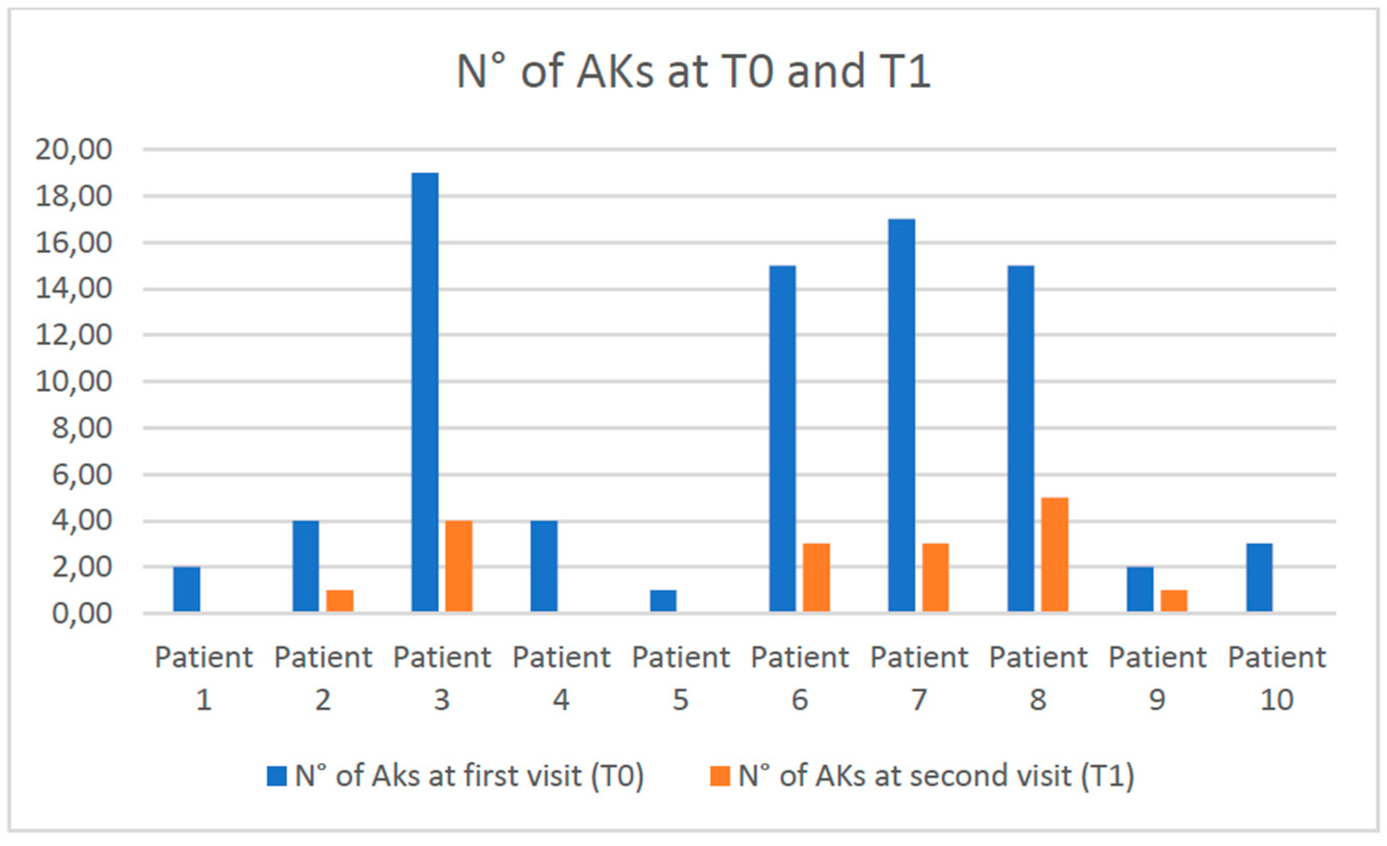
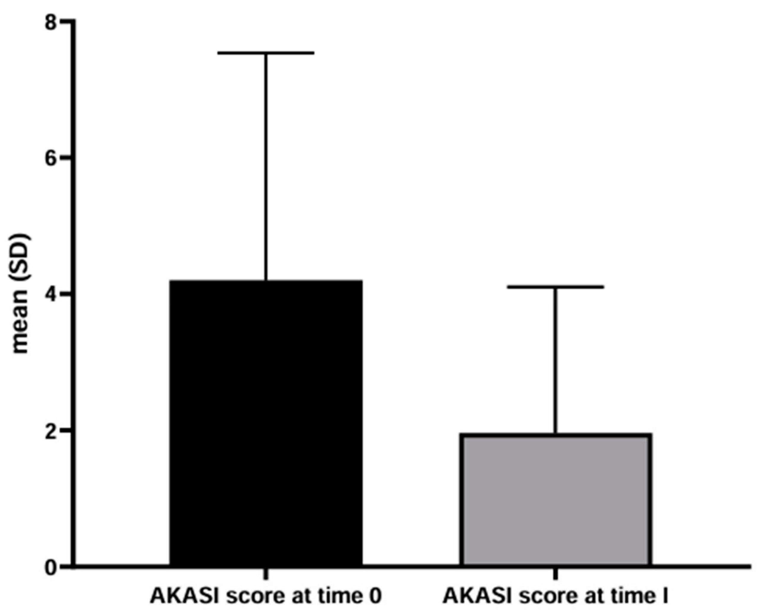
Background/Objectives: People living with HIV (PLWH) are more susceptible than immunocompetent people to non-melanoma skin cancers. These tumors can arise de novo or from precancerous lesions, such as actinic keratosis (AKs). The AKs management in PLWH has not been widely discussed in the literature. More specifically, the efficacy of AKs treatment in PLWH with modern topical drugs, such as tirbanibulin, is limited. The present work aims to evaluate the efficacy and tolerability of AKs treatment with tirbanibulin 1% ointment in PLWH. Methods: We retrospectively collected the data of the PLWH who visited the Dermatology Department of the Policlinico Riuniti (Foggia, Italy) between September 2023 and September 2024. PLWH who received the diagnosis of AKs and underwent treatment with tirbanibulin 1% ointment were studied. To assess the severity of AKs, the number of AKs and the AKs area and severity index (AKASI) score were calculated at the time of diagnosis (T0) and after treatment (T1). Results: Ten PLWH were found to have AKs and received topical therapy with tirbanibulin 1% ointment. On average, al T0, the number of lesions was 8.2 and the AKASI score was 4.20; at T1, the number of AKs was 1.7 and the AKASI score was 1.5. Only two patients reported a mild inflammatory reaction to applying tirbanibulin 1% ointment. Conclusions: The rate of satisfactory responses was 80%, in line with a recent multicentric Italian study performed on immunocompetent patients. Our results confirm the efficiacy and tolerability of tirbanibulin 1% ointment in treating AKs also in PLWH.

Keywords: 
;  ;  ;  

1. Introduction

Actinic keratosis (AK), also referred to as ‘solar keratosis’ or ‘senile keratosis’, is a cutaneous lesion that typically begins to appear in the fourth and fifth decades, increasing in number over the years. AKs are one of the most common potentially dangerous manifestations of chronic ultraviolet radiation (UV) exposure, even if it may appear after exposure to radiation, psoralens plus ultraviolet light, chemotherapeutic agents and arsenic. AKs are poorly circumscribed erythematous flat macules and variably rough or scaly papules, measuring from a few millimeters to a few centimeters and usually arising in the photo-exposed areas. Histologically, the typical erythematous AK is characterized by foci of atypical, pleomorphic keratinocytes confined to the lower third of the epidermal skin layer and budding into the upper papillary dermis. AK is considered either a precancerous lesion that may progress to an invasive squamous cell carcinoma (SCC) by some or an in situ SCC (intraepidermal proliferation of atypical keratinocytes) that may progress to an invasive stage by others [1,2]. Indeed, AK is cytologically indistinguishable from in situ SCC and has several molecular features ascribed to SCC [3,4].
Excessive chronic exposure to UV, particularly UV-B (280-320 nm), is the main environmental factor leading to AKs and SCC development. Indeed, AKs are located mainly in areas with chronically sun-damaged skin, such as the face, scalp, head and neck area, hands, and forearms [5]. Mutations and deregulation of tumor suppressor proteins such as p53, p16INK4a, and PTEN induced by UVB radiation are considered crucial pathogenetic mechanisms in the development of AK and SCC [5,6,7].
AK is among the most prevalent precancerous skin conditions worldwide. The prevalence varies significantly across countries, depending on the study setting, UV radiation level and patient characteristics. In Australia, up to 60% of people over 40 have AKs, while in Italy, the prevalence is estimated at 27.4% [2].
Factors associated with an increased risk of AK in immunocompetent individuals are: male sex, age >45 years, fair skin type, light hair and eye color, the presence of freckles on face and arms, positive history of non-melanoma skin cancer (NMSC), repeated sunburns occurred in childhood and adulthood, chronic occupational and/or recreational sun exposure, severe baldness, and use of potentially photosensitizing drugs such as thiazide diuretics or other cardiac drugs [2,8]. Conversely, factors associated with a reduced risk of AK are regular sunscreen use and probably history of atopy [2,9].
Although the role of human papillomavirus (HPV) infection in AKs development is still controversial, as for other skin and mucosal cancers like melanoma [10], a meta-analysis showed that HPV (mainly the beta HPV species) may act as a co-carcinogen with other factors to amplify the risk of developing SCC [11]. Notably, an increase in HPV prevalence was found in tumors from immunosuppressed patients compared to immunocompetent patients. The lack of HPV presence in all SCCs may imply the involvement of HPV in the initiation of oncogenesis rather than tumor promotion or maintenance. Several studies have shown that HPV DNA is more prevalent in sun-exposed versus non-sun-exposed skin, suggesting a link between the two factors. HPV could disturb cellular DNA repair or apoptosis mechanisms, making the cells more susceptible to UV-induced damage. Furthermore, UV light may have a transient immunosuppressive effect on the skin, allowing HPV to evade the immune system. However, it has also been assumed that HPV may be an innocent bystander and not a real factor in the pathogenesis of cutaneous SCC [11].
The diagnosis of AK is usually made by clinical examination. Dermatoscopy, confocal microscopy, optical coherence tomography or line-field confocal-OCT can help in the differential diagnosis of AK and other skin neoplasms. Individual AK lesions have been clinically graded based on their thickness using the Olsen classification system. Grade 1 lesions are slightly palpable and more easily felt than seen; grade 2 lesions are moderately thick and easily seen and felt, and grade 3 lesions are very thick and hyperkeratotic [2,12]. A recent study suggested that the Olsen classification is strongly associated with the risk of SCC development [13].
It is well known that a weakened immune system can reduce the ability to detect and eliminate cancer and precancerous cells, leading to the development of cancers (de novo or starting from precursor lesions). Among the reasons for immunosuppression are inherited and acquired diseases, medications, some medical procedures, and several infections, such as the human immunodeficiency virus (HIV).
HIV infection increases the likelihood of developing certain cancers, such as Kaposi sarcoma and non-Hodgkin’s lymphoma, alone or in association with other infectious agents like the Epstein-Barr virus. [14,15,16,17].
People living with HIV (PLWH) may also be especially susceptible to non-melanoma skin cancers (NMSCs), as well as other immunocompromised groups, such as solid organ transplant recipients (OTR) [18,19,20].
Despite the introduction of antiretroviral therapies, PLWHs are at higher risk of developing non–acquired immunodeficiency syndrome (AIDS)-related cancers than the general population. Indeed, a study found that PLWH had a 2.6 times higher incidence of SCC compared to those without HIV [21]. This increased risk can be attributed to several factors, such as their longer life expectancy, accelerated aging, and impaired control of oncogenic infections caused by HIV-related immune suppression [15,21]. Although it has been shown that taking antiretroviral therapy reduces the risk of developing skin cancers compared to those who do not take it, PLWHs still undergo chronic antigenic stimulation by the virus, suffering a state of chronic inflammation and cytokine dysregulation (even when HIV replication is suppressed and CD4+ T cell count is preserved) that can contribute to the development of lymphomas and other tumors [22,23].
Based on these factors, precancerous lesions of the skin, such as AKs, may put PLWH at greater risk of developing skin cancers than the immunocompetent population, as they do in transplanted patients [24]; therefore, the treatment of AKs in these immunosuppressed populations is crucial.
Usually, the treatment of AKs involves several superficial or ablative procedures such as curettage, electrocoagulation, cryotherapy, laser therapy, topical photodynamic therapy and topical chemotherapeutic or immune response modifiers therapies. More specifically, using the former methods is preferentially reserved for isolated lesions, whereas topical therapies are preferred in cases where several lesions are present [2]. The latter type of intervention guarantees less invasiveness and is intended to act on the so-called “field of cancerization.” The “field of cancerization” is defined as an area of subclinical histological change located at the periphery of actinic keratoses and detectable by molecular biology, histology or confocal microscopy [25]. From a clinical point of view, it is configured as a chronically photo-damaged area in which recurrent AKs and other signs of sun damage appear, such as wrinkles, solar elastosis, lentigines, and telangiectasias.
In light of the clinic-pathological substrate underlying AKs in PLWH, the picture of multiple AKs in the context of a “field of cancerization” is more frequent in this type of patient, making the use of topical therapies preferable.
Among the superficial or ablative procedures, cryotherapy, laser therapy, curettage and electrocoagulation cause tissue destruction by liquid nitrogen, laser (mainly CO2), and curettage combined with electrocoagulation, respectively. Photodynamic therapy, on the other hand, uses aminolevulinic acid; the latter is applied to the area to be treated, is activated through an appropriate light, and, at this point, selectively destroys the tumor cells [26].
Regarding topical therapies, the most recent European Guidelines on diagnosis, treatment, and prevention of AKs mention 5-fluorouracil, imiquimod, diclofenac, and tirbanibulin 1% ointment as effective modalities [2].
5-fluorouracil is a cytotoxic agent that inhibits RNA transcription. Imiquimod activates innate immunity against precancerous cells, and Diclofenac inhibits the Cox pathway, leading to altered cell apoptosis [5]. Tirbanibulin is a novel synthetic chemical entity that has shown potent antiproliferative and antitumoral effects by inducing cell cycle arrest and, ultimately, apoptotic cell death [27]. These effects are attributed to tirbanibulin's role as a microtubule and Src kinase inhibitor with potent antiproliferative activity against keratinocyte growth.
In UV-damaged skin, a cascade of events activates Src oncogene expression, increases Src kinase activity, and enhances the EGFR/Erk1/2 signaling pathway, increasing epithelial-to-mesenchymal transition (EMT) marker expression [28]. Elevated levels of Src have been linked to AKs and SCCs and play a role in both primary tumor growth and metastases.
In 2020 and 2021, the Food and Drug Administration (FDA) and the European Medicines Agency (EMA) approved tirbanibulin 1% ointment for treating Olsen grade 1 AKs of the scalp and face [29].
Tirbanibulin has a short application course, once daily for five consecutive days, on a maximum of 25 cm2 of the affected area. The most common adverse events are localized skin reactions at the application site. They range from mild to severe, including erythema, flaking or scaling, crusting, and swelling. These manifestations have been shown to resolve promptly with discontinuation of therapy [29].
In two identically designed double-blind trials, complete clearance occurred in 44% (Trial 1) and in 54% (Trial 2) of the patients treated with tirbanibulin 1% ointment; partial clearance (≥75% reduction in the number of lesions) at day 57 occurred in 68% (Trial 1) and 76% (Trial 2) of the patients [29].
The most recent guidelines have widely discussed the treatment of AKs in immunosuppressed patients, with particular attention to transplant patients [2]. Regarding PLWH, the management of AKs has not been widely discussed and validated in the literature.
Cancer screening in PLWH should align with the general population [30] and mainly concerns the prevention of carcinomas of the following types: colorectal, breast, prostate, and cervix. Since lung, liver, and anal cancers have a higher incidence and mortality in PLWH, screening for the early diagnosis of these tumors is also indicated [31]. Screening for skin cancers is generally recommended in PLWH, but national guidelines do not provide specific recommendations [32]. Furthermore, evidence on the efficacy and tolerability of AK treatment in PLWH, especially with modern topical chemotherapy drugs such as tirbanibulin 1% ointment, is limited.
Therefore, the present work aims to evaluate the efficacy and tolerability of AK treatment with Tirbanibulin 1% ointment in PLWH.

2. Materials and Methods

We retrospectively collected the clinical data of the PLWH who visited the Dermatology Department of the Policlinico Riuniti of Foggia, Italy, for skin cancer screening between 1st September 2023 and 30th September 2024.
PLWH who received the diagnosis of AKs during the study period and underwent at least one treatment cycle with tirbanibulin 1% ointment were included in the study. Treatment with tirbanibulin had been proposed for patients with single or multiple, non-hyperkeratotic, non-hypertrophic AKs (Olsen grade I) and moderately thickened, palpable AKs (Olsen grade II).
AKs were diagnosed based on the clinical and dermoscopic features of the skin lesions.
To assess the severity of AK/AKs, the actinic keratosis area and severity index (AKASI) score was calculated at the time of diagnosis using the calculator available on the website https://akasicalculator.com/it/#nav-onepage [33].
The number of lesions at the first visit (time 0, T0) was also counted.
At the follow-up visit, 4-8 weeks after the end of treatment, the number of lesions was counted again and the AKASI score was calculated. In addition, the patients were asked to rate the severity of the skin reaction following the application of the tirbanibulin 1% ointment with a value ranging from 0 to 5 (0 = “no reaction”; 5 = “significant erythema, edema, desquamation, itching, pain”).
The response to treatment was considered satisfactory if the number of lesions found at follow-up was zero (complete healing) or reduced by more than 75% (partial healing) [29]; when the number of lesions did not reduce by ≥75% the patient was considered not healed.
In patients who consented, photographic documentation of the AKs was performed at the time of diagnosis (time 0) and after therapy (time 1).
This observational retrospective study adhered to the ethical standards of the institutional and national research committees, the 1964 Helsinki Declaration, and its later amendments. All the patients gave informed consent to participate in the study.
Categorical variables were expressed in terms of absolute numbers and percentages, whereas continuous variables were expressed as mean and standard deviations (SD).

3. Results

During the study period, 90 PLWH consented to be visited for skin cancer screening. Ten of these patients (11.1%) were found to have AKs and received topical therapy with tirbanibulin 1% ointment as first-line treatment. These patients have been retrospectively studied in detail.
They were all males with a mean age of 69.3 years (range 53-75 years, SD 5.8). In all cases, the diagnosis of HIV infection had been discovered more than 10 years earlier; on average, the diagnosis of HIV infection had been known for 27.2 years (range 12-39 years, SD 8.7). All the patients were regularly taking oral or injectable antiretroviral therapy prescribed by the infectious disease specialist. The HIV RNA load in plasma measured within the last month before the dermatological visit was undetectable in all patients (<20 copies/ml).
Six out of 10 patients had extra-dermatological comorbidities, including hypercholesterolemia (3 patients), hypertension (2 patients), hepatitis B virus (HBV), hepatitis C virus (HCV) infection and gastroesophageal reflux (1 patient).
In addition, one patient was further immunosuppressed because he had undergone liver transplantation 7 years earlier (following HCV-related cirrhosis), for which he was also taking oral tacrolimus therapy. This patient was the only one with a personal history of skin cancers (two basal cell carcinomas and one squamous cell carcinoma surgically removed in the previous 4 years).
Eight patients had Fitzpatrick phototype II, and two had Fitzpatrick phototype III. Six patients (60%) had a positive history of sunburns during childhood. Only one patient used potentially photosensitizing thiazide diuretics for hypertension. None of the patients were regular sunscreen users.
Two out of ten patients had already received treatments for AKs with topical drugs and liquid nitrogen cryotherapy in the previous two years at other hospital facilities.
At the first dermatological visit (T0), AKs were located in 9 out of 10 patients on the face and/or scalp. Only one patient had AKs exclusively on the back of the hands (Table 1).
On average, al T0, the number of lesions was 8.2 (range 1-19; DV 7.28) and the average AKASI score was 4.20 (range 1.2-8.8; DV 3.34). Most patients (7/10) had Olsen grade I and II AKs; 3 had only grade I AKs (Table 1).
At T0, two of the ten patients with AKs also had several skin lesions suspicious for skin cancers: histological examinations confirmed the clinical diagnoses of cutaneous melanoma and basal cell carcinoma in one patient and of basal cell carcinoma in another patient.
Follow-Up After Treatment
On average, the follow-up visits were performed 18 weeks after the first visit (8-40 weeks, DV 11.75). Only two patients reported a mild inflammatory skin reaction (rated “1” on a scale of 0 to 5) to applying tirbanibulin 1% ointment, which resolved within 3-4 days. Eight patients did not experience any skin reactions at the site of the ointment application.
At the follow-up visit (T1), the average number of AKs was 1.7 (range 0-5; DV 1.88) (Figure 1) and the average AKASI score was 1.5 (range 0-3.4; DV 1.7).
Based on the number of lesions, the response to treatment was considered satisfactory in 80% of the patients who obtained a reduction in the number of lesions ≥75%; conversely, in two out of ten patients (20%), the tirbanibulin treatment did not determine a reduction in the number of lesions more significant than 75%. Therefore, they were not considered healed (Figure 1, patients n° 8 and 9). However, these patients presented at T1 a lesion number that had been reduced by at least 50% compared to T0 (from 15 to 5 lesions in case 8 and from 2 to 1 lesion in case 9).
Figure 2 shows the pre-and post-treatment conditions in some AK patients treated with tirbanibulin 1% ointment; figure 3 expresses the mean AKASI scores before and after treatment.

4. Discussion

The results obtained in this study confirm the efficacy and tolerability of tirbanibulin 1% ointment in treating AKs. Noteworthy, the present work is the first to evaluate the efficacy of this drug in PLWH. Overall, in the present series, the AKASI score was significantly reduced after therapy: from a pre-treatment mean AKASI score of 4.20 ± 3.34 to a post-treatment mean AKASI score of 1.57 ± 1.7. Notably, tirbanibulin 1% ointment was well tolerated in all our patients, as none reported severe adverse reactions (grade 4-5 on a scale of 1 to 5). In our series, the rate of satisfactory responses was 80%, according with a recent multicentric Italian study performed in a setting of immunocompetent patients [34]. Nazzaro et al. studied 250 patients affected by AKs recruited in 15 Italian dermatological clinics, including the Dermatology Unit of the Policlinico Riuniti of Foggia; treatment with Tirbanibulin led to a satisfactory response in 88.8% of cases, and the response rates were higher (97%) when the follow-up was closer (8 weeks). Only 7 (2.8%) grade 4 adverse events were recorded, whereas we did not detect them in our smaller case series of PLWH [34]. The efficacy of tirbanibulin 1% ointment in treating skin field cancerization has recently been described in 2 immunosuppressed patients following solid organ transplantation [35]. These data support the positive outcome obtained in one of our patients who was immunosuppressed for pharmacological and infectious reasons (liver transplantation and HIV infection).
Indeed, it is well known that chronically immunosuppressed patients, especially OTRs, have a higher risk of developing AK and SCC. The increase in skin cancer in OTRs is secondary to the long-term immunosuppressive therapies required for transplanted organ survival, which impairs the immune system’s ability to eliminate atypical cells. This increased risk in OTRs includes many types of cutaneous cancers and is exceptionally high for SCC, which some studies estimated to be 100-fold higher compared to the general population [36,37].
The most recent European guidelines on the diagnosis, treatment, and prevention of AKs provided ample space for managing AKs in immunosuppressed patients; however, they only referred to solid OTRs, and immunosuppression for other reasons, such as HIV infection, was not mentioned.
The authors underlined that immunosuppressed subjects frequently show multiple, metachronous evolving lesions over vast skin fields. Therefore, purely lesion-directed therapies may not be sufficient to obtain disease control. Furthermore, repetition of the treatment is often necessary due to a more aggressive disease course and a higher portion of treatment-resistant lesions in this population. The threshold to biopsy cutaneous and mucosal lesions to rule out progression to cutaneous SCC should be lower in immunosuppressed subjects than in immunocompetent individuals.
Based on randomized controlled trials and systematic reviews, the treatments that should be offered to immunocompromised patients with single and multiple AKs and a field of cancerization (grade of recommendation B) are conventional PDT, 5-FU 5% cream and diclofenac sodium 3% in hyaluronic acid gel 2.5% [2]. Cohort studies on the efficacy of tirbanibulin 1% ointment in treating AKs in immunocompromised patients are lacking.
Currently, tirbanibulin 1% ointment has been indicated for treating single or multiple AKs and field cancerization of the face and scalp only in immunocompetent patients (grade of recommendation B, level of evidence 1) [2].
PLWH represents a unique subpopulation with sociodemographic features and dermatologic conditions different from the general population [38]. Paradoxically, the longevity achieved thanks to the use of antiretroviral therapy in PLWH has resulted in higher rates of cancers that prevail in older age or are induced by prolonged exposure to physical (skin cancer), chemical (lung, skin cancers), and biological (skin, anal, genital, liver cancers) mutagens [39].
Compared to immunocompetent subjects, PLWHs have a more than two-fold higher incidence rate of keratinocyte carcinomas, especially SCC [40].
The systemic and cutaneous immune impairment induced by HIV has several different pathogenetic mechanisms that explain the link between this infection and skin cancers.
First, HIV shows a tropism for human immune system cells, such as macrophages, dendritic cells, and T lymphocytes (CD4 and CD8) [41]. Direct contact between CD4 T cells and HIV-pulsed dendritic antigen-presenting cells triggers the virus's replication, leading to the death of both cell types. When CD4 T cells and antigen-presenting cells count decrease meaningfully, skin becomes susceptible to opportunistic infections and neoplastic diseases.
In addition, HIV seems to activate proto-oncogenes, cause alterations in cell cycle regulation, and downregulate tumor suppressor genes, including p53 [42]. HIV could also determine microsatellite gene instability and genetic alterations, promoting the development of different cancers, including NMSCs. Finally, HIV infection may stimulate pro-angiogenesis signaling that could lead to endothelial abnormalities. These alterations could promote tumor growth and metastasis [41]. Skin cancers are the majority of neoplasias among PLWH, and NMSCs are the most frequent skin cancers in this population. NMSCs are usually more aggressive, as evidenced by an increased risk of metastatic disease and mortality compared to immunocompetent subjects. They tend to occur at a younger age, have an increased recurrence rate, and have an overall poorer outcome than the general population [41]. A low CD4 count (< 200/microL) and high viral load (>10000 copies/mL) in PLWH are associated with a greater risk of developing a primary NMSC and of having a recurrence [43]. Interestingly, an increased skin cancer risk has been documented not only among Caucasian PLWH but also among a South African Black population of PLWH [44]. On these grounds, PLWH, especially those with severe immunosuppression and those with prior NMSC (or both), should visit a dermatologist at least annually for a full-body skin check [41].
Regrettably, studies on managing AKs in PLWH are scarce. Our work demonstrated that PLWH on antiretroviral therapy can benefit from tirbanibulin 1% ointment therapy, presenting response rates similar to those of immunocompetent patients. Therefore, this treatment, which is the most manageable among the topical therapies suggested by the international guidelines since it requires only five consecutive days of application, can also be indicated in other contexts of immunodeficiency.
Remarkably, our results confirm compliance with the antiviral treatment and adherence to skin cancer screening in PLWH are crucial for promptly detecting and treating precancerous lesions and reducing the tumor burden.
The treatment of precancerous lesions in PLWH needs further investigation. More extensive studies with longer follow-up times could further confirm these preliminary results.

Author Contributions

Conceptualization, Giulia Ciccarese and F.D.; methodology, Gerardo Cazzato, B.T.; software, Gerardo Cazzato; investigation, Giulia Ciccarese, M. M., L. L., M. G.; resources, S. L. C.; data curation, Giulia Ciccarese, Gerardo Cazzato; writing—original draft preparation, Giulia Ciccarese, B.T., C.C., R.B.; writing—review and editing, C.F.; visualization, F.D.; supervision, C.F., F.D. All authors have read and agreed to the published version of the manuscript.

Funding

This research received no external funding.

Institutional Review Board Statement

The study was conducted in accordance with the Declaration of Helsinki, and approved by the Institutional Review Board of OSPEDALI RIUNITI DI FOGGIA (protocol code 104 / C.E. / 2020 and date of approval 30.09.2020)”.

Informed Consent Statement

Informed consent was obtained from all subjects involved in the study.

Data Availability Statement

The data supporting this study's findings are available from the corresponding author upon reasonable request.

Acknowledgments

none.

Conflicts of Interest

The authors declare no conflicts of interest.

Abbreviations

The following abbreviations are used in this manuscript:
AK Actinic keratosis
UV Ultraviolet
HIV Human immunodeficiency virus
PLWH People living with HIV
SCC Squamous cell carcinoma
NMSC Non-melanoma skin cancer
HPV Human papillomavirus
AIDS Acquired immunodeficiency syndrome
FDA Food and Drug Administration
AKASI Actinic keratosis area and severity index

References

  1. Röwert-Huber, J.; Patel, M.J.; Forschner, T.; Ulrich, C.; Eberle, J.; Kerl, H.; Sterry, W.; Stockfleth, E. Actinic keratosis is an early in situ squamous cell carcinoma: a proposal for reclassification. Br J Dermatol 2007, 156 Suppl 3, 8–12. [Google Scholar] [CrossRef]
  2. Kandolf, L.; Peris, K.; Malvehy, J.; Mosterd, K.; Heppt, M.V.; Fargnoli, M.C.; Berking, C.; Arenberger, P.; Bylaite-Bučinskiene, M. , Del Marmol, V.; et al. European consensus-based interdisciplinary guideline for diagnosis, treatment and prevention of actinic keratoses, epithelial UV-induced dysplasia and field cancerization on behalf of European Association of Dermato-Oncology, European Dermatology Forum, European Academy of Dermatology and Venereology and Union of Medical Specialists (Union Européenne des Médecins Spécialistes). J Eur Acad Dermatol Venereol 2024, 18, 1024–1047. [Google Scholar]
  3. Ackerman, A.B.; Mones, J.M. Solar (actinic) keratosis is squamous cell carcinoma. Br J Dermatol 2006, 155, 9–22. [Google Scholar] [CrossRef] [PubMed]
  4. Padilla, R.S.; Sebastian, S.; Jiang, Z.; Nindl, I.; Larson, R. Gene expression patterns of normal human skin, actinic keratosis, and squamous cell carcinoma: a spectrum of disease progression. Arch Dermatol 2010, 146, 288–93. [Google Scholar] [CrossRef] [PubMed]
  5. Thamm, J.R.; Schuh, S.; Welzel, J. Epidemiology and Risk Factors of Actinic Keratosis. What is New for The Management for Sun-Damaged Skin. Dermatol Pract Concept 2024, 14, e2024146S. [Google Scholar] [CrossRef]
  6. Ming, M.; Han, W.; Maddox, J.; Soltani, K.; Shea, C.R.; Freeman, D.M.; He, Y.Y. UVB-induced ERK/AKT-dependent PTEN suppression promotes survival of epidermal keratinocytes. Oncogene 2010, 29, 492–502. [Google Scholar] [CrossRef]
  7. Kanellou, P.; Zaravinos, A.; Zioga, M.; Stratigos, A.; Baritaki, S.; Soufla, G.; Zoras, O.; Spandidos, D.A. Genomic instability, mutations and expression analysis of the tumour suppressor genes p14(ARF), p15(INK4b), p16(INK4a) and p53 in actinic keratosis. Cancer Lett 2008, 264, 145–61. [Google Scholar] [CrossRef]
  8. Flohil, S.C.; van der Leest, R.J.; Dowlatshahi, E.A.; Hofman, A.; de Vries, E.; Nijsten, T. Prevalence of actinic keratosis and its risk factors in the general population: the Rotterdam Study. J Invest Dermatol 2013, 133, 1971–8. [Google Scholar] [CrossRef]
  9. Li, Y.; Wang, J.; Xiao, W.; Liu, J.; Zha, X. Risk Factors for Actinic Keratoses: A Systematic Review and Meta-Analysis. Indian J Dermatol 2022, 67, 92. [Google Scholar] [CrossRef]
  10. Ciccarese, G.; Drago, F.; Broccolo, F.; Pastorino, A.; Pizzatti, L.; Atzori, L.; Pilloni, L.; Santinelli, D.; Urbani, A.; Parodi, A.; et al. Oncoviruses and melanomas: A retrospective study and literature review. J Med Virol 2023, 95, e27924. [Google Scholar] [CrossRef]
  11. Wang, J.; Aldabagh, B.; Yu, J.; Arron, S.T. Role of human papillomavirus in cutaneous squamous cell carcinoma: a meta-analysis. J Am Acad Dermatol 2014, 70, 621–629. [Google Scholar] [CrossRef] [PubMed]
  12. Olsen, E.A.; Abernethy, M.L.; Kulp-Shorten, C.; Callen, J.P.; Glazer, S.D.; Huntley, A.; McCray, M.; Monroe, A.B.; Tschen, E.; Wolf, J.E. Jr. A double-blind, vehicle-controlled study evaluating masoprocol cream in the treatment of actinic keratoses on the head and neck. J Am Acad Dermatol 1991, 24, 738–43. [Google Scholar] [CrossRef] [PubMed]
  13. Ahmady, S.; Jansen, M.H.E.; Nelemans, P.J.; Kessels, J.P.H.M.; Arits, A.H.M.M.; de Rooij, M.J.M.; Essers, B.A.B.; Quaedvlieg, P.J.F.; Kelleners-Smeets, N.W.J.; et al. Risk of Invasive Cutaneous Squamous Cell Carcinoma After Different Treatments for Actinic Keratosis: A Secondary Analysis of a Randomized Clinical Trial. JAMA Dermatol 2022, 158, 634–640. [Google Scholar] [CrossRef]
  14. Ciccarese, G.; Trave, I.; Herzum, A.; Parodi, A.; Drago, F. Dermatological manifestations of Epstein-Barr virus systemic infection: a case report and literature review. Int J Dermatol 2020, 59, 1202–1209. [Google Scholar] [CrossRef]
  15. Wang, C.C.; Silverberg, M.J.; Abrams, D.I. Non-AIDS-Defining Malignancies in the HIV-Infected Population. Curr Infect Dis Rep 2014, 16, 406. [Google Scholar] [CrossRef]
  16. Shiels, M.S.; Cole, S.R.; Kirk, G.D.; Poole, C. A meta-analysis of the incidence of non-AIDS cancers in HIV-infected individuals. J Acquir Immune Defic Syndr 2009, 52, 611–22. [Google Scholar] [CrossRef]
  17. Drago, F.; Ciccarese, G.; Merlo, G.; Trave, I.; Javor, S.; Rebora, A.; Parodi, A. Oral and cutaneous manifestations of viral and bacterial infections: Not only COVID-19 disease. Clin Dermatol 2021, 39, 384–404. [Google Scholar] [CrossRef]
  18. Trave, I.; Ciccarese, G.; Gasparini, G.; Canta, R.; Serviddio, G.; Herzum, A.; Drago, F.; Parodi, A. Skin cancers in solid organ transplant recipients: a retrospective study on 218 patients. Transpl Immunol 2023, 80, 101896. [Google Scholar] [CrossRef]
  19. Ojute, F.; Zakaria, A.; Zullo, S.; Klufas, D.; Amerson, E. Skin cancer risk across racial/ethnic groups among solid organ transplant recipients in the United States, 2000-2022. J Am Acad Dermatol 2024, S0190-9622, 03235-3. [Google Scholar] [CrossRef]
  20. Korzeniowski, P.; Griffith, C.; Young, P.A. Update on skin cancer prevention modalities and screening protocols in solid organ transplant recipients: a scoping review. Arch Dermatol Res 2024, 317, 52. [Google Scholar] [CrossRef]
  21. Silverberg, M.J.; Chao, C.; Leyden, W.A.; Xu, L.; Tang, B.; Horberg, M.A.; Klein, D.; Quesenberry, C.P. Jr.; Towner, W.J.; Abrams, D.I. HIV infection and the risk of cancers with and without a known infectious cause. AIDS 2009, 23, 2337–45. [Google Scholar] [CrossRef] [PubMed]
  22. Zhao, H.; Shu, G.; Wang, S. The risk of non-melanoma skin cancer in HIV-infected patients: new data and meta-analysis. Int J STD AIDS 2016, 27, 568–75. [Google Scholar] [CrossRef] [PubMed]
  23. Yarchoan, R.; Uldrick, T.S. HIV-Associated Cancers and Related Diseases. N Engl J Med 2018, 378, 1029–1041. [Google Scholar] [CrossRef]
  24. Drago, F.; Ciccarese, G.; Parodi, A. Nicotinamide for Skin-Cancer Chemoprevention. N Engl J Med 2016, 374, 789–90. [Google Scholar]
  25. Dréno, B.; Amici, J.M.; Basset-Seguin, N.; Cribier, B.; Claudel, J.P.; Richard, M.A. AKTeam™. Management of actinic keratosis: a practical report and treatment algorithm from AKTeam™ expert clinicians. J Eur Acad Dermatol Venereol 2014, 28, 1141–1149. [Google Scholar] [CrossRef]
  26. Reinehr, C.P.H.; Bakos, R.M. Actinic keratoses: review of clinical, dermoscopic, and therapeutic aspects. An Bras Dermatol 2019, 94, 637–657. [Google Scholar] [CrossRef]
  27. DeTemple, V.K.; Walter, A.; Bredemeier, S.; Gutzmer, R.; Schaper-Gerhardt, K. Anti-tumor effects of tirbanibulin in squamous cell carcinoma cells are mediated via disruption of tubulin-polymerization. Arch Dermatol Res 2024, 316, 341. [Google Scholar] [CrossRef]
  28. Montagner, A.; Delgado, M.B.; Tallichet-Blanc, C.; Chan, J.S.; Sng, M.K.; Mottaz, H.; Degueurce, G.; Lippi, Y.; Moret, C.; Baruchet, M.; Antsiferova, M.; et al. Src is activated by the nuclear receptor peroxisome proliferator-activated receptor β/δ in ultraviolet radiation-induced skin cancer. EMBO Mol Med 2014, 6, 80–98. [Google Scholar] [CrossRef]
  29. Blauvelt, A.; Kempers, S.; Lain, E.; Schlesinger, T.; Tyring, S.; Forman, S.; Ablon, G.; Martin, G.; Wang, H.; Cutler, D.L.; et al. Phase 3 Trials of Tirbanibulin Ointment for Actinic Keratosis. N Engl J Med 2021, 384, 512–520. [Google Scholar] [CrossRef]
  30. European AIDS Clinical Society Guidelines. Version 11.1, October 2022. Available online: https://www.eacsociety.org/guidelines/eacs-guidelines/ (accessed on day month year).
  31. Masiá, M.; Gutiérrez-Ortiz de la Tabla, A.; Gutiérrez, F. Cancer screening in people living with HIV. Cancer Med 2023, 12, 20590–20603. [Google Scholar] [CrossRef]
  32. Kosche, C.; Chio, M.T.W.; Arron, S.T. Skin cancer and HIV. Clin Dermatol 2023, S0738-081X, 00258-4. [Google Scholar] [CrossRef] [PubMed]
  33. Dirschka, T.; Pellacani, G.; Micali, G.; Malvehy, J.; Stratigos, A.J.; Casari, A.; Schmitz, L.; Gupta, G. Athens AK Study Group. A proposed scoring system for assessing the severity of actinic keratosis on the head: actinic keratosis area and severity index. J Eur Acad Dermatol Venereol 2017, 31, 1295–1302. [Google Scholar] [CrossRef] [PubMed]
  34. Nazzaro, G.; Carugno, A.; Bortoluzzi, P.; Buffon, S.; Astrua, C.; Zappia, E.; Trovato, E.; Caccavale, S.; Pellegrino, V.; Paolino, G.; et al. Efficacy and tolerability of tirbanibulin 1% ointment in the treatment of cancerization field: a real-life Italian multicenter observational study of 250 patients. Int J Dermatol 2024, 63, 1566–1574. [Google Scholar] [CrossRef] [PubMed]
  35. Pellacani, G.; Schlesinger, T.; Bhatia, N.; Berman, B.; Lebwohl, M.; Cohen, J.L.; Patel, G.K.; Kunstfeld, R.; Hadshiew, I.; et al. Efficacy and safety of tirbanibulin 1% ointment in actinic keratoses: Data from two phase-III trials and the real-life clinical practice presented at the European Academy of Dermatology and Venereology Congress 2022. J Eur Acad Dermatol Venereol 2024, 38 Suppl 1, 3–15. [Google Scholar] [CrossRef]
  36. Roewert-Huber, J.; Stockfleth, E.; Kerl, H. Pathology and pathobiology of actinic (solar) keratosis - an update. Br J Dermatol 2007, 157, 18–20. [Google Scholar] [CrossRef]
  37. Jiyad, Z.; Marquart, L.; O'Rourke, P.; Green, A.C. The natural history of actinic keratoses in organ transplant recipients. J Am Acad Dermatol 2017, 76, 162–164. [Google Scholar] [CrossRef]
  38. Paul, S.; Evans, R.; Maurer, T.; Muhe, L.M.; Freeman, E.E. Treatment of Dermatological Conditions Associated with HIV/AIDS: The Scarcity of Guidance on a Global Scale. AIDS Res Treat 2016, 2016, 3272483. [Google Scholar] [CrossRef]
  39. Sanchez M. Skin Cancers in Patients with HIV. In: Nouri K. eds. Skin Cancer: A Comprehensive Guide. McGraw-Hill Education; 2023. Available online: https://dermatology.mhmedical.com/content.aspx?bookid=3257&sectionid=271456253 (Accessed December 21, 2024).
  40. Silverberg, M.J.; Leyden, W.; Warton, E.M.; Quesenberry, C.P. Jr.; Engels, E.A.; Asgari, M.M. HIV infection status, immunodeficiency, and the incidence of non-melanoma skin cancer. J Natl Cancer Inst 2013, 105, 350–60. [Google Scholar] [CrossRef]
  41. Venanzi Rullo, E.; Maimone, M.G.; Fiorica, F.; Ceccarelli, M.; Guarneri, C.; Berretta, M.; Nunnari, G. Non-Melanoma Skin Cancer in People Living With HIV: From Epidemiology to Clinical Management. Front Oncol 2021, 11, 689789. [Google Scholar] [CrossRef]
  42. Li, C.J.; Wang, C.; Friedman, D.J.; Pardee, A.B. Reciprocal modulations between p53 and Tat of human immunodeficiency virus type 1. Proc Natl Acad Sci U S A 1995, 92, 5461–4. [Google Scholar] [CrossRef]
  43. Reinhart, J.P.; Leslie, K.S. Skin cancer risk in people living with HIV: a call for action. Lancet HIV 2024, 11, e60–e62. [Google Scholar] [CrossRef]
  44. Stein, L.; Urban, M.I.; O'Connell, D.; Yu, X.Q.; Beral, V.; Newton, R.; Ruff, P.; Donde, B.; Hale, M.; Patel, M.; et al. The spectrum of human immunodeficiency virus-associated cancers in a South African black population: results from a case-control study, 1995-2004. Int J Cancer 2008, 122, 2260–5. [Google Scholar] [CrossRef]
Figure 1. Number of actinic keratosis and actinic keratosis area and severity index (AKASI) score in the ten patients living with HIV included in the study.
Preprints 144779 g001
Figure 2. Actinic keratosis of the left temple before treatment with tirbanibulin 1% ointment (A) and complete resolution after therapy (B) in one patient; actinic keratosis of the nose before treatment (C) and complete resolution after therapy (D) in another patient.
Preprints 144779 g002
Figure 3. Mean Actinic Keratosis Area and Severity Index ± SD before and after treatment with tirbanibulin 1% ointment in the 10 patients of our study.
Preprints 144779 g003
Table 1. Demographic and clinical features of the ten patients living with HIV affected by actinic keratoses.
Baseline features of the studied patients
Number of patients 10
Male sex 10
Mean age ± SD (years) 69.5 ± 5.8
Year from HIV diagnosis (years) 27.2 ± 8.7
Fitzpatrick phototype
II 8 (80%)
III 2 (20%)
History of sunburns in childhood 6 (60%)
Comorbidities
hypertension 2 (20%)
hypercholesterolemia 3 (30%)
liver transplantation 1 (10%)
HBV, HCV infection 1 (10%)
Use of potentially photosensitizing drugs 1 (10%)
History of non melanoma skin cancers 1 (10%)
Previous AKs 2 (20%)
Site of Aks
only face 4 (40%)
only scalp 1 (10%)
face and scalp 3 (30%)
other sites 1 (10%)
face and other sites 1 (10%)
Olsen grade of Aks at T0
I 3 (30%)
I-II 7 (70%)
II 0 (0%)
Disclaimer/Publisher’s Note: The statements, opinions and data contained in all publications are solely those of the individual author(s) and contributor(s) and not of MDPI and/or the editor(s). MDPI and/or the editor(s) disclaim responsibility for any injury to people or property resulting from any ideas, methods, instructions or products referred to in the content.
Copyright: This open access article is published under a Creative Commons CC BY 4.0 license, which permit the free download, distribution, and reuse, provided that the author and preprint are cited in any reuse.
Prerpints.org logo

Preprints.org is a free preprint server supported by MDPI in Basel, Switzerland.

Subscribe

Disclaimer

Terms of Use

Privacy Policy

Privacy Settings

© 2025 MDPI (Basel, Switzerland) unless otherwise stated