
Submitted:
22 December 2024
Posted:
23 December 2024
You are already at the latest version
Abstract
Currently, nanotechnology is the most promising science, engineering, and technology conducted at the nanoscale (nm) which is used in several sectors. The interest in nanomaterials is strongly increased during the last two decades and can be easily evaluated by considering the number of studies present in literature, which include 764,279 experimental works developed in the years 2009-2024. During such a period, implying the last 15 years, our group contributed to the field of applicative nanotechnology with several experimental and review articles, which we hope could have relevantly enhanced the knowledge of the scientific community. In this new publication, an exhaustive overview regarding the main types of developed nanomaterials and their applications, with particular attention to those employed for the enhancement of bioavailability and delivery of bioactive molecules, and to those used for ameliorating traditional food packaging have been provided. Then, we briefly reviewed all our experimental works on the development of nanoparticles (NPs), dendrimers, micelles and liposomes for biomedical applications and not only, by collecting inherent details in a reader-friendly Table. A brief excursus about our reviews on the topic has been also provided followed by the stinging question of nanotoxicology. Indeed, although the application of nanotechnology translates into a great improvement of properties of non-nanosized pristine materials, there may be a still not totally predictable risk for humans, animals and the environment associated with an extensive application of NPs to bioactive molecules and food. Nanotoxicology is a science in rapid expansion, but several sneaky risks are not yet fully disclosed. The final part of this work discusses the pending issue related to the possible toxic effects of NPs and their impact on customers’ acceptance, in a scenario of limited knowledge.
Keywords:
nanotechnology
; nanoparticles (NPs) applications
; nano packaging in food
; dendrimers
; nanosized polymers
; liposomes
; micelles
; nanotoxicology
1. Introduction
Currently, nanotechnology is the most promising science, engineering, and technology conducted at the nanoscale and is used in several sectors. The interest in nanomaterials is strongly increased during the two last decades and can be easily evaluated by considering the number of studies present in literature. From a survey conducted using the Scopus database (https://www.scopus.com/, accessed on 06 November 2024), the number of experimental studies intended as full articles, conference papers and letters, found using keyword “nanoparticles”, published in the last fifteen years (2009-2024) was 764,279 (Figure 1, light blue line).
That found summing the results obtained by using as keywords “antimicrobial AND nanoparticles” and then “antibacterial AND nanoparticles” was 82,286 (Figure 1 and Figure 2, blue lines).
That found summing the results obtained by using “anticancer AND nanoparticles”, “antitumor AND nanoparticles” and then “antitumour AND nanoparticles” resulted 42,390 (Figure 1 and Figure 2, red lines), that obtained using “biomedical AND nanoparticles” was 24,056 (Figure 1 and Figure 2, purple lines), that using “environment AND nanoparticles” was 42,845 (Figure 1 and Figure 2, green lines) and finally that obtained using “nanomedicine” was 21,555 (Figure 1 and Figure 2, pink lines). As observable in Figure 1, the number of studies published on NPs (764,279) is very large and the sum of publications dealing more specifically with definite applications of NPs, considered in Figure 1 and Figure 2 (213,132) does not reach it, thus leaving a great clean space between the light blue line of NPs and all other lines grouped below. This establishes how the number of NPs applications missing in the graph is high. Particularly, among the applications of NPs randomly chosen and considered in the graphs of Figure 1 and Figure 2, those dealing with human health and medicine (170,287) are almost 4-fold more numerous than those dealing with environment (42,845). However, several sectors of both these areas have not been reported, including those of food, food packaging, diagnosis, imaging, sensors, electric and electronic components, thermal and electric conductors, fertilizers, pesticides, soil improvers and so on. They would account for 225,129 other publications, for a total of only 57% of publications found on nanomaterials. During the period reported in Figure 1 and Figure 2, our group contributed to the field of applicative nanotechnology with several experimental and review articles, which we hope could have relevantly enhanced the knowledge of the scientific community in the field. In this new work, an extensive overview concerning the main types of NPs and their applications in the medical and food sector developed so far, with particular attention to those regarding the enhancement of bioavailability, target delivery and reduction of possible toxicity of bioactive molecules, and those used for ameliorating the traditional food packaging, have been provided. Then, we have reviewed all our experimental works on nanosized materials, both in the form of nanoparticles (NPs), dendrimers, micelles and liposomes developed in the years 2009-2024 [1,2,3,4,5,6,7,8,9,10,11,12,13,14,15,16,17,18,19,20,21,22,23,24,25,26,27,28,29,30,31,32,33,34,35,36,37,38,39,40,41,42,43,44,45,46,47,48,49,50,51]. Additionally, a list of review articles composed by us along the same years and concerning the same topics, particularly useful because collecting the most relevant advances made by other eminent scientists in nanotechnology has been reported [52,53,54,55,56,57,58,59,60,61,62]. Anyway, paradoxically, although the application of nanotechnology translates in a great improvement of properties of pristine materials, there may be a still not predictable risk, for humans, animals and the environment associated with an extensive application of NPs. Unfortunately, despite nanotoxicology is a science in large expansion, such risks are not yet fully disclosed. On this state, the final part of this work discusses the pending issue relating to the possible toxic effect of NPs and their impact on customers’ acceptance, in a scenario of limited knowledge.
2. Among the Nanotechnology Applications
2.1. Application of Nanomaterials to Natural and Synthetic Bioactive Molecules
Before passing on reviewing our fifteen years of working in the field of nanotechnology, an exhaustive overview about its employment in the medicine and food sectors has been provided in the following sections, with particular attention to the use of nanotechnology to improve the characteristics, often not favorable, of bioactive principles (APs).
Nanotechnology, the different techniques of nano-formulation, and nanomaterials are strongly implied in the current methods used to address the drawbacks concerning bioactive molecules bioavailability [63]. The solubility, delivery, and cell uptake of APs can be strongly improved by using NPs, as well as their protection from early degradation and fast metabolism. In this context, although the existence of several critical challenges, including reproducibility, proper characterization, and biological evaluation via proper assays, are still associated with their use, the Food and Drug Administration (FDA) and the European Medicines Agency (EMA) have approved several nanomedicines, which are now commercially available[63]. Rigorous studies besides stringent guidelines are warranted, for effective and safe nanomedicine development and use [63]. Moreover, by the nanotechnological manipulation of bioactive molecules is possible to prepare food products enriched with them and therefore with improved health properties without interfering with the sensory and qualities of the original food. It is foreseen that the market for nanotechnological items produced in the food and beverage sectors as health promoters will be incessantly increasing[64]. The nanomanipulation of APs, regardless of their natural or synthetic origin, can allow them to more easily bypass the physiological barriers that commonly limit their oral delivery. It is forecast that synthetic nanomedicines will have nonpareil advantages in drug delivery, as well as in clinical practice in the future [65]. Several factors could in fact affect oral absorption of APs, including poor aqueous solubility and therefore a slow dissolution rate in gastrointestinal (GIT) fluids, instability in the acidic environment of the stomach, the presence of degrading enzymes in GIT, the presence of food, biological barriers, and finally, first-pass metabolism in the liver[66]. Moreover, also when systemic circle and/or cells are reached, other issues could consist in the tendency of APs to bind irreversibly to blood proteins. Furthermore, APs could tie permanently to cellular DNA and proteins or form weakly soluble complexes with calcium and magnesium ions, which greatly reduce transcellular absorption[67], thus reducing their health effects. The lack of pathogenesis-targeting effects in neurodegenerative diseases such as Parkinson disease (PD), Alzheimer disease (AD), the varies form of sclerosis and dementia is principally due to the limiting effects of the blood–brain barrier (BBB), which keeps out of the brain about 99% of all “foreign substances”[68]. Nanotechnology, when correctly applied to drugs suffering from the above-mentioned drawbacks can enhance their efficacy and in vivo stability, while reducing their toxicity, thus aligning the excellent results commonly observed in vitro with those found in vivo, which are usually much less satisfactory[54]. Carrying bioactive compounds in NPs favors their distribution in specific brain areas, thus providing more valuable benefits in neuro-regenerative treatments, while minimizing their accumulation in the systemic circulation, as well as the related toxic side effects[69]. Specifically, by loading neurotrophin in NPs, its distribution in specific brain areas has been favoured, thus providing more valuable benefits in different types of neuro-regenerative treatments [69]. Moreover, NPs formulation of APs protects them from early degradation and rapid metabolism. Several are the natural APs extracted by fruits, seeds and vegetables, which are endowed with several health-promoting properties. These activities of natural APs have been extensively improved by engineering using nanotechnology, thus ameliorating their very poor solubility, as well as the many pharmacokinetic drawbacks associated with their pristine form [54]. Several studies have reported on the development of appropriate nanomaterial-based devices used to enhance the solubility of strong antioxidants polyphenols, their hydrophilic–lipophilic balance (HLB), GIT absorbability and/or to protect them from early oxidation and/or metabolism [11,70,71,72,73,74,75]. It is the case of the insoluble ellagic acid (EA), which was gifted with high water solubility using cyclodextrins [73,74,75], pectin[11], and polyester-based dendrimers[11]. In a paper, the effects of phospholipid composition on pharmacokinetics and biodistribution of epirubicin-loaded liposomes were examined, proving a significantly prolonged circulating time, reduced clearance, and reduced heart toxicity[76].
2.1.1. NPs-Mediated Controlled Release of APs
The controlled and targeted release of APs from NPs has been recognized as a pivotal step for realizing their effective administration. A controlled and targeted delivery of APs allows to reach their higher concentration at the desired site, thus permitting to reduce the overall dosage and the systemic toxicity affecting the pristine AP. Many are the parameters, which can be optimized to control the specific release of APs, including pH, temperature, ultrasound or magnetic fields applications, light incidence, type and physicochemical features of NPs, chemical structure, physicochemical features[77].
APs-loaded stimuli-sensitive nano-capsules possessing an oil core were shown to improve the effects following the oral administration of pristine APs, while the dose and the administration frequency, thus ameliorating the patient compliance, was reduced [77].
Rhodamine-loaded poly-alkylene glycol (PAG)-NPs were applied to SH-SY5Y NB cells or prostate cancer DU145 cells and were visualized by fluorescence. PAG-NPs were visualized in the cytoplasm, suggesting that they have been internalized via endocytosis, overcoming, without damage, the phospholipidic barrier of the cell membrane, which represents an impediment for hydrophilic compounds to enter the cells [76].
2.1.2. Main NPs Developed to Nano Formulate Natural and Synthetic APs
To provide readers several information using a tool as much as possible reader-friendly, the details on this topic have been organized in Table 1, that summarizes the most used engineered NPs for biomedical uses and/or in food sector.
The conventional techniques to prepare NSs consist of the bottom-up and top-down methods. Figure A1, in Appendix A provides a schematic representation of both techniques and additional information has been included in the Figure A1 caption [62].
β-carotene was formulated as nanomaterial by precipitation from pressurized ethyl acetate-on-water emulsions for application as natural colorant[78]. Quercetin was instead subjected to high pressure homogenization (HPH), achieving an NS of amorphous NPs[79]. Using spray drying (SD) technique and maltodextrin as encapsulating agent, water-re-dispersible powders loaded with the products derived from acai fruit were prepared. They demonstrated improved nutritional values, extended shelf life, and radicals scavenging activity[80]. A stable aqueous NPs (150 nm) suspension of α-tocopherol, with improved solubility and bioavailability was obtained by supercritical assisted process [81]. To improve the performances of the single approaches, combination methods were born by merging the top-down and bottom-up techniques. They include Nanoedge™ Technique (Baxter Healthcare) [52,121], H 69 Technology, H 42 Technology, H 96 Technology[52] and Combination Technology (CT)
CT, which is suitable for scaling up[122], was used to formulate hesperidin. In vitro studies have established its antioxidant activity and when assumed with the diet, it has proven to be a valid vase protector. NPs were characterized by improved solubility and long-term stability. They were suitable both for oral administration and topical application [52]. Hesperidin nanocrystals are in the Platinum Rare cosmetic product (La Prairie, Volketswil, Switzerland). Furthermore, rutin and apigenin were processed with the CT technology. Rutin NPs of about 600 nm, suitable both for oral and topical administration and apigenin NPs of 275 nm were obtained. Rutin nanocrystals can be found in a cosmetic product launched by Juvena, St. Margrethen, Switzerland [52].
Nanoedge-like techniques were employed to formulate all-trans retinoic acid (ATRA) in 155-nm-sized particles, suitable for oral administration, in 30′ operation time [52].
H 69 Technology (SmartCrystal® technology group) was approached to formulate resveratrol (RES) in particles of 150 nm, suitable for oral administration [62].
H 42 Technology is like H 69 [121]. When H 42 was used for formulating RES, particles of 200 nm eligible for oral administration were obtained [52,121]. Differently, through NEs technology, APs are encapsulated in small droplets mixing an aqueous phase (w) with an oil one (o) and obtaining water in oil (w/o), oil in water (o/w), or bi-continuous colloidal dispersions. The colloids are stabilized using specific additives, such as generally-regarded-as-safe (GRAS) pharmaceutical surfactants, co-surfactants, and emulsifiers (5–10%). Oils utilized in NEs encompass Captex 355, Captex 8000, Witepsol, Myritol 318, Isopropyl myristate, Capryol 90, Sefsol-218, triacetin, isopropyl myristate, castor oil, olive oil, etc. In bi-continuous colloidal dispersions, microdomains of oil and water are inter-dispersed in the system.
NEs can be achieved by high and low energy methods, such as high-pressure homogenization, ultrasonication, phase inversion temperature and emulsion inversion point, as well as recently developed approaches such as bubble bursting methods[82]. High drug loading (DL%) is possible, and solutions are isotropic, transparent, and kinetically stable, even if NEs stability is lower than that of micro-emulsions, due to the very small droplets initially obtained, which tend to re-aggregate along time with the formation and growth of undesired great crystals[123,124,125]. Using NEs-based delivery systems, herbal drugs, whole plant extracts or their constituents, as well as food-related APs, unstable in highly acidic pH and/or undergoing liver metabolism if administered as free, were formulated. NE techniques were considered to reduce possible side effects due to the accumulation of some APs in the non-targeted areas. For this characteristic, NEs are authorized also for paediatric and geriatric oral the administration [123].
Self-emulsifying drug delivery systems (SEDDSs) represent a particular type of NE. SEDDSs are generally suitable for orally delivering LBACs and include SNEDDSs and SMEDDSs based on their droplets size[52]. They can be taken orally by either solubilizing them in water and drinking the obtained NE or by ingesting capsules filled with gelatin as schematized in Figure A2 in Appendix A.
Many formulation parameters, including surfactants concentrations, the oil/surfactant ratio, the polarity of the emulsion, the droplets’ size and charge, the physicochemical properties of APs, such as pKa, log P, molecular structure, MW, presence, and quantity of ionizable groups, have remarkable effects on the performances of SEDDSs.
The group of Hu manufactured a self-double-emulsifying drug delivery system (SDEDDS) loaded with epigallocatechin-3-gallate (EGCG), having improved photo-stability in respect of free EGCG[90].
NE techniques were used to nano-formulate turmeric, curcumin (diferuloylmethane), and di-benzoyl-methane (a structural analogue of curcumin). Curcumin, also used as GRAS food supplement, possesses antiseptic, analgesic, antimalarial, and insect-repellent activities. Triacylglycerol was chosen as the oil phase and Tween-20 as an emulsifier to formulate curcumin in NE, achieving NPs with reduced toxicity, improved bioavailability and bioactivity and strong anti-inflammatory properties[83]. Turmeric is instead commonly used to treat biliary disorders, jaundice, anorexia, cough, diabetic ulcers, liver disorders, rheumatism, inflammation, sinusitis, menstrual disorders, haematuria, and haemorrhage[52]. Furthermore, tannins, stilbenes, and flavonoids, possessing at least in vitro antioxidant effects, have been encapsulated in NEs[84]. Differently from in vitro results, the in vivo antioxidant activity shown by EGCG was very poor. However, it was significantly increased by formulating it in small NPs using NE technique[85]. Bioactive lipids and carotenoids were converted in NEs achieving respectively, more stability against autoxidation and increased bio-accessibility[52]. NEs were successful also in saving lactic acid bacteria from degradation and in restoring the proper microbiota in diverse intestinal diseases conditions[126].
Pomegranate peel ethyl acetate extracts containing several polyphenols, including high levels of ellagic acid (EA) were beat together with pomegranate seed oil, achieving polyphenols-loaded NEs, suitable for topical applications. The NE possessed the capability to avoid or delay UV radiation damage, thus being suitable as anti-photo-aging cosmetic [52,86]. Lemongrass essential oil (LEO), often found in soaps and other personal care products as flavour, is traditionally used to treat digestive problems and high blood pressure, as a tool in aromatherapy to relieve stress, anxiety, and depression, or like an antimicrobial. Unfortunately, LEO is prone to autooxidation and easy degradation, by which it loses activity, and provides smelly or even harmful compounds, responsible for allergic reactions and skin irritation.
To address such drawbacks, NE formulations of LEO were prepared with reduced undesired sensory impact, while enhancing its antimicrobial activity. Edible carnauba wax and LEO NEs were developed, achieving a coat packaging for protecting plums, which proved to inhibit the growth of food-borne Salmonella spp. and E. coli [87].
By using APs-loaded solid nanoparticles delivery systems (SNDSs), highly soluble bioactive nanomaterials were obtained. Further details are reported in the previous Table 1 [91], [92]. The SNDSs’ digestibility in the GIT or in others body districts controls the release of APs in that precise body area, thus realizing a targeted release. In this regard, materials of carrier agents should be selected based on their physicochemical features, which should be appropriate to permit SNDSs degradation where desired [91]. Starch-based NPs are digested at an oral level by the activity of amylase, while polysaccharide- and protein/polysaccharide-based NPs are assimilated in the small intestine, due to variations of pH and salt concentrations[93]. According to these degradative processes, the APs release happens in the oral cavity from starch-based NPs, while in the small intestine from polysaccharide and protein/polysaccharide-based NPs. On the contrary, lipid-based NPs will release APs in the small intestine simultaneously with the digestion of triglycerides[91].
Particles sizes of SNDSs can positively influence the transport of APs through enterocytes by transcellular endocytosis, while their surface charge could be responsible for the formation of hydrogen bonds with the mucosal surfaces, contributing to momentary retention[127]. On the other hand, the presence of surface cell-penetrating ligands could enhance transmembrane transport efficiency[91], thus influencing positively the effectiveness and bioactivity of the transported APs. Additionally, NPs equipped with a lipid phase can access the bloodstream via mesenteric lymph and thoracic ducts, avoiding hepatic first-pass metabolism, thus extending the half-life of APs-loaded SNDSs.
APs-loaded SNDSs were prepared in the form of micelles (MICs) using some of the polymers reported in Table 1 (row 4). They are present in many therapeutic devices approved by the Food and Drug Administration (FDA) or in clinical trials Phases II-IV [94].
Different APs-loaded SNDSs were prepared also in form of hydrogels using different polymers or co-polymers, including PCL-b-PEG-b-PCL (10 nm), PLGA-b-PEG-b-PLGA (77–84 nm), PLA-b-PEG (<200 nm), Pluronics® (<60 nm), PGA-b-PAE (100–200 nm), PLL-b-DOCA-b-mPEG (<200 nm), PEG-b-Pasp (22 to 60 nm), PLH-b-PEG (112 nm), PEI-g-PVP (142 nm), PDMAEMA-PCL (<150 nm), PEG-b-PLL-b-PLLeu (100–125 nm), PIHCA-Tween80 (<320 nm), sodium alginate-HPMC, PEO-b-PHB-b-PEO, OncoGelTM, PAH/Chitosan and are already approved and marketed as medical treatments for different diseases [94]. Interestingly, regarding dendrimers characterized by a core-shell structure, when APs were physically encapsulated, the resulting AP-enriched dendrimers were characterized by having a bioactive functional core and a dendrimer shell. On the contrary, when APs were covalently bound on the surface of dendrimers, the dendrimer formulations were typified by a dendrimer core and a bioactive shell. The drug-loaded dendrimers showed a favourable drug release profile protracted in time and improved biological activities.
More specifically, with organic solid nanoparticles (OSNPs), organic nano carriers are intended, whose classification is based on their physicochemical nature, production method, properties, free energy, interactions type, and typology, etc. [128]. To date, the most adopted OSNPs for APs encapsulation are those reported in Table 1. Anyway, inorganic metal oxide-based and clay-based NPs are also extensively used.
The main typologies of lipid-based nanoparticles (LNPs) have been reported both in Table 1 and in Figure A3 in Appendix A[129].
SLNPs are emerging products of lipid nanotechnology [130,131], are commercially available NPs and are suitable for delivering LAPs. The lipids used to obtain SLNPs include triglycerides (tristearin), diglycerides (glycerol bahenate), monoglycerides (glycerol monostearate), fatty acids (stearic acid), steroid molecules (cholesterol), and waxes (cetyl palmitate) [52].
LPs have demonstrated remarkable therapeutic benefits in clinical applications, even if their approval is still limited by all stages necessary for the liposomal development and for the production process that encompasses manufacturing methods, regulatory approval by the competent authorities and intellectual property[132]. Anyway, due to intensive research in the development of liposomal formulations for clinical use, a few liposomes have entered the market as commercialized liposomal products [133]. The main liposomal products approved and marketed have been reported in Figure 3 [133], while the main APs contained in such clinical approved liposomal formulation have been reported in Table 1[96].
Anyway, even if not marketed util now, several other or similar APs have been already formulated in several other liposome carriers and are currently in clinical trial Phase I-III. Those that are in Phase III clinical trials include APs such as amikacin, tecemotide, T4 endonuclease V, prostaglandin E-1 (PGE-1), doxorubicin, and cisplatin. Those in Phase II encompass platinum analogue cis-(trans-R,R-1,2-diaminocyclohexane) bis (neo-decanoato), platinum (II) semi-synthetic doxorubicin analogue, annamycin, cisplatin, lurtotecan, potent topoisomerase I inhibitor, irinotecan’s active metabolite, paclitaxel and ATRA. Finally, those in Phase I include APs such as mitoxantrone, antisense oligodeoxynucleotide growth factor receptor bound protein 2 (Grb-2), vinorelbine tartrate, topotecan, PLK1 siRNA, PKN3 siRNA, doxorubicin, CEBPA siRNA, docetaxel, cisplatin, doxorubicin, p53 gene, and vinorelbine[96]. Furthermore, LNPs have been used to entrap essential oils (EOs), ferulic acid and tocopherol achieving loaded lipid NPs, which showed the capability of reaching different types of cells and improved antioxidant activity[134]. Micelles (MICs) are tiny spherical lipid particles made using both hydrophilic and hydrophobic copolymers like those reported for SDDSs. Usually, PEG-PLGA micelles are normal micelles (n-MICs), while PLC-P2VP micelles are inverse micelles (i-MICs). MICs-based delivery systems allow the intravenous administration of HAPs, without using solubilizing adjuvants which can cause undesired toxic symptoms[97]. The release of APs from MICs can be voluntarily provoked at the target site by local stimuli, as variation in pH, temperature, or the application of ultrasounds or light. Drug-loaded MICs found applications especially in the treatment of cancer disease, and the selection of stimuli-sensitive polymers used in MICs preparation is based on the specific conditions found in the tumor microenvironment[97]. The current clinical trends in using stimuli-responsive MICs to treat cancer have been reported and discussed in a relevant work by Wang et al [97]. Stimuli include pH, ROS, hypoxia, enzymes, thermic and magnetic stimuli[97].
Anyway, many factors including MIC intrinsic stability, APs diffusion rate, their partition coefficient, the copolymers biodegradation rate, APs concentration within the MICs, their MW, physicochemical features, and location within the MICs can also influence their release[52].
Fatty acid-based micelles were used to solubilize and transport plant oxylipins, phytoprostanes, and phytofurans, which were derived by the non-enzymatic oxidation of linolenic acid [91].
Niosomes (NIOs) are vesicles osmotically active and stable, representing an alternative option to LPs specifically used for ameliorating oral bioavailability of APs with limited absorption in GIT. They can act as reservoir systems capable to providing controlled and sustained delivery of encapsulated APs. NIOs made with non-ionic surfactants demonstrated low levels of toxicity for cells, due to their uncharged structure [98]. NIOs, as MICs and LPs, have been employed to formulate APs such as those reported in Table 1, clinically applied to treat different forms of cancer including breast, lung, colorectal, prostate and skin cancer [98]. By mixing Span 60 and Tween 60, with 15% PEG 400 as a solvent, a dermal delivery system consisting of EA-loaded NIOs was prepared. It exhibited very high EE% and high efficacy in delivering EA to human epidermis and dermis[135].
ONPs consist mainly of cyclodextrins (CDs) which are commonly used as host molecules for encapsulating and delivery LAPs by the monomolecular inclusion complex technique[104,105]. CDs are cyclic oligosaccharides of different dimensions obtained through enzymatic degradation of amylose by the enzyme cyclodextrin glucosyl transferase[52]. They have a truncated cone structure and can accommodate hydrophobic molecules inside their hydrophobic interior cavity. CDs’ outer side, due to the presence of several OH groups, forms a hydrophilic layer, which confers CDs high water solubility (Figure A4 in Appendix A).
Low doses of CDs are well tolerated by humans, but high doses may cause some adverse effects such as diarrhoea and soft stools. β-CDs are currently mostly used as devices for drug delivery and loading several non-polar APs[103] and found applications as carriers in the food, pharmaceutical, and cosmetic industries. Different methods are available to prepare the inclusion complexes (ICPXs) of LAPs using CDs[104,105].
Most studies assert that by encapsulation in CDs, significant improvements were observed in polyphenols, such as flavonoids or other APs from plants, including those reported in Table 1 [52,62,106,107,108]. The improvements concerned mainly ameliorated water solubility, water dispersibility, stability, antioxidant and anti-inflammatory activity, drug loading (DL%), controlled release, oral bioavailability, while possible bitter taste perception and degradation were reduced.
Polysaccharides NPs (PNPs) are instead synthesized from natural polyelectrolytes or non-electrolytes hydrophilic polysaccharides such as alginate, chitosan, hyaluronic acid, pectin, and cellulose derivatives (hydroxyethyl cellulose and carboxymethylcellulose) and proper cross linkers or other substances inducing polymer–polymer interactions, as schematized in Figure A5 in Appendix A.
APs were either physically entrapped during NPs formation, covalently attached to the precursor materials, or absorbed into NPs after their preparation. PNPs can be freeze-dried (FD) in the presence of a suitable cryoprotectant or spray-dried (SD) into a microparticulate powder. PNPs have a high affinity to mucosal layers of the cells present in the respiratory tract and GIT, thus being capable of long residence time in these districts. Moreover, their biodegradability, biocompatibility, mucoadhesive features, and tunable properties make them attractive as carriers for formulating colon-targeted drug delivery systems[136]. PNPs, mainly those made using cationic chitosan, anionic alginate or combinations of alginate and chitosan allowed the administration of several APs, also food-derived, for treating diseases in several body compartments such as nasal, oral, ocular, and dermal with enhanced circulation time[62]. Anionic hyaluronic acid-based NPs are particularly efficient for targeting delivery of anticancer drugs, due to hyaluronic acid affinity for hyaluronan receptors, which are highly expressed in tumour cells. Finally, neutral PNPs, made of dextran, maltodextrin, pullulan, pectin, were used to prepare delivery systems able to escape the reticuloendothelial system, thus possessing long systemic residence time, circulation permanence, and higher efficiency[62]. Maltodextrin, that is digested like glucose; is massively used by the bodybuilding industry to increase the intake of carbohydrates in the diet without resorting to sugar[62]. Some plant extracts and APs encapsulated in PNPs have been listed in Table 1 [109,110,111,112,113,114,115,116,117,118,119,120].
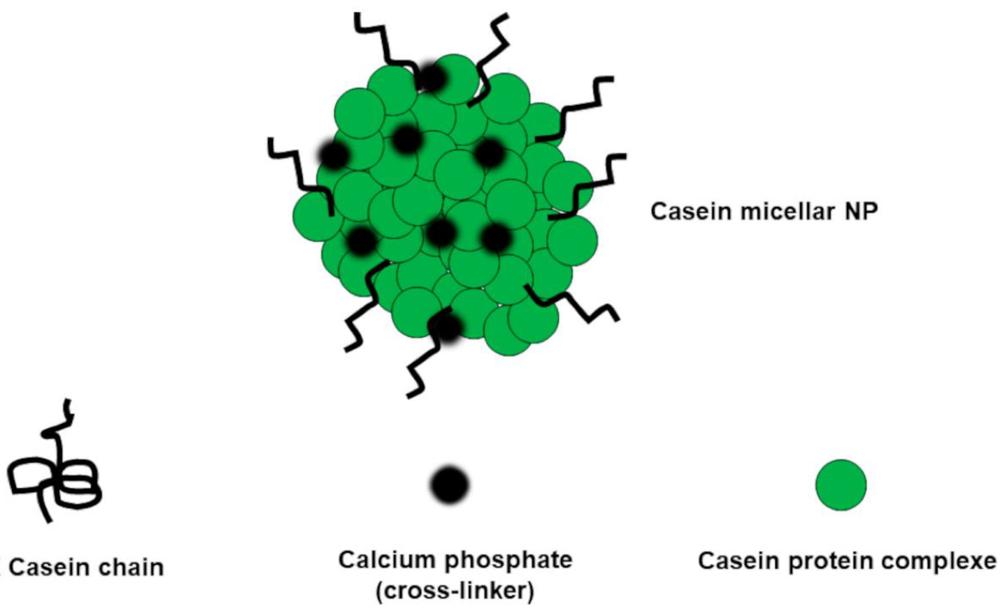
Protein-based NPs (ProNPs) can be prepared through proteins precipitation methods including de-solvation, coacervation, emulsification, nanoprecipitation, SD, NP albumin-bound technology, self-assembly, electro-spraying, salting out, and crosslinking[62]. Specifically, proteins are dissolved in a suitable solvent and a non-solvent is added. Also, by changing the physicochemical parameters of the protein solution (pH, salinity, or temperature) precipitation of the pristine protein can be caused[99]. De-solvating agents are often added to promote the dehydration of the system. The stability of the pristine protein is increased using chemical, ionic, thermal, and enzymatic crosslinking agents, among which 8% glutaraldehyde aqueous solution or calcium phosphate are the most used. Figure A6 in Appendix A, shows a casein protein complex stabilized with calcium phosphate [62].
An innovative method was introduced, which allows to produce crosslinked and sterilized ProNPs in a one-step procedure is based on γ-irradiation of ProNPs in phosphate buffer (pH = 7.2), in the absence and/or presence of ethanol and methanol at 30% and 40% (v/v). The results showed that by controlling the irradiation dose, it was possible to modulate the crosslinking density and the particle size[100]. Moreover, to enhance their circulation residence time, ProNPs have been surface modified with PEG [52].
Several food-related APs have been formulated using ProNPs, some of which have been included in Table 1, such as EGCG, GA, and probiotics microorganisms[52,101].
Concerning organo-synthetic biodegradable polymer nanoparticles (OBP-NPs) they are already described when SNPs made with biodegradable polymers were discussed. They can load different APs, either by physical interactions or by covalently binding by utilizing their several chemical functions. Depending on the hydrophilic/hydrophobic balance (HLB) of polymers or copolymers, NPs characterized by various shapes and morphologies can be prepared.
They are also suitable for oral administration of nutraceuticals and phytochemicals and for producing food-grade smart nanocomposites for food-packaging (FP), able to preserve food quality, looks, and taste along with storage.
In the food industry, a topical ointment was prepared with PEG and 5% pomegranate rind extract, with excellent release profile and skin-permeation capability of EA and anti-inflammatory effects in a mouse model of contact dermatitis[137,138]. NPs (150–300 nm) made of PLGA, chitosan, and PEG, were loaded with EA (up to 100 μM), achieving EA-loaded PLGA-chitosan-PEG NPs, that were able to potentiate apoptosis-mediated cell death in HepG2 human hepatoma cells[139]. PLGA-based NPs stabilized by PEG were used to encapsulate anthocyanins, obtaining anthocyanins-loaded biodegradable NPs that showed an EE% of 60%, improved stability, extended life, and a biphasic release profile in vitro. In vivo, they proved anti-inflammatory and anti-neurodegenerative capacities, preventing memory losses in estrogen-deficient rats, and showed a neuroprotective power against Alzheimer’s dementia[140,141,142]. Finally, anthocyanins formulated as NPs significantly upregulated endogenous antioxidant genes, thus helping in the prevention of oxidative stress (OS), with consequent attenuation of the clinical symptoms of the Alzheimer’s dementia and reduction of DNA damage to a higher extent than the native non-conjugated AP[140].
The oral administration of EA-loaded PLC-NPs (EA-NPs), which proved to have high EE% and DL%, produced an EA plasma concentration 3.6-fold higher than that produced administering free EA[143].
Several structurally different eco-friendly soybean-oil-based cationic polyurethanes (PURs) were prepared to develop edible food coatings with antimicrobial properties toward a panel of bacterial pathogens including Listeria monocytogenes NADC 2045, Salmonella typhimurium ATCC 13311, and S. minnesota R613. Tested against the same strains of wild-type, the PURs-based NPs exhibited better antibacterial activity on the Gram-positive L. monocytogenes than on the Gram-negative S. minnesota and excellent activity against S. Minnesota R613 [144].
With the aim of ameliorating their performances, several EOs and their constituents have been subjected to modifications by nanotechnology and converted into NPs formulations for improving their antimicrobial activity, thus allowing their exploitation to extend food shelf-life and to minimize the growth of foodborne pathogens.
More in Deep about Nanotechnology Applications to Nutraceuticals (Nuts) and Phytochemicals (Phys): in Vivo Experimental Advances
To exploit phytochemicals as health enhancers, researchers extensively engineered nanomaterials and resorted to nanotechnology and nanostructures with dimensions of nano meters (nm). Formulation of Phys using NPs has allowed their controlled and targeted release, which is essential for effective administration [70]. Controlled nano delivery translates in a higher concentration at the target, thus allowing a reduction in the overall administered dose and consequently systemic toxicity [1]. Both internal and external factors, such as pH, temperature, ultrasound or magnetic field application, light incidence, and the type and physicochemical features of NPs, as well as the chemical structure and the physicochemical features of the bioactive compounds themselves, can control their specific release [1].
Stimuli-sensitive nano-capsules containing a bioactive derivative of paclitaxel and possessing an oil core showed the capability to improve the anticancer effects of the encapsulated compound taken by oral administration, thanks to targeted delivery and controlled long-term release[77]. The improved effects allowed a decrease in the dosage and the administration frequency, thus improving patient compliance[77]. Starting from biocompatible pH-dependent polyelectrolytes, nontoxic nanocarriers with high permeability were designed[77].
The layer-by-layer self-assembly of pH-sensitive building blocks proved to be a promising approach to obtaining Phys-based biomaterials with customized properties, which were successfully applied as stimuli-responsive nanocarriers[145]. The encapsulation of bioactive compounds contained in food in properly functionalized NPs permitted increased cellular uptake and slower drug release, thus improving their bioactivity and contributing to sustained therapy[52].
According to a not recent but relevant research paper, only up to the year 2019, while liposomes were the most studied NPs for nano-manipulating Phys, nano-emulsions (NEs) were little considered as nanotechnological approach, while nano-suspensions (NSs) were not even reported [146]. Additionally, NSs and NEs are usually produced using regarded as safe (GRAS) ingredients like liposomes, so that they should be considered among the less toxic and the most suitable tactics for developing Phys-loaded NPs finalized to clinical application. Anyway, due to this statistic, a relevant Review dedicated to these too little considered nanotechnologies has been published in 2023. Such work could be of interest to readers particularly attracted by the topic [54].
The poor solubility, permeability, and negative pharmacokinetics of a series of nutraceuticals (Nuts) were enhanced by developing different nanosized delivery systems[147]. Table 2 and Table 3 summarize some examples of Nuts nanotechnologically formulated using different NPs and methods, associated to their activity as demonstrated by in vivo experiments and/or structural characteristics.
In addition to enhance bioavailability of Phys and Nuts, using different techniques, nanotechnology was and is used in the food sector to prepare NPs finalized to act as colour additives, flavourings, and preservatives, as well as to prepare improved food packaging, with the aim to enhance food shelf-life, taste, and appearance [70].
More in Deep about Nanotechnology Applications in Food-Packaging (FP) Industry
In order to improve the mandatory properties of traditional materials for FP, which have been listed in in studies by Kuswandy, 2017, Kuswandi and Moradi, 2019 and more recently reviewed by Alfei et al in 2020 [61,192], nanotechnology is nowadays intensively studied, also for application in the FP industry. It has been demonstrated that the nanoencapsulation of bioactive natural compounds, by using particles with diameters ranging from 1 to 100 nm, leads to a remarkable increase of their solubility and stability, as well as to a decrease of their inactivation rate, thus offering the possibility of preparing better performant food packaging exploitable as preservative agents in comparison to conventional ones[193]. The inclusion in food packaging of NPs with intrinsic antioxidant, antimicrobial and antifungal properties or capable to release antioxidant, antimicrobial, preservative APs, flavours or enzymes, nutraceuticals and/or phytochemicals previously entrapped, can allow further improvements, including longer shelf life and higher food overall quality[194].
Collectively, the use of nanomaterials has improved FP both in physical and in biochemical characteristics [192,195].
Note that both natural and synthetic polymers have been employed in the past to produce conventional FPs. Natural biopolymers include lipids-based, polysaccharides-based and proteins-based polymers like those previously discussed but obtained by the action of living organisms. They are completely degradable, while synthetic ones comprehend both petroleum-based plastics and eco-friendly bio-based polymers, that can be in turn degradable and not degradable materials. Degradable bio-based polymers and natural degradable biopolymers allow to reduce the levels of environmental pollution, but lack of suitable mechanical properties. Resorting to nanotechnology, both eco-friendly bio-based and petroleum-based nanocomposites have been prepared, which allow the development of nanomaterial-based physically improved FP.
3. Our Dealing with Nanotechnology Applications: Last 15 Years Studies
The following Table 5 collects the main details about the nanosized materials developed in our laboratories in the years 2009-2024, while additional twelve review articles we have published, concerning the most relevant nanomaterials for specific applications such as improvement of solubility and bioavailability of natural and/or synthetic APs, food sectors, food packaging, etc., developed by other eminent scientist in the same years [52,53,54,55,56,57,58,59,60,61,62].
4. Nanotoxicology
Nowadays, it is universally recognized that the application of nanotechnology can allow to achieve many nonpareil advantages, regardless the sector of application. Concerning APs, their nanomanipulation has permitted to efficiently administer not soluble and not bioavailable bioactive compounds and to prepare unconventional food-related therapeutics. The protective action of nanoencapsulations of unstable APs has provided bioactive NPs capable to undergo the strong conditions of processes necessary to enrich food with them, thus achieving functional foods (FFs), food supplements (FSs), as well as manufacturing active food packaging (FP) or preservative additives.
Many nanocomposites are still at the lab stage, but several products have been already approved by EFSA and by the Member States and the European Commission[199]. Nanotechnology application to APs, food and beverages has had an exponential growing over the past 15 years, and due to the obtained advantages, the presence in the market of APs and foods nanotechnologically manipulated is destined to increase further. Anyway, a major problem remains to be solved, which concerns the poor knowledge about the possible risky effects on health of humans, animals and environment, which can derive from ingestion and massive exposure to NPs, and from the possible migration of NPs from FP into foodstuff [199]. In this context, the amplification of the development of nanomaterial-based food-related products is a topic debated incessantly among researchers with contrasting opinions, thus spreading concern and prejudice both among producers in various sectors and among consumers[200].
The Possible Migration of NPs From FP to Food and Toxicity of Ingesting Them
The migration rate of NPs from nanocomposites used to manufacture innovative food packaging (FP) into food or food simulants has been measured using European and U.S. (FDA) standard migration tests. Anyway, numerous issues have emerged that complicated the determination and interpretation of NPs migration studies and related results [199]. Commonly, “migration into foodstuff” indicates the process by which the constituents initially present in the package, possibly including NPs, are liberated into the food or beverage packaged [61]. Limits of safety and the list of authorized substances for manufacturing polymeric food-contact nanomaterials have been established by the European Regulation (European Commission, 2011) [199]. Data of migration are available mainly for inorganic NPs, thus evidencing a very restricted knowledge on the question. They mainly include nano-clay, titanium nitride, nano-silver, silanated silicon dioxide, titanium oxide, zinc oxide, and iron oxide NPs. Collectively, concerning these NPs, the reported findings have established that the migration of NPs from FP would be low and slow[61]. Additionally, the migration rate of a system increases when NPs size and polymer dynamic viscosity decrease[201].
The practice to insert NPs in the FP materials is still at its infancy, therefore only a few studies are available in the literature and current information on their possible migration and on their toxicity upon exposure are limited. Further fundamental studies on toxicity, ecotoxicity, migration tendency and on risk of the intake of nanocomposite materials are needed, to authorize massive application of nanomaterials in the FP field[202]. What is the actual behaviour of NPs once inserted in packaging and in contact with packaged foodstuff? What are the eventual mechanisms involved in the migration and how can the diffusion process change the size and morphology of nanomaterials?
With the aim at answering to these questions, a standardized food model (SFM) for evaluating the toxicity and fate of NPs migrated in food matrix after ingestion was proposed for the first time by Zhang et al, in the year 2019 and its efficacy was assessed by examining the impact of food matrix on the toxicity of TiO2NPs[203]. SFM is an oil-in-water emulsion usable both in wet and in dried forms. Using a simulated GIT model was observed that all the SFM food components were well digested and that the potential toxicity of TiO2NPs was reduced in the presence of the SFM, by underling the importance of food matrix effects on the actual toxicity of NPs[203]. Table 6 collects the results achieved by evaluations made on some NPs, while Table 7 reports detailed migration results expressed as wt/volume or as wt/wt of numerous inorganic NPs from different food contact materials (FCMs) into food or foods simulants[199].
The overall migration of nanoclay/starch nanoparticles into vegetables was in conformity with European directives, thus establishing that these materials are suitable for utilization in FP sector [61]. Similarly, the migration of PLA/laurate LDH-C12-modified NPs used to reduce gas permeability of a packaging in modified atmosphere for conservation of meat, resulted largely below the total legislative migration limits established for all materials [204]. Experimental results and theoretical considerations about the migration of two types of CNTs/LDPE/PS NPs, in different food simulants, established that such NPs do not migrate from the polymer matrix into food[205]. Even if a commercially available FP improved with AgNPs intended to package chicken meatballs, under common domestic storage conditions, demonstrated no significant antibacterial effects, evidenced a migration rate encouragingly slow[206]. AgNPs did not migrate from AgNPs/PEF into chicken breast or distilled water, after 168 and 72 hours respectively[207]. Inductively coupled plasma mass spectrometry (ICP-MS) analytical technique evidenced that, migration of AgNPs from AgNPs/PE packaging films into an acidic food simulant (3% acetic acid) was promoted by the presence of organic additives, as Irganox 1076, Irgafos 168, Chimassorb 944, Tinuvin 622, UV-531 and UV-P, and by Ag oxidation endorsed by high humidity and temperature treatment[208].
The evaluation of the degree of migration of NPs from active/smart/mechanically improved FP into foods and into the environment should be associate with an all-round knowledge of the possible toxic outcomes that the NPs eventually could have on humans and animals when ingested, as well as on the environment. NPs are not normally eaten and metabolized by humans and animal species and, paradoxically, even if by adding NPs in FP, new advantageous opportunities can be achieved such as ingesting higher-quality and more safe aliments, new risks to human health and the environment can occur[199]. In this regard, the overall risks potentially associated to the intake of food containing NPs is still unclear. Currently, we know that smaller particles are usually absorbed easier and faster and are more promptly distributed into the organs. Here, NPs can damage cells and tissues by reactive oxygen species (ROS) generation or by other type of direct or indirect toxicity[209]. As represented in Figure 4, the toxicological effects of NPs and their mechanisms have been evaluated in vitro and in vivo [210]. For evaluations in vitro, human and/or rodent cell lines from intestine, liver, lung and skin, and plants cells, have been used. The cytotoxicity tests comprised the lactate dehydrogenase (LHD) release assay, live-dead assay, cell counting, Alamar blue assay, neutral red uptake, the evaluation of protein content and, trypan blue dye exclusion test. Specific mechanisms are generally studied by observing the changes in different biomarkers, such as ROS production, glutathione (GSH) levels, inflammation response, DNA damage and cell death. On the contrary, for assessing potential genotoxicity Comet assay, Ames test and micronucleus essay have been adopted.
In vivo evaluations used mainly rodents and macro-toxic or histopathological effects, following the sub-chronic/chronic exposure to different kind of NPs were assessed[210]. Unfortunately, the results from both in vitro and in vivo analyses are often contrasting and questionable. As an example, findings on nano organoclay have established that their toxicological evaluation, case by case, should be performed [210]. In addition, since the average amount of clay added as reinforcement of polymers should be around 5 wt%, it is important that the concentrations tested would be pertinent with a real oral exposure scenario. Collectively, more toxicity data from studies on an increasing number of nanocomposites are necessary for a more reliable evaluation of nanotoxicology and exposure estimation[211].
Table 8 collects the results from in vitro studies we have reviewed. Toxicity depended mainly on concentrations, type of cation of metal NPs and especially morphology of NPs, (nano wires, nano roads, nano spheres etc.). Similarly, Table 9 reports the results from in vivo evaluations of different NPs with different morphologies on different animal models.
Although the potential toxicity by ingestion of Ag NPs is still debated, it has been found that the cytotoxicity reported in Table 8 (first row) can be nullified by the addition of carboxymethylcellulose (CMC) to the colloidal solutions of Ag NPs, while the genotoxic effects of Ag NP dispersions was observed at concentration of 12.4 ppm [212]. One of the most accredited mechanisms by which SPM iron oxide NPs, also referred to as USPIO or SPION (USPIO = ultrasmall superparamagnetic iron oxide; SPION = superparamagnetic iron oxide nanoparticles) can induce cytotoxicity consists in causing an aberrant increasing of ROS. By crossing mitochondrial membrane, the free iron in the form of ferrous ions (Fe2 + ) can react with hydrogen peroxide and oxygen produced by the mitochondria to produce highly reactive hydroxyl radicals and ferric ions (Fe3 + ) via the Fenton reaction. Hydroxyl radicals generated could indirectly damage DNA, causes peroxidation of proteins and lipids and generates inflammation [214]. Physiological doses of SiO2NPs tested in an in vitro model, to assess their effects on gastrointestinal function and health, evidenced damage to the brush border membrane and both acute and chronic adverse effects in gastrointestinal tract (GIT) cells[224]. Furthermore, the results of an in vitro study have demonstrated that ZnO2 NPs may cause a decrement in the transport of Fe and glucose and affect the microvilli of the intestinal cells [225]. Anyway, the toxicity of ZnO2 NPs decreased when a surface modification by a silica coating was performed [226]and proposed as a possible solution to broaden the applications of ZnO2 NPs as antibacterial agent in FP.
Although both in vitro and in vivo studies have established that TiO2 NPs accumulate in the tissues of mammals and are eliminated very slowly, the results both on their accumulation and toxicity are conflicting. More reliable in vivo toxicokinetic data are needed to provide conclusions concerning the risk of TiO2NPs oral exposure[199].
Some in vivo studies on SiO2 NPs showed that they may cause cytotoxicity and ROS generation and may accumulate in liver causing hepatotoxicity evidenced by alterations in morphometry, biochemistry, hematology, liver tissues and the expression of drug-metabolizing enzyme genes[241]. On the contrary, other studies, performed by administrating SiO2NPs, as well as Fe NPs to both female and male rats over a 13-week period, reported no accumulation or toxicity[199].
5. Conclusions
The biomedical revolution is governed by nanotechnology, which aids resolve several issues associated with the most part of natural and-or synthetic APs, thus limiting their effectiveness, as well as their actual applicability in vivo. Target treatment administration and maximized therapeutic effectiveness can be achieved, while side effects can be reduced by formulating APs using nanomaterials. Applying nanotechnology, vaccines, anticancer drugs, antimicrobial agents and other nanomedicines can be engineered, while wearable equipment, diagnostic and imaging equipment can be realized. Combining standard medicines with nanoscale technologies, the blood brain barrier (BBB) can be crossed intact and nanodrugs can circulate inside the brain. This technology offers enormous potential markets and benefits to whole classes of current drugs. It is possible to develop tailored mechanisms for medication administration, new diagnostic methods, and nanoscale medical devices with high residence time. Technological advancements in the fields of nanoscience and nanotechnology have led to a remarkable innovation also in the food sector that could bring wide-ranging benefits to the whole food chain. Such innovations include the development of new tastes, textures, mouth sensations and consistencies of food products. The reduction in the amount of fat and certain additives, such as salt, an enhancement in the absorption and bioavailability of nutrients and supplements, the preservation of food quality and freshness represent other innovative challenges which are progressively solved by nanotechnological studies. The research for novel nanomaterial-based packaging solutions, allowing better traceability and security of food products in the supply chain, is incessantly in rapid expansion. Food packaging (FP) applications currently represent the largest portion of the nanofood market, following the nanosized and/or nano-encapsulated ingredients and additives for healthy food production. Although still new and scarce in Europe and other regions, a discrete amount of nanomaterial-improved food-contact materials (FCMs) and more functional food products containing nanosized ingredients and additives are already available in some countries. It is anyway rational to imagine that such products will be available on the global market in increasing numbers and variety in the coming years. Their expansion will depend mainly on the price, quality and, above all, acceptance by consumers. Although nanotechnology applications for the food, healthy food and biomedicine sectors have undoubtedly unlocked up enormous opportunities for innovation and new developments, they have also opened new happenstances for ensuring safety and evidencing all the potential risks of this new technology, without highlighting only the benefits. Reliable strategies to better know the risks for human, animals and the environment, which can derive from a massive exposition to nanomaterials and to regulate them in a globally harmonized manner are needed. Unfortunately, food laws in different countries may not conform to each other and this fact is a challenge to the regulatory authorities. In this regard, it would desirable that in due course, such issues could be addressed through the development of frameworks relating to key international trade agreements, such as those administered by the World Trade Organization. We hope that this new review could provide much-needed insights into the various aspects and issues relating to the new and exciting developments that nanotechnologies are offering to the food, medicine and related sectors.
Author Contributions
The two authors have read and agreed to the published version of the manuscript
Funding
This research received no external funding.
Data Availability Statement
Not applicable.
Conflicts of Interest
The authors declare no conflicts of interest
Appendix A
Figure A1.
Bottom-up and top-down methods [62]. The top-down techniques start from large-dimension particles and reduce their size to nanometers by a media milling technique, high-pressure homogenization (HPH), ultra-high-pressure homogenization (UHPH), or supercritical fluid processes. The bottom-up methods start instead from the pristine PA and subject it to precipitation, melt emulsification, coacervation, inclusion complexation, or supercritical fluid extraction, thus causing self-association and self-organization, forming nanosized materials. Reproduced from our work [62].
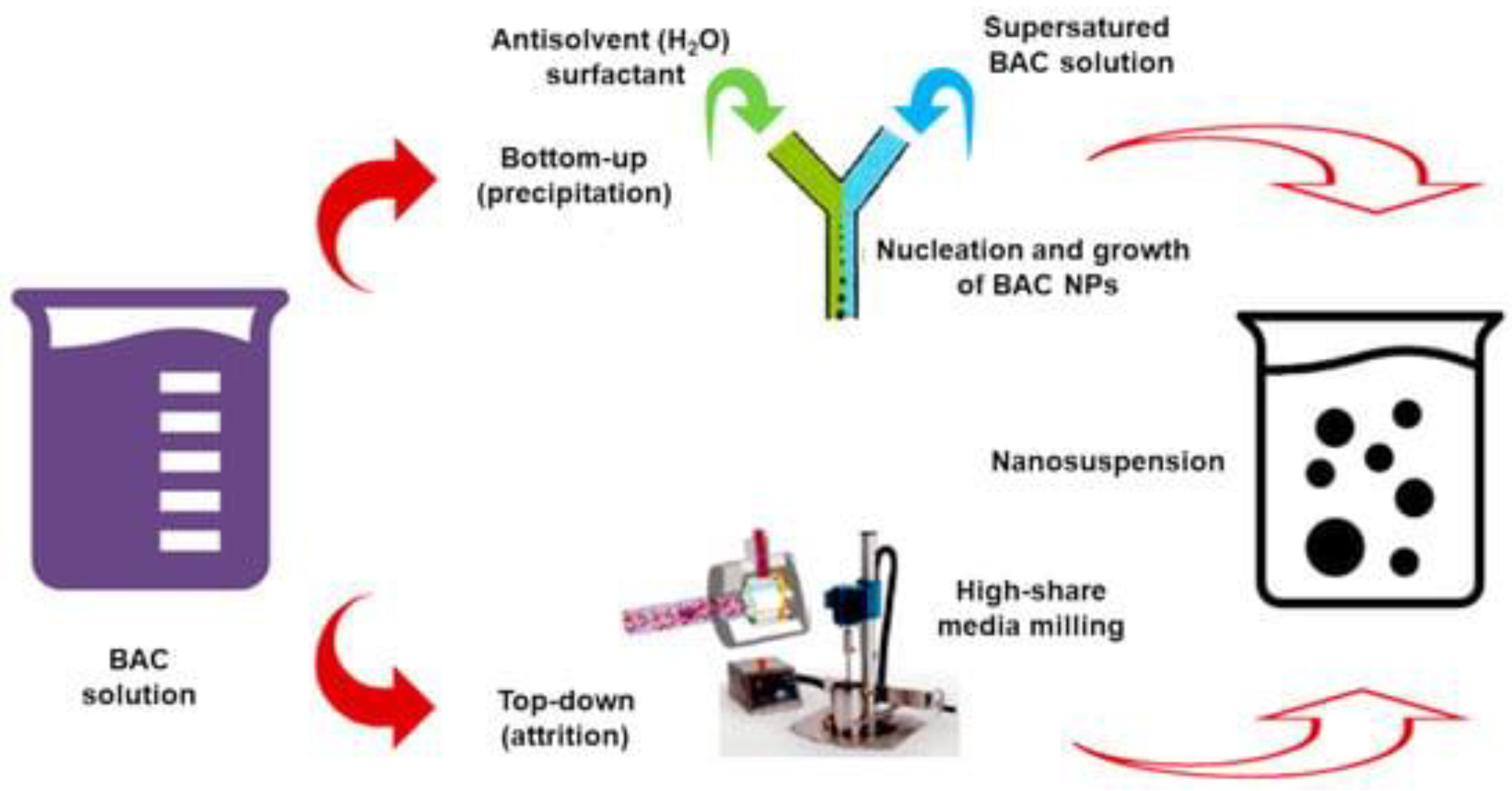
Figure A2.
Schematic process for preparing SEEDSs and formation of NE in GIT fluids. Reproduced from our work [62].
Figure A2.
Schematic process for preparing SEEDSs and formation of NE in GIT fluids. Reproduced from our work [62].
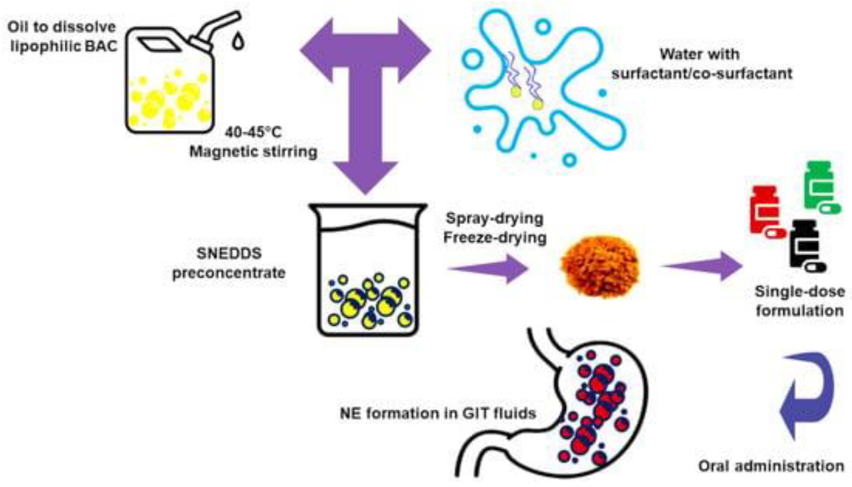
Figure A3.
Examples of lipid-based NPs. Reproduced from our work [62].
Figure A3.
Examples of lipid-based NPs. Reproduced from our work [62].
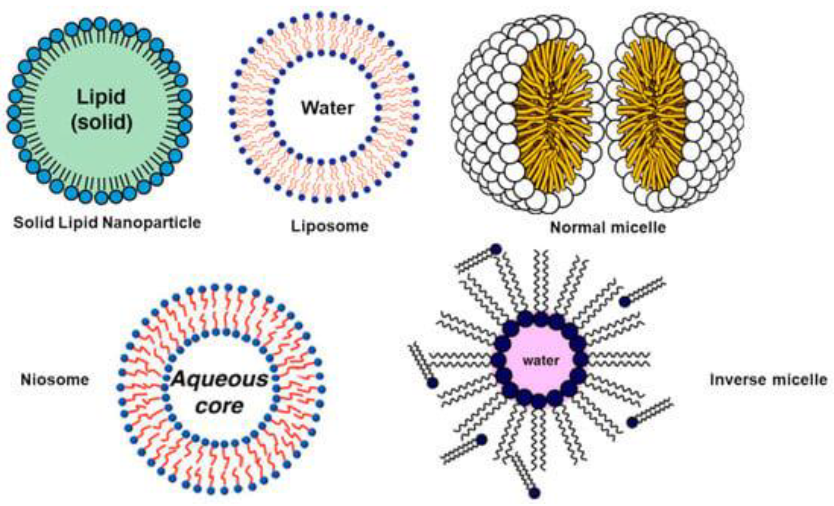
Figure A4.
Chemical structure, spatial arrangement, and size of CDs. Reproduced from our work [62].
Figure A4.
Chemical structure, spatial arrangement, and size of CDs. Reproduced from our work [62].
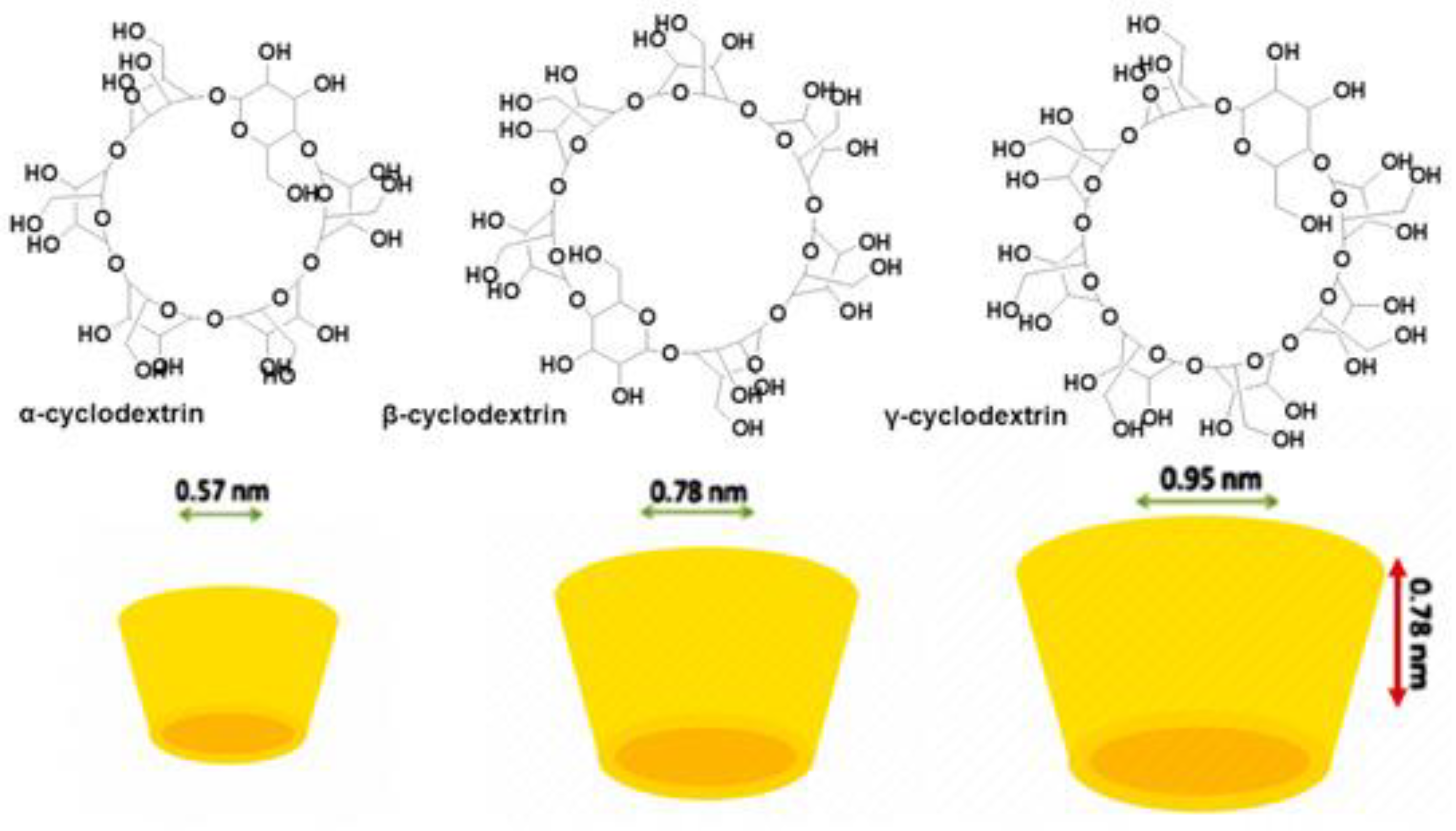
Figure A5.
Process to prepare PNPs. Reproduced from our work [62].
Figure A5.
Process to prepare PNPs. Reproduced from our work [62].
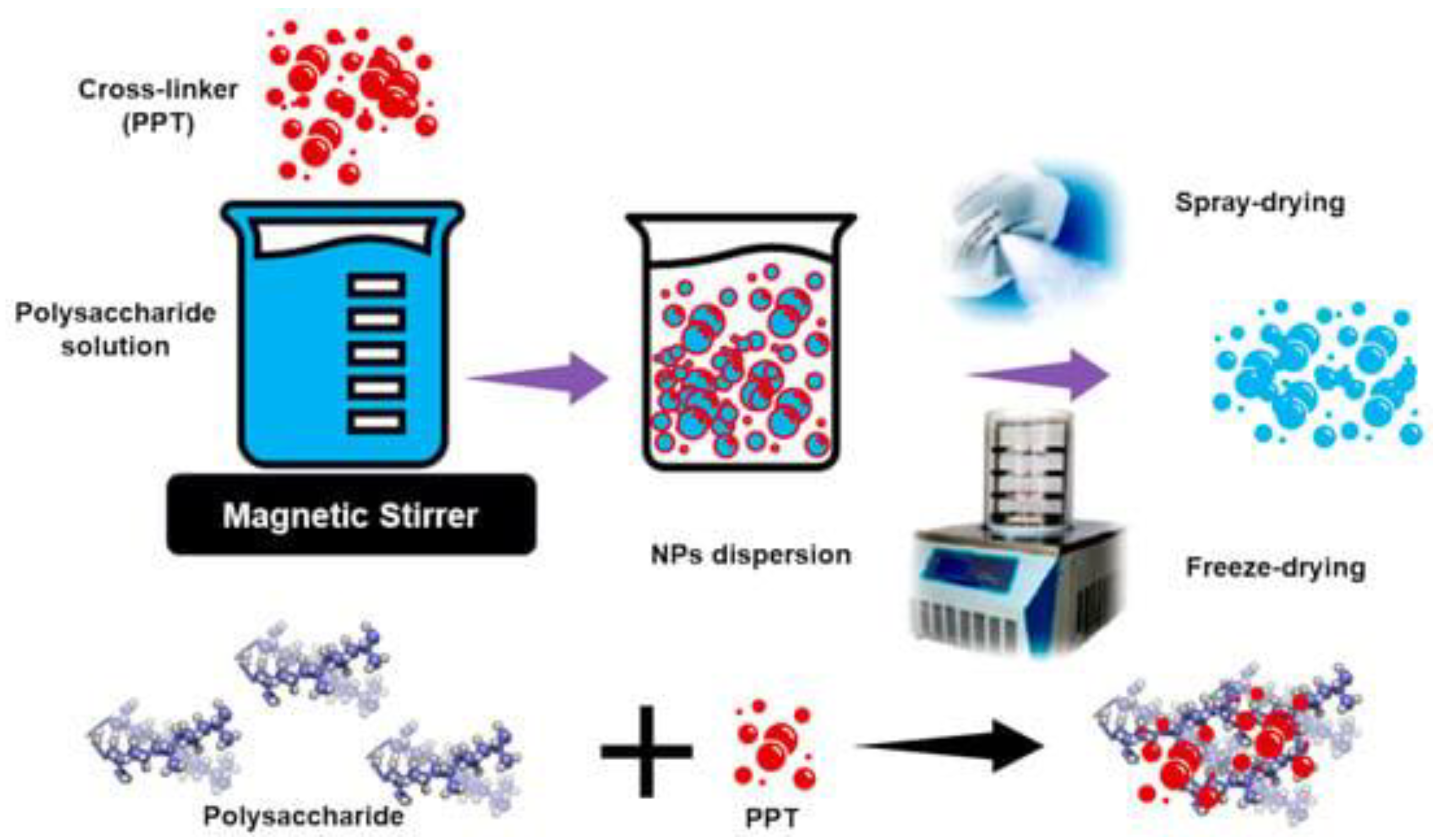
Figure A6.
Example of a casein micellar NPs cross-linked with calcium phosphate. Reproduced from our work [62].
Figure A6.
Example of a casein micellar NPs cross-linked with calcium phosphate. Reproduced from our work [62].
References
- Pocci, M.; Alfei, S.; Lucchesini, F.; Bertini, V.; Idini, B. Nanostructured Styrenic Co-Polymers Containing Glucopyranosyl Residues and Their Functionalization. Tetrahedron 2009, 65, 5684–5692. [Google Scholar] [CrossRef]
- Pocci, M.; Alfei, S.; Lucchesini, F.; Castellaro, S.; Bertini, V. Synthesis and NMR Investigation of Styrene Glycopolymers Containing <scp>d</Scp> -Galactose Units Functionalized with 4-(4-Hydroxybutoxy)Benzylamine Residues. Polym. Chem. 2013, 4, 740–751. [Google Scholar] [CrossRef]
- Pocci, M.; Alfei, S.; Lucchesini, F.; Castellaro, S.; Bertini, V. Synthesis, Glycosylation and NMR Characterization of Linear Peracetylated <scp>d</Scp> -Galactose Glycopolymers. RSC Adv 2015, 5, 23835–23846. [Google Scholar] [CrossRef]
- Alfei, S.; Castellaro, S. Synthesis and Characterization of Polyester-Based Dendrimers Containing Peripheral Arginine or Mixed Amino Acids as Potential Vectors for Gene and Drug Delivery. Macromol Res 2017, 25, 1172–1186. [Google Scholar] [CrossRef]
- Alfei, S.; Castellaro, S.; Taptue, G.B. Synthesis and NMR Characterization of Dendrimers Based on 2, 2-Bis-(Hydroxymethyl)-Propanoic Acid (Bis-HMPA) Containing Peripheral Amino Acid Residues for Gene Transfection. Organic Communications 2017, 10, 144–177. [Google Scholar] [CrossRef]
- Alfei, S.; Catena, S. Synthesis and Characterization of Versatile Amphiphilic Dendrimers Peripherally Decorated with Positively Charged Amino Acids. Polym Int 2018, 67, 1572–1584. [Google Scholar] [CrossRef]
- Alfei, S.; Catena, S. Synthesis and Characterization of Fourth Generation Polyester-based Dendrimers with Cationic Amino Acids-modified Crown as Promising Water Soluble Biomedical Devices. Polym Adv Technol 2018, 29, 2735–2749. [Google Scholar] [CrossRef]
- Alfei, S.; Taptue, G.B.; Catena, S.; Bisio, A. Synthesis of Water-Soluble, Polyester-Based Dendrimer Prodrugs for Exploiting Therapeutic Properties of Two Triterpenoid Acids. Chinese Journal of Polymer Science 2018, 36, 999–1010. [Google Scholar] [CrossRef]
- Alfei, S.; Catena, S.; Ponassi, M.; Rosano, C.; Zoppi, V.; Spallarossa, A. Hydrophilic and Amphiphilic Water-Soluble Dendrimer Prodrugs Suitable for Parenteral Administration of a Non-Soluble Non-Nucleoside HIV-1 Reverse Transcriptase Inhibitor Thiocarbamate Derivative. European Journal of Pharmaceutical Sciences 2018, 124, 153–164. [Google Scholar] [CrossRef] [PubMed]
- Alfei, S.; Signorello, M.G.; Schito, A.; Catena, S.; Turrini, F. Reshaped as Polyester-Based Nanoparticles, Gallic Acid Inhibits Platelet Aggregation, Reactive Oxygen Species Production and Multi-Resistant Gram-Positive Bacteria with an Efficiency Never Obtained. Nanoscale Adv 2019, 1, 4148–4157. [Google Scholar] [CrossRef]
- Alfei, S.; Turrini, F.; Catena, S.; Zunin, P.; Parodi, B.; Zuccari, G.; Pittaluga, A.M.; Boggia, R. Preparation of Ellagic Acid Micro and Nano Formulations with Amazingly Increased Water Solubility by Its Entrapment in Pectin or Non-PAMAM Dendrimers Suitable for Clinical Applications. New Journal of Chemistry 2019, 43, 2438–2448. [Google Scholar] [CrossRef]
- Alfei, S.; Oliveri, P.; Malegori, C. Assessment of the Efficiency of a Nanospherical Gallic Acid Dendrimer for Long-Term Preservation of Essential Oils: An Integrated Chemometric-Assisted FTIR Study. ChemistrySelect 2019, 4, 8891–8901. [Google Scholar] [CrossRef]
- Alfei, S.; Marengo, B.; Domenicotti, C. Polyester-Based Dendrimer Nanoparticles Combined with Etoposide Have an Improved Cytotoxic and Pro-Oxidant Effect on Human Neuroblastoma Cells. Antioxidants 2020, 9, 50. [Google Scholar] [CrossRef]
- Alfei, S.; Marengo, B.; Domenicotti, C. Development of a Fast, Low-Cost, Conservative and Ecological Method for Quantifying Gallic Acid in Polymeric Formulations by FTIR Spectroscopy in Solution. ChemistrySelect 2020, 5, 4381–4388. [Google Scholar] [CrossRef]
- Alfei, S.; Marengo, B.; Zuccari, G.; Turrini, F.; Domenicotti, C. Dendrimer Nanodevices and Gallic Acid as Novel Strategies to Fight Chemoresistance in Neuroblastoma Cells. Nanomaterials 2020, 10, 1243. [Google Scholar] [CrossRef] [PubMed]
- Alfei, S.; Catena, S.; Turrini, F. Biodegradable and Biocompatible Spherical Dendrimer Nanoparticles with a Gallic Acid Shell and a Double-Acting Strong Antioxidant Activity as Potential Device to Fight Diseases from “Oxidative Stress. ” Drug Deliv Transl Res 2020, 10, 259–270. [Google Scholar] [CrossRef] [PubMed]
- Schito, A.M.; Alfei, S. Antibacterial Activity of Non-Cytotoxic, Amino Acid-Modified Polycationic Dendrimers against Pseudomonas Aeruginosa and Other Non-Fermenting Gram-Negative Bacteria. Polymers (Basel) 2020, 12, 1818. [Google Scholar] [CrossRef]
- Alfei, S.; Piatti, G.; Caviglia, D.; Schito, A. Synthesis, Characterization, and Bactericidal Activity of a 4-Ammoniumbuthylstyrene-Based Random Copolymer. Polymers (Basel) 2021, 13, 1140. [Google Scholar] [CrossRef]
- Alfei, S.; Marengo, B.; Valenti, G.; Domenicotti, C. Synthesis of Polystyrene-Based Cationic Nanomaterials with Pro-Oxidant Cytotoxic Activity on Etoposide-Resistant Neuroblastoma Cells. Nanomaterials 2021, 11, 977. [Google Scholar] [CrossRef] [PubMed]
- Schito, A.M.; Schito, G.C.; Alfei, S. Synthesis and Antibacterial Activity of Cationic Amino Acid-Conjugated Dendrimers Loaded with a Mixture of Two Triterpenoid Acids. Polymers (Basel) 2021, 13, 521. [Google Scholar] [CrossRef]
- Alfei, S.; Brullo, C.; Caviglia, D.; Piatti, G.; Zorzoli, A.; Marimpietri, D.; Zuccari, G.; Schito, A.M. Pyrazole-Based Water-Soluble Dendrimer Nanoparticles as a Potential New Agent against Staphylococci. Biomedicines 2021, 10, 17. [Google Scholar] [CrossRef]
- Alfei, S.; Brullo, C.; Caviglia, D.; Zuccari, G. Preparation and Physicochemical Characterization of Water-Soluble Pyrazole-Based Nanoparticles by Dendrimer Encapsulation of an Insoluble Bioactive Pyrazole Derivative. Nanomaterials 2021, 11, 2662. [Google Scholar] [CrossRef] [PubMed]
- Zuccari, G.; Alfei, S.; Zorzoli, A.; Marimpietri, D.; Turrini, F.; Baldassari, S.; Marchitto, L.; Caviglioli, G. Increased Water-Solubility and Maintained Antioxidant Power of Resveratrol by Its Encapsulation in Vitamin E TPGS Micelles: A Potential Nutritional Supplement for Chronic Liver Disease. Pharmaceutics 2021, 13, 1128. [Google Scholar] [CrossRef]
- Schito, A.M.; Caviglia, D.; Piatti, G.; Zorzoli, A.; Marimpietri, D.; Zuccari, G.; Schito, G.C.; Alfei, S. Efficacy of Ursolic Acid-Enriched Water-Soluble and Not Cytotoxic Nanoparticles against Enterococci. Pharmaceutics 2021, 13, 1976. [Google Scholar] [CrossRef] [PubMed]
- Zuccari, G.; Baldassari, S.; Alfei, S.; Marengo, B.; Valenti, G.E.; Domenicotti, C.; Ailuno, G.; Villa, C.; Marchitto, L.; Caviglioli, G. D-α-Tocopherol-Based Micelles for Successful Encapsulation of Retinoic Acid. Pharmaceuticals 2021, 14, 212. [Google Scholar] [CrossRef] [PubMed]
- Alfei, S.; Schito, A.M.; Zuccari, G. Considerable Improvement of Ursolic Acid Water Solubility by Its Encapsulation in Dendrimer Nanoparticles: Design, Synthesis and Physicochemical Characterization. Nanomaterials 2021, 11, 2196. [Google Scholar] [CrossRef] [PubMed]
- Schito, A.M.; Piatti, G.; Caviglia, D.; Zuccari, G.; Alfei, S. Broad-Spectrum Bactericidal Activity of a Synthetic Random Copolymer Based on 2-Methoxy-6-(4-Vinylbenzyloxy)-Benzylammonium Hydrochloride. Int J Mol Sci 2021, 22, 5021. [Google Scholar] [CrossRef]
- Alfei, S.; Caviglia, D.; Piatti, G.; Zuccari, G.; Schito, A.M. Bactericidal Activity of a Self-Biodegradable Lysine-Containing Dendrimer against Clinical Isolates of Acinetobacter Genus. Int J Mol Sci 2021, 22, 7274. [Google Scholar] [CrossRef] [PubMed]
- Schito, A.M.; Piatti, G.; Caviglia, D.; Zuccari, G.; Zorzoli, A.; Marimpietri, D.; Alfei, S. Bactericidal Activity of Non-Cytotoxic Cationic Nanoparticles against Clinically and Environmentally Relevant Pseudomonas Spp. Isolates. Pharmaceutics 2021, 13, 1411. [Google Scholar] [CrossRef]
- Alfei, S.; Caviglia, D.; Piatti, G.; Zuccari, G.; Schito, A.M. Synthesis, Characterization and Broad-Spectrum Bactericidal Effects of Ammonium Methyl and Ammonium Ethyl Styrene-Based Nanoparticles. Nanomaterials 2022, 12, 2743. [Google Scholar] [CrossRef] [PubMed]
- Alfei, S.; Zuccari, G.; Caviglia, D.; Brullo, C. Synthesis and Characterization of Pyrazole-Enriched Cationic Nanoparticles as New Promising Antibacterial Agent by Mutual Cooperation. Nanomaterials 2022, 12, 1215. [Google Scholar] [CrossRef] [PubMed]
- Alfei, S.; Spallarossa, A.; Lusardi, M.; Zuccari, G. Successful Dendrimer and Liposome-Based Strategies to Solubilize an Antiproliferative Pyrazole Otherwise Not Clinically Applicable. Nanomaterials 2022, 12, 233. [Google Scholar] [CrossRef] [PubMed]
- Alfei, S.; Caviglia, D.; Zorzoli, A.; Marimpietri, D.; Spallarossa, A.; Lusardi, M.; Zuccari, G.; Schito, A.M. Potent and Broad-Spectrum Bactericidal Activity of a Nanotechnologically Manipulated Novel Pyrazole. Biomedicines 2022, 10, 907. [Google Scholar] [CrossRef]
- Alfei, S.; Zorzoli, A.; Marimpietri, D.; Schito, A.M.; Russo, E. Mutual Jellification of Two Bactericidal Cationic Polymers: Synthesis and Physicochemical Characterization of a New Two-Component Hydrogel. Pharmaceutics 2022, 14, 2444. [Google Scholar] [CrossRef] [PubMed]
- Schito, A.M.; Caviglia, D.; Brullo, C.; Zorzoli, A.; Marimpietri, D.; Alfei, S. Enhanced Antibacterial Activity of a Cationic Macromolecule by Its Complexation with a Weakly Active Pyrazole Derivative. Biomedicines 2022, 10, 1607. [Google Scholar] [CrossRef]
- Alfei, S.; Zorzoli, A.; Marimpietri, D.; Zuccari, G.; Russo, E.; Caviglia, D.; Schito, A.M. A Self-Forming Hydrogel from a Bactericidal Copolymer: Synthesis, Characterization, Biological Evaluations and Perspective Applications. Int J Mol Sci 2022, 23, 15092. [Google Scholar] [CrossRef]
- Zuccari, G.; Russo, E.; Villa, C.; Zorzoli, A.; Marimpietri, D.; Marchitto, L.; Alfei, S. Preparation and Characterization of Amorphous Solid Dispersions for the Solubilization of Fenretinide. Pharmaceuticals 2023, 16, 388. [Google Scholar] [CrossRef] [PubMed]
- Zuccari, G.; Zorzoli, A.; Marimpietri, D.; Brullo, C.; Alfei, S. Pyrazole-Enriched Cationic Nanoparticles Induced Early- and Late-Stage Apoptosis in Neuroblastoma Cells at Sub-Micromolar Concentrations. Pharmaceuticals 2023, 16, 393. [Google Scholar] [CrossRef] [PubMed]
- Valenti, G.E.; Marengo, B.; Milanese, M.; Zuccari, G.; Brullo, C.; Domenicotti, C.; Alfei, S. Imidazo-Pyrazole-Loaded Palmitic Acid and Polystyrene-Based Nanoparticles: Synthesis, Characterization and Antiproliferative Activity on Chemo-Resistant Human Neuroblastoma Cells. Int J Mol Sci 2023, 24, 15027. [Google Scholar] [CrossRef] [PubMed]
- Alfei, S.; Milanese, M.; Brullo, C.; Valenti, G.E.; Domenicotti, C.; Russo, E.; Marengo, B. Antiproliferative Imidazo-Pyrazole-Based Hydrogel: A Promising Approach for the Development of New Treatments for PLX-Resistant Melanoma. Pharmaceutics 2023, 15, 2425. [Google Scholar] [CrossRef] [PubMed]
- Alfei, S.; Giannoni, P.; Signorello, M.G.; Torazza, C.; Zuccari, G.; Athanassopoulos, C.M.; Domenicotti, C.; Marengo, B. The Remarkable and Selective In Vitro Cytotoxicity of Synthesized Bola-Amphiphilic Nanovesicles on Etoposide-Sensitive and -Resistant Neuroblastoma Cells. Nanomaterials 2024, 14, 1505. [Google Scholar] [CrossRef] [PubMed]
- Alfei, S.; Zuccari, G.; Bacchetti, F.; Torazza, C.; Milanese, M.; Siciliano, C.; Athanassopoulos, C.M.; Piatti, G.; Schito, A.M. Synthesized Bis-Triphenyl Phosphonium-Based Nano Vesicles Have Potent and Selective Antibacterial Effects on Several Clinically Relevant Superbugs. Nanomaterials 2024, 14, 1351. [Google Scholar] [CrossRef]
- Alfei, S.; Giordani, P.; Zuccari, G. Synthesis and Physicochemical Characterization of Gelatine-Based Biodegradable Aerogel-like Composites as Possible Scaffolds for Regenerative Medicine. Int J Mol Sci 2024, 25, 5009. [Google Scholar] [CrossRef] [PubMed]
- Alfei, S.; Zuccari, G.; Athanassopoulos, C.M.; Domenicotti, C.; Marengo, B. Strongly ROS-Correlated, Time-Dependent, and Selective Antiproliferative Effects of Synthesized Nano Vesicles on BRAF Mutant Melanoma Cells and Their Hyaluronic Acid-Based Hydrogel Formulation. Int J Mol Sci 2024, 25, 10071. [Google Scholar] [CrossRef] [PubMed]
- Orienti, I.; Zuccari, G.; Carosio, R.; G. Montaldo, P. Improvement of Aqueous Solubility of Fenretinide and Other Hydrophobic Anti-Tumor Drugs by Complexation with Amphiphilic Dextrins. Drug Deliv 2009, 16, 389–398. [Google Scholar] [CrossRef]
- Zuccari, G.; Bergamante, V.; Carosio, R.; Gotti, R.; Montaldo, P.G.; Orienti, I. Micellar Complexes of All-Trans Retinoic Acid with Polyvinylalcohol-Nicotinoyl Esters as New Parenteral Formulations in Neuroblastoma. Drug Deliv 2009, 16, 189–195. [Google Scholar] [CrossRef] [PubMed]
- Carosio, R.; Pistoia, V.; Orienti, I.; Formelli, F.; Cavadini, E.; Mangraviti, S.; Montaldo, P.G.; Ognio, E.; Emionite, L.; Zuccari, G. Enhanced Anti-Neuroblastoma Activity of a Fenretinide Complexed Form after Intravenous Administration. Journal of Pharmacy and Pharmacology 2012, 64, 228–236. [Google Scholar] [CrossRef]
- Orienti, I.; Zuccari, G.; Falconi, M.; Teti, G.; Illingworth, N.A.; Veal, G.J. Novel Micelles Based on Amphiphilic Branched PEG as Carriers for Fenretinide. Nanomedicine 2012, 8, 880–890. [Google Scholar] [CrossRef] [PubMed]
- Di Paolo, D.; Pastorino, F.; Zuccari, G.; Caffa, I.; Loi, M.; Marimpietri, D.; Brignole, C.; Perri, P.; Cilli, M.; Nico, B.; et al. Enhanced Anti-Tumor and Anti-Angiogenic Efficacy of a Novel Liposomal Fenretinide on Human Neuroblastoma. Journal of Controlled Release 2013, 170, 445–451. [Google Scholar] [CrossRef] [PubMed]
- Zuccari, G.; Milelli, A.; Pastorino, F.; Loi, M.; Petretto, A.; Parise, A.; Marchetti, C.; Minarini, A.; Cilli, M.; Emionite, L.; et al. Tumor Vascular Targeted Liposomal-Bortezomib Minimizes Side Effects and Increases Therapeutic Activity in Human Neuroblastoma. Journal of Controlled Release 2015, 211, 44–52. [Google Scholar] [CrossRef]
- Parise, A.; Milelli, A.; Tumiatti, V.; Minarini, A.; Neviani, P.; Zuccari, G. Preparation, Characterization and in Vitro Evaluation of Sterically Stabilized Liposome Containing a Naphthalenediimide Derivative as Anticancer Agent. Drug Deliv 2015, 22, 590–597. [Google Scholar] [CrossRef]
- Alfei, S. Nanotechnology Applications to Improve Solubility of Bioactive Constituents of Foods for Health-Promoting Purposes. In; 2020; pp. 189–257.
- Alfei, S. Cationic Materials for Gene Therapy: A Look Back to the Birth and Development of 2,2-Bis-(Hydroxymethyl)Propanoic Acid-Based Dendrimer Scaffolds. Int J Mol Sci 2023, 24, 16006. [Google Scholar] [CrossRef] [PubMed]
- Zuccari, G.; Alfei, S. Development of Phytochemical Delivery Systems by Nano-Suspension and Nano-Emulsion Techniques. Int J Mol Sci 2023, 24, 9824. [Google Scholar] [CrossRef]
- Alfei, S.; Caviglia, D. Prevention and Eradication of Biofilm by Dendrimers: A Possibility Still Little Explored. Pharmaceutics 2022, 14, 2016. [Google Scholar] [CrossRef] [PubMed]
- Alfei, S.; Zuccari, G. Attempts to Improve Lipophilic Drugs’ Solubility and Bioavailability: A Focus on Fenretinide. Pharmaceutics 2024, 16, 579. [Google Scholar] [CrossRef] [PubMed]
- Alfei, S.; Caviglia, D. Prevention and Eradication of Biofilm by Dendrimers: A Possibility Still Little Explored. Pharmaceutics 2022, 14, 2016. [Google Scholar] [CrossRef]
- Alfei, S.; Schito, A.M. Positively Charged Polymers as Promising Devices against Multidrug Resistant Gram-Negative Bacteria: A Review. Polymers (Basel) 2020, 12, 1195. [Google Scholar] [CrossRef]
- Alfei, S.; Schito, G.C. Nanotubes: Carbon-Based Fibers and Bacterial Nano-Conduits Both Arousing a Global Interest and Conflicting Opinions. Fibers 2022, 10, 75. [Google Scholar] [CrossRef]
- Alfei, S.; Schito, A.M. From Nanobiotechnology, Positively Charged Biomimetic Dendrimers as Novel Antibacterial Agents: A Review. Nanomaterials 2020, 10, 2022. [Google Scholar] [CrossRef] [PubMed]
- Alfei, S.; Marengo, B.; Zuccari, G. Nanotechnology Application in Food Packaging: A Plethora of Opportunities versus Pending Risks Assessment and Public Concerns. Food Research International 2020, 137, 109664. [Google Scholar] [CrossRef] [PubMed]
- Alfei, S.; Schito, A.M.; Zuccari, G. Nanotechnological Manipulation of Nutraceuticals and Phytochemicals for Healthy Purposes: Established Advantages vs. Still Undefined Risks. Polymers (Basel) 2021, 13, 2262. [Google Scholar] [CrossRef]
- Thapa, R.K.; Kim, J.O. Nanomedicine-Based Commercial Formulations: Current Developments and Future Prospects. J Pharm Investig 2023, 53, 19–33. [Google Scholar] [CrossRef]
- Jagtiani, E. Advancements in Nanotechnology for Food Science and Industry. Food Front 2022, 3, 56–82. [Google Scholar] [CrossRef]
- Li, J.; Kataoka, K. Chemo-Physical Strategies to Advance the in Vivo Functionality of Targeted Nanomedicine: The Next Generation. J Am Chem Soc 2021, 143, 538–559. [Google Scholar] [CrossRef] [PubMed]
- Ganta, S.; Deshpande, D.; Korde, A.; Amiji, M. A Review of Multifunctional Nanoemulsion Systems to Overcome Oral and CNS Drug Delivery Barriers. Mol Membr Biol 2010, 27, 260–273. [Google Scholar] [CrossRef]
- González-Sarrías, A.; García-Villalba, R.; Núñez-Sánchez, M.Á.; Tomé-Carneiro, J.; Zafrilla, P.; Mulero, J.; Tomás-Barberán, F.A.; Espín, J.C. Identifying the Limits for Ellagic Acid Bioavailability: A Crossover Pharmacokinetic Study in Healthy Volunteers after Consumption of Pomegranate Extracts. J Funct Foods 2015, 19, 225–235. [Google Scholar] [CrossRef]
- Alfei, S.; Zuccari, G. Ellagic Acid (EA): A Green Multi-Target Weapon Reducing Oxidative Stress and Inflammation Thus Preventing and Ameliorating Alzheimer Disease (AD) Condition. Preprints 2024, 2024111130. [Google Scholar]
- Angelova, A.; Angelov, B.; Drechsler, M.; Lesieur, S. Neurotrophin Delivery Using Nanotechnology. Drug Discov Today 2013, 18, 1263–1271. [Google Scholar] [CrossRef] [PubMed]
- Zuccari, G.; Baldassari, S.; Ailuno, G.; Turrini, F.; Alfei, S.; Caviglioli, G. Formulation Strategies to Improve Oral Bioavailability of Ellagic Acid. Applied Sciences 2020, 10, 3353. [Google Scholar] [CrossRef]
- Avachat, A.M.; Patel, V.G. Self Nanoemulsifying Drug Delivery System of Stabilized Ellagic Acid–Phospholipid Complex with Improved Dissolution and Permeability. Saudi Pharmaceutical Journal 2015, 23, 276–289. [Google Scholar] [CrossRef]
- Madrigal-Carballo, S.; Lim, S.; Rodriguez, G.; Vila, A.O.; Krueger, C.G.; Gunasekaran, S.; Reed, J.D. Biopolymer Coating of Soybean Lecithin Liposomes via Layer-by-Layer Self-Assembly as Novel Delivery System for Ellagic Acid. J Funct Foods 2010, 2, 99–106. [Google Scholar] [CrossRef]
- Hajipour, H.; Hamishehkar, H.; Rahmati-yamchi, M.; Shanehbandi, D.; Nazari Soltan Ahmad, S.; Hasani, A. Enhanced Anti-Cancer Capability of Ellagic Acid Using Solid Lipid Nanoparticles (SLNs). Int J Cancer Manag 2018, 11. [Google Scholar] [CrossRef]
- Bulani, V.D.; Kothavade, P.S.; Kundaikar, H.S.; Gawali, N.B.; Chowdhury, A.A.; Degani, M.S.; Juvekar, A.R. Inclusion Complex of Ellagic Acid with β-Cyclodextrin: Characterization and in Vitro Anti-Inflammatory Evaluation. J Mol Struct 2016, 1105, 308–315. [Google Scholar] [CrossRef]
- Mady, F.M.; Ibrahim, S.R.-M. Cyclodextrin-Based Nanosponge for Improvement of Solubility and Oral Bioavailability of Ellagic Acid. Pak J Pharm Sci 2018, 31, 2069–2076. [Google Scholar]
- Sha, X.; Guo, J.; Chen, Y.; Fang, X. Effect of Phospholipid Composition on Pharmacokinetics and Biodistribution of Epirubicin Liposomes. J Liposome Res 2012, 22, 80–88. [Google Scholar] [CrossRef]
- Liu, Y.; Li, K.; Liu, B.; Feng, S.-S. A Strategy for Precision Engineering of Nanoparticles of Biodegradable Copolymers for Quantitative Control of Targeted Drug Delivery. Biomaterials 2010, 31, 9145–9155. [Google Scholar] [CrossRef] [PubMed]
- de Paz, E.; Martín, Á.; Estrella, A.; Rodríguez-Rojo, S.; Matias, A.A.; Duarte, C.M.M.; Cocero, M.J. Formulation of β-Carotene by Precipitation from Pressurized Ethyl Acetate-on-Water Emulsions for Application as Natural Colorant. Food Hydrocoll 2012, 26, 17–27. [Google Scholar] [CrossRef]
- Karadag, A.; Ozcelik, B.; Huang, Q. Quercetin Nanosuspensions Produced by High-Pressure Homogenization. J Agric Food Chem 2014, 62, 1852–1859. [Google Scholar] [CrossRef] [PubMed]
- Vargas, V.; Saldarriaga, S.; Sánchez, F.S.; Cuellar, L.N.; Paladines, G.M. Effects of the Spray-Drying Process Using Maltodextrin on Bioactive Compounds and Antioxidant Activity of the Pulp of the Tropical Fruit Açai (Euterpe Oleracea Mart.). Heliyon 2024, 10, e33544. [Google Scholar] [CrossRef]
- Campardelli, R.; Reverchon, E. α-Tocopherol Nanosuspensions Produced Using a Supercritical Assisted Process. J Food Eng 2015, 149, 131–136. [Google Scholar] [CrossRef]
- Gupta, A.; Eral, H.B.; Hatton, T.A.; Doyle, P.S. Nanoemulsions: Formation, Properties and Applications. Soft Matter 2016, 12, 2826–2841. [Google Scholar] [CrossRef]
- Scalbert, A.; Williamson, G. Dietary Intake and Bioavailability of Polyphenols. J Nutr 2000, 130, 2073S–2085S. [Google Scholar] [CrossRef] [PubMed]
- Koutelidakis, A.E.; Argyri, K.; Sevastou, Z.; Lamprinaki, D.; Panagopoulou, E.; Paximada, E.; Sali, A.; Papalazarou, V.; Mallouchos, A.; Evageliou, V.; et al. Bioactivity of Epigallocatechin Gallate Nanoemulsions Evaluated in Mice Model. J Med Food 2017, 20, 923–931. [Google Scholar] [CrossRef]
- Martínez-Ballesta, Mc.; Gil-Izquierdo, Á.; García-Viguera, C.; Domínguez-Perles, R. Nanoparticles and Controlled Delivery for Bioactive Compounds: Outlining Challenges for New “Smart-Foods” for Health. Foods 2018, 7, 72. [Google Scholar] [CrossRef]
- Baccarin, T.; Lemos-Senna, E. Potential Application of Nanoemulsions for Skin Delivery of Pomegranate Peel Polyphenols. AAPS PharmSciTech 2017, 18, 3307–3314. [Google Scholar] [CrossRef] [PubMed]
- Kim, I.; Lee, H.; Kim, J.E.; Song, K. Bin; Lee, Y.S.; Chung, D.S.; Min, S.C. Plum Coatings of Lemongrass Oil-incorporating Carnauba Wax-based Nanoemulsion. J Food Sci 2013, 78. [Google Scholar] [CrossRef]
- Salazar, J.; Müller, R.H.; Möschwitzer, J.P. Combinative Particle Size Reduction Technologies for the Production of Drug Nanocrystals. J Pharm (Cairo) 2014, 2014, 1–14. [Google Scholar] [CrossRef]
- Magnuson, B.A.; Jonaitis, T.S.; Card, J.W. A Brief Review of the Occurrence, Use, and Safety of Food-Related Nanomaterials. J Food Sci 2011, 76. [Google Scholar] [CrossRef]
- Simion, V.; Stan, D.; Constantinescu, C.A.; Deleanu, M.; Dragan, E.; Tucureanu, M.M.; Gan, A.-M.; Butoi, E.; Constantin, A.; Manduteanu, I.; et al. Conjugation of Curcumin-Loaded Lipid Nanoemulsions with Cell-Penetrating Peptides Increases Their Cellular Uptake and Enhances the Anti-Inflammatory Effects in Endothelial Cells. Journal of Pharmacy and Pharmacology 2016, 68, 195–207. [Google Scholar] [CrossRef] [PubMed]
- Xiao, J.; Cao, Y.; Huang, Q. Edible Nanoencapsulation Vehicles for Oral Delivery of Phytochemicals: A Perspective Paper. J Agric Food Chem 2017, 65, 6727–6735. [Google Scholar] [CrossRef] [PubMed]
- Kumari, A.; Yadav, S.K.; Yadav, S.C. Biodegradable Polymeric Nanoparticles Based Drug Delivery Systems. Colloids Surf B Biointerfaces 2010, 75, 1–18. [Google Scholar] [CrossRef] [PubMed]
- Zou, L.; Zheng, B.; Zhang, R.; Zhang, Z.; Liu, W.; Liu, C.; Xiao, H.; McClements, D.J. Food-Grade Nanoparticles for Encapsulation, Protection and Delivery of Curcumin: Comparison of Lipid, Protein, and Phospholipid Nanoparticles under Simulated Gastrointestinal Conditions. RSC Adv 2016, 6, 3126–3136. [Google Scholar] [CrossRef]
- Osorno, L.; Brandley, A.; Maldonado, D.; Yiantsos, A.; Mosley, R.; Byrne, M. Review of Contemporary Self-Assembled Systems for the Controlled Delivery of Therapeutics in Medicine. Nanomaterials 2021, 11, 278. [Google Scholar] [CrossRef]
- Thatipamula, R.; Palem, C.; Gannu, R.; Mudragada, S.; Yamsani, M. Formulation and in Vitro Characterization of Domperidone Loaded Solid Lipid Nanoparticles and Nanostructured Lipid Carriers. Daru 2011, 19, 23–32. [Google Scholar] [PubMed]
- Bulbake, U.; Doppalapudi, S.; Kommineni, N.; Khan, W. Liposomal Formulations in Clinical Use: An Updated Review. Pharmaceutics 2017, 9, 12. [Google Scholar] [CrossRef]
- Wang, Q.; Atluri, K.; Tiwari, A.K.; Babu, R.J. Exploring the Application of Micellar Drug Delivery Systems in Cancer Nanomedicine. Pharmaceuticals 2023, 16, 433. [Google Scholar] [CrossRef] [PubMed]
- Liga, S.; Paul, C.; Moacă, E.-A.; Péter, F. Niosomes: Composition, Formulation Techniques, and Recent Progress as Delivery Systems in Cancer Therapy. Pharmaceutics 2024, 16, 223. [Google Scholar] [CrossRef]
- Miladi, K.; Ibraheem, D.; Iqbal, M.; Sfar, S.; Fessi, H.; Elaissari, A. Particles from Preformed Polymers as Carriers for Drug Delivery. EXCLI J 2014, 13, 28–57. [Google Scholar] [PubMed]
- Tarhini, M.; Greige-Gerges, H.; Elaissari, A. Protein-Based Nanoparticles: From Preparation to Encapsulation of Active Molecules. Int J Pharm 2017, 522, 172–197. [Google Scholar] [CrossRef]
- Ruan, J.; Yang, Y.; Yang, F.; Wan, K.; Fan, D.; Wang, D. Novel Oral Administrated Ellagic Acid Nanoparticles for Enhancing Oral Bioavailability and Anti-Inflammatory Efficacy. J Drug Deliv Sci Technol 2018, 46, 215–222. [Google Scholar] [CrossRef]
- Zhu, X.-F.; Zheng, J.; Liu, F.; Qiu, C.-Y.; Lin, W.-F.; Tang, C.-H. The Influence of Ionic Strength on the Characteristics of Heat-Induced Soy Protein Aggregate Nanoparticles and the Freeze–Thaw Stability of the Resultant Pickering Emulsions. Food Funct 2017, 8, 2974–2981. [Google Scholar] [CrossRef] [PubMed]
- Marques, H.M.C. A Review on Cyclodextrin Encapsulation of Essential Oils and Volatiles. Flavour Fragr J 2010, 25, 313–326. [Google Scholar] [CrossRef]
- Sarabia-Vallejo, Á.; Caja, M. del M.; Olives, A.I.; Martín, M.A.; Menéndez, J.C. Cyclodextrin Inclusion Complexes for Improved Drug Bioavailability and Activity: Synthetic and Analytical Aspects. Pharmaceutics 2023, 15, 2345. [Google Scholar] [CrossRef] [PubMed]
- Recharla, N.; Riaz, M.; Ko, S.; Park, S. Novel Technologies to Enhance Solubility of Food-Derived Bioactive Compounds: A Review. J Funct Foods 2017, 39, 63–73. [Google Scholar] [CrossRef]
- Fang, Z.; Bhandari, B. Encapsulation of Polyphenols – a Review. Trends Food Sci Technol 2010, 21, 510–523. [Google Scholar] [CrossRef]
- Nerome, H.; Machmudah, S.; Wahyudiono; Fukuzato, R. ; Higashiura, T.; Youn, Y.-S.; Lee, Y.-W.; Goto, M. Nanoparticle Formation of Lycopene/β-Cyclodextrin Inclusion Complex Using Supercritical Antisolvent Precipitation. J Supercrit Fluids 2013, 83, 97–103. [Google Scholar] [CrossRef]
- de Lima Petito, N.; da Silva Dias, D.; Costa, V.G.; Falcão, D.Q.; de Lima Araujo, K.G. Increasing Solubility of Red Bell Pepper Carotenoids by Complexation with 2-Hydroxypropyl-β-Cyclodextrin. Food Chem 2016, 208, 124–131. [Google Scholar] [CrossRef] [PubMed]
- Nitta, S.; Numata, K. Biopolymer-Based Nanoparticles for Drug/Gene Delivery and Tissue Engineering. Int J Mol Sci 2013, 14, 1629–1654. [Google Scholar] [CrossRef]
- Salatin, S.; Jelvehgari, M. Natural Polysaccharide Based Nanoparticles for Drug/Gene Delivery. Pharmaceutical Sciences 2017, 23, 84–94. [Google Scholar] [CrossRef]
- Amidi, M.; Mastrobattista, E.; Jiskoot, W.; Hennink, W.E. Chitosan-Based Delivery Systems for Protein Therapeutics and Antigens. Adv Drug Deliv Rev 2010, 62, 59–82. [Google Scholar] [CrossRef]
- Duceppe, N.; Tabrizian, M. Advances in Using Chitosan-Based Nanoparticles for in Vitro and in Vivo Drug and Gene Delivery. Expert Opin Drug Deliv 2010, 7, 1191–1207. [Google Scholar] [CrossRef] [PubMed]
- Ojea-Jiménez, I.; Tort, O.; Lorenzo, J.; Puntes, V.F. Engineered Nonviral Nanocarriers for Intracellular Gene Delivery Applications. Biomedical Materials 2012, 7, 054106. [Google Scholar] [CrossRef] [PubMed]
- Sun, J.; Tan, H. Alginate-Based Biomaterials for Regenerative Medicine Applications. Materials 2013, 6, 1285–1309. [Google Scholar] [CrossRef] [PubMed]
- Chan, S.Y.; Choo, W.S.; Young, D.J.; Loh, X.J. Pectin as a Rheology Modifier: Origin, Structure, Commercial Production and Rheology. Carbohydr Polym 2017, 161, 118–139. [Google Scholar] [CrossRef] [PubMed]
- Holscher, H.D. Dietary Fiber and Prebiotics and the Gastrointestinal Microbiota. Gut Microbes 2017, 8, 172–184. [Google Scholar] [CrossRef]
- Cho, Y.-S.; Kim, S.-K.; Ahn, C.-B.; Je, J.-Y. Preparation, Characterization, and Antioxidant Properties of Gallic Acid-Grafted-Chitosans. Carbohydr Polym 2011, 83, 1617–1622. [Google Scholar] [CrossRef]
- Gopalakrishnan, L.; Ramana, L.N.; Sethuraman, S.; Krishnan, U.M. Ellagic Acid Encapsulated Chitosan Nanoparticles as Anti-Hemorrhagic Agent. Carbohydr Polym 2014, 111, 215–221. [Google Scholar] [CrossRef]
- Rocha, D.S.; Casagrande, L.; Model, J.F.A.; dos Santos, J.T.; Hoefel, A.L.; Kucharski, L.C. Effect of Yerba Mate (Ilex Paraguariensis) Extract on the Metabolism of Diabetic Rats. Biomedicine & Pharmacotherapy 2018, 105, 370–376. [Google Scholar] [CrossRef]
- Boggia, R.; Turrini, F.; Roggeri, A.; Olivero, G.; Cisani, F.; Bonfiglio, T.; Summa, M.; Grilli, M.; Caviglioli, G.; Alfei, S.; et al. Neuroinflammation in Aged Brain: Impact of the Oral Administration of Ellagic Acid Microdispersion. Int J Mol Sci 2020, 21, 3631. [Google Scholar] [CrossRef] [PubMed]
- Al Shaal, L.; M. R.H.; S.R. Smart Crystal Combination Technology – Scale up from Lab to Pilot Scale and Long Term Stability. Die Pharmazie - An International Journal of Pharmaceutical Sciences 2010, 65, 877–884. [Google Scholar]
- McClements, D.J.; Li, Y. Review of in Vitro Digestion Models for Rapid Screening of Emulsion-Based Systems. Food Funct 2010, 1, 32. [Google Scholar] [CrossRef] [PubMed]
- Odriozola-Serrano, I.; Oms-Oliu, G.; MartÃn-Belloso, O. Nanoemulsion-Based Delivery Systems to Improve Functionality of Lipophilic Components. Front Nutr 2014, 1. [Google Scholar] [CrossRef] [PubMed]
- Lee, D.R.; Ho, M.J.; Choi, Y.W.; Kang, M.J. A Polyvinylpyrrolidone-Based Supersaturable Self-Emulsifying Drug Delivery System for Enhanced Dissolution of Cyclosporine A. Polymers (Basel) 2017, 9, 124. [Google Scholar] [CrossRef] [PubMed]
- Qian, C.; McClements, D.J. Formation of Nanoemulsions Stabilized by Model Food-Grade Emulsifiers Using High-Pressure Homogenization: Factors Affecting Particle Size. Food Hydrocoll 2011, 25, 1000–1008. [Google Scholar] [CrossRef]
- Chow, P.Y.; Gue, S.Z.; Leow, S.K.; Goh, L.B. Solid Self-Microemulsifying System (S-SMECS) for Enhanced Bioavailability and Pigmentation of Highly Lipophilic Bioactive Carotenoid. Powder Technol 2015, 274, 199–204. [Google Scholar] [CrossRef]
- Barua, S.; Mitragotri, S. Challenges Associated with Penetration of Nanoparticles across Cell and Tissue Barriers: A Review of Current Status and Future Prospects. Nano Today 2014, 9, 223–243. [Google Scholar] [CrossRef]
- Khan, I.; Saeed, K.; Khan, I. Nanoparticles: Properties, Applications and Toxicities. Arabian Journal of Chemistry 2019, 12, 908–931. [Google Scholar] [CrossRef]
- Naseri, N.; Valizadeh, H.; Zakeri-Milani, P. Solid Lipid Nanoparticles and Nanostructured Lipid Carriers: Structure, Preparation and Application. Adv Pharm Bull 2015, 5, 305–313. [Google Scholar] [CrossRef] [PubMed]
- Mashaghi, S.; Jadidi, T.; Koenderink, G.; Mashaghi, A. Lipid Nanotechnology. Int J Mol Sci 2013, 14, 4242–4282. [Google Scholar] [CrossRef]
- Pardeshi, C.; Rajput, P.; Belgamwar, V.; Tekade, A.; Patil, G.; Chaudhary, K.; Sonje, A. Solid Lipid Based Nanocarriers: An Overview / Nanonosači Na Bazi Čvrstih Lipida: Pregled. Acta Pharmaceutica 2012, 62, 433–472. [Google Scholar] [CrossRef]
- Saraf, S.; Jain, A.; Tiwari, A.; Verma, A.; Panda, P.K.; Jain, S.K. Advances in Liposomal Drug Delivery to Cancer: An Overview. J Drug Deliv Sci Technol 2020, 56, 101549. [Google Scholar] [CrossRef]
- Moosavian, S.A.; Bianconi, V.; Pirro, M.; Sahebkar, A. Challenges and Pitfalls in the Development of Liposomal Delivery Systems for Cancer Therapy. Semin Cancer Biol 2021, 69, 337–348. [Google Scholar] [CrossRef] [PubMed]
- Chhipa, H. Applications of Nanotechnology in Agriculture. In; 2019; pp. 115–142.
- Junyaprasert, V.B.; Singhsa, P.; Suksiriworapong, J.; Chantasart, D. Physicochemical Properties and Skin Permeation of Span 60/Tween 60 Niosomes of Ellagic Acid. Int J Pharm 2012, 423, 303–311. [Google Scholar] [CrossRef]
- Kurl, S.; Kumar, A.; Reena; Mittal, N. ; Singh, D.; Bassi, P.; Kaur, G. Challenges, Opportunities, and Future Prospects of Polysaccharide-Based Nanoparticles for Colon Targeting: A Comprehensive Review. Carbohydrate Polymer Technologies and Applications 2023, 6, 100361. [Google Scholar] [CrossRef]
- Mo, J.; Panichayupakaranant, P.; Kaewnopparat, N.; Songkro, S.; Reanmongkol, W. Topical Anti-inflammatory Potential of Standardized Pomegranate Rind Extract and Ellagic Acid in Contact Dermatitis. Phytotherapy Research 2014, 28, 629–632. [Google Scholar] [CrossRef] [PubMed]
- Mo, J.; Kaewnopparat, N.; Songkro, S.; Panichayupakaranant, P.; Reanmongkol, W. Physicochemical Properties, in Vitro Release and Skin Permeation Studies of a Topical Formulation of Standardized Pomegranate Rind Extract. Pak J Pharm Sci 2015, 28, 29–36. [Google Scholar] [PubMed]
- Abd-Rabou, A.A.; Ahmed, H.H. CS-PEG Decorated PLGA Nano-Prototype for Delivery of Bioactive Compounds: A Novel Approach for Induction of Apoptosis in HepG2 Cell Line. Adv Med Sci 2017, 62, 357–367. [Google Scholar] [CrossRef] [PubMed]
- LI, L.; LI, W.; JUNG, S.-W.; LEE, Y.-W.; KIM, Y.-H. Protective Effects of Decursin and Decursinol Angelate against Amyloid β-Protein-Induced Oxidative Stress in the PC12 Cell Line: The Role of Nrf2 and Antioxidant Enzymes. Biosci Biotechnol Biochem 2011, 75, 434–442. [Google Scholar] [CrossRef]
- Jiménez-Aguilar, D.M.; Ortega-Regules, A.E.; Lozada-Ramírez, J.D.; Pérez-Pérez, M.C.I.; Vernon-Carter, E.J.; Welti-Chanes, J. Color and Chemical Stability of Spray-Dried Blueberry Extract Using Mesquite Gum as Wall Material. Journal of Food Composition and Analysis 2011, 24, 889–894. [Google Scholar] [CrossRef]
- Amin, F.U.; Shah, S.A.; Badshah, H.; Khan, M.; Kim, M.O. Anthocyanins Encapsulated by PLGA@PEG Nanoparticles Potentially Improved Its Free Radical Scavenging Capabilities via P38/JNK Pathway against Aβ1–42-Induced Oxidative Stress. J Nanobiotechnology 2017, 15, 12. [Google Scholar] [CrossRef]
- Mady, F.; Shaker, M. Enhanced Anticancer Activity and Oral Bioavailability of Ellagic Acid through Encapsulation in Biodegradable Polymeric Nanoparticles. Int J Nanomedicine 2017, Volume 12, 7405–7417. [Google Scholar] [CrossRef]
- Xia, Y.; Zhang, Z.; Kessler, M.R.; Brehm-Stecher, B.; Larock, R.C. Antibacterial Soybean-Oil-Based Cationic Polyurethane Coatings Prepared from Different Amino Polyols. ChemSusChem 2012, 5, 2221–2227. [Google Scholar] [CrossRef]
- Kumar, G.; Virmani, T.; Sharma, A.; Pathak, K. Codelivery of Phytochemicals with Conventional Anticancer Drugs in Form of Nanocarriers. Pharmaceutics 2023, 15, 889. [Google Scholar] [CrossRef] [PubMed]
- Han, H.S.; Koo, S.Y.; Choi, K.Y. Emerging Nanoformulation Strategies for Phytocompounds and Applications from Drug Delivery to Phototherapy to Imaging. Bioact Mater 2022, 14, 182–205. [Google Scholar] [CrossRef] [PubMed]
- Ting, Y.; Jiang, Y.; Ho, C.-T.; Huang, Q. Common Delivery Systems for Enhancing in Vivo Bioavailability and Biological Efficacy of Nutraceuticals. J Funct Foods 2014, 7, 112–128. [Google Scholar] [CrossRef]
- Huang, Y.-B.; Tsai, M.-J.; Wu, P.-C.; Tsai, Y.-H.; Wu, Y.-H.; Fang, J.-Y. Elastic Liposomes as Carriers for Oral Delivery and the Brain Distribution of (+)-Catechin. J Drug Target 2011, 19, 709–718. [Google Scholar] [CrossRef] [PubMed]
- Takahashi, M.; Uechi, S.; Takara, K.; Asikin, Y.; Wada, K. Evaluation of an Oral Carrier System in Rats: Bioavailability and Antioxidant Properties of Liposome-Encapsulated Curcumin. J Agric Food Chem 2009, 57, 9141–9146. [Google Scholar] [CrossRef] [PubMed]
- ELSAMALIGY, M.; AFIFI, N.; MAHMOUD, E. Evaluation of Hybrid Liposomes-Encapsulated Silymarin Regarding Physical Stability and in Vivo Performance. Int J Pharm 2006, 319, 121–129. [Google Scholar] [CrossRef] [PubMed]
- Akhtar, F.; Rizvi, M.M.A.; Kar, S.K. Oral Delivery of Curcumin Bound to Chitosan Nanoparticles Cured Plasmodium Yoelii Infected Mice. Biotechnol Adv 2012, 30, 310–320. [Google Scholar] [CrossRef]
- Chen, Z.; Sun, J.; Chen, H.; Xiao, Y.; Liu, D.; Chen, J.; Cai, H.; Cai, B. Comparative Pharmacokinetics and Bioavailability Studies of Quercetin, Kaempferol and Isorhamnetin after Oral Administration of Ginkgo Biloba Extracts, Ginkgo Biloba Extract Phospholipid Complexes and Ginkgo Biloba Extract Solid Dispersions in Rats. Fitoterapia 2010, 81, 1045–1052. [Google Scholar] [CrossRef] [PubMed]
- Maiti, K.; Mukherjee, K.; Gantait, A.; Saha, B.P.; Mukherjee, P.K. Curcumin–Phospholipid Complex: Preparation, Therapeutic Evaluation and Pharmacokinetic Study in Rats. Int J Pharm 2007, 330, 155–163. [Google Scholar] [CrossRef] [PubMed]
- Tan, Q.; Liu, S.; Chen, X.; Wu, M.; Wang, H.; Yin, H.; He, D.; Xiong, H.; Zhang, J. Design and Evaluation of a Novel Evodiamine-Phospholipid Complex for Improved Oral Bioavailability. AAPS PharmSciTech 2012, 13, 534–547. [Google Scholar] [CrossRef] [PubMed]
- YANYU, X.; YUNMEI, S.; ZHIPENG, C.; QINENG, P. The Preparation of Silybin–Phospholipid Complex and the Study on Its Pharmacokinetics in Rats. Int J Pharm 2006, 307, 77–82. [Google Scholar] [CrossRef]
- Hüsch, J.; Gerbeth, K.; Fricker, G.; Setzer, C.; Zirkel, J.; Rebmann, H.; Schubert-Zsilavecz, M.; Abdel-Tawab, M. Effect of Phospholipid-Based Formulations of Boswellia Serrata Extract on the Solubility, Permeability, and Absorption of the Individual Boswellic Acid Constituents Present. J Nat Prod 2012, 75, 1675–1682. [Google Scholar] [CrossRef] [PubMed]
- FILBURN, C.R.; KETTENACKER, R.; GRIFFIN, D.W. Bioavailability of a Silybin–Phosphatidylcholine Complex in Dogs. J Vet Pharmacol Ther 2007, 30, 132–138. [Google Scholar] [CrossRef] [PubMed]
- Lin, W.; Hong, J.; Shen, G.; Wu, R.T.; Wang, Y.; Huang, M.; Newmark, H.L.; Huang, Q.; Khor, T.O.; Heimbach, T.; et al. Pharmacokinetics of Dietary Cancer Chemopreventive Compound Dibenzoylmethane in Rats and the Impact of Nanoemulsion and Genetic Knockout of Nrf2 on Its Disposition. Biopharm Drug Dispos 2011, 32, 65–75. [Google Scholar] [CrossRef] [PubMed]
- Hatanaka, J.; Chikamori, H.; Sato, H.; Uchida, S.; Debari, K.; Onoue, S.; Yamada, S. Physicochemical and Pharmacological Characterization of α-Tocopherol-Loaded Nano-Emulsion System. Int J Pharm 2010, 396, 188–193. [Google Scholar] [CrossRef] [PubMed]
- Gui, Sh.Y. ; G. Sh.Y.; G.Sh.Y.; W.L.; W.L.; P.D.Y.; P.D.Y.; P.D.Y.; L.Q.Y.; L.Q.Y.; Y.B.P.; S.J.Zh. Preparation and Evaluation of a Microemulsion for Oral Delivery of Berberine. 2008, 63, 516–519. [Google Scholar]
- Yu, A.; Wang, H.; Wang, J.; Cao, F.; Gao, Y.; Cui, J.; Zhai, G. Formulation Optimization and Bioavailability After Oral and Nasal Administration in Rabbits of Puerarin-Loaded Microemulsion. J Pharm Sci 2011, 100, 933–941. [Google Scholar] [CrossRef] [PubMed]
- Yang, S.; Zhu, J.; Lu, Y.; Liang, B.; Yang, C. Body Distribution of Camptothecin Solid Lipid Nanoparticles after Oral Administration. Pharm Res 1999, 16, 751–757. [Google Scholar] [CrossRef] [PubMed]
- Li, H.; Zhao, X.; Ma, Y.; Zhai, G.; Li, L.; Lou, H. Enhancement of Gastrointestinal Absorption of Quercetin by Solid Lipid Nanoparticles. Journal of Controlled Release 2009, 133, 238–244. [Google Scholar] [CrossRef]
- MEI, Z.; LI, X.; WU, Q.; HU, S.; YANG, X. The Research on the Anti-Inflammatory Activity and Hepatotoxicity of Triptolide-Loaded Solid Lipid Nanoparticle. Pharmacol Res 2005, 51, 345–351. [Google Scholar] [CrossRef] [PubMed]
- Yu, H.; Huang, Q. Improving the Oral Bioavailability of Curcumin Using Novel Organogel-Based Nanoemulsions. J Agric Food Chem 2012, 60, 5373–5379. [Google Scholar] [CrossRef] [PubMed]
- S. Chopraa, K.K.S.A.R.K.K. In-Situ Nano-Emulsification Technique for Enhancing Oral Bioavailability of Curcumin and Thereby Evaluating Its Anticancer Efficacy on Human Lung Adeno-Carcinoma Epithelial Cell Line. J Pharm Res 2011, 4, 4087–4093. [Google Scholar]
- Tang, J.; Sun, J.; He, Z.-G. Self-Emulsifying Drug Delivery Systems: Strategy for Improving Oral Delivery of Poorly Soluble Drugs. Curr Drug ther 2007, 2, 85–93. [Google Scholar] [CrossRef]
- Shao, B.; Cui, C.; Ji, H.; Tang, J.; Wang, Z.; Liu, H.; Qin, M.; Li, X.; Wu, L. Enhanced Oral Bioavailability of Piperine by Self-Emulsifying Drug Delivery Systems: In Vitro, in Vivo and in Situ Intestinal Permeability Studies. Drug Deliv 2015, 22, 740–747. [Google Scholar] [CrossRef] [PubMed]
- Liu, W.; Tian, R.; Hu, W.; Jia, Y.; Jiang, H.; Zhang, J.; Zhang, L. Preparation and Evaluation of Self-Microemulsifying Drug Delivery System of Baicalein. Fitoterapia 2012, 83, 1532–1539. [Google Scholar] [CrossRef] [PubMed]
- You, J.; Cui, F.; Li, Q.; Han, X.; Yu, Y.; Yang, M. A Novel Formulation Design about Water-Insoluble Oily Drug: Preparation of Zedoary Turmeric Oil Microspheres with Self-Emulsifying Ability and Evaluation in Rabbits. Int J Pharm 2005, 288, 315–323. [Google Scholar] [CrossRef] [PubMed]
- Zhang, P.; Liu, Y.; Feng, N.; Xu, J. Preparation and Evaluation of Self-Microemulsifying Drug Delivery System of Oridonin. Int J Pharm 2008, 355, 269–276. [Google Scholar] [CrossRef] [PubMed]
- Lambert, J.D.; Sang, S.; Hong, J.; Kwon, S.-J.; Lee, M.-J.; Ho, C.-T.; Yang, C.S. Peracetylation as a Means of Enhancing in Vitro Bioactivity and Bioavailability of Epigallocatechin-3-Gallate. Drug Metabolism and Disposition 2006, 34, 2111–2116. [Google Scholar] [CrossRef] [PubMed]
- Liang, L.; Liu, X.; Wang, Q.; Cheng, S.; Zhang, S.; Zhang, M. Pharmacokinetics, Tissue Distribution and Excretion Study of Resveratrol and Its Prodrug 3,5,4′-Tri-O-Acetylresveratrol in Rats. Phytomedicine 2013, 20, 558–563. [Google Scholar] [CrossRef]
- Mulholland, P.J.; Ferry, D.R.; Anderson, D.; Hussain, S.A.; Young, A.M.; Cook, J.E.; Hodgkin, E.; Seymour, L.W.; Kerr, D.J. Pre-Clinical and Clinical Study of QC12, a Water-Soluble, pro-Drug of Quercetin. Annals of Oncology 2001, 12, 245–248. [Google Scholar] [CrossRef]
- Belluti, F.; Fontana, G.; Bo, L.D.; Carenini, N.; Giommarelli, C.; Zunino, F. Design, Synthesis and Anticancer Activities of Stilbene-Coumarin Hybrid Compounds: Identification of Novel Proapoptotic Agents. Bioorg Med Chem 2010, 18, 3543–3550. [Google Scholar] [CrossRef] [PubMed]
- Dube, A.; Nicolazzo, J.A.; Larson, I. Chitosan Nanoparticles Enhance the Plasma Exposure of (−)-Epigallocatechin Gallate in Mice through an Enhancement in Intestinal Stability. European Journal of Pharmaceutical Sciences 2011, 44, 422–426. [Google Scholar] [CrossRef] [PubMed]
- Deng, Y. ; Sun; Wang; Sui; She; Zhai; Wang Effect of Particle Size on Solubility, Dissolution Rate, and Oral Bioavailability: Evaluation Using Coenzyme Q10 as Naked Nanocrystals. Int J Nanomedicine 2012, 5733. [Google Scholar] [CrossRef] [PubMed]
- Lau, Y.T.; Chen, N.; Ko, K.M.; Ng, K.M. Product Design: A Nanomized Nutraceutical with Enhanced Bioactivity and Bioavailability. Ind Eng Chem Res 2012, 51, 7320–7326. [Google Scholar] [CrossRef]
- Ghosh, D.; Choudhury, S.T.; Ghosh, S.; Mandal, A.K.; Sarkar, S.; Ghosh, A.; Saha, K. Das; Das, N. Nanocapsulated Curcumin: Oral Chemopreventive Formulation against Diethylnitrosamine Induced Hepatocellular Carcinoma in Rat. Chem Biol Interact 2012, 195, 206–214. [Google Scholar] [CrossRef] [PubMed]
- Wang, X.; Jiang, Y.; Wang, Y.-W.; Huang, M.-T.; Ho, C.-T.; Huang, Q. Enhancing Anti-Inflammation Activity of Curcumin through O/W Nanoemulsions. Food Chem 2008, 108, 419–424. [Google Scholar] [CrossRef] [PubMed]
- MAITI, K., M. K., G.A., A.H.N., P.S.B., & M.P.K. ENHANCED THERAPEUTIC BENEFIT OF QUERCETIN–PHOSPHOLIPID COMPLEX IN CARBON TETRACHLORIDE–INDUCED ACUTE LIVER INJURY IN RATS: A COMPARATIVE STUDY. Https://Sid.Ir/Paper/297090/En. IRANIAN JOURNAL OF PHARMACOLOGY AND THERAPEUTICS (IJPT) 2005, 4, 84–90. [Google Scholar]
- Date, A.A.; Nagarsenker, M.S.; Patere, S.; Dhawan, V.; Gude, R.P.; Hassan, P.A.; Aswal, V.; Steiniger, F.; Thamm, J.; Fahr, A. Lecithin-Based Novel Cationic Nanocarriers (Leciplex) II: Improving Therapeutic Efficacy of Quercetin on Oral Administration. Mol Pharm 2011, 8, 716–726. [Google Scholar] [CrossRef] [PubMed]
- Ghosh, A.; Mandal, A.K.; Sarkar, S.; Panda, S.; Das, N. Nanoencapsulation of Quercetin Enhances Its Dietary Efficacy in Combating Arsenic-Induced Oxidative Damage in Liver and Brain of Rats. Life Sci 2009, 84, 75–80. [Google Scholar] [CrossRef] [PubMed]
- Guazelli, C.F.S.; Fattori, V.; Colombo, B.B.; Georgetti, S.R.; Vicentini, F.T.M.C.; Casagrande, R.; Baracat, M.M.; Verri, W.A. Quercetin-Loaded Microcapsules Ameliorate Experimental Colitis in Mice by Anti-Inflammatory and Antioxidant Mechanisms. J Nat Prod 2013, 76, 200–208. [Google Scholar] [CrossRef] [PubMed]
- ELSAMALIGY, M.; AFIFI, N.; MAHMOUD, E. Evaluation of Hybrid Liposomes-Encapsulated Silymarin Regarding Physical Stability and in Vivo Performance. Int J Pharm 2006, 319, 121–129. [Google Scholar] [CrossRef]
- Yen, F.-L.; Wu, T.-H.; Lin, L.-T.; Cham, T.-M.; Lin, C.-C. Nanoparticles Formulation of Cuscuta Chinensis Prevents Acetaminophen-Induced Hepatotoxicity in Rats. Food and Chemical Toxicology 2008, 46, 1771–1777. [Google Scholar] [CrossRef] [PubMed]
- Yen, F.-L.; Wu, T.-H.; Lin, L.-T.; Cham, T.-M.; Lin, C.-C. Naringenin-Loaded Nanoparticles Improve the Physicochemical Properties and the Hepatoprotective Effects of Naringenin in Orally-Administered Rats with CCl4-Induced Acute Liver Failure. Pharm Res 2009, 26, 893–902. [Google Scholar] [CrossRef]
- Pathan, R.A.; Bhandari, U.; Javed, S.; Nag, T.C. Anti-Apoptotic Potential of Gymnemic Acid Phospholipid Complex Pretreatment in Wistar Rats with Experimental Cardiomyopathy. Indian J Exp Biol 2012, 50, 117–127. [Google Scholar] [PubMed]
- Wang, Y.; Ma, Y.; Zheng, Y.; Song, J.; Yang, X.; Bi, C.; Zhang, D.; Zhang, Q. In Vitro and in Vivo Anticancer Activity of a Novel Puerarin Nanosuspension against Colon Cancer, with High Efficacy and Low Toxicity. Int J Pharm 2013, 441, 728–735. [Google Scholar] [CrossRef] [PubMed]
- Larrosa, M.; Tomé-Carneiro, J.; Yáñez-Gascón, M.J.; Alcántara, D.; Selma, M. V.; Beltrán, D.; García-Conesa, M.T.; Urbán, C.; Lucas, R.; Tomás-Barberán, F.; et al. Preventive Oral Treatment with Resveratrol Pro-Prodrugs Drastically Reduce Colon Inflammation in Rodents. J Med Chem 2010, 53, 7365–7376. [Google Scholar] [CrossRef] [PubMed]
- Chiou, Y.-S.; Sang, S.; Cheng, K.-H.; Ho, C.-T.; Wang, Y.-J.; Pan, M.-H. Peracetylated (−)-Epigallocatechin-3-Gallate (AcEGCG) Potently Prevents Skin Carcinogenesis by Suppressing the PKD1-Dependent Signaling Pathway in CD34 + Skin Stem Cells and Skin Tumors. Carcinogenesis 2013, 34, 1315–1322. [Google Scholar] [CrossRef] [PubMed]
- Kuswandi, B.; Moradi, M. Improvement of Food Packaging Based on Functional Nanomaterial. In Nanotechnology: Applications in Energy, Drug and Food; Springer International Publishing: Cham, 2019; pp. 309–344. [Google Scholar]
- Pal, S.; Sarantsev, A. A Note on Transportation Cost Inequalities for Diffusions with Reflections. Electronic Communications in Probability 2019, 24. [Google Scholar] [CrossRef]
- Ashfaq, A.; Khursheed, N.; Fatima, S.; Anjum, Z.; Younis, K. Application of Nanotechnology in Food Packaging: Pros and Cons. J Agric Food Res 2022, 7, 100270. [Google Scholar] [CrossRef]
- Kuswandi, B. Environmental Friendly Food Nano-Packaging. Environ Chem Lett 2017, 15, 205–221. [Google Scholar] [CrossRef]
- Pocci, M.; Alfei, S.; Lucchesini, F.; Castellaro, S.; Bertini, V. Synthesis, Glycosylation and NMR Characterization of Linear Peracetylated d-Galactose Glycopolymers. RSC Adv 2015, 5. [Google Scholar] [CrossRef]
- Alfei, S.; Pintaudi, F.; Zuccari, G. Synthesis and Characterization of Amine and Aldehyde-Containing Copolymers for Enzymatic Crosslinking of Gelatine. Int J Mol Sci 2024, 25, 2897. [Google Scholar] [CrossRef]
- Zuccari, G.; Zorzoli, A.; Marimpietri, D.; Alfei, S. Development of Mixed Micelles for Enhancing Fenretinide Apparent Solubility and Anticancer Activity Against Neuroblastoma Cells. Curr Drug Deliv 2024, 22. [Google Scholar] [CrossRef] [PubMed]
- Enescu, D.; Cerqueira, M.A.; Fucinos, P.; Pastrana, L.M. Recent Advances and Challenges on Applications of Nanotechnology in Food Packaging. A Literature Review. Food and Chemical Toxicology 2019, 134, 110814. [Google Scholar] [CrossRef] [PubMed]
- Bumbudsanpharoke, N.; Ko, S. Nano-Food Packaging: An Overview of Market, Migration Research, and Safety Regulations. J Food Sci 2015, 80. [Google Scholar] [CrossRef]
- Cushen, M.; Kerry, J.; Morris, M.; Cruz-Romero, M.; Cummins, E. Nanotechnologies in the Food Industry – Recent Developments, Risks and Regulation. Trends Food Sci Technol 2012, 24, 30–46. [Google Scholar] [CrossRef]
- Huang, Y.; Mei, L.; Chen, X.; Wang, Q. Recent Developments in Food Packaging Based on Nanomaterials. Nanomaterials 2018, 8, 830. [Google Scholar] [CrossRef] [PubMed]
- Zhang, Z.; Zhang, R.; Xiao, H.; Bhattacharya, K.; Bitounis, D.; Demokritou, P.; McClements, D.J. Development of a Standardized Food Model for Studying the Impact of Food Matrix Effects on the Gastrointestinal Fate and Toxicity of Ingested Nanomaterials. NanoImpact 2019, 13, 13–25. [Google Scholar] [CrossRef]
- Schmidt, B.; Katiyar, V.; Plackett, D.; Larsen, E.H.; Gerds, N.; Koch, C.B.; Petersen, J.H. Migration of Nanosized Layered Double Hydroxide Platelets from Polylactide Nanocomposite Films. Food Additives & Contaminants: Part A 2011, 28, 956–966. [Google Scholar] [CrossRef]
- Bott, J.; Störmer, A.; Franz, R. Migration of Nanoparticles from Plastic Packaging Materials Containing Carbon Black into Foodstuffs. Food Additives & Contaminants: Part A 2014, 31, 1769–1782. [Google Scholar] [CrossRef]
- Gallocchio, F.; Cibin, V.; Biancotto, G.; Roccato, A.; Muzzolon, O.; Carmen, L.; Simone, B.; Manodori, L.; Fabrizi, A.; Patuzzi, I.; et al. Testing Nano-Silver Food Packaging to Evaluate Silver Migration and Food Spoilage Bacteria on Chicken Meat. Food Additives & Contaminants: Part A 2016, 33, 1063–1071. [Google Scholar] [CrossRef]
- Tiimob, B.J.; Mwinyelle, G.; Abdela, W.; Samuel, T.; Jeelani, S.; Rangari, V.K. Nanoengineered Eggshell–Silver Tailored Copolyester Polymer Blend Film with Antimicrobial Properties. J Agric Food Chem 2017, 65, 1967–1976. [Google Scholar] [CrossRef]
- Su, Q.-Z.; Lin, Q.-B.; Chen, C.-F.; Wu, L.-B.; Wang, Z.-W. Effect of Organic Additives on Silver Release from Nanosilver–Polyethylene Composite Films to Acidic Food Simulant. Food Chem 2017, 228, 560–566. [Google Scholar] [CrossRef] [PubMed]
- Han, W.; Yu, Y.; Li, N.; Wang, L. Application and Safety Assessment for Nano-Composite Materials in Food Packaging. Chinese Science Bulletin 2011, 56, 1216–1225. [Google Scholar] [CrossRef]
- Maisanaba, S.; Pichardo, S.; Puerto, M.; Gutiérrez-Praena, D.; Cameán, A.M.; Jos, A. Toxicological Evaluation of Clay Minerals and Derived Nanocomposites: A Review. Environ Res 2015, 138, 233–254. [Google Scholar] [CrossRef] [PubMed]
- Cushen, M.; Kerry, J.; Morris, M.; Cruz-Romero, M.; Cummins, E. Evaluation and Simulation of Silver and Copper Nanoparticle Migration from Polyethylene Nanocomposites to Food and an Associated Exposure Assessment. J Agric Food Chem 2014, 62, 1403–1411. [Google Scholar] [CrossRef] [PubMed]
- Becaro, A.A.; Siqueira, M.C.; Puti, F.C.; de Moura, M.R.; Correa, D.S.; Marconcini, J.M.; Mattoso, L.H.C.; Ferreira, M.D. Cytotoxic and Genotoxic Effects of Silver Nanoparticle/Carboxymethyl Cellulose on Allium Cepa. Environ Monit Assess 2017, 189, 352. [Google Scholar] [CrossRef] [PubMed]
- Demir, E. A Review on Nanotoxicity and Nanogenotoxicity of Different Shapes of Nanomaterials. Journal of Applied Toxicology 2021, 41, 118–147. [Google Scholar] [CrossRef]
- Singh, N.; Jenkins, G.J.S.; Asadi, R.; Doak, S.H. Potential Toxicity of Superparamagnetic Iron Oxide Nanoparticles (SPION). Nano Rev 2010, 1, 5358. [Google Scholar] [CrossRef] [PubMed]
- Lucarelli, M.; Gatti, A.M.; Savarino, G.; Quattroni, P.; Martinelli, L.; Monari, E.; Boraschi, D. Innate Defence Functions of Macrophages Can Be Biased by Nano-Sized Ceramic and Metallic Particles. Eur Cytokine Netw 2004, 15, 339–346. [Google Scholar] [PubMed]
- Huang, L.-H.; Sun, X.-Y.; Ouyang, J.-M. Shape-Dependent Toxicity and Mineralization of Hydroxyapatite Nanoparticles in A7R5 Aortic Smooth Muscle Cells. Sci Rep 2019, 9, 18979. [Google Scholar] [CrossRef] [PubMed]
- Jia, Y.-P.; Ma, B.-Y.; Wei, X.-W.; Qian, Z.-Y. The in Vitro and in Vivo Toxicity of Gold Nanoparticles. Chinese Chemical Letters 2017, 28, 691–702. [Google Scholar] [CrossRef]
- Perez, J.E.; Contreras, M.F.; Vilanova, E.; Felix, L.P.; Margineanu, M.B.; Luongo, G.; Porter, A.E.; Dunlop, I.E.; Ravasi, T.; Kosel, J. Cytotoxicity and Intracellular Dissolution of Nickel Nanowires. Nanotoxicology 2016, 10, 871–880. [Google Scholar] [CrossRef] [PubMed]
- Hanley, C.; Thurber, A.; Hanna, C.; Punnoose, A.; Zhang, J.; Wingett, D.G. The Influences of Cell Type and ZnO Nanoparticle Size on Immune Cell Cytotoxicity and Cytokine Induction. Nanoscale Res Lett 2009, 4, 1409. [Google Scholar] [CrossRef] [PubMed]
- Peters, K.; Unger, R.E.; Kirkpatrick, C.J.; Gatti, A.M.; Monari, E. Effects of Nano-Scaled Particles on Endothelial Cell Function in Vitro: Studies on Viability, Proliferation and Inflammation. J Mater Sci Mater Med 2004, 15, 321–325. [Google Scholar] [CrossRef] [PubMed]
- Jin, C.-Y.; Zhu, B.-S.; Wang, X.-F.; Lu, Q.-H. Cytotoxicity of Titanium Dioxide Nanoparticles in Mouse Fibroblast Cells. Chem Res Toxicol 2008, 21, 1871–1877. [Google Scholar] [CrossRef]
- Seabra, A.B.; Paula, A.J.; de Lima, R.; Alves, O.L.; Durán, N. Nanotoxicity of Graphene and Graphene Oxide. Chem Res Toxicol 2014, 27, 159–168. [Google Scholar] [CrossRef] [PubMed]
- Lin, Y.-S.; Haynes, C.L. Impacts of Mesoporous Silica Nanoparticle Size, Pore Ordering, and Pore Integrity on Hemolytic Activity. J Am Chem Soc 2010, 132, 4834–4842. [Google Scholar] [CrossRef] [PubMed]
- Guo, Z.; Martucci, N.J.; Liu, Y.; Yoo, E.; Tako, E.; Mahler, G.J. Silicon Dioxide Nanoparticle Exposure Affects Small Intestine Function in an in Vitro Model. Nanotoxicology 2018, 12, 485–508. [Google Scholar] [CrossRef] [PubMed]
- Moreno-Olivas, F.; Tako, E.; Mahler, G.J. Retracted Article: ZnO Nanoparticles Affect Intestinal Function in an in Vitro Model. Food Funct 2018, 9, 1475–1491. [Google Scholar] [CrossRef] [PubMed]
- Chia, S.L.; Leong, D.T. Reducing ZnO Nanoparticles Toxicity through Silica Coating. Heliyon 2016, 2, e00177. [Google Scholar] [CrossRef] [PubMed]
- Scanlan, L.D.; Reed, R.B.; Loguinov, A. V.; Antczak, P.; Tagmount, A.; Aloni, S.; Nowinski, D.T.; Luong, P.; Tran, C.; Karunaratne, N.; et al. Silver Nanowire Exposure Results in Internalization and Toxicity to Daphnia Magna. ACS Nano 2013, 7, 10681–10694. [Google Scholar] [CrossRef] [PubMed]
- Durairaj, K.; Roy, B.; Chandrasekaran, N.; Krishnan, S.P.; Mukherjee, A. Silver Nanorods Induced Oxidative Stress and Chromosomal Aberrations in the Allium Cepa Model. IET Nanobiotechnol 2020, 14, 161–166. [Google Scholar] [CrossRef]
- Rajeshwari, A.; Roy, B.; Chandrasekaran, N.; Mukherjee, A. Cytogenetic Evaluation of Gold Nanorods Using Allium Cepa Test. Plant Physiology and Biochemistry 2016, 109, 209–219. [Google Scholar] [CrossRef]
- Alaraby, M.; Hernández, A.; Marcos, R. Systematic in Vivo Study of NiO Nanowires and Nanospheres: Biodegradation, Uptake and Biological Impacts. Nanotoxicology 2018, 12, 1027–1044. [Google Scholar] [CrossRef] [PubMed]
- Demir, E. An in Vivo Study of Nanorod, Nanosphere, and Nanowire Forms of Titanium Dioxide Using Drosophila Melanogaster : Toxicity, Cellular Uptake, Oxidative Stress, and DNA Damage. J Toxicol Environ Health A 2020, 83, 456–469. [Google Scholar] [CrossRef] [PubMed]
- Park, E.-J.; Shim, H.-W.; Lee, G.-H.; Kim, J.-H.; Kim, D.-W. Comparison of Toxicity between the Different-Type TiO2 Nanowires in Vivo and in Vitro. Arch Toxicol 2013, 87, 1219–1230. [Google Scholar] [CrossRef] [PubMed]
- Demir, E.; Marcos, R. Antigenotoxic Potential of Boron Nitride Nanotubes. Nanotoxicology 2018, 12, 868–884. [Google Scholar] [CrossRef] [PubMed]
- Fukushima, S.; Kasai, T.; Umeda, Y.; Ohnishi, M.; Sasaki, T.; Matsumoto, M. Carcinogenicity of Multi-Walled Carbon Nanotubes: Challenging Issue on Hazard Assessment. J Occup Health 2018, 60, 10–30. [Google Scholar] [CrossRef] [PubMed]
- Adolfsson, K.; Schneider, M.; Hammarin, G.; Häcker, U.; Prinz, C.N. Ingestion of Gallium Phosphide Nanowires Has No Adverse Effect on Drosophila Tissue Function. Nanotechnology 2013, 24, 285101. [Google Scholar] [CrossRef] [PubMed]
- Mattsson, K.; Adolfsson, K.; Ekvall, M.T.; Borgström, M.T.; Linse, S.; Hansson, L.-A.; Cedervall, T.; Prinz, C.N. Translocation of 40 Nm Diameter Nanowires through the Intestinal Epithelium of Daphnia Magna. Nanotoxicology 2016, 10, 1160–1167. [Google Scholar] [CrossRef] [PubMed]
- Liu, L.; Sun, M.; Li, Q.; Zhang, H.; Alvarez, P.J.J.; Liu, H.; Chen, W. Genotoxicity and Cytotoxicity of Cadmium Sulfide Nanomaterials to Mice: Comparison Between Nanorods and Nanodots. Environ Eng Sci 2014, 31, 373–380. [Google Scholar] [CrossRef] [PubMed]
- Anzai, Y.; Piccoli, C.W.; Outwater, E.K.; Stanford, W.; Bluemke, D.A.; Nurenberg, P.; Saini, S.; Maravilla, K.R.; Feldman, D.E.; Schmiedl, U.P.; et al. Evaluation of Neck and Body Metastases to Nodes with Ferumoxtran 10–Enhanced MR Imaging: Phase III Safety and Efficacy Study. Radiology 2003, 228, 777–788. [Google Scholar] [CrossRef] [PubMed]
- Bhasin, G.; Kauser, H.; Athar, M. Iron Augments Stage-I and Stage-II Tumor Promotion in Murine Skin. Cancer Lett 2002, 183, 113–122. [Google Scholar] [CrossRef]
- Sadeghiani, N.; Barbosa, L.S.; Silva, L.P.; Azevedo, R.B.; Morais, P.C.; Lacava, Z.G.M. Genotoxicity and Inflammatory Investigation in Mice Treated with Magnetite Nanoparticles Surface Coated with Polyaspartic Acid. J Magn Magn Mater 2005, 289, 466–468. [Google Scholar] [CrossRef]
- Almansour, M.; Alarifi, S.; Jarrar, B. In Vivo Investigation on the Chronic Hepatotoxicity Induced by Intraperitoneal Administration of 10-Nm Silicon Dioxide Nanoparticles. Int J Nanomedicine 2018, Volume 13, 2685–2696. [Google Scholar] [CrossRef]
- Bulte, J.W.M.; Douglas, T.; Witwer, B.; Zhang, S.-C.; Strable, E.; Lewis, B.K.; Zywicke, H.; Miller, B.; van Gelderen, P.; Moskowitz, B.M.; et al. Magnetodendrimers Allow Endosomal Magnetic Labeling and in Vivo Tracking of Stem Cells. Nat Biotechnol 2001, 19, 1141–1147. [Google Scholar] [CrossRef] [PubMed]
- Veranth, J.M.; Kaser, E.G.; Veranth, M.M.; Koch, M.; Yost, G.S. Cytokine Responses of Human Lung Cells (BEAS-2B) Treated with Micron-Sized and Nanoparticles of Metal Oxides Compared to Soil Dusts. Part Fibre Toxicol 2007, 4, 2. [Google Scholar] [CrossRef] [PubMed]
- Häfeli, U.O.; Riffle, J.S.; Harris-Shekhawat, L.; Carmichael-Baranauskas, A.; Mark, F.; Dailey, J.P.; Bardenstein, D. Cell Uptake and in Vitro Toxicity of Magnetic Nanoparticles Suitable for Drug Delivery. Mol Pharm 2009, 6, 1417–1428. [Google Scholar] [CrossRef] [PubMed]
- Ankamwar, B.; Lai, T.C.; Huang, J.H.; Liu, R.S.; Hsiao, M.; Chen, C.H.; Hwu, Y.K. Biocompatibility of Fe 3 O 4 Nanoparticles Evaluated by in Vitro Cytotoxicity Assays Using Normal, Glia and Breast Cancer Cells. Nanotechnology 2010, 21, 075102. [Google Scholar] [CrossRef] [PubMed]
- Faux, S.P.; Francis, J.E.; Smith, A.G.; Chipman, J.K. Induction of 8-Hydroxydeoxyguanosine in Ah -Responsive Mouse Liver by Iron and Aroclor 1254. Carcinogenesis 1992, 13, 247–250. [Google Scholar] [CrossRef]
- Ansar, S.; Abudawood, M.; Hamed, S.S.; Aleem, M.M. Exposure to Zinc Oxide Nanoparticles Induces Neurotoxicity and Proinflammatory Response: Amelioration by Hesperidin. Biol Trace Elem Res 2017, 175, 360–366. [Google Scholar] [CrossRef] [PubMed]
- Senapati, V.A.; Gupta, G.S.; Pandey, A.K.; Shanker, R.; Dhawan, A.; Kumar, A. Zinc Oxide Nanoparticle Induced Age Dependent Immunotoxicity in BALB/c Mice. Toxicol Res (Camb) 2017, 6, 342–352. [Google Scholar] [CrossRef]
Figure 1.
Number of publications found in literature and published in the last fifteen years (2009-2024), achieved from a survey conducted using the Scopus database (https://www.scopus.com/, accessed on 06 November 2024). The values on y axis are in logarithmic scale to reduce the space between light blue line, and the others. Only experimental studies were considered intended as full articles, conference papers and letters. They were found using keyword as “nanoparticles” (light blue line), using as keywords “antimicrobial AND nanoparticles” and then “antibacterial AND nanoparticles” (blue line), using “anticancer AND nanoparticles”, “antitumor AND nanoparticles” and then “antitumor AND nanoparticles” (red line), using “biomedical AND nanoparticles” (purple lines), using “environment AND nanoparticles” (green line) and finally that obtained using “nanomedicine” (pink line).
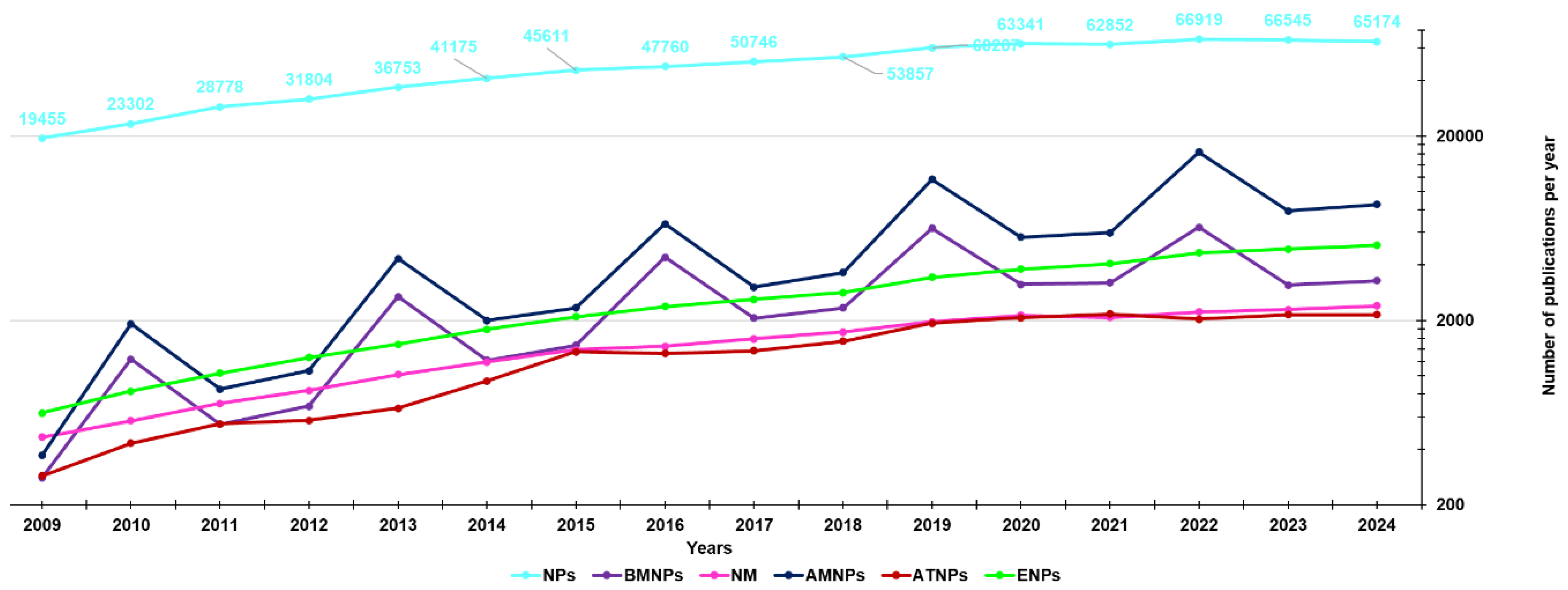
Figure 2.
Number of publications found in literature and published in the last fifteen years (2009-2024), achieved from a survey conducted using the Scopus database (https://www.scopus.com/, accessed on 06 November 2024). The values on y axis are in logarithmic scale for a better distribution of lines within the graph. Only experimental studies were considered intended as full articles, conference papers and letters. They were found using as keywords “antimicrobial AND nanoparticles” and then “antibacterial AND nanoparticles” (blue line), “anticancer AND nanoparticles”, “antitumor AND nanoparticles” and then “antitumor AND nanoparticles” (red line), using “biomedical AND nanoparticles” (purple lines), using “environment AND nanoparticles” (green line) and finally that obtained using “nanomedicine” (pink line).
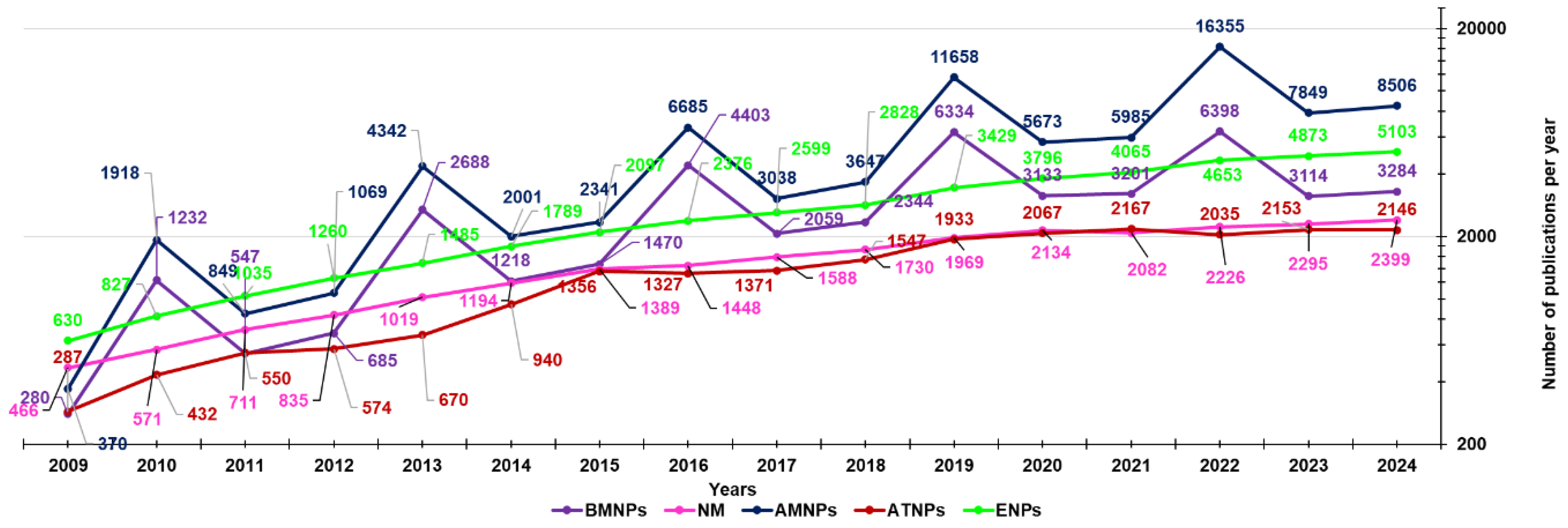
Figure 3.
Marketed liposomal formulations. Reproduced from [133] with permission of Copyright Clearance Center’s RightsLink® service.
Figure 3.
Marketed liposomal formulations. Reproduced from [133] with permission of Copyright Clearance Center’s RightsLink® service.
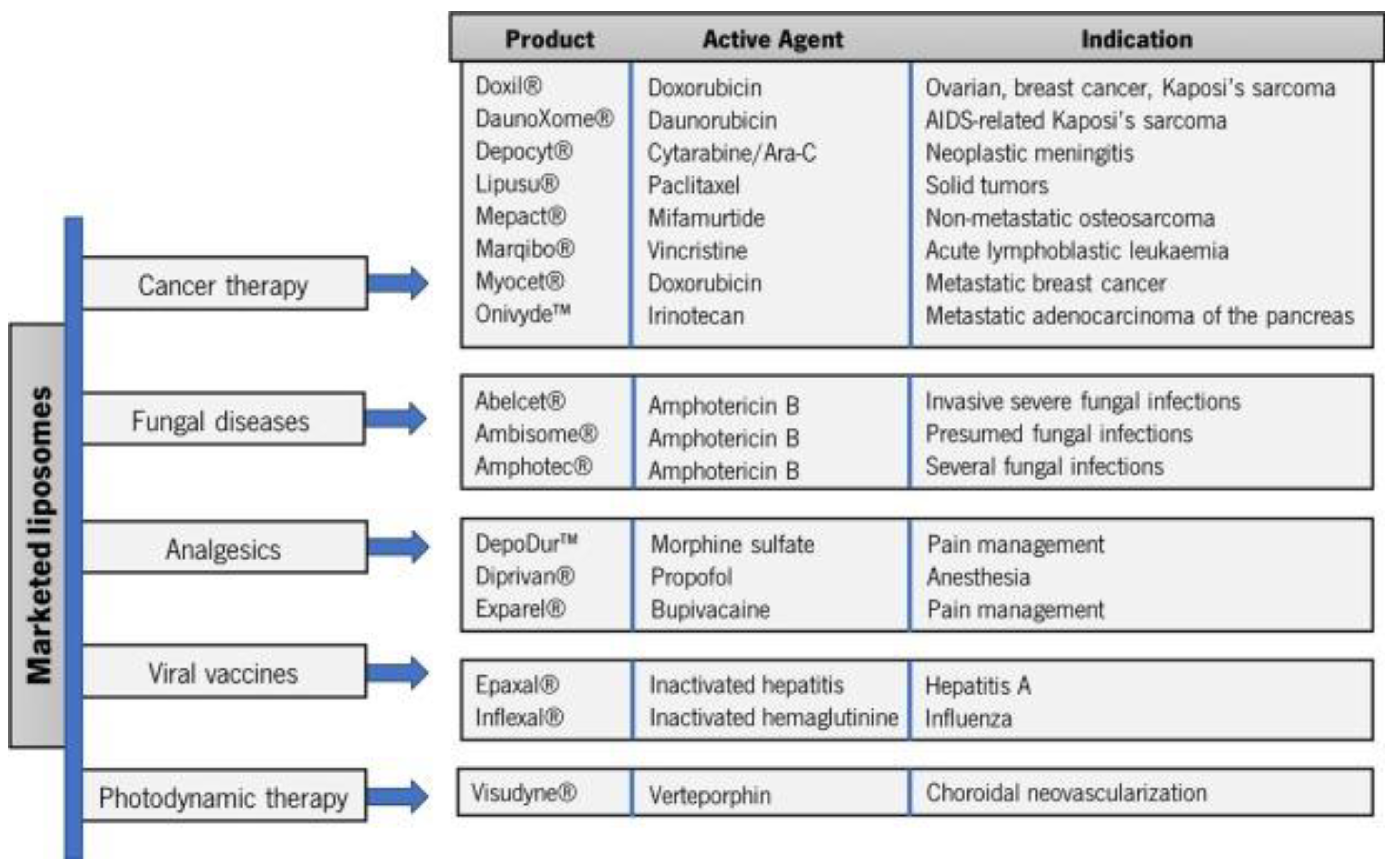
Figure 4.
Developed in vitro and in vivo toxicological evaluations of NPs.
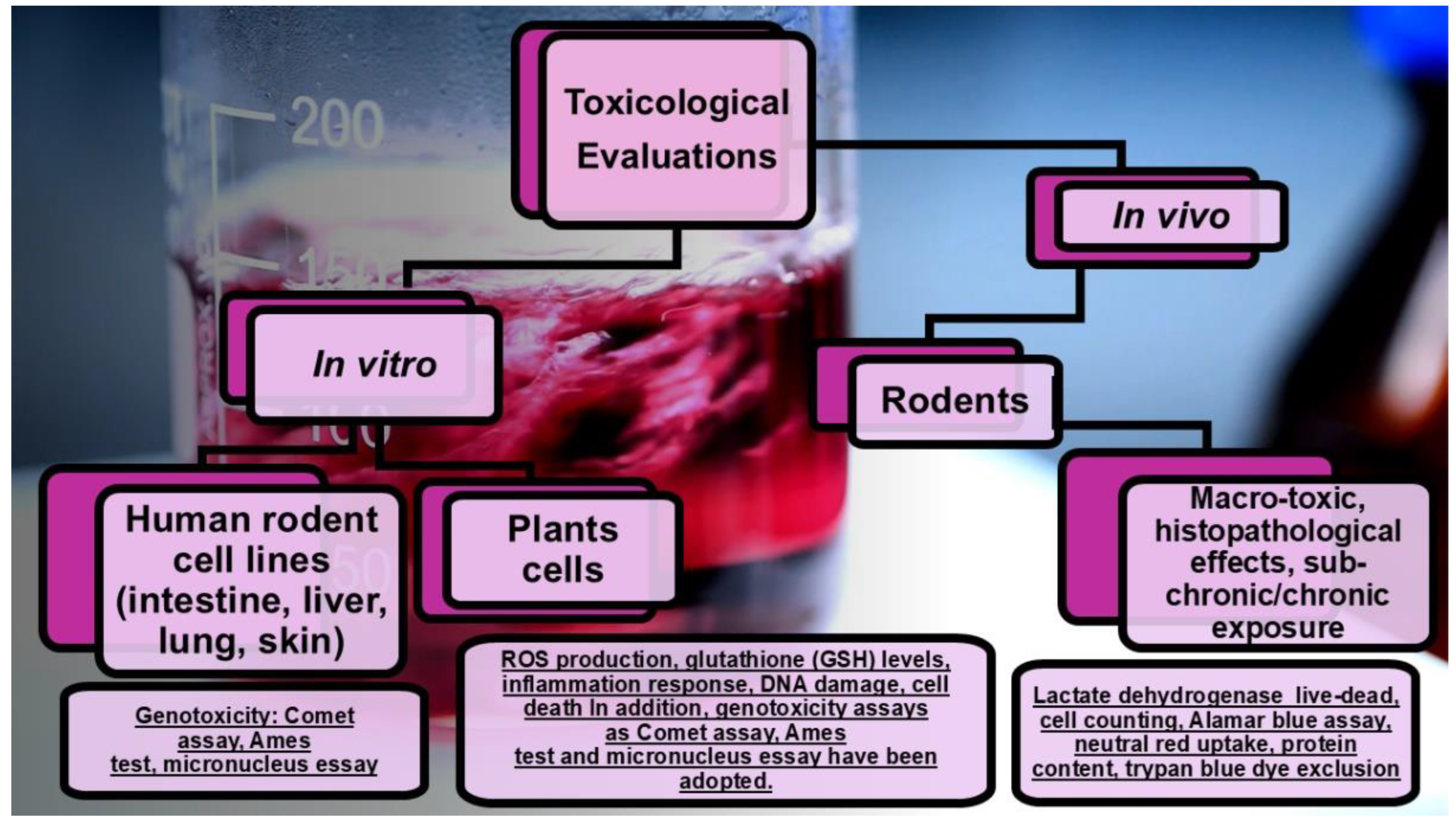

Table 1.
Most used nanomaterials engineered for encapsulating natural and synthetic APs for biomedical uses and health purposes.
Table 1.
Most used nanomaterials engineered for encapsulating natural and synthetic APs for biomedical uses and health purposes.
| Method | Description | General Indications/Uses | Properties | Main APs | Refs. | |||
|---|---|---|---|---|---|---|---|---|
| NSs | Colloidal dispersion of NPs (10-900 nm) in water Surfactants, co-surfactants, polymers * |
To improve solubility/bioavailability of both HAPs/LAPs | ↑Dispersibility, ↑solubility Sustained, controlled, targeted delivery ↑Stability ↑Therapeutic effects in cells and tissues |
[52] | ||||
| β-carotene | [78] | |||||||
| Quercetin | [79] | |||||||
| Acai fruits | [80] | |||||||
| α-Tocopherol | [81] | |||||||
| NEs | Kinetically stable liquid-in-liquid dispersions Droplet sizes 100-500 nm |
↓Particles size of HAPs/LAPs, H/L food additives ** Orally administrable drugs Protected drug delivery Suitable for food, cosmetics, pharmaceuticals Suitable for material synthesis |
↑Solubility/bioavailability Sustained, controlled, targeted delivery Extended half-life Obtained either by low energy techniques or by high energy techniques |
[82] | ||||
| Turmeric | [52] | |||||||
| Curcumin | [83] | |||||||
| di-Benzoyl-methane | [52] | |||||||
| Tannins Stilbene Flavonoids |
[84] | |||||||
| ECGC | [85] | |||||||
| Lipids Carotenoids |
[52] | |||||||
| Pomegranate extracts | [52,86] | |||||||
| LEO | [87] | |||||||
| SEDDSs | SMEDDSs 100–200 nm |
Anhydrous nano-dispersions achieved by drying A an oil phase, surfactants, co-surfactants/co-solvents, and LAPs Powders will spontaneously arrange in colloidal NEs when merged with water or with GIT fluids by small agitation or by the digestive motility of the stomach and intestine |
For orally delivering LAPs, food-grade chemicals, additives, drugs For low therapeutic dose APs |
↑Oral bioavailability improvement Possibility of an easy scale-up ↑DL% Allow delivering peptides and lipids without the risk of lipid digestion |
[88,89] | |||
| SNEDDSs < 50 nm |
EGCG | [90] | ||||||
| SDDSs | ↑ Soluble bioactive NPs with the AP physically entrapped or covalently linked (20–1000 nm) Nano carriers can be made of PEG, PUR, PCL, PLGA, PVA, P2VP, PLA, PPO, Pluronics®, PGA, PAE, PLL, mPEG, PasP, PLH, PEI, PVP, PLLeu, DOCA, HPMC, PHB, PEO, PBLG, PS, PIHCA, PAH, biocompatible polyester-based dendrimers |
For delivering HAPs/LAPs, food-grade chemicals additives, drugs For low therapeutic dose APs |
↑Solubility, bioavailability, dispersion and stability in GIT ↑APs systemic spread, transportation through the endothelial cell layer ↑Release at the target site Controlled microbiota metabolism ↑ APs’ bio-efficacy ↑Cellular uptake Favourable drug release profile protracted in time |
[91,92,93] | ||||
| Paclitaxel B Doxorubicin B mPEG-PLGA-Paclitaxel B PEGylated factor VII C Estradiol C PEGylated antibody fragment C Erythropoiesis stimulating agent C PEGylated IFNbeta-1a C |
[94] | |||||||
| DexamethasoneDocetaxel Rifampin Genistein + paclitaxel + quercetinHydrochlorothiazide Cisplatin Curcumin Diminazen aceturate Paclitaxel Folic acid siRNA + paclitaxel Docetaxel +siRNA-Bcl-2 Doxorubicin Lidocaine Cripofloxacin HCl DexamethasoneInsulin FITC-Dextran LevonorgestrelDNA | ||||||||
| OSNPs | LNPs | SLNPs | An external lipid monolayer with a solid-lipid core Spherical morphology (10–1000 nm) Surfactants/emulsifiers to stabilize Ideal fat/aqueous medium ratio 0.1/30.0 (w/w) |
For delivering LAPs | Biocompatible | Domperidone | [95] | |
| LPs | Artificial vesicles achieved by mixing phospholipids + cholesterol Lipid bilayer enclosing an aqueous core |
Immunological adjuvants and drug carriers | ↑ EE% of APs with different polarities Preserve APs from enzymes activity and degrading agents Biodegradable, biologically inactive Non-antigenic, non-pyrogenic, no intrinsic toxicity, instability in plasma D |
Irinotecan Amphotericin B Verteporfin Morphine sulphate Bupivacaine Inactivated hepatitis A Inactivated hemagglutinin of influenza A and B |
[52] [96] |
|||
| n-MIC | Very slim, spherical lipid particles (10–400 nm) | n-MICs form in aqueous medium n-MIC can solubilize LAPs |
↑ Bioavailability ↑Systemic residence time Protect APs from early inactivation ↑DL% and good stability |
Paclitaxel Doxorubicin Curcumin Dextran/Doxorubicin Doxorubicin/SN-38 Podophyllotoxin LCA Doxorubicin/siPD-L1 β-Lapachone/camptothecin Doxorubicin/CD147 miR-34a mimic/volasertib (BI6727) siRNA siRNA/ Doxorubicin Docetaxel Sorafenib Camptothecin Paclitaxel/siRNA Dexamethasone JQ1 Estradiol Adriamycin Doxorubicin/Fe3O4 NPs |
[97] | |||
| i-MIC | i-MICs form in oil medium i-MIC solubilize HAPs |
|||||||
| NIOs | Uncharged or charged lipid-based lamellar nanostructures Merge non-ionic E, cationic F or anionic G surfactants + cholesterol Vesicles osmotically active/stable |
For ↑oral bioavailability of APs with limited absorption | ↓Toxicity for cells *** Act as reservoir systems Provide controlled and sustained delivery |
Tamoxifen Docetaxel Metformin Celecoxib Gemcitabine Ascorbic acid Geranium oil Curcumin Cisplatin, Epirubicin Folic acid Letrozole Cyclophosphamide, Farnesol Gingerol Doxorubicin Hyaluronic acid Morusin Melittin Paclitaxel 2,5-Diketopiperazine Carnosine Trastuzumab Mcl-1 Nioplex Nintedanib Artemisin Silibinin Sunitinib 5-Fluorouracil Oxaliplatin Saccharomyces Cerevisiae Lycopene Hippadine γ-Oryzanol Amygdalin Ozonated olive oil |
[98] | |||
| Pro NPs | Made of both animal I and vegetable proteins L through proteins precipitation and crosslinking agents § De-solvating agents M |
For carrying several molecules | Simple manufacturing Compatible with the ↑-pressure Emulsification processes ↑ Freeze–thaw stability Suitable to being transformed Biocompatibility ↑ Stability ↑ Permeation ability in vitro Sustained delivering of APs ↓Toxicity for cells # ↑Shelf-life of APs ↑Resistance of APs to acidic gastric pH |
EGCG, GA Probiotics |
[15] [99,100,101,102] |
|||
| ONPs | CDs | Cyclic oligosaccharides consisting of six (α-CD), seven (β-CD), eight (γ-CD), or more glucopyranose units linked by α-(1, 4) bonds | For preparing FFs, FSs, IFT, APs, by the monomolecular inclusion complex technique To delivery different LAPs |
↑Hydrophilicity and water solubility of LAPs ↑Chemical stability ↓Early degradation and metabolism Can modify unpleasant tastes and flavours Realize a controlled release of LAPs |
Linoleic acid Resveratrol Carotenoids Lycopene (Lyc) Hesperidin Olive leaf extracts 1 Quercetin Myricetin Kaempferol 3-Hydroxyflavone Morin Rutin Curcumin Ferulic acid Ellagic acid Amino acids Hydrolysed soy pro 2 |
[52] [103] [104,105] [62] [106,107,108] |
||
| PNPs | Prepared from natural hydrophilic polysaccharides Comprise polyelectrolytes (cationic, anionic, neutral saccharides) and non-polyelectrolytes |
To deliver different APs To develop APs-load FFs and additives |
↑Solubility ↑Controlled and target release ↑Stability, ↑food shelf-life ↑Cellular uptake |
Olive leaf extract Gallic acid Caffeic acid Yerba mate H Caffeine Theobromine Saponins Polyphenols Probiotics Flavors Anthocyanins Procyanidins Ellagic acid Gliadin |
[52] [109,110,111,112,113,114,115,116,117,118,119,120] |
|||
* To stabilize the system; HAP = hydrophilic active principle; LAP = hydrophilic active principle; ** not high-melting APs; SEDDSs = self-emulsifying drug delivery systems; A = through proper procedures, including spray dry (SD) or freeze drying (FD); SNEDDSs = self-nanoemulsifying drug delivery systems; SMEDDSs = self-micro-emulsifying drug delivery systems; ECCG = epigallocatechin-3-gallate; LEO = lemongrass oil; PEG = polyethylene glycol; PUR = poly urethane; PCL = poly capro-lactone; PLGA = poly lactic-co-glycolic acid; PVA = polyvinyl alcohol; P2VP = poly 2-vinyl pyridine; PLA = poly(lactic acid); PPO = poly(propylene oxide); Pluronics® = PPO-PEO; PGA = poly(γ-L-glutamic); PAE = poly(L-phenylalanine ethyl ester); PLL = poly(L-Lysine); mPEG = methyl-PEG; PasP = poly(aspartamic acid); PLH = poly(L-histidine); PEI = poly(ethylene amine); PVP = poly(N-vinylpyrrolidone); PLLeu = poly(L-Leucine); DOCA = deoxycholic acid; HPMC = hydroxy propyl methyl cellulose; PHB = poly(hydroxy butyrate); PEO = poly(ethylene oxide) ; PBLG = poly(γ-benzyl-L-glutamate); PS = phosphatidylserine; PIHCA = poly(isohexyl-cyanoacrylate); PAH = poly(allylamine hydrochloride); B = in clinical trial; C = approved; SDDSs = solid drug delivery systems; OSNPs = organic solid nanoparticles; LNPs = non-synthetic lipid-based NPs; ProNPs = protein-based polymeric NPs; ONPs = oligosaccharides-based NPs; PNPs = polysaccharides-based NPs; SLNPs = solid-lipid nanoparticles; LPs = liposomes ; n-MICs = normal micelles; i-MIC = inverse micelles; NIOs = niosomes; D = sterically stabilized LPs have been developed; E = alkyl or di-alkyl polyglycerol ethers; F = stearyl pyridinium salts; G = di-acetyl phosphate; *** for non-charged niosomes; CDs = cyclodextrins; FFs = functional foods; FSs = food supplements; IFT = innovative food-related therapeutics; H = Ilex paraguariensis; I = gelatin, collagen, albumin, casein, and silk protein; L = zein, gliadin, and soy protein; M = alcohol or acetone; § = chemical, ionic, thermal, and enzymatic (8% glutaraldehyde aqueous solution or calcium phosphate); ↓ = reduced, decreased, low, lower; ↑ improved, increased, high, higher.
Table 2.
In vivo experiments with Nuts-loaded delivery systems.
| NPs | Nuts | Bioactivities | (nm) | Animal | Refs. |
|---|---|---|---|---|---|
| Phospholipid-based delivery systems | |||||
| Liposome | (+)-Catechin | Antioxidant, neuro-protective | 35–70 | Wistar Albino rats | [148] |
| Liposome | Curcumin | Anti-HIV, anti-tumour, antioxidant Anti-inflammatory |
263 | Sprague-Dawley rats | [149] |
| HL | Silymarin | Hepatoprotective | 660 | Albino rats | [150] |
| PLS | Silymarin | 196 | Beagle dogs | [151] | |
| PPC | Ginkgo biloba * | ↓Platelet aggregation Radical scavenger, antioxidant, protection of CNS |
N/A | Sprague-Dawley rats | [152] |
| PPC | Curcumin | Antioxidant, anti-inflammatory Anti-carcinogenic, anti-bacteria ↓Cholesterol, antitumor, antispasmodic Wound healing, anti-coagulant, hepatoprotective |
Wistar Albino rats | [153] | |
| PPC | Evodiamine | Anti-tumour, anti-inflammatory, anti-obesity Anti-nociceptive, thermoregulatory |
Sprague-Dawley rats | [154] | |
| PPC | Silybin | Hepatoprotective | Rats | [155] | |
| PPC | Boswellic acid | Anti-inflammatory, hepatoprotective ↓5-Lipoxygenase |
Rats | [156] | |
| PPC | Silybin | Hepatoprotective, antioxidant | Dogs | [157] | |
| Emulsion-based delivery systems | |||||
| NEs | DBM | Anti-cancer activities, anti-proliferation | 70 | Sprague-Dawley rats | [158] |
| NEs | α-Tocopherol | Antioxidant, neuroprotective | 85 | Wistar rats | [159] |
| MEs | Berberine | Anti-bacteria, anti-tumour, anti-diabetes ↑Cerebral ischemia |
24 | Sprague-Dawley rats | [160] |
| MEs | Puerarin | Cardiovascular diseases, antioxidant, anti-diabetes | 40 | Kunming mice | [161] |
| SLNPs | Camptothecin | Anti-cancer | 197 | C57BL/6J mice | [162] |
| SLNPs | Quercetin | Antioxidant, ↓blood lipid, anti-cancer ↓Platelet aggregation, anti-anaemia Anti-inflammation, anti-anaphylaxis Dilate coronary arteries |
155 | Wistar rats | [163] |
| SLNPs | Triptolide | Immune-suppressive activity Anti-fertility, anti-neoplastic activity |
116 | Wistar rats | [164] |
| OG NEs | Curcumin | Anti-cancer, anti-inflammatory, antioxidant | 218 | CD-1 mice | [165] |
| SEDDSs | Curcumin | Anti-inflammatory, antioxidant, anti-cancer | 85 | Wistar rats | [166] |
| SEDDSs | Ginkgo biloba * | ↓Platelet aggregation, radical scavenging Antioxidant, protection of CNS |
∼100 | Dogs | [167] |
| SEDDSs | Wurenchun | Antihepatotoxic, hepatoprotective | 240 | Sprague-Dawley rats | [168] |
| SEDDSs | Baicalein | Anti-inflammatory, anti-cancer, antioxidant Anti-virus, anti-allergic |
27-54 | Sprague-Dawley rats | [169] |
| SEDDSs | ZTO | Hepatoprotective, ↓tumour, anti-bacteria ↑White blood cell, anti-thrombosis |
182 | Rabbits | [170] |
| SEDDSs | Oridonin | Anti-tumour, anti-bacteria, antioxidant Anti-inflammatory |
24 | SD rats | [171] |
| Chemical modifications | |||||
| PAc | EGCG | Antioxidant, anti-viral, anti-inflammatory Cardio-protective, neuro-protective Anti-cancer |
N/A | CF-1 mice | [172] |
| 3,5,4′-TAR** | Resveratrol | Anti-cardiovascular disease, antioxidant Anti-inflammatory, anti-tumour |
Rats | [173] | |
| QC-12** | Quercetin | Antioxidant, ↓blood lipid, anti-anaemia ↓Platelet aggregation, anti-cancer Anti-inflammation, anti-anaphylaxis Dilate coronary arteries |
Human | [174] | |
| HAA | Tricin | Antioxidant, anti-viral, anti-inflammatory Antihistamine, anti-cancer |
SD rats | [175] | |
| Other delivery methods | |||||
| Ch NPs | EGCG | Antioxidant, anti-viral, anti-inflammatory Cardio-protective, neuro-protective, anti-cancer |
440 | Swiss outbred mice | [176] |
| Ch NPs | Curcumin | Antioxidant, anti-inflammatory, anti-proliferative, anti-angiogenic | 178 | Swiss mice | [151] |
| Naked NCs | Coenzyme Q10 | Co-factor of the mitochondrial electron transport chain, antioxidant, cardio-protective neuro-protective |
400, 700 | Beagle dogs | [177] |
| NCs | Schisandrin B | Hepatoprotective, neuroprotective | 45, 168 | Sprague-Dawley rats |
[178] |
* Extracts; ** prodrug; HL = hybrid liposome; MEs = micro emulsions; CNS = central nervous system; DBM = di-benzoyl methane; OG NEs = organo-gel nano emulsions; PPC = phosphatidylcholine; SEDDSs = self-emulsifying drug delivery systems; SLNPs = solid-lipid nanoparticles; Ch NPs = chitosan nanoparticles; NPs = nanoparticles; 3,5,4’-TAR = 3,5,4’-tri-O-acetyl resveratrol; ZTO = Zedoary turmeric oil; HAA = hybrid with amino acids; NC = nanocrystals; EGCG = epigallocatechin gallate; PLSs = proliposome; PPCs = phospholipid complex; PAc = peracetylation.
Table 3.
In vivo experiments with a specific Nut nano-formulated using different nanomaterials with enhanced activity.
Table 3.
In vivo experiments with a specific Nut nano-formulated using different nanomaterials with enhanced activity.
| Nuts | Technologies | Efficacy evaluated | Models | Refs. |
|---|---|---|---|---|
| Curcumin | PPC | Antioxidant, hepatoprotective | CCl4-I liver OD in mice | [153] |
| Nanoencapsulation | Chemo preventive | DENA-I liver carcer in rat | [179] | |
| NE | Anti-inflammation | TPA-I acute mouse ear edema | [180] | |
| Quercetin | PPC | Antioxidant, hepatoprotective | CCl4-I liver OD in rats | [181] |
| Cationic NPs | Anti-tumorigenic | B16F10 melanoma cells subcutaneously injected into C57BL/6 mice | [182] | |
| Nanoencapsulation | Antioxidant, protective against liver and brain damage | As-I liver/brain OD in rats | [183] | |
| Microcapsules | Anti-inflammatory, antioxidant | AA-I acute colitis in mice | [184] | |
| Silymarin | Liposomes | Hepatoprotective | CCl4-I liver damage in rats | [185] |
| Triptolide | Solid lipid NPs | Anti-inflammatory | Carrageenan-I rat paw edema | [164] |
| CU | NPs | Hepatoprotective | AcAPh-I hepatotoxicity in rats | [186] |
| Naringenin | NPs | Hepatoprotective | CCl4-I acute rat liver failure | [187] |
| α-TPh | NE | Anti-diabetes, antioxidant | STZ-I diabetes | [159] |
| GA | PPC | Anti-apoptotic, cardioprotective | DOX-I cardiac toxicity in rats | [188] |
| Puerarin | Nanodispersion | Anti-colorectal cancer | HT-29 human colon carcinoma cell subcutaneously injected to BALB/c nude mice | [189] |
| Resveratrol | Pro-drug | Anti-inflammation | 1% DSS-I in drinking water for 8 days mice colon inflammation | [190] |
| EGCG | Peracetylation | Anti-inflammation, anti-tumorigenesis | DSS-I mice colitis/tumour | [191] |
α-TPh = α-Tocopherol; GA = gymnemic acid; NPs = nanoparticles; NEs = nano emulsions; EGCG = epigallocatechin gallate; CU = Cuscuta chinensis; DENA = Diethylnitrosamine; DSS = dextran sulphate sodium; DOX = doxorubicin; AcAPh = acetaminophen; AA = acetic acid; I = induced; As = arsenic; OD = oxidative damage; PPCs = phospholipid complex.
Table 4.
Main advanced FP types achieved using functional nanomaterials [61].
Table 4.
Main advanced FP types achieved using functional nanomaterials [61].
| Advanced FPs type | Purposes and Description | Key nanomaterials currently used |
|---|---|---|
| Physically improved packaging | Packaging materials incorporated with NPs to improve physical properties, such as temperature and moisture stability, mechanical strength, gas barrier, durability and flexibility | Metal oxides NPs, nano clays Carbon nanotubes, metallic NPs |
| Active packaging | NPs endowed with antimicrobial or other functionalities (e.g. antioxidant, UV absorbents) and the ability to release them into-packaging. Food packaged into AP results improved in term of taste, freshness and shelf life | Ag NPs, Au NPs, metal oxides NPs, antimicrobial and/or antioxidant NPs functionalized NPs |
| Smart packaging | Packaging materials incorporated with nano-sensors to monitor and report on the condition of the food (e.g. oxygen indicators, freshness indicators and pathogen) | ↑ Variability of nano-sensors |
↑ = High.
Table 5.
Main details about the nanosized materials developed in our laboratories in the years 2005-2009.
Table 5.
Main details about the nanosized materials developed in our laboratories in the years 2005-2009.
| NPs Type | Size (nm), ζP (mV), PDI EE%,MW * |
Bioactivity | Characteristics | APs | Refs. |
|---|---|---|---|---|---|
| Sty-CPs (C8) |
589 nm | Adaptable to biochemical interaction studies with CAOs | Soluble, positive Schiff's fuchsin-sulphite reagent Spherical morphology |
Functionalized Glucopyranose | 2009 [1] |
| 4-HPR-A-DEX NPs | 150-350 nm 0.112-0.176 (PDI) |
↑Cytotoxicity to HTLA-230 | CPXs with 4-HPR were prepared by kneading method TGA suggested a ↑↑ thermal stability, ↑ DL%, ↑EE% Sustained drug release, possible parenteral administration ↑ Cytotoxicity to HTLA-230 than free 4-HPR ↑ Drug bioavailability, biodegradable |
Entrapped 4-HPR | 2009 [45] |
| ATRA-NIC-PVA | < 400 nm 0.202-0.450 (PDI) |
Cytotoxic to NB cells (LAN-5 cells) |
↑ATRA solubilization and release ↑↑ATRA aqueous solubility, drug fractional release < 8% ↑↑ Growth inhibition effect than free ATRA Suitable for parenteral injection, drug targeting Long-term storage |
Entrapped ATRA | 2009 [46] |
| 4-HPR-OL-DEX NPs | ~ 310 nm | In vitro (HTLA-230, LAN-5 and IMR32 NB cells) and in vivo antitumor activity | Tested both in vitro and in vivo, ↑↑ Cytotoxicity in vitro ↑↑Fraction of sub-G1 cells, no haemolytic activity Suitable for injections, ↑↑AUC, ↓ clearance ↑↑Lifespan and long-term survival of treated mice ↑↑Aqueous solubility, ↑↑ bioavailability |
Entrapped 4-HPR | 2012 [47] |
| 4-HPR-C14-PEG NPs |
50-137 nm 0.165-0.221 (PDI) |
In vitro (SH-SY5Y and NGP NB cells) antitumor activity | ↑↑Aqueous solubility, suitable for injection Stable aggregates, drug-targeting to solid tumors No release of free 4-HPR in an aqueous environment asso ↑↑Intracellular concentrations and activity than 4-HPR ↓↓4-HPR early metabolism, protracted release |
Complexed 4-HPR | 2012 [48] |
| StyGlyco-CCPs (R1) |
~ 300 nm | Suitable for interaction with CAOs | Cross linked resins, spherical morphology | Functionalized D-Glucose | 2013 [2] |
| 4-HPR-NGR-NLs | 142 nm -19.2 mV 0.073 (PDI) 69% (EE) |
In vivo ↑↑ the life of NB mice by apoptotic/anti-angiogenic effects ↓↓of tumour progression ↓↓of intra-tumoral vessels ↓↓ of VEGF expression ↓↓ of metalloproteinases MMP2/MMP9 |
By reverse phase evaporation method ↑↑ Structural integrity of NL in organic fluids Target the tumour endothelial cell marker |
Complexed 4-HPR | 2013 [49] |
| StyGlyco-LCPs (P1G2) |
N.R. | Substrates/inhibitors of CAOs | Soluble | Benlyl amine D-Glactose | 2015 [196] |
| SL[LM-BTZ] | 179 nm -33.9 mV 0.070 (PDI) |
In vivo ↑↑ the life of NB mice | Lyophilization with cryoprotectants Targeted drug delivery systems, ↑↑ therapeutic index ↑↑ Efficacy, ↑↑ EE%, suitable for intravenous injection ↓↓of BTZ systemic adverse effects |
Complexed BTZ | 2015 [50] |
| NGR-SL[LM-BTZ]) | 173 nm -30.2 mV 0.093 (PDI) |
||||
| AN169-PEG-NLs | 143.9-153.8 nm 0.052-0.077 (PDI) |
In vitro antitumor activity in human cancer cell lines (HTLA-230, Mel 3.0, OVCAR-3, SV620) | By thin-film hydration method, slow drug release Antitumour activity as free AN169 (72h) Lyophilization with cryoprotectants Long-term stability, ↑↑EE%, for intravenous injection |
Entrapped AN169 | 2015 [51] |
| PolyE-Ds | 4.4-5.4 nm 31.2-51.8 mV |
Carriers for gene and drug delivery | ↑ Water-soluble, excellent β Well-defined sizes, shapes, ↑controlled architecture Polycationic, biodegradable, 13593-25661 * |
Linked amino acids | 2017 [4] |
| b-HMPA-Ds. | N.R. | For gene transfection with p-DNA and si-RNA | ↑ Water-soluble, excellent β Well-defined sizes, shapes, ↑Controlled architecture, polycationic, biodegradable Not cytotoxic, 9834-60725* |
2017 [5] |
|
| PolyE-ADs | 3.3-3.6 nm 5.4-5.9 mV |
Drug delivery, gene transfection | Well-tailored polymeric structure, excellent β Symmetric tree-like shape, ↑functional groups Inner cavities, hydrolysable, amphiphilic by a C-18 chain Polycationic, biodegradable, not cytotoxic, 2932-6762 * |
2018 [6] |
|
| 4G PolyE-HDs | 4.5-4.6 nm 32.8-33.4 mV |
Water-soluble biomedical devices. | Well-tailored polymeric structure; excellent β Symmetric tree-like shape, ↑functional groups Inner cavities, hydrolysable, hydrophilic; ↑ solubility Polycationic, biodegradable, not cytotoxic, 2932-6762 * |
2018 [7] |
|
| 4G-5G PolyE-DPX | 4.4-36.3 nm 15.5-51.8 mV |
Several beneficial effects of UOA | Well-tailored polymeric structure; excellent β Symmetric tree-like shape, inner cavities, hydrolysable ↑Water solubility, polycationic, biodegradable Not cytotoxic, 1360-3130 * |
Entrapped UOA | 2018 [8] |
| PolyE-A/H-DPX | 18.9-47.7 nm 2.4-15.3 mV |
Non-nucleoside HIV-1 reverse transcriptase inhibitor Suitable for parenteral administration |
↑ Water-soluble, excellent β, well-defined sizes, shapes, ↑Controlled architecture, polycationic, biodegradable, Not cytotoxic, sustained release, 5851-24203 * |
Entrapped (1) | 2018 [9] |
| 5G PE-PD-D/GA (GAD) |
348.6 nm | Platelet aggregation inhibition ** ROS production inhibition ** Antibacterial (Gram-positive) ** |
↑ Water-soluble, excellent β, well-defined sizes, Spherical morphology, ↑controlled architecture 192 OH groups, biodegradable, not cytotoxic, 17010 * |
Linked GA | 2019 [10] |
| PolyE-A/H-DPX | 60–70 nm | Beneficial effects of EA For clinical applications RSA |
300-1000-fold ↑water solubility, non-PAMAM dendrimers Biodegradable, not cytotoxic, excellent β, 14287-25604 * Sustained release, ↑ DL% |
Entrapped EA | 2019 [11] |
| 5G PE-PD-D/GA (GAD) |
348.6 nm | Long-Term Preservation of EOs | Nano-spherical dendrimer, preservative power ↑than GA No pro-oxidant action, spherical morphology (SEM) ↑Compatible with lipids and oily matrices RSA 4-fold ↑than GA (DPPH), biodegradable by esterase hydrolytic actions, release GA, not cytotoxic, 17010 * |
Linked GA | 2019 [12] |
| 5G PE-PD-D/GA (GAD) |
348.6 nm | To treat diseases by OS | Nano-spherical dendrimer, no pro-oxidant action Spherical morphology (SEM), RSA 4-fold ↑than GA, biodegradable (lipase), release GA, not cytotoxic, 17010 * |
Linked GA | 2020 [16] |
| 5G-PE-PD-D-OH | 44.5 nm -21.2 mV 0.208 (PDI) |
ROS-dependent per se cytotoxicity against NB cells sensitive to ETO | Per se activity, nano-spheric, ↑ solubility, 7275 * | Empty dendrimer | 2020 [13] |
| 5G PE-PD-DPX (CPX 5) |
70 nm -45 mV |
Prevention and treatment of NB cells | ↑ Cytotoxicity and Pro-Oxidant Effects than ETO Protection of ETO, synergistic action with ETO Sustained release of ETO, ↑ DL%, ↑EE Nano-spherical morphology, ↑ solubility |
Entrapped ETO | |
| 5G-PE-PD-D-OH | 44.5 nm -21.2 mV 0.208 (PDI) |
ROS-dependent per se cytotoxicity against NB cells both sensitive and resistant to ETO | ↑ Cytotoxicity and Pro-Oxidant Effects than ETO Nano-spherical morphology, 7275 *, 64 peripherals OH Water soluble |
Empty dendrimer | 2020 [15] |
| 5G PE-PD-D/GA (GAD) |
348.6 nm | Nano-formulation nullify the pro-oxidant activity of GA To treat diseases by OS To prevent DNA oxidative damage and tumors onset |
Nano-spherical dendrimer, no pro-oxidant action Spherical morphology (SEM), RSA 4-fold ↑than GA Biodegradable (lipase), release GA by hydrolysis, Water-soluble, not cytotoxic, 17010 * |
Linked GA | |
| 5G PE-PD-DPX (GALD) |
349.9 nm -29.2 mV 0.708 (PDI) |
Nano-spherical hygroscopic dendrimer, ↑water soluble no pro-oxidant action, ↑ DL%, ↑EE, Spherical morphology (SEM), biodegradable (lipase) Sustained and quantitative release of GA Not cytotoxic, 28610 * |
Entrapped GA | ||
| PolyE-Ds | N.R. | Bactericidal (Pseudomonas aeruginosa, Acinetobacter baumannii, Stenotrophomonas maltophilia) |
Non-Cytotoxic, amino acid-modified polycationic Ds More potent than colistin against P. aeruginosa (5GK) Not lithic behaviour, membrane disruptor Broad spectrum of action |
Linked K, H, KH | 2020 [17] |
| 4-AMSTY-CP (P5) | 334 nm +57.6 mV 1.012 (PDI) |
Bactericidal (Enterococcus, Staphylococcus, Pseudomonas, Klebsiella, Escherichia coli, A. baumannii, S. maltophilia | Random copolymer, ↑water soluble Rapid (0.5h) and broad spectrum non-lytic bactericidal activity, stability in solution, excellent buffer capacity Activity by membrane disruption, 5,100 (Mn) |
No AP NH3+ groups |
2021 [18] |
| 4-AMSTY-CP (P5) | 334 nm +57.6 mV 1.012 (PDI) |
ROS-dependent cytotoxic activity on ETO-resistant NB | Random copolymer, ↑water soluble Stability in solution, excellent buffer capacity Membrane disruptor, cause ↑ROS generation, 5,100 (Mn) |
No AP NH3+ groups |
2021 [19] |
| Sty-CP (P7) | 220 nm +49.8 mV 0.809 (PDI) |
Random copolymer, ↑water soluble Stability in solution, excellent buffer capacity Membrane disruptor, cause ↑ROS generation, 13,719 (Mn) |
|||
| 4G-5G-PolyE-Ds | 16.1-24.9 nm +24.8-34.0 mV |
↑Antibacterial effects (MIC = 0.5–8.7 µM vs. Enterococci, Staphylococci) | Activity depended on the density and on the type of cationic amino acid-conjugated dendrimers and not on the presence and the release of UOA, ↑water soluble Stability in solution, excellent buffer capacity Protracted release of UOA, membrane disruptor 14,600-29,300 * |
Linked K, R, KR Entrapped UOA |
2021 [20] |
| 4G-BBB4-PolyE-Ds | 112.1 nm +28.9 mV 0.289 (PDI) |
Antibacterial vs. Staphylococci To treat skin infections |
↑↑↑Water solubility than BBB4, good DL and EE SI = 1.4-5.5, protracted release of BBB4 Membrane disruptor ↓Cytotoxicity on HaCat than BBB4, 21176 * |
Linked K Entrapped BBB4 |
2021 [21] |
| N.D. | ↑↑↑Water solubility than BBB4, good DL and EE Sustained/protracted release of BBB4, 21176 * |
Linked K Entrapped BBB4 |
2021 [22] |
||
| RES-TPGS | 9.6-12.7 nm § -1.6—4.8 mV § 0.13-0.26 (PDI)§ |
Antioxidant Anti-inflammatory Protective action in the liver |
Micellar NPs, ↑↑↑water solubility than RES Good DL% and EE% Sustained/protracted release of RES ↓Cytotoxicity on HaCat than RES, 21176 |
Entrapped RES | 2021 [23] |
| UA-4G PolyE-D (UA-G4K NPs) |
577.5 vs. 333.4 nm 4GK -42.6 vs +66.1 mV 4GK 0.235 vs 0.286 4GK (PDI) |
N.D. | Biodegradable, not cytotoxic (Hela cells), ↑DL%, spheric Protracted release profile governed by diffusion Water solubility 1868-fold ↑ than UA Clinical applicability, 30,069 * |
Linked K Entrapped UA |
2021 [26] |
| Antibacterial vs. enterococci (MICs =0.5-4.3 µM) Bactericidal vs. E. faecium |
↓Cytotoxicity on HaCat (IC50 96.4 µM), SIs = 22-193 Valuable as a novel oral-administrable therapeutic option to-treat enterococcal infections. |
2021 [24] |
|||
| ATRA-TPGS NPs | 14.1-21.0 nm -7.2—-13.0 mV 0.19-0.32 (PDI, water) |
To prepare topical gel Treatment for skin diseases Treatment for melanoma |
↓ATRA cutaneous side effects, ↑stability than ATRA ↑ATRA solubilization, good EE% 22 ± 4 µ cm-2 permeation after 24 h ↑Cytotoxic effects on melanoma cells |
Entrapped ATRA | 2021 [25] |
| Sty-CP (P7) | 220 nm +49.8 mV 0.809 (PDI) |
Antibacterial activity vs. enterococci, staphylococci Acinetobacters, Pseudomonas Klebsielle, Escherichia coli Stenotrophomonas maltophylia Rapid bactericidal effects on S. aureus, K. pneumoniae, P. aeruginosa |
Random copolymer, ↑water soluble ↑Stability in solution, excellent buffer capacity Membrane disruptor, ↓tendency to develop resistance ↓Toxicity, long-term activity, 13,719 (Mn) Lowest MICs = 0.6–1.2 µM |
No AP NH3+ groups |
2021 [27] |
| 5G-PolySty-D (5G-PDK) |
203.0 nm +19.2 mV 0.282 (PDI) |
Rapid bactericidal affects vs. A. baumannii, A. pittii A. ursingii |
Membrane disrupters, MICs = 3.2-12.7 µM Electrostatic interactions with bacterial surface Self-biodegradable, 20,145.3 * |
64 Linked K | 2021 [28] |
| Antibacterial and bactericidal vs. Pseudomonadaceae | MICs depending on pigment production P. aeruginosa = 1.6-> 6.4 µM P. putida producing pyoverdine = 3.2-6.4 µM P. putida producing non-pigmented colonies = 0.2-1.6 µM ↓Cytotoxicity on HaCat, ↑↑↑ SIs (13-404) |
2021 [29] |
|||
| 4-AMSTY CP (CP1) |
833.4 nm +27.3 mV 0.2235 (PDI) 157,306 * |
Potent broad-spectrum antibacterial effects Kill pathogens rapidly |
Cationic macromolecules acting as membrane disruptors CP1 MICs = 0.1–0.8 µM, OP2 MICs = 0.35–2.8 µM Promising ingredients for the development of novel antibacterial dosage forms for topical applications (hydrogel) Spherical morphology |
No AP NH3+ groups |
2022 [30] |
| 4-AESTY OP (OP2) |
163.4 nm +31.1 mV 0.301 (PDI) 44,514 * |
||||
| CB1H-P7 NPs | 142.9 nm +36.7 mV 0.626 (PDI) |
N.D. | Spherical morphology, positive surface charge ↑↑↑ DL%, ↑↑↑ EE%, protracted release profile, 26,623.9 * |
CB1H | 2022 [31] |
| Antibacterial vs. G+/G- Bactericidal vs. S. aureus E. coli, P. aeruginosa. |
MICs ↓↓ of pristine CB1H and matrix P7 NPs displayed MICs = 0.6-4.8 µM on 34 out of 36 isolates ↓Cytotoxicity on HaCat, SIs up to 2.4 |
2022 [35] |
|||
| New curative option vs. NB (IMR 32 and SHSY 5Y cells) |
Membrane disruptors, IC50 = 0.43-0.54 µM vs. IMR 32 and SHSY 5Y cells Early-stage (66-85%) and late-stage apoptosis (52-65%) Effects of CB1H and P7 ↑ by 54-57 and 2.5-4-times (IMR32) ↑ By 53-61 and 1.3-2 times against SHSY 5Y 1-12-fold more potent than fenretinide ↓Cytotoxicity on HaCat, SIs = 2.8-3.3 |
2023 [38] |
|||
| CR232-SUVs | 173.4 nm +17.8 mV 0.118 (PDI) |
N.D. | Biocompatible, DL%, EE% ↑ with ↑ lipids/CR232 ratio Prolonged release profile ruled by zero-order kinetics 1764-fold more soluble than the untreated CR232 |
Linked K Entrapped CR232 |
2022 [32] |
| 5GK PoliE-D NPs (CR232-G5K NPs) | 529.7 nm +37.2 mV 0.472 (PDI) |
N.D. | 2311-fold more water-soluble than pristine CR232 No use of harmful organic solvents/additives, spheric Biodegradable, ↑↑↑ DL%, ↑↑↑EE%, ↓Cytotoxicity on Hela Quantitative release profile (Weibull kinetics), 44,153.1 * |
||
| Antibacterial vs G+ and G-Rapid bactericidal activity | MICs = 0.36–2.89 µM vs. all of the considered G+ and G- MICs = 0.72 µM vs. colistin-resistant P. aerginosa and K. pneumoniae carbapenemases (KPCs)-producing ↓Cytotoxicity on HaCat, SIs up to 8 |
2022 [33] |
|||
| 4-HPR-P5 | 249 nm +41.3 mV 0.210 (PDI) |
Antiproliferative activity IC50 = 1.25 µM (IMR32), 1.93 µM (SH-SY5Y) |
Molecularly dispersed 4-HPR using P5 as solubilizing agent by antisolvent co-precipitation method ↑↑↑Clinical outcomes of 4-HPR 4-HPR apparent solubility 1134-fold ↑, faster dissolution Suitable for intravenous administration, ↑↑↑ DL% Extended release over time, excellent β |
Dispersed 4-HPR | 2023 [37] |
| P5PA-4I NPs | 541 nm +8.39 mV 0.194(PDI) |
New promising treatment for chemo-resistant NB Cytotoxic to ETO-sensitive (HTLA-230) and to ETO-resistant (HTLA-ER) cells |
↑↑↑ Activity of 4I, ↑↑↑ DL%, ↑↑↑ EE% ↑↑↑ hydrophilic–lipophilic balance (HLB) Excellent buffer capacity, ↑↑↑ residence time inside cells Chemically stable in an aqueous medium > 40 days Assumed low hemolytic toxicity ROS-dependent cytotoxic effects ↑↑↑ than 4I ↑↑↑Efficacy than ETO in HTLA-ER cells |
Loaded 4I | 2023 [39] |
| TPP-BA-NVs | 49.3 nm +18.2 mV 0.529 (PDI) 852.7 * |
Cytotoxic to MDR HR-NB IC50 = 0.2 µM (HTLA-230) IC50 = 1.1 µM (HTLA-ER) |
Tested on HTLA-230 human stage-IV NB cells and HTLA-ER NB cells resistant to ETO, DOX, etc. IC50 = 538-fold ↓ than ETO (HTLA-ER) Limited cytotoxic effects against mammalian cell lines (Cos-7, IC50 = 4.9 µM, HepG2, IC50 = 9.6 µM, MRC-5, IC50 = 2.8 µM, RBCs, IC50 = 14.9 µM, SIs = 2.5–74.6 |
No additional AP Linked BPPB |
2024 [41] |
| ↑↑↑ Antibacterial effects on 50 G+ and G- MDR and ESKAPE pathogens | Characterization of BPPB by ATR-FTIR, NMR, UV, FIA-MS (ESI), elemental analysis, potentiometric titrations. Spherical vesicles, MICs = 0.250-32 µg/mL, SIs > 10 | 2024 [42] |
|||
| New treatments for CMM by MeOV and MeTRAV IC50 = 49 nM on MeOV (72 h) |
Cytoplasmic membrane disruptors trigging OS ROS-correlated apoptotic effects ↓Cytotoxicity to non-tumoral cells and RBCs SIs up to 299 on MeOV (72 h) |
2024 [44] |
|||
| Biodegradable HA-based hydrogel formulation HA-BPPB-HA possesses ↑↑↑ swelling capability ↑↑↑ Porosity, viscous elastic rheological behavior | |||||
| CP5 (P5)/DMAA 11b,c/DMAA CP MA/DMAA CP |
334, 2590, 373, 112 nm +58, +6.5, +25, +18 mV 1.012, 0.281, 0.326, 0.590PDI |
Possibility to develop M21 as a new scaffold for TE | Amine or aldehyde containing CPs were developed CPs by 5 (P5), 11b, and 11c excellent substrates for LO CPs by 5, 11b, and 11c and MA crosslinked Gel B M21 by P5/DMMA have 71% crosslinking M21 is biocompatible |
NH3+ groups CHO groups |
2024 [43,197] |
| 4-HPR-TPGS- DSPE-PEG | 11.4-15.7 nm -4- -14 mV 0.12-0.46 (PDI) |
Cytotoxic to MDR HR-NB | Micelles prepared using the solvent casting technique Good DL%, ↑↑↑ EE %, stable colloidal dispersions Apparent solubility 363-fold ↑ than 4-HPR Slow-release behaviour of about 28% (24 h) ↑ Cytotoxicity than 4-HPR on SK-N-BE-2C NB cells |
Entrapped 4-HPR | 2024 [198] |
APs = Active principles; CAO = copper-containing amine oxidases; Sty-CPs = Styrene based copolymers; StyGlyco-CCPs = Glyco-styrene-based crosslinked copolymers; StyGlyco-LCPs = Glyco-styrene-based linear copolymers; N.R. = not reported; PolyE-Ds = polyester-based dendrimers; ↑ = High, highly; ζP = Zeta potential; PDI = polydispersity index; * = molecular weight; b-HMPA-Ds.= polyester-based dendrimers based on 2, 2-bis-(hydroxymethyl)-propanoic acid; PolyE-ADs = poly-ester-based amphiphilic dendrimers; 4G PolyE-HDs = fourth generation polyester-based hydrophilic dendrimers; β = buffer capacity; UOA = ursolic mixed with oleanolic triterpenoid acids; 4G-5G PolyE-DPX = G4-G5 polyester-based dendrimer dendriplexes loaded with UOA; PolyE-A/H-DPX = polyester-based hydrophilic and amphiphilic dendriplexes (DPX) loaded with insoluble (1) (thiocarbamate derivative); GA = gallic acid; 5G PE-PD-D/GA (GAD) = 5G polyester-based propane diol (PD) dendrimer linked to GA; ** 7-50-fold more active than GA alone; EA = ellagic acid; PolyE-A-DPX = polyester-based amphiphilic/hydrophilic DPX loaded with the insoluble EA; DL = drug loading; RSA = radical scavenging activity; EOs = essential oils; OS = oxidative stress; 5G-PE-PD-D-OH = polyester-based propane diol dendrimer with 64 peripheral OH; ETO = etoposide; 5G PE-PD-DPX (CPX 5) = polyester-based propane diol dendriplex loaded with ETO; CPX = complex; NB = neuroblastoma; 5G PE-PD-DPX (GALD) = polyester-based propane diol dendriplex loaded with GA; EE = encapsulation efficiency; K = lysine; H = histidine; 4-AMSTY-CP = 4-ammoniumbuthylstyrene-based random copolymer; R = arginine; BBB4 = 2-(4-bromo-3,5-diphenyl-pyrazol-1-yl)-ethanol; SI = selectivity index; N.D. = not yet determined; RES-TPGS = D-α-tocopheryl-polyethylene-glycol-succinate micelles loaded with resveratrol (RES); HaCaT = human keratinocytes; § = fresh loaded; UA-4G PolyE-D = fourth generation lysine polyester dendrimer (4GK9) loaded with ursolic acid (UA); ATRA = all-trans-retinoic acid; ATRA-TPGS NPs = D-α-tocopheryl-polyethylene-glycol-succinate (TPGS) NPs loaded with ATRA; ° = lyophilized; MICs = minimal inhibitory concentrations; 5G-PDK = fifth generation lysine-modified cationic polyester-based dendrimer with a propane diol (PD) core; 4-AMSTY CP = 4-ammoniummethylstyrene copolymer; 4-AESTY OP = 4-ammoniumethylstyrene homopolymer; CB1H = pyrazole derivative as hydrochloride salt; CB1H-P7 NPs = copolymer P7 loaded with CB1H; G+ = Gram-positive; G- = Gram-negative; CR232 = 3-(4-chlorophenyl)-5-(4-nitrophenylamino)-1H-pyrazole-4-carbonitrile; 5GK PoliE-D NPs = fifth-generation polyester-based dendrimer containing lysine (5GK) loaded with CR232; CR232-SUVs = CR232-loaded liposomes; 4-HPR = fenretinide; 4-HPR-P5 = solid dispersion of 4-HPR using P5; 4I = synthesized imidazo-pyrazole (IMP); PA = palmitic acid; 4I-loaded cationic NPs achieved by using P5 and PA as nanosized matrices; TPPs = triphenyl phosphonium salts; TPP-BA-NVs = TPP-based bola amphiphilic (BA) nanovesicles (NVs); HR-NB = high-risk NB; BPPB = sterically hindered quaternary bis-phosphonium bromide; DOX = doxorubicin; CMM = cutaneous metastatic melanoma; HA = hyaluronic acid; 11b, c = acryloyl amidoamine monomers; MA =methacrolein monomer; DMAA = dimethylacrylamide; CP5 (P5)/DMAA = ammonium CP of P5 with DMAA; 11a, c/DMAA CPs = ammonium acrylic CPs of 11b and 11c with DMAA; MA/DMAA CP = aldehyde CP of MA with DMAA; LO = lysil oxidase; TE = tissue engineering; DSPE-PEG = 1,2- dis-tearoyl-glycero-3-phosphoethanola-mine-N-[methoxy(polyethyleneglycol)-2000]; 4-HPR-TPGS- DSPE-PEG = mixed micelles made of TPGS and DSPE-PEG loaded with 4-HPR; PCLX = paclitaxel; CPTC = camptothecin; 4-HPR-A-DEX NPs = Amphiphilic dextrins NPs loaded with 4-HPR; TGA = thermogravimetric analysis; NIC = nicotinoyl; ATRA-NIC-PVA = amphiphilic polymeric micelles made of NIC-esterified polyvinyl alcohol (PVA) complexed with ATRA; 4-HPR-OL-DEX NPs = amphiphilic dextrin oleate NPs loaded with 4-HPR; PEG = polyethylene glycol; bPEG = branched PEG; 4-HPR-C14-bPEG NPs = alkylated bPEG micelles loaded with 4-HPR; 4-HPR-NGR-NLs = nano liposomes (NLs) functionalized with NGR peptides and loaded with 4-HPR; SL[LM-BTZ] = stealth liposomes (SL) complexed with an amino-lactose (LM) and loaded with bortezomib (BTZ); NGR-SL[LM-BTZ] = stealth liposomes (SL) functionalized with NGR peptides, complexed with an amino-lactose (LM) and loaded with bortezomib (BTZ); AN169-PEG-NLs = pegylated nano liposomes loaded with a naphthalenediimide derivative (AN169); ↓ = reduced, decreased, low, lower; ↑ improved, increased, high, higher; More than one arrows means an higher effect.Since Table 5 appeared exhaustive in the description of the main structural, physicochemical characteristics and pharmacological effects of nanomaterials prepared by us, we did not dedicate further time to additional dissertations on them, and preferred to drive readers attention towards a thorny and still foggy topic, such as the possible toxicity of nanoparticles.
Table 6.
Some general findings achieved by evaluations made on some NPs.
| Food/simulant | NPs/Nanocomposite | Additive/Fortifier | Migration | Ref. |
|---|---|---|---|---|
| Vegetables | Starch/clay | None | In conformity with European directive | [61] |
| Fatty food simulant * | PLA/laurate | LHD-C12 | Below legal migration limits | [204] |
| Food simulant | CNT/LDPE/PS | None | No migration | [205] |
| Chicken meatballs | AgNPs ** | None | Slow migration | [206] |
| Chicken breast | AgNPs/co-PEFs | None | No transfer | [207] |
| Distilled water | ||||
| Acidic food simulants *** | AgNPs/PE | Irganox 1076 Irgafos 168 Chimassorb 944 Tinuvin 622 UV-531, UV-P |
Transfer promoted by organic additives | [208] |
LDH-C12 = layered double hydroxide (LDH-C12); * to predict possible migration in meat; CNT = carbon nano-tube NPs; LDPE = low-density polyethylene; PS = polystyrene; ** present in an already marketed FP; PEF = polyester films; *** 3% acetic acid; PE = polyethylene.
Table 7.
Detailed migration results expressed as wt/volume or as wt/wt of numerous inorganic NPs from different food contact materials (FCMs) into food or foods simulants.
Table 7.
Detailed migration results expressed as wt/volume or as wt/wt of numerous inorganic NPs from different food contact materials (FCMs) into food or foods simulants.
| NPs | Polymer | FCMs | Migration result |
|---|---|---|---|
| ZnONPs | LDPE | Films | 0.009–3.416 mg/L |
| Ti2ONPs | PET | Films | 1.88–3.32 ng/kg |
| AgNPs | LDPE | Baby products | 1.05–2.25 ng/L |
| Nanoclay | LDPE-EVA | Films | N.D. |
| AgNPs | LDPE | Commercial cutting board | 0.24–0.60 µg/g |
| Ti2ONPs | PLA | Films | 2.19–3.5 µg/kg |
| Ti2ONPs/AgNPs | PLA | Films | 2.36 µg/kg |
| Ti2ONPs/AgNPs | Films | 0.593–0.8 µg/kg | |
| graphene | LDPE | Films | 1.02–1.29 MG/kg |
| Ti2ONPs | LDPE | Films | 0.61 mg/kg |
| ZnONPs | LDPE | Films | 14.17 mg/kg |
| ZnONPs | LDPE | Plaques | 0.05–2 mg/kg |
| AgNPs | PP | Tow plastic containers | 62–18887 ng/dm2 |
| PC | Baby feeding bottle | ||
| Food pox | |||
| AgNPs | PE, HDPE | Food storage boxex Commercial storage boxex |
< 0.04–0.31 µg/g |
| Commercial container Commercial bags |
0.5–46 µg/L | ||
| ZnONPs | PE, HDPE | Commercial containers, bags, dishes, cups | 0.54–46 µg/L |
| AgNPs | PE | Commercial containers | 3.17–5.66 µg/L |
| PE with a 10 µm AgNPs coating | Commercial cing films | 0.01–28.92 µg/L | |
| PP, LDPE, PS | baby bottles cutting boards food storage bags food storage containers |
6.60–35.8 μg/g | |
| AgNPs | PP, LDPE | Commercial food containers | <0.0001–0.1 ng/g |
| SiNPs | LDPE | Films | N.D. |
| Cloisit 20 A | PET | Bottle | 0.18–9.5 mg/kg |
| Carbon black | LDPE, PS | Injection moulded plaques | N.D. |
| TiN | LDPE | Films | 0.09–0.24 μg/kg |
| AgNPs | PE | Films | 0.003–0.005 mg/dm2 |
| LDPE | 0.30–1.43 mg/kg | ||
| CuNPs | PE | Films | 0.024–0.049 mg/dm2 |
| Ti2ONPs | PE | Films | 0.5–12.1 µg/kg |
| AgNPs | PP, PE | Commercial plastic container | 4.75–9.5 ng/cm2 |
| AgNPs | Plasticized PVC | Commercial plastic bags | 0.5 ng/cm2 |
| Film | 0.01–0.37 mg/dm2 | ||
| AgNPs | LDPE | Commercial bags, containers | 3.1x10⁻³-3.74 ng/cm2 |
| PP | Commercial bags, containers | 50.3 x 10⁻³-31.46 ng/cm2 | |
| PE | Commercial bags | 1–4 µg/dm−2 | |
| AgNPs | PE | Commercial food contact film | 0.22–5.6% |
| ZnONPs | LDPE | Film | 0.11–0.68 μg/L |
| Cloisite | PLA | Film | N.D. |
N.D. = Not detected.
Table 8.
Results from in vitro studies on possible toxic effects to different cells lines of different NPs at different concentrations having different morphology (nano wires, nano roads, nano spheres etc.).
Table 8.
Results from in vitro studies on possible toxic effects to different cells lines of different NPs at different concentrations having different morphology (nano wires, nano roads, nano spheres etc.).
| NPs | Size/shape/concentration | Effects on cells | Refs. |
|---|---|---|---|
| Ag NPs | 2-8 nm | Genotoxic and cytotoxic effects on root meristematic cells of Allium cepa (A. cepa) ↓ Mitotic index ↑ Chromosomal aberration number |
[212] |
| Ag NWs | 20,000 × 65 nm 0.39-25 μg/mL |
Less toxic than nanoplates Toxicity was not only caused by Ag+ release No established LC50 value |
[213] |
| 100 nm 4 μg/cm2 |
Toxicity to human monocyte-derived macrophage THP-1 cells ↓Cell proliferation, ↑ increase of membrane instability |
||
| 40 nm 5-30 μg/mL |
Toxicity to RBCs Cell deformability, aggregation, haemolysis in a dose-dependent manner |
||
| Ag NSs | 30 nm 0.05-5 μg/cm2 |
Cytotoxic and genotoxic to fish OLHNI2 cells Chromosomal aberrations |
|
| 10, 20, 40 nm 0.39-25 μg/mL |
Cytotoxicity and superoxide generation in a fish gill cell line ↓Toxic than nanoplates None of these contributions established an AgNW LD or LC50 value |
||
| SPM Fe NPs | > 100 µg/mL | Impaired DNA, nucleus, mitochondria in different cell lines Causes ↑ ROS, inflammation |
[214] |
| Fe NWs | 50 nm 10,000 NWs per cell |
Toxicity to HeLa cells No significant effect MTT assay Up to 10,000 NWs per cell (72 h) ↑ cell viability of about 80%. |
[213] |
| Zr NPs | 5-30 nm | ↑ Viral receptor expressions, causes inflammation | [215] |
| Ce NRs | >200 nm | Progressive pro-inflammatory effect and cytotoxicity in THP-1 cells | [213] |
| HAP | Crystals, H-rod H-needle, H-sphere H-plate |
Decreased cell viability and consequent necrosis in rat aortic smooth muscle cells | [216] |
| AuNPs | 5 nm | Toxicity, ↑cytokine production in mouse fibroblasts | [217] |
| Au NRs | 54 nm 30-100 μM |
Toxicity to human prostate cancer cell line DU145, cervix carcinoma cell line HeLa, male C57/BL6 mice No genotoxicity, induction of autophagy Destabilization of lysosomes, alterations of actin cytoskeleton Impairments in cell migration |
[213] |
| 65 nm 0.5 mM |
No toxicity in HeLa cells > 90% viability after 24 h |
||
| Ni NWs | 33 nm diameter 5 μg/mL |
↓ Viability in human colorectal carcinoma HCT 116 cells | [218] |
| 200 nm 106 NPs/mL |
Toxicity to rat marrow stromal cells (MSC), MC3T3-E1 osteoblast cells, UMR-106 osteosarcoma cells Binding to cytoplasm metalloproteins Trigger lysosome reorganization around the nucleus Cell viability was more than 95% up to 5 days after internalization |
[213] | |
| 200 nm 35,000 NPs/mL |
Cytotoxicity to L929 mouse fibroblast cells No cytotoxicity. |
||
| 33 nm 5 μg/mL |
Cytotoxicity to HCT 116 cells Viability of HCT 116 cells ↓↓ at 24, 48, and 72 h exposure |
||
| Al2O3 NWs | 200-400 nm 50-200 mg/mL |
Viability of L929 and RAW264 cells was not ↓↓, no ↑ LDH release Not cytotoxic, no nuclei damage |
[213] |
| Zn NPs | 4–20 nm | Low viability, ROS production, cytotoxicity in human immune cells | [219] |
| Ti NPs | 70 nm 50 μg/mL |
Inflammation ↑ IL-8 in human microvascular endothelial cells |
[220] |
| 3/600 μg/mL | Shrinking of cells, lower metabolic activity, releasing of LDH ROS production in mouse fibroblastL929 |
[221] | |
| 70 nm | ↑ Viral receptor expressions, causes inflammation | [215] | |
| Ti NWs | 10 μg/mL | RAW264.7, H9C2, Chang human liver cells HACAT, MH-S, HEK-293, TM3, BEAS-2B cells Toxicity depended on the surface area of TiO2NWs |
[213] |
| <10 nm 12.5-350 μg/mL |
Toxicity to Caco-2/HT29 intestinal cells Non-cytotoxic damage was detected (24 h) Viability was above 80% Different interactions and cellular responses related to differently shaped TiO2NPs |
||
| Ti NRs: | <100 nm 12.5-350 μg/mL |
||
| Ti NSs | 25 nm 12.5-350 μg/mL |
||
| GO NPs | Up to 25 μg/mL | Effected antigen inhibition ↓ Intracellular levels of immune proteasome |
[222] |
| Co NPs | 50-200 nm | Pro-inflammatory effect on naïve macrophages ↓ Anti-inflammatory IL-1Ra ↑ Inflammatory TNF-a |
[215] |
| Co NWs | 12.5-175 μg/mL | Apparent cytotoxicity to 3T3 and 4T1 cells after 9 h (50 μg/ml) | [213] |
| MSP Si NPs | 100 nm | Membrane deformities and haemolysis in RBC | [223] |
| 15 nm | Strongly biased naïve macrophages towards inflammation ↑ Inflammatory cytokines IL-1b, TNF-a. ↑ Inflammatory phenotype of LPS-polarised M1 macrophages |
[215] | |
| Si NWs | 100 nm 6.25-100 μg/mL |
Pre-osteoblast subclones (MC3T3-E1) cells Induce apoptosis due to OS in MC3T3-E1 cells (48 h) Cell viability remains ↑ after 24 h |
[213] |
| 50/150 mg/mL | HeLa, HepG2, HEK293T, human normal liver-7702 cells Cytotoxicity ↑dependent on cell lines, concentration, incubation time |
||
| Cloisite® Na+ | 0–125 mg/mL | Cytotoxicity and mutagenicity in the HUVEC No cytotoxic or mutagenic effect |
[210] |
| Cloisite®130B | 0-250 mg/mL | Cytotoxicity and mutagenicity in the HUVEC toxic effects |
NRs = nano roads; NW = nano wires; SPM = super paramagnetic; HAP = hydroxyapatite; GO = graphene oxide; MSP = mesoporous; OS = oxidative stress; NSs = nano spheres; RBC = red blood cells; ↓ = reduced, decreased, low, lower; ↑ improved, increased, high, higher; HUVEC = human umbilical vein endothelial cells.
Table 9.
Results from in vivo studies on possible toxic effects to different animal models of different NPs at different concentrations having different morphology (nano wires, nano roads, nano spheres etc.).
Table 9.
Results from in vivo studies on possible toxic effects to different animal models of different NPs at different concentrations having different morphology (nano wires, nano roads, nano spheres etc.).
| NPs | Concentration(s) | Organism | Cytotoxicity/genotoxicity/Assays | Findings | Refs. |
|---|---|---|---|---|---|
| Ag NWs 100 nm | 4 μg/cm2 | D. magna | Toxicity varied as a function of AgNW dimension, coating and solution chemistry | ↑Toxic ↓Toxic than Au+ |
[227] |
| Ag NRs | 5-15 μM | Allium cepa | Mitotic index, chromosomal aberrations ROS assays |
↑ROS and chromosomal damage (15 μM) | [228] |
| Au NRs TAB-capped 46.4 nm PEG-capped 48.1nm |
0.1-10 μg/mL | A. cepa | OS, lipid peroxidation assay | ↑Mitotic index and OS | [229] |
| Ni NWs 20 nm Ni NSs < 50 nm |
0.016-10 mM | D. melanogaster | Insignificant toxic effects | No toxic or mutagenic impacts | [230] |
| Ti NRs <100 nm Ti NWs <10 nm Ti NSs <25 nm |
0.01-10 mM | D. melanogaster | Viability, internalization, intracellular ROS production, genotoxicity (comet assay) | ROS and DNA damage (10 mM) Dose–effect in hemocytes |
[231] |
| Ti NWs 14-95 m2/g | 10 μg/mL | 5-week-old ICR mice | Toxicity depended on the surface area of TiO2NWs | ↑Th2-type inflammatory cytokines ↑ Interleukin (IL)-1, ↑Tumor necrosis factor-alpha (TNFα) ↑ IL-6 |
[232] |
| BNNTs 4.56 nm | 0.01-10 mM | D. melanogaster | Non-significant toxic effects. | BNNTs ↓ genotoxic effects of K2CrO7 ↓Intracellular levels of ROS | [233] |
| CNTs | Mice/rats | Injected into the animal peritoneal cavity | Peritoneal mesothelioma | [234] | |
| GaP NWs 80 nm | * 10 NWs nL−1 * ** 50 μL 6 × 10⁷NWs mL−1 |
D. melanogaster | Not significantly affected life span or somatic mutation rate | Not taken up into Drosophila tissues No measurable immune response No changes in genome-wide gene expression |
[235] |
| GaP NWs 40 nm | 6.2 × 10¹⁰ NWs/L | D. magna | No mortality was observed | Penetration of biological barriers governed by the NW diameter. | [236] |
| CdS NRs 30–50 nm | 1,000-10,000 mg/kg | Kunming mice (17-22 g/mouse) | Apparent toxic effects | OS and DNA damage | [237] |
| USPIO | N.R. | Humans | Phase III clinical trial | Urticaria, diarrhea, nausea | [238] |
| SPION | 1.0 mg Fe/mouse per day for 15 days | Mice | Trigger skin cancer | ↑ Stage-I, stage-II skin tumor | [239] |
| PAMNPs 8.5 nm | 0.6-1.6 × 10¹⁰ Ps/mL | Swiss mice | Time and dose-dependent toxicity | ↑Micronucleus frequency | [240] |
| SiO2 NPs 10 nm | *** 2 mg/kg | Rats | Alterations in morphometry, biochemistry, hematology, liver tissues and the expression of drug-metabolizing enzyme genes | ↑Alkaline phosphatase, LDH, low-density lipids, procalcitonin, aspartate aminotransferase, alanine aminotransferase ↑K, P, Fe concentrations |
[241] |
NRs = nano roads; NW = nano wires; OS = oxidative stress; NSs = nano spheres; ↓ = reduced, decreased, low, lower; ↑ improved, increased, high, higher, highly; USPIO = ultrasmall superparamagnetic iron oxide; SPION = superparamagnetic iron oxide nanoparticles; N.R. = not reported; TBIS = total body iron stores; PAMNPs = magnetite NPs coated with poly aspartic acid; BNNTs = boron nitride nanotubes; CNTs = carbon nanotubes; *** 20, 35 and 50 repeated injections; As evidenced in Table 9, the number of in vivo studies performed in humans is very limited. One investigation found that Ferumoxtran-10, an iron-dextran compound coated with ultrasmall superparamagnetic iron oxide (USPIO) used in diagnostic, induced only side effects such as urticaria, diarrhea and nausea, all of which were mild and short in duration [238]. In vivo and in vitro studies using rats, mice or lung, glia and breast cells evidenced that the iron released from superparamagnetic iron oxide nanoparticles (SPION) could lead to an iron overload in specific targeted organs or tissues with toxic implications such as an imbalance in iron homeostasis and can cause aberrant cellular responses including cytotoxicity, DNA damage, OS, epigenetic events and inflammatory processes [242,243,244,245]. The deleterious cellular disruption in the form of DNA damage caused by iron accumulation may initiate carcinogenesis due to a hyper-generation of ROS that can potentiate direct damage to DNA, proteins and lipid peroxidation[214]. Iron-overload following intra-muscular injections of an iron–dextran complex has been associated with spindle cell sarcoma and pleomorphic sarcoma in rats [239]. It has been found that iron overload is associated with the production of hydroxyl radicals in rats, which react with membrane lipids giving rise to breakdown products including malondialdehyde (MDA) and 4-hydroxy-2-nonenal (HNE), both of which can bind to DNA and are mutagenic[214]. Furthermore, an increased number of DNA breaks have been demonstrated in rats subjected to dietary iron overload, whilst oxidative damage to DNA have been observed in mice administered with iron–dextran[246]. An in vivo study on Swiss mice using poly aspartic acid-coated magnetite NPs demonstrated a time and dose-dependent increase in micronucleus frequency[240]. CNTs caused peritoneal mesothelioma when injected into the rats or mice peritoneal cavity[234]. Different mineral clays were assayed in vitro using human umbilical vein endothelial cells (HUVEC) and their possible mutagenicity was assessed by Ames test. While unmodified Cloisite® Na+ did not show any cytotoxic or mutagenic effect, Cloisite®130B) showed both toxic and mutagenic effects[210].
Disclaimer/Publisher’s Note: The statements, opinions and data contained in all publications are solely those of the individual author(s) and contributor(s) and not of MDPI and/or the editor(s). MDPI and/or the editor(s) disclaim responsibility for any injury to people or property resulting from any ideas, methods, instructions or products referred to in the content. |
© 2024 by the authors. Licensee MDPI, Basel, Switzerland. This article is an open access article distributed under the terms and conditions of the Creative Commons Attribution (CC BY) license (http://creativecommons.org/licenses/by/4.0/).
Copyright: This open access article is published under a Creative Commons CC BY 4.0 license, which permit the free download, distribution, and reuse, provided that the author and preprint are cited in any reuse.


