
Submitted:
18 December 2024
Posted:
18 December 2024
You are already at the latest version
Abstract
Introduction. Hyperspectral Imaging (HSI) combined with an augmented model-based data processing enables the measurement of the depth-resolved perfusion of burn wounds. With this methods the fundamental problem of the wound dynamics (wound conversion or progression) in the first 4 days should be parametrically analysed and evaluated.
Keywords:
Burn depth assessment
; burn wound dynamics
; hyperspectral imaging
1. Introduction
The treatment of burn wounds continues to face the fundamental challenge of early diagnosis, which is crucial for optimizing treatment effectiveness and outcomes [1, 2]. The classification of the degree of damage ("burn degrees") in thermally induced wounds typically involves three categories:
- -
- 1st degree: superficial
- -
- 2nd degree: partial thickness
- -
- 3rd degree: full thickness
Building upon the anticipated findings of the Hyperspectral Imaging analysis of burn measurements [15], these results necessitate a further differentiation of the aforementioned classification into four distinct classes:
- -
- 1st degree: superficial ⇒ “2a”: superficial dermal (exceeding the severity of a sun burn).
- -
- 2nd degree: partial thickness ⇒ “2b1”: mid-dermal, and “2b2”: deep dermal.
- -
- 3rd degree: full thickness ⇒ „3“.
The degrees correspond to an increasing depth of damage („burn depth“) with increasing degradation of the skin structure including the microcirculatory perfusion network [3].
Damages up to and including mid-dermal (2a-2b1) normally possess a sufficient healing potential to be treated conservatively with good results. For deep dermal damages (2b2-3) normally a surgical treatment is necessary to remove necrotic tissue and ensure a healing without difficulties and with good results.
However, burn wounds often show a dynamic development over the first 3-4 days known as „burn wound progression“ or „wound conversion“ [3-7]: at day 0, using standard clinical methods of wound assessment, the degree of damage is often perceived as rather superficial or medium (2b1) and then develops to a deeper damage (2b2). The intrinsic wound processes in this period are very complex and also not yet sufficiently researched [8, 9].
The experience shows, that especially for 2nd-degree damages (2b1 ↔ 2b2), the usual experience-based and subjectively biased visual estimation of the burn depth at the first 3 days is very unreliable and only yields from the third day onwards a reliable classification [10, 11]. Therefore, it is often waited until the damage degree is unmasked at the third or fourth day.
Also available measuring methods like Laser-Doppler-Imaging, LDI [10, 12], yield reliable classifications only from the third day on. For a more detailed analysis of the burn dynamics about the first 2-3 days, measurement methods providing more extensive information about the wound state are needed [13].
With Hyperspectral Imaging (HSI) as a non-contact imaging measurement technique, primary the perfusion state of the skin resp. wound can be measured [14]. An augmented processing of the remission spectra enables a depth-resolved presentation of the perfusion up to 6 depth layers, which is considered as the essential feature describing the damage state resp. the residual healing potential of the wounds.
The aims of the presented project were
- -
- firstly, the analysis and evaluation of the perfusion states and, described by this parameter, the wound dynamics over the first 4 days after burn depending on the degree of damage, and
- -
- secondly, based on this analysis, the evaluation of an estimation process of the degree of damage as early as possible (day 0-1).
2. Materials and Methods
Measurement Method
Hyperspectral Imaging (HSI) is based on optical remission spectroscopy in a spectral range from 450 to 1000 nm. In this range especially hemoglobin is the main absorber of light. By multiple scattering of the irradiated white light and the specific absorption of the main components (beside hemoglobin melanin, collagen, water, fat) spectrally modified backscattered light (remission) is created, which can be split into the spectral parts by a spectroscopic module and subsequently be detected by an image sensor. In our measuring project the HSI-camera TIVITA (Diaspective Vision GmbH, Germany) was used. A description of the camera system and the measurement procedure can be found in [14, 15].
Data Processing Methods
From the remission spectra theoretically the volume parts of the skin or wound components can be calculated. Spatial structures (layer system), biochemical components (hemoglobin, collagen, melanin, water, fat etc.) contribute with specific spectral features to the remission.
For the specific information retrieval, a 6-layer-model of the skin/wound and an approximate solution process for the inverse problem (calculation of the system parameter from the remission spectra) was developed, which allows for the calculation of so-called perfusion profiles [15]. These profiles consist of the two components volume part (vHb) and oxygen saturation (xHbO2) of the hemoglobin for each of the layers. This inverse procedure does not allow for an exact determination of the actual layer depths, the profiles are accordingly presented as a series of balks with equal width (Figure 1):
For the scaling of the vHb-values, depending on the width of the layers, globally fixed scale factors are used. Main benefit of this method is that nearly the complete information of the spectra is transformed uniquely into the model system.
From the perfusion profiles secondary parameter are calculated, describing the composed volume part and the oxygen saturation of hemoglobin in the upper (_1) and deeper (_2) layers (vHb_1,2, xHbO2_1,2), moreover the blood flow (flow_1 = , v1=vHb_1, x1=xHbO2_1, xa=arterial oxygen saturation, flow_2 = , v2=vHb_2, x2=xHbO2_2) through the upper and lower layers, as well as the oxygen consumption rate (xRate = , v3=volume part in the middle layers 3-4) in the upper layers. In the following, for a clearer presentation of the parameter distributions, two 2-dimensional parameter spaces are used (PS_1,2), exhibiting the best discriminant separation of the burn class distributions (Figure 2):
PS_1: Superficial perfusion (x: vHb_1, y: xRate)
- o PS_2: Deep perfusion (x: vHb_2, y: flow_2)
- o All parameter have a value range of [0..1].
Clinical Reference Classification
To establish a clinical reference classification, patients with burn wounds treated conservatively were monitored for a two-week period following the initial trauma, during which the extent of wound healing was documented. The time taken for spontaneous wound closure, indicated by the presence of a continuous epithelial cell covering, was used as an objective indicator of burn depth. Segments that achieved spontaneous wound closure by day 14 after injury were categorized as 2a. Category 2b2 was defined as secondary wound closure occurring after 21 days or based on clinical decisions for burn wound excision and grafting. Segments that healed between 14 and 21 days were clinically assigned to category 2b1.
This clinical classification is assigned on day 3 or 4 when a recognizable steady state is reached, as evidenced by distinct patterns observed in the parameter spaces [16], and then retrospectively assigned to the preceding days (day 0 to 3). Wounds, clinically being classified as to be treated surgically, are classified as 2b2 in all cases.

- The hypothesis to be tested posits that thermal damage initiates a specific burn or damage class that remains consistent but exhibits class-specific wound dynamics over the subsequent 3-4 days. Consequently, these classes are associated with day-specific characteristics that can be described using the parameters.
The following analysis of the wound dynamics primarily concerns the classes 2b1 and 2b2, because a superficial 2a-damage and also a deep class 3-damage can be clinically estimated with higher reliability.
Segments as Basic Wound Elements
In order to analyze heterogeneous wounds and facilitate statistical evaluation, the available wounds have been partitioned into homogeneous segments. Hyperspectral Imaging (HSI) offers a comprehensive range of up to 24 parameters that enable a depth-resolved and localized characterization of burn wounds [18]. Appropriate segmentation methods have been employed to divide the heterogeneous wound area into segments that exhibit sufficient homogeneity in terms of their parameter values [16].
These segments are then classified and serve as the foundation for conducting distribution analysis within the parameter spaces.
Classification of Wound Segments
Class 2b1 represents positive developments in the initial three days following the burn, while class 2b2 signifies negative developments. This demarcates the shift from conservative treatment (2b1) to surgical intervention (2b2).
Already known results of the analysis of perfusion profiles of burn wounds [16] show characteristic differences of the profile form and changes in time over the first 4 days for the 4 classes. The clinical reference classification exhibits no contradictions concerning the discrimination in the classes and in conservatively and surgically treatment decisions in our study.
Measurements
In a prospective single center study, all patients with thermal burns who fulfilled the inclusion criteria (age ≥ 18 years, 2nd or 3rd degree thermal burns, written informed consent and presentation within 24 hours after injury) were included [16]. Patients with facial burns or infected burns as well as patients who were treated with enzymatic debridement were excluded. Included were wounds of maximum medium size to avoid systemic influences of large wound sizes on the healing process.
Alle patients have been treated according to the normal standard at the conducting clinic without surgical intervention before day 3: When the patient is admitted, initially a debridement, removal of blisters, a shave and the wound cleaning with an antiseptic (Polyhexanid) is carried out followed by a first assessment of the state of wound damage. Localization, pattern of the injuries, wound assessment and other relevant data are registered in a documentation system. The wound is dressed with a moist and antiseptic bandage. After 24 hours the re-evaluation of the wound state and assessment is carried out.
The clinical treatment in our clinic was conducted independently of the hyperspectral assessments, and the assessments did not play a role in guiding clinical decision making. Data were collected on days 0, 1, 2, and 3 following the burn injury. Patients with conservatively treated burn wounds were followed up for two weeks, during which the extent of wound healing was recorded.
59 patients have been included in the study. The wound segmentation analysis yields 281 homogeneous wound segments with a consistent clinical reference classification.
Statistics:
- ○
- Patients: N = 59; mean age: 44,15 ± 19,2
- ○
-
Cause of burns:
- -
- Scalds: 32
- -
- Flames: 22
- -
- Explosions: 5
- ○
-
Localization of the wounds:
- -
- Hand: 19
- -
- Foot: 13
- -
- Lower leg: 9
- -
- Thigh: 7
- -
- Forearm: 3
- -
- Upper arm: 3
- -
- Torso front: 5
- ○
- Segments per wound: mean: 4.6
- ○
- Wound area: 50 - 175 cm2, mean: 82,4 cm2;
- ○
- Segment area: 1,5 - 20 cm2, mean: 3,4 cm2
Segment selection: Significant segments, which can be continuously identified also by clinical inspection, and being sufficiently large to minimize the influence of the surrounding wound area - although this influence principally cannot be totally ruled out.
3. Results
3.1. Wound Dynamics
Each of the 2D-parameter spaces depicted in Figures 2, 3, 4, 5 and 7 exhibits either PS_1 (superficial perfusion) or PS_2 (deep perfusion). The abstracted distribution areas shown in Figure 4 and 5 are obtained by deriving a convex hull of the kernel class areas from the original distributions depicted in Figure 3. The class distributions in the parameter spaces at every day (0, 1, 2, 3) (Figure 3a-d) are derived from the homogeneous classified segments. At day 3 the distributions are compact and well separated particularly in PS_2 (deep perfusion).
For a better overview, Figure 4 and Figure 5 schematically show the main distribution areas of the four classes for the upper (Figure 4) and deeper perfusion (Figure 5) depicting the kernel class areas corresponding to Figure 3. The class dynamics in the upper layers (Figure 4) are principally similar to those in the deeper layers (Figure 5), but the distribution areas are larger for all classes, as well as the overlaps of the class areas.
The course of the distributions for the single classes in PS_2 (deep perfusion, Figure 5) over the days shows:
- ▪
- Initially at day 0 a compact global distribution in the lower left area of the parameter space with strongly overlapping classes; in the following days the distribution subsequently spreads towards the upper right area with reduced overlaps of the classes (increased class discrimination).
- ▪
-
The individual classes show significant different dynamics over the days:
- -
- 2a: initially medium values, then a strong movement to the top right and a widening of the class area at day 1; the parameter value variance at day 0 seems to be larger as for the other classes, the final distribution at day 3 is significantly reduced;
- -
- 2b1: initially some lower values than 2a, a strong movement to the top right with a strong widening at day 1; the main part of the distribution remains underneath of the 2a-area;
- -
- 2b2: initially lower values than 2b1; significant lower dynamic than 2b1, no widening of the area; overall, the smallest distribution area;
- -
- 3: nearly stationary with a widening to the bottom left (= lower values).
- ▪
- The class dynamics not only leads to a shift of the mean values, but also to a widening of the distribution areas (increasing variability).
- ▪
- Only from day 2 an increasing discrimination of the classes occurs, whereby 2b2 is still widely covered by 2b1 and 2a.
- ▪
-
At day 3 the classes are relatively well separated in PS_2.Wound physiological interpretation (deep perfusion):
- -
- The hyperemic reaction of the wound is expressed by an increasing vHb_2 and increasing flow_2;
- -
- 2a: The initial reaction is limited, the hyperemic reaction develops over the next 3 days, also significantly strong in the upper layers, because the capillary perfusion is still widely intact;
- -
- 2b1: Initially similar values as 2a, a significant dynamic over the days in form of a hyperemic reaction, which is also still recognizable in the upper layers, that means the damaging is still limited;
- -
- 2b2: Initially on average lower values than 2b1, afterwards a limited hyperemic reaction, clearly lower than with 2b1-wounds; conspicuous the clearly smaller distribution area compared to 2b1, maybe due to the limited dynamic development;
- -
- 3: The perfusion is strongly reduced due to the deep layer damages and decreases furthermore over the days.
At day 0 the classes strongly overlap, a discrimination between 2b1 and 2b2 seems not possible. At day 1 2a and 2b1 start to separate from 2b2 and 3.
Figure 6 shows typical perfusion profiles for an 2b1 and a 2b2-segment over the days 0 to 4. Characteristic for 2b2 (lower row) is the vHb-peak at the upper layer 2 or 3 describing a blood congestion underneath the capillary system, due to the damage of the latter. The final xHbO2 values for 2b2 are lower than for 2b1.
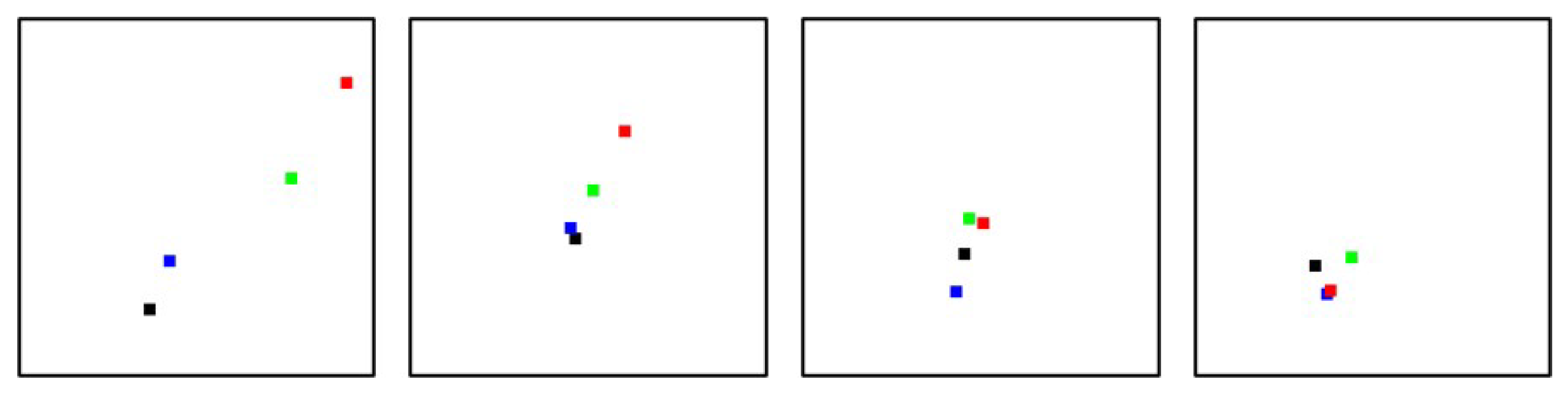
Figure 7 shows examples of the segment dynamics for the 4 classes in PS_2. In this example the 2a- segment starts with relatively low values but shows a strong increase at day 2 and 3. The hyperemic development of the 2b2 is more limited than for 2b1.
3.2. Classification
The aim of this article is not to present a sophisticated method for burn wound classification based on the available parameter, this is the object of further projects. The special attention is here paid to the discrimination of 2b1 and 2b2, because this is decisive for the kind of treatment. The comparison of Figure 4 (upper) to Figure 5 (deep) clearly shows the importance of the deep perfusion for classification.
The qualitative assessment of class discrimination shows, that at day 1 a differentiation between 2a-2b1 and 2b2-3 is possible with some uncertainty. This is hardly not possible at day 0 due to the strong overlapping of the distributions. The discrimination is further significantly improved at day 2. From day 3 onwards the class distributions are nearly stationary with a good discrimination.
Figures
Figure 1.
Perfusion profiles: left: vHb (volume part hemoglobin), right: xHbO2 (oxygen saturation hemoglobin).
Figure 1.
Perfusion profiles: left: vHb (volume part hemoglobin), right: xHbO2 (oxygen saturation hemoglobin).
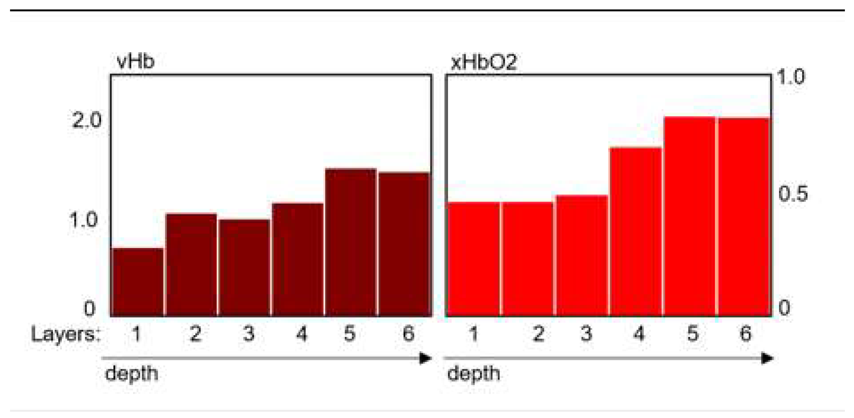
Figure 2.
Left: PS_1: vHb_1 – xRate, right: PS_2: vHb_2 – flow_2 The parameter values range from 0 to 1, left to right and bottom to top; class distributions at day 3 after burn; classes: 2a: blue, 2b1: green, 2b2: yellow, 3: red; in this 2-dim. presentation the higher classes partially mask the lower ones: 3 > 2b2 > 2b1 > 2a.
Figure 2.
Left: PS_1: vHb_1 – xRate, right: PS_2: vHb_2 – flow_2 The parameter values range from 0 to 1, left to right and bottom to top; class distributions at day 3 after burn; classes: 2a: blue, 2b1: green, 2b2: yellow, 3: red; in this 2-dim. presentation the higher classes partially mask the lower ones: 3 > 2b2 > 2b1 > 2a.
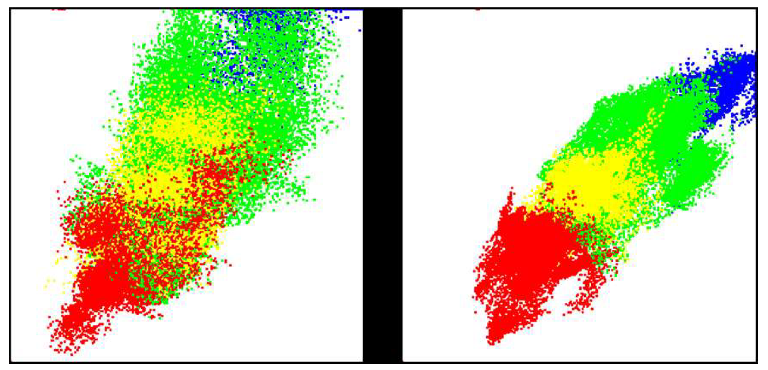
Figure 3.
Class distributions in the parameter spaces PS_1 and PS_2 at day 0 (a), 1 (b), 2 (c) and 3 (d); first row: PS_1, second row: PS_2; the colours depict the distribution density from gray to red.
Figure 3.
Class distributions in the parameter spaces PS_1 and PS_2 at day 0 (a), 1 (b), 2 (c) and 3 (d); first row: PS_1, second row: PS_2; the colours depict the distribution density from gray to red.
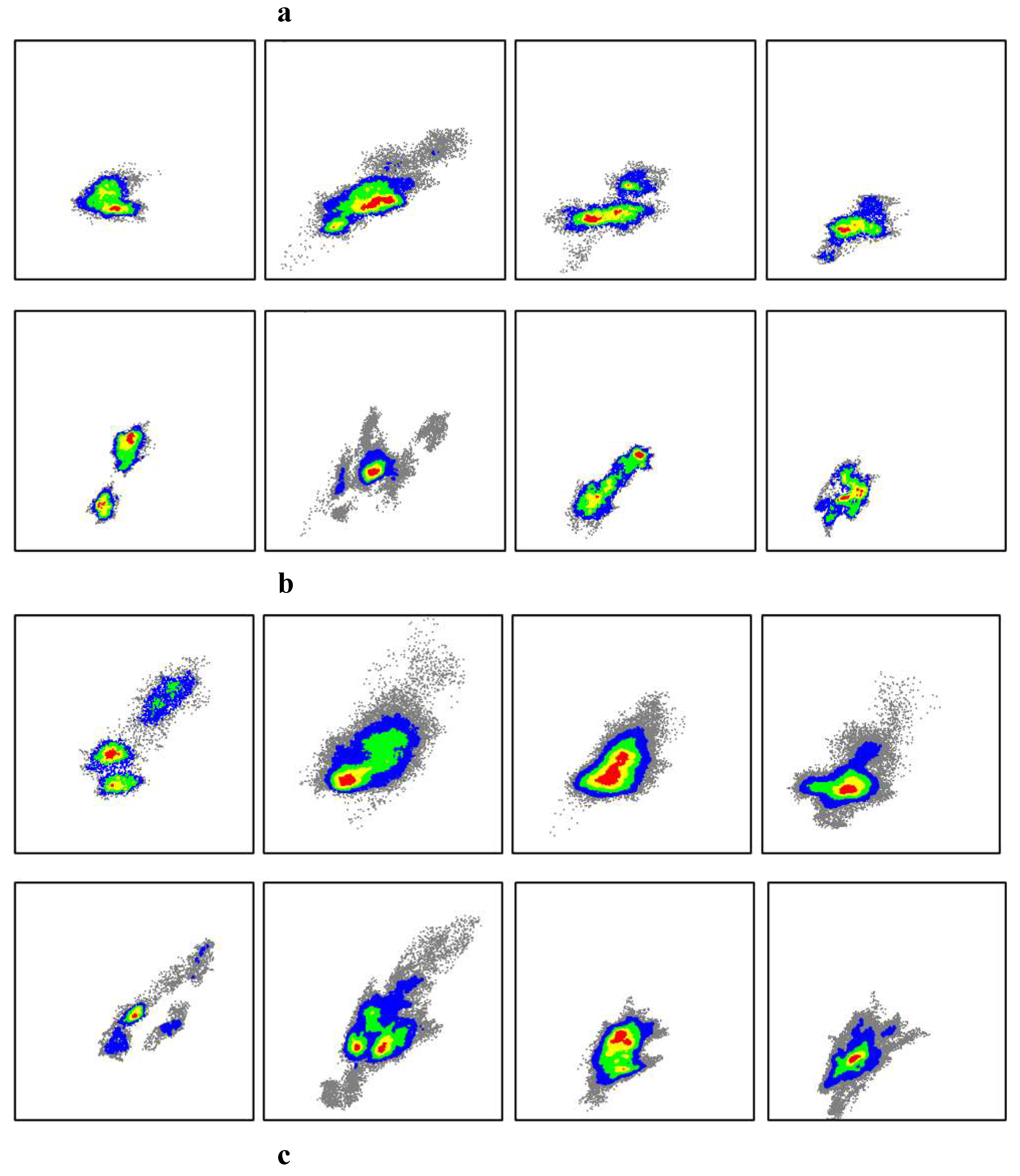
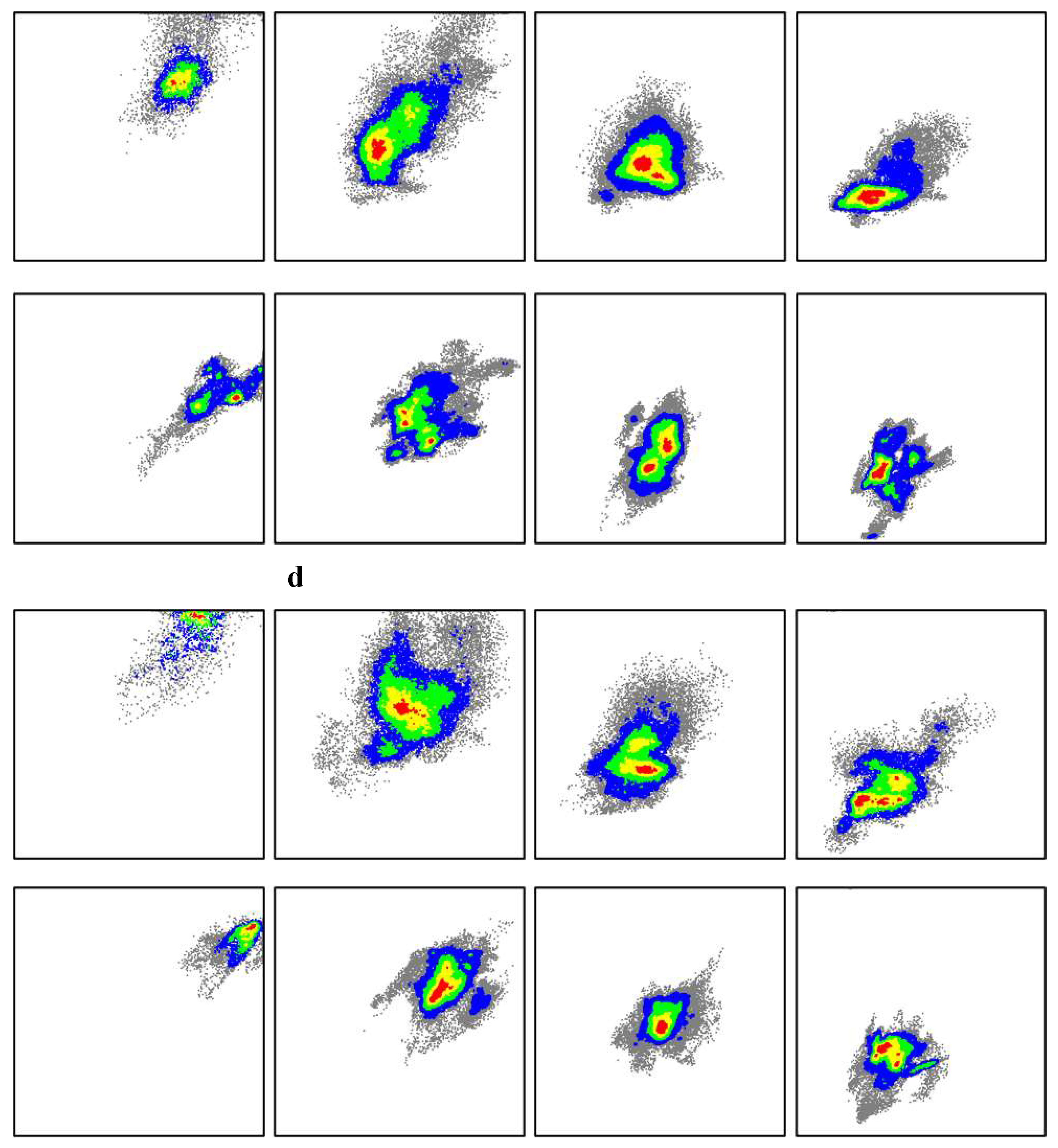
Figure 4.
Abstracted class areas for upper perfusion (PS_1) at day 0, 1, 2 ,3 (from left to right).
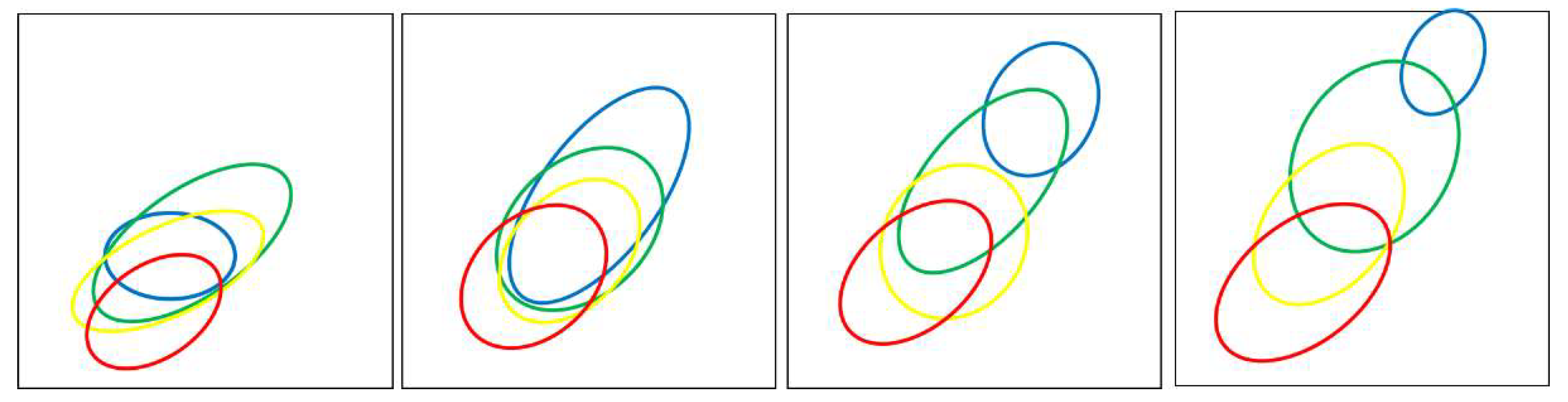
Figure 5.
Abstracted class areas for deep perfusion (PS_2) at day 0, 1, 2 ,3 (from left to right).
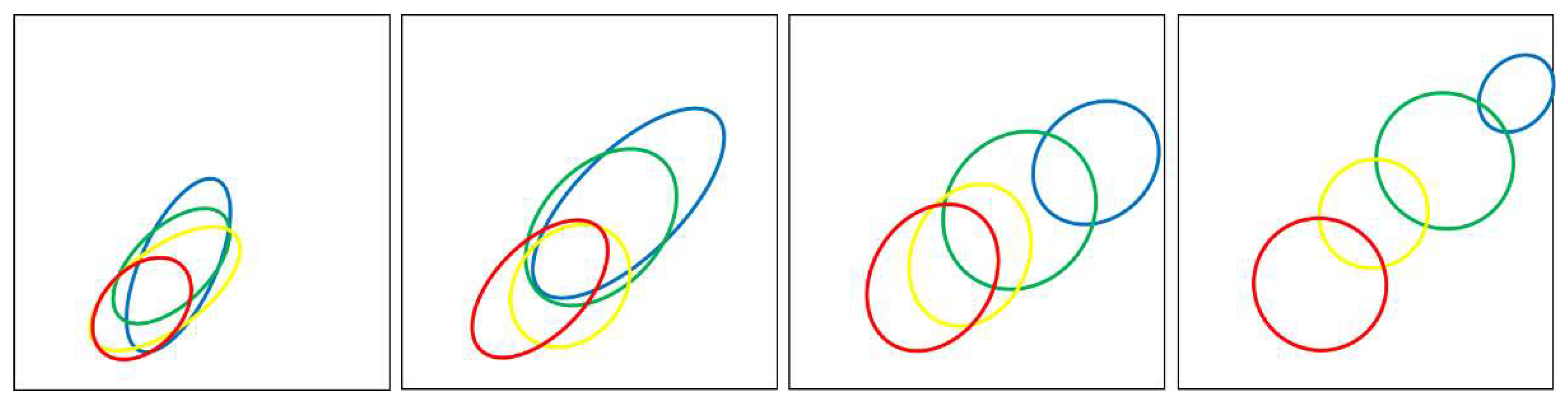
Figure 6.
Typical perfusion profiles for class 2b1 (upper row) and 2b2 (lower row) for the day 0 to 3 (left to right), measured at wounds at lower legs and hands.
Figure 6.
Typical perfusion profiles for class 2b1 (upper row) and 2b2 (lower row) for the day 0 to 3 (left to right), measured at wounds at lower legs and hands.
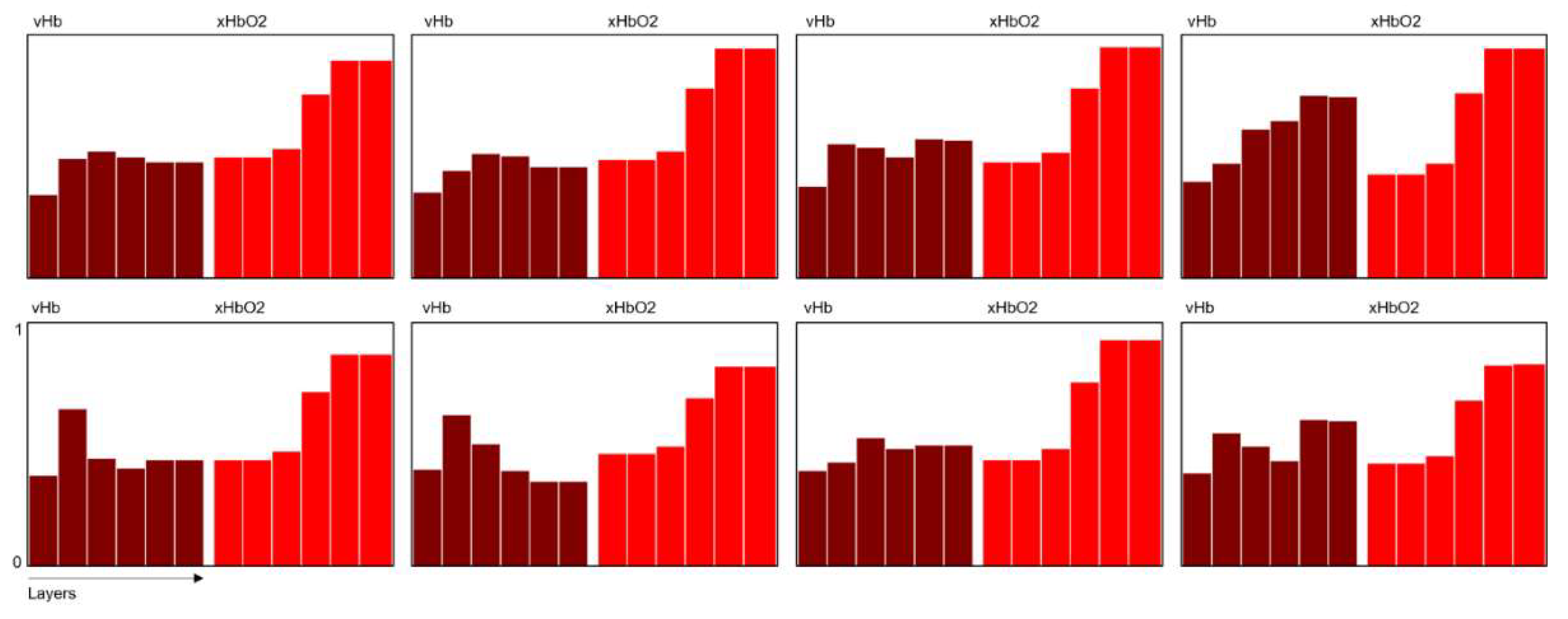
Figure 7.
Examples of segment dynamics in PS_2 (deep perfusion) for the classes 2a, 2b1, 2b2 and 3 (from left to right); day 0: black, day 1: blue, day 2: green, day 3: red; measured at wounds at femur and feet.
Figure 7.
Examples of segment dynamics in PS_2 (deep perfusion) for the classes 2a, 2b1, 2b2 and 3 (from left to right); day 0: black, day 1: blue, day 2: green, day 3: red; measured at wounds at femur and feet.
4. Discussion
Numerous articles have addressed the early dynamics of burn wounds, examining cellular and molecular processes [3-6]. However, due to the absence of objective measurement techniques capable of capturing relevant wound parameters during the initial period (day 0 to 3 post-burn), these intrinsic processes cannot be observed to facilitate treatment decisions, such as choosing between conservative or surgical approaches.
Assessments of Laser Doppler Imaging (LDI) accuracy for burn depth evaluation have been conducted at various time points (days 0, 1, 3, 5). The accuracy of LDI consistently surpassed that of clinical assessment, but its efficacy was limited to 54% on day 0, 79% on day 1, and 95% on day 3 [10]. These findings align qualitatively with our own results, considering the adoption of distinct class definitions.
HSI offers the potential to obtain extensive information about burn wounds, particularly on a macroscopic level, primarily through the assessment of perfusion parameters.
The analysis of the wound dynamics, described by this parameter, exemplifies the fundamental problem of an early estimation of the degree of damage of burn wounds. Figure 5 clearly shows the development of the class distributions over the first four days from a compact global distribution area at day 0 until an untangled distribution with good discrimination of the classes at day 3. This corresponds to the clinical experience that classifications (2b1 – conservative and 2b2 – surgical treatment) generally are not reliable until day 3.
With a mostly visual inspection of the wound, only superficial features are recognizable (Figure 4), with a limited discrimination of 2b1 and 2b2 also at day 3. The „visual border“ tends to run in the upper part of the 2b2-distribution (yellow), so that a larger part of the 2b1-distribution (green) may be wrongly estimated as 2b2. This correlates with our experience, that with using a more objective measuring method, which also captured the deeper perfusion (like LDI and HSI), the surgical treatments are reduced in favour of conservative treatments.
Concerning the parameter distributions some relevant influencing factors have to be considered:
- ▪
- Measuring and data processing method:
The information gathered by the measuring method does not allow an exact determination of the depth structure of the skin or wound (layer thickness) [16]. Therefore, the depth profiles represent only relative quantities with globally fixed scale factors. This results in a limited adaptivity to actual skin/wound structures and a widening of the parameter distributions due to non-adapted variations.
- ▪
-
Projection on 2-dimensional parameter spaces:The projections on PS_1 and PS_2 have been primary used to achieve an easy to view representation of the class distributions. The use of higher dimensional parameter spaces might result in a better discrimination of the classes but requests a significantly larger number of referenced data.
- ▪
- Clinical reference classification:
The clinical classification based on the healing time is rather inaccurate but is supported here by the parameter-based classification at day 3.
The transition towards a positive or negative development, particularly in 2b1-segments, becomes evident from day 0 to day 1, possibly originating from initial states that are indecisive - contrary to the hypothesis - or from initial classes that are indistinguishable based on the HSI parameter at day 0.
The distributions presented in Figure 4 clearly demonstrate that a clinical estimation at day 0 has minimal chances of correctly identifying a deep 2b2-degree, and there is a high probability of misinterpretations as 2b1 at this early stage.
Discriminating between the distributions of different classes, as an indicator of classification quality, is insufficient for a reliable early assessment of burn class on day 0. The classes exhibit significant overlap at day 0; however, Figure 5 illustrates distinct developments, with 2a-2b1 and 2b2-3 becoming significantly separated as early as day 1. This separation can be crucial for making early decisions regarding conservative or surgical treatment.
Overall, currently, there is no foreseeable progress in effectively discriminating between the classes on day 0. The overlapping of class distributions appears to result primarily from the considerable global variability of wound characteristics and intrinsic dynamics that cannot be captured solely by the HSI-perfusion parameter.
5. Conclusions
HSI represents a practical and highly informative measurement method that provides a comprehensive understanding of the burn wound condition. It allows for the parametric analysis of wound dynamics based on the degree of damage, leading to novel insights into the estimation of burn class within the initial four days.
The study findings introduce several noteworthy contributions:
- ○
- The utilization of HSI enables the parametric analysis of dynamics associated with different burn classes, presenting a novel approach that examines the dynamics of these classes using physiological perfusion parameters.
- ○
- The significance of deep perfusion in relation to distinct burn classes is clearly emphasized, with HSI being the only method capable of distinguishing between superficial and deep perfusion.
- ○
- The critical timeframe for determining positive or negative developments in 2b-wound segments is identified as the initial 24 hours, under the standard treatment protocol.
- ○
- The study also sheds light on the (limited) potential of HSI in facilitating early assessments of resulting burn class.
Author Contributions
Methodology, Jörg Marotz; Validation, Thomas Wild; Investigation, Ahmed Aljowder; Resources, Frank Siemers; Writing – original draft, Thomas Wild and Jörg Marotz; Writing – review & editing, Ahmed Aljowder; Supervision, Frank Siemers. All authors have read and agreed to the published version of the manuscript.
Funding
This research did not receive any specific grant from funding agencies in the public, commercial, or not-for-profit sectors.
Institutional Review Board Statement
The data collection procedures adhered to the principles outlined in the Declaration of Helsinki. The study protocol received approval from the Ethics Committee of the Ärztekammer Sachsen-Anhalt, Germany (47/19). The trial was officially registered on DRKS (German Clinical Trials Register) and can be accessed on the public website under the registration number DRKS00022843.
Informed Consent Statement
Informed consent was obtained from all subjects involved in the study.
Data Availability Statement
The original contributions presented in this study are included in the article/supplementary material. Further inquiries can be directed to the corresponding author(s).
Conflicts of Interest
The authors declare no conflicts of interests relevant to this article.
References
- Brusselaers N., Monstrey S., Vogelaers D. et al. Severe burn injury in europe: a systematic review of the incidence, etiology, morbidity, and mortality. Crit Care 14, R188 (2010). [CrossRef]
- Jaspers MEH, van Haasterecht L, van Zuijlen PPM, Mokkink LB. A systematic review on the quality of measurement techniques for the assessment of burn wound depth or healing potential. Burns 45(2), 2019: 261-281.
- Singh V, Devgan L, Bhat S, Milner S; The Pathogenesis of Burn Wound Conversion; Annaly of Plastic Surgery, Vol 59, No 1, July 2007, 109-115.
- Lanier ST, McClain SA, Lin F, Singer AJ, Clark RA. Spatiotemporal progression of cell death in the zone of ischemia surrounding burns. Wound Repair Regen. 2011 Sep-Oct;19(5): 622-32. [CrossRef]
- Salibian AA, Rosario ATD, Severo LAM, Nguyen L, Banyard DA, Toranto JD, Evans GRD, Widgerow AD. Current concepts on burn wound conversion-A review of recent advances in understanding the secondary progressions of burns. Burns. 2016 Aug;42(5): 1025-1035. [CrossRef]
- Bumbuc RV, Korkmaz HI, van Zuijlen PPM, Vermeulen L and Sheraton VM; Understanding the Dynamics of the Proliferative Phase in Local Burn Wound Healing: A Computational Model; 2023 IEEE International Conference on Bioinformatics and Biomedicine (BIBM), Istanbul, Turkiye, 2023:. 3676-3683.
- Meiqi L, Jie Z, Xiaochuan W, Jingjuan Z, Fei S, Duyin J; Research advances in prevention and treatment of burn wound deepening in early stage; Frontiers in Surgery 9, 2022: 1015411.
- Palackic, A.; Jay, J.W.; Duggan, R.P.; Branski, L.K.; Wolf, S.E.; Ansari, N.; El Ayadi, A. Therapeutic Strategies to Reduce Burn Wound Conversion. Medicina 2022, 58: 922. [CrossRef]
- Schmauss D, Rezaeian F, Finck T, Machens HG, Wettstein R, Harder Y. Treatment of secondary burn wound progression in contact burns-a systematic review of experimental approaches. Journal of burn care & research: official publication of the American Burn Association. 2015; 36: e176–89. [CrossRef]
- Hoeksema H, Van de Sijpe K, Tondu T, Hamdi M, Van Landuyt K, Blondeel P, et al. Accuracy of early burn depth assessment by laser Doppler imaging on different days post burn. Burns 35, 2009: 36-45. [CrossRef]
- Jeschke MG, van Baar ME, Choudhry MA, Chung KK, Gibran NS, Logsetty S. Burn injury. Nat Rev Dis Primers. 2020 Feb 13;6(1): 11.
- Ru Wang, Juan Zhao, Zhenyu Zhang, Chang Cao, Yange Zhang, Yu Mao, Diagnostic Accuracy of Laser Doppler Imaging for the Assessment of Burn Depth: A Meta-analysis and Systematic Review, Journal of Burn Care & Research, Volume 41, Issue 3, May/June 2020: 619–25. [CrossRef]
- Lertsakdadet B, Kennedy G, Stone R et al; Assessing the Need for Multimodal Optical Imaging of Perfusion in Burn Wounds; Burns 48(4), 2021: 799-807.
- Marotz J, Siafliakis A, Holmer A, et al. First results of a new hyperspectral camera system for chemical based wound analysis. Wound Med 10-11, 2015: 17-22. [CrossRef]
- Marotz J, Kulcke A, Siemers F, Cruz D, Aljowder A, Promny D, Daeschlein G, Wild T (2019): Extended Perfusion Parameter Estimation from Hyperspectral Imaging Data for Bedside Diagnostic in Medicine. In: Molecules 24, 2019: 4164. [CrossRef]
- Marotz J, Schulz T, Seider S, Cruz D, Aljowder A, Promny D, Daeschlein G, Wild T, Siemers F (2020): 3D-Perfusion analysis of burn wounds using Hyperspectral Imaging; Burns 47, 2021: 157-170.
- Schulz T, Marotz J, Seider S, Langer S, Leuschner S, Siemers F. Burn depth assessment using hyperspectral imaging in a prospective single center study. Burns 48(5), 2022: 1112-1119. [CrossRef]
- Liu H, Yue K, Cheng S, Li W, Fu Z. A Framework for Automatic Burn Image Segmentation and Burn Depth Diagnosis Using Deep Learning. Comput Math Methods Med. 7, 2021: 5514224. [CrossRef]
Disclaimer/Publisher’s Note: The statements, opinions and data contained in all publications are solely those of the individual author(s) and contributor(s) and not of MDPI and/or the editor(s). MDPI and/or the editor(s) disclaim responsibility for any injury to people or property resulting from any ideas, methods, instructions or products referred to in the content. |
© 2024 by the authors. Licensee MDPI, Basel, Switzerland. This article is an open access article distributed under the terms and conditions of the Creative Commons Attribution (CC BY) license (http://creativecommons.org/licenses/by/4.0/).
Copyright: This open access article is published under a Creative Commons CC BY 4.0 license, which permit the free download, distribution, and reuse, provided that the author and preprint are cited in any reuse.


