Submitted:
16 December 2024
Posted:
18 December 2024
Read the latest preprint version here
Abstract
Metabolic syndrome is a global health challenge, marked by visceral obesity, hyperglycemia, dyslipidemia, hypertension, and chronic low-grade systemic inflammation. The limitations of existing therapeutic options, including variable efficacy and significant risk of adverse events, have driven growing interest in natural products, particularly vegetable extracts as food supplements or functional food ingredients to prevent metabolic syndrome risk factors in high-risk individuals. This study provides a comprehensive review of the role of vegetable extracts in mitigating metabolic syndrome risk factors, addressing their safety concerns, and exploring associated technological advancements. Evidence from the literature highlights the potential of these extracts to combat the pathogenesis of metabolic dysfunctions. However, challenges such as safety concerns, lack of standardized regulations, and the possibility of drug-food interactions impede their clinical application. Robust, long-term clinical studies are essential to confirm the efficacy and safety of vegetable extracts before they can be established as a preventive strategy for managing metabolic syndrome.
Keywords:
vegetable extracts
; functional foods
; metabolic syndrome
; efficacy
; safety
; technological advancements
1. Introduction
A group of diseases and illnesses known as metabolic syndrome include obesity, hyperglycemia, hypertension, hyperlipidemia, and pro-inflammatory state. These factors are known to raise the risk of cardiovascular diseases, type 2 diabetes mellitus (DM2), and cerebrovascular accidents. A few additional terms that are also used to refer to metabolic syndrome are Reaven’s syndrome, Syndrome X, the deadly quartet, and insulin resistance syndrome [1,2]. The definition and diagnostic criteria of metabolic syndrome varies among different organizations such as World Health Organization (WHO), National Cholesterol Education Program’s Adult Treatment Panel III (NCEP: ATP III), and International Diabetes Federation (IDF). However, all these emphasize importance of visceral obesity, dyslipidemia, hypertension, and hyperglycemia [3,4,5]. Metabolic syndrome has become more prevalent worldwide during the last few decades, particularly among urban population of developing countries. As estimated by WHO, 890 million individuals worldwide suffer from obesity, 422 million from diabetes, and 1.28 billion from hypertension [6,7,8,9].
Potential risk factors that predispose individual to these metabolic abnormalities may include female sex, advanced age (>50 years), sedentary lifestyle, positive family history, middle socioeconomic status, illiteracy, unemployment, omnivore diet, stress, insomnia, and increased body mass index (BMI) [10]. The syndrome may slowly progress to major health issues such as coronary heart disease (CHD), heart failure, stroke, hepatic steatosis, and liver failure, if it persists or is not properly treated, which make it a major challenge for healthcare system worldwide [11,12]. Aerobic exercise, modification in dietary patterns, and therapeutic interventions are the strategies that remain the cornerstone of managing patients with metabolic syndrome [13,14]. At the moment, a combination of therapeutic agents with different mechanistic targets such as such as insulin sensitizers, hypotensive drugs, and hypolipidemic agents are used to treat the individual components of metabolic syndrome and to slow the syndrome’s progression towards serious complications [15]. The multitude of adverse drug reactions (ADRs) linked to the therapeutic choices currently employed for the management of metabolic syndrome may outweigh the benefits of treatment in certain cases. Metformin-induced lactic acidosis, thiazolidinediones-induced congestive heart failure, renin-angiotensin-aldosterone system (RAAS) inhibitors-induced renal toxicities and hyperkalemia, aspirin-induced peptic ulcer, and hypolipidemics-induced myopathies are few of the most common ADRs [16,17,18,19].
Natural products derived from both marine and terrestrial sources might be essential for preserving human health. They are effective for treating and/or preventing the most challenging disorders [20]. According to a recent market analysis, the global market for natural substances is growing, and this expansion may be linked to consumers’ growing inclination towards preventative care [21]. In 2014, Ekor [22] reported that over 80% of the global population is currently relying on the use of vegetable products for their primary healthcare. Herbal products have demonstrated therapeutic efficacy in randomized controlled trials in decreasing risk factors for metabolic syndrome by positively regulating blood pressure, serum glucose, waist circumference, and lipid levels [23]. As a result, they may offer an alternative treatment option for metabolic syndrome. In this review, vegetable extracts and their bioactive components as valuable ingredients of functional foods have been explored to prevent the risk factors for metabolic syndrome and to mitigate the syndrome’s impact. Fair discussions on their safety concerns and associated technological advancements are part of this review.
2. Vegetable Extracts as Functional Food Ingredients
Consuming a variety of fruits and vegetables in appropriate quantities may help reduce metabolic syndrome risk factors. Enriched with bioactive ingredients, these foods show promise in improving glucose and lipid homeostasis, reducing lipid deposition, decreasing inflammatory markers, and modulating gut microbiome [24,25]. These ingredients can also alter signaling pathways at various levels i.e., enzyme activity, gene expression, epigenetic regulation, and protein expression [26]. Incorporation of vegetable extracts and derived bioactive compounds in food supplements or functional foods may offer a novel strategy of protection against the development of metabolic syndrome risk factors, particularly in subjects carrying high risk for metabolic disorders.
2.1. Obesity and Hyperglycemia
Obesity is characterized by fats accumulation in the body associated with adverse alterations in adipose tissues such as decrease in lipid turnover and accumulation of inflammatory macrophages [27]. Adopise tissues in abdominal or visceral regions possess more negative consequences on metabolic and insulin signaling pathways, thus providing a roadmap to the pathogenesis of obesity and other risk factors of metabolic syndrome [28]. A chronic hyperglycemia due to defective insulin secretion and/or action may progresses silently to the development of diabetes mellitus, where persistent and chronic state of diabetes could result in serious complications i.e., retinopathy, neuropathy, nephropathy, atherosclerosis, peripheral arterial disease, and cerebrovascular accidents. In majority of cases, insulin resistance and impaired insulin secretion may occur concomitantly, affecting metabolism of carbohydrates, proteins and fats [29,30,31].
Ullah, et al. [32] studied the hydroethanolic extract of Prunus domestica L. to modulate the molecular mechanisms of cardiometabolic disorders using in vitro experimental models. The extract rich in polyphenols (mainly carcinogenic acid derivatives) inhibited enzymes associated with glucose and lipids metabolism i.e., α-amylase, α-glucosidase, HMG-CoA reductase, and pancreatic lipase with IC50 values of 7.01 mg/mL, 6.4 mg/mL, 21.4 mg/mL, and 20.4 mg/mL, respectively. Prunus persica (L.) Stokes flowers demonstrated anti-obesity effects in high-fat diet induced obesity in male C57BL/6 mice, supplemented with 0.2% or 0.6% flower extract for eight weeks [33]. The anti-obesity effects were linked to improved hepatic lipid metabolism resulted in reduction of body weight, visceral fat mass, and serum levels of glucose, alanine transaminase (ALT), and aspartate transaminase (AST). Berberis species, enriched with berberine alkaloids have been showed promising anti-diabetic effects in various cell and animal models as well as clinical trials [34]. Berberine promoted glucose uptake and prevented gluconeogenesis by inhibition of NAD-dependent deacetylase sirtuin-3 (SIRT3) [35]. Berberine also showed to mimic insulin-sensitizing effects via down regulation of protein tyrosine 1B activity, both on adipocytes and myocytes [36,37]. In vivo study demonstrated regulation of hepatic lipid utilization and maintenance of whole-body energy metabolism via mediating autophagy and fibroblast growth factor (FGF) 21 activation [38]. Extracts from leafy vegetables (Hibiscus sabdariffa L., Vigna unguiculata L. Walp, and Solanum nigrum L.) significantly decreased fasting blood glucose levels in high fat diet-streptozotocin induced diabetic Wistar rats [39].
A randomized, placebo-controlled, cross-over trial demonstrated significant reduction of body weight and body mass index with a considerable increase in adiponectin and decrease in leptin blood levels in healthy participants (aged 18–65 years) supplemented with anthocyanin-rich Queen Garnet plum juice for 28 days [40]. In another randomized, placebo-controlled, cross-over trial, brewer’s spent grain rich in soluble fibers showed a significant reduction of postprandial glycemia and insulinemia in healthy subjects with slightly impaired glucose tolerance [41]. Oral administration of olive leaf extract (500 mg/day) for 14-weeks significantly decreased HbA1c and fasting insulin levels in diabetic subjects in randomized, placebo-controlled, trial [42]. Gupta et al. [43] observed significant improvement of area under curve of glucose and insulin sensitivity in diabetic patients supplemented either with fenugreek seeds (1 g/day) or placebo for two months, though no significant differences were noted in fasting blood glucose levels and oral glucose tolerance test among the groups. Another study with cross-over design showed a considerable decrease in fasting blood glucose levels and improvement in oral glucose tolerance test in diabetic subjects supplemented with diet containing fenugreek seeds (100 g/day) for 10 days [44]. Supplementation of diabetic subjects with fenugreek seeds (15 g/day) soaked in water resulted in significant reduction of postprandial glucose levels [45]. Green tea, one of most common beverages in the world used for maintaining normal body weight and glucose metabolism, showed controversial results in clinical trials. A randomized, placebo-controlled study exhibited significant decrease in HbA1c levels with no considerable effects on fasting blood glucose levels in healthy subjects consumed daily a packet of green tea extract (containing 544 mg polyphenols) for two months [46]. A combination of exercise and green tea extract (containing 890 mg polyphenols) resulted in significant decrease in area under curve for insulin with increase in insulin sensitivity in healthy subjects [47]. Conversely, another study showed no effects on fasting blood glucose and HbA1c levels, insulin sensitivity and secretion, and glucose tolerance in healthy subjects supplemented with epigallo-catechin-3-gallate (800 mg/day) for eight weeks [48].
2.2. Dyslipidemia
Dyslipidemia, one of the important risk factors of atherosclerosis-induced cardiovascular damage, is characterized by abnormal lipid profiles including high levels of total cholesterol (TC), low-density lipoprotein (LDL), very low-density lipoprotein (VLDL) and triglycerides (TGs) coupled with low levels of high-density lipoprotein (HDL). A clinically significant condition of dyslipidemia is most commonly seen in subjects with obesity and/or diabetes mellitus [49,50]. Smoking, chronic alcohol intake, obesity, diabetes mellitus, or certain medications i.e., steroids are certain factors increasing risk for dyslipidemia [51]. Mediterranean diet composed of high intake of fruits, vegetables legumes, complex carbohydrates, and unsaturated fatty acids, moderate intake of wine and fish, and low intake of meat and dairy products, is most widely studied dietary pattern for its capacity to protect against dyslipidemia [52,53].
The extracts from fruits, leaves, and bark from Zanthoxylum armatum DC (500 mg/kg) demonstrated significant hypolipidemic effects in mice treated for 15-days. A reduction of TC, TG, and LDL levels was observed [54]. A novel multi-targeted herbal formula Schisandrae Fructus, milk thistle, hawthorn, and bitter melon) ameliorates diet-induced metabolic syndrome [55]. The results indicated potent inhibitory effects of Crataegus Fructus on 3T3-L1 preadipocytes differentiation and cholesterol uptake into Caco-2 cells, inhibitory effects of Schisandrae Fructus and milk thistle on oleic acid-induced fatty liver in HepG2 cells, and inhibitory effects of bitter melon on glucose uptake in brush border membrane vesicles. In vivo experiments suggested reduction of diet-induced increase in body weight and fat pad mass, with a significant decrease in liver weight, and hepatic and plasma lipid levels in dose-dependent manner. Fixed oils from spices (Alpinia galanga (L.) Willd., Cinnamomum zeylanicum var. cassia, Trigonella foenum-graecum L., Foeniculum vulgare Mill., and Myristica fragrans Houtt.) showed in vitro reduction of accumulated lipid droplets in 3T3-L1 cell lines, and in vivo improvement of lipid profiles, anti-oxidant enzymes and reduced droplets in liver and adipose tissues in C57BL/6 mice [56]. Another study showed a significant decrease in TC, LDL, TG, atherogenic index and increase in HDL levels in diet induced dyslipidemia in Wistar rats treated with m Mangifera indica L. leaves extract [57].
De Lellis et al. [58] observed hypolipidemic effects of food supplement based on monacolins, γ-oryzanol, and γ-aminobutyric acid (bioactive ingredients from rice fermented with the Monascus purpureus) in participants with mild dyslipidemia. In a randomized, double-blind, placebo-controlled trial, enrolled subjects were treated either with supplement or placebo for three months, where the results indicated significant decrease of TC and LDL and increase in HDL levels in supplement-treated group. Daily consumption of prunes (100 g) for eight weeks resulted in significant reduction of serum LDL levels and fecal bile concentration of lithocholic acid as compared to grape juice (control) in a cross-over study [59]. A randomized clinical study demonstrated a considerable improvement of TC, TG, LDL, and HDL in hyperlipidemic subjects treated with lettuce seed extract (1000 mg/day) for 12-weeks [60]. Eight weeks intake of a nutraceutical supplement based on bergamot extract (120 mg flavonoids), vitamin C, phytosterols, and chlorogenic acid from dry artichoke extract significantly improved the levels of TC, TG, LDL, non-HDL cholesterol, high sensitivity C-reactive protein (hs-CRP), and tumor necrosis factor alpha (TNF-α) in a three-arm, placebo-controlled trial in dyslipidemic overweight subjects [61]. A three-times daily intake of bitter melon extract (100 mg) for 30-days significantly reduced LDL levels as compared to placebo in Japanese adults, though no significant difference was observed among the groups in TC, TG, and blood glucose levels [62].
2.3. Hypertension, Endothelial Dysfunction, and Pro-Inflammatory State
Etiology of hypertension in metabolic syndrome is multifactorial and several elements like insulin resistance, obesity, hyperglycemia, and dyslipidemia may contribute to the cause. It is one of the key risks for cardiovascular complications and cerebrovascular accidents [63,64,65]. As suggested by large body of evidence, dietary modifications or nutritional interventions, such as diets rich in fruits, vegetables, whole grains, and low-fat dairy products, with low intake of sodium could be a fruitful strategy in preventing or managing hypertension [66]. In addition, suppressing pro-inflammatory state and improving endothelial function could be significant part of metabolic syndrome therapy and to prevent and/or delay the development of chronic complications associated with the syndrome [67].
Polyphenols enriched diets such as tea, red wines, vegetables, and fruits possess ability to regulate vascular tone via upregulation of nitric oxide-cyclic guanosine monophosphate (NO-cGMP) pathway. They may also counter oxidative stress by decreasing the production of endogenous pro-oxidants like NADPH oxidase [24]. A study conducted by Luna-Vazquez et al. [68] demonstrated a significant reduction of oxidative stress markers and systolic blood pressure in L-nitro-arginine-methyl ester (L-NAME) induced hypertensive rats treated with chemically characterized black cherry fruit extracts (300 mg/kg/day), contained high polyphenolic contents (mainly chlorogenic acid and anthocyanins). Treatment of L-NAME induced hypertensive rats with a methanolic extract of Adansonia digitata L. (200 mg and 400 mg/kg/day) resulted in dose-dependent reduction of systolic and diastolic blood pressure, mean arterial pressure, and heart rate towards the normal physiological values [69]. In addition, A. digitata extract considerably reduces biomarkers associated with endothelial dysfunction (i.e., angiotensin converting enzyme activity), inflammatory and oxidative stress mediators (i.e., C-reactive protein, IL-1β, and malondialdehyde), and cardiac injury markers (i.e., creatine kinase-MB and lactate dehydrogenase activities).
Kim et al. [70] studied the vasorelaxant effects of Prunus persica extract on endothelium-denuded aortic rings from rat thoracic aorta, using concentrations ranging from 0.5 to 20 μg/mL. They found that the extract’s vasorelaxation involved the nitric oxide-soluble guanylate cyclase-cyclic guanosine monophosphate (NO-sGC-cGMP) pathway, vascular prostacyclin, and muscarinic receptor transduction. Additionally, the extract reduced calcium-induced vasoconstriction via inositol triphosphate receptors (IP3R) in the endoplasmic reticulum membrane. A randomized, controlled, cross-over clinical trial showed a significant reduction of systolic blood pressure in adults with mildly elevated blood pressure who consumed cruciferous vegetables (300 g/day) as compared to root and squash vegetables [71]. Supplementation of subjects with mild hypertension with Nigella sativa L. seed extracts (200 and 400 mg per day) for eight weeks resulted in significant reduction of systolic and diastolic blood pressure in a randomized, placebo-controlled, clinical trial [72]. Consumption of garlic may enhance nitric oxide production, improve endothelial function, and reduce oxidative stress, thereby improving blood pressure [73]. A double-blind, randomized, placebo-controlled, clinical trial showed supplementation of hypertensive subjects with aged garlic extract (960 mg/day, containing 2.4 mg S-allylcysteine) for 12 weeks to reduce systolic blood pressure in treated patients but with uncontrolled hypertension [74].
A supplementation of anthocyanin-rich queen garnet plum juice alleviates platelet aggregation via reduced P-selectin expression of activated de-granulated platelets, increased activated partial thromboplastin clotting time, and decreased blood levels of fibrinogen and malondialdehyde in healthy volunteers in a randomized, placebo-controlled clinical trial [75,76]. An experimental study designed to verify the potential of dietary berries (Viburnum trilobum Marshall, Amelanchier alnifolia, Shepherdia argentea (Pursh) Nutt., and Prunus virginiana L.) in alleviating diabetic microvascular complications and pro-inflammatory gene expression, showed potent inhibition of aldose reductase with nonpolar fraction (rich in carotenoids) and strong inhibition of IL-1β and COX-2 gene expression with polar fraction (rich in anthocyanins, phenolic acids, and proanthocyanidins) [77]. Aldose reductase enzyme is reportedly involved in the pathogenesis of diabetic microvascular complications.
3. Safety Concerns
Safety of vegetable extracts and derived bioactive ingredients is one of the crucial factors in their utilization as food supplements or functional food ingredients. While these extracts are derived from natural sources, they are generally considered safe for use; however, certain factors need to be evaluated to ensure their safety for human use. In majority of cases, the toxicity of vegetable extracts has not been systematically evaluated, while scientific studies have revealed that a large number of plants used as food or in traditional medicine are potentially toxic, mutagenic, and carcinogenic [78]. Adewunmi and Ojewole [79] observed that toxic or lethal compounds may be found in some complementary and alternative medicines such as lectins, viscotoxins, aristolochic acids, pyrrolizidine alkaloids, benzophenanthrine alkaloids, saponins, diterpenes, cyanogenic glycosides, and furanocoumarins. Unlike conventional medicines, dose-dependent toxicity and long-term safety evaluation of vegetable extracts are lacking, particularly for novel extracts or high-potency formulations. For instance, some of the most common vegetable extracts used to maintain good health are associated with increased risk of hepatotoxicity i.e., Curcuma longa L., Camellia sinensis (L.) Kuntze, Withania somnifera (L.) Dunal, Garcinia gummi-gutta (L.) N.Robson, Monascus purpureus, and Actaea racemosa L. [80].
In addition, safety concerns are also linked to microbial or heavy metal contamination of vegetable extracts. Strict safety and sterility regulations are typically not guaranteed when herbal medicines are collected, stored, and prepared, and thus they are more likely to expose to microorganisms from the soil, water, and improper handling or storage techniques [81]. The extensive dispersion of heavy metals in plants, soil, and water is a result of several industrial, agricultural, medical, and technological applications, where research studies have demonstrated the frequently presence of heavy metals in herbal products in concentration exceeding the permitted limits [82]. These metals are known carcinogenic and could increase the risk of internal organ toxicities i.e., brain, heart, lungs, liver, and kidneys [82].
Campbell-Tofte et al. [83] reported that the World Health Organization (WHO) has urged the establishment of international standards and procedures for the assessment of the safety and therapeutic efficacy of traditional medicinal approaches. Even though pharmacological and toxicological analyses of medicinal plants are essential for the development of drugs and standardized herbal remedies, reports of scientific studies describing the effectiveness of traditional claims far outnumber those of toxicological analyses and toxicity [84,85,86,87]. Plant preparations and phytochemicals obtained from medicinal plants should be studied in vivo for their safety and effectiveness using appropriate animal models or in specially designed randomized, placebo-controlled clinical trials [83,88]. Unquestionably, safety assessments of herbal remedies, particularly those that assess the effects of long-term usage are essential to the development of standardized herbal medicines and their adoption by healthcare stakeholders for therapeutic use.
Lastly, the growing clinical safety concerns about interactions between botanicals and life-saving medications pose a threat to the scientific consensus. These interactions can be either pharmacokinetic, affecting absorption, distribution, metabolism, and elimination of drugs, or pharmacodynamic, resulting in antagonistic, synergistic, or additive effect on the drugs, or vice versa [89]. Grapefruit products are reported to interact with certain drugs via intestinal cytochrome P450 3A4 inhibition due to the presence of furanocoumarins or modulation of P-glycoprotein and uptake transporters due to the presence of flavonoids, that could alter the bioavailability of substrate drugs such as calcium channel blockers, statins, antihistamines, and immunosuppressants [90]. American ginseng may decrease and cranberry juice may increase effects of warfarin, thus altering the coagulation and bleeding response of the drug [91,92]. Gingko products alter the transport of drugs across P-glycoprotein, thus decreasing the blood and tissue concentrations of several drugs like colchicine, doxorubicin, digoxin, quinidine, tacrolimus, verapamil, and rosuvastatin [89].
4. Technological Aspects
Vegetable extracts are being using as preventive and therapeutic approach since centuries for the management of metabolic syndrome risk factors. These extracts while being rich in bioactive ingredients hold promising potential to mitigate obesity, hyperglycemia, hypertension, and hyperlipidemia [93]. However, successful application of vegetable extracts in routine practice requires deeper understanding of their technological processes involving in their production, stability, and delivery. In recent years, advances in these technological processes including extraction and processing techniques tend to improve stability, bioavailability, efficacy, and safety of these bioactive ingredients, thus enhancing their effectiveness both in food supplements and pharmaceutical formulations [94,95]. The technological aspects of vegetable extracts encompass a broad range of considerations, from selection of appropriate extraction technique to the development of innovative delivery method. Unlike traditional extraction techniques, emerging technologies such as ultrasound-assisted extraction, supercritical fluid extraction, and encapsulation techniques offer considerable advantages, like improved efficiency, higher yields, improved compound stability, and reduced environmental impact. This section focuses on different technological aspects of vegetable extracts including extraction techniques, delivery systems, stability improvement, formulation, and industrial scale application.
4.1. Extraction Techniques
Extraction is the initial step in separating and purifying bioactive components from botanical and food sources. Extracting soluble compounds is easier than extracting insoluble secondary metabolites like flavonoids and phenolic acids. Though Soxhlet, maceration, and heat reflux are all useful methods for extracting bioactive compounds, their requisite equipment varies. The technology for extracting bioactive components from vegetable sources should prioritize product quality without compromising efficiency, cost-effectiveness, or sustainability. The food industry is researching new extraction technologies to meet consumer demand for chemical-free goods and address sustainability concerns [96]. Ultrasound-assisted extraction (UAE), enzyme-assisted extraction, microwave-assisted extraction (MAE), pressurized liquid extraction (PLE) and super critical fluids (SCFs) are among the cutting-edge technologies that are rapidly replacing traditional procedures. Innovative and integrated innovative technologies may result in enhanced yields and extraction rates. It uses a variety of inorganic solvents, requires less energy, and preserves thermosensitive compounds in the final extract [96]. Figure 1 summarizes different extraction techniques.
Traditionally, liquid-liquid extraction, solid-phase extraction, and solid-phase microextraction methods were used for extracting bioactive components from vegetable sources. Liquid-liquid extraction involves utilization of two immiscible solvents (i.e., aqueous and organic) and the analyte is supposed to be partitioned between these solvents based on their relative solubility [97]. In solid-phase extraction method, the analytes from a liquid sample are extracting and/or adsorbing while passing through a solid stationary phase [98]. A solid-phase microextraction approach involves exposing a sample to a solid phase dispersed in a modest amount of extracting phase for a predetermined time. Coated fibers or sorbents should be exposed to the target material or sample for a specific duration before being transported for gas chromatography or high-performance liquid chromatography (HPLC). Solid-phase microextraction is effective for detecting trace amounts of bioactive compounds in vegetable extracts [99].
UAE is one of the most common cutting-edge techniques that uses sound frequencies between 18 to 100 kHz, which are inaudible to humans. Ultrasound boosts mass transfer and breaks the cellular matrix, resulting in increased extraction yield [100,101]. Enzyme-assisted extraction involves adding enzymes to the extraction medium to improve recovery rates. Enzymes extracted from plant matter are typically used to breakdown or liquefy cell walls. This approach allows active chemicals to access the solvent [102]. MAE is an efficient technique since it can heat a matrix both internally and externally without creating a thermal gradient. Molecules with a persistent dipole moment, such as phenolic compounds and ionic solutions, absorb microwave energy. Furthermore, microwaves produce internal superheating of a sample’s water molecules, boosting cellular disruption and increasing the recovery of target substances from the matrix [103]. PLE relies on the link between pressure and the boiling point of the extracted liquid. Raising the extraction system’s pressure first, followed by temperature, ensures the solution remains liquid throughout the procedure. The PLE runs at temperatures ranging from 50 to 200 ℃ [104]. Using SCF is innovative approach that replaces organic solvents in a wide range of operations. SCF’s specificity is determined by their physical properties, which can be adjusted beyond critical levels through temperature or pressure adjustments. SCF offers substantial advantages over traditional techniques due to its liquid-like density and solvating power. A fluid reaches its critical state when treated with heat and pressure above its critical pressure and critical temperature [105].
Using ultrasound energy to extract the tea solids from dried leaves with water increased the extraction yield by 20%. Using several solvents, such as ethanol, ethyl acetate, and butanone, UAE also demonstrated superior carnosic acid extraction and decreased extraction time [106]. Jadhav et al. [107] demonstrated better extraction of vanillin in shorter time period for different solvents using UAE technique as compared to Soxhlet method. Cho et al. [108] showed UAE as very effective method for extracting resveratrol from grapes, where degradation of resveratrol during extraction process was negligible within a specified time period. When compared to maceration and Soxhlet extraction, UAE provides the maximum extraction yield of some flavonoids, including tectoridin, iristectorin B, iristectorin A, tectorigenin, iris-tectorigenin A, and total isoflavones, in a shorter amount of time [109]. MAE, as alternate method for extracting tanshinones from the root of Saliva miltiorrhiza Bunge yields higher extraction efficiency in less time [110]. A kinetic analysis of the impact of solvent composition, solvent volume, extraction temperature, and matrix properties on the MAE of peppermint and rosemary leaves showed that using pure, microwave-transparent solvents like hexane could lead to the quick extraction of essential oil components from sample matrices that contain water. This resulted from the direct contact of microwaves with the cell’s free water molecules, which ruptured the cell and released the essential oil into the hexane [111]. The MAE-prepared extract had the highest scavenging activity and the highest phenolic and tannin concentration. MAE was found to be more effective than UAE in terms of extraction efficiency, especially when it came to extracting phenolic and tannin content. Additionally, a notable 20% increase in antioxidant activity was seen [112]. SFE is used to extract volatile or aromatic chemicals from plant materials, including caffeine and essential oils. Several variables are crucial for extraction by SFE, including temperature, pressure, sample volume, cosolvent addition, and flow and pressure control [106]. Hexane, pentane, butane, nitrous oxide, sulfur hexafluoride, and fluorinated hydrocarbons are among the solvents that can be utilized for SFE, with CO2 being the most widely used extraction solvent [113]. There are several benefits to using SFE with CO2 for grape seed oil extraction in terms of both process efficiency and extracted oil quality. Supercritical CO2 extraction produces oil devoid of organic solvents, and it also takes less processing time than traditional solvent extraction. Today’s oil technology requires the extract to be completely free of organic solvents; otherwise, it takes a lot of time and effort [114]. Kothari et al. [115] conducted a comparative analysis of different extraction techniques for extracting phenolic and antibacterial components from plant seeds (Annona squamosa, Manilkara zapota, Phoenix sylvestris, Syzygium cumini, and Tamarindus indica). These techniques included the Soxhlet method, UAE, extraction by continuous shaking at room temperature, and MAE, both with and without intermittent cooling. Soxhlet technique was more effective in terms of high extraction efficiency and phenolic compound extraction. MAE with intermittent cooling, room temperature extraction by shaking, and UAE showed promising effects in extracting antibacterial components from plant seeds.
4.2. Encapsulation and Delivery Systems
Despite having wide range of health benefits, the use of vegetable extracts and derived bioactive ingredients in functional foods and food supplements has been limited due to their low bioaccessibility and bioavailability, owing to their physiochemical properties (i.e., limited liberation from the matrix), poor solubility in the gastrointestinal fluids, low permeability across the epithelial cells, and molecular transformation in the gastrointestinal tract [116,117,118,119,120]. Many of these compounds are sensitive to environmental factors like oxygen, heat, and gastrointestinal tract that may result in their degradation and thus reduced effectiveness of the extract. Encapsulation and advanced delivery systems are innovative techniques that can overcome these limitations by improving stability and bioavailability of these vegetable extracts [121,122].
Encapsulation involves coating an active chemical or mixture with a polymer to protect it from external impacts and control the release of bioactive compounds to specific sites [123]. It may also cover bad odor and taste, thus contributing to improve overall appearance and physical properties of the bioactive compound [124]. While considering low bioavailability of bioactives as a limiting factor in clinical use of vegetable extracts, encapsulation process could be an interesting concept, as it safeguards the bioactive compounds while passing through the gastrointestinal tract and guide the delivery and release of bioactives at the site of action. Based on the size of capsules, encapsulation can be termed as nano-capsule (capsule size < 1 µm) or microcapsule (capsule size = 3–800 µm) [125,126]. Different types of encapsulation techniques are being explored including spray-drying, freeze-drying, extrusion, emulsification, coacervation, molecular inclusion, and ionic gelation (Table 1).
Spray-drying is most commonly method due to low cost, easy scale-up, and better final product features, and it involves preparing the wall material by mixing components and passing them through a nozzle or spinning wheel in a hot chamber to atomize the mixture. When the mixture comes into contact with heated air, the solvent evaporates and the active ingredient solidifies. As a result, the mixture becomes powdery and settles at the bottom of the instrument [127]. Freeze-drying involves freezing active ingredients to form ice, which is then transferred to vacuum chambers. Additionally, it produces a porous substance that results in a high-quality powdered product [127]. This approach is simple and is mostly implies for the encapsulation of compounds that are sensitive to high temperature like aromas or volatile oils. In extrusion, a gel solution passes through a nozzle together with the solution containing the polymer and material. Sodium alginate is utilized as a wall material in this procedure, which uses syringes as nozzles for an extrusion process. The capsules are then created in a calcium chloride solution [128]. Emulsification is the process of adding one of two immiscible liquids, like water and oil, in the form of droplets. An emulsifier is needed to properly stabilize this method. With this approach, the final stage of the encapsulated bioactive compounds might be either liquid or powder following the emulsification process [127]. Coacervation is the process by which polyelectrolyte phases separate and form coacervate from solution and core material. The firmness and stability of wall material can be improved by using cross-linking agents, chemicals, and enzymes [126]. In molecular inclusion, polar molecules form bonds with one another by electrostatic interactions, Van der Waals bonds, and hydrogen bonds. This method, which uses cyclodextrins as the most popular material, is ideal for encasing polar molecules since it has a hydrophilic exterior and a hydrophobic inside [129,130]. Ionic gelation is a simple encapsulation method in which a biopolymer-based microbead is used to encapsulate the active ingredient. In this method, calcium alginate gel is the most often utilized gel. A syringe, spraying or vibrating nozzle, atomizing disk, and jet cutter are used to pass the active substance dissolved in a polymer solution [129].
In vivo studies by Ezzat et al. [131] and Peng et al. [132] showed an increased oral bioavailability of encapsulated tea polyphenols in rats. Similarly, coacervated fisetin resulted in improved oral bioavailability and increased peak plasma concentration in C57BL6 mice [133]. Nano-formulated tea mixture demonstrated promising anti-obesity effects in rats via upregulation of AMP-activated protein kinase/sirtuin-1/glucose transporter type 4 (AMPK/Sirt-1/Glut-4) and peroxisome proliferator-activated receptor gamma (PPAR-γ) pathways [134]. A freeze-drying assisted encapsulation of Mulberry fruit extract resulted in improved body weight, adiposity index, glucose intolerance, lipid profiles, atherogenic index, and oxidative stress status in an animal model of menopause with metabolic syndrome [135]. Niosomes of anthocyanins from Andean blueberry prepared via thin-film hydration and ultrasonication method demonstrated anti-obesity effects by decreasing fasting blood glucose and insulin levels, glucose intolerance, and animal weight [136]. Ionic gelation of powdered black carrot anthocyanin extract showed restriction of lipid peroxidation, increased activity of antioxidant enzymes (i.e., superoxide dismutase and catalase) increased, and decreased lipogenesis [137]. In a clinical trial, a spray drying of peanut skin extract demonstrated reduced postprandial glucose spike associated with glucose load [138]. Acute toxicity of spray-dried green coffee fruit extracts [139], polyherbal formulations (PHF) rich in natural polyphenolic compounds [140], and Moringa oleifera leaf polyphenols [141] were investigated in animal models for 14 days, where no adverse effects were observed and the results showed that encapsulated extracts could be safe for daily intake.
4.3. Stabilization and Shelf-Life Improvement
One of the most critical challenges in the application of vegetable extracts as functional food ingredients is preserving their shelf life during storage. As bioactive components like phenols and carotenoids are sensitive to environmental factors such as oxygen, heat and light, and thus they are susceptible to degradation and a subsequent loss of bioactivity. Shelf-life improvement technologies such as encapsulation, spray-drying, freeze-drying, and use of natural stabilizers and antioxidants can play a vital role in maintaining the shelf-life of vegetable extracts and their functionality during storage [142,143]. Microencapsulation through extrusion is reported to extend shelf life of oxidation-prone flavor compounds such as citrus oils, as atmospheric gases diffuse through the hydrophilic glassy matrix very slowly, creating an almost impenetrable barrier against oxygen. According to reports, extruded flavor oils can last up to five years on the shelf, while spray-dried flavors usually last a year and unencapsulated citrus oils only last a few months [144]. The spray-drying of Euterpe oleraceae Mart. Powder reduced the moisture contents and improved the stability, which could limit the microbial growth and chemical deterioration, thus increasing the shelf life of powders [145]. Spray-drying of grape skin phenolic extract resulted in decreased moisture contents, water activity, and improved solubility while freeze-drying reduced the hygroscopicity of the powders [146].
4.4. Vegetable Extracts as Functional Food Ingredients
The Materials and Methods should be described with sufficient details to allow others to replicate and build on the published results. Please note that the publication of your manuscript implicates that you must make all materials, data, computer code, and protocols associated with the publication available to readers. Please disclose at the submission stage any restrictions on the availability of materials or information. New methods and protocols should be described in detail while well-established methods can be briefly described and appropriately cited.
4.5. Formulation Into Functional Foods, Scalability, and Industrial Applications
Incorporation of vegetable extracts into functional foods gained considerable attention as a preventive strategy to address the rising prevalence of metabolic disorders and associated health concerns. However, successful integration of these extracts into functional foods require careful consideration due to certain limitations and food industry face numerous challenges in this regard. Their characteristics may alter as the yield increases from small to high amounts when used in food industry, that may affect flavor, consistency, and nutritional value of the final products [147]. Moreover, the bioactive compounds are often unstable and can degrade during processing and storage, resulting in loss of efficacy and reduction of potential health benefits [148]. To fulfill the increasing demand for functional foods, food production, including the manufacturing of functional foods, must be scaled up. This is a complicated process that requires careful planning and execution. Multiplying the amounts is not the only way to scale up functional foods fortified with bioactives. Several aspects alter as the yield rises from modest to large quantities. To get the intended effect, the related distribution mechanisms may need to be modified because the bioactive substances that have been added to the food product may not react in the same way. For instance, bioactive compounds like polyphenols, carotenoids, phytosterols, and bioactive peptides, which can be added to foods to increase their nutritious content, have trouble retaining their bioactivity under specific processing and storage circumstances [147]. The final functional product may taste different, and the stability of a particular bioactive component may not satisfy the requirements of the new food manufacturing process. Scaling up functional foods with bioactive ingredients presents a number of hurdles, including changes in sourcing techniques, formula modifications, and operational complications. [147,149]. Encapsulation techniques, particularly spray-drying, freeze-drying, and coacervation are currently employed by food industries to address these challenges and to successfully incorporate the functional ingredients in food products [150,151].
5. Conclusions
This study highlights the potential of vegetable extracts in mitigating the risk factors associated with metabolic syndrome. Evidence from the literature underscores their ability to regulate glucose and lipid metabolism, enhance vascular function, and counteract oxidative stress. Incorporating vegetable extracts into food supplements or functional foods holds significant promise, as it can enrich these products with health-promoting properties and improve their capacity to prevent chronic illnesses. However, several limitations hinder the clinical application of vegetable extracts, including concerns regarding efficacy, safety, and bioavailability. Emerging technologies, such as non-conventional extraction methods and advanced delivery systems, offer promising solutions to these challenges by enhancing their effectiveness and safety. Additional obstacles include the lack of standardized regulations and the potential for drug-food interactions. To fully realize the benefits of vegetable extracts, long-term clinical studies employing robust, randomized designs are necessary to confirm their efficacy and safety for widespread use.
Author Contributions
Conceptualization, H.U. and M. Daglia; methodology, H.U., M. Dacrema and A.D.M.; validation, M. Dacrema, D.G.B. and L.F.D.L.; formal analysis, D.G.B., M.F., L.F.D.L. and A.B.; resources, M.F., M.V.M. and A.B.; data curation, D.G.B. and A.B.; writing—original draft preparation, H.U., M. Dacrema and M.F.; writing—review and editing, H.U., L.F.D.L., M.V.M., A.D.M. and M. Daglia; visualization, A.D.M. and M. Daglia; supervision, H.U. and M. Daglia. All authors have read and agreed to the published version of the manuscript.
Funding
This research received no external funding.
Institutional Review Board Statement
Not applicable.
Informed Consent Statement
Not applicable.
Conflicts of Interest
The authors declare no conflicts of interest.
References
- Costa, L.A.; Canani, L.H.; Lisboa, H.R.K.; Tres, G.S.; Gross, J.L. Aggregation of features of the metabolic syndrome is associated with increased prevalence of chronic complications in Type 2 diabetes. Diabet. Med. 2004, 21, 252–255. [Google Scholar] [CrossRef]
- McCracken, E.; Monaghan, M.; Sreenivasan, S. Pathophysiology of the metabolic syndrome. Clin. Dermatol. 2018, 36, 14–20. [Google Scholar] [CrossRef]
- Alberti, K.G.M.M.; Zimmet, P.Z. Definition, diagnosis and classification of diabetes mellitus and its complications. Part 1: Diagnosis and classification of diabetes mellitus. Provisional report of a WHO consultation. Diabet. Med. 1998, 15, 539–553. [Google Scholar] [CrossRef]
- Expert Panel on Detection, E. Executive summary of the third report of the National Cholesterol Education Program (NCEP) expert panel on detection, evaluation, and treatment of high blood cholesterol in adults (Adult Treatment Panel III). JAMA, 2001, 285, 2486. [Google Scholar]
- IDF, 2005. International Diabetes Federation: The IDF consensus worldwide definition of the metabolic syndrome. Available online: http://www.idf.org/webdata/docs/Metabolic_syndrome_def.pdf (accessed on 9 August 2024).
- Belwal, T.; Bisht, A.; Devkota, H.P.; Ullah, H.; Khan, H.; Pandey, A.; Bhatt, I.D.; Echeverría, J. Phytopharmacology and clinical updates of Berberis species against diabetes and other metabolic diseases. Front. Pharmacol. 2020, 11, 41. [Google Scholar] [CrossRef] [PubMed]
- WHO, 2018. Obesity and overweight. Available online: https://www.who.int/news-room/fact-sheets/detail/obesity-and-overweight (accessed on 9 August 2024).
- WHO, 2019. Diabetes. Available online: https://www.who.int/news-room/fact-sheets/detail/diabetes (accessed on 9 August 2024).
- WHO, 2019. Hypertension. Available online: https://www.who.int/news-room/fact-sheets/detail/hypertension (accessed on 9 August 2024).
- Kaur, J. Assessment and screening of the risk factors in metabolic syndrome. Med. Sci. 2014, 2, 140–152. [Google Scholar] [CrossRef]
- Åberg, F.; Helenius-Hietala, J.; Puukka, P.; Färkkilä, M.; Jula, A. Interaction between alcohol consumption and metabolic syndrome in predicting severe liver disease in the general population. Hepatology, 2018, 67, 2141–2149. [Google Scholar] [CrossRef]
- Mongraw-Chaffin, M.; Foster, M.C.; Anderson, C.A.; Burke, G.L.; Haq, N.; Kalyani, R.R.; Ouyang, P.; Sibley, C.T.; Tracy, R.; Woodward, M.; Vaidya, D. Metabolically healthy obesity, transition to metabolic syndrome, and cardiovascular risk. J. Am. Coll. Cardiol. 2018, 71, 1857–1865. [Google Scholar] [CrossRef]
- Prasad, H.; Ryan, D.A.; Celzo, M.F.; Stapleton, D. Metabolic syndrome: Definition and therapeutic implications. Postgrad. Med. 2012, 124, 21–30. [Google Scholar] [CrossRef] [PubMed]
- Rask Larsen, J.; Dima, L.; Correll, C.U.; Manu, P. The pharmacological management of metabolic syndrome. Expert Rev. Clin. Pharmacol. 2018, 11, 397–410. [Google Scholar] [CrossRef] [PubMed]
- Matfin, G. Developing therapies for the metabolic syndrome: Challenges, opportunities, and... the unknown. Ther. Adv. Endocrinol. Metab. 2010, 1, 89–94. [Google Scholar] [CrossRef] [PubMed]
- Behzad, M.; Negah, R.; Suveer, B.; Neda, R. A review of thiazolidinediones and metformin in the treatment of type 2 diabetes with focus on cardiovascular complications. Vasc. Health Risk Manag. 2007, 3, 967–973. [Google Scholar]
- Cheung, K.S.; Chan, E.W.; Wong, A.Y.; Chen, L.; Seto, W.K.; Wong, I.C.; Leung, W.K. Aspirin and risk of gastric cancer after Helicobacter pylori eradication: A territory-wide study. J. Natl. Cancer Inst. 2018, 110, 743–749. [Google Scholar] [CrossRef]
- Pasnoor, M.; Barohn, R.J.; Dimachkie, M.M. Toxic myopathies. Curr. Opin. Neurol. 2018, 31, 575–582. [Google Scholar] [CrossRef] [PubMed]
- Weir, M.R.; Bakris, G.L.; Bushinsky, D.A.; Mayo, M.R.; Garza, D.; Stasiv, Y.; Wittes, J.; Christ-Schmidt, H.; Berman, L.; Pitt, B. Patiromer in patients with kidney disease and hyperkalemia receiving RAAS inhibitors. N. Engl. J. Med. 2015, 372, 211–221. [Google Scholar] [CrossRef]
- Guo, Z. The modification of natural products for medical use. Acta Pharm. Sin. B. 2017, 7, 119–136. [Google Scholar] [CrossRef]
- Williamson, E.M.; Liu, X.; Izzo, A.A. Trends in use, pharmacology, and clinical applications of emerging herbal nutraceuticals. British J. Pharmacol. 2020, 177, 1227–1240. [Google Scholar] [CrossRef] [PubMed]
- Ekor, M. The growing use of herbal medicines: Issues relating to adverse reactions and challenges in monitoring safety. Front. Pharmacol. 2014, 4, 177. [Google Scholar] [CrossRef]
- Jang, S.; Jang, B.H.; Ko, Y.; Sasaki, Y.; Park, J.S.; Hwang, E.H.; Song, Y.K.; Shin, Y.C.; Ko, S.G. Herbal medicines for treating metabolic syndrome: A systematic review of randomized controlled trials. Evid. Based Complement. Alternat. Med. 2016, 2016, 5936402. [Google Scholar] [CrossRef]
- Ullah, H.; De Filippis, A.; Khan, H.; Xiao, J.; Daglia, M. An overview of the health benefits of Prunus species with special reference to metabolic syndrome risk factors. Food Chem. Toxicol. 2020, 144, 111574. [Google Scholar] [CrossRef]
- Ullah, H.; Daglia, M. Phytonutrients in the management of glucose metabolism. In The Role of Phytonutrients in Metabolic Disorders; Khan, H., Akkol, E., Daglia, M., Eds.; Academic Press: Cambridge, UK, 2022; pp. 163–193. [Google Scholar]
- Ullah, H.; De Filippis, A.; Santarcangelo, C.; Daglia, M. Epigenetic regulation by polyphenols in diabetes and related complications. Med. J. Nutrition. Metab. 2020, 13, 289–310. [Google Scholar] [CrossRef]
- Sam, S.; Mazzone, T. Adipose tissue changes in obesity and the impact on metabolic function. Transl. Res. 2014, 164, 284–292. [Google Scholar] [CrossRef]
- Blüher, M.; Paschke, R. Visceral adipose tissue and metabolic syndrome. Dtsch. Med. Wochenschr. 2003, 128, 2319–23. [Google Scholar] [PubMed]
- Lotfy, M.; Adeghate, J.; Kalasz, H.; Singh, J.; Adeghate, E. Chronic complications of diabetes mellitus: A mini review. Curr. Diabetes Rev. 2017, 13, 3–10. [Google Scholar] [CrossRef] [PubMed]
- Gandhi, J.; Dagur, G.; Warren, K.; Smith, N.L.; Khan, S.A. Genitourinary complications of diabetes mellitus: An overview of pathogenesis, evaluation, and management. Curr. Diabetes Rev. 2017, 13, 498–518. [Google Scholar] [CrossRef] [PubMed]
- Ozougwu, J.; Obimba, K.; Belonwu, C.; Unakalamba, C. The pathogenesis and pathophysiology of type 1 and type 2 diabetes mellitus. J. Physiol. Pathophysiol. 2013, 4, 46–57. [Google Scholar] [CrossRef]
- Ullah, H.; Sommella, E.; Santarcangelo, C.; D’Avino, D.; Rossi, A.; Dacrema, M.; Minno, A.D.; Di Matteo, G.; Mannina, L.; Campiglia, P.; Magni, P.; Daglia, M. Hydroethanolic extract of Prunus domestica L.: Metabolite profiling and in vitro modulation of molecular mechanisms associated to cardiometabolic diseases. Nutrients, 2022, 14, 340. [Google Scholar] [CrossRef]
- Song, J.; Kim, Y.S.; Kim, L.; Park, H.J.; Lee, D.; Kim, H. Anti-obesity effects of the flower of Prunus persica in high-fat diet-induced obese mice. Nutrients, 2019, 11, 2176. [Google Scholar] [CrossRef] [PubMed]
- Belwal, T.; Bisht, A.; Devkota, H.P.; Ullah, H.; Khan, H.; Pandey, A.; Bhatt, I.D.; Echeverría, J. Phytopharmacology and clinical updates of Berberis species against diabetes and other metabolic diseases. Front. Pharmacol. 2020, 11, 41. [Google Scholar] [CrossRef]
- Zhang, B.; Pan, Y.; Xu, L.; Tang, D.; Dorfman, R.G.; Zhou, Q.; Yin, Y.; Li, Y.; Zhou, L.; Zhao, S.; Zou, X. Berberine promotes glucose uptake and inhibits gluconeogenesis by inhibiting deacetylase SIRT3. Endocrine, 2018, 62, 576–587. [Google Scholar] [CrossRef]
- Chen, C.; Zhang, Y.; Huang, C. Berberine inhibits PTP1B activity and mimics insulin action. Biochem. Biophys. Res. Commun. 2010, 397, 543–547. [Google Scholar] [CrossRef] [PubMed]
- Ko, B.-S.; Choi, S.B.; Park, S.K.; Jang, J.S.; Kim, Y.E.; Park, S. Insulin sensitizing and insulinotropic action of berberine from Cortidis rhizoma. Biol. Pharm. Bull. 2005, 28, 1431–1437. [Google Scholar] [CrossRef] [PubMed]
- Sun, Y.; Xia, M.; Yan, H.; Han, Y.; Zhang, F.; Hu, Z.; Cui, A.; Ma, F.; Liu, Z.; Gong, Q.; Chen, X. Berberine attenuates hepatic steatosis and enhances energy expenditure in mice by inducing autophagy and fibroblast growth factor 21. British J. Pharmacol. 2018, 175, 374–387. [Google Scholar] [CrossRef] [PubMed]
- Asekenye, C.; Alele, P.E.; Ogwang, P.E.; Olet, E.A. Hypoglycemic effect of leafy vegetables from Ankole and Teso sub-regions of Uganda: Preclinical evaluation using a high fat diet-streptozotocin model. Res. Sq. [Online ahead of print]. 2024. [Google Scholar]
- Tucakovic, L.; Colson, N.; Santhakumar, A.B.; Kundur, A.R.; Shuttleworth, M.; Singh, I. The effects of anthocyanins on body weight and expression of adipocyte’s hormones: Leptin and adiponectin. J. Funct. Foods, 2018, 45, 173–180. [Google Scholar] [CrossRef]
- Ullah, H.; Esposito, C.; Piccinocchi, R.; De Lellis, L.F.; Santarcangelo, C.; Minno, A.D.; Baldi, A.; Buccato, D.G.; Khan, A.; Piccinocchi, G.; Sacchi, R.; Daglia, M. Postprandial glycemic and insulinemic response by a Brewer’s spent grain extract-based food supplement in subjects with slightly impaired glucose tolerance: A monocentric, randomized, cross-over, double-blind, placebo-controlled clinical trial. Nutrients, 2022, 14, 3916. [Google Scholar] [CrossRef]
- Wainstein, J.; Ganz, T.; Boaz, M.; Bar Dayan, Y.; Dolev, E.; Kerem, Z.; Madar, Z. Olive leaf extract as a hypoglycemic agent in both human diabetic subjects and in rats. J. Med. Food, 2012, 15, 605–610. [Google Scholar] [CrossRef] [PubMed]
- Gupta, A.; Gupta, R.; Lal, B. Effect of Trigonella foenum-graecum (fenugreek) seeds on glycaemic control and insulin resistance in type 2 diabetes mellitus: A double blind placebo controlled study. J. Assoc. Physicians India, 2001, 49, 1057–61. [Google Scholar] [PubMed]
- Sharma, R.D.; Raghuram, T.C. Hypoglycaemic effect of fenugreek seeds in non-insulin dependent diabetic subjects. Nutr. Res. 1990, 10, 731–9. [Google Scholar] [CrossRef]
- Madar, Z.; Abel, R.; Samish, S.; Arad, J. Glucose-lowering effect of fenugreek in non-insulin dependent diabetics. Eur. J. Clin. Nutr. 1988, 42, 51–4. [Google Scholar]
- Fukino, Y.; Ikeda, A.; Maruyama, K.; Aoki, N.; Okubo, T.; Iso, H. Randomized controlled trial for an effect of green tea-extract powder supplementation on glucose abnormalities. Eur. J. Clin. Nutr. 2008, 62, 953–60. [Google Scholar] [CrossRef]
- Venables, M.C.; Hulston, C.J.; Cox, H.R.; Jeukendrup, A.E. Green tea extract ingestion, fat oxidation, and glucose tolerance in healthy humans. Am. J. Clin. Nutr. 2008, 87, 778–84. [Google Scholar] [CrossRef] [PubMed]
- Brown, A.L.; Lane, J.; Coverly, J.; Stocks, J.; Jackson, S.; Stephen, A.; Bluck, L.; Coward, A.; Hendrickx, H. Effects of dietary supplementation with the green tea polyphenol epigallocatechin-3-gallate on insulin resistance and associated metabolic risk factors: Randomized controlled trial. Br. J. Nutr. 2009, 101, 886–94. [Google Scholar] [CrossRef] [PubMed]
- Hirano, T. Pathophysiology of diabetic dyslipidemia. J. Atherosclerosis Thromb. 2018, 25, 771–782. [Google Scholar] [CrossRef]
- Lin, C.-F.; Chang, Y.-H.; Chien, S.-C.; Lin, Y.-H.; Yeh, H.-Y. Epidemiology of dyslipidemia in the Asia pacific region. Int. J. Gerontol. 2018, 12, 2–6. [Google Scholar] [CrossRef]
- Mbikay, M. Therapeutic potential of Moringa oleifera leaves in chronic hyperglycemia and dyslipidemia: A review. Front. Pharmacol. 2012, 3, 24. [Google Scholar] [CrossRef]
- Haimeur, A.; Ulmann, L.; Mimouni, V.; Guéno, F.; Pineau-Vincent, F.; Meskini, N.; Tremblin, G. The role of Odontella aurita, a marine diatom rich in EPA, as a dietary supplement in dyslipidemia, platelet function and oxidative stress in high-fat fed rats. Lipids Health Dis. 2012, 11, 147. [Google Scholar] [CrossRef]
- Vormund, K.; Braun, J.; Rohrmann, S.; Bopp, M.; Ballmer, P.; Faeh, D. Mediterranean diet and mortality in Switzerland: An alpine paradox? Eur. J. Nutr. 2015, 54, 139–148. [Google Scholar] [CrossRef]
- Alam, F.; Saqib, Q.N.U.; Ashraf, M. Zanthoxylum armatum DC extracts from fruit, bark and leaf induce hypolipidemic and hypoglycemic effects in mice-in vivo and in vitro study. BMC Complement. Altern. Med. 2018, 18, 68. [Google Scholar] [CrossRef] [PubMed]
- Wat, E.; Wang, Y.; Chan, K.; Law, H.W.; Koon, C.M.; Lau, K.M.; Leung, P.C.; Yan, C.; San Lau, C.B. An in vitro and in vivo study of a 4-herb formula on the management of diet-induced metabolic syndrome. Phytomedicine, 2018, 42, 112–125. [Google Scholar] [CrossRef]
- Manasa, V.; Tumaney, A.W. Evaluation of the anti-dyslipidemic effect of spice fixed oils in the in vitro assays and the high fat diet-induced dyslipidemic mice. Food Biosci. 2022, 46, 101574. [Google Scholar] [CrossRef]
- Sandoval-Gallegos, E.M.; Ramírez-Moreno, E.; Lucio, J.G.D.; Arias-Rico, J.; Cruz-Cansino, N.; Ortiz, M.I.; Cariño-Cortés, R. In vitro bioaccessibility and effect of Mangifera indica (Ataulfo) leaf extract on induced dyslipidemia. J. Med. Food, 2018, 21, 47–56. [Google Scholar] [CrossRef]
- De Lellis, L.F.; Morone, M.V.; Buccato, D.G.; Cordara, M.; Larsen, D.S.; Ullah, H.; Piccinocchi, R.; Piccinocchi, G.; Balaji, P.; Baldi, A.; Di Minno, A.; El-Seedi, H.R.; Sacchi, R.; Daglia, M. Efficacy of food supplement based on monacolins, γ-oryzanol, and γ-aminobutyric acid in mild dyslipidemia: A randomized, double-blind, parallel-armed, placebo-controlled clinical trial. Nutrients, 2024, 16, 2983. [Google Scholar] [CrossRef]
- Tinker, L.F.; Schneeman, B.O.; Davis, P.A.; Gallaher, D.D.; Waggoner, C.R. Consumption of prunes as a source of dietary fiber in men with mild hypercholesterolemia. Am. J. Clin. Nutr. 1991, 53, 1259–1265. [Google Scholar] [CrossRef]
- Moghadam, M.H.; Ghasemi, Z.; Sepahi, S.; Rahbarian, R.; Mozaffari, H.M.; Mohajeri, S.A. Hypolipidemic effect of Lactuca sativa seed extract, an adjunctive treatment, in patients with hyperlipidemia: A randomized double-blind placebo-controlled pilot trial. J. Herb. Med. 2020, 23, 100373. [Google Scholar] [CrossRef]
- Cicero, A.F.G.; Fogacci, F.; Bove, M.; Giovannini, M.; Borghi, C. Three-arm, placebo-controlled, randomized clinical trial evaluating the metabolic effect of a combined nutraceutical containing a bergamot standardized flavonoid extract in dyslipidemic overweight subjects. Phytother. Res. 2019, 33, 2094–2101. [Google Scholar] [CrossRef]
- Kinoshita, H.; Ogata, Y. Effect of bitter melon extracts on lipid levels in Japanese subjects: A randomized controlled study. Evid. Based Complement. Alternat. Med. 2018, 2018, 4915784. [Google Scholar] [CrossRef]
- Morse, S.A.; Zhang, R.; Thakur, V.; Reisin, E. Hypertension and the metabolic syndrome. Am. J. Med. Sci. 2005, 330, 303–310. [Google Scholar] [CrossRef] [PubMed]
- Conn, V.S.; Ruppar, T.M.; Chase, J.-A.D. Blood pressure outcomes of medication adherence interventions: Systematic review and meta-analysis. J. Behav. Med. 2016, 39, 1065–1075. [Google Scholar] [CrossRef]
- McCormack, T.; Krause, T.; O’Flynn, N. Management of hypertension in adults in primary care: NICE guideline. Br. J. Gen. Pract. 2012, 62, 163–164. [Google Scholar] [CrossRef] [PubMed]
- Schwingshackl, L.; Schwedhelm, C.; Hoffmann, G.; Knüppel, S.; Iqbal, K.; Andriolo, V.; Bechthold, A.; Schlesinger, S.; Boeing, H. Food groups and risk of hypertension: A systematic review and doseresponse meta-analysis of prospective studies. Adv. Nutr. 2017, 8, 793–803. [Google Scholar] [CrossRef] [PubMed]
- Tziomalos, K.; Athyros, V.G.; Karagiannis, A.; Mikhailidis, D.P. Endothelial dysfunction in metabolic syndrome: Prevalence, pathogenesis and management. Nutr. Metabol. Cardiovasc. Dis. 2010, 20, 140–146. [Google Scholar] [CrossRef] [PubMed]
- Luna-Vázquez, F.J.; Ibarra-Alvarado, C.; Rojas-Molina, A.; Rojas-Molina, J.I.; Yahia, E.M.; Rivera-Pastrana, D.M.; Rojas-Molina, A.; Zavala-Sánchez, M.Á. Nutraceutical value of black cherry Prunus serotina Ehrh. fruits: Antioxidant and antihypertensive properties. Molecules, 2013, 18, 14597–14612. [Google Scholar] [CrossRef]
- Liman, A.A.; Salihu, A.; Onyike, E. Effect of methanol extract of baobab (Adansonia digitata L.) fruit pulp on NG-Nitro-L-arginine methyl ester (L-NAME) induced hypertension in rats. High Blood Press. Cardiovasc. Prev. 2021, 28, 291–300. [Google Scholar] [CrossRef]
- Kim, B.; Kim, K.W.; Lee, S.; Jo, C.; Lee, K.; Ham, I.; Choi, H.Y. Endothelium-dependent vasorelaxant effect of Prunus persica branch on isolated rat thoracic aorta. Nutrients, 2019, 11, 1816. [Google Scholar] [CrossRef]
- Connolly, E.L.; Liu, A.H.; Radavelli-Bagatini, S.; Shafaei, A.; Boyce, M.C.; Wood, L.G.; McCahon, L.; Koch, H.; Sim, M.; Hill, C.R.; Parmenter, B.H. Cruciferous vegetables lower blood pressure in adults with mildly elevated blood pressure in a randomized, controlled, crossover trial: The VEgetableS for vaScular hEaLth (VESSEL) study. BMC Med. 2024, 22, 353. [Google Scholar] [CrossRef]
- Dehkordi, F.R.; Kamkhah, A.F. Antihypertensive effect of Nigella sativa seed extract in patients with mild hypertension. Fundam. Clin. Pharmacol. 2008, 22, 447–452. [Google Scholar] [CrossRef]
- Sleiman, C.; Daou, R.M.; Al Hazzouri, A.; Hamdan, Z.; Ghadieh, H.E.; Harbieh, B.; Romani, M. Garlic and Hypertension: Efficacy, Mechanism of Action, and Clinical Implications. Nutrients, 2024, 16, 2895. [Google Scholar] [CrossRef] [PubMed]
- Ried, K.; Frank, O.R.; Stocks, N.P. Aged garlic extract lowers blood pressure in patients with treated but uncontrolled hypertension: A randomised controlled trial. Maturitas, 2010, 67, 144–150. [Google Scholar] [CrossRef]
- Santhakumar, A.B.; Kundur, A.R.; Fanning, K.; Netzel, M.; Stanley, R.; Singh, I. Consumption of anthocyanin-rich Queen Garnet plum juice reduces platelet activation related thrombogenesis in healthy volunteers. J. Funct. Foods, 2015, 12, 11–22. [Google Scholar] [CrossRef]
- Santhakumar, A.B.; Kundur, A.R.; Sabapathy, S.; Stanley, R.; Singh, I. The potential of anthocyanin-rich Queen Garnet plum juice supplementation in alleviating thrombotic risk under induced oxidative stress conditions. J. Funct. Foods, 2015, 14, 747–757. [Google Scholar] [CrossRef]
- Burns Kraft, T.F.; Dey, M.; Rogers, R.B.; Ribnicky, D.M.; Gipp, D.M.; Cefalu, W.T.; Raskin, I.; Lila, M.A. Phytochemical composition and metabolic performance-enhancing activity of dietary berries traditionally used by native North Americans. J. Agric. Food Chem. 2008, 56, 654–660. [Google Scholar] [CrossRef] [PubMed]
- Akindele, A.J.; Adeneye, A.A.; Salau, O.S.; Sofidiya, M.O.; Benebo, A.S. Dose and time-dependent sub-chronic toxicity study of hydroethanolic leaf extract of Flabellaria paniculata Cav.(Malpighiaceae) in rodents. Front. Pharmacol. 2014, 5, 78. [Google Scholar] [CrossRef] [PubMed]
- Adewunmi, C.O.; Ojewole, J.A.O. Safety of traditional medicines, complementary and alternative medicines in Africa. Afr. J. Tradit. Complement. Altern. Med. 2004, 1, 1–3. [Google Scholar] [CrossRef]
- Likhitsup, A.; Chen, V.L.; Fontana, R.J. Estimated exposure to 6 potentially hepatotoxic botanicals in US adults. JAMA Netw. Open, 2024, 7, e2425822–e2425822. [Google Scholar] [CrossRef] [PubMed]
- de Sousa Lima, C.M.; Fujishima, M.A.T.; de Paula Lima, B.; Mastroianni, P.C.; de Sousa, F.F.O.; da Silva, J.O. Microbial contamination in herbal medicines: A serious health hazard to elderly consumers. BMC Complement. Med. Ther. 2020, 20, 17. [Google Scholar] [CrossRef]
- Luo, L.; Wang, B.; Jiang, J.; Fitzgerald, M.; Huang, Q.; Yu, Z.; Li, H.; Zhang, J.; Wei, J.; Yang, C.h.; Zhang, H.; Dong, L.; Chen, S. Heavy metal contaminations in herbal medicines: Determination, comprehensive risk assessments, and solutions. Front. Pharmacol. 2021, 11, 1–13. [Google Scholar] [CrossRef]
- Campbell-Tofte, J.I.A.; Mølgaard, P.; Winther, K. Harnessing the potential clinical use of medicinal plants as anti-diabetic agents. Botanics Targets Ther. 2012, 2, 7–19. [Google Scholar] [CrossRef]
- Hellión-lbarrola, M.C.; Montalbetti, Y.; Heinichen, O.; Alvarenga, N.; Figueredo, A.; Ferro, E.A. Isolation of hypotensive compounds from Solanum sisymbriifolium. J. Ethnopharmacol. 2000, 70, 301–307. [Google Scholar] [CrossRef] [PubMed]
- Ahmed, M.; Khan, M.A.; Arshad, M.; Zafar, M. Ethnophytotherapical approaches for the treatment of diabetes by the local inhabitants of district Attock (Pakistan). Ethnobotanical Leafl. 2006, 10, 41–48. [Google Scholar]
- Perera, L.M.S.; Escobar, A.; Souccar, C.; Remigio, M.A.; Mancebo, B. Pharmacological and toxicological evaluation of Rhizophora mangle L., as a potential antiulcerogenic drug: Chemical composition of active extract. J. Pharmacognosy Phytother. 2010, 2, 56–63. [Google Scholar]
- Afolabi, S.O.; Akindele, A.J.; Awodele, O.; Anunobi, C.C.; Adeyemi, O.O. A 90 day chronic toxicity study of Nigerian herbal preparation DAS77 in rats. BMC Complement. Altern. Med. 2012, 12, 79. [Google Scholar] [CrossRef]
- Verpoorte, R.; Choi, Y.H.; Kim, H.K. Ethnopharmacology and systems biology: A perfect holistic match. J. Ethnopharmacol. 2005, 100, 53–56. [Google Scholar] [CrossRef] [PubMed]
- Ijinu, T.P.; Rani, M.P.; Sasidharan, S.P.; Shanmugarama, S.; Govindarajan, R.; George, V.; Pushpangadan, P. Clinical significance of herb–drug interactions. In Nutraceuticals: A Holistic Approach to Disease Prevention; Ullah, H., Rauf, A., Daglia, M., Eds.; De Gruyter: Germany, 2024; p. 103. [Google Scholar]
- Seden, K.; Dickinson, L.; Khoo, S.; Back, D. Grapefruit-drug interactions. Drugs, 2010, 70, 2373–2407. [Google Scholar] [CrossRef] [PubMed]
- Yuan, C.S.; Wei, G.A.N.G.; Dey, L. American ginseng reduces warfarin’s effect in healthy patients: A randomized, controlled trial. ACC Curr. J. Rev. 2004, 13, 9–10. [Google Scholar] [CrossRef]
- Mohammed Abdul, M.I.; Jiang, X.; Williams, K.M.; Day, R.O.; Roufogalis, B.D.; Liauw, W.S.; Xu, H.; McLachlan, A.J. Pharmacodynamic interaction of warfarin with cranberry but not with garlic in healthy subjects. British J. Pharmacol. 2008, 154, 1691–1700. [Google Scholar] [CrossRef]
- Hosseinpour-Niazi, S.; Malmir, H.; Mirmiran, P.; Shabani, M.; Hasheminia, M.; Azizi, F. Fruit and vegetable intake modifies the association between ultra-processed food and metabolic syndrome. Nutr. Metab. 2024, 21, 58. [Google Scholar] [CrossRef] [PubMed]
- Joana Gil-Chávez, G.; Villa, J.A.; Fernando Ayala-Zavala, J.; Basilio Heredia, J.; Sepulveda, D.; Yahia, E.M.; González-Aguilar, G.A. Technologies for extraction and production of bioactive compounds to be used as nutraceuticals and food ingredients: An overview. Compr. Rev. Food Sci. Food Saf. 2013, 12, 5–23. [Google Scholar] [CrossRef]
- Mohammad Azmin, S.N.H.; Abdul Manan, Z.; Wan Alwi, S.R.; Chua, L.S.; Mustaffa, A.A.; Yunus, N.A. Herbal processing and extraction technologies. Sep. Purif. Rev. 2016, 45, 305–320. [Google Scholar] [CrossRef]
- Jha, A.K.; Sit, N. Extraction of bioactive compounds from plant materials using combination of various novel methods: A review. Trends Food Sci. Technol. 2021, 119, 579–591. [Google Scholar] [CrossRef]
- Wells, M.J. Principles of extraction and the extraction of semivolatile organics from liquids. In Sample Preparation Techniques in Analytical Chemistry; Mitra, S., Ed.; Wiley & Sons, Inc.: New Jersey, USA, 2003; pp. 37–138. [Google Scholar]
- Murakami, H.; Omiya, M.; Miki, Y.; Umemura, T.; Esaka, Y.; Inoue, Y.; Teshima, N. Evaluation of the adsorption properties of nucleobase-modified sorbents for a solid-phase extraction of watersoluble compounds. Talanta, 2020, 217, 121052. [Google Scholar] [CrossRef]
- Merkle, S.; Kleeberg, K.K.; Fritsche, J. Recent developments and applications of solid phase microextraction (SPME) in food and environmental analysis—A review. Chromatography, 2015, 2, 293–381. [Google Scholar] [CrossRef]
- Chemat, F.; Tomao, V.; Virot, M. Ultrasound-assisted extraction in food analysis. In Handbook of Food Analysis Instruments; Otles, S., Ed.; CRC Press: Boca Raton, Florida, 2008; pp. 85–103. [Google Scholar]
- Jambrak, A.R.; Mason, T.J.; Lelas, V.; Herceg, Z.; Herceg, I.L. Effect of ultrasound treatment on solubility and foaming properties of whey protein suspensions. J. Food Eng. 2008, 86, 281–287. [Google Scholar] [CrossRef]
- Nadar, S.S.; Rao, P.; Rathod, V.K. Enzyme assisted extraction of biomolecules as an approach to novel extraction technology: A review. Food Res. Int. 2018, 108, 309–330. [Google Scholar] [CrossRef]
- Yahya, N.A.; Attan, N.; Wahab, R.A. An overview of cosmeceutically relevant plant extracts and strategies for extraction of plant-based bioactive compounds. Food Bioprod. Process. 2018, 112, 69–85. [Google Scholar] [CrossRef]
- Pasrija, D.; Anandharamakrishnan, C. Techniques for extraction of green tea polyphenols: A review. Food Bioprod. Process. 2015, 8, 935–950. [Google Scholar] [CrossRef]
- Brunner, G. Supercritical fluids: Technology and application to food processing. J. Food Eng. 2005, 67, 21–33. [Google Scholar] [CrossRef]
- Gupta, A.; Naraniwal, M.; Kothari, V. Modern extraction methods for preparation of bioactive plant extracts. Int. J. Appl. Nat. Sci. 2012, 1, 8–26. [Google Scholar]
- Jadhav, D.; Rekha, B.N.; Gogate, P.R.; Rathod, V.K. Extraction of vanillin from vanilla pods: A comparison study of conventional Soxhlet and ultrasound assisted extraction. J. Food Eng. 2009, 93, 421–426. [Google Scholar] [CrossRef]
- Cho, Y.J.; Hong, J.Y.; Chun, H.S.; Lee, S.K.; Min, H.Y. Ultrasonication assisted extraction of resveratrol from grapes. J. Food Eng. 2006, 77, 725–730. [Google Scholar] [CrossRef]
- Sun, Y.; Liu, Z.; Wang, J. Ultrasound-assisted extraction of five isoflavones from Iris tectorum Maxim. Sep. Purif. Technol. 2011, 78, 49–54. [Google Scholar] [CrossRef]
- Pan, X.; Niu, G.; Lio, H. Comparision of microwave assisted extraction and conventional extraction techniques for the extraction of tanshinones from Saliva miltiorrhiza bunge. Biochem. Eng. J. 2002, 12, 71–77. [Google Scholar] [CrossRef]
- Huie, C.W. A review of modern sample-preparation techniques for the extraction and analysis of medicinal plants. Anal. Bioanal. Chem. 2002, 373, 23–30. [Google Scholar] [CrossRef]
- Thomas, R.; Tripathi, R.; Kamat, S.D.; Kamat, D.V. Comparative study of phenolics and antioxidant activity of phytochemicals of T. chebula extracted using microwave and ultrasonication. Int. J. Pharm. Sci. Res. 2012, 3, 194–197. [Google Scholar]
- Reverchon, E.; Marco, I. Supercritical fluid extraction and fractionation of natural matter. J. Supercrit Fluids. 2006, 38, 146–166. [Google Scholar] [CrossRef]
- Aleksovski, S.; Sovova, H.; Urapova, B.; Poposka, F. Supercritical CO2 extraction and Soxhlet extraction of grape seeds oil. Bull. Chem. Technol. Macedonia, 1998, 17, 129–134. [Google Scholar]
- Kothari, V.; Gupta, A.; Naraniwal, M. Comparative study of various methods for extraction of antioxidant and antibacterial compounds from plant seeds. J. Nat. Remedies, 2012, 12, 162–173. [Google Scholar]
- Moelants, K.R.; Lemmens, L.; Vandebroeck, M.; Van Buggenhout, S.; Van Loey, A.M.; Hendrickx, M.E. Relation between particle size and carotenoid bioaccessibility in carrot-and tomato-derived suspensions. J. Agric. Food Chem. 2012, 60, 11995–12003. [Google Scholar] [CrossRef]
- Porter, C.J.; Trevaskis, N.L.; Charman, W.N. Lipids and lipidbased formulations: Optimizing the oral delivery of lipophilic drugs. Nat. Rev. Drug Discov. 2007, 6, 231–248. [Google Scholar] [CrossRef]
- Pouton, C.W.; Porter, C.J. Formulation of lipid-based delivery systems for oral administration: Materials, methods and strategies. Adv. Drug Deliv. Rev. 2008, 60, 625–637. [Google Scholar] [CrossRef] [PubMed]
- Martinez, M.N.; Amidon, G.L. A mechanistic approach to understanding the factors affecting drug absorption: A review of fundamentals. J. Clin. Pharmacol. 2002, 42, 620–643. [Google Scholar] [CrossRef] [PubMed]
- Actis-Goretta, L.; Leveques, A.; Rein, M.; Teml, A.; Schäfer, C.; Hofmann, U.; Li, H.; Schwab, M.; Eichelbaum, M.; Williamson, G. Intestinal absorption, metabolism, and excretion of (-)-epicatechin in healthy humans assessed by using an intestinal perfusion technique. Am. J. Clin. Nutr. 2013, 98, 924–933. [Google Scholar] [CrossRef] [PubMed]
- Ozkan, G.; Ceyhan, T.; Çatalkaya, G.; Rajan, L.; Ullah, H.; Daglia, M.; Capanoglu, E. Encapsulated phenolic compounds: Clinical efficacy of a novel delivery method. Phytochem. Rev. 2024, 23, 781–819. [Google Scholar] [CrossRef]
- Khan, H.; Ullah, H.; Martorell, M.; Valdes, S.E.; Belwal, T.; Tejada, S.; Sureda, A.; Kamal, M.A. Flavonoids nanoparticles in cancer: Treatment, prevention and clinical prospects. Semin. Cancer Biol. 2021, 69, 200–211. [Google Scholar] [CrossRef]
- Dias, D.R.; Botrel, D.A.; Fernandes, R.V.D.B.; Borges, S.V. Encapsulation as a tool for bioprocessing of functional foods. Curr. Opin. Food Sci. 2017, 13, 31–37. [Google Scholar] [CrossRef]
- Naczk, M.; Shahidi, F. Phenolics in cereals, fruits and vegetables: Occurrence, extraction and analysis. J. Pharm. Biomed. Anal. 2006, 41, 1523–1542. [Google Scholar] [CrossRef]
- Del Rio, D.; Rodriguez-Mateos, A.; Spencer, J.P.; Tognolini, M.; Borges, G.; Crozier, A. Dietary (poly) phenolics in human health: Structures, bioavailability, and evidence of protective effects against chronic diseases. Antioxid. Redox. Signal. 2013, 18, 1818–1892. [Google Scholar] [CrossRef]
- Ezhilarasi, P.N.; Karthik, P.; Chhanwal, N.; Anandharamakrishnan, C. Nanoencapsulation techniques for food bioactive components: A review. Food Bioproc. Tech. 2013, 6, 628–647. [Google Scholar] [CrossRef]
- Fang, Z.; Bhandari, B. Encapsulation of polyphenols–a review. Trends Food Sci. Technol. 2010, 21, 510–523. [Google Scholar] [CrossRef]
- Munin, A.; Edwards-Le’vy, F. Encapsulation of natural polyphenolic compounds; a review. Pharmaceutics, 2011, 3, 793–829. [Google Scholar] [CrossRef]
- Mishra, M. Materials of natural origin for encapsulation. In Handbook of Encapsulation and Controlled Release; Mishra, M., Ed.; CRC Press: Boca Raton, 2015; pp. 517–540. [Google Scholar]
- Fernandes, A.; Sousa, A.; Azevedo, J.; Mateus, N.; de Freitas, V. Effect of cyclodextrins on the thermodynamic and kinetic properties of cyanidin-3-O-glucoside. Food Res. Int. 2013, 51, 748–755. [Google Scholar] [CrossRef]
- Ezzat, H.M.; Elnaggar, Y.S.R.; Abdallah, O.Y. Improved oral bioavailability of the anticancer drug catechin using chitosomes: Design, in-vitro appraisal and in-vivo studies. Int. J. Pharm. 2019, 565, 488–498. [Google Scholar] [CrossRef] [PubMed]
- Peng, Y.; Meng, Q.; Zhou, J.; Chen, B.; Xi, J.; Long, P.; Zhang, L.; Hou, R. Nanoemulsion delivery system of tea polyphenols enhanced the bioavailability of catechins in rats. Food Chem. 2018, 242, 527–532. [Google Scholar] [CrossRef]
- Kadari, A.; Gudem, S.; Kulhari, H.; Bhandi, M.M.; Borkar, R.M.; Kolapalli, V.R.M.; Sistla, R. Enhanced oral bioavailability and anticancer efficacy of fisetin by encapsulating as inclusion complex with HPbCD in polymeric nanoparticles. Drug Deliv. 2017, 26, 224–232. [Google Scholar] [CrossRef]
- Salem, M.A.; Aborehab, N.M.; Abdelhafez, M.M.; Ismail, S.H.; Maurice, N.W.; Azzam, M.A.; Alseekh, S.; Fernie, A.R.; Salama, M.M.; Ezzat, S.M. Anti-obesity effect of a tea mixture nano-formulation on rats occurs via the upregulation of AMP-activated protein kinase/sirtuin-1/glucose transporter type 4 and peroxisome proliferator-activated receptor gamma pathways. Metabolites, 2023, 13, 871. [Google Scholar] [CrossRef]
- Wattanathorn, J.; Kawvised, S.; Thukham-mee, W. Encapsulated mulberry fruit extract alleviates changes in an animal model of menopause with metabolic syndrome. Oxid. Med. Cell. Longev. 2019, 2019, 1–23. [Google Scholar] [CrossRef] [PubMed]
- Colorado, D.; Fernandez, M.; Orozco, J.; Lopera, Y.; Muñoz, D.L.; Acín, S.; Balcazar, N. Metabolic activity of anthocyanin extracts loaded into non-ionic niosomes in diet-induced obese mice. Pharm. Res. 2020, 37, 152. [Google Scholar] [CrossRef] [PubMed]
- Sreerekha, P.R.; Dara, P.K.; Vijayan, D.K.; Chatterjee, N.S.; Raghavankutty, M.; Mathew, S.; Ravishankar, C.N.; Anandan, R. Dietary supplementation of encapsulated anthocyanin loaded-chitosan nanoparticles attenuates hyperlipidemic aberrations in male Wistar rats. Carbohydr. Polym. Technol. Appl. 2021, 2, 100051. [Google Scholar] [CrossRef]
- Christman, L.M.; Dean, L.L.; Allen, J.C.; Godinez, S.F.; Toomer, O.T. Peanut skin phenolic extract attenuates hyperglycemic responses in vivo and in vitro. PLoS ONE, 2019, 14, e0214591. [Google Scholar] [CrossRef]
- Faria, W.C.S.; da Silva, A.A.; Veggi, N.; Kawashita, N.H.; de França Lemes, S.A.; de Barros, W.M.; da Conceiçao Cardoso, E.; Converti, A.; de Melo Moura, W.; Bragagnolo, N. Acute and subacute oral toxicity assessment of dry encapsulated and nonencapsulated green coffee fruit extracts. J. Food Drug Anal. 2020, 28, 337. [Google Scholar] [PubMed]
- Hussain, S.A.; Hameed, A.; Nazir, Y.; Naz, T.; Wu, Y.; Suleria, H.A.R.; Song, Y. Microencapsulation and the characterization of polyherbal formulation (PHF) rich in natural polyphenolic compounds. Nutrients, 2018, 10, 843. [Google Scholar] [CrossRef] [PubMed]
- Wanjiru, J.; Gathirwa, J.; Sauli, E.; Swai, H.S. Formulation, optimization, and evaluation of Moringa oleifera leaf polyphenol-loaded phytosome delivery system against breast cancer cell lines. Molecules, 2022, 27, 4430. [Google Scholar] [CrossRef] [PubMed]
- Shahbaz, M.U.; Arshad, M.; Mukhtar, K.; Nabi, B.G.; Goksen, G.; Starowicz, M.; Nawaz, A.; Ahmad, I.; Walayat, N.; Manzoor, M.F.; Aadil, R.M. Natural plant extracts: An update about novel spraying as an alternative of chemical pesticides to extend the postharvest shelf life of fruits and vegetables. Molecules, 2022, 27, 5152. [Google Scholar] [CrossRef]
- Zabot, G.L.; Schaefer Rodrigues, F.; Polano Ody, L.; Vinícius Tres, M.; Herrera, E.; Palacin, H.; Córdova-Ramos, J.S.; Best, I.; Olivera-Montenegro, L. Encapsulation of bioactive compounds for food and agricultural applications. Polymers, 2022, 14, 4194. [Google Scholar] [CrossRef] [PubMed]
- Gouin, S. Microencapsulation: Industrial appraisal of existing technologies and trends. Trends Food Sci. Technol. 2004, 15, 330–347. [Google Scholar] [CrossRef]
- Tonon, R.V.; Brabet, C.; Hubinger, M.D. Influence of process conditions on the physicochemical properties of açai (Euterpe oleraceae Mart.) powder produced by spray drying. J. Food Eng. 2008, 88, 411–418. [Google Scholar] [CrossRef]
- Kuck, L.S.; Noreña, C.P.Z. Microencapsulation of grape (Vitis labrusca var. Bordo) skin phenolic extract using gum Arabic, polydextrose, and partially hydrolyzed guar gum as encapsulating agents. Food Chem. 2016, 194, 569–576. [Google Scholar] [CrossRef]
- Rashidinejad, A. The road ahead for functional foods: Promising opportunities amidst industry challenges. Future Postharvest and Food, 2024, 1, 266–273. [Google Scholar] [CrossRef]
- Kandemir, K.; Piskin, E.; Xiao, J.; Tomas, M.; Capanoglu, E. Fruit juice industry wastes as a source of bioactives. J. Agric. Food Chem. 2022, 70, 6805–6832. [Google Scholar] [CrossRef] [PubMed]
- Galanakis, C.M. Functionality of food components and emerging technologies. Foods, 2021, 10, 128. [Google Scholar] [CrossRef] [PubMed]
- Alu’datt, M.H.; Alrosan, M.; Gammoh, S.; Tranchant, C.C.; Alhamad, M.N.; Rababah, T.; Alzoubi, H.; Ghatasheh, S.; Ghozlan, K.; Tan, T.C. Encapsulation-based technologies for bioactive compounds and their application in the food industry: A roadmap for food-derived functional and health-promoting ingredients. Food Biosci. 2022, 50, 101971. [Google Scholar] [CrossRef]
- Zhang, R.; Zhang, Z.; McClements, D.J. Nanoemulsions: An emerging platform for increasing the efficacy of nutraceuticals in foods. Colloids Surf. B. Biointerfaces, 2020, 194, 111202. [Google Scholar] [CrossRef] [PubMed]
Figure 1.
Extraction techniques and their basic principles. Ultrasound-assisted extraction (UAE); microwave-assisted extraction (MAE); pressurized liquid extraction (PLE); super critical fluid (SCF).
Figure 1.
Extraction techniques and their basic principles. Ultrasound-assisted extraction (UAE); microwave-assisted extraction (MAE); pressurized liquid extraction (PLE); super critical fluid (SCF).
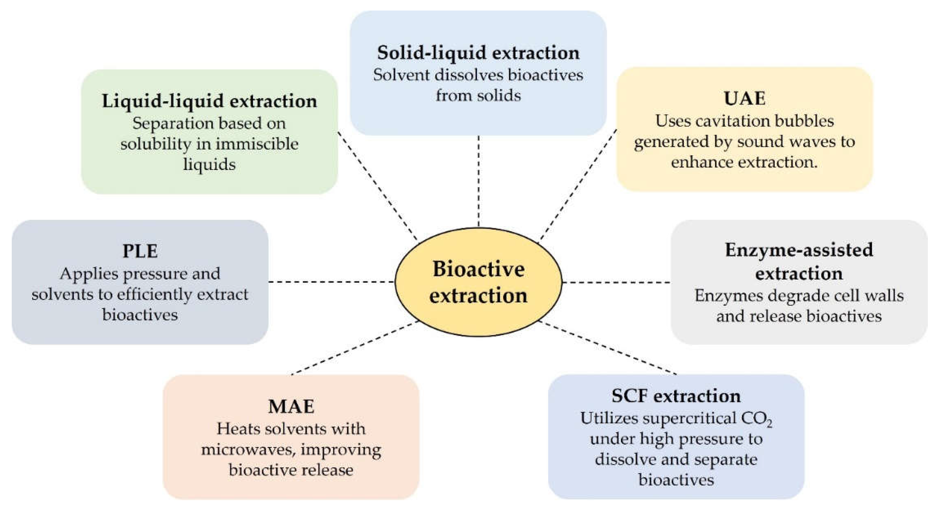

Table 1.
Encapsulation techniques, their definitions, uses, advantages, and disadvantages [121].
Table 1.
Encapsulation techniques, their definitions, uses, advantages, and disadvantages [121].
| Encapsulation technique | Definition | Uses | Advantages | Disadvantages |
|---|---|---|---|---|
| Spray-drying | A method where active ingredients are mixed with a wall material, atomized in a hot chamber, and dried into powder. | Used for shelf-life enhancement and encapsulation of various active compounds. | Low cost, easy scalability, and improved product stability. | Limited number of wall materials can be used. |
| Freeze-drying | Freezing active materials to form ice, followed by sublimation in vacuum to create porous, powdered products. | Encapsulation of temperature-sensitive materials like aromas and volatile oils. | Simple process, preserves sensitive compounds effectively. | Time-consuming and high energy costs. |
| Extrusion | Polymer solution containing active material is extruded through a nozzle into a gel solution. | Used for encapsulating both hydrophilic and hydrophobic compounds. | Simple, laboratory-friendly, produces high shelf-life capsules. | Difficult and expensive to scale up. |
| Emulsification | Involves creating emulsions of two immiscible liquids (water and oil) stabilized by emulsifiers. | Encapsulation of oil-soluble compounds like dietary fats and sterols. | Provides both liquid and powder encapsulation options. | Requires specific emulsifiers for stabilization. |
| Coacervation | Separation of phases leading to the formation of encapsulated materials within polymeric walls. | High-efficiency encapsulation with controlled release properties. | High encapsulation efficiency and control over material release. | Capsules are often unstable and production cost is high. |
| Molecular inclusion | Based on hydrogen bonding and electrostatic interactions between polar molecules. | Encapsulation of polar molecules, commonly using cyclodextrins. | Compatible with a wide range of polar compounds. | Limited use outside of specific polar interactions. |
| Ionic gelation | Encapsulation using microbeads in biopolymer gels, formed by methods like spraying or extrusion. | Commonly used for suspending active materials in polymer solutions. | Simple and adaptable to various active materials. | Limited by the biopolymer’s properties and stability in different environments. |
Disclaimer/Publisher’s Note: The statements, opinions and data contained in all publications are solely those of the individual author(s) and contributor(s) and not of MDPI and/or the editor(s). MDPI and/or the editor(s) disclaim responsibility for any injury to people or property resulting from any ideas, methods, instructions or products referred to in the content. |
© 2024 by the authors. Licensee MDPI, Basel, Switzerland. This article is an open access article distributed under the terms and conditions of the Creative Commons Attribution (CC BY) license (http://creativecommons.org/licenses/by/4.0/).
Copyright: This open access article is published under a Creative Commons CC BY 4.0 license, which permit the free download, distribution, and reuse, provided that the author and preprint are cited in any reuse.


