Preprint
Article

This version is not peer-reviewed.

Valproic Acid and Lamotrigine Differentially Modulate the Telomere Length in Epilepsy Patients

A peer-reviewed article of this preprint also exists.

Submitted:

13 December 2024

Posted:

16 December 2024

You are already at the latest version

Abstract

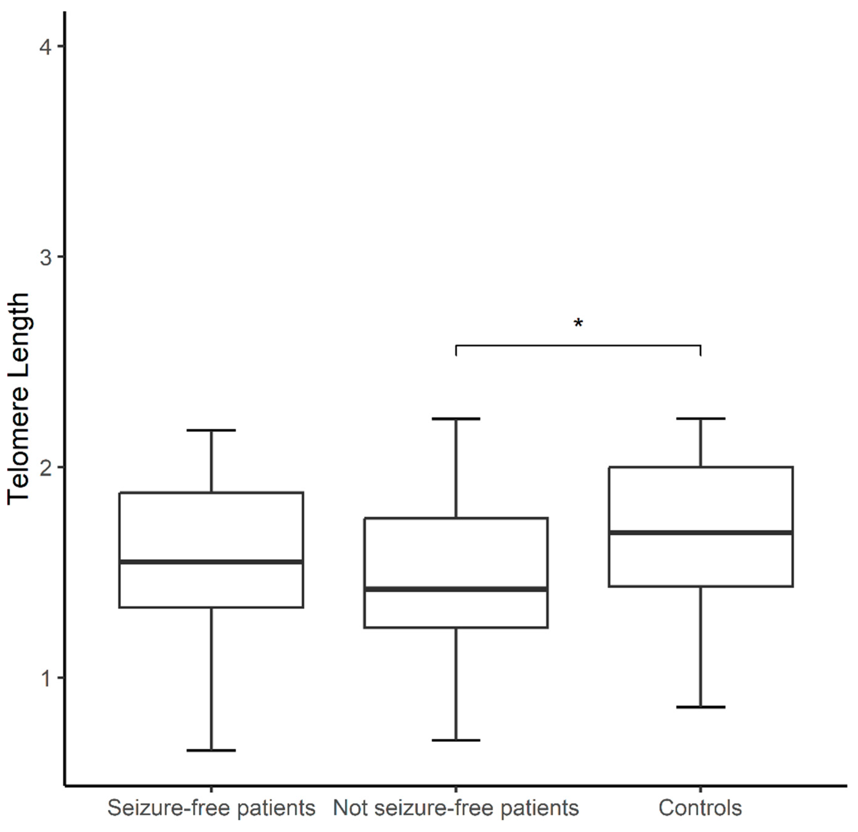
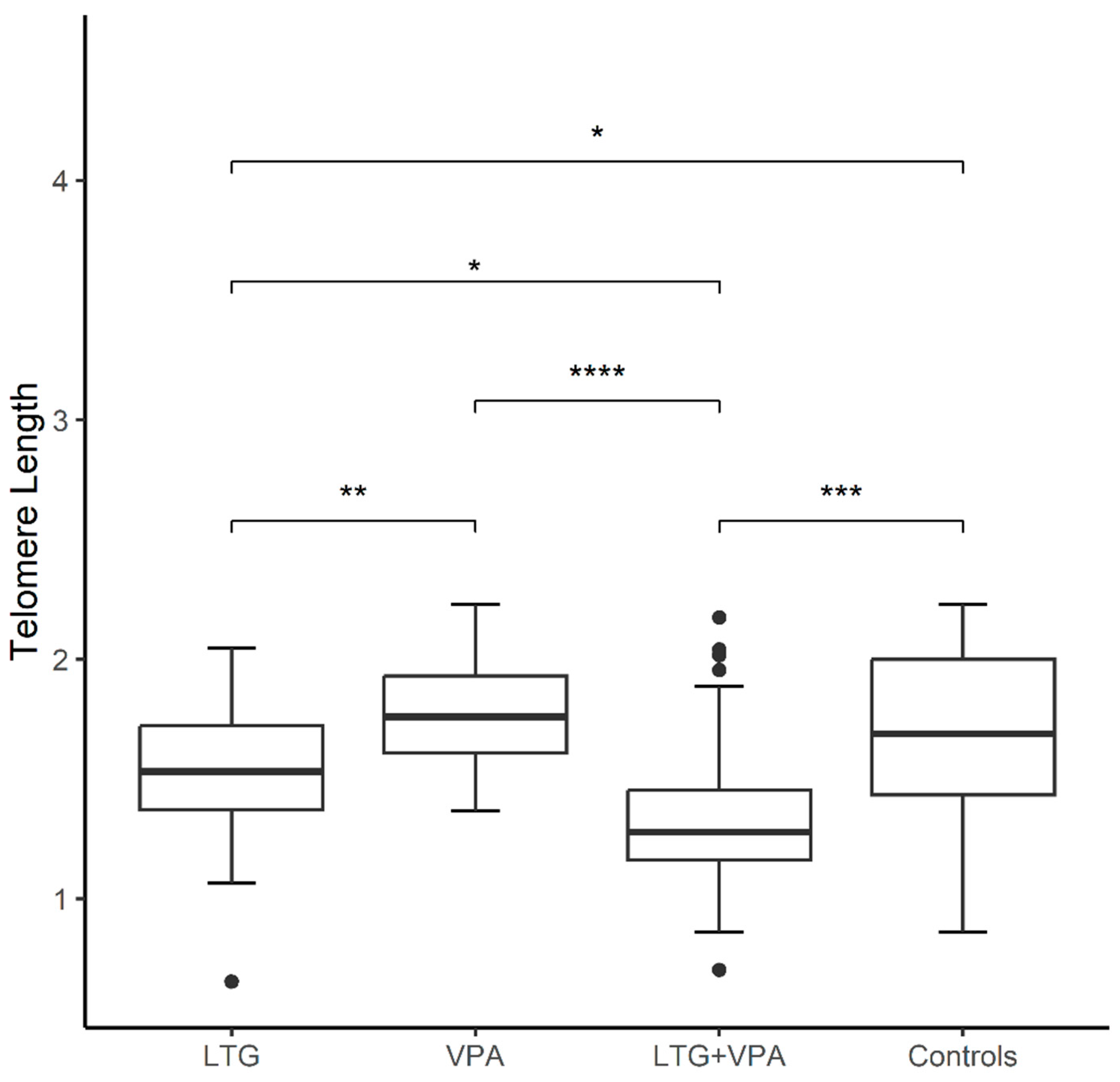
Background/Objectives: Antiseizure drugs (ASDs) are the primary therapy for epilepsy and the choice varies according to the seizure type. Epilepsy patients experience chronic mitochondrial oxidative stress and increased levels of pro-inflammatory mediators, recognizable hallmarks of biological aging, however few studies have explored aging markers in epilepsy. Herein, we addressed for the first time the impact of ASDs on molecular aging by measuring telomere length (TL) and mtDNA copy number (mtDNA-CN). Methods: Using QPCR, in epilepsy patients compared to matched healthy controls (CT), and its association with plasma levels of ASDs and other clinical variables. The sample comprised 64 epilepsy patients and 64 CT. Patients were grouped on monotherapy with lamotrigine (LTG) or valproic acid (VPA), and those treated with a combination therapy (LTG+VPA). Multivariable logistic regression was applied to analyze obtained data. Results: mtDNA-CN was similar between patients and controls, and none of the comparisons were significant for this marker. TL was shorter in not seizure-free patients than CT (1.50±0.35 vs. 1.68±0.34, p<0.05), regardless of the ASD therapy. These patients exhibited the highest proportion of adverse drug reactions. TL was longer in patients on VPA monotherapy, followed by patients on LTG monotherapy and by patients on LTG+VPA combined scheme (1.77±0.24; 1.50±0.32; 1.36±0.37 respectively, p<0.05), suggesting that ASD treatment differentially modulates TL. Conclusions: Our findings suggest that clinicians could consider TL measurements to decide the best ASD treatment option (VPA and/or LTG) to help predict ASD response in epilepsy patients.

Keywords: 
;  ;  ;  ;  

1. Introduction

Epilepsy is one of the most common neurological diseases worldwide, affecting around 50 million people of all ages around the world. The prevalence of epilepsy varies between countries and nearly 80% of people with epilepsy live in low- and middle-income countries [1]. For the vast majority of people with epilepsy, the initial therapy consists of pharmacological treatment where there is a wide variety of antiseizure drugs (ASDs) [2], and the choice of them varies according to the different types of seizures and epileptic syndromes [3]. Epilepsy is the leading cause of outpatient consultation at the Manuel Velasco Suárez National Institute of Neurology and Neurosurgery (INNNMVS), a third level hospital in Mexico City, where valproic acid (VPA) and lamotrigine (LTG) are two of the most widely used ASDs [4].
Telomeres are repetitive noncoding DNA sequences that protect the ends of chromosomes and provide genome stability. Telomere length (TL) decreases with each cell division due to incomplete replication of linear chromosomes [5,6] and has been suggested as a "mitotic clock" indicating cell age [7]. There are reports supporting a relationship between some medicines used in the management of cardiovascular diseases, diabetes and menopause and their positive protective effects against TL shrinking [8]. Similarly, some psychotropic medications may modulate TL; for instance, clinical evidence suggests that lithium may attenuate telomere shortening in patients with bipolar disorder (BD) [9,10,11,12]. Length of leukocyte TL has also been associated with response to some antidepressant drugs [13,14]. Antipsychotics could prolong or retain TL in peripheral blood mononuclear cells [15] and might influence telomerase activity [16]. In this regard, some studies have found no effect of VPA, LTG, carbamazepine or any combination of them on TL in BD patients [10,17]; while VPA, in a rat model for autism, induced an increased gene expression of telomerase in vivo and in vitro [18]. There is only one observational study that has shown telomere shortening in drug-resistant epilepsy patients [19].
Evidence indicates the existence of a strong link between TL shortening and metabolic and mitochondrial compromise, central mechanisms to cell functional decline during aging [20,21,22]. Telomere shortening may affect mitochondria activity through nuclear signaling to mitochondria [21], inducing alterations in the electron transport chain, which will result in an increased generation of reactive oxygen species (ROS), reduced ATP levels, and more DNA damage induced. In turn, the oxidative stress will cause greater telomere shrinkage [23]. Indeed, a Mendelian randomization (MR) analysis found a positive causal effect of TL on mtDNA-CN, suggesting a complex interrelationship between these two markers in the aging process [22].
Mitochondrial dysfunction has been implicated in the pathophysiology of neurological and psychiatric disorders and, particularly in epilepsy, it has been identified as one potential cause of epileptic seizures [23]. On the other hand, a relation between psychotropic medication and mtDNA copy number (mtDNA-CN), a measure of the number of mitochondrial genomes per cell, has also been postulated. It is known that antipsychotics and mood stabilizers (including VPA and LTG) may affect the function of mitochondria in BD [12,24,25,26]. Antipsychotics may have an effect in reducing mtDNA-CN in patients with major depression disorder [27] or with psychosis [28]. In contrast, VPA concentration has been positively correlated with peripheral mtDNA-CN and better cognitive performance in BD patients, distinguishing responders vs. non-responders to that ASD [29].
TL and mtDNA-CN are markers of biological aging that have been studied in several neurological disorders [19,28,29,30,31,32,33], reporting differences when compared with healthy controls. The accumulation of adverse effects of some conventional ASDs over the time, as well as pro-inflammatory and oxidative stress conditions produced by epileptic seizures, all together may contribute to the biological aging in patients with epilepsy [19,34]. To the best of our knowledge, no report has examined the effect of ASDs treatment on TL and mtDNA-CN in patients with epilepsy. Herein, we investigated whether ASDs modulate biological aging by comparing TL and CN-mtDNA in patients with epilepsy to that of age- and sex-matched healthy controls.

2. Materials and Methods

2.1. Study Participants

Sixty-four unrelated patients with epilepsy (18–72 years old; 33 females) were consecutively recruited from the Epilepsy Clinic at the INNNMVS. Sixty-four clinically healthy controls were matched by age and sex. Controls enrolled in the study were college students or unrelated companions of patients, with no family history of epilepsy or any neurological disease, and who were not taking any ASD. All participants were Mexican Mestizos (MM), with at least the two previous generations born in Mexico. This study was carried out in accordance with the latest version of the Declaration of Helsinki, and the study design was reviewed and approved by the local Research and Ethics Committees (registration numbers: INNNMVS_38/19 and UAM-X #34605034). Written informed consent was obtained from all participants after the nature of the procedures had been fully explained.

2.2. Clinical Data of Patients

Diagnosis of epilepsy was established for each patient by an expert neurologist in this study based on international criteria [35,36]. All patients were under pharmacological treatment categorized in two groups including monotherapy (LTG or VPA) and combined therapy (LTG+VPA), as follows: with LTG (n=18), with VPA (n=19), and 27 patients with combined therapy. Patients were taking the mentioned ASD treatment at least six months before the study. Data regarding dose and adjusted plasma concentrations of ASD, number of epileptic crises per year, and type of seizure (focal vs. generalized) were extracted from their clinical records. In compliance with the operational definition of seizure freedom of the International League Against Epilepsy (ILAE), patients were considered ‘seizure-free’ following an intervention after a period without seizures has elapsed equal to three times the longest pre-intervention inter-seizure interval over the previous year [37]. Adverse drug reactions (ADR) to ASDs were personally interrogated with a questionnaire ad hoc. ADR were classified into general, gastrointestinal, cutaneous, neurological and psychiatric reactions for all patients and patients categorized by ASD (Table 2).

2.3. Relative Quantification of Telomere Length and mtDNA Copy Number

Peripheral blood samples (12 mL) were collected from all subjects and genomic DNA was isolated by standard procedures. The relative quantification of the leukocyte TL [38] and mtDNA-CN [39] were assessed by real-time quantitative PCR (QPCR) as previously described. For both quantifications, a standard curve of serial dilutions of a commercial DNA from CEPH individual 1347–02 was included in each run (Thermofisher, Écublens, Switzerland). A relative measure of TL was calculated as a telomere hexanucleotide repeat / single copy gene (T/S) ratio, and as a mtDNA / nuclear DNA (ND3/TH) ratio. All PCRs were performed using KAPA SYBR Fast ABI Prism qPCR Master Mix on a QuantStudio™ 5 Real-Time PCR System (Applied Biosystems, Waltham, MA USA). All DNA samples were run in four replicates on separate plates for each sequence of interest, but in the same well positions. The inter-assay and intra-assay coefficients of variation were calculated and accepted when <10%.

2.4. Statistical Analyses

Statistical analyses were performed using R version 4.4.2 (R Core team, Vienna, Austria) [40,41]. The normality of the data was estimated with Kolmogorov-Smirnov and Shapiro-Wilk tests and then, mean values were compared between groups using student´s t test, U Mann-Whitney test, or Kruskal-Wallis test, accordingly. Correlation analyses were done using simple linear correlation approaches. Subsequently, for the patient´s data a multivariate regression analysis was used. Covariates included in the analyzes were age and sex for all participants; while the seizure freedom, dose and plasma concentrations (PC) of each ASD only for patients. A p-value< 0.05 was considered statistically significant in all analyses.

3. Results

The socio-demographic characteristics of the 128 participants (64 patients with epilepsy and 64 controls) are presented in Table 1. Both groups of study included 31 males and 33 females of comparable ages.
All patients were being treated with ASDs, and 34.4% of them were seizure-free. According to the ASD treatment received 28%, 53% and 26% were seizure-free patients on LTG, VPA or combined therapy, respectively. The seizure types were 66.13% focal onset, and 33.87% generalized onset. The most frequent ADRs were neurological and psychiatric (memory failure and aggression/irritability, respectively). Then, when patients were grouped by ASD, the most frequent ADRs by group were: headache in LTG monotherapy; humor changes in VPA monotherapy; and nervousness or distress in the group receiving combined therapy (Table 2). The adjusted plasma ASD concentrations were 1.5±1.5, 4.4±1.3, and 3.7±1.8 / 2.7±1.6 µg/mL/dose/Kg for LTG, VPA and combined therapy (LTG+VPA), respectively (Table 1). Seven patients on LTG monotherapy were found to have subtherapeutic PC (<3 µg mL-1) and eleven were in therapeutic levels (3–15 µg mL-1) [42]. All patients on VPA monotherapy had levels that fell within the therapeutic range (8.9–115 µg mL-1) [42,43]. Regarding the group receiving combined therapy, most of the patients were found on therapeutic levels for LTG (21/26, 81%), one had subtherapeutic levels and five were in supratherapeutic levels. The therapeutic range for VPA was found in therapeutic levels for all but one patient who was in supratherapeutic levels (>115 µg mL-1).
Epilepsy patients presented shorter telomeres when compared to controls, however this difference was only significant between not seizure-free patients, regardless of the ASD treatment, and controls (1.50±0.35 vs. 1.68±0.34 p<0.01, Figure 1). Among controls, women exhibited shorter TL than men (1.09±0.22 vs. 1.04±0.22, p= 0.04), but this was not observed in patients. After comparing patients categorized by seizure type (focal vs. generalized, groups of similar age, p>0.05) to controls, the group of patients with focal seizures exhibited shorter TL compared to controls (1.48 ± 0.36 vs. 1.68 ± 0.34, p< 0.01); however no difference was observed between patients with focal and generalized seizures. mtDNA-CN was similar between patients and controls (0.41±0.11 vs. 0.42±0.10; p= 0.07), and none of the above comparisons (seizure type and seizure freedom) was significant for this marker (data not shown).
Both aging markers were compared between controls and patients categorized as per ASD treatment: LTG (18), VPA (19), LTG+VPA (27). Kruskal-Wallis test revealed TL differences among patients grouped by ASD treatment (p< 0.05), according to the following ranking: TL of patients on LTG+VPA < TL of patients on LTG < TL of patients on VPA (1.36±0.37 vs. 1.50±0.32 vs. 1.77±0.24, respectively); whereas controls exhibited a mean value of TL of 1.68±0.34. Using Mann-Whitney U test, differences were observed between TL of patients receiving a combined ASD therapy vs. controls (1.36±0.37 vs. 1.68±0.34, p< 0.001), and TL of patients on LTG vs. controls (1.50±0.32 vs. 1.68±0.34, p< 0.05) (Figure 2). In contrast, the mtDNA-CN of controls vs. patients categorized by ASD treatment did not show differences (Kruskal-Wallis test, 0.42±0.10 vs. 0.45±0.12, 0.40±0.09 and 0.39±0.10 for patients on VPA, LTG and LTG+VPA, respectively).
Correlation analysis between TL and chronological age (in years) in both groups of patients studied showed moderate negative correlation: R= -0.30, p= 0.02 in patients, and R= -0.39, p< 0.01 in controls. Then, the correlation of TL vs. mtDNA-CN was significant in patients with epilepsy (R= 0.27, p= 0.03) (Figure 3), and the analysis by ASD treatment revealed that this observation only persisted in the group receiving LTG+VPA therapy.
We performed multiple logistic regressions (MLR) adjusting for confounding factors in all patients (Figure 3), and the patients grouped by ASD treatment (Figure 4). TL was considered the independent variable; whereas ASD treatment, sex, age, seizure freedom, ASD dose, and adjusted plasma concentrations (ADJ PC), ADRs and mtDNA-CN were dependent variables. The p-values obtained with this analysis were only significant for TL vs. age (R= -0.30, p<0.05), and TL correlating with mtDNA-CN (R= 0.27, p< 0.05) (Figure 3).
We then performed MLR analysis on patients based on the ASD therapy that included doses and adjusted plasma concentrations. The analysis on LTG monotherapy showed strong negative correlations between TL and seizure freedom (R= -0.51, p< 0.05), and LTG-dose with seizure freedom (R= -0.48, p< 0.05) (Figure 4A). The output for this analysis in patients on VPA monotherapy revealed a strong positive correlation between mtDNA-CN and sex (women with a higher copy number) (R= 0.60, p< 0.01) and a significant negative relation between VPA-dose and TL (R= -0.46, p< 0.05) (Figure 4B). MLR analysis in the group of patients on LTG+VPA also showed a strong correlation between both studied aging markers (mtDNA-CN vs. TL, R= 0.55, p< 0.01), while adjusted plasma concentrations of VPA negatively correlated with mtDNA-CN (R= -0.40, p< 0.05) and with TL (R= -0.68, p< 0.01) (Figure 4C).
Table 2. Adverse drug reactions observed in patients with epilepsy treated with antiseizure drugs.
Adverse drug reactions
(ADRs)
Total patients (%)
(n=64)
Patients on LTG (%)
(n=18)
Patients on VPA (%)
(n=19)
Patients on LTG+VPA (%)
(n=27)
General
Nervousness or distress 38.9 25.0 5.9 69.2
Fatigue or tiredness 35.6 50.0 11.8 42.3
Drowsiness 35.6 18.8 41.2 42.3
Weight gain 39.0 31.3 47.1 38.5
Insomnia 32.2 43.8 11.8 38.5
Alopecia 25.4 18.8 11.8 38.5
Feeling groggy 22.0 12.5 5.9 38.5
Thick or swollen gums 20.7 31.3 11.8 20.0
Hyperactivity 13.6 6.3 5.9 23.1
Sexual dysfunction 10.2 12.5 5.9 11.5
Weight loss 8.6 12.5 5.9 8.0
Hirsutism 6.8 6.3 0 11.5
Gastrointestinal
Abdominal pain or gastritis 33.9 18.8 11.8 57.7
Constipation 23.7 12.5 17.6 34.6
Diarrhea 13.6 6.3 17.6 15.4
Nausea and/or vomiting 13.6 0 11.8 23.1
Cutaneous
Allergy (mild rash) 5.1 0 5.9 7.7
Allergy (moderate or severe rash or Steven Johnson) 0 0 0 0
Facial edema 0 0 0 0
Toxic epidermal necrolysis (Lyell syndrome) 0 0 0 0
Neurological
Memory failure 50.8 43.8 35.3 65.4
Difficulty concentrating 45.8 31.3 35.3 61.5
Headache 42.4 56.3 17.6 50.0
Shaking (tremors) 35.6 18.8 29.4 50.0
Dizziness or vertigo 27.1 12.5 17.7 42.3
Trouble speaking 25.4 25.0 17.6 30.8
Slow thinking 23.7 25.0 11.8 30.8
Confusion 18.6 18.8 29.4 11.5
Double or blurred vision or nystagmus 16.9 18.8 5.9 23.1
Difficulty walking (ataxia) 13.6 12.5 11.8 15.4
Instability 10.2 0 11.8 15.4
Paresthesia 8.5 6.3 0 15.4
Parkinsonism 8.5 6 11.8 7.7
Diplopia 0 0 0 0
Choreoathetosis 0 0 0 0
Psychiatric
Aggression or irritability 50.8 50 29.4 65.4
Depression or sadness 47.5 43.8 47.1 50.0
Humor changes 37.3 31.3 52.9 30.8
Hallucinations, agitation, delirium 13.6 6.3 5.9 23.1
Manic episode 6.8 0 5.9 11.5
Suicidal ideation 6.8 6.3 17.6 0

4. Discussion

Mortality rates are 2–3 times higher in epilepsy patients than in the general population. The average reduction in life expectancy in the patients is 10.91–11.84 years (i.e., symptomatic cases) compared to the general population [44]. Telomere shortening and mtDNA-CN are associated with human disease and a reduced lifespan. The observed reduction in life expectancy in these patients could be linked to the concept of accelerated cellular aging and being differentially modulated by ASDs. Some reports suggest potential aging-modulating properties for VPA and LTG: (i) increasing lifespan in experimental models [45,46], and (ii) VPA decelerating epigenetic aging associated in peripheral samples of BD patients; however further investigation is required [47].
This is the first study in which two aging markers are simultaneously evaluated in controls and patients with epilepsy under different ASD treatment, monotherapy (LTG or VPA) or in a combination scheme (LTG+VPA). Our results demonstrated shorter TL in epilepsy patients compared with controls, but far from associating this marker with an increased risk for the disease, we highlight the potential differential aging-modulating properties of these ASDs, which might be dose dependent. In fact, a research study using pooled statistics from genome-wide association studies (GWAS) and a Mendelian randomization approach found no evidence of a causal relationship between epilepsy and TL [48]. However, that study included data for seven subtypes of epilepsy in European children and adult patients, did not consider ASD therapy, and their results may not be appropriate to all ethnicities [48].
After categorizing patients by seizure freedom and type, the shorter TL was shown in those not seizure-free patients (i.e., “non-responders”, regardless of the ASD treatment) and in those with focal seizures. This agrees with a previous work where authors compared TL of patients and controls and observed significantly shorter TL in the group of drug-resistant epilepsy patients [19].
After comparing patients categorized by ASD therapy, significant differences of TL among them persisted, particularly for patients on LTG monotherapy and combination therapy, suggesting a different modulation of TL depending on the ASD studied. Of note, these two groups included a high percentage of not seizure-free patients (72% and 74%, respectively). In particular, the LTG monotherapy group showed a negative correlation between LTG dose and seizure freedom, and between TL and seizure freedom. This last finding is in accordance with that reported in animal studies showing that shortened TL increased seizure frequency [16,49].
Beyond the therapeutic effects as ASD, VPA and LTG exhibit potential effects as mood stabilizers. In this regard, there are previous reports of their use for BD and its relationship with TL and mtDNA-CN. Some of them suggest that LTG [50] and VPA [49] may protect against oxidative stress and possibly TL shortening, while others found no differences in TL between these ASDs-treated BD patients and controls [10]. However, there are no reports in epilepsy patients of the impact of LTG and VPA treatment on TL and/or mtDNA-CN. This work demonstrated that epilepsy patients treated with LTG or LTG+VPA therapy had shorter TL than patients on VPA monotherapy and controls. Also, MLR analysis showed a positive correlation between both studied aging markers only in the group of patients on LTG+VPA. A similar correlation has been previously documented mainly in old patients with cancer [51] and in patients with Parkinson’s disease [52]. The three groups of patients on different ASD treatment did not show differences in age or sex among them, ruling them out as variables associated with this correlation. The strong correlation observed might be a compensation for the insufficient cellular energy supply due to the mitochondrial dysfunction in epilepsy patients on polytherapy, whereby increasing mtDNA-CN occurs to keep normal mitochondrial functions [52].
Regarding the TL results observed in patients treated with VPA monotherapy, its neuroprotective properties could be involved [53]. VPA functions as an inhibitor of histone deacetylases (HDACi) activating the transcription from many promoters [54]; also, it has been reported that the neural progenitor cells of rat exposed to VPA in vitro exhibited increased telomerase expression [18,55]. Nonetheless, we also observed that the higher VPA dose on monotherapy, the shorter TL (Figure 4B), and plasma VPA concentrations negatively correlated with TL and mtDNA-CN in the combined scheme (Figure 4C). In contrast, VPA concentration has been positively correlated with mtDNA-CN, better cognitive performance and response to VPA in BD patients [56]. We did not evaluate cognition, but VPA concentrations were on therapeutic levels, and more than half of individuals were seizure-free patients in the VPA monotherapy group. TL is modulated by a plethora of intrinsic and extrinsic factors, including the effect of pharmacologically active substances. Indeed, a recent review proposes that integrating TL measurements into personalized medicine procedures could aid significantly individualize the treatment plan [8].
The synergistic scheme of LTG and VPA is an effective treatment of refractory epilepsy in children and adults and has been considered the best combination therapy in a retrospective study [57]. However, VPA can decrease LTG clearance by 54% in combination therapy [58]. In this sense, the patients with the combined therapy showed the highest percentage of ADRs (Table 2), and somehow their accumulation over the time, as well as risks factors early mentioned herein, jointly could accelerate the biological aging in patients with epilepsy. Another research study also found that female patients had higher oxidative stress levels than controls; and that this was more pronounced in patients on ASD polytherapy vs. those who were on monotherapy [59] Therefore, we cannot rule out that higher ASD doses or the combined ASD therapy may lead to shortening of telomeres in patients with epilepsy as was observed in the present study.
Little is known about ASDs interference with mitochondria, e.g., in vitro, the mtDNA-CN increased after VPA treatment in a dose-dependent manner [60,61]. Conversely, it has been documented that LTG has toxic effects on mitochondria [62]. On the one hand, ASDs are known to induce epigenetic modifications with unknown consequences; while VPA, LTG, and other ASDs are known to exert HDACi properties [63,64]. For instance, LTG monotherapy led to lower serum folate levels in patients with epilepsy, which in turn may affect the DNA methylation process [65]. Some differences in epigenetic age acceleration in BD patients taking combinations of mood stabilizers (including VPA) vs. those taking no medication/monotherapy have been reported, as well [47]. The comparison of TL between patients and controls, and the positive correlation of TL with mtDNA-CN exclusively found in patients on combined therapy (LTG+VPA), indicates a mechanism related to biological aging participating in the physiopathology of epilepsy where one of the potential implicated factors might be oxidative stress. In fact, the marked oxidative stress in epilepsy may accelerate the telomere erosion observed, which has been previously shown in epilepsy patients [19,66].
Contrary to TL, mtDNA-CN was similar between controls and patients, and among patients, regardless of type of crisis, or ASD treatment received. The MLR analysis only demonstrated that female patients on VPA monotherapy had higher mtDNA-CN than females on other ASD therapy. Therefore, VPA or VPA+LTG schemes differentially influenced this marker in female patients, so the function of mitochondrial response to this ASD therapy is unresolved.
The aging markers we have analyzed here are a measurement proxy of personal health outcomes, and are easily damaged by ROS, systemic inflammation and stress [67]. TL and mtDNA-CN may be also coregulated via stress by the hypothalamic–pituitary–adrenal axis activity, which in turn exacerbates seizure occurrence, thus playing an important role in the development of epilepsy [68]. All the above features are present in patients with epilepsy and might be modulated by ASD therapy, but unfortunately, we were not able to evaluate them; so further studies are warranted to address them.
Limitations of the present study were a limited sample size, lack of clinical data in patients regarding time of use of previous ASDs, duration of the illness, cotreatments and comorbidities, weight and other metabolic data, and lifestyle; the design was transversal, and a peripheral tissue was explored for aging markers. However, regarding this last point, a group of researchers published a correlation of TL in brain tissue with peripheral tissues (e.g., blood, saliva, buccal) in living human subjects with intractable epilepsy [69]; thus, posing the possibility that at least leukocyte TL might provide insight into brain TL in these patients with a neurological disorder. In addition, measurement of peripheral mtDNA-CN has been associated with gene expression in other tissues, suggesting that blood-derived mtDNA-CN can reflect metabolic health across multiple tissues [66]. Although this study evaluated a relatively small sample size, study design allowed the comparative analysis of ASD monotherapy vs. combined therapy on aging markers in epilepsy patients. Also, our results suggest that TL might predict the studied ASD treatment responsiveness in epilepsy patients.

5. Conclusions

Treatment with VPA in monotherapy or combined scheme differentially influenced only TL in our patients. These findings should be confirmed in future studies to clarify the mechanisms of ASDs on biological aging. This type of research will allow the identification of new mechanisms of action of ASDs, will reveal cellular alterations in epilepsy and will allow the development of new therapeutic strategies. In addition, further longitudinal studies are required to achieve full understanding on the role of ASD therapy (not only VPA and LTG) on different aging markers in patients with epilepsy.

Author Contributions

“Conceptualization, N.M.-J. and A.O-V.; methodology, S.S.-B.; software, S.S.-B.; validation, S.S.-B. and A.O-V.; formal analysis, S.S.-B.; investigation, N.M.-J. and M.L.-L.; resources, N.M.-J. and M.L.-L.; data curation, S.S.-B.; writing—original draft preparation, N.M.-J.; writing—review and editing, S.S.-B., A.O-V., M.L.-L., and N.M.-J.; visualization, X.X.; supervision, N.M.-J.; project administration, A.O-V.; funding acquisition, N.M.-J., and M.L.-L. All authors have read and agreed to the published version of the manuscript.” .

Funding

This research was partially funded by Universidad Autónoma Metropolitana Unidad Xochimilco, grant number no. 34605034, awarded to M.L.-L.

Institutional Review Board Statement

The study was conducted in accordance with the Declaration of Helsinki and approved by the Institutional Review Boards (Research and Ethics Committees) of the Instituto Nacional de Neurología y Neurocirugía, Manuel Velasco Suárez (INNN_38/19, date of approval 19 August 2019) and Universidad Autónoma Metropolitana Unidad Xochimilco (UAMX_#34605034, date of approval 10 October 2017).

Informed Consent Statement

Written informed consent has been obtained from all subjects involved in the study to publish this paper.

Data Availability Statement

The data presented in this study are available upon request from the corresponding author. The data are not publicly available due to ethical issues.

Acknowledgments

The authors thank Iris E. Martinez-Juarez, M.D., for her collaboration in the recruitment of patients with epilepsy. The authors would also like to thank the patients for their participation in this study.

Conflicts of Interest

The authors declare no conflicts of interest. The funders had no role in the design of the study; in the collection, analyses, or interpretation of data; in the writing of the manuscript; or in the decision to publish the results.

References

  1. Organization WH. Epilepsy: a public health imperative: summary. 2019 [cited 2024 Nov 19]; Available from: https://iris.who.int/handle/10665/325440.
  2. Málaga I, Sánchez-Carpintero R, Roldán S, Ramos-Lizana J, García-Peñas JJ. [New anti-epileptic drugs in Paediatrics]. An Pediatr. 2019 Dec;91(6):415.e1-415.e10. [CrossRef]
  3. Hakami T. Neuropharmacology of Antiseizure Drugs. Neuropsychopharmacol Rep. 2021 Sep;41(3):336–51. [CrossRef]
  4. Martínez-Juárez IE, López-Zapata R, Gómez-Arias B, Bravo-Armenta E, Romero-Ocampo L, Estévez-Cruz Z, et al. [Refractory epilepsy: use of the new definition and related risk factors. A study in the Mexican population of a third-level centre]. Rev Neurol. 2012 Feb 1;54(3):159–66.
  5. Barrett JH, Iles MM, Dunning AM, Pooley KA. Telomere length and common disease: study design and analytical challenges. Hum Genet. 2015 Jul;134(7):679–89. [CrossRef]
  6. Eitan E, Hutchison ER, Mattson MP. Telomere shortening in neurological disorders: an abundance of unanswered questions. Trends Neurosci. 2014 May;37(5):256–63. [CrossRef]
  7. Ridout KK, Ridout SJ, Price LH, Sen S, Tyrka AR. Depression and telomere length: A meta-analysis. J Affect Disord. 2016 Feb;191:237–47. [CrossRef]
  8. Apetroaei MM, Fragkiadaki P, Velescu B Ștefan, Baliou S, Renieri E, Dinu-Pirvu CE, et al. Pharmacotherapeutic Considerations on Telomere Biology: The Positive Effect of Pharmacologically Active Substances on Telomere Length. Int J Mol Sci. 2024 Jul 13;25(14):7694. [CrossRef]
  9. Martinsson L, Wei Y, Xu D, Melas PA, Mathé AA, Schalling M, et al. Long-term lithium treatment in bipolar disorder is associated with longer leukocyte telomeres. Transl Psychiatry. 2013 May 21;3(5):e261. [CrossRef]
  10. Powell TR, Dima D, Frangou S, Breen G. Telomere Length and Bipolar Disorder. Neuropsychopharmacol Off Publ Am Coll Neuropsychopharmacol. 2018 Jan;43(2):445–53. [CrossRef]
  11. Squassina A, Pisanu C, Congiu D, Caria P, Frau D, Niola P, et al. Leukocyte telomere length positively correlates with duration of lithium treatment in bipolar disorder patients. Eur Neuropsychopharmacol J Eur Coll Neuropsychopharmacol. 2016 Jul;26(7):1241–7. [CrossRef]
  12. Courtes AC, Jha R, Topolski N, Soares JC, Barichello T, Fries GR. Exploring accelerated aging as a target of bipolar disorder treatment: A systematic review. J Psychiatr Res. 2024 Oct 23;180:291–300. [CrossRef]
  13. Hough CM, Bersani FS, Mellon SH, Epel ES, Reus VI, Lindqvist D, et al. Leukocyte telomere length predicts SSRI response in major depressive disorder: A preliminary report. Mol Neuropsychiatry. 2016 Jul;2(2):88–96. [CrossRef]
  14. Rasgon N, Lin KW, Lin J, Epel E, Blackburn E. Telomere length as a predictor of response to Pioglitazone in patients with unremitted depression: a preliminary study. Transl Psychiatry. 2016 Jan 5;6(1):e709. [CrossRef]
  15. Polho GB, Cardillo GM, Kerr DS, Chile T, Gattaz WF, Forlenza OV, et al. Antipsychotics preserve telomere length in peripheral blood mononuclear cells after acute oxidative stress injury. Neural Regen Res. 2022 May;17(5):1156–60. [CrossRef]
  16. Bersani FS, Lindqvist D, Mellon SH, Penninx BWJH, Verhoeven JE, Révész D, et al. Telomerase activation as a possible mechanism of action for psychopharmacological interventions. Drug Discov Today. 2015 Nov;20(11):1305–9. [CrossRef]
  17. Elvsåshagen T, Vera E, Bøen E, Bratlie J, Andreassen OA, Josefsen D, et al. The load of short telomeres is increased and associated with lifetime number of depressive episodes in bipolar II disorder. J Affect Disord. 2011 Dec;135(1–3):43–50. [CrossRef]
  18. Kim KC, Choi CS, Gonzales ELT, Mabunga DFN, Lee SH, Jeon SJ, et al. Valproic Acid Induces Telomerase Reverse Transcriptase Expression during Cortical Development. Exp Neurobiol. 2017 Oct;26(5):252–65.
  19. Miranda DM, Rosa DV, Costa BS, Nicolau NF, Magno L a. V, de Paula JJ, et al. Telomere shortening in patients with drug-resistant epilepsy. Epilepsy Res. 2020 Oct;166:106427. [CrossRef]
  20. Zhu Y, Liu X, Ding X, Wang F, Geng X. Telomere and its role in the aging pathways: telomere shortening, cell senescence and mitochondria dysfunction. Biogerontology. 2019 Feb;20(1):1–16. [CrossRef]
  21. Sahin E, Colla S, Liesa M, Moslehi J, Müller FL, Guo M, et al. Telomere dysfunction induces metabolic and mitochondrial compromise. Nature. 2011 Feb 17;470(7334):359–65. [CrossRef]
  22. Zole E, Ranka R. Mitochondria, its DNA and telomeres in ageing and human population. Biogerontology. 2018 Jul;19(3–4):189–208. [CrossRef]
  23. Zsurka G, Kunz WS. Mitochondrial dysfunction and seizures: the neuronal energy crisis. Lancet Neurol. 2015 Sep 1;14(9):956–66. [CrossRef]
  24. Bachmann RF, Wang Y, Yuan P, Zhou R, Li X, Alesci S, et al. Common effects of lithium and valproate on mitochondrial functions: Protection against methamphetamine-induced mitochondrial damage. Int J Neuropsychopharmacol. 2009;12(6):805–22. [CrossRef]
  25. Scaini G, Rezin GT, Carvalho AF, Streck EL, Berk M, Quevedo J. Mitochondrial dysfunction in bipolar disorder: Evidence, pathophysiology and translational implications. Neurosci Biobehav Rev. 2016 Sep 1;68:694–713. [CrossRef]
  26. Kageyama Y, Deguchi Y, Kasahara T, Tani M, Kuroda K, Inoue K, et al. Intra-individual state-dependent comparison of plasma mitochondrial DNA copy number and IL-6 levels in patients with bipolar disorder. J Affect Disord. 2022 Feb 15;299:644–51. [CrossRef]
  27. Chang CC, Jou SH, Lin TT, Lai TJ, Liu CS. Mitochondria DNA Change and Oxidative Damage in Clinically Stable Patients with Major Depressive Disorder. PLoS ONE. 2015 May 6;10(5):e0125855. [CrossRef]
  28. Kumar P, Efstathopoulos P, Millischer V, Olsson E, Wei YB, Brüstle O, et al. Mitochondrial DNA copy number is associated with psychosis severity and anti-psychotic treatment. Sci Rep. 2018 Aug 24;8(1):12743. [CrossRef]
  29. Fernandez-Egea E, Bernardo M, Heaphy CM, Griffith JK, Parellada E, Esmatjes E, et al. Telomere length and pulse pressure in newly diagnosed, antipsychotic-naive patients with nonaffective psychosis. Schizophr Bull. 2009 Mar;35(2):437–42. [CrossRef]
  30. Shivakumar V, Rajasekaran A, Subbanna M, Kalmady SV, Venugopal D, Agrawal R, et al. Leukocyte mitochondrial DNA copy number in schizophrenia. Asian J Psychiatry. 2020 Oct;53:102193. [CrossRef]
  31. Lee EH, Han MH, Ha J, Park HH, Koh SH, Choi SH, et al. Relationship between telomere shortening and age in Korean individuals with mild cognitive impairment and Alzheimer’s disease compared to that in healthy controls. Aging. 2020 Dec 15;13(2):2089–100. [CrossRef]
  32. Pyle A, Anugrha H, Kurzawa-Akanbi M, Yarnall A, Burn D, Hudson G. Reduced mitochondrial DNA copy number is a biomarker of Parkinson’s disease. Neurobiol Aging. 2016 Feb;38:216.e7-216.e10. [CrossRef]
  33. Petersen MH, Budtz-Jørgensen E, Sørensen SA, Nielsen JE, Hjermind LE, Vinther-Jensen T, et al. Reduction in mitochondrial DNA copy number in peripheral leukocytes after onset of Huntington’s disease. Mitochondrion. 2014 Jul;17:14–21. [CrossRef]
  34. Breuer LEM, Boon P, Bergmans JWM, Mess WH, Besseling RMH, de Louw A, et al. Cognitive deterioration in adult epilepsy: Does accelerated cognitive ageing exist? Neurosci Biobehav Rev. 2016 May;64:1–11. [CrossRef]
  35. Fisher RS, Acevedo C, Arzimanoglou A, Bogacz A, Cross JH, Elger CE, et al. ILAE official report: a practical clinical definition of epilepsy. Epilepsia. 2014 Apr;55(4):475–82. [CrossRef]
  36. Najafi MR, Malekian M, Akbari M, Najafi MA. Magnetic resonance imaging and electroencephalography findings in a sample of Iranian patients with epilepsy. J Res Med Sci Off J Isfahan Univ Med Sci. 2018;23:106. [CrossRef]
  37. Westover MB, Cormier J, Bianchi MT, Shafi M, Kilbride R, Cole AJ, et al. Revising the “Rule of Three” for inferring seizure freedom. Epilepsia. 2012 Feb;53(2):368–76. [CrossRef]
  38. Cawthon RM. Telomere measurement by quantitative PCR. Nucleic Acids Res. 2002 May 15;30(10):e47. [CrossRef]
  39. Refinetti P, Warren D, Morgenthaler S, Ekstrøm PO. Quantifying mitochondrial DNA copy number using robust regression to interpret real time PCR results. BMC Res Notes. 2017 Nov 13;10(1):593. [CrossRef]
  40. RStudio Team. RStudio: Integrated Development Environment for R [Internet]. Boston, MA: RStudio, PBC.; 2024. Available from: http://www.rstudio.com/.
  41. Olivoto T, Lúcio AD. metan: An R package for multi-environment trial analysis. Jarman S, editor. Methods Ecol Evol. 2020 Jun;11(6):783–9. [CrossRef]
  42. Rahman M, Awosika AO, Nguyen H. Valproic Acid. In: StatPearls [Internet]. Treasure Island (FL): StatPearls Publishing; 2024 [cited 2024 Nov 19]. Available from: http://www.ncbi.nlm.nih.gov/books/NBK559112/.
  43. Douglas-Hall P, Dzahini O, Gaughran F, Bile A, Taylor D. Variation in dose and plasma level of lamotrigine in patients discharged from a mental health trust. Ther Adv Psychopharmacol. 2017 Jan 1;7(1):17–24. [CrossRef]
  44. Dreier JW, Laursen TM, Tomson T, Plana-Ripoll O, Christensen J. Cause-specific mortality and life years lost in people with epilepsy: a Danish cohort study. Brain. 2023 Jan 4;146(1):124–34. [CrossRef]
  45. Avanesian A, Khodayari B, Felgner JS, Jafari M. Lamotrigine extends lifespan but compromises health span in Drosophila melanogaster. Biogerontology. 2010 Feb;11(1):45–52. [CrossRef]
  46. Evason K, Collins JJ, Huang C, Hughes S, Kornfeld K. Valproic acid extends Caenorhabditis elegans lifespan. Aging Cell. 2008 Jun;7(3):305–17. [CrossRef]
  47. Okazaki S, Numata S, Otsuka I, Horai T, Kinoshita M, Sora I, et al. Decelerated epigenetic aging associated with mood stabilizers in the blood of patients with bipolar disorder. Transl Psychiatry. 2020 May 4;10(1):129. [CrossRef]
  48. Luo X, Ruan Z, Liu L. Causal relationship between telomere length and epilepsy: A bidirectional Mendelian randomization study. Epilepsia Open. 2023 Dec;8(4):1432–9. [CrossRef]
  49. Shao L, Young LT, Wang JF. Chronic treatment with mood stabilizers lithium and valproate prevents excitotoxicity by inhibiting oxidative stress in rat cerebral cortical cells. Biol Psychiatry. 2005 Dec 1;58(11):879–84. [CrossRef]
  50. Eren I, Naziroğlu M, Demirdaş A. Protective effects of lamotrigine, aripiprazole and escitalopram on depression-induced oxidative stress in rat brain. Neurochem Res. 2007 Jul;32(7):1188–95. [CrossRef]
  51. Park WJ, Lee JH. Positive correlation between telomere length and mitochondrial copy number in breast cancers. Ann Transl Med. 2019 Apr;7(8):183. [CrossRef]
  52. Ortega-Vázquez A, Sánchez-Badajos S, Ramírez-García MÁ, Alvarez-Luquín D, López-López M, Adalid-Peralta LV, et al. Longitudinal Changes in Mitochondrial DNA Copy Number and Telomere Length in Patients with Parkinson’s Disease. Genes. 2023 Oct 7;14(10):1913. [CrossRef]
  53. Russo R, Kemp M, Bhatti UF, Pai M, Wakam G, Biesterveld B, et al. Life on the battlefield: Valproic acid for combat applications. J Trauma Acute Care Surg. 2020 Aug;89(2S Suppl 2):S69–76. [CrossRef]
  54. Romoli M, Mazzocchetti P, D’Alonzo R, Siliquini S, Rinaldi VE, Verrotti A, et al. Valproic Acid and Epilepsy: From Molecular Mechanisms to Clinical Evidences. Curr Neuropharmacol. 2019;17(10):926–46. [CrossRef]
  55. Salamati A, Majidinia M, Asemi Z, Sadeghpour A, Oskoii MA, Shanebandi D, et al. Modulation of telomerase expression and function by miRNAs: Anti-cancer potential. Life Sci. 2020 Oct 15;259:118387. [CrossRef]
  56. Chang CC, Chen PS, Lin JR, Chen YA, Liu CS, Lin TT, et al. Mitochondrial DNA Copy Number Is Associated With Treatment Response and Cognitive Function in Euthymic Bipolar Patients Receiving Valproate. Int J Neuropsychopharmacol. 2022 Aug 4;25(7):525–33. [CrossRef]
  57. Pipek LZ, Pipek HZ, Castro LHM. Seizure control in mono- and combination therapy in a cohort of patients with Idiopathic Generalized Epilepsy. Sci Rep. 2022 Jul 19;12(1):12350. [CrossRef]
  58. Costa B, Vale N. Understanding Lamotrigine’s Role in the CNS and Possible Future Evolution. Int J Mol Sci. 2023 Mar 23;24(7):6050. [CrossRef]
  59. Deepa D, Jayakumari N, Thomas SV. Oxidative stress is increased in women with epilepsy: Is it a potential mechanism of anti-epileptic drug-induced teratogenesis? Ann Indian Acad Neurol. 2012 Oct;15(4):281–6. [CrossRef]
  60. Jang EH, Lee JH, Kim SA. Acute Valproate Exposure Induces Mitochondrial Biogenesis and Autophagy with FOXO3a Modulation in SH-SY5Y Cells. Cells. 2021 Sep 23;10(10):2522. [CrossRef]
  61. Sitarz KS, Elliott HR, Karaman BS, Relton C, Chinnery PF, Horvath R. Valproic acid triggers increased mitochondrial biogenesis in POLG-deficient fibroblasts. Mol Genet Metab. 2014 May;112(1):57–63. [CrossRef]
  62. Finsterer J. Toxicity of Antiepileptic Drugs to Mitochondria. Handb Exp Pharmacol. 2017;240:473–88. [CrossRef]
  63. Kong FC, Ma CL, Zhong MK. Epigenetic Effects Mediated by Antiepileptic Drugs and their Potential Application. http://www.eurekaselect.com [Internet]. [cited 2024 Dec 3]; Available from: https://www.eurekaselect.com/article/101309. [CrossRef]
  64. Stettner M, Krämer G, Strauss A, Kvitkina T, Ohle S, Kieseier BC, et al. Long-term antiepileptic treatment with histone deacetylase inhibitors may reduce the risk of prostate cancer. Eur J Cancer Prev Off J Eur Cancer Prev Organ ECP. 2012 Jan;21(1):55–64. [CrossRef]
  65. Ni G, Qin J, Li H, Chen Z, Zhou Y, Fang Z, et al. Effects of antiepileptic drug monotherapy on one-carbon metabolism and DNA methylation in patients with epilepsy. PloS One. 2015;10(4):e0125656. [CrossRef]
  66. Yang N, Guan QW, Chen FH, Xia QX, Yin XX, Zhou HH, et al. Antioxidants Targeting Mitochondrial Oxidative Stress: Promising Neuroprotectants for Epilepsy. Oxid Med Cell Longev. 2020;2020:6687185. [CrossRef]
  67. Li Y, et al. Association of Mitochondrial DNA Copy Number and Telomere Length with Prevalent and Incident Cancer and Cancer Mortality in Women: A Prospective Swedish Population-Based Study, Cancers, vol. 13, no. 15, Art. no. 15, Jan. 2021. [CrossRef]
  68. Espinosa-Garcia C, Zeleke H, Rojas A. Impact of Stress on Epilepsy: Focus on Neuroinflammation—A Mini Review. Int J Mol Sci. 2021 Jan;22(8):4061. [CrossRef]
  69. Carver AJ, Hing B, Elser BA, Lussier SJ, Yamanashi T, Howard MA, et al. Correlation of telomere length in brain tissue with peripheral tissues in living human subjects. Front Mol Neurosci [Internet]. 2024 Mar 7 [cited 2024 Dec 3];17. Available from: https://www.frontiersin.org/journals/molecular-neuroscience/articles/10.3389/fnmol.2024.1303974/full. [CrossRef]
Figure 1. Boxplot showing comparison of telomere length (TL) of seizure-free patients (n=22), not seizure-free patients (n=42), and controls (n=64). Student’s t-test was used. Significance is denoted by asterisks as follows: * p < 0.05, ** p < 0.01, and *** p < 0.001.
Preprints 142883 g001
Figure 2. Boxplot showing comparison of telomere length (TL) among epilepsy patients grouped by antiseizure drug treatment (ASD) and controls (n=64), as follows: patients on lamotrigine (LTG, n=18); patients on valproic acid (VPA, n=19); patients on LTG+VPA (n=27). TL of each group of epilepsy patients compared to controls and TL differences among patients grouped by ASD treatment was performed with Kruskal-Wallis test. Significance is denoted by asterisks as follows: * p < 0.05, ** p < 0.01, and *** p < 0.001.
Preprints 142883 g002
Figure 3. Heatmap matrix depicting the multiple Pearson correlation between TL, mtDNA-CN, sex, age, and clinical variables with significant value in epilepsy patients included (n=64). Significance is denoted by asterisks as follows: * p < 0.05, ** p < 0.01, and *** p < 0.001
Preprints 142883 g003
Figure 4. Heatmap matrix depicting the multiple Pearson correlation between TL, mtDNA-CN, sex, age, and clinical variables with significant value in epilepsy patients included (n=64). Significance is denoted by asterisks as follows: * p < 0.05, ** p < 0.01, and *** p < 0.001.
Preprints 142883 g004
Table 1. Demographic and clinical characteristics of patients with epilepsy (n= 64) and healthy controls (n= 64).
Characteristics
Patients (n= 64) Controls (n = 64)
Total (n= 64) Male (n= 31) Female (n= 33) Total (n= 64) Male (n= 31) Female (n= 33)
Sex (%) 100 48 52 100 48 52
Age in years mean ± SD (range) 32.0 ± 13.10 (18-73) 32.48 ± 14.20 (18-72) 31.6 ± 12.3 (18-73) 32.0 ± 13.0 (18-73) 32.5 ± 14.30 (73-18) 31.4 ± 11.9 (19-72)
Not seizure-free patients* 42 19 23 NA NA NA
Seizure-free patients* 22 12 10 NA NA NA
LTG monotherapy group Patients (n= 18) Controls (n = 18)
Total (n= 18) Male (n= 7) Female (n= 11) Total (n=18) Male (n=7) Female (n=11)
Sex (%) 100 39 61 100 39 61
Age in years, mean ± SD (range) 34.2 ± 14.9 (18-72) 39.6 ± 27.7 (18-72) 30.8 ± 9.39 (19-49) 34.3 ± 15.3 (18-73) 40.0 ± 21.4 (18-73) 30.6 ± 9.17 (19-47)
Not seizure-free patients* 13 5 8 NA NA NA
Seizure-free patients* 5 2 3 NA NA NA
LTG dose in mg; mean ± SD 225.0 ± 113.0 236.0 ± 103.0 218.0 ± 123.0 NA NA NA
LTG PC, n = (Subtherapeutic / therapeutic /supratherapeutic) (7 / 11 / 0) (3 / 8 / 0) (4 / 3 / 0) NA NA NA
LTG PC μg mL−1; mean ± SD 4.6 ± 3.6 2.8 ± 1.6 5.74 ± 4.1 NA NA NA
LTG adjusted PC (µg mL-1 dose Kg-1) 1.5 ± 1.5 0.9 ± 0.3 2.0 ± 1.8 NA NA NA
VPA monotherapy group Patients (n= 19) Controls (n = 18)
Total (n= 19) Male (n=9) Female (n= 10) Total (n= 19) Male (n= 9) Female (n= 10)
Sex (%) 100 57 43 100 57 43
Age in years, mean ± SD (range) 32.8 ± 12.3 (20-67) 34.4 ± 14.8 (21-67) 31.0 ± 9.42 (20-49) 32.7 ± 12.0 (21-66) 34.2 ± 14.5 (21-66) 31.0 ± 9.19 (21-49)
Not seizure-free patients* (n= 9) 9 4 5 NA NA NA
Seizure-free patients* (n = 10) 10 5 5 NA NA NA
VPA dose in mg; mean ± SD⤉ 932.0 ± 437.0 1100.0 ± 477.0 844.0 ± 397.0 NA NA NA
VPA PC, n = (Subtherapeutic / therapeutic / supratherapeutic) (0 / 17 / 0) (0 / 9 / 0) (0 / 8 / 0) NA NA NA
VPA PC, μg mL−1; mean ± SD 68.1 ± 20.6 60.3 ± 16.7 76.9 ± 22.0 NA NA NA
VPA adjusted PC (µg mL-1 dose Kg-1) 4.4 ± 1.3 4.0 ± 1.2 5.0 ± 1.2 NA NA NA
LTG+VPA combined therapy group Patients (n= 27) Controls (n= 27)
Total (n= 27) Male (n= 14) Female (n=13) Total (n= 27) Male (n=14) Female (n= 13)
Sex (%) 100 52 48 100 52 48
Age in years, mean ± SD (range) 30.1 ± 12.6 (18-73) 27.6 ± 7.64 (19-48) 32.8 ± 16.4 (18-73) 29.9 ± 12.3 (18-72) 27.6 ± 7.44 (18-47) 32.4 ± 15.9 (19-72)
Not seizure-free patients* 20 9 11 NA NA NA
Seizure-free patients* 7 5 2 NA NA NA
LTG dose in mg; mean ± SD 189.0 ± 84.7 211.0 ± 92.4 165.0 ± 71.8 NA NA NA
LTG PC, n = (Subtherapeutic / therapeutic / supratherapeutic) (1 / 21 / 5) (1 / 10 / 3) (0 / 11 / 2) NA NA NA
LTG PC, μg mL−1; mean ± SD 9.8 ± 4.3 9.8 ± 4.6 9.7 ± 4.0 NA NA NA
LTG adjusted PC (µg mL-1 dose Kg-1) 3.7 ± 1.8 3.2 ± 0.8 4.1 ± 2.4 NA NA NA
VPA dose in mg; mean ± SD 1039.0 ± 496.0 1189.0 ± 476.0 950 ± 122.0 NA NA NA
VPA PC, n = (Subtherapeutic / therapeutic / supratherapeutic) § (0 / 15 / 1) (0 / 6 / 0) (0 / 9 / 1) NA NA NA
VPA PC, μg mL−1; mean ± SD § 74.8 ± 25.5 76.8 ± 15.9 80.0 ± 33.9 NA NA NA
VPA adjusted PC (µg mL-1 dose Kg-1) § 2.7 ± 1.6 1.1 ± 1.5 2.1 ± 2.0 NA NA NA
*Seizure-free patients: according to the operational definition of International League Against Epilepsy (ILAE): once patients have gone without a seizure for at least 3 times the duration of their longest pre-intervention inter-seizure interval in the preceding 12 months (www.ilae.org). PC: plasma concentration; Subtherapeutic PC of LTG (< 3 µg mL-1); Therapeutic PC of LTG (3-15 µg mL-1); Supratherapeutic PC of LTG (> 15 µg mL-1); Subtherapeutic PC of VPA (< 8.9 µg mL-1); Therapeutic PC of VPA ( 8.9-115 µg mL-1); Supratherapeutic PC of VPA (> 115 µg mL-1); ⤉ data from 17 patients; § Data from 16 patients; NA: Not applicable.
Disclaimer/Publisher’s Note: The statements, opinions and data contained in all publications are solely those of the individual author(s) and contributor(s) and not of MDPI and/or the editor(s). MDPI and/or the editor(s) disclaim responsibility for any injury to people or property resulting from any ideas, methods, instructions or products referred to in the content.
Copyright: This open access article is published under a Creative Commons CC BY 4.0 license, which permit the free download, distribution, and reuse, provided that the author and preprint are cited in any reuse.
Prerpints.org logo

Preprints.org is a free preprint server supported by MDPI in Basel, Switzerland.

Subscribe

Disclaimer

Terms of Use

Privacy Policy

Privacy Settings

© 2026 MDPI (Basel, Switzerland) unless otherwise stated