Preprint
Article

This version is not peer-reviewed.

Cardioprotective Activity of the Ethanolic Leaf Extract of Amaranthus viridis on Verapamil-Induced Heart Failure in Zebrafish (Danio rerio)

Submitted:

05 February 2025

Posted:

06 February 2025

You are already at the latest version

Abstract

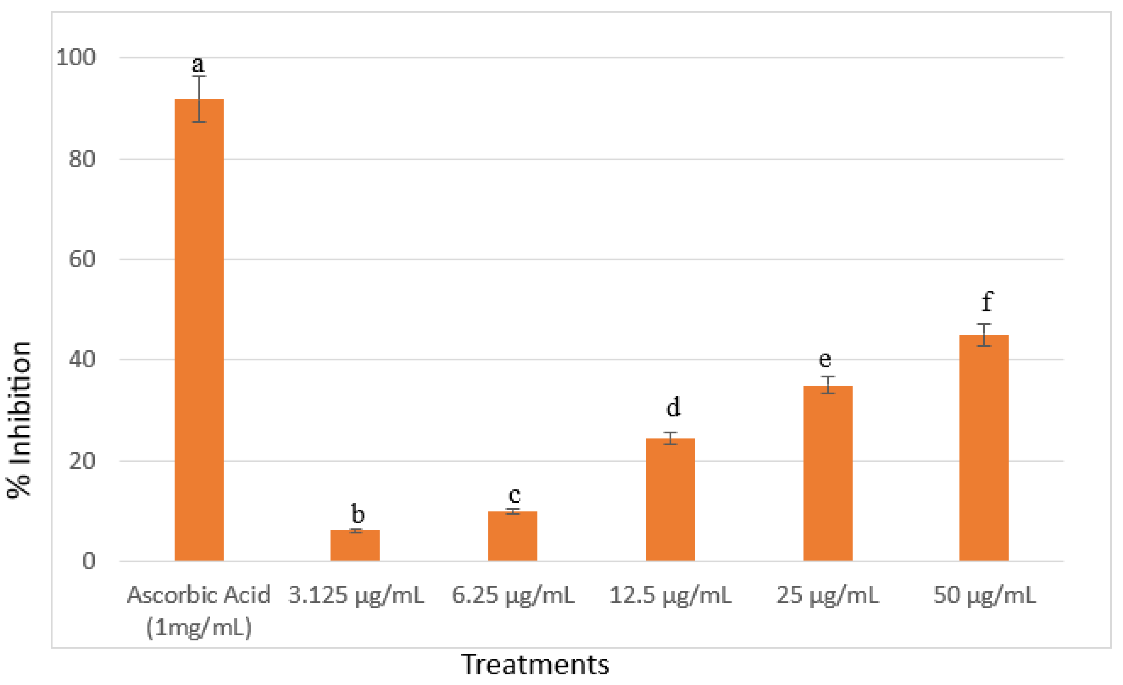
Cardiovascular disease (CVD), a major global health concern, is characterized by cardiac complications that can lead to death. The commonly used treatments for this condition are synthetic drugs, but these often come with risky side effects. A potential alternative is the use of traditional medicinal plants, such as Amaranthus viridis, which is rich in bioactive compounds. This study aimed to determine the non-toxic concentration of A. viridis ethanolic extract, investigate its cardioprotective effects on zebrafish (Danio rerio) heart rate and cardiac phenotype, qualitatively assess the presence of phytochemicals, and assess its free radical scavenging activity using 2,2-diphenyl-1-picrylhydrazyl (DPPH). Zebrafish larvae at 72 hours post-fertilization (hpf) were used to evaluate mortality and optimize dosing. Physio-morphological screening was conducted by pre-treating zebrafish larvae with the extract 4 hours prior to administering a heart failure inducer, verapamil. The maximum non-toxic concentration was found to be 25 µg/mL, as all zebrafish survived after 24 hours. Mortality began at 50 µg/mL, and concentrations from 100 µg/mL to 400 µg/mL resulted in 100% mortality. All tested concentrations of A. viridis leaf extract showed cardioprotective activity in the physio-morphological analysis. Phytochemical analysis detected the presence of alkaloids, flavonoids, and saponins. Furthermore, A. viridis exhibited free radical scavenging activity from all tested concentrations. Based on the results, A. viridis exhibited cardioprotective effects against verapamil-induced cardiotoxicity, as evidenced by the recovery of heart rate and cardiac phenotype in the zebrafish model.

Keywords: 
;  ;  ;  ;  

Introduction

Cardiovascular disease (CVD) is a significant global health concern, consistently impacting human life and overall well-being. It is the leading cause of mortality worldwide, affecting millions of individuals of all ages and demographics. According to the World Health Organization (WHO), CVD is responsible for a staggering 9.6 million deaths annually in men and 8.9 million in women, collectively constituting nearly one-third of all deaths attributed to diseases. In the Philippines, the primary causes of death in 2021 were coronary heart disease (CHD), other cerebrovascular diseases or stroke, and various forms of cancer. Notably, there were over half a million reported cases of CHD in 2021, making up approximately 19% of the total deaths in the country (Bray et al., 2021; Corpuz, 2023; Daniels et al., 2014; Nguyen & Cheng-Lai, 2013; Timmis et al., 2020; Timmis et al., 2022).
Conventional treatments for heart failure involve a combination of medications such as angiotensin converting enzyme inhibitors (ACEIs), beta-blockers, diuretics, corticosteroid receptor antagonists, sodium-glucose cotransporter two inhibitors, and others. These drugs aim to reduce the heart's workload, enhance efficiency, alleviate symptoms, and target specific aspects of heart failure. Despite the extensive pharmaceutical options, heart failure remains a significant medical challenge, with persistently high rates of illness and mortality suggesting that current treatments, while valuable, often fall short in completely managing the condition (Chia et al., 2016; Krum et al., 2011). Amid this ongoing challenge, interest in herbal medicines as complementary treatments has grown. Amaranthus viridis, commonly referred to as "Spinach Tagalog" or "Kolitis," is a locally available vegetable in the Philippines and widely distributed in Asia. Abundant in bioactive compounds like flavonoids, alkaloids, saponins, and polyphenols, this plant has been linked to various pharmacological activities. Significantly, the bioactive compounds present in Amaranthus viridis demonstrates noteworthy properties including antioxidants, anti-inflammatory, antithrombotic, antiarrhytmic, and anti-hypertensive effect which collectively contribute to cardiovascular health (Bachheti et al., 2022; Pirdhankar et al., 2023; Saravanan & Ponmurugan, 2012; Shah et al., 2019).
The zebrafish (Danio rerio) is a versatile model organism in cardiovascular research. Native to Southeast Asia, this small tropical freshwater fish is particularly valuable for studying cardiac function and disease. A key feature is the transparency of zebrafish embryos, enabling non-invasive, real-time visualization of internal organs, including the developing heart. This transparency facilitates detailed observations of cardiac morphogenesis and function during various developmental stages, as early as 24 hours post-fertilization (Irion & Nüsslein-Volhard, 2022; Shen & Zuo, 2020; Tavares & Lopes, 2013).
Furthermore, zebrafish exhibit external development, and their embryos undergo rapid and synchronous growth. This characteristic makes them particularly suitable for studies involving developmental biology and the effects of various compounds on embryonic development, including potential therapeutic agents. The external development also allows for easy manipulation and observation, making zebrafish a cost-effective and time-efficient model for cardiovascular studies (Eimon and Ashkenazi, 2010). However, to date, no studies have evaluated Amaranthus viridis using zebrafish larvae model. Hence, this study was conducted to evaluate the cardioprotective activity of Amaranthus viridis in verapamil-induced heart failure in zebrafish model. The findings from this research are expected to offer valuable insights that can contribute to further exploration and understanding within the field of cardiovascular health.
This study aims to determine the maximum non-toxic concentration of the ethanolic leaf extract of Amaranthus viridis in 72-hours post-fertilization (hpf) zebrafish larvae. Additionally, it seeks to identify the lowest effective concentration and evaluate the cardioprotective activity of A. viridis against verapamil-induced heart failure through physio-morphological assessments. The study also qualitatively examines the presence of phytochemicals, including saponins, flavonoids, phenols, and alkaloids, which may contribute to cardioprotective effects. Furthermore, the free radical scavenging activity of the leaf extract is assessed using the 2,2-diphenyl-1-picrylhydrazyl (DPPH) assay.

Materials and Methods

Collection and Authentication of Plant Material

Approximately 2 kilograms of fresh young leaves from the actively growing tops of Amaranthus viridis were collected at Barangay Tabunan, Cebu City as depicted in Figure 1. Subsequently, a whole plant sample was transported to the Department of Biology at the University of San Carlos for botanical authentication by Mr. Val Salares. A voucher specimen was submitted to the University of San Carlos Herbarium.

Plant Extraction

The leaves were washed with distilled water to remove any external impurities and will be air-dried in the shade at 21-27°C for 2 weeks to remove excess moisture. The leaves were grounded to a fine powder using an Osterizer blender for 5 minutes to achieve 68-200 µm particle size. After grinding, straining was done to obtain a finely powdered particle using a 100 µm sieve strain (Alsaud and Farid, 2020). The finely powdered leaves were stored in Erlenmeyer flasks and covered with wooden cork to prevent contamination from moisture and other pollutants. Twenty grams of dried powder were macerated in 200 mL of ethanol solvent at room temperature for 72 hours. The extract was filtered using a Whatman filter paper number 1. Subsequently, the filtrate was transferred to a Heidolph rotary evaporator for evaporation, resulting in the collection of a solid concentrate (Gahlot et al., 2018). Following this, the extract was placed in amber vials and stored at a temperature of 4°C.
Preparations were made for various concentrations of 25, 50, 100, 200, and 400 µg/mL. The final crude extracts were resuspended to dimethyl sulfoxide (DMSO) and subjected to sonication to ensure homogeneity. They were stored in appropriately labeled amber vials for safety and future use (Lee and Yang, 2021).

Preparation of Egg Water

Throughout the experiment, the medium for zebrafish embryos was consisted of egg water. The egg water was prepared by thoroughly mixing 40 g of sea salt (Mars Fishcare Europe, UK) with 1 L of distilled water. Following this, 1.5 mL from the stock salts were dissolved in another 1 L of distilled water to attain a final concentration of 60 μg/ml (Westerfield, 2007).

Ethics Declaration

All protocols were subjected to approval by the Institutional Animal Care and Use Committee (IACUC) at the University of San Carlos. Furthermore, future experiments related to zebrafish were taken place at the Medical Biology Laboratory of the Department of Biology at the University of the Visayas.

Establishment of Zebrafish Aquarium and Husbandry

An aquarium with a divider in the center, measuring 30 cm in length, 20 cm in height, and 20 cm in width, was constructed using acrylic glass containing 5 liters of dechlorinated water. The tank was equipped with Precision aquarium air pumps (PR-7500 and PR-2500, China), Xinyou aquarium sponge filters (China), thermometers, and Zetlight control units (China). The water temperature in the tank was maintained between 24-29 ℃. Light conditions, following a 10-hour dark and 14-hour light cycle (10:14), were regulated using a timer. Tap water with a pH range of 6.5 to 8 was utilized for water quality. Monitoring and daily documentation of pH, total hardness, nitrate, nitrite, free chloride, and carbonate were carried out using 6in1 Aquarium freshwater test strips (China). To ensure water quality, a 25% water change was performed daily using AquaCare aquarium water conditioner (Seachem, Philippines), ParaGuard Parasite control (Seachem, Philippines), and anti-chlorine (SeaQuest). Weekly testing of ammonia levels was also conducted with Ammonia aquarium freshwater test kits (China). Zebrafish was housed at a density of 4-10 adult fish per liter and fed twice daily with dry feeds dispensed by a food timer (Warmtone, China), supplemented by one feeding of decapsulated brine shrimp. Routine cleaning, including the cleaning of sponge filters, was conducted weekly on the fish tanks. (Avdesh et al., 2012; Evidente et al., 2021).

Procurement of Zebrafish

A total of 420 at 72hpf zebrafish larvae were used for the entire study. To facilitate the breeding, 2 males and 2 females adult wild type zebrafish (> 6 months old with 3-4 cm in length) were purchased from a local breeder since a 1:1 ratio of male and female will give the highest production of embryos around 200 - 300 fertilized eggs and lesser aggression in the tank (Ruhl et al., 2009). The zebrafish was further identified by Mr. Dave Valles of the University of San Carlos. The identification of male and female zebrafish will be based on specific characteristics, as illustrated in Figure 2. Upon acquisition, the differentiation between males and females were based on specific characteristics. Females were identified by their larger underbelly, while males were recognized for their slimmer and darker coloration compared to females. Subsequently, male and female zebrafish were separated and individually placed in containers.
For transportation, the zebrafish were carefully placed inside a container within a box and transported using an air-conditioned delivery car. Upon acquisition, a thorough inspection of the zebrafish was conducted to identify potential signs of sickness, with a focus on behaviors such as decreased movement, lethargy, low food intake, and anxiety. Daily records were diligently maintained to monitor their food intake. Additionally, the zebrafish's anxiety behavior was observed as they initially dive to the bottom of a novel environment, followed by monitoring their exploration of the upper portion after six minutes, as indicated by Kirsten et al. (2018). The zebrafish were maintained according to standard protocols outlined by Alestrom et al. (2019) and Avdesh et al. (2012), with confirmation from the Institutional Animal Care and Use Committee (IACUC). During transportation, the protocol involved placing two adult zebrafish per 500 mL and maintaining a 1:1 ratio of air to water in the vessel. Additionally, adult zebrafish underwend a 24-hour period of fasting before transportation to reduce excretion and fouling in the transportation vessel.

Breeding and Embryo Isolation

The process of zebrafish breeding and isolation were conducted according to the protocol of outlined by the Zebrafish Information Network (ZFIN). Before spawning, a tank with a central divider housed a 1:2 ratio of male and female adult zebrafish. Separate breeding boxes, each holding less than 1 L of water, was arranged in a separate container filled with fresh tap water. In the evening, the zebrafish were introduced into the tank, and the following morning, when spawning occurs, fertilized eggs were collected on the enclosure floor. Zebrafish typically lay eggs within the first two hours after lights are turned on.
Post-spawning, the upper compartment, along with the adult fish, were transferred to a new tank or returned using a fishnet. The water containing settled eggs were siphoned and passed through a 0.10 mm strainer. The collected fertilized eggs were washed 3 times with egg water to remove debris. Subsequently, all fertilized eggs were transferred into each 1000 mL beakers filled with 100 mL egg water. The beakers were maintained at in a room temperature, providing an environment for the embryos to develop over a period of 72 hours. (Avdesh et al., 2012; Mason, 2016). Selection of embryos for analysis were based on symmetrical appearance in the one-cell stage or the presence of well-defined dark masses within a chorion, indicating fertility. Only embryos without morphological defects were chosen for further analysis.

Preliminary Mortality Test of A. viridis Extracts Against Zebrafish Larvae

Seventy zebrafish larvae at 72 hours post-fertilization were carefully distributed into a 96-well plate, with each well accommodating one larva. The larvae were subjected to 100 µL A. viridis leaf extracts at 25, 50, 100, 200, and 400 µg/mL concentration as a soaking drug, along with a vehicle control containing the highest concentration of DMSO used in the experimental treatments and a control group treated with egg water without any leaf extract. Each treatment concentration was replicated 10 times to ensure statistical reliability. The zebrafish larvae were left in a room temperature and were inspected after 24 hours. The concentration exhibiting the least number of dead larvae were recorded and then discarded. Death was judged by the coagulation of larvae, the absence of the heartbeat, or the lack of movement observed for 20 seconds. The extract associated with the zero-mortality rate or did not lead to any observable side effect under stereomicroscope was selected as the best extract for further investigation into its cardioprotective activity (Li et al., 2021).

Treatment Protocol for Assessing Cardioprotective Activity

A total of 240 zebrafish larvae (72 hpf) were used across 3 trials with 10 replicates per treatment. Each larva was placed in a well of a 96-well plate. The first treatment group received 100 µL of A. viridis extract at concentrations of 3.125, 6.25, 12.5, 25, and 50 µg/mL. The second group (positive control) received 100 µL of 200 µM eplerenone, the third group (negative control) received 100 µL of 200 µM verapamil, and the fourth group was exposed to egg water. After 4 hours of treatment, all groups were exposed to 100 µL of 200 µM verapamil for 30 minutes to induce heart failure. (Kossack et al., 2017; Maciag et al., 2022).

Heart Rate Assessment

The heart rate of each zebrafish larva per treatment were assessed. The zebrafish larvae have easily observable hearts, which can be seen through a stereomicroscope. A 15-second video of each specimen were recorded using a camera attached to the stereomicroscope. These videos were played in slow motion to accurately count the number of heartbeats. The heart rate was calculated using the formula (Heideman et al., 2005; Hoage et al., 2012):
heart   rate = Number   of   heart   beats 15   sec onds x   4

Scoring on Cardiac Phenotypes

After the treatment, the zebrafish larvae were analyzed using a dissecting microscope. For imaging purposes, photographs were captured using a Nikon D750 camera. The acute heart failure (AHF) phenotypes were classified into 4 groups as shown in Figure 7. Category I end stage of AHF: heart with no contraction; Category II severe AHF: heart with contraction only in the atrium and no circulation; Category III mild AHF: heart with a distinct contraction in both cardiac chambers but morphological abnormalities, including edema, accumulated blood cells in front of the heart, stretched heart; Category IV normal heart. Larvae were tallied into the four AHF categories and the heart failure attenuation was determined by the percentage of each category (Haege et al., 2021; Hoyberghs et al., 2020; Maciag et al., 2022).

Disposal of Zebrafish Carcass

Disposal of the dead zebrafish followed the NIH guidelines (2013) by placing them into 1 part sodium hypochlorite and five parts water for 5 minutes. Then, the larvae were set into a tightly sealed bag and disposing of them in a yellow bin as clinical waste.

Qualitative Phytochemical Analysis

The qualitative phytochemicals analyses were involved in determining the primary group of bioactive compounds such as saponins, flavonoids, phenols, and alkaloids which are known to have cardioprotective properties. The most cardioprotective concentration were used for phytochemical analyses. All experiments were conducted in triplicate to ensure the reliability of results.

Statistical Analyses

The analysis began with descriptive statistics to summarize mortality rates at various concentrations. This involves calculating the mean mortality rate and standard deviation for each concentration of the verapamil and ethanolic leaf extract. Additionally, Kruskal-Wallis Test (Minitab 21.1.1) was applied to describe the phenotypic differences in zebrafish hearts, followed by pairwise comparison. To compare the means of cardiac function indicators between the control group and the group treated with the candidate concentration extract, one-way analysis of variance (ANOVA), followed by Dunnett’s t-test will be utilized. A p-value of <0.05 were considered statistically significant.

Results and Discussion

In vivo maximum non-toxic concentration of the ethanolic leaf extracts of A. viridis against 72hpf zebrafish A. viridis leaf extract exhibited mortality and toxicity at concentrations starting from 50 μg/mL. As shown in Figure 7 and Table 4, concentrations ranging from 100 μg/mL to 400 μg/mL, 100% mortality occurred with distinct morphological abnormalities such as hemorrhage, bent tails, pericardial edema, yolk sac edema, coagulation and a cloudy appearance. In contrast, all larvae exposed to 25 μg/mL remained alive, indicating this as the non-toxic concentration of A. viridis leaf extract.
Table 2. Percentage mortality of zebrafish treated with A. viridis leaf extract.
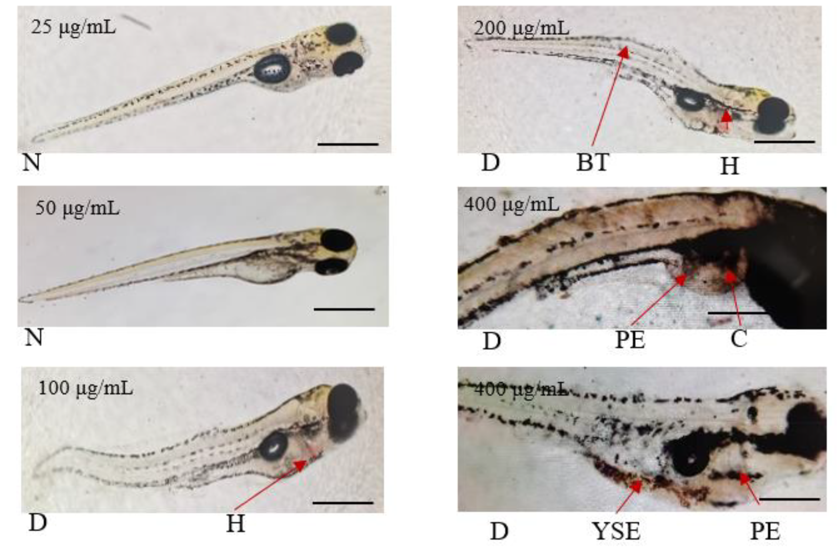
Concentrations
(μg/mL)
A. viridis (%)
0.5% DMSO 0
0 0
25 0
50 13.33%
100 100%
200 100%
400 100%
The results demonstrated a direct proportional relationship between the concentration of A. viridis extract and mortality rates among zebrafish larvae. Specifically, higher concentrations of the extract were associated with increased mortality, while lower concentrations resulted in fewer to zero mortality. This observation aligns with the findings of Zahir et al. (2021), who reported that higher concentrations of Aervasan guinolenta, Amaranthus viridis, and Cynodon dactylon reduced the survival rates of zebrafish embryos, particularly with prolonged exposure. Aervasan guinolenta and Amaranthus viridis, both belonging to the family Amaranthaceae, and Cynodon dactylon belonging to the family of Poaceae caused mortality in zebrafish embryos at various concentrations (100, 200, 300, 400, and 600 µg/mL). The study demonstrated that mortality rates increased with longer exposure durations. Prolonged exposure may result in the accumulation of bioactive compounds, which, at specific concentrations, can induce toxicity (Alafiatayo et al., 2019).
Similarly, Chen et al. (2018) observed toxicity in zebrafish embryos exposed to ethanol and aqueous extracts of Sutherlandia frutescens at concentrations of 20, 30, 50, 100, and 200 μg/mL. Chronic teratogenic effects, including pericardial edema, yolk sac swelling, and other developmental abnormalities, were documented.
Furthermore, these findings suggest that the compounds present in the leaf extract of A. viridis may interfere cellular processes within the heart of zebrafish, potentially inducing toxicity by affecting mechanisms such as cell proliferation, differentiation, and migration. For instance, the observed hemorrhages could be linked to damage to endothelial cells, which from the lining of blood vessels (Rajendran et al., 2013). Pericardial edema may arise from increased vascular permeability or impaired cardiac function (Wiegand et al., 2023). The bent tail phenotype observed suggests additional neurotoxic effects, as the development of the spinal cord and nervous system is closely linked to heart formation (Zhao et al., 2024). Coagulation abnormalities could result from direct damage to blood cells or interference with the coagulation cascade (Kretz et al., 2015). Yolk sac edema likely reflects impaired fluid balance and potential to the circulatory system (Sant et al., 2018).

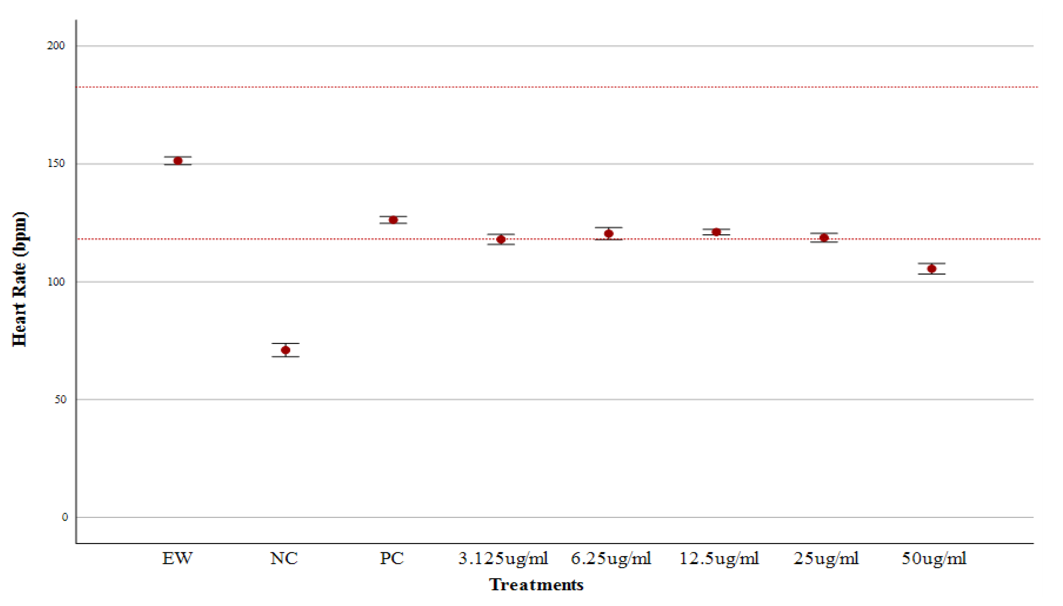
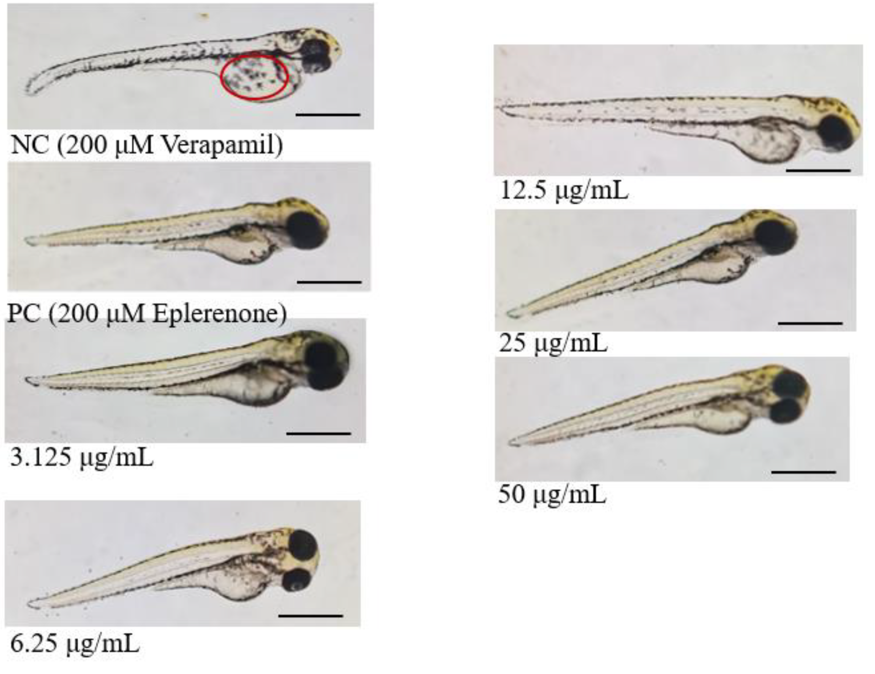
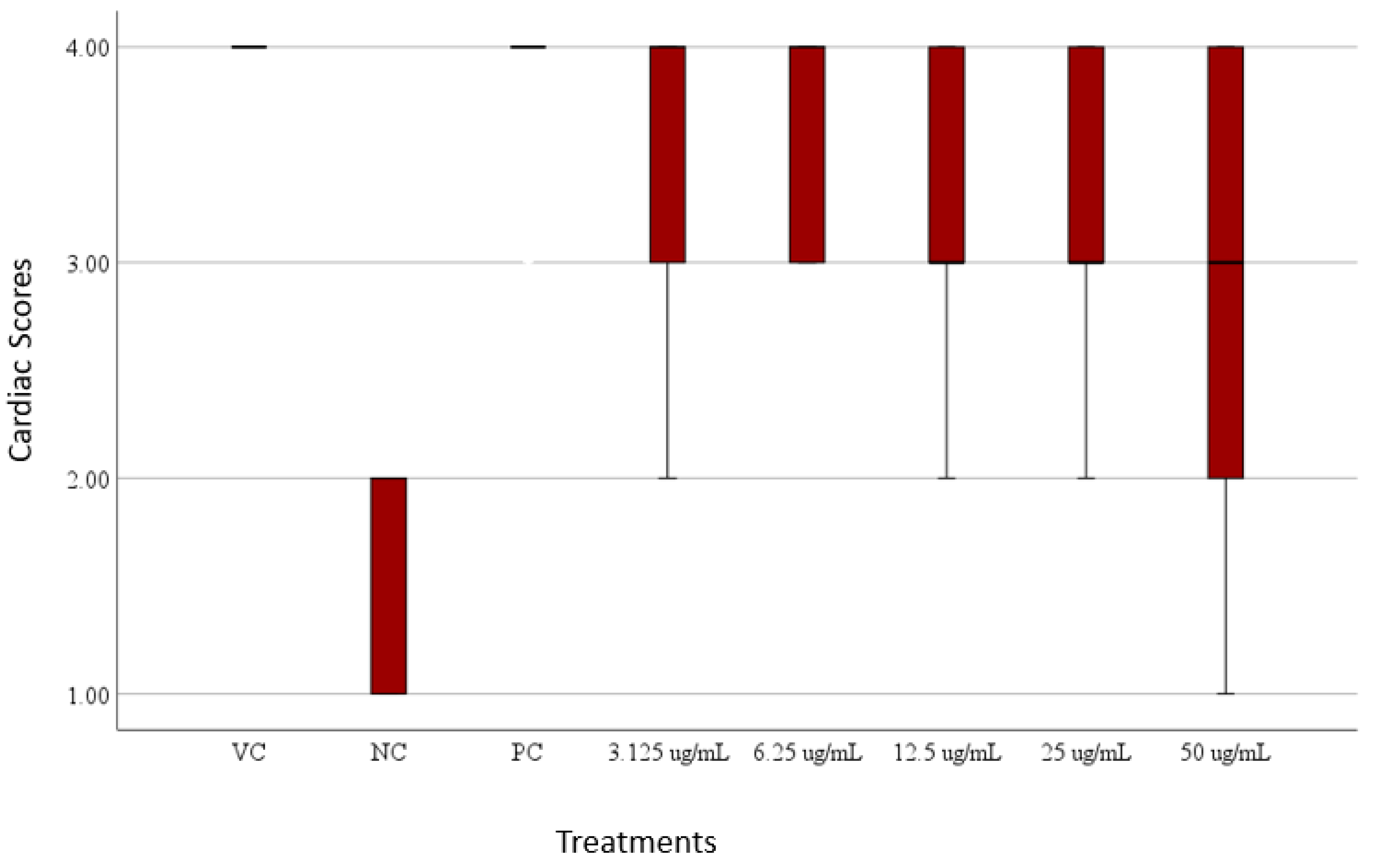
Heart Rate and Cardiac Phenotype Response of Zebrafish larvae Protected by Amaranthus viridis Leaf Ethanolic Extracts Against Verapamil-Induced Heart Failure

As shown in Figure 8, the heart rate of zebrafish larvae in egg water (control) was within the normal range of 120–180 bpm (De Luca et al., 2014), recorded at 151.57 ± 3.86 bpm. Verapamil treatment significantly reduced the heart rate to 71.60 ± 6.74 bpm, confirming heart failure (Li et al., 2024). However, eplerenone provided protection from heart damage, resulting in a moderate heart rate recovery to 126.37 ± 3.65 bpm. The cardioprotective effect of A. viridis leaf extract was evident across all tested concentrations, from 50 μg/mL to 3.125 μg/mL, with heart rates of 111.47 ± 6.85 bpm, 118.20 ± 5.37 bpm, 121.20 ± 2.75 bpm, 120.30 ± 6.09 bpm, and 117.60 ± 6.16 bpm, respectively. These values indicate an improvement in heart rate, suggesting cardioprotective properties at each of these concentrations. However, the heart rate at 50 μg/mL was not within the normal range, as mortality began at this concentration. The study initiated testing at 50 μg/mL to identify the lowest effective concentration, using it as a baseline for subsequent evaluations. The results suggest that concentrations below 50 μg/mL not only improved heart rate but also minimized mortality, highlighting the efficacy of lower doses of the extract.
Identifying lower effective doses is critical in drug discovery and development research as it ensures both efficacy and safety. Lower concentrations reduce the risk of adverse effects, minimize toxicity, and are often more sustainable for large-scale production. Additionally, determining the minimum effective dose allows for the optimization of therapeutic regimens, improving patient outcomes while reducing the likelihood of drug resistance or cumulative toxicity. This approach is particularly important when developing natural product-based therapies, where the balance between bioactivity and safety is essential for clinical applicability (Mohs & Greig, 2017).
For the cardiac phenotypes, the verapamil-induced zebrafish larvae (negative control) had lower scores between 1 and 2, indicating heart failure in this group (Li et al., 2024). In contrast, the eplerenone-treated larvae (positive control) scored higher, between 3 and 4, suggesting protection from verapamil-induced damage. As shown in Figure 10, which highlights the observed cardiac phenotypes in zebrafish across various treatments, Table 3, which summarizes the cardiac functions and phenotypes, and in Figure 11, which highlights the cardiac scores of zebrafish, A. viridis demonstrated promising results, consistently scoring in the higher range across all tested concentrations (3.125, 6.25, 12.5, 25, and 50 μg/mL). Some larvae showed lower scores in the extract treatments, indicating individual variations in tolerance (Meyer et al., 2013). The Kruskal-Wallis test and pairwise comparisons revealed a highly significant difference between the negative control and the treatments (p < 0.001). The positive control did not differ significantly from any treatments. These results suggest that the treatments provide cardiac protection for the larvae against verapamil-induced heart failure.
Table 3. Cardiac function and phenotypes of 72hpf zebrafish in different treatments.
Treatments Cardiac functions and phenotypes
Egg water Normal function and no visible abnormality
NC – Verapamil No contraction in the heart; Weak contraction in the atria; No circulation; Pericardial edema
PC – Eplerenone + Verapamil Some showed slow circulation, while most had no visible abnormalities
3.125 μg/mL A. viridis + Verapamil Some showed slow circulation, while most had no visible abnormalities
6.25 μg/mL A. viridis + Verapamil Some showed slow circulation, while most had no visible abnormalities
12.5 μg/mL A. viridis + Verapamil Some showed slow circulation, while most had no visible abnormalities
25 μg/mL A. viridis + Verapamil Some showed slow circulation, while most had no visible abnormalities
50 μg/mL A. viridis + Verapamil No contraction in the heart; Weak contraction in the atria; No circulation;
Verapamil is a calcium channel blocker used in humans to manage hypertension and certain heart conditions. In zebrafish larvae, verapamil induces heart damage by disrupting calcium ion influx into cardiac cells. Calcium is crucial for maintaining normal heart contractions and overall cardiac health, so this disruption impairs cardiac function and causes structural damage (Horng et al., 2024). The damage is further increased by oxidative stress, the downregulation of cardiomyocyte biomarkers, and the obstruction of protein synthesis (Chatterjee et al., 2010; Abbate, 2021). Prior studies indicated that the verapamil (200 μM)-induced larval zebrafish heart failure model is a useful tool for whole animal heart failure studies and for screening safe agents of heart failure. Zebrafish treated with 200uM verapamil exhibited obvious heart failure phenotypes and weakened cardiac function. These data indicated that the heart failure zebrafish model was successfully established and can be utilized as an alternative, novel and simple experimental animal model of heart failure (Li et al., 2022; Li et al., 2023; Narumanchi et al., 2021; Zhu et al., 2018).
Cardiac damage or abnormalities are directly linked to deviations in heart rate, as the heart's rhythm and rate are critical indicators of its functional integrity. An elevated heart rate, often indicative of stress or pathological conditions, can lead to increased myocardial workload and oxygen demand, potentially causing structural and functional damage over time. Conversely, bradycardia, or abnormally slow heart rate, can impair adequate blood flow, resulting in tissue hypoxia and systemic complications (Fatisson et al., 2016; Sessa et al., 2018). In zebrafish models, alterations in heart rate often reflect underlying cardiac phenotypes, such as edema, chamber malformations, or disrupted contractility, which are common indicators of cardiotoxicity or cardiac dysfunction (Wang et al., 2017).
Sedighi et al. (2019) demonstrated that different concentrations of Melissa officinalis L. (25, 50, and 100 mg/kg) significantly improved ischemia/reperfusion-induced arrhythmia and heart injury in an in vitro rat model. The treatment, administered for five days following reperfusion, effectively stabilized heart rate and mitigated cardiac damage. Similarly, Zhou et al. (2020) found that 100 μg/mL of timosaponin B-II extracted from Anemarrhena improved heart function in rats due to its anti-inflammatory properties. Additionally, Chen et al. (2021) reported that 5 μg/mL of Gardenia jasminoides extract reduced inflammation in a zebrafish embryo model. Likewise, Rafacho et al. (2017) demonstrated that a 0.02% concentration of Rosmarinus officinalis L. effectively mitigated cardiac damage in male Wistar rats. The study revealed that this concentration improved cardiac metabolism and significantly reduced oxidative stress. Furthermore, it enhanced diastolic function and decreased cardiac hypertrophy following myocardial infarction.
These studies highlight the potential of natural plant extracts and their active compounds to protect the heart by reducing inflammation, minimizing oxidative stress, and improving overall heart function. Their ability to prevent cardiac damage offers promising insights for developing effective treatments for heart-related conditions.
Based on the results of this study, cardiac analysis revealed significant improvements in both heart rate and cardiac phenotypes, in all tested concentrations of A. viridis leaf ethanolic extract.

Phytochemical Composition of A. viridis Crude Leaf Extracts

For the phytochemical screening, qualitative tests were conducted to detect the presence of alkaloids, flavonoids, phenols, and saponins in A. viridis. As shown in Table 4, the results revealed that A. viridis contains alkaloids, flavonoids, and saponins, while no phenols were detected.
Table 4. Qualitative phytochemical composition of A. viridis. (‘+’ indicates presence and ‘-’ indicates absence).
A. viridis
Alkaloids +
Flavonoids +
Phenols -
Saponins +
A. viridis was evaluated for its cardioprotective potential due to its bioactive compounds, which contribute to its medicinal properties. It is important to note that the phytochemical composition of A. viridis can vary due to environmental factors such as climate, seasonal changes, soil conditions, and geographical location (Kumar, 2017).
Phytochemical analysis is essential for identifying the specific bioactive compounds responsible for the observed cardioprotective effects. In this study, alkaloids, flavonoids, and saponins were detected in the A. viridis leaf ethanolic extracts, which align with findings from Fouad et al. (2024) who reported the presence of alkaloids, flavonoids, phenols, and saponins in A. viridis extracts and highlighted their role in various therapeutic activities. Furthermore, Kumari et al., (2018) confirmed the presence of various phytochemicals in A. viridis leaf extract across different solvents, including aqueous, methanol, chloroform, and hexane. These phytochemicals included flavonoids, alkaloids, phenolics, steroids, terpenoids, saponins, cardiac glycosides, and tannins.
The cardioprotective activity of A. viridis leaf extracts were notably linked to its alkaloid, flavonoid, and saponin content. Alkaloids are bioactive compounds known for their cardiovascular benefits, including the regulation of heart rate and promotion of vasodilation, which help maintain cardiac stability and reduce the risk of heart dysfunction. Flavonoids, on the other hand, are well-documented for their antioxidant properties, which play a critical role in neutralizing reactive oxygen species (ROS), preserving cardiac cell integrity, and reducing inflammation within the heart. Additionally, flavonoids enhance endothelial function, promoting vascular relaxation and lowering blood pressure (Ullah et al., 2020). This mechanism aligns with the findings of Kumar et al. (2013), who demonstrated that flavonoids from A. viridis significantly reduced oxidative stress and inflammation in rat models, thereby mitigating cardiotoxicity.
Saponins, another key component of A. viridis leaf extract, further support cardioprotection through their anti-inflammatory and antihypertensive properties. They modulate immune responses and suppress the production of pro-inflammatory cytokines, alleviating inflammation in cardiovascular tissues. Saponins also enhance vascular function by reducing blood vessel constriction, thereby aiding in blood pressure regulation. Saravanan et al. (2013) observed that saponins from A. viridis mitigated inflammation and improved cardiac function in isoproterenol (ISO)-induced heart failure in rats. The combined effects of alkaloids, flavonoids, and saponins underscore the potential of A. viridis as a natural cardioprotective agent.
The mechanisms underlying these effects involve several biochemical processes. Alkaloids play a vital role by promoting vasodilation and regulating heart rate, which contribute to cardiovascular stability. Flavonoids reduce oxidative stress by scavenging reactive oxygen species (ROS) and inhibiting inflammatory pathways, thereby maintaining cardiovascular health. Saponins further enhance this protective effect by reducing inflammation and improving vascular health (Li et al., 2020). Together, alkaloids, flavonoids, and saponins synergistically support heart function and protection.
Supporting these observations, Krishna et al. (2023) reported that alkaloids, flavonoids, and saponins from A. viridis alleviated verapamil-induced cardiotoxicity in rat models through their antioxidant and anti-inflammatory actions.

In Vitro Free Radical Scavenging Activity of A. viridis Ethanolic Leaf Extracts

The DPPH radical scavenging activity of A. viridis ethanolic leaf extracts at various concentrations is presented in Figure 11. The extract exhibited the highest scavenging activity at 50 μg/mL, achieving an inhibition rate of 45.06% ± 10.90%. A concentration-dependent decline in scavenging activity was observed at lower concentrations, with inhibition rates of 34.95% ± 4.16% at 25 μg/mL, 24.36% ± 2.69% at 12.5 μg/mL, 9.92% ± 4.31% at 6.25 μg/mL, and 6.04% ± 4.66% at 3.125 μg/mL, respectively.
This suggests that as the concentration of A. viridis decreases, the free radical scavenging activity also diminishes, indicating that higher concentrations of the extract possess greater free radical scavenging potential. This trend underscores the importance of bioactive compounds, such as alkaloids, flavonoids, and saponins, in neutralizing reactive oxygen species (ROS) and protecting cells from oxidative damage (Soni & Sosa, 2013). The observed reduction in antioxidant activity at lower concentrations highlights the importance of determining optimal dosages for therapeutic applications to maximize the efficacy of A. viridis in combating oxidative stress.
The results demonstrated significant free radical scavenging activity of A. viridis ethanolic leaf extract in all concentrations tested, which contributes to its cardioprotective properties. This free radical scavenging potential plays a crucial role in preventing oxidative stress-induced cardiac damage and supports the traditional use of A. viridis in treating cardiovascular diseases (Valaei et al., 2021).
The antioxidant property of A. viridis can be primarily attributed to its phytochemical composition such as alkaloids, flavonoids, and saponins. These compounds are known for their ability to chelate metal ions and inhibit lipid peroxidation, which are crucial mechanisms in preventing oxidative stress-induced cardiac damage. The presence of these bioactive compounds supports the extract's observed cardioprotective effects in verapamil-induced heart failure (Kumari et al., 2018).
Furthermore, the ability of A. viridis leaf extract to scavenge free radicals correlates with its protective effects against verapamil-induced heart failure in zebrafish. This correlation is supported by the study of Nowak et al., (2018), who demonstrated that natural antioxidants could effectively protect cardiac damage from oxidative stress.
The observed free radical scavenging activity supports the traditional use of A. viridis in managing various cardiovascular conditions. As highlighted by Lalhminghlui and Jagetia (2023), natural antioxidants from plant sources often provide additional benefits beyond their primary antioxidant activity, including anti-inflammatory and membrane-stabilizing properties, which enhance their overall therapeutic efficacy.
The crude preparation of the extract likely contributed to its strong free radical scavenging activity, with 50 μg/mL demonstrating the highest inhibition rate of 45.06% ± 10.90%. However, the presence of other bioactive compounds in the crude extract may also explain its observed toxicity to zebrafish larvae at higher concentrations. While 50 μg/mL emerged as the most effective concentration for antioxidant activity, the potential toxicity associated with the crude extract highlights the importance of isolating and identifying specific bioactive compounds to optimize therapeutic applications while minimizing adverse effects (Cordero-Maldonado et al., 2013; Mohamad-Shariff et al., 2020).

Summary, Conclusion, and Recommendation

Cardiovascular disease (CVD) remains one of the most pressing health challenges worldwide, requiring immediate and effective management. With the incidence of CVDs continuing to rise, there is an increasing need for alternative treatments to complement existing therapies. Natural products, which are abundant in bioactive compounds, offer promising potential in addressing this issue. These bioactive compounds, derived from plants and other natural sources, have shown potential for therapeutic use, making them a valuable resource in the search for new, effective treatments for cardiovascular conditions.
The 72-hpf zebrafish larvae were able to tolerate concentration of up to 25 μg/mL of A. viridis ethanolic leaf extract, indicating that their tolerance to the extract is dose-dependent. All tested concentrations exhibited cardioprotective activity, as shown through heart rate and cardiac phenotype analyses in zebrafish larvae exposed to verapamil-induced heart damage.
Additional improvements for future research are advised to enrich the pharmacological application of A. viridis on a verapamil-induced zebrafish research model. It is encouraged to isolate and identify the specific cardioprotective compounds of the whole plant extracts. Since only the end-stage (72 hpf) is applied in both survival and physiomorphological screening, future studies can increase the time frame and include observations during 96, 120, and 144 hpf. It is also recommended to incorporate blood flow in addition to the heart rate of the treated zebrafish larvae. Histopathology analysis of the heart s advised to provide a deeper understanding of tissue-level effects. Lastly, quantitative analysis on the levels of Reactive Oxygen Species (ROS) could also be performed to measure the oxidative stress and inflammatory pathways.

References

  1. Abbate, F., Maugeri, A., Laurà, R., Levanti, M., Navarra, M., Cirmi, S., Germanà, A. (2021). Zebrafish as a useful model to study oxidative stress-linked disorders: Focus on flavonoids. Antioxidants (Basel), 10(5), 668. [CrossRef]
  2. Abbate, F., Maugeri, A., Laurà, R., Levanti, M., Navarra, M., Cirmi, S., & Germanà, A. (2021). Zebrafish as a useful model to study oxidative stress-linked disorders: Focus on flavonoids. Antioxidants (Basel), 10(5), 668. [CrossRef]
  3. Abdel-alim, M. E., Serag, M. S., Moussa, H. R., Elgendy, M. A., Mohesien, M. T., & Salim, N. S. (2023). Phytochemical screening and antioxidant potential of Lotus corniculatus and Amaranthus viridis. Egyptian Journal of Botany, 63(2), 665–681.
  4. Ahmed, S. A., Hanif, S., & Iftkhar, T. (2013). Phytochemical profiling with antioxidant and antimicrobial screening of Amaranthus viridis L. leaf and seed extracts. Open Journal of Medical Microbiology.
  5. Alafiatayo, A. A., Lai, K.-S., Syahida, A., Mahmood, M., & Shaharuddin, N. A. (2019). Phytochemical evaluation, embryotoxicity, and teratogenic effects of Curcuma longa extract on zebrafish (Danio rerio). Evidence-Based Complementary and Alternative Medicine, 1–10. [CrossRef]
  6. Aronson, J. K. (2014). Meyler's side effects of drugs 15E: The international encyclopedia of adverse drug reactions and interactions. Newnes.
  7. Bachheti, R. K., Worku, L. A., Gonfa, Y. H., Zebeaman, M., Pandey, D. P., & Bachheti, A. (2022). Prevention and treatment of cardiovascular diseases with plant phytochemicals: A review. Evidence-Based Complementary and Alternative Medicine, 2022.
  8. Bagher, P., & Segal, S. S. (2011). Regulation of blood flow in the microcirculation: Role of conducted vasodilation. Acta Physiologica, 202(3), 271–284.
  9. Barteková, M., Adameová, A., Görbe, A., Ferenczyová, K., Pecháňová, O., Lazou, A., & Giricz, Z. (2021). Natural and synthetic antioxidants targeting cardiac oxidative stress and redox signaling in cardiometabolic diseases. Free Radical Biology and Medicine, 169, 446–477. [CrossRef]
  10. Bhardwaj, K., Chopra, C., Bhardwaj, P., Dhanjal, D. S., Singh, R., Najda, A., & Manickam, S. (2022). Biogenic metallic nanoparticles from seed extracts: Characteristics, properties, and applications. Journal of Nanomaterials, 2022.
  11. Bowley, G., Kugler, E., Wilkinson, R., Lawrie, A., van Eeden, F., Chico, T. J. A., Evans, P. C., Noël, E. S., & Serbanovic-Canic, J. (2022). Zebrafish as a tractable model of human cardiovascular disease. British Journal of Pharmacology, 179(5), 900–917. [CrossRef]
  12. Bray, F., Laversanne, M., Cao, B., Varghese, C., Mikkelsen, B., Weiderpass, E., & Soerjomataram, I. (2021). Comparing cancer and cardiovascular disease trends in 20 middle- or high-income countries 2000–19: A pointer to national trajectories towards achieving sustainable development goal target 3.4. Cancer Treatment Reviews, 100, 102290.
  13. Brittijn, S. A., Duivesteijn, S. J., Belmamoune, M., Bertens, L. F., Bitter, W., Debruijn, J. D., & Richardson, M. K. (2009). Zebrafish development and regeneration: New tools for biomedical research. International Journal of Developmental Biology, 53(5-6), 835–850.
  14. Brown, D. R., Samsa, L. A., Qian, L., & Liu, J. (2016). Advances in the study of heart development and disease using zebrafish. Journal of Cardiovascular Development and Disease, 3(2), 13. [CrossRef]
  15. Bucciarelli, V., Caterino, A. L., Bianco, F., Caputi, C. G., Salerni, S., Sciomer, S., & Gallina, S. (2020). Depression and cardiovascular disease: The deep blue sea of women’s heart. Trends in Cardiovascular Medicine, 30(3), 170–176.Chatterjee, S., Min, L., Karuturi, R.K., and Lufkin, T. (2010) The role of post-transcriptional RNA processing and plasmid vector sequences on transient transgene expression in zebrafish. Transgenic Research. 19(2):299-304.
  16. Chen, J., Tchivelekete, G. M., Zhou, X., Tang, W., Liu, F., Liu, M., Zhao, C., Shu, X., & Zeng, Z. (2021). Anti-inflammatory activities of Gardenia jasminoides extracts in retinal pigment epithelial cells and zebrafish embryos. Experimental and Therapeutic Medicine, 22(1), 700. [CrossRef]
  17. Chen, L., Xu, M., Gong, Z. (2018). Comparative cardio and developmental toxicity induced by the popular medicinal extract of Sutherlandia frutescens (L.) R.Br. detected using a zebrafish Tuebingen embryo model. BMC Complement Altern Med 18, 273. [CrossRef]
  18. Choy, K. W., Murugan, D., & Mustafa, M. R. (2018). Natural products targeting ER stress pathway for the treatment of cardiovascular diseases. Pharmacological Research, 132, 119–129.
  19. Cordero-Maldonado, M. L., Siverio-Mota, D., Vicet-Muro, L., Wilches-Arizábala, I. M., Esguerra, C. V., de Witte, P. A., & Crawford, A. D. (2013). Optimization and pharmacological validation of a leukocyte migration assay in zebrafish larvae for the rapid in vivo bioactivity analysis of anti-inflammatory secondary metabolites. PLoS One, 8(10), e75404.
  20. Corpuz, J. C. (2023). Cardiovascular disease in the Philippines: a new public health emergency? Journal of Public Health, fdad175.
  21. Daniels, M., Donilon, T., & Bollyky, T. J. (2014). The emerging global health crisis: noncommunicable diseases in low-and middle-income countries. Council on Foreign Relations independent task force report, (72).
  22. Dlugos, C. A., & Rabin, R. A. (2010). Structural and Functional Effects of Developmental Exposure to Ethanol on the Zebrafish Heart. Alcoholism: Clinical and Experimental Research, 34(6), 1013–1021 . [CrossRef]
  23. Dubey, A., Ghosh, N. S., & Singh, R. (2022). Zebrafish as An Emerging Model: An Important Testing Platform for Biomedical Science. Journal of Pharmaceutical Negative Results. Volume, 13(3), 2.
  24. Dubińska-Magiera M, Daczewska M, Lewicka A, Migocka-Patrzałek M, Niedbalska-Tarnowska J, Jagla K. (2016). Zebrafish: A Model for the Study of Toxicants Affecting Muscle Development and Function. Int J Mol Sci. [CrossRef]
  25. Dweck, M. R., Williams, M. C., Moss, A. J., Newby, D. E., & Fayad, Z. A. (2016). Computed tomography and cardiac magnetic resonance in ischemic heart disease. Journal of the American College of Cardiology, 68(20), 2201-2216.
  26. Echeazarra, L., Hortigón-Vinagre, M. P., Casis, O., & Gallego, M. (2021). Adult and developing zebrafish as suitable models for cardiac electrophysiology and pathology in research and industry. Frontiers in Physiology, 11, 607860. [CrossRef]
  27. Eimon, P. M., & Ashkenazi, A. (2010). The zebrafish as a model organism for the study of apoptosis. Apoptosis, 15, 331–349.
  28. Fatisson, J., Oswald, V., & Lalonde, F. (2016). Influence diagram of physiological and environmental factors affecting heart rate variability: an extended literature overview. Heart international, 11(1), heartint-5000232.
  29. FitzSimons, M., Beauchemin, M., Smith, A. M., Stroh, E. G., Kelpsch, D. J., Lamb, M. C., Tootle, T. L., & Yin, V. P. (2020). Cardiac injury modulates critical components of prostaglandin E2 signaling during zebrafish heart regeneration. Scientific Reports, 10(1), 3095. [CrossRef]
  30. Forbes, E. L., Preston, C. D., & Lokman, P. M. (2010). Zebrafish (Danio rerio) and the egg size versus egg number trade off: effects of ration size on fecundity are not mediated by orthologues of the Fec gene. Reproduction, Fertility and Development, 22(6), 1015-1021.
  31. Fouad, M., Ghareeb, M., Aidy, E., Tabudravu, J., Sayed, A., Tammam, M., & Mwaheb, M. (2024). Exploring the antioxidant, anticancer, and antimicrobial potential of Amaranthus viridis L. collected from Fayoum depression: Phytochemical and biological aspects. South African Journal of Botany, 166. [CrossRef]
  32. Fukuta, H., & Little, W. C. (2008). The cardiac cycle and the physiologic basis of left ventricular contraction, ejection, relaxation, and filling. Heart Failure Clinics, 4(1), 1-11.
  33. Gandhi, P., Samarth, R. M., & Peter, K. (2020). Bioactive Compounds of Amaranth (Genus Amaranthus). Bioactive Compounds in Underutilized Vegetables and Legumes, 1-3.
  34. Geetha, R. G., & Ramachandran, S. (2021). Recent advances in the anti-inflammatory activity of plant-derived alkaloid rhynchophylline in neurological and cardiovascular diseases. Pharmaceutics, 13(8), 1170.
  35. Genge, C. E., Lin, E., Lee, L., Sheng, X., Rayani, K., Gunawan, M., & Tibbits, G. F. (2016). The zebrafish heart as a model of mammalian cardiac function. Reviews of Physiology, Biochemistry and Pharmacology, Vol. 171, 99-136.
  36. Giardoglou, P., & Beis, D. (2019). On zebrafish disease models and matters of the heart. Biomedicines, 7(1), 15.
  37. González-Rosa JM, Burns CE, Burns CG. Zebrafish heart regeneration: 15 years of discoveries. Regeneration (Oxf). 2017 Sep 28;4(3):105-123. [CrossRef]
  38. González-Rosa, J. M. (2022). Zebrafish models of cardiac disease: From fortuitous mutants to precision medicine. Circulation Research, 130(12), 1803-1826.
  39. Haege, E. R., Huang, H. C., & Huang, C. C. (2021). Identification of Lactate as a Cardiac Protectant by Inhibiting Inflammation and Cardiac Hypertrophy Using a Zebrafish Acute Heart Failure Model. Pharmaceuticals (Basel, Switzerland), 14(3), 261. [CrossRef]
  40. Horng J.L., Hsiao B.Y., Lin W.T., Lin T.T., Chang C.Y., & Lin L.Y. (2024). Investigation of verapamil-induced cardiorenal dysfunction and compensatory ion regulation in zebrafish embryos. Comp Biochem Physiol C Toxicol Pharmacol. [CrossRef]
  41. Hou Y, Liu X, Qin Y, Hou Y, Hou J, Wu Q, Xu W. (2023). Zebrafish as model organisms for toxicological evaluations in the field of food science. Compr Rev Food Sci Food Saf. [CrossRef]
  42. Hu, N., Sedmera, D., Yost, H. J., & Clark, E. B. (2000). Structure and function of the developing zebrafish heart. The Anatomical Record: An Official Publication of the American Association of Anatomists, 260(2), 148-157.
  43. Huang, C.C., Monte, A., Cook, J. M., Kabir, M. S., & Peterson, K. P. (2013). Zebrafish Heart Failure Models for the Evaluation of Chemical Probes and Drugs. ASSAY and Drug Development Technologies, 11(9-10), 561–572. [CrossRef]
  44. Iamonico, D., Hussain, A. N., Sindhu, A., Saradamma Anil Kumar, V. N., Shaheen, S., Munir, M., & Fortini, P. (2023). Trying to Understand the Complicated Taxonomy in Amaranthus (Amaranthaceae): Insights on Seeds Micromorphology. Plants, 12(5), 987.
  45. Imam, M. U., Zhang, S., Ma, J., Wang, H., & Wang, F. (2017). Antioxidants mediate both iron homeostasis and oxidative stress. Nutrients, 9(7), 671.
  46. Iqbal, M. J., Hanif, S., Mahmood, Z., Anwar, F., & Jamil, A. (2012). Antioxidant and antimicrobial activities of Chowlai (Amaranthus viridis L.) leaf and seed extracts. J. Med. Plants Res, 6(27), 4450-4455.
  47. Irion, U., & Nüsslein-Volhard, C. (2022). Developmental genetics with model organisms. Proceedings of the National Academy of Sciences, 119(30), e2122148119. ISSN 0254-6299. [CrossRef]
  48. Jewhurst, K., & McLaughlin, K. A. (2015). Beyond the mammalian heart: Fish and amphibians as a model for cardiac repair and regeneration. Journal of Developmental Biology, 4(1), 1.
  49. Jin, L., Pan, Y., Li, Q., Li, J., & Wang, Z. (2021). Elabela gene therapy promotes angiogenesis after myocardial infarction. Journal of Cellular and Molecular Medicine, 25(17), 8537-8545.
  50. Jing, L., Xi, L., Chen, Y., Chen, Z., Zhang, L. H., Fang, F., & Jiang, L. X. (2013). National survey of doctor-reported secondary preventive treatment for patients with acute coronary syndrome in China. Chinese medical journal, 126(18), 3451-3455.
  51. Jyotsna, F. N. U., Ahmed, A., Kumar, K., Kaur, P., Chaudhary, M. H., Kumar, S., & Kumar, F. K. (2023). Exploring the complex connection between diabetes and cardiovascular disease: analyzing approaches to mitigate cardiovascular risk in patients with diabetes. Cureus, 15(8).
  52. Kebler, M., Just, S., & Rottbauer, W. (2012). Ion flux dependent and independent functions of ion channels in the vertebrate heart: lessons learned from zebrafish. Stem Cells International, 2012.
  53. Kirshenblatt-Gimblett, B., & Fernandez, D. G. (2003). Culture ingested: On the indigenization of Phillipine food.
  54. Kossack, M., Hein, S., Juergensen, L., Siragusa, M., Benz, A., Katus, H. A., & Hassel, D. (2017). Induction of cardiac dysfunction in developing and adult zebrafish by chronic isoproterenol stimulation. Journal of Molecular and Cellular Cardiology, 108, 95-105.
  55. Kretz, C.A., Weyand, A.C., & Shavit, J.A. Modeling Disorders of Blood Coagulation in the Zebrafish. Curr Pathobiol Rep. 2015 Jun;3(2):155-161. [CrossRef]
  56. Kris-Etherton, P. M., Petersen, K. S., Velarde, G., Barnard, N. D., Miller, M., Ros, E., & Freeman, A. M. (2020). Barriers, opportunities, and challenges in addressing disparities in diet-related cardiovascular disease in the United States. Journal of the American Heart Association, 9(7), e014433.
  57. Krishna, P.S., Nenavath, R.K., Sudha-Rani, S., Anupalli, R.R. (2023). Cardioprotective action of Amaranthus viridis methanolic extract and its isolated compound Kaempferol through mitigating lipotoxicity, oxidative stress and inflammation in the heart. 3 Biotech. [CrossRef]
  58. Krum, H., Massie, B., Abraham, W. T., Dickstein, K., Kober, L., McMurray, J. J., & atmosphere investigators. (2011). Direct renin inhibition in addition to or as an alternative to angiotensin converting enzyme inhibition in patients with chronic systolic heart failure: rationale and design of the Aliskiren Trial to Minimize OutcomeS in Patients with HEart failure (Atmosphere) study. European journal of heart failure, 13(1), 107-114.
  59. Kumar, S., & Pandey, A. K. (2013). Chemistry and Biological Activities of Flavonoids: An Overview. The Scientific World Journal, 2013, 1–16. [CrossRef]
  60. Kumar, S., Yadav, M., Yadav, A., & Yadav, J. P. (2017). Impact of spatial and climatic conditions on phytochemical diversity and in vitro antioxidant activity of Indian Aloe vera (L.) Burm.f. South African Journal of Botany, 111, 50–59. [CrossRef]
  61. Kumari S, Elancheran R, Devi R. (2018). Phytochemical screening, antioxidant, antityrosinase, and antigenotoxic potential of Amaranthus viridis extract. Indian J Pharmacol. [CrossRef]
  62. Kumari, M., Zinta, G., Chauhan, R., Kumar, A., Singh, S., & Singh, S. (2023). Genetic resources and breeding approaches for improvement of amaranth (Amaranthus spp.) and quinoa (Chenopodium quinoa). Frontiers in Nutrition, 10.
  63. Lahera, V., Goicoechea, M., Garcia de Vinuesa, S., Miana, M., Heras, N. D. L., Cachofeiro, V., & Luno, J. (2007). Endothelial dysfunction, oxidative stress and inflammation in atherosclerosis: beneficial effects of statins. Current medicinal chemistry, 14(2), 243-248.
  64. Lalhminghlui, K., & Jagetia, G.C. (2018). Evaluation of the free-radical scavenging and antioxidant activities of Chilauni, Schima wallichii Korth in vitro. Future Sci OA. FSO272. [CrossRef]
  65. Lee, R. T., & Walsh, K. (2016). The future of cardiovascular regenerative medicine. Circulation, 133(25), 2618-2625.
  66. Li, Q., Wang, P., Chen, L., Gao, H., & Wu, L. (2016). Acute toxicity and histopathological effects of naproxen in zebrafish (Danio rerio) early life stages. Environ Sci Pollut Res Int. [CrossRef]
  67. Li, Q., Wang. Y., Mai, Y., Li, H., Wang, Z., Xu, J., & He, X. (2020). Health Benefits of the Flavonoids from Onion: Constituents and Their Pronounced Antioxidant and Anti-neuroinflammatory Capacities. J Agric Food Chem. [CrossRef]
  68. Li, J., Zhu, Y., Zhao, X., Zhao, L., Wang, Y., & Yang, Z. (2022). Screening of anti-heart failure active compounds from fangjihuangqi decoction in verapamil-induced zebrafish model by anti-heart failure index approach. Frontiers in Pharmacology, 13, 999950.
  69. Li, S., Liu, H., Li, Y., Qin, X., Li, M., Shang, J., & Zhou, M. (2021). Shen-Yuan-Dan capsule attenuates verapamil-induced zebrafish heart failure and exerts antiapoptotic and anti-inflammatory effects via reactive oxygen species–induced NF-κB pathway. Frontiers in Pharmacology, 12, 626515.
  70. Liu, W., Di Giorgio, C., Lamidi, M., Elias, R., Ollivier, E., & De Méo, M. P. (2011). Genotoxic and clastogenic activity of saponins extracted from Nauclea bark as assessed by the micronucleus and the comet assays in Chinese Hamster Ovary cells. Journal of Ethnopharmacology, 137(1), 176-183.
  71. Low Wang, C. C., Hess, C. N., Hiatt, W. R., & Goldfine, A. B. (2016). Clinical update: cardiovascular disease in diabetes mellitus: atherosclerotic cardiovascular disease and heart failure in type 2 diabetes mellitus–mechanisms, management, and clinical considerations. Circulation, 133(24), 2459-2502.
  72. Mahmood, A., Eqan, M., Pervez, S., Javed, R., Ullah, R., Islam, A., & Rafiq, M. (2021). Drugs Resistance in Heart Diseases. Biochemistry of Drug Resistance, 295-334.
  73. Mai Sayed Fouad, Mosad A. Ghareeb, Ahmed A. Hamed, Esraa A. Aidy, Jioji Tabudravu, Ahmed M. Sayed, Mohamed A. Tammam, Mai Ali Mwaheb. (2024). Exploring the antioxidant, anticancer and antimicrobial potential of Amaranthus viridis L. collected from Fayoum depression: Phytochemical, and biological aspects, South African Journal of Botany,Volume 166,Pages 297-310.
  74. Meid, H., & S Haddad, P. (2017). The antidiabetic potential of quercetin: underlying mechanisms. Current medicinal chemistry, 24(4), 355-364.
  75. Minamino, T. (2012). Cardioprotection From Ischemia/Reperfusion Injury–Basic and Translational Research–. Circulation Journal, 76(5), 1074-1082.
  76. Mohamad Shariff, N. F. S., Singgampalam, T., Ng, C. H., & Kue, C. S. (2020). Antioxidant activity and zebrafish teratogenicity of hydroalcoholic Moringa oleifera L. leaf extracts. British Food Journal, 122(10), 3129-3137.
  77. Mohs, R. C., & Greig, N. H. (2017). Drug discovery and development: Role of basic biological research. Alzheimer's & dementia (New York, N. Y.), 3(4), 651–657. [CrossRef]
  78. Monteiro, L. M., Vasques-Nóvoa, F., Ferreira, L., Pinto-do-Ó, P., & Nascimento, D. S. (2017). Restoring heart function and electrical integrity: closing the circuit. NPJ Regenerative medicine, 2(1), 9.
  79. Baky, M., Elsaid, M., & Farag, M. (2022). Phytochemical and biological diversity of triterpenoid saponins from family Sapotaceae: A comprehensive review. Phytochemistry, Volume 202, , 113345, ISSN 0031-9422,. [CrossRef]
  80. Murino-Rafacho, B. P., Portugal dos Santos, P., Gonçalves, A. de F., Fernandes, A. A. H., Okoshi, K., Chiuso-Minicucci, F., Rupp de Paiva, S. A. (2017). Rosemary supplementation (Rosmarinus oficinallis L.) attenuates cardiac remodeling after myocardial infarction in rats. PLOS ONE, 12(5), e0177521. [CrossRef]
  81. Narumanchi, S., Wang, H., Perttunen, S., Tikkanen, I., Lakkisto, P., & Paavola, J. Zebrafish Heart Failure Models. (2021). Front Cell Dev Biol.20;9:662583. [CrossRef]
  82. Nguyen, C., & Cheng-Lai, A. (2013). The polypill: a potential global solution to cardiovascular disease. Cardiology in review, 21(1), 49-54.
  83. Nowak, D., Gośliński, M., Wojtowicz, E., & Przygoński, K. (2018). Antioxidant Properties and Phenolic Compounds of Vitamin C-Rich Juices. J Food Sci. [CrossRef]
  84. Nowbar, A. N., Gitto, M., Howard, J. P., Francis, D. P., & Al-Lamee, R. (2019). Mortality from ischemic heart disease: Analysis of data from the World Health Organization and coronary artery disease risk factors From NCD Risk Factor Collaboration. Circulation: cardiovascular quality and outcomes, 12(6), e005375.
  85. Omondi, J. O. (2017). Phenotypic variation in morphology, yield and seed quality in selected accessions of leafy Amaranths (Doctoral dissertation, Maseno University).
  86. Pearson, T. A., Mensah, G. A., Alexander, R. W., Anderson, J. L., Cannon III, R. O., Criqui, M. & Vinicor, F. (2003). Markers of inflammation and cardiovascular disease: application to clinical and public health practice: a statement for healthcare professionals from the Centers for Disease Control and Prevention and the American Heart Association. circulation, 107(3), 499-511.
  87. Peter, K., & Gandhi, P. (2017). Rediscovering the therapeutic potential of Amaranthus species: A review. Egyptian Journal of Basic and Applied Sciences, 4(3), 196-205.
  88. Pichaivel, M., Dhandayuthapani, D., Porwal, O., Sharma, P. K., & Manickam, D. (2022). Phytochemical And Pharmacological Evaluation Of Hydroalcoholic Extract Of Amaranthus Viridis Linn. Journal of Pharmaceutical Negative Results, 2250-2269.
  89. Pirdhankar, M. S., Sadamate, P. S., & Tasgaonkar, D. R. (2023). Cardioprotective Herbal Plants: A Review. International Journal for Research in Applied Science & Engineering Technology, 11(3), 1114-1118.
  90. Plein, S., Greenwood, J. P., Ridgway, J. P., Cranny, G., Ball, S. G., & Sivananthan, M. U. (2004). Assessment of non–ST-segment elevation acute coronary syndromes with cardiac magnetic resonance imaging. Journal of the American College of Cardiology, 44(11), 2173-2181.
  91. Poon, K. L., & Brand, T. (2013). The zebrafish model system in cardiovascular research: A tiny fish with mighty prospects. Global Cardiology Science and Practice, 2013(1), 4.
  92. Pruthvi, N. (2010). Phytochemical Studies And Evaluation Of Leaves Of Amaranthus Viridis (Amaranthaceae) For Antidiabetic Activity (Doctoral dissertation, Rajiv Gandhi University of Health Sciences (India).
  93. Raggi, P., Genest, J., Giles, J. T., Rayner, K. J., Dwivedi, G., Beanlands, R. S., & Gupta, M. (2018). Role of inflammation in the pathogenesis of atherosclerosis and therapeutic interventions. Atherosclerosis, 276, 98-108.
  94. Rajendran P, Rengarajan T, Thangavel J, Nishigaki Y, Sakthisekaran D, Sethi G, Nishigaki I. The vascular endothelium and human diseases. Int J Biol Sci. 2013 Nov 9;9(10):1057-69. [CrossRef]
  95. Ribas, L., & Piferrer, F. (2014). The zebrafish (Danio rerio) as a model organism, with emphasis on applications for finfish aquaculture research. Reviews in Aquaculture, 6(4), 209-240.
  96. Rotariu, D., Babes, E. E., Tit, D. M., Moisi, M., Bustea, C., Stoicescu, M., & Bungau, S. G. (2022). Oxidative stress–Complex pathological issues concerning the hallmark of cardiovascular and metabolic disorders. Biomedicine & Pharmacotherapy, 152, 113238.
  97. Salvamani, S., Gunasekaran, B., Shukor, M. Y., Shaharuddin, N. A., Sabullah, M. K., & Ahmad, S. A. (2016). Anti-HMG-CoA reductase, antioxidant, and anti-inflammatory activities of Amaranthus viridis leaf extract as a potential treatment for hypercholesterolemia. Evidence-Based Complementary and Alternative Medicine, 2016.
  98. Sant KE, Timme-Laragy AR. Zebrafish as a Model for Toxicological Perturbation of Yolk and Nutrition in the Early Embryo. Curr Environ Health Rep. 2018 Mar;5(1):125-133. [CrossRef]
  99. Santoso, F., Farhan, A., Castillo, A. L., Malhotra, N., Saputra, F., Kurnia, K. A., & Hsiao, C. D. (2020). An overview of methods for cardiac rhythm detection in zebrafish. Biomedicines, 8(9), 329.
  100. Sapna, F. N. U., Raveena, F. N. U., Chandio, M., Bai, K., Sayyar, M., Varrassi, G., & Mohamad, T. (2023). Advancements in Heart Failure Management: A Comprehensive Narrative Review of Emerging Therapies. Cureus, 15(10).
  101. Saravanan, G., & Ponmurugan, P. (2012). Amaranthus viridis Linn., a common spinach, modulates C-reactive protein, protein profile, ceruloplasmin and glycoprotein in experimental induced myocardial infarcted rats. Journal of the Science of Food and Agriculture, 92(12), 2459-2464.
  102. Sarker, U., & Oba, S. (2019). Nutraceuticals, antioxidant pigments, and phytochemicals in the leaves of Amaranthus spinosus and Amaranthus viridis weedy species. Scientific reports, 9(1), 20413.
  103. Schwitter, J., Wacker, C. M., Wilke, N., Al-Saadi, N., Sauer, E., Huettle, K., & MR-IMPACT Investigators. (2013). MR-IMPACT II: Magnetic Resonance Imaging for Myocardial Perfusion Assessment in Coronary artery disease Trial: perfusion-cardiac magnetic resonance vs. single-photon emission computed tomography for the detection of coronary artery disease: a comparative multicentre, multivendor trial. European heart journal, 34(10), 775-781.
  104. Sedighi, M., Faghihi, M., Rafieian-Kopaei, M., Rasoulian, B., & Nazari, A. (2019). Cardioprotective Effect of Ethanolic Leaf Extract of Melissa Officinalis L Against Regional Ischemia-Induced Arrhythmia and Heart Injury after Five Days of Reperfusion in Rats. Iranian journal of pharmaceutical research: IJPR, 18(3), 1530–1542. [CrossRef]
  105. Sedmera, D., Reckova, M., DeAlmeida, A., Sedmerova, M., Biermann, M., Volejnik, J., & Thompson, R. P. (2003). Functional and morphological evidence for a ventricular conduction system in zebrafish and Xenopus hearts. American Journal of Physiology-Heart and Circulatory Physiology, 284(4), H1152-H1160.
  106. Sessa, F., Anna, V., Messina, G., Cibelli, G., Monda, V., Marsala, G., ... & Salerno, M. (2018). Heart rate variability as predictive factor for sudden cardiac death. Aging (Albany NY), 10(2), 166.
  107. Shah, S. M. A., Akram, M., Riaz, M., Munir, N., & Rasool, G. (2019). Cardioprotective potential of plant-derived molecules: a scientific and medicinal approach. Dose-response, 17(2), 1559325819852243.
  108. Shen, C., & Zuo, Z. (2020). Zebrafish (Danio rerio) as an excellent vertebrate model for the development, reproductive, cardiovascular, and neural and ocular development toxicity study of hazardous chemicals. Environmental Science and Pollution Research, 27(35), 43599-43614.
  109. Singh, G. M., Danaei, G., Farzadfar, F., Stevens, G. A., Woodward, M., Wormser, D., & Prospective Studies Collaboration (PSC). (2013). The age-specific quantitative effects of metabolic risk factors on cardiovascular diseases and diabetes: a pooled analysis. PloS one, 8(7), e65174.
  110. Sogorski, A., Spindler, S., Wallner, C., Dadras, M., Wagner, J. M., Behr, B., & Kolbenschlag, J. (2021). Optimizing remote ischemic conditioning (RIC) of cutaneous microcirculation in humans: number of cycles and duration of acute effects. Journal of Plastic, Reconstructive & Aesthetic Surgery, 74(4), 819-827.
  111. Srivastava, D., & Baldwin, H. S. (2001). Molecular determinants of cardiac development. Moss and Adams’ Heart Disease in Infants, Children, and Adolescents, Hugh D. Allen, Howard P. Gutgesell, et al, 3-23.
  112. Stajer, D., Bervar, M., & Horvat, M. (2001). Cardiogenic shock following a single therapeutic oral dose of verapamil. International journal of clinical practice, 55(1), 69–70.
  113. Stevens, C. M., Rayani, K., Genge, C. E., Singh, G., Liang, B., Roller, J. M., & Tibbits, G. F. (2016). Characterization of zebrafish cardiac and slow skeletal troponin C paralogs by MD simulation and ITC. Biophysical journal, 111(1), 38-49.
  114. Sullivan-Brown, J., Bisher, M. E., & Burdine, R. D. (2011). Embedding, serial sectioning and staining of zebrafish embryos using JB-4 resin. Nature protocols, 6(1), 46–55. [CrossRef]
  115. Swathy, M., & Saruladha, K. (2022). A comparative study of classification and prediction of Cardio-vascular diseases (CVD) using Machine Learning and Deep Learning techniques. ICT Express, 8(1), 109-116.
  116. Tavares, B., & Lopes, S. S. (2013). The importance of Zebrafish in biomedical research. Acta medica portuguesa, 26(5), 583-592.
  117. Timmis, A., Townsend, N., Gale, C. P., Torbica, A., Lettino, M., Petersen, S. E., & Vardas, P. (2020). European Society of Cardiology: cardiovascular disease statistics 2019. European heart journal, 41(1), 12-85.
  118. Timmis, A., Vardas, P., Townsend, N., Torbica, A., Katus, H., De Smedt, D., & Achenbach, S. (2022). European Society of Cardiology: cardiovascular disease statistics 2021. European Heart Journal, 43(8), 716-799.
  119. Ullah, A., Munir, S., Badshah, S.L., Khan, N., Ghani, L., Poulson, B.G., Emwas, A.H, & Jaremko, M. (2020). Important Flavonoids and Their Role as a Therapeutic Agent. Molecules. [CrossRef]
  120. Valaei, K., Taherkhani, S., Arazi, H., & Suzuki, K. (2021). Cardiac Oxidative Stress and the Therapeutic Approaches to the Intake of Antioxidant Supplements and Physical Activity. Nutrients. [CrossRef]
  121. Wang, L. W., Huttner, I. G., Santiago, C. F., Kesteven, S. H., Yu, Z. Y., Feneley, M. P., & Fatkin, D. (2017). Standardized echocardiographic assessment of cardiac function in normal adult zebrafish and heart disease models. Disease models & mechanisms, 10(1), 63-76.
  122. Ward, E., Jemal, A., Cokkinides, V., Singh, G. K., Cardinez, C., Ghafoor, A., & Thun, M. (2004). Cancer disparities by race/ethnicity and socioeconomic status. CA: a cancer journal for clinicians, 54(2), 78-93.
  123. Weichao Zhao, Yuna Chen, Nan Hu, Dingxin Long, Yi Cao. (2042). The uses of zebrafish (Danio rerio) as an in vivo model for toxicological studies: A review based on bibliometrics, Ecotoxicology and Environmental Safety, Volume 272, 2024, 116023, ISSN 0147-6513. [CrossRef]
  124. White, R., Rose, K., & Zon, L. (2013). Zebrafish cancer: the state of the art and the path forward. Nature Reviews Cancer, 13(9), 624-636.
  125. Wiciński, M., Socha, M., Walczak, M., Wódkiewicz, E., Malinowski, B., Rewerski, S., & Pawlak-Osińska, K. (2018). Beneficial effects of resveratrol administration—Focus on potential biochemical mechanisms in cardiovascular conditions. Nutrients, 10(11), 1813.
  126. Wiegand, J., Avila-Barnard, S., Nemarugommula, C., Lyons, D., Zhang, S., Stapleton, H.M., & Volz, D.C. (2023). Triphenyl phosphate-induced pericardial edema in zebrafish embryos is dependent on the ionic strength of exposure media. Environ Int. ;172:107757. [CrossRef]
  127. Williams, J. S., Walker, R. J., & Egede, L. E. (2016). Achieving equity in an evolving healthcare system: opportunities and challenges. The American journal of the medical sciences, 351(1), 33-43.
  128. World Health Organization. (2007). Prevention of cardiovascular disease. Pocket guidelines for assessment and management of cardiovascular risk. Africa: Who/Ish cardiovascular risk prediction charts for the African region. World Health Organization.
  129. Zhu, X.Y., Wu, S.Q., Guo, S.Y., Xia, B., Li, P., & Li, C.Q. (2018). A Zebrafish Heart Failure Model for Assessing Therapeutic Agents. Zebrafish.243-253.http://doi.org/10.1089/zeb.2017.1546.
  130. Yumnamcha, T., Nongthomba, U., & Devi, M. D. (2014). Phytochemical screening and evaluation of genotoxicity and acute toxicity of aqueous extract of Croton tiglium L. International Journal of Scientific and Research Publications, 4(1), 2250-3153.
  131. Yumnamcha, T., Roy, D., Devi, M. D., & Nongthomba, U. (2015). Evaluation of developmental toxicity and apoptotic induction of the aqueous extract of Millettia pachycarpa using zebrafish as model organism. Toxicological & Environmental Chemistry, 97(10), 1363-1381.
  132. Zahir, S., Pal, T. K., Sengupta, A., Biswas, S., Bar, S., & Bose, S. (2021). Determination of lethal concentration fifty (LC50) of whole plant ethanolic extract of Amaranthus viridis, Cynodon dactylon & Aerva sanguinolenta on Zebrafish (Danio rerio) embryos. Int J Pharm Sci & Res 2021; 12(4): 2394-04. [CrossRef]
  133. Zahra, M., Abrahamse, H., & George, B.P. (2021). Flavonoids: Antioxidant Powerhouses and Their Role in Nanomedicine. Antioxidants (Basel). [CrossRef]
  134. Zainol-Abidin, I.Z., Fazry, S., Jamar, N.H., Ediwar, Dyari, H.R., Zainal-Ariffin, Z., Johari, A.N., Ashaari, N.S, Johari NA, Megat Abdul Wahab R, Zainal Ariffin SH. (2020). The effects of Piper sarmentosum aqueous extracts on zebrafish (Danio rerio) embryos and caudal fin tissue regeneration. Sci Rep. [CrossRef]
Figure 7. Representative visual images of zebrafish larvae at 72hpf treated with A. viridis ethanolic leaf extracts for survival and mortality. Images were captured at 400x magnification using stereomicroscope. N: Normal, H: Hemorrhage, BT: Bent Tail, PE: Pericardial Edema, C: Coagulation, YSE: Yolk Sack Edema, D: Dead. Scale bar: 3mm.
Preprints 148391 g001
Figure 8. Heart rate response of 72 hpf zebrafish treated with A. viridis leaf extract in verapamil-induced heart failure, expressed as mean ± SD. Means without 'A' differ significantly from the control (p < 0.001). Means without ‘a' differ significantly from the EW (p < 0.001). '---' indicates the normal heart rate range of 120–180 bpm.
Preprints 148391 g002
Figure 9. Representative images of 72 hpf zebrafish larve treated with various concentrations of A. viridis leaf extract for cardioprotective response, captured at 400x magnification using a stereomicroscope. --- indicates pericardial edema. Scale bar: 3mm.
Preprints 148391 g003
Figure 10. Cardiac scores of 72hpf zebrafish treated with A. viridis leaf extract in verapamil-induced heart failure model. Kruskal-Wallis test, followed by pairwise comparison with Dunn Bonferroni corrections were used in cardiac scoring for the cardiac phenotypes of the larvae. VC – Vehicle Control (0.5% DMSO), NC – Negative Control (Verapamil), PC – Positive Control (Eplerenone). n = 80 larvae per treatment, divided into three independent experiments.
Preprints 148391 g004
Figure 11. Percentage inhibition of DPPH radical scavenging activity of various concentrations. Data were expressed as mean ± standard deviation. Means with different letters indicate a significant difference, p = <0.001.
Preprints 148391 g005
Disclaimer/Publisher’s Note: The statements, opinions and data contained in all publications are solely those of the individual author(s) and contributor(s) and not of MDPI and/or the editor(s). MDPI and/or the editor(s) disclaim responsibility for any injury to people or property resulting from any ideas, methods, instructions or products referred to in the content.
Copyright: This open access article is published under a Creative Commons CC BY 4.0 license, which permit the free download, distribution, and reuse, provided that the author and preprint are cited in any reuse.
Prerpints.org logo

Preprints.org is a free preprint server supported by MDPI in Basel, Switzerland.

Subscribe

Disclaimer

Terms of Use

Privacy Policy

Privacy Settings

© 2025 MDPI (Basel, Switzerland) unless otherwise stated