Submitted:
10 December 2024
Posted:
11 December 2024
You are already at the latest version
Abstract
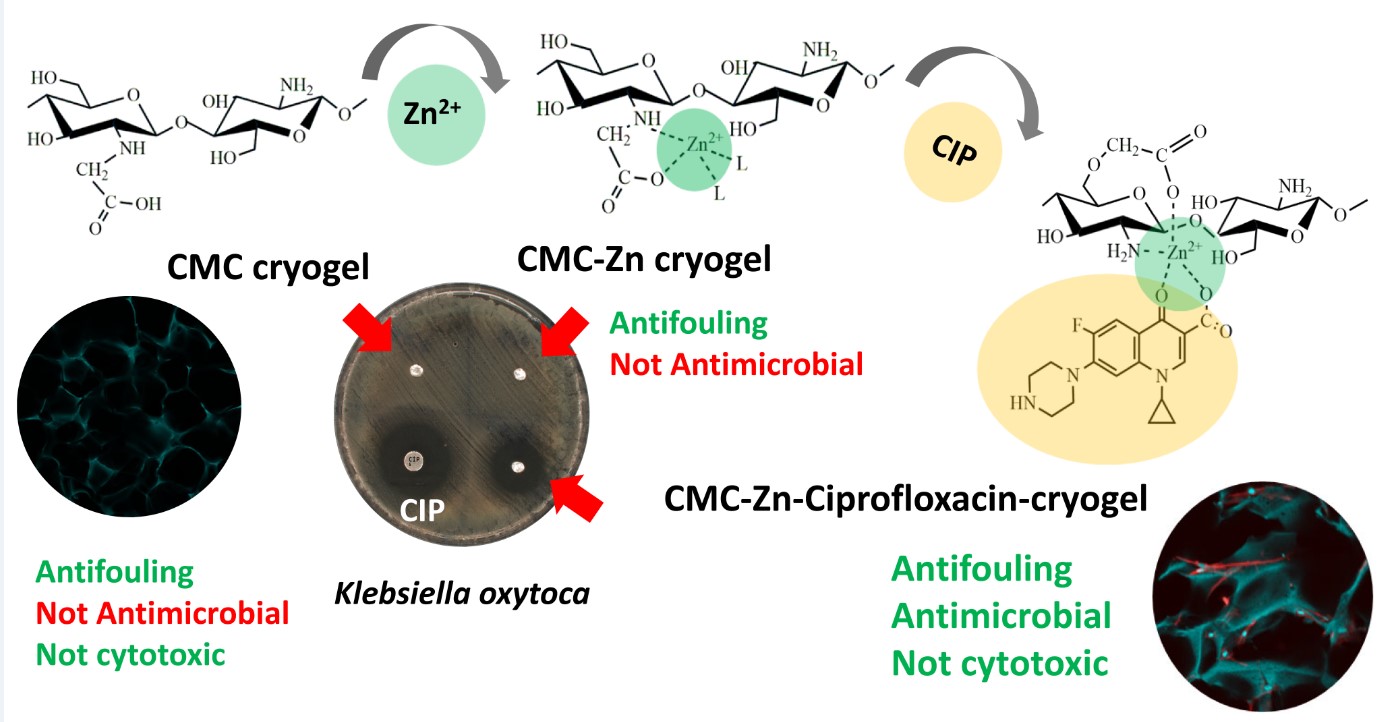
Loading of antibiotics to the biodegradable polymers is a straightforward approach to design drug-delivery systems for prevention and treatment of infections, associated with surgical implants. Recently, composites containing Zn2+ ions and antibiotics in polymeric matrices have gained attention as multifunctional materials, which are capable to modulate antimicrobial activity and bioavailability of antibiotics and promote the vascular remodeling, and collagen deposition. Here we suggest a novel approach to fabrication of antimicrobial CMC-Zn-CIP cryogel via stepwise Zn2+ and ciprofloxacin (CIP) loading to supermacroporous carboxymethyl chitosan (CMC) scaffolds cross-linked with diglycidyl ether of 1,4-butanediol with inherent antifouling properties against biofilm formation. To find a balance between antimicrobial activity and biocompatibility of CMC-Zn-CIP cryogels, cytotoxicity to the fibroblasts and antibacterial properties against clinically isolated strain of Klebsiella oxytoca were investigated in vitro for Zn2+, CIP, and cryogels with different loadings of Zn and CIP. Kinetics of CIP release from the composite cryogels was fitted with Korsmeyer–Peppas model, the release exponent value of 0.69-0.70 indicates non-Fickian diffusion mechanism. Relatively fast initial release of CIP with the half-life time of 60-123 min and release of 78±1% of CIP after 72 h in PBS buffer at 37°C are potentially beneficial to prevent development of implant-associated infections.
Keywords:
carboxymethyl chitosan
; zinc
; ciprofloxacin
; drug release
; cytotoxicity
; FT-IR spectroscopy
; binding mechanism
1. Introduction
Local application of antibiotics in wound healing, prevention and treatment of infections, associated with surgical implants, is an alternative to systemic administration of antibiotics, which can provide higher efficiency at the reduced dose [1,2]. Loading of antibiotics to the biodegradable polymers is a straightforward approach to design drug-delivery systems with prolonged effect. Polysaccharide-based hydrogels are often mentioned as materials with inherent antimicrobial properties. However, in general, they are not efficient enough to combat infections alone, but can be used as a matrix for tunable loading and release of different antimicrobial factors [3].
Chitosan, carboxymetyl chitosan (CMC) and other chitosan derivatives, have been applied to fabricate films, hydrogels, cryogels, and fibers for tissue engineering [4,5,6,7,8,9,10,11]. CMC often demonstrate superior to chitosan properties in biomedical applications: CMC hydrogels enhance the migration and proliferation of epithelial cells and fibroblasts, stimulate production of collagen and regulate gene expression [12]. CMC cryogels were shown to be more efficient than chitosan cryogels in preventing the adhesion and colonization of both Pseudomonas fluorescence and Staphylococcus aureus on the surface, thus demonstrating antifouling properties rather than the ability to kill bacteria [13], despite earlier reported antimicrobial activity of CMC with different substitution patterns and molecular weights [14,15].
The ability of chitosan and its derivatives to form stable complexes with heavy metal ions characterized with the antimicrobial activity [16,17,18] have promoted development of several types of multifunctional materials containing metal ions or nanoparticles [8,10,11,19], antibiotics [8], and other bioactive factors [11,19] to provide a synergetic effect for tissue remodeling and preventing infections. Although bacteria were reported to be more sensitive to Cu2+ compared to Zn2+ ions, Cu2+ is more toxic for the fibroblasts [20], making Zn-containing materials more promising for biomedical applications.
At concentrations from 0.001 to 0.1 μg/ml Zn increased cell proliferation in human fibroblasts (CRL2522) and keratinocytes (HaCaT) and demonstrated antimicrobial properties in a simulated nutrient-deficient environment in vitro [21]. 3D-printable Zn/tannic acid-reinforced glycol functionalized chitosan hydrogel was reported to accelerate M2 polarization of macrophages through the activation of anti-inflammatory transcription factors that provided this material with robust antibiofilm and wound healing properties [11]. Zinc ions and ciprofloxacin-encapsulated chitosan/poly(ε-caprolactone) composite nanofibers promoted wound healing via enhanced antibacterial and immunomodulatory activity [8]. Supplementing chitosan chitosan/poloxamer-based thermosensitive hydrogels with zinc gluconate/recombinant human epidermal growth factor promoted the vascular remodeling and collagen deposition, facilitated fibrogenesis, and reduced the level of interleukin 6 for wound basement repair [19]. Double-syringe injection device was used for continuous production of CMC-Zn2+ supramolecular hydrogel fiber, which demonstrated antimicrobial activity against S. aureus and Escherichia coli, but at relatively high Zn loadings, whose possible negative effect on fibroblasts have not been assessed [7].
Interest to combination of metal ions and antibiotics, which are capable to form stable metal complexes, is a route to modulate antimicrobial activity [22,23] and bioavailability [24] of antibiotics. In this regard, fluoroquinolones (FQ), synthetic antimicrobial drugs with a broad spectrum of activity [25], represent a promising class of antibiotics for the design of multifunctional biomaterials via coordination bonding between FQ, chitosan derivatives, and metal ions [7,26].
Here we have reported fabrication of the novel CMC-Zn cryogel, which can be loaded with precise amounts of Zn and one of the most widely used FQ – ciprofloxacin (CIP) to yield porous scaffolds with antimicrobial properties for tissue engineering. To find a balance between antimicrobial activity and biocompatibility of CMC-Zn-CIP cryogels, cytotoxicity to the fibroblasts and antibacterial properties against clinically isolated strain of K. oxytoca were investigated in vitro for Zn2+, CIP, and cryogels with different loadings of Zn and CIP. The mechanisms of Zn and CIP binding and release from the polymer matrix were also assessed.
2. Results and Discussion
2.1. Fabrication of CMC-Zn and CMC-Zn-CIP Cryogels
Scheme 1 shows two-step route of CMC-Zn-CIP cryogels fabrication starting from the earlier developed by our group CMC cryogel covalently cross-linked using diglycidyl ether of 1,4-butandiol. The original CMC cryogel was relatively soft material with Young modulus of 6.9±15 kPa and supermacroporous morphology with the average pore size of 156±40 µm, which demonstrated high elasticity, high total swelling (5276±407%), high permittivity for the cells and low cytotoxicity [27].
At the first step, CMC-Zn cryogels with Zn contents from 2 to 85 mg/g were obtained via Zn2+ ions adsorption from Zn(NO3)2 solutions with concentrations optimized to provide metal uptake efficiency close to 100% due to the high affinity of CMC to Zn2+ ions (Figure 1A). Although, a part of CMC functional groups was involved in the covalent cross-linking reaction, the sorption capacity of the cryogel for Zn2+ ions remained sufficiently high (85 mg/g) in comparison with earlier reported CMC-based materials [16,17].
Formation of coordination bonds between Zn2+ and functional groups of CMC resulted in doubly crosslinked network that significantly changed mechanical properties and swelling of the original CMC cryogel. Starting from the Zn loading of 20 mg/g (CMC-Zn20), we have observed gradually increasing hysteresis between loading and unloading strain–stress curves (Figure 1B). Observations during uniaxial compression measurements revealed that to a great extent hysteresis originates from disability of more rigid double cross-linked CMC-Zn cryogels to rapidly absorb water at the unloading stage, most likely, due to the changes of capillary structure of polymer walls upon cross-linking with Zn2+ ions. Densification of the pore walls after Zn loading is also evident from the difference in strain–stress curves shape, namely, from the shift of the point, when stiffness starts to grow exponentially, to the lower deformations (>30% for CMC-Zn65 compared to >55% for original CMC and CMC-Zn20). This was accompanied with significant increase of the compressive strength, which reached at maximum deformation of 75% the value of 30 kPa and 41kPa for CMC-Zn45 and CMC-Zn65, respectively, while for CMC-Zn20 it remained at the level of original CMC (12±2 kPa).
Earlier we have shown that metal-affine sorbents based on carboxyalkyl chitosans cross-linked with epichlorohydrin or hexamethylene diisocyanate and loaded with Cu2+ and Al3+ ions can efficiently bind ciprofloxacin (CIP) via ligand-exchange reaction [26,29,30]. Although stability constants of CIP-Zn2+ complexes is 2-4 orders of magnitude lower than those of CIP-Cu2+ and CIP-Al3+ complexes [31], one can still expect that CMC-Zn cryogels can be loaded with CIP via formation of the mixed ligand complexes CMC- Zn2+ -CIP.
At the second step of CMC-Zn-CIP cryogel fabrication (Scheme 1) CIP was loaded to CMC-Zn cryogels with Zn contents from 6.5 to 73 mg/g. pH of the CIP solution was fixed to 7.5, since we have earlier shown that this is the optimal pH value for CIP uptake by CMC-based metal-affine sorbents [29]. Figure 1C shows good linear correlation (r2=0.97) between Zn and CIP loadings that allows precise control of CIP content in the cryogels under the fixed sorption conditions (CIP concentration, ionic strength, thickness of the cryogel disk, mixing rate and contact time). Due to the high affinity type of CIP sorption isotherms on CMC-based metal-affine sorbents [29], the sorption conditions can be optimized to provide CIP loading efficiency close to 100% and to vary CIP contents at the fixed Zn loadings. Release of Zn2+ from CMC-Zn cryogels upon CIP sorption was 0.83±0.06% for the full range of Zn loadings shown in Figure 1A. This result correlates to the literature data that fluoroquinolones are not effective competitors to amino acids for Zn binding in blood plasma [32].
2.2. Antimicrobial Activity and Cytotoxicity
Low cytotoxicity, high permittivity for the cells and strong antifouling properties of the unmodified CMC cryogel were earlier demonstrated in 3D cell culturing applications [13,27]. Here we have focused on the effects of Zn and CIP loadings on antimicrobial properties of the composite cryogel and possible dose-dependent effect of these antimicrobial factors on viability of fibroblasts.
First, antimicrobial activity of CMC, CMC-Zn, and CMC-Zn-CIP cryogels against clinical strain K. oxytoca #20 was investigated by the disk diffusion method. Figure 2A shows that neither CMC nor CMC-Zn cryogel with Zn loading of 65 mg/g (CMC-Zn65) inhibited bacterial growth. Earlier reported antibacterial activity of CMC-Zn supramolecular hydrogel fibers [33] is, most likely, related to the very high Zn content, which can be toxic to human cells. Figure 2B demonstrates that Zn2+ ions at concentrations 15-150 µg/ml did not inhibit K. oxytoca growth efficiently. The notable effect of Zn2+ on bacteria proliferation was observed after 24 h cultivation starting from Zn concentration of 60 μg/ml. Bacteria growth inhibition by 55% was reached at 150 μgZn/ml. In this concentration range cytotoxic effect of Zn on human fibroblasts was much more significant: at 60 μgZn/ml the fibroblast viability did not exceed 2% and decreased even lower at 150 μgZn/ml.
It should be noted that the low antibacterial effect of CMC-Zn cryogels with high Zn loadings can be related to the strong binding of Zn2+ ions by CMC. Thus, antibacterial properties of CMC-Zn20-CIP5 cryogel evident from the disk diffusion test can be associated solely with the loaded CIP. In comparison with the commercial disk with the same CIP content (5 µg), CMC-Zn20-CIP5 exhibited the smaller inhibition zone – 28 and 22 mm, respectively, that can result from the diffusion limitations for the CIP release from the polymer matrix to agar.
To further elucidate antimicrobial activity of CMC-Zn cryogels, a series of cryogel disks with total CIP contents of 1, 5, 15, or 40 µg were fabricated and tested in bacterial suspension in DMEM using dissolved CIP as a reference (Figure 2C). At the lowest tested CIP concentration in DMEM (0.4 µg/ml equivalent to the total CIP content 1 μg), which is higher than MIC concentration for K. oxytoca #20 (0.125 µg/ml), bacteria growth was inhibited after 24 h by 92.3% with dissolved CIP, and only by 7.7% with CMC-Zn-CIP cryogel loaded with the same CIP content. When CIP loading to the cryogel disk was increased up to 5 µg, efficiency of bacterial growth inhibition increased to 98%. At higher CIP loading there was no difference in performance of CIP dissolved or loaded to the cryogel. The bacterial growth suppression with efficiency close to 100% remained constant during 72 h.
We have earlier shown that CMC cryogels did not suppress S. aureus and P. fluorescens growth in the suspension but efficiently prevented bacteria adhesion to the surface during 12 days of cultivation [13]. Figure 2D shows no difference in surface morphology of cryogels, which did not prevent bacterial growth (CMC) or efficiently suppressed it (CMC-Zn20-CIP5). No signs of K. oxytoca #20 adhesion and colonization was detected after cultivation for 72 h in the presence of unmodified CMC cryogel, despite high turbidity of the bacterial suspension. Thus, CMC-Zn-CIP cryogels can be considered as dual function materials with antimicrobial and antifouling properties. It is advantageous, since contact-killing antimicrobial surfaces often lead to accumulation of dead bacteria and other debris, which provide nutrients and sites for other bacteria to attach and proliferate [34].
To investigate, which loadings of Zn and CIP in CMC cryogel can be used without negative effects on human fibroblast viability and proliferation, cytotoxicity tests were performed in relatively small volume of DMEM, simulating local in vivo environment around implanted material. Since MIC value for the tested K. oxytoca strain was sufficiently lower than 1 µg/ml, at which microorganisms are still considered susceptible to CIP [35], it was important to investigate cytotoxicity of CMC-Zn-CIP cryogels at higher loadings of Zn and CIP than were found to be efficient against K. oxytoca (Figure 2C).
Figure 3A shows that after 5 days of cultivation the viability of fibroblasts in the adhesive culture was about 95%, which is slightly higher than that of cells in the cryogel – about 70%. Probably, fibroblasts were more difficult to detach from the cryogel surface, especially in the case of the most viable ones, and they were dropped from the viability calculations. Nevertheless, at Zn loadings of 20 mg/g (CMC-Zn20) the relative positive effect on fibroblast viability in comparison with unmodified CMC cryogel was noticeable: viability increased to 80%. Confocal microscopy images of human fibroblasts in CMC and CMC-Zn cryogels (Figure 3C) show that fibroblasts are arranged individually, without forming aggregates or clusters, indicating an even distribution of cells within the three-dimensional porous structure of cryogels. The majority of the cells exhibit a spindle-like morphology, with a less frequent polygonal shape. The visualization of the actin cytoskeleton reveals a well-developed network of actin filaments. The cells typically maintain close contact with the surface of the gel and are occasionally flattened, indicating their ability for adhesion and strong interaction with the matrix. Frequently, fibroblasts show contact areas on the walls of the cryogel's pores, suggesting active stretching within the space between the walls. This morphology may indicate high mechanical activity, facilitating the migration of cells within the three-dimensional matrix. Visually the general fibroblast density was slightly higher in CMC-Zn cryogel comparing to CMC cryogel.
In comparison with earlier reported gelatin and gelatin-pectin cryogels doped with Zn, which resulted in 50% inhibition of human skin fibroblasts growth at Zn loadings of 11 mg/g [36], CMC cryogels demonstrated lower cytotoxicity at higher Zn loadings of 20 mg/g, most likely due to the higher stability of CMC-Zn2+ complexes. After loading CMC-Zn20 cryogel disk with 40 µg CIP, fraction of viable fibroblasts increased up to 85-90% (Figure 3A). High activity and normal morphology of fibroblasts in CMC-Zn20-CIP40 cryogel was also confirmed by confocal microscopy (Figure 3C). When Zn and CIP loadings in CMC cryogel simultaneously increased 3.25-fold (CMC-Zn65-CIP130), a cytotoxic effect on fibroblasts was evident from the reduced population of viable cells to 50-55% (Figure 3A) and changes in cell morphology (Figure 3C). The number of cells observed by confocal microscopy in CMC-Zn65-CIP130 cryogel was extremely low. The cells appeared anchored and flattened on the pore walls surface but did not exhibit the same degree of elongation as in other cryogels.
To elucidate, which component of CMC-Zn65-CIP130 cryogel was responsible for increased cytotoxicity, we have cultivated human skin fibroblasts in the presence of CIP for three days (Figure 3B). Increase of CIP concentration from 5 to 120 μg/ml did not result in a noticeable decrease in fibroblast viability. However, proliferative capacity of the cells significantly decreased at CIP concentration >20 μg/ml. After culturing in the presence of 40 μgCIP/ml cell density decreased from 47-50×103 cells/cm2 (in control or at CIP< 20 μg/ml) to 30-35×103 cells/cm2. At CIP concentration 80 μg/ml and higher the density did not exceed 13×103 cells/cm2. Since after incubation of CMC-Zn65-CIP130 cryogel at 37°C in PBS buffer for 24 h, 75-80% of CIP and less than 1% of Zn was released (see section 2.3), one can assume more significant contribution of CIP to the reduced cryogel cytocompatibility at high Zn and CIP loadings. Indeed, release of 80% of CIP from CMC-Zn65-CIP130 cryogel results in CIP concentration in the medium of ~70 μg/ml, at which notable negative effect of CIP on fibroblast proliferation was observed (Figure 3B).
2.3. Binding and Release Mechanisms
Antimicrobial activity and cytotoxicity of polymeric composite materials strongly depend on binding strength and release profiles of loaded active components. Information about mechanism of Zn2+ complexation with N,O-CMC reported by different research groups is ambiguous [17,18,28]. Binding of Zn2+ to CMC via carboxylic groups only was suggested in [18] that resulted in higher solubility and enhanced antimicrobial activity of the O,N-CMC chitosan–zinc complex compared to chitosan–zinc complex against S. aureus and E. coli. Formation of soluble CMC-Zn2+ complexes without participation of amino groups (Scheme 1, structures 5 and 6) and of water-insoluble chelates via amino and carboxylic groups was suggested in [28].
In our case CMC contains 46% of monomer units with primary amino group (Table 1), so their participation in Zn2+ binding cannot be excluded. The fact that Zn2+ ions were not released upon CIP adsorption (release <1%) suggest that stability of CMC-Zn-CIP complexes (Scheme 1, structures 7 and 8) is higher that of Zn2+ -CIP complex under the experimental conditions used for CIP loading.
Most notable difference between FTIR spectra of CMC, CMC-Zn, and CMC-Zn-CIP cryogels was observed in the region 1200-1700 cm-1 (Figure 4). Stretching band of unionized and uncoordinated carboxylic group at 1723 cm-1 was observed only for CMC cryogel in protonated form (CMC-H+). Band near 1580 cm-1, which can be assigned to stretching of ionized or coordinated carboxylic groups shifted from 1579 cm-1 in CMC cryogel to 1584 cm-1 in CMC-Zn cryogel, indicating participation of carboxylic groups of CMC in Zn2+ ions binding. Although bands near 1380 cm-1, which shifts from 1403 cm-1 in CMC cryogel to 1396 cm-1 in CMC-Zn, can be assigned to symmetrical vibrations of carboxylic groups, however, in amino carboxylate - metal complexes these bands can overlap with N–H deformation [37]. The asymmetry of the bands centered near 1580 cm-1, most likely, results from overlapping of bands corresponding to carboxylic and amino groups, whose binding with Zn2 + in chitosan resulted in the shift of -NH band from 1624 cm−1 to 1628 cm−1 [38]. The above discussed changes, presence of band at 1378 cm-1 in both polymers – CMC and chitosan, and increased intensity of the band at 1257 cm-1 in protonated CMC-cryogel, which can be assigned to C–N stretching in amines [37], support possibility of several possible structures of CMC-Zn, in which amino and carboxylic groups can be coordinated to Zn2+ (Scheme 1, structures 3 and 4). Low antibacterial effect of CMC-Zn cryogels with high Zn loadings (Figure 1C) also correlates with the strong binding of Zn2+ ions by CMC.
Binding of CIP to CMC-Zn cryogel via ligand exchange reactions (Scheme 1, structures 7 and 8) does not result in significant changes of FT-IR spectra due to the numerous overlapping bands in CIP and CMC spectra. However, presence of CIP in CMC-Zn-CIP cryogel can be confirmed by notable increase of band intensity at 1267 cm-1 and visible asymmetry of the band centered in CMC-Zn45 cryogel at 1584 cm-1 due to the overlapping with intensive CIP band at 1623 cm-1 (Figure 4).
In comparison with the earlier reported CIP loading to the polymer matrices via passive diffusion [8,10,39], sorption of CIP on CMC-Zn cryogel via ligand exchange reaction is beneficial in terms of loading efficiency and precise control of loaded CIP amounts. Since the CIP loading is proportional to Zn content, nearly 100% of loading efficiency can be reached, while CIP encapsulation efficiency in both chitosan/poly(ɛ-caprolactone) and chitosan/ Zn2+ /poly(ɛ-caprolactone) fibers was below 40% and was not notably improved in the presence of Zn2+ [8]. This supports drastic difference of CIP binding to CMC- Zn2+ and chitosan- Zn2+ complexes.
Saturation of the polymers with CIP via passive diffusion and subsequent release of antibiotic is based on the pH-dependence of CIP solubility, which is minimal at pH 7.4 and significantly increases at pH <5.5 resulting in the burst effect of CIP release in the first 5–7 min [9]. In contrast, CIP release from CMC-Zn-CIP cryogels was triggered by increase of the ionic strength (Figure 6). Kinetic parameters of CIP release from the disks of CMC-Zn65-CIP130 cryogel calculated using Korsmeyer–Peppas model gave a value of release exponent (n) of 0.69-0.70 and release constants of 0.45 and 0.56 (min-1) for the disks with thickness of 1.7 and 3.6 mm, respectively, in PBS buffer at 37°C. The half-time of CIP release increased from 60 to 123 min, when the disk thickness was doubled.
The value of release exponent corresponds to the anomalous non-Fickian behavior, which can be related to the strong contribution of swelling and relaxation in polymer matrix to the drug-release mechanism. It is not surprising, considering high swelling degree of cryogels (> 5000 %) and presence of not only free (“squeezable”) water in macropores but also water bound to the polymer. This structure assumes the existence of the stagnation zones inside the swollen supermacroporous disks, where mass transfer is hindered even at high rates of external stirring, as we have demonstrated earlier [40]. This suggests that release rate from CMC-Zn-CIP cryogels can be tuned via modification of the porous structure and pore wall thickness.
Despite the difference in CIP release profiles depending on the disk thickness, in both cases 78±1% of CIP were released after 72 h. This CIP fraction can be associated with the most stable mixed ligand CMC-Zn-CIP complexes, which will be released only upon bioresorption of the CMC cryogel that would be beneficial for prevention of biofilm formation during tissue remodeling process, while relatively fast release within first 24 h is essential to stop development of possible implant-associated infection [41].
3. Materials and Methods
3.1. Materials
N,O-(carboxymethyl)chitosan (CMC) with degree of carboxyalkyl substitution 1.49 was purchased from BioLog Heppe GmbH (Landsberg, Germany) in the form of sodium salt. The CMC monomer composition was determined by 1H NMR spectroscopy (Table 1). Cross-linking agent—1,4-butanediol diglycidyl ether (BDDGE) 95% and dry ciprofloxacin (CIP) were purchased from Sigma-Aldrich (St. Louis, USA). PBS tablets and CIP solution for infusion were purchased from “Paneco” Ltd. (Moscow, Russia) and “Ist-Farm” Ltd. (Ussuriysk, Russia), respectively. Other reagents were of the analytical grade, Milli Q Ultrapure Water was used for preparation of the solutions.
3.2. Fabrication of CMC and CMC-Zn Cryogels
3% CMC solution was prepared in distilled water, the resulting pH of the solution was 10.2. BDDE (0.143 g) and CMC solution (10 g) were mixed and left for 10 min under constant stirring at 25 °C. Then, solution was placed into the plastic syringes with inner diameter of ~0.85 cm and 0.44 cm (for the disk diffusion test only) and kept in a freezer (Liebherr, Kirchdorf an der Iller, Germany) at –10 °C during 7 days. After thawing at 25 °C, the CMC cryogels were thoroughly washed with distilled water using a peristaltic pump (Ismatec, Wertheim, Germany). The CMC cryogels were cut to the disks with thickness of ~1.7 or 3.6 mm for the further modification.
Disks of CMC-Zn cryogels with Zn content from 2 to 85 mg/g were obtained by adsorption from Zn(NO3)2 solutions with concentrations 0.15-4.5 mM. The precise contents of Zn in cryogels were calculated from the differences in initial and equilibrium concentrations of Zn determined by the atomic absorption spectrometry (AAS) using a Shimadzu 7000 spectrometer (Shimadzu, Kyoto, Japan). The obtained CMC-Zn cryogels were thoroughly washed with distilled water and stored swollen and sealed until used. Cryogels are labeled as CMC-Zn with content of Zn in mg/g: e.g. CMC-Zn20 means CMC cryogel with Zn loading 20 mg/g.
3.3. Ciprofloxacin Loading and Release
A stock solution (50 ml) was prepared by dissolving 4.14 mg of Ciprofloxacin (CIP) in 0.2 ml of 1M HCl, followed by addition of ultrapure water and pH adjustment to 7.5 with 1M NaOH solution. CIP was loaded to CMC-Zn cryogels with different Zn contents from 0.25 mM or 0.05 mM CIP solutions at solid:liquid ratio of 1:1000 and 120 rpm (Orbital shaker Biosan PSU-10i (SIA Biosan, Riga, Latvia). To find correlation between CIP and Zn loadings, the cryogel disks with thickness of 1.7 mm in the swollen state were used, and the contact time was fixed to 18 h. The equilibrium concentration of CIP was determined spectrophotometrically at a wavelength of 272 nm using a UV-1650PC spectrophotometer (Shimadzu, Japan). The precise contents (µg) of CIP in the cryogel disks were calculated using the difference in initial and equilibrium CIP concentrations. The CMC-Zn-CIP cryogels were labeled with content of Zn in mg/g and total content of CIP in the disk: e.g. CMC-Zn20-CIP5 means disk of CMC cryogel with Zn loading of 20 mg/g and CIP content of 5µg.
CIP release from CMC-Zn65-CIP130 cryogels with thickness of 1.7 mm and 3.6 mm was investigated as follows: disks were immersed in 4 ml of PBS buffer (pH 7.4) and thermostated at 37°С; CIP concertation in the solutions were determined spectrophotometrically as described above at time points from 5 min to 24 h. Zn concentration in PBS after solution after CIP desorption was determined by AAS. Kinetics of CIP release was fitted using Korsmeyer–Peppas model [42]:
where Mt and M∞ are cumulative amounts of CIP released at time t and the maximum amount released under the experimental conditions used; n is the release exponent indicating release mechanism; K is the release rate constant.
To confirm material balance in CIP release experiments, cryogel disks were removed from PBS after 72 h, washed with ultrapure water and immersed in 0.01M HCl solution for release of CIP and Zn from the polymer matrix.
3.4. Mechanical Properties of Cryogels
Uniaxial compression tests at a constant speed of 0.01 mm/s were performed for the swollen cylindrically shaped cryogels with a diameter of 10-15 mm and height of 8–9 mm using Physica MCR 301 rheometer (Anton Paar GmbH, Graz, Austria). Loading and unloading strain-stress curves have been calculated from the measured normal force (FN) and gap. Compressive strength was determined at 75% strain.
3.5. Antibacterial Activity
Clinically isolated Klebsiella oxytoca #20 strain from the collection of the Medical Complex (Center for Laboratory Research) of the Far Eastern Federal University was grown on Agar containing 5% sheep blood («Sredoff», Saint-Petersburg, Russia) for 24 h, suspended in the liquid medium – Miller's LB Broth (MP Biomedicals, LCC, Illkirch-Graffenstaden, France) and grown overnight. Then overnight culture was diluted with LB Broth to adjust turbidity to 0.5 McFarland standard using MicroScan Turbidity Meter (Siemens AG, Munich, Germany).
To investigate antibacterial properties of the cryogels, disks with thickness of 1.5 mm and diameter of 5 mm were placed on the surface of agar plate with spread bacterial suspension. Bio-Rad antimicrobial susceptibility disk with CIP content of 5 µg was used as a reference. The inverted plates were incubated at 37°C for 24 h. The images of the inhibition zones were captured using ADAGIO BioRad analyzer (Marnes-la-Coquette, France), diameters of the inhibition zones and MIC for ciprofloxacin were calculated using the built-in software.
Quantitative analysis of bacteria sensitivity to Zn2+ ions was performed in LB Broth, to soluble form of CIP (“Ist-Farm” Ltd., Ussuriysk, Russia) and CIP bound to cryogels in DMEM (#D5648, Sigma-Aldrich, St. Louis, MO, USA). 25 µL of bacterial suspension (turbidity of 0.5 MFU) was added to the test tubes with 2.5 ml of liquid medium containing i) zinc nitrate at concentrations of 15-150 µgZn/mL, ii) 1, 5, 15, 40 µg of CIP; iii) cryogel disks (2.5 mg of dry weight) – unmodified CMC, CMC with Zn content 65 mg/g (CMC-Zn65); CMC with Zn content 20 mg/g (CMC-Zn20) loaded with 1, 5, 15, 40 µg of CIP per disk. 25 µL of bacterial suspension in 2.5 ml of medium without additives was used as a control (Tcontrol). Test tubes were incubated at 37°C for 24 h under aerobic conditions, then the turbidity was measured using MicroScan Turbidity Meter. Turbidity of medium (T0) was used as a background signal.
Bacterial growth inhibition (I) was calculated using the following equation:
where Tsample, Tcontrol, and T0 – turbidity of the sample, control, and background, respectively.
3.6. Cytotoxicity
3.6.1. Isolation and Cultivation of Human Dermal Fibroblasts (HDF)
The skin punch biopsy of 3 mm wide and 3 mm depth from the shoulder area was conducted at the Medical Center of Far Eastern Federal University with the voluntary consent of the donor. The skin tissue was minced and enzymatically disaggregated into cells with 2 units/ml collagenase IV type (Sigma-Aldrich, St. Louis, MO, USA) for 90 min at +37°C. The resulting cell suspension was seeded in T75 flasks (TPP, Trasadingen, Switzerland) in Dulbecco’s modified Eagle’s medium (DMEM, #D5648, Sigma-Aldrich) supplemented with 10% (v/v) fetal bovine serum (FBS, HyClone, Logan, UT, USA), 3.7 mg/ml sodium bicarbonate (Sigma-Aldrich), 1× mixture of non-essential amino acids (MEM NEAA, Sigma-Aldrich), 100 U/ml penicillin (Sigma-Aldrich), and 100 µg/ml streptomycin (Sigma-Aldrich). The cells were cultivated at +37°C, 5% CO2, and 90% relative humidity until 90% confluency. Then HDFs were detached with the solution of 0.05% (w/v) trypsin - 0.02% (w/v) EDTA at +37°C for 10 min, centrifuged at 500 g for 5 min, resuspended in fresh medium without antibiotics and used for cytotoxicity tests or seeding into cryogel disks.
3.6.2. Cytotoxicity Tests with Zn2+ and CIP
HDFs were seeded in adhesive 24-well culture plates at a density 100 × 103 cells/well in 1 ml of DMEM with additives. Then 0.01M Zn(NO3)2 solution or CIP solution for infusion (“Ist-Farm” Ltd., Ussuriysk, Russia) were added to the wells to the final concentrations of 15, 30, 60, 80, 120, 150 µg/ml for Zn; and 5, 20, 40, 80, 100, 120 µg/ml for CIP.
The fibroblasts were cultivated at +37°C, 5% CO2, and 90% relative humidity for 24 h, detached from the wells with the solution of 0.25% (w/v) trypsin - 0.05% (w/v) EDTA at +37°C for 20 min, triple washed with 2 ml of DPBS, and centrifuged at 500 g for 5 min. Their viability was assessed as described in sections 3.5.4 and 3.5.5.
3.6.3. Cultivation of Human Dermal Fibroblast in Cryogels
Swallen cryogel disks (dry weight equivalent of 3 mg) were placed in wells of a 24-well culture plate, consistently washed with 5 mL of Dulbecco's phosphate buffer saline (DPBS, Sigma-Aldrich) without Ca2+ and Mg2+, and 5 ml of DMEM with additives and without antibiotics. All liquid was squeezed from the cryogel. HDFs were suspended in DMEM with additives. The cells were seeded on a top of cryogel disk at a density 100 × 103 cells/disk. The samples were cultivated in 1.5 ml of DMEM per well at +37°C, 5% CO2, and 90% relative humidity. The medium has been replaced on the 2nd and 4th day.
After 5 days of cultivation the cryogel disks with cells were either fixed for confocal laser scanning microscopy or transferred to a new 24-well culture plate, washed twice with 1 ml of DPBS, and minced with scalpel (for viability assay only). Cells were detached from the cryogel disks with the solution of 0.25% (w/v) trypsin - 0.05% (w/v) EDTA at +37°C for 20 min, triple washed with 2 ml of DPBS, filtered through a 40 µm nylon mesh, and centrifuged at 500 g for 5 min.
3.6.4. Assessing of Fibroblast Viability
A pellet of cells from a single well of culturing plate or cryogel disk was re-suspended in 100 µl of DPBS with 1 µM SYTO 9 (Sigma-Aldrich) to stain only cells and separate from cryogel fragments and 10 µM 2',7'-dichlorodihydrofluorescein diacetate (H2DCFDA) (Sigma-Aldrich) to assess the mitochondrial activity (fibroblast viability). The cell suspension was incubated in the dark at room temperature for 10 min, diluted with 100 µl of DPBS, then flow cytometric analyses was performed using a CytoFLEX flow cytometer (Beckman-Coulter, Brea, CA, USA) connected to a computer running CytExpert software (version 2.6, Beckman-Coulter). More detailed description of the analysis is given in [43].
3.6.5. Confocal Laser Scanning Microscopy (CLSM)
Cryogel disks with cultured fibroblasts were fixed in 4% paraformaldehyde (PFA; Sigma-Aldrich) in DPBS for 60 min at +4°C and rinsed three times with cold DPBS. To detect filamentous actin, gels with cells were incubated overnight in a solution of tetramethylrhodamine-conjugated phalloidin (Santa Cruz Biotechnology, Dallas, TX, USA) at +4°C; then, they were washed three times with cold DPBS followed by staining with 10 μg/ml 4′,6′-diamidino-2-phenylindole (DAPI, Sigma-Aldrich) to reveal the nuclei. The stained material was stored in DPBS with added preservative (ProClin, Sigma-Aldrich) in the dark at +4°C. Immediately before the microscopy analysis, the gel section was placed in Vectashield medium in a Petri dish with a thin (0.17 mm) glass bottom. The fibroblast morphology was investigated using a LSM 800 confocal laser scanning microscope (Carl Zeiss, Germany). For each sample, several fields were scanned at a magnification 10× and at a magnification 20× to visualize the interaction of cells with the matrix. Z-projections and multichannel images were assembled using the ImageJ software (NIH, USA).
3.7. Scanning Electron Microscopy
Cryogel disks with cultured bacteria were fixed with 2.5% glutaraldehyde (EMS, USA) in 0.1 M cacodylate buffer (Sigma-Aldrich), passed through the increasing concentrations of ethanol (15, 30, 50, 70, and 96%) with 30-minute incubation at each stage, dried at the critical point and coated with chromium, then analyzed using scanning electron microscope Sigma VP (Carl Zeiss, Germany).
Author Contributions
Conceptualization, S.B.; methodology, S.B., A.B., T.B, R.G.; investigation, S.B., A.B., T.B., Y.P., D.M., M.M., A.S., A.S.; visualization, S.B., M.M., A.B.; data curation, S.B., A.B., Y.P.; writing—original draft preparation, S.B., A.B.; writing—review and editing, S.B., A.B., R.G.; project administration, S.B., R.G. All authors have read and agreed to the published version of the manuscript.
Funding
Financial support from Russian Science Foundation (project No. 20-13-00399) is gratefully acknowledged.
Institutional Review Board Statement
The study was conducted in accordance with the Declaration of Helsinki, and approved by the Institutional Ethics Committee of School of Medicine and Life Sciences Far Eastern Federal University (protocol #12, June 14, 2024).
Informed Consent Statement
Informed consent was obtained from all subjects involved in the study.
Data Availability Statement
The original contributions presented in this study are included in the article/supplementary material. Further inquiries can be directed to the corresponding author.
Acknowledgments
Investigations using FT-IR spectroscopy were conducted at the facilities of the Far East Center of Structural Studies (Institute of Chemistry, FEB RAS, Vladivostok, Russia) under government assignment of the Ministry of Science and Higher Education of the Russian Federation (project no. FWFN(0205)-2022-0002). The authors are grateful to the staff of the Electron Microscopy Department of NSCMB FEB RAS, Denis V. Fomin and Kirill A. Shefer, for assistance with scanning electron microscopy.
Conflicts of Interest
The authors declare no conflicts of interest.
References
- Yang, K.; Han, Q.; Chen, B.; Zheng, Y.; Zhang, K.; Li, Q.; Wang, J. Antimicrobial Hydrogels: Promising Materials for Medical Application. Int. J. Nanomedicine 2018, 13, 2217–2263. [Google Scholar] [CrossRef] [PubMed]
- Darouiche, R.O. Antimicrobial Approaches for Preventing Infections Associated with Surgical Implants. Clin. Infect. Dis. 2003, 36, 1284–1289. [Google Scholar] [CrossRef] [PubMed]
- Kapusta, O.; Jarosz, A.; Stadnik, K.; Giannakoudakis, D.A.; Barczyński, B.; Barczak, M. Antimicrobial Natural Hydrogels in Biomedicine: Properties, Applications, and Challenges—A Concise Review. Int. J. Mol. Sci. 2023, 24. [Google Scholar] [CrossRef]
- Agarwal, T.; Narayan, R.; Maji, S.; Behera, S.; Kulanthaivel, S.; Maiti, T.K.; Banerjee, I.; Pal, K.; Giri, S. Gelatin/Carboxymethyl Chitosan Based Scaffolds for Dermal Tissue Engineering Applications. Int. J. Biol. Macromol. 2016, 93, 1499–1506. [Google Scholar] [CrossRef]
- Hendradi, E.; Hariyadi, D.; Adrianto, M. The Effect of Two Different Crosslinkers on in Vitro Characteristics of Ciprofloxacin-Loaded Chitosan Implants. Res. Pharm. Sci. 2018, 13, 38. [Google Scholar] [CrossRef]
- Zare, S.; Eskandani, M.; Vandghanooni, S.; Hossainpour, H.; Jaymand, M. Ciprofloxacin-Loaded Chitosan-Based Nanocomposite Hydrogel Containing Silica Nanoparticles as a Scaffold for Bone Tissue Engineering Application. Carbohydr. Polym. Technol. Appl. 2024, 7, 100493. [Google Scholar] [CrossRef]
- Zhou, T.; Zhou, H.; Wang, F.; Zhang, P.; Shang, J.; Shi, L. An Injectable Carboxymethyl Chitosan Hydrogel Scaffold Formed via Coordination Bond for Antibacterial and Osteogenesis in Osteomyelitis. Carbohydr. Polym. 2024, 324, 121466. [Google Scholar] [CrossRef]
- Zhou, F.; Sun, S.; Cui, C.; Li, X.; Wu, S.; Ma, J.; Chen, S.; Li, C.M. Zinc Ions and Ciprofloxacin-Encapsulated Chitosan/Poly(ɛ-Caprolactone) Composite Nanofibers Promote Wound Healing via Enhanced Antibacterial and Immunomodulatory. Int. J. Biol. Macromol. 2023, 253, 127086. [Google Scholar] [CrossRef] [PubMed]
- Pamfil, D.; Butnaru, E.; Vasile, C. Poly (Vinyl Alcohol)/Chitosan Cryogels as PH Responsive Ciprofloxacin Carriers. J. Polym. Res. 2016, 23. [Google Scholar] [CrossRef]
- Kumar, B.; Kumar, P. Synthesis and Characterization of PH-Sensitive Nanocarrier Based Chitosan-g-Poly(Itaconic Acid) for Ciprofloxacin Delivery for Anti-Bacterial Application. Int. J. Biol. Macromol. 2024, 268, 131604. [Google Scholar] [CrossRef]
- Patil, T. V.; Jin, H.; Dutta, S.D.; Aacharya, R.; Chen, K.; Ganguly, K.; Randhawa, A.; Lim, K.T. Zn@TA Assisted Dual Cross-Linked 3D Printable Glycol Grafted Chitosan Hydrogels for Robust Antibiofilm and Wound Healing. Carbohydr. Polym. 2024, 344, 122522. [Google Scholar] [CrossRef] [PubMed]
- Kruczkowska, W.; Kłosiński, K.K.; Grabowska, K.H.; Gałęziewska, J.; Gromek, P.; Kciuk, M.; Kałuzińska-Kołat, Ż.; Kołat, D.; Wach, R.A. Medical Applications and Cellular Mechanisms of Action of Carboxymethyl Chitosan Hydrogels. Molecules 2024, 29, 4360. [Google Scholar] [CrossRef]
- Boroda, A.; Privar, Y.; Maiorova, M.; Beleneva, I.; Eliseikina, M.; Skatova, A.; Marinin, D.; Bratskaya, S. Chitosan versus Carboxymethyl Chitosan Cryogels: Bacterial Colonization, Human Embryonic Kidney 293T Cell Culturing and Co-Culturing. Int. J. Mol. Sci. 2022, 23. [Google Scholar] [CrossRef] [PubMed]
- Anitha, A.; Divya Rani, V.V.; Krishna, R.; Sreeja, V.; Selvamurugan, N.; Nair, S.V.; Tamura, H.; Jayakumar, R. Synthesis, Characterization, Cytotoxicity and Antibacterial Studies of Chitosan, O-Carboxymethyl and N,O-Carboxymethyl Chitosan Nanoparticles. Carbohydr. Polym. 2009, 78, 672–677. [Google Scholar] [CrossRef]
- Chen, L.; Xie, Y.; Chen, X.; Li, H.; Lu, Y.; Yu, H.; Zheng, D. O-Carboxymethyl Chitosan in Biomedicine: A Review. Int. J. Biol. Macromol. 2024, 275, 133465. [Google Scholar] [CrossRef] [PubMed]
- Shoukry, A. a.; Hosny, W.M. Coordination Properties of N,O-Carboxymethyl Chitosan (NOCC). Synthesis and Equilibrium Studies of Some Metal Ion Complexes. Ternary Complexes Involving Cu(II) with (NOCC) and Some Biorelevant Ligand. Cent. Eur. J. Chem. 2012, 10, 59–70. [Google Scholar] [CrossRef]
- Sun, S.; Wang, A. Adsorption Properties and Mechanism of Cross-Linked Carboxymethyl-Chitosan Resin with Zn(II) as Template Ion. React. Funct. Polym. 2006, 66, 819–826. [Google Scholar] [CrossRef]
- Patale, R.L.; Patravale, V.B. O,N-Carboxymethyl Chitosan-Zinc Complex: A Novel Chitosan Complex with Enhanced Antimicrobial Activity. Carbohydr. Polym. 2011, 85, 105–110. [Google Scholar] [CrossRef]
- Lin, S.; Pei, L.; Zhang, W.; Shu, G.; Lin, J.; Li, H.; Xu, F.; Tang, H.; Peng, G.; Zhao, L.; et al. Chitosan-Poloxamer-Based Thermosensitive Hydrogels Containing Zinc Gluconate/Recombinant Human Epidermal Growth Factor Benefit for Antibacterial and Wound Healing. Mater. Sci. Eng. C 2021, 130, 112450. [Google Scholar] [CrossRef]
- Padaga, S.G.; Bhatt, H.; Ch, S.; Paul, M.; Itoo, A.M.; Ghosh, B.; Roy, S.; Biswas, S. Glycol Chitosan-Poly(Lactic Acid) Conjugate Nanoparticles Encapsulating Ciprofloxacin: A Mucoadhesive, Antiquorum-Sensing, and Biofilm-Disrupting Treatment Modality for Bacterial Keratitis. ACS Appl. Mater. Interfaces 2024, 16, 18360–18385. [Google Scholar] [CrossRef] [PubMed]
- Rembe, J.D.; Boehm, J.K.; Fromm-Dornieden, C.; Hauer, N.; Stuermer, E.K. Comprehensive Analysis of Zinc Derivatives Pro-Proliferative, Anti-Apoptotic and Antimicrobial Effect on Human Fibroblasts and Keratinocytes in a Simulated, Nutrient-Deficient Environment in Vitro. Int. J. Mol. Cell. Med. 2020, 9, 165–179. [Google Scholar] [CrossRef]
- Psomas, G.; Kessissoglou, D.P. Quinolones and Non-Steroidal Anti-Inflammatory Drugs Interacting with Copper(Ii), Nickel(Ii), Cobalt(Ii) and Zinc(Ii): Structural Features, Biological Evaluation and Perspectives. Dalt. Trans. 2013, 42, 6252–6276. [Google Scholar] [CrossRef]
- Zarkan, A.; MacKlyne, H.R.; Chirgadze, Di.Y.; Bond, A.D.; Hesketh, A.R.; Hong, H.J. Zn(II) Mediates Vancomycin Polymerization and Potentiates Its Antibiotic Activity against Resistant Bacteria. Sci. Rep. 2017, 7, 1–9. [Google Scholar] [CrossRef]
- Tewes, F.; Bahamondez-Canas, T.F.; Moraga-Espinoza, D.; Smyth, H.D.C.; Watts, A.B. In Vivo Efficacy of a Dry Powder Formulation of Ciprofloxacin-Copper Complex in a Chronic Lung Infection Model of Bioluminescent Pseudomonas Aeruginosa. Eur. J. Pharm. Biopharm. 2020, 152, 210–217. [Google Scholar] [CrossRef]
- Uivarosi, V. Metal Complexes of Quinolone Antibiotics and Their Applications: An Update. Molecules 2013, 18, 11153–11197. [Google Scholar] [CrossRef] [PubMed]
- Bratskaya, S.; Privar, Y.; Slobodyuk, A.; Shashura, D.; Marinin, D.; Mironenko, A.; Zheleznov, V.; Pestov, A. Cryogels of Carboxyalkylchitosans as a Universal Platform for the Fabrication of Composite Materials. Carbohydr. Polym. 2019, 209, 1–9. [Google Scholar] [CrossRef] [PubMed]
- Boroda, A.; Privar, Y.; Maiorova, M.; Skatova, A.; Bratskaya, S. Sponge-like Scaffolds for Colorectal Cancer 3D Models: Substrate-Driven Difference in Micro-Tumors Morphology. Biomimetics 2022, 7, 56. [Google Scholar] [CrossRef] [PubMed]
- Tang, L.G.; Hon, D.N.S. Chelation of Chitosan Derivatives with Zinc Ions. II. Association Complexes of Zn2+ onto O,N-Carboxymethyl Chitosan. J. Appl. Polym. Sci. 2001, 79, 1476–1485. [Google Scholar] [CrossRef]
- Privar, Y.; Shashura, D.; Pestov, A.; Modin, E.; Baklykov, A.; Marinin, D.; Bratskaya, S. Metal-Chelate Sorbents Based on Carboxyalkylchitosans: Ciprofloxacin Uptake by Cu(II) and Al(III)-Chelated Cryogels of N-(2-Carboxyethyl)Chitosan. Int. J. Biol. Macromol. 2019, 131, 806–811. [Google Scholar] [CrossRef]
- Privar, Y.; Shashura, D.; Pestov, A.; Ziatdinov, A.; Azarova, Y.; Bratskaya, S. Effect of Regioselectivity of Chitosan Carboxyalkylation and Type of Cross-Linking on the Metal-Chelate Sorption Properties toward Ciprofloxacin. React. Funct. Polym. 2020, 150, 104536. [Google Scholar] [CrossRef]
- Uivarosi, V. Metal Complexes of Quinolone Antibiotics and Their Applications: An Update. Molecules 2013, 18, 11153–11197. [Google Scholar] [CrossRef] [PubMed]
- Djurdjevic, P.; Jakovljevic, I.; Joksovic, L.; Ivanovic, N.; Jelikic-Stankov, M. The Effect of Some Fluoroquinolone Family Members on Biospeciation of Copper(II), Nickel(II) and Zinc(II) Ions in Human Plasma. Molecules 2014, 19, 12194–12223. [Google Scholar] [CrossRef]
- Wang, Y.L.; Zhou, Y.N.; Li, X.Y.; Huang, J.; Wahid, F.; Zhong, C.; Chu, L.Q. Continuous Production of Antibacterial Carboxymethyl Chitosan-Zinc Supramolecular Hydrogel Fiber Using a Double-Syringe Injection Device. Int. J. Biol. Macromol. 2020, 156, 252–261. [Google Scholar] [CrossRef] [PubMed]
- Katsikogianni, M.G.; Wood, D.J.; Missirlis, Y.F. Biomaterial Functionalized Surfaces for Reducing Bacterial Adhesion and Infection. In Handbook of Bioceramics and Biocomposites; Antoniac, I.V., Ed.; Springer International Publishing Switzerland 2015, 2015; pp. 1–28 ISBN 9783319092300.
- Baudry-Simner, P.J.; Singh, A.; Karlowsky, J.A.; Hoban, D.J.; Zhanel, G.G. Mechanisms of Reduced Susceptibility to Ciprofloxacin in Escherichia Coli Isolates from Canadian Hospitals. Can. J. Infect. Dis. Med. Microbiol. 2012, 23, 60–64. [Google Scholar] [CrossRef]
- Luong, D.; Yergeshov, A.A.; Zoughaib, M.; Sadykova, F.R.; Gareev, B.I.; Savina, I.N.; Abdullin, T.I. Transition Metal-Doped Cryogels as Bioactive Materials for Wound Healing Applications. Mater. Sci. Eng. C 2019, 103, 109759. [Google Scholar] [CrossRef]
- Lanigan, K.C.; Pidsosny, K. Reflectance FTIR Spectroscopic Analysis of Metal Complexation to EDTA and EDDS. Vib. Spectrosc. 2007, 45, 2–9. [Google Scholar] [CrossRef]
- Rogina, A.; Lončarević, A.; Antunović, M.; Marijanović, I.; Ivanković, M.; Ivanković, H. Tuning Physicochemical and Biological Properties of Chitosan through Complexation with Transition Metal Ions. Int. J. Biol. Macromol. 2019, 129, 645–652. [Google Scholar] [CrossRef]
- Kioomars, S.; Heidari, S.; Malaekeh-Nikouei, B.; Shayani Rad, M.; Khameneh, B.; Mohajeri, S.A. Ciprofloxacin-Imprinted Hydrogels for Drug Sustained Release in Aqueous Media. Pharm. Dev. Technol. 2017, 22, 122–129. [Google Scholar] [CrossRef] [PubMed]
- Malakhova, I.; Golikov, A.; Azarova, Y.; Bratskaya, S. Extended Rate Constants Distribution (RCD) Model for Sorption in Heterogeneous Systems: 2. Importance of Diffusion Limitations for Sorption Kinetics on Cryogels in Batch. Gels 2020, 6, 15. [Google Scholar] [CrossRef] [PubMed]
- Federico, S.; Pitarresi, G.; Palumbo, F.S.; Fiorica, C.; Catania, V.; Schillaci, D.; Giammona, G. An Asymmetric Electrospun Membrane for the Controlled Release of Ciprofloxacin and FGF-2: Evaluation of Antimicrobial and Chemoattractant Properties. Mater. Sci. Eng. C 2021, 123. [Google Scholar] [CrossRef]
- Aguzzi, C.; Cerezo, P.; Salcedo, I.; Sánchez, R.; Viseras, C. Mathematical Models Describing Drug Release from Biopolymeric Delivery Systems. Mater. Technol. 2010, 25, 205–211. [Google Scholar] [CrossRef]
- Bratskaya, S.; Skatova, A.; Privar, Y.; Boroda, A.; Kantemirova, E.; Maiorova, M.; Pestov, A. Stimuli-Responsive Dual Cross-Linked N-Carboxyethylchitosan Hydrogels with Tunable Dissolution Rate. Gels 2021, 7, 188. [Google Scholar] [CrossRef] [PubMed]
Scheme 1.
Formation of CMC-Zn-CIP cryogel via coordination bonds: structure of N,O-CMC (1), fragment of CMC cryogel cross-linked with BDDE and its confocal microscopy image (2); possible structures of Zn2+ complexes with O-CMC (3) and N-CMC (4) fragments in CMC-Zn cryogel, and corresponding structures of O-CMC-Zn-CIP (7) and N-CMC-Zn-CIP (8) fragments in CMC-ZN-CIP cryogel, images on the photo are for CMC-Zn65 (left) and CMC-Zn65-CIP130-cryogel. Structures of soluble Zn2+-CMC complexes (5 and 6), adapted from [28].
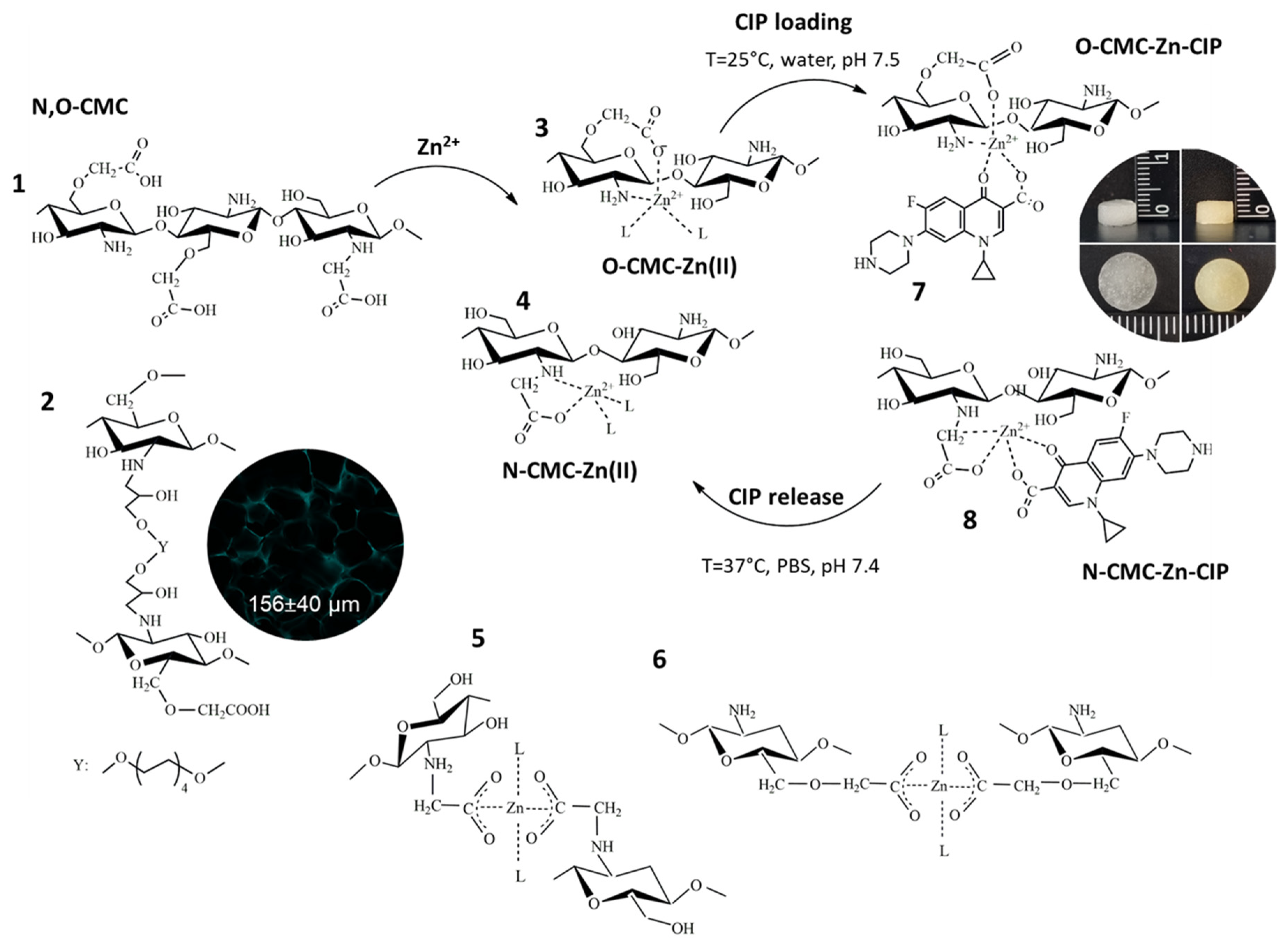
Figure 1.
Efficiency of Zn loading to CMC cryogel (A). Strain–stress curves of uniaxial compression of CMC and CMC-Zn cryogels with different Zn contents: images in the insert correspond to CMC cryogel before compression (a), after removal of water (b), and after reswelling (c) (B). Dependence of CIP loaded to CMC-Zn cryogel on Zn content (C). .
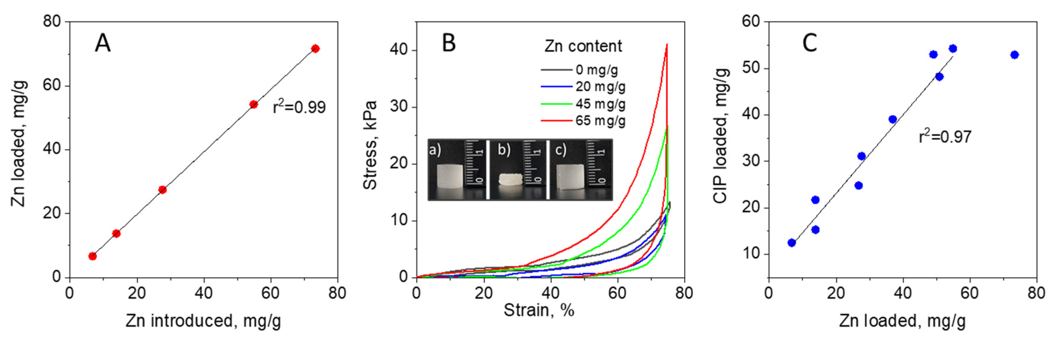
Figure 2.
Bacterial growth inhibition on agar plate with CMC cryogel, CMC-Zn65 (CMC with Zn content 65 mg/g ) and CMC-Zn20-CIP5 (CMC with Zn content 20 mg/g loaded with 5 μg of CIP per disk) – A. Growth inhibition of K. oxytoca #20 in LB medium and viability of human fibroblasts in DMEM depending on Zn2+ concentration (B). Growth inhibition of K. oxytoca #20 in DMEM in the presence of cryogels or dissolved CIP. CIP loadings per disk of CMC-Zn20 cryogel were equal to the doses of the dissolved CIP (C). SEM images of CMC and CMC-Zn20-CIP5 cryogels after cultivation of K. oxytoca #20 in DMEM for 3 days (scale bar 100 µm). .
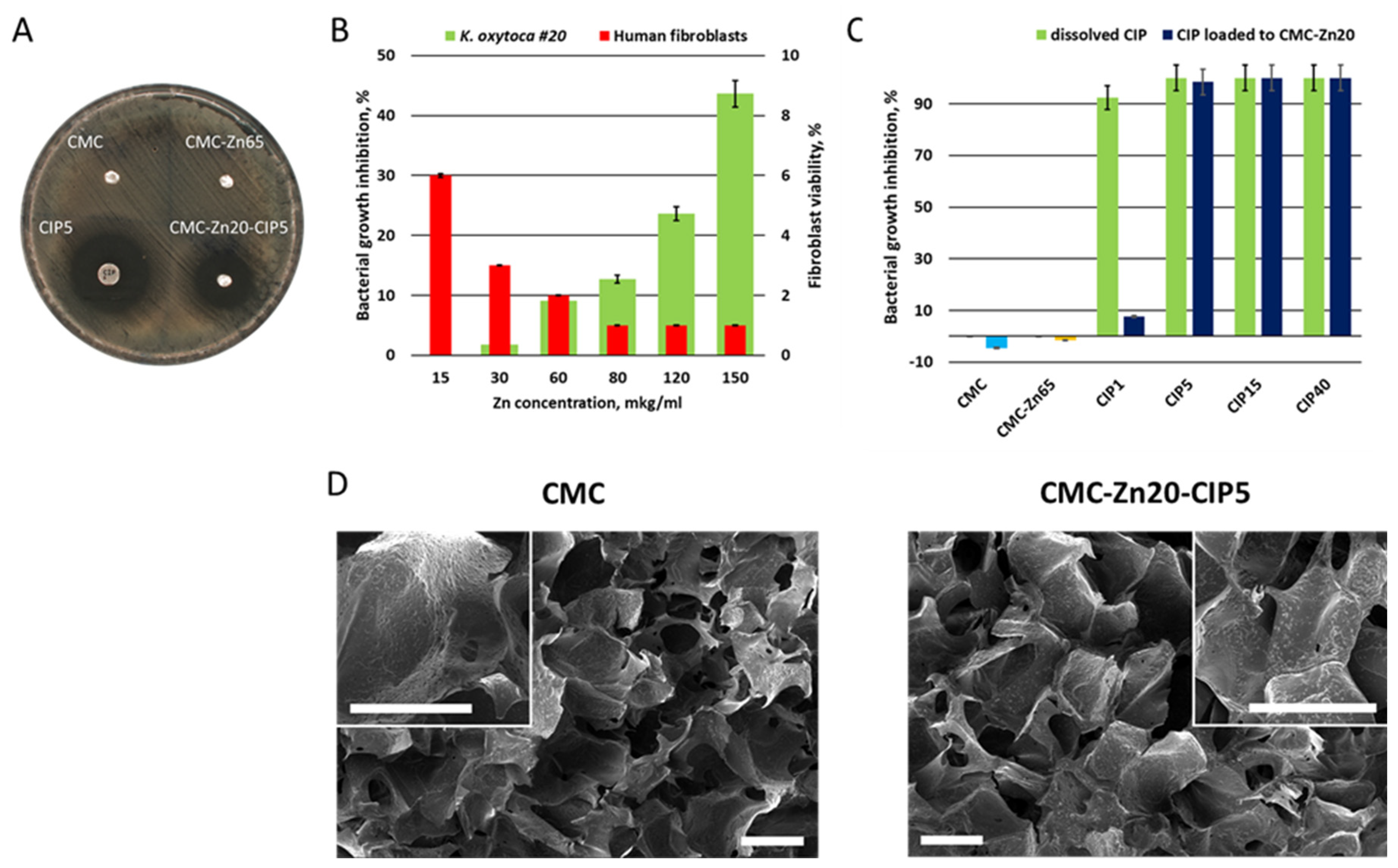
Figure 3.
In vitro viability of human fibroblasts cultivated for 5 days in CMC, CMC-Zn, and CMC-Zn-CIP cryogels (A). Viability of human fibroblasts cultivated for 24 h in DMEM containing 5-120 μg/l of CIP (B). Confocal microscopy images of human fibroblasts cultured in CMC, CMC-Zn, and CMC-Zn-CIP cryogels, and visualized using fluorescently-labeled phalloidin for staining filamentous actin (red color) and DAPI for nuclear staining (cyan color). Autofluorescence of the polymer (shown in cyan color) can also be seen (C).
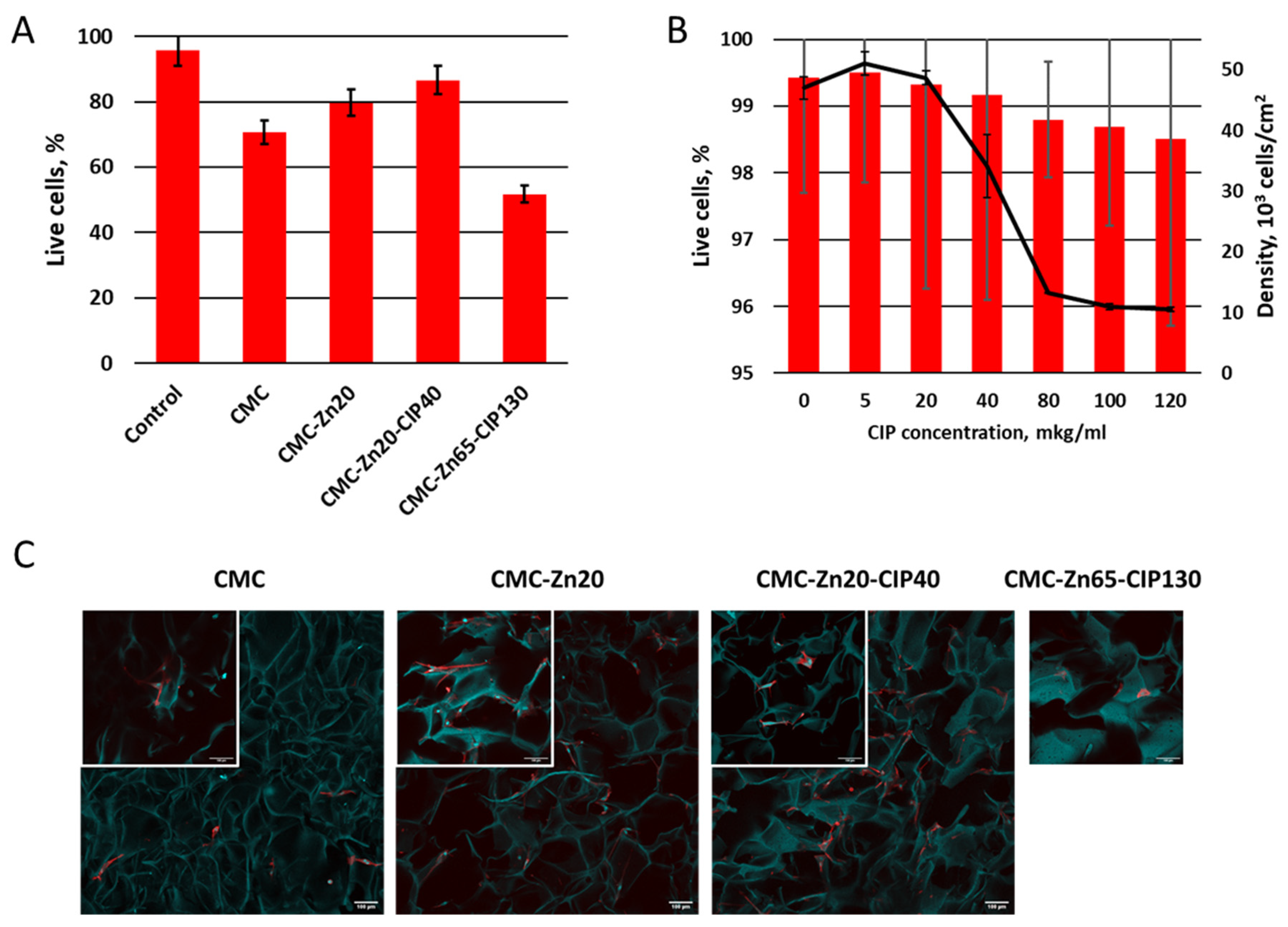
Figure 4.
FT-IR spectra of CMC polymer and dry CIP, and of cryogels: CMC cryogel as synthesized (CMC), after protonation at pH 2 (CMC-H+), after loading with Zn2+ (CMC-Zn45) and Zn2+ and CIP (CMC-Zn45-CIP130).
Figure 4.
FT-IR spectra of CMC polymer and dry CIP, and of cryogels: CMC cryogel as synthesized (CMC), after protonation at pH 2 (CMC-H+), after loading with Zn2+ (CMC-Zn45) and Zn2+ and CIP (CMC-Zn45-CIP130).
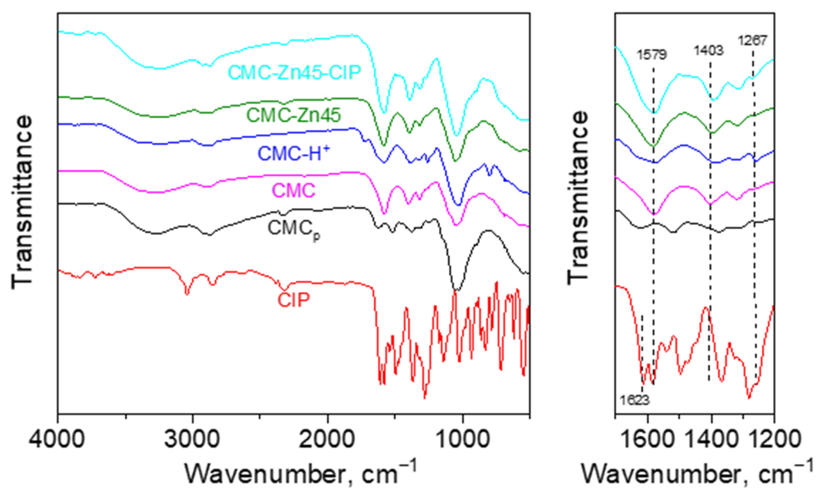
Figure 6.
Profiles of CIP release from the disks of CMC-Zn65-CIP130 cryogels with thickness of 1.1 and 3.7 mm in PBS buffer, pH 7.4 and ultrapure (MQ) water, pH 6.3, T=37°C.
Figure 6.
Profiles of CIP release from the disks of CMC-Zn65-CIP130 cryogels with thickness of 1.1 and 3.7 mm in PBS buffer, pH 7.4 and ultrapure (MQ) water, pH 6.3, T=37°C.
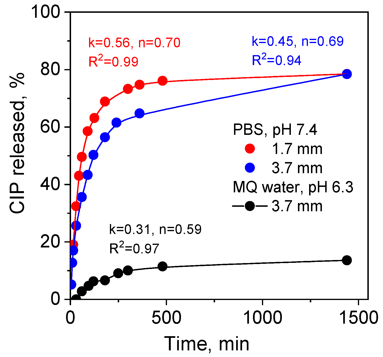

Table 1.
Monomer composition of carboxymethyl chitosan (CMC).
| DA1 | DStot2 | N-DS3 | O-DS4 | Monomer composition | |||
| NH2 | NHR | NR2 | NHCOCH3 | ||||
| 0.25 | 1.49 | 0.29 | 1.20 | 0.46 | 0.29 | 0 | 0.25 |
1 - Degree of acetylation, 2 - Degree of carboxyalkyl substitution (total), 3 - Degree of N-carboxyalkyl substitution, 4 - Degree of O-carboxyalkyl substitution.
Disclaimer/Publisher’s Note: The statements, opinions and data contained in all publications are solely those of the individual author(s) and contributor(s) and not of MDPI and/or the editor(s). MDPI and/or the editor(s) disclaim responsibility for any injury to people or property resulting from any ideas, methods, instructions or products referred to in the content. |
© 2024 by the authors. Licensee MDPI, Basel, Switzerland. This article is an open access article distributed under the terms and conditions of the Creative Commons Attribution (CC BY) license (http://creativecommons.org/licenses/by/4.0/).
Copyright: This open access article is published under a Creative Commons CC BY 4.0 license, which permit the free download, distribution, and reuse, provided that the author and preprint are cited in any reuse.


