Preprint
Article

This version is not peer-reviewed.

Comparative Functional and Morphological Data of Different IOL Dislocation Undo Methods

A peer-reviewed article of this preprint also exists.

Submitted:

02 December 2024

Posted:

04 December 2024

You are already at the latest version

Abstract

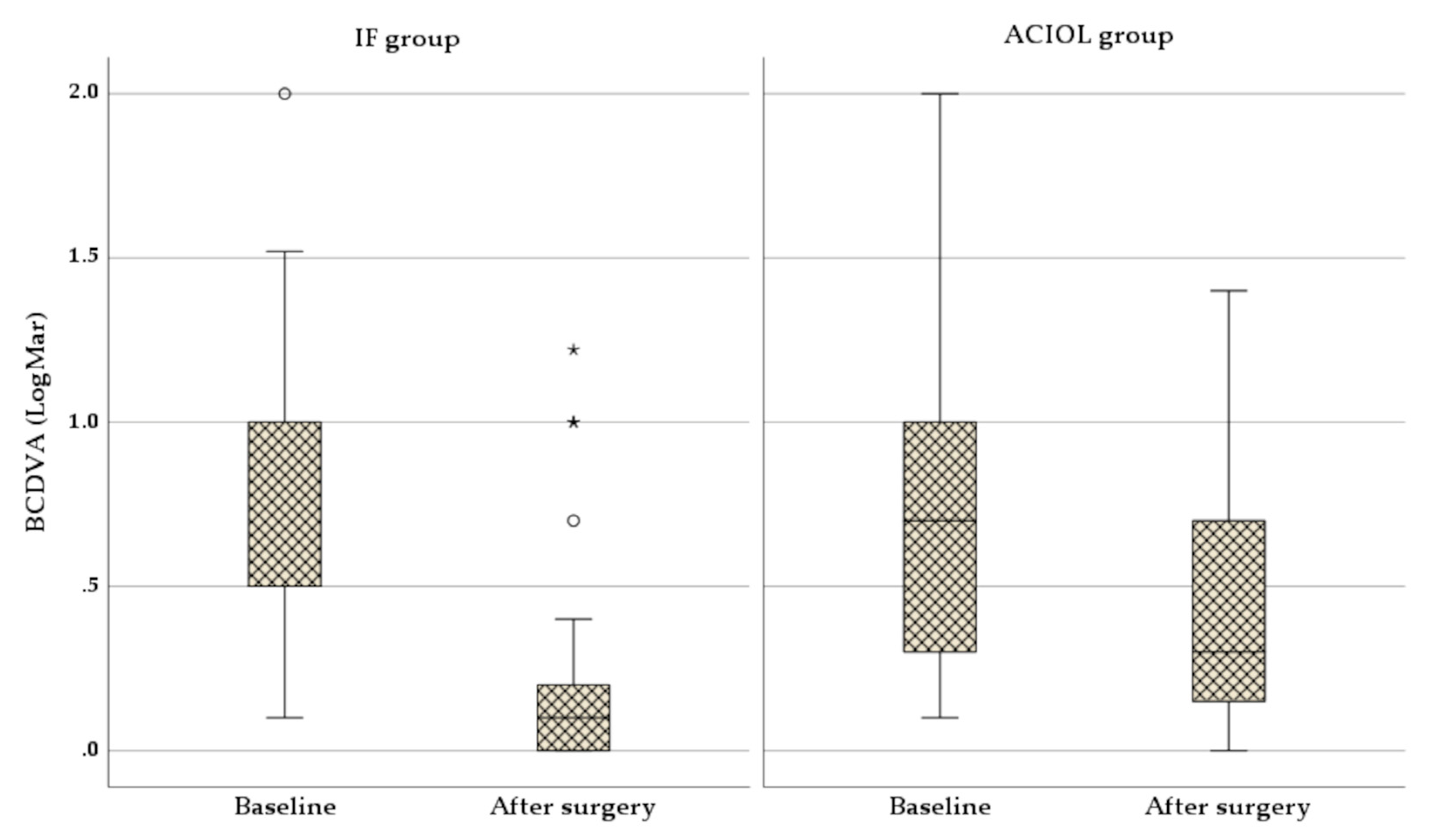
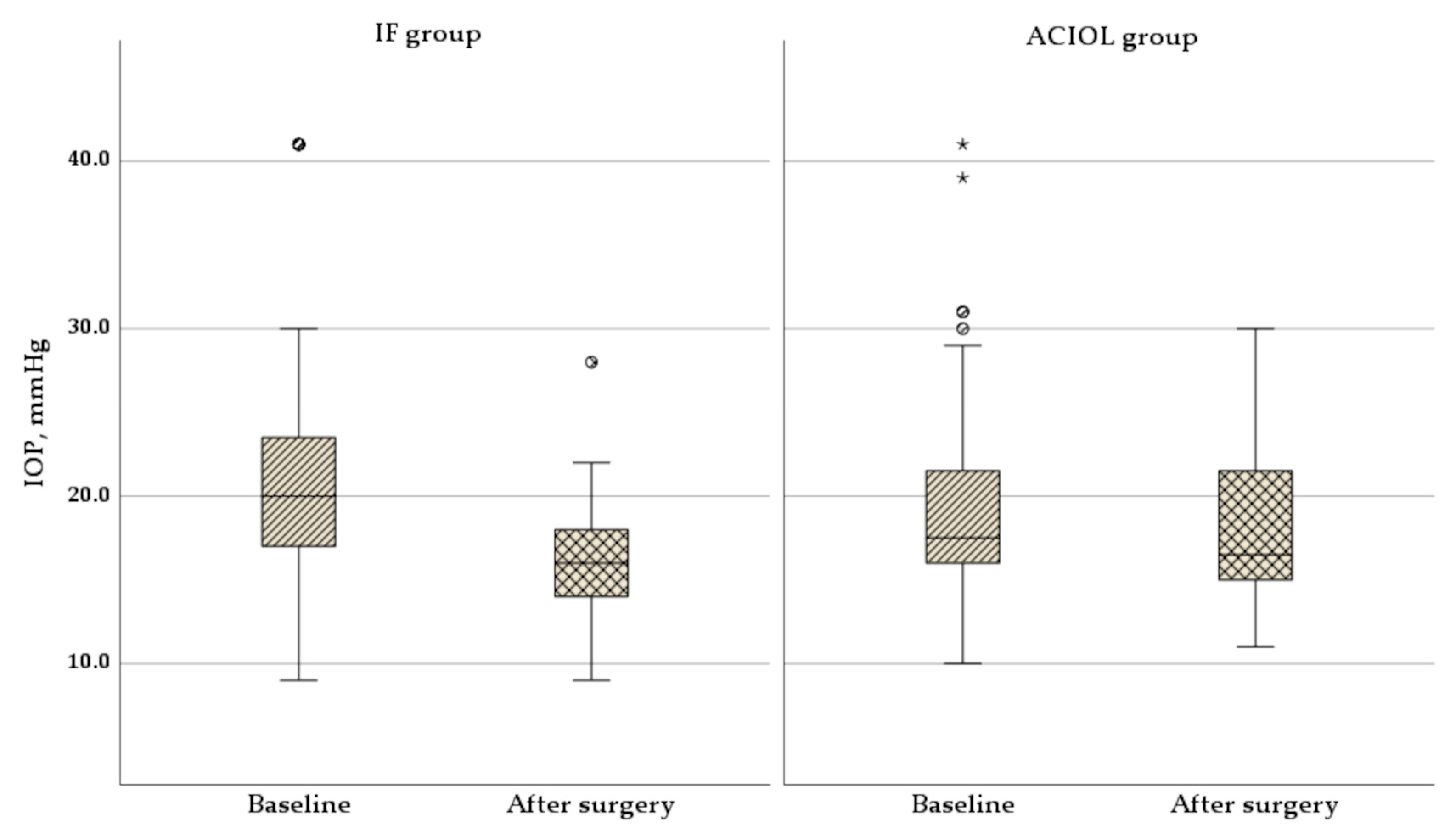
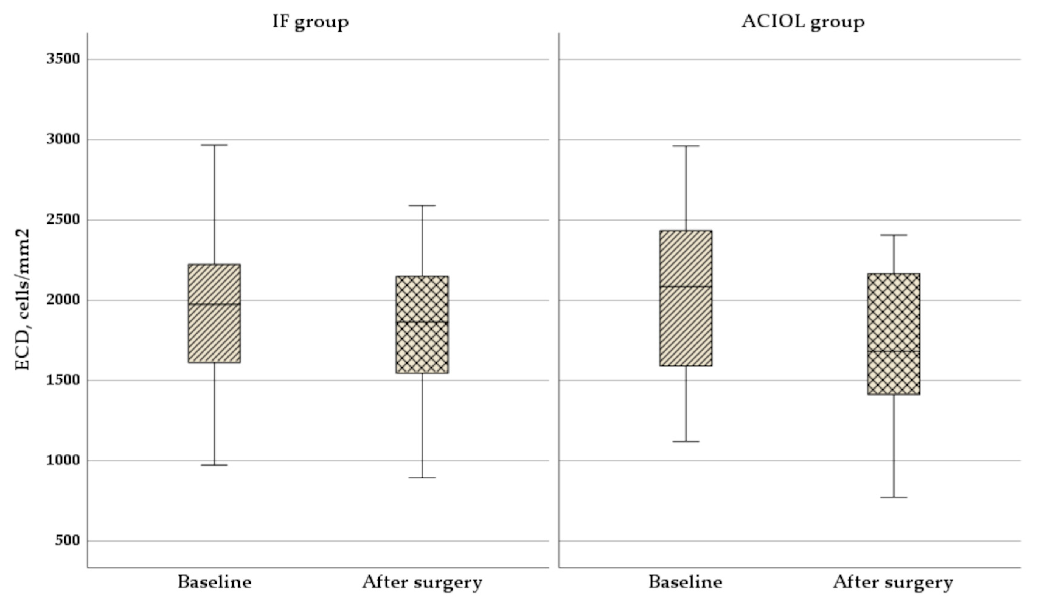
Background/Objectives: This study compared the results of iris-sutured, late spontaneously dislocated IOL-capsular bag complexes with IOL exchange using anterior chamber intraocular lenses (ACIOL) in a tertiary reference center in Lithuania. Methods: A prospective observational study was conducted between 2017 and 2019 involving 80 patients (83 eyes) with late spontaneous IOL-capsular bag dislocation. Patients underwent repositioning and fixation of the dislocated IOL to the iris (IF group) or IOL exchange with an ACIOL implant (ACIOL group). Pre- and postoperative assessments included best-corrected distance visual acuity (BCDVA), intraocular pressure (IOP), corneal endothelial cell density (ECD), and macular thickness (evaluating whether cystoid macular edema (CME) has occurred). Results: Both groups showed a significant improvement in BCDVA, with a more remarkable improvement in the IF group (median: 0.1 logMAR) than in the ACIOL group (median: 0.3 logMAR), p=0.001. Corneal astigmatism increased significantly in the ACIOL group (p<0.001) but remained stable in the IF group. IOP management outcomes were better in the IF group as fewer eyes required additional glaucoma treatment. ECD decreased in both groups but was significantly greater in the ACIOL group (p<0.001). Postoperative CME occurred in 4.4% of IF eyes and 39% of ACIOL eyes (p=0.01). Conclusions: Iris fixation of late dislocated IOL-capsular bag complexes is a safe and minimally invasive technique that offers better visual outcomes, less astigmatism, and fewer complications than ACIOL exchange.

Keywords: 
;  ;  

1. Introduction

Cataract surgery with implantation of an intraocular lens (IOL) is a highly successful surgical procedure that restores patient's vision and quality of life [1]. A sutureless incisions, a continuous curvilinear capsulorhexis, phacoemulsification and placement of a foldable IOL into the capsular bag are now the gold standard for routine cataract surgery, with the number of operations increasing each year [2]. In addition, improvement of surgical techniques and development of new tools such as capsular support devices, iris and capsular hooks together with intraocular sophisticated microsurgical instruments offer possibilities for the safe placement of an IOL in the capsular bag, even in difficult cases [3,4,5,6]. Unfortunately, there are a number of known complications following uncomplicated cataract surgery and spontaneous dislocation of the IOL-capsular bag complex is one of them. The growing number of population with pseudophakic eyes increases the number of patients with the above complication. The incidence of IOL dislocation is reported to be 0.2% to 3%, with late spontaneous dislocation of the IOL-capsular bag complex occurring many years after uneventful cataract surgery [1,7,8,9,10,11,12,13,14]. However, the exact number of late spontaneous IOL-capsular bag complex dislocations is not known, as most studies have included all types of dislocations, including cases of early or even immediate dislocations at the time of cataract surgery [15]. In addition, the prevalence of IOL dislocations varies by region and other factors, such as pseudoexfoliative syndrome [16].
IOL-capsular bag complex dislocation presents significant surgical management challenges and questions for ophthalmologists when it comes to surgical treatment. Two main options are removal of the IOL and replacement with a new IOL or repositioning and fixation of the same IOL in the eye. One of the advantages of IOL repositioning and fixation is that extensive surgical maneuvers, including large incisions and vitreous surgery, can be avoided. This may lead to less corneal endothelial damage and lower postoperative astigmatism [17]. More, replacing of IOL-capsular bag complex with an anterior or posterior chamber IOL with additional fixation is challenging and carries the risk of complications such as choroidal hemorrhage, macular edema etc. development [17].
It seems that the need to preserve the IOL is becoming more common according to the literature [18,19,20,21,22]. Consecvently much attention is being paid to the development of surgical techniques to expand the options in different cases, especially for anterior segment surgeons [18,19,20,21,22].
The current study focuses on the outcomes of the iris sutured late spontaneously dislocated IOL-capsular bag complex and compares it with IOL exchange in a large tertiary reference center in Lithuania.

2. Materials and Methods

The prospective observational study was conducted between 2017 and 2019 year at the Department of Ophthalmology, Lithuanian University of Health Sciences, Kaunas, Lithuania. All study procedures were conducted in accordance with the Declaration of Helsinki. Kaunas Regional Ethics Committee approved the study protocol for biomedical research. Written informed consent was obtained from all patients.
The inclusion criteria were a late spontaneous IOL-capsular bag complex dislocation, with visible IOL in the pupil area more than 6 months after uncomplicated cataract surgery (by phacoemulsification). Totally dislocated IOLs into vitreous body were not included. Pregnant or nursing women, patients younger than 18 years old, patients with uncontrolled systemic illness, congenital or terminal eye diseases as well as patients with a history of eye trauma or vitrectomy were excluded from the study.
A total of 80 patients (83 eyes) with late spontaneous IOL-capsular bag complex dislocation were enrolled. The surgical technique chosen depended on the extent of the IOL dislocation and the patient's preference to retain their own IOL.

2.1. Preoperative Examination

The time interval between cataract surgery and dislocation of the IOL-capsular bag complex, the IOL type, the presence or absence of a capsular tension ring (CTR) and concomitant ocular diseases were recorded before IOL-capsular bag complex dislocation surgery. In addition, previous glaucoma diagnoses, filtering surgeries, and glaucoma medication requirements were recorded, indicating the number of different glaucoma medication drops the patient was taking during the preoperative visit.
Refractive status was examined using autokeratometry (Tonoref III, Nidek, Japan), and spherical and cylindrical powers were converted to the spherical equivalent (SE) (sphere plus half cylinder). Other investigations including: examination of best corrected distance visual acuity (BCDVA) using the Early Treatment Diabetic Retinopathy Study visual acuity chart from a distance of 5 meters, applanation tonometry, using a Goldmann tonometer, slit lamp biomicroscopy and ophthalmoscopy, slit lamp photography of the anterior segment of the eye, measurement of corneal endothelial cell density (ECD) with confocal microscopy of the cornea in vivo (Heidelberg Retina Tomography with III Rostock Cornea Module; Heidelberg Engineering GmbH, Heidelberg, Berlin, Germany).

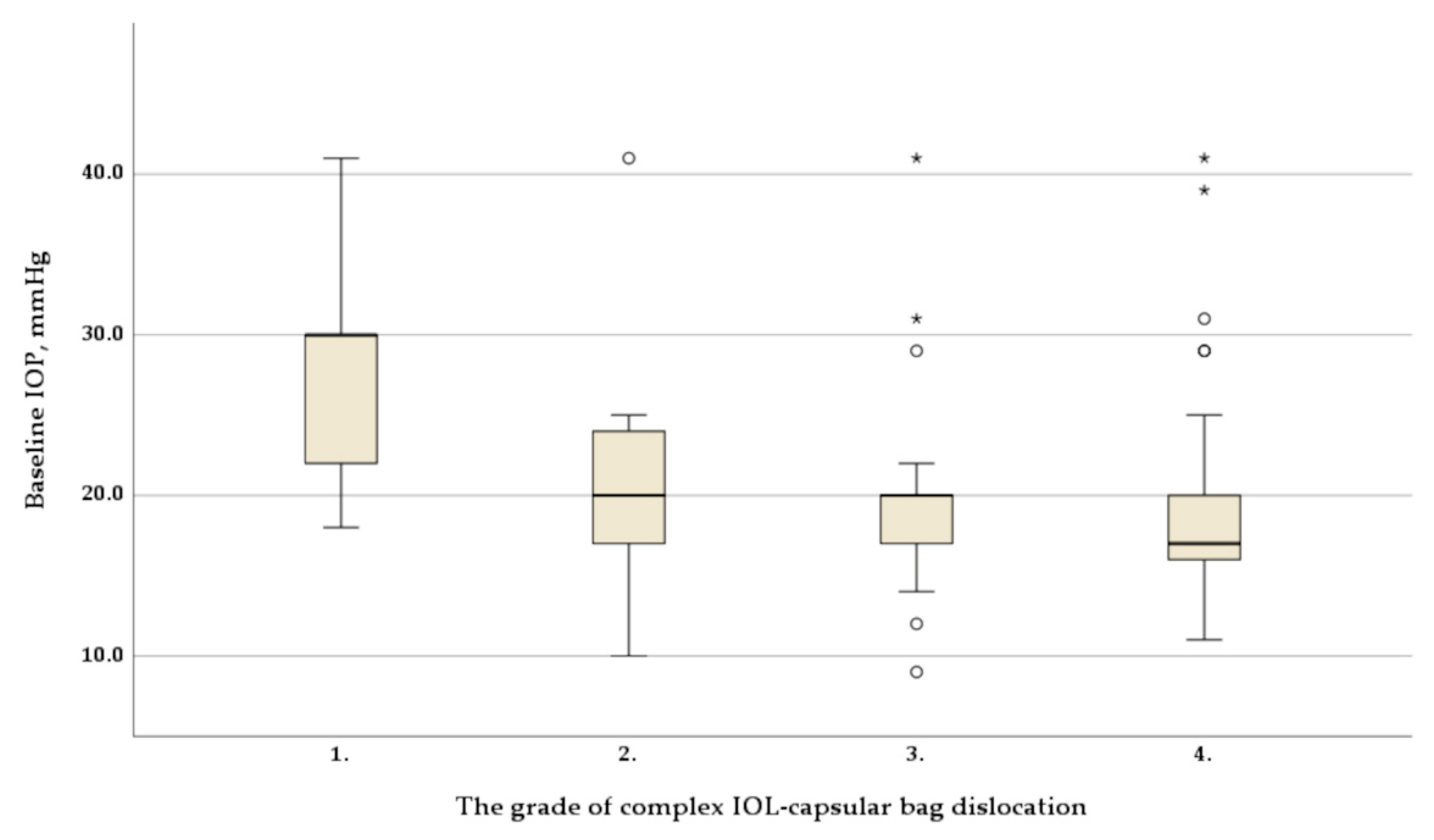
2.1.1. The Grade of IOL-Capsular Bag Complex Dislocation

The degree of IOL-capsular bag complex dislocation was evaluated by slit-lamp examination and photography of the anterior segment were taken. The dislocation was classified as follows:
  • Grade 1: pseudophacodonesis.
  • Grade 2: small decentration. The IOL is slightly decentered, but the equator of the IOL optic's is located behind the iris outside the pupillary zone. With the narrow pupil, only the decentration of the capsulorhexis (no IOL equator) can be observed.
  • Grade 3: moderate dislocation. The equator of the IOL optic's is above or coincides with the line drawn through the centre of the pupil.
  • Grade 4: advanced dislocation. The IOL is more dislocated than grade 3 and the equator of the IOL optic's is below the line horisontaly drawn through the centre of the pupil.
The assessment of the degree of dislocation methodology is described in more detail by our group [23].

2.2. Surgical Procedure

2.2.1. IOL Repositioning and Fixation to the Iris Procedure (IF Group)

Two paracentesis in the clear cornea are performed along the anticipated path of the needle’s entry and exit. 10-0 Polypropylene suture with a long curved needle (PC 10, Alcon) is used. The needle is passed through the first stab incision and the pupil beneath the haptics of IOL and guided into the anterior chamber through the iris in a matched place for IOL fixation. IOL-capsular bag complex can be temporarily stabilized by planting a spatula behind it to facilitate the puncture of a fibrosed capsular bag and to prevent subsequent additional damaging of residual Zinn’s zonules. A 27-gauge cannula is introduced through the distal paracentesis, and the needle is docked to facilitate exit. The needle is passed back into the eye through the second stab incision, iris (close to the first perforation), above IOL haptics, through the pupil into the anterior chamber and out of the eye through the first stab incision. At this moment, IOL haptic is encircled by the suture. The first double throw is performed outside the eye. Both suture ends are pulled to make the first floor of the knot behind the iris. The long end of the thread is pulled outside the eye trough the second stab incision and double throw is pulled inside of the eye. A gently tied knot is formed inside the eye and beneath the iris. The long suture is repeatedly drawn through the first stab incision with a microhook and second single throw is performed outside the eye. The third single throw is performed in the same manner. The ends of the suture are pulled away, bringing the third throw snugly into position without twisting. The ends of the suture are cut-off with vitrectomy scissors or knife inside the eye and the knot is buried behind the iris. Using this fixation method the pupil remains circular after the procedure. This modified Siepser knot methodology was described earlier [24].

2.2.2. Replacing the IOL-Capsular Bag Complex with Anterior Chamber Intraocular Lens Implant Procedure (ACIOL Group)

Three sclerotomies are performed by 25 G troacars, two paracenteses – by 20 G knife. Infusion line is inserted into troacar and after checking its full penetration trough pars plana balanced saline solution (BSS) infusion is started. After full pars plana vitrectomy is done an iridotomy at 12 o’clock is made by vitrector at slow cutting rate.
The main 6 mm corneal incision is made by knife. The old IOL is removed and new IOL (Kelman Multiflex III (Alcon Laboratories, Inc)) is implanted into the anterior chamber of the eye. The IOL is rotated into a horizontal position. The main corneal incision is sutured with three single-knotted 10/0 Ethylon sutures. The paracenteses are closed by injecting a BSS into the corneal stroma. The trocars are removed from the sclerotomies.

2.3. Postoperative Examination

All patients underwent corneal suture removal 3 months after replacing the IOL-Capsular bag complex with anterior chamber lens surgery. Six months after the surgery, all participants underwent the same ophthalmic examination as before surgery. Postoperative complications were recorded. After surgery, to evaluate status of macular area optical coherence tomography (DRI OCT Triton plus (Ver.10.13), Topcon, Japan) was performed. We investigated whether cystoid macular edema (CME) was present and measured the postoperative central macular thickness.

2.4. Statistical Analysis

Statistical analysis was performed using IBM SPSS Statistics for Windows, Version 29.0.2.0 Armonk, NY: IBM Corp. The groups were compared using Mann–Whitney U test, T test, chi-square test and Kruskal-Wallis test. Pre- and postoperative data were compared using the Wilcoxon signed-rank test and T test. The data are presented as mean (standard deviation (SD)), minimum (min.) and maximum (max.) values, median (interquartile range (IQR)) or number (percentage), and p < 0.05 was considered statistically significant.

3. Results

3.1. Demographic Data

In total, 80 patients (83 eyes) (30 female, 50 male) were included in to the study. Forty-three eyes were in IF group, and 40 eyes were in the ACIOL group. Mean age at the moment of diagnosed IOL dislocation was 77.08 ± 7.28 years. The mean patients’ age at the moment of cataract surgery was 69.45 ± 8.02 years. The mean time from cataract surgery to IOL dislocation was 7.64 ± 3.88 years. Baseline characteristics and preoperative study parameters are presented in Table 1.
Age, gender, and time since cataract surgery did not differ between the two groups. Also, the data related to the cataract surgery (IOL type, CTR presence and previous laser capsulotomy) did not differ between the groups. Trabeculectomy (TB) prior to IOL dislocation was performed more frequently in the IF group than in the ACIOL group. However, this difference was not significant (Table 1).

3.2. Visual Outcomes

As shown in Figure 1, median BCDVA statistically significantly improved throughout the follow-up period in both groups, increasing from 0.5 (1.0 – 0.5) logMAR at baseline to 0.1 (0.2 – 0.0) logMAR at 6 months in the IF group (Z = -5.13, p < 0.001), and from 0.7 (1.0 – 0.3) logMAR at baseline to 0.3 (0.7 – 0.13) logMAR at 6 months in the ACIOL group (Z = -3.223, p = 0.001). No statistically significant differences were observed between the two groups at baseline BCDVA (p = 0.508), but it was significantly better in IF group after 6 months (p = 0.001).
During the follow-up period, there was no statistically significant change in corneal astigmatism in the IF group (median cylinder 1.10 (0.65 – 1.55) D at baseline and 0.86 (0.41 – 1.39) D at 6 months, (Z = -1.863, p = 0.62)). However, the change was significant in the ACIOL group 6 months after surgery (median cylinder 1.01 (0.53 – 1.51) D at baseline and 2.8 (1.51 – 4.72) D at 6 months, (Z = -4.888, p=0.001)). No significant differences in corneal astigmatism were found between the two groups at baseline (p = 0.613), but after 6 months corneal astigmatism was significantly higher in the ACIOL group (p < 0.001).
The median SE changed statistically significant from 1.38 (-0.63 – 6.38) D at baseline to –0.75 (-1.63 – -0.13) D at 6 months (Z = -3.797, p < 0.001) in the IF group and from 5.38 (0.47 – 10.63) D at baseline to –1.31 (-1.97 – -0.63) D at 6 months (Z = -5.498, p < 0.001) in the ACIOL group. The SE was significantly higher in the ACIOL group at baseline (p = 0.002) and did not differ significantly between the groups after 6 months (p = 0.001).

3.3. IOP and Glaucoma Treatment Outcomes

45 (54.22%) eyes were diagnosed with primary open angle glaucoma before the IOL-capsular bag complex dislocation. 15 (18.1%) eyes were diagnosed with secondary glaucoma at the time of the IOL-capsular bag complex dislocation. Taken together, this represents 60 (72.32%) eyes of the study participants. All investigated eyes (n = 83) had a median preoperative IOP of 19.0 (16.0 - 22.0) mmHg. Despite medical treatment an IOP ≥ 21 mmHg before surgery of IOL dislocation was found in 24 (28.9%) eyes and even half of them (12 (50%) eyes) had an IOP ≥30 mmHg.
The analysis of the preoperative IOP in relation to the grade of IOL dislocation revealed a statistically significantly higher IOP with a grade 1 dislocation compared to grades 2, 3 and 4 (χ2 = 13.783, df = 3, p = 0.003) (Figure 2).
As shown in Figure 3, IOP decreased throughout the follow-up period in both groups, decreasing from 20.0 (17.0 – 25.0) mmHg at baseline to 16.0 (14.0 – 18.0) mmHg at 6 months in IF group (Z = -4.710, p < 0.01), and from 17.5 (16.0 – 21.75) mmHg at baseline to 16.5 (15.0 – 21.75) mmHg at 6 months in the ACIOL group (Z = -0.684, p = 0.494). There was no significant difference in IOP between the groups before or after IOL dislocation surgery (p = 0.248 and p = 0.057, respectively), but the change was statistically significantly bigger in the IF group (p = 0.004).
The changes in IOP-lowering medical treatment between the preoperative and postoperative visits are shown in Table 2.
The amount of antiglaucoma medication did not change statistically significantly in either group when comparing the results before and after IOL dislocation treatment (IF group p = 0.519, ACIOL group p = 0.159). In the IF group, the amount of antiglaucoma drops did not change in 22 (70.97%) eyes, increased in 4 (12.90%) eyes and decreased in 5 (16.13%) eyes. In the IF group, 3 of 12 eyes that were not diagnosed with glaucoma before IOL dislocation treatment started with antiglaucoma therapy after surgical IOL dislocation treatment. In group ACIOL, the amount of antiglaucoma drops did not change in 18 (62.07%) eyes, increased in 8 (27.59%) eyes and decreased in 3 (10.34%) eyes.
4 (9.3%) eyes from the IF group and 1 (2.5%) eye from the ACIOL group underwent filtering surgery for subcompensated IOP during 6 month period after IOL dislocation treatment (p = 0.193). In the IF group, the mean IOP of these eyes was 38.0 mmHg before IOL dislocation treatment and 11.5 mmHg 6 months after IOL dislocation treatment and filtering surgery, without antiglaucoma medication. In ACIOL group, the IOP of the eye with additional filtering surgery was 31.0 mmHg before IOL dislocation and 15.0 mmHg 6 months after IOL dislocation treatment filtering surgery. 3 drops of an antiglaucoma medication were administered daily to support the IOP.

3.4. Other Postoperative Outcomes

As shown in Figure 4, mean ECD significantly decreased throughout the follow-up period in both groups, from 1922 ± 468 cells/mm2 at baseline to 1800 ± 433 cells/mm2 at 6 months in IF group (p < 0.001), and from 2021 ± 480 cells/mm2 at baseline to 1683 ± 446 at 6 months in the ACIOL group (p < 0.001). There was no significant difference in ECD between the groups before or after IOL dislocation surgery (p = 0.347 and p = 0.230, respectively), but the change was statistically significantly bigger in the ACIOL group (p < 0.001).
CME was found in 18 of 83 eyes (21.69%) after surgery. Statistically significantly less cases occurred in the IF group than in the ACIOL group (16 (39%) and 2 (4.4%), respectively, p = 0.01). The central macular thickness was higher in the ACIOL group (p = 0.01) (Table 2). In 3 (6.7%) of the 43 eyes in the IF group, the IOL was re-fixated due to insufficient centering of the IOL.
There was no intraocular hemorrhage, fluid misdirection syndrome during surgery, bullous keratopathy, anterior pigmentary dispersion syndrome, chronic uveitis, retinal detachment, or endophthalmitis during 6-month period after surgery in both groups.

4. Discussion

Over the decades, a number of surgical techniques have been developed to treat IOL dislocations. These include removal of the dislocated IOL and secondary IOL implantation (anterior chamber IOL, iris-claw IOL, iris-fixated IOL, scleral-fixated IOL with suture, scleral-fixated IOL without suture) or repositioning and fixation of the same IOL to the sclera or iris, each with its benefits and risks [25,26,27,28,29]. Despite numerous studies on this topic, however, no single technique has gained an advantage over the others [26,27,28,29,30,31]. Nevertheless, complications such as corneal decompensation, glaucoma, chronic inflammation and CME are more common with IOL exchange (due to the large incision wounds and longer operating times) than with other treatment options, including repositioning of a dislocated IOL using a scleral or iris fixation in the globe as a closed system [32]. Scleral fixation of IOLs, especially sutureless technique, reduces the risk of many of these complications [28]. However, it is considered the most technically challenging technique requiring a posterior segment surgeon [28]. When performing to deep scleral groove, a microvitreoretinal knife or needle can traumatize the ciliary vessels, which carries the risk of suprachoroidal hemorrhage (incidence after sutured scleral fixation is up to 3 % and rarely after sutureless scleral fixation) [33]. On the other hand, extrusion of the haptics (slippage of the haptics from the scleral tunnel) may occur due to thin scleral flaps, poor scleral tunnel construction or adhesion, excessive use of scleral cauterization, and congenital or acquired fragility of the sclera [34]. Reported rates vary from 0.8% to 12.5% [34]. Permanent externalized sutures or ends of cauterized haptics may also increase the risk of late endophthalmitis [35,36]. Another common problem with sutureless fixation is the instability of the IOL [37]. Also, the increased iris flutter caused by eye movement can lead to a reverse pupillary block due to the absence of the barrier behind the iris, which prevents the aqueous humor and vitreous humor from flowing into the anterior chamber [38].
To expand the options for anterior segment surgeons, we have focused on the technique of the iris fixation for dislocated IOL-capsular bag complex. Our technique we have presented is safe and minimally invasive. Performing IOL fixation with this technique, there are no blind movements of the needle behind the iris. Haptics (with surrounding capsule) being sutured to the iris under direct observation under the microscope, precisely in the anticipated location with no or minimal pupil deformation with the knot tied behind the iris. Fixation of dislocated IOL to the iris using McCannel sutures is generally performed in a 'blind' manner (when the needle is behind the iris) [39]. This may cause an improper localization of the haptic and suturing to the iris in the unintended place. Furthermore, it is challenging to penetrate the iris from behind close to the first prick. In addition, the presence of fibrosed capsular bag makes the procedure more risky due to IOL luxation into the vitreous. The described surgical technique of additional stabilization of IOL-capsular bag complex with an instrument (spatula) during surgery prevents damage to the remaining Zinn’s zonules. In addition, another unique feature is that the Siepser slipknot remains behind the iris (between the posterior surface of the iris and the IOL-capsular bag complex). We believe that creating this small space between these structures has positive properties. After such fixation, none of the patients had pigment dispersion syndrome or uveitis-glaucoma-hyphema syndrome. This technique also does not require large incisions in the cornea or sclera and does not harm the conjunctiva [20]. As 72.3% of patients had concomitant glaucoma, it was important to find the least traumatic method of repositioning the IOL to spare the superior conjunctiva and sclera in patients if due to glaucoma should subsequent filtration surgery be required. This technique can be used by anterior segment surgeons as no additional surgical intervention is required. However, experience with McCannel suturing and tying a Siepser slipknot is essential to maintain the minimally invasive nature of this technique.
Our prospective study is unique because we have included cases of late spontaneous IOL-capsular bag complex dislocation treated with repositioning of the same IOL using iris suture fixation or IOL exchange into the anterior chamber IOL. Early in our study, the IOL exchange into the anterior chamber IOL was preferred. Still, the trend shifted to the IOL exchange to iris-claw IOL or IOL repositioning as these techniques developed and improved.
In the IF group, very encouraging visual and refractive results were achieved. This supports other authors' findings from studies using iris-suturing of modified McCannel sutures in cases with IOLs without capsular support or in cases with IOL-capsular bag complex dislocation [39,40]. In most patients in IF group (with the exception of 3 patients (6.7%) who required re-fixation due to inadequate IOL centration), we observed an improvement in BCDVA. Significant improvement in visual acuity was also observed in the ACIOL group. However, it was significantly less than in the IF group due to the significant increase in corneal astigmatism. Often, anterior chamber or iris-claw IOLs are implanted, but large corneal incisions (approximately 6 mm) are required, which can increase corneal astigmatism. Our technique of IOL repositioning with iris fixation does not require a large corneal incision, and this was the reason for minor changes in the corneal cylinder. Condon et al included forty-six patients who underwent foldable acrylic IOL implantation using peripheral iris suture fixation for aphakia without capsule support. They also observed no significant changes in corneal astigmatism [39]. The results of the study by Michaeli et al. also confirm these findings [41]. Furthermore, the more anterior position of the lens caused a myopic shift, which was acceptable for the patients. In a retrospective study, Faria et al. examined 36 eyes with dislocated IOLs treated by an iris suture and also found a myopic shift in all patients [42]. Our study data also supports this.
IOP and the need for glaucoma medication did not change statistically significantly in either group. However, using of anterior chamber IOLs implants, may be associated with IOP elevation and glaucoma development, meanwhile eyes with iris sutured IOLs were less prone to IOP increase. In our series, we found 15 patients (18.07%) with postoperatively IOP elevation that was resolved with topical therapy (7 in the IF group and 8 in the ACIOL group). In 5 patients with high preoperative IOP (>30 mmHg), filtering surgery was performed within 6 months after treatment of IOL dislocation to control IOP. We cannot extrapolate from a few cases to others, but our technique of IOL repositioning with iris fixation does not harm the sclera and conjunctiva as we expected. In 4 cases, the target IOP was achieved after IOL repositioning with iris fixation and filtering surgery without medication. After IOL exchange and filtering surgery in 1 case, 3 drops of antiglaucoma medication were administered daily to support the target IOP.
A decrease in corneal ECD was observed 6 months after IOL dislocation surgery in both groups, but change being significantly bigger in the ACIOL group. Dzhaber et al. retrospectively examined 117 eyes in which iris fixation was used and recorded a decrease in ECD of almost 200 cells/mm2 [43]. Kim et al. recorded a similar decrease (12.7% ± 8.7%) in ECD after iris fixation, and this change was not different from the transscleral fixation group (10.9% ± 9.2%) (p = 0.16) [44].
CME is of particular concern because it is the most common cause of postoperative visual acuity deterioration in iris- and scleral-fixated IOLs. As found in previous studies, the rate of CME in iris-fixated IOLs is equal to or lower than in scleral-fixated and anterior chamber IOLs [45]. A randomized trial comparing the 3 IOL fixation strategies in 176 patients lacking adequate capsule support has been published [46]. Although visual outcomes were similar in the 3 groups, it was found that iris-sutured IOLs were associated with significantly less CME (20%) versus anterior chamber IOLs (38%) or scleral-sutured IOLs (41%) (P=0.02) [46]. Or study results do not contradict these findings.
In a retrospective study, Caporossi et al. reported on 41 eyes with IOL dislocation treated with repositioning of the same IOL using iris suture fixation in two groups (acrylic and rigid one-piece IOL group and acrylic three-piece IOL group) [21]. They achieved good refractive results, a stable IOL, no increase in surgically induced astigmatism and a low number of complications (no intraoperative complications, endothelial dysfunction and pigment dispersion). Results did not differ between groups. In summary, they concluded that the iris fixation technique for one- and three-piece IOLs is a safe and valid option for treating IOL dislocations. Our results do not contradict these findings.
In the literature overview we did not find methods for sutureless/automated fixation technique of subluxated IOL to the iris.

5. Conclusions

According to our report, the iris fixation technique seems to be safe for treating of dislocated IOL-capsular bags complex. We have experienced the distinct advantage of using the same IOL in a closed eye without large incision. Small number of complications, good morphological and functional outcomes enables to recommend this technique for the clinical use. Development of more elegant sutureless/automated technique for dislocated IOL fixation to the iris is desirable.

Author Contributions

Conceptualization, V.J. and R.V.; methodology, V.J., M.S. and R.V.; software, R.V.; validation, R.V.; formal analysis, R.V. and U.R.; investigation, R.V.; resources, R.V. and M.S.; data curation, V.J.; writing—original draft preparation, R.V.; writing—review and editing, V.J., M.S. and U.R.; visualization, R.V.; supervision, V.J.; project administration, V.J.; funding acquisition, V.J. All authors have read and agreed to the published version of the manuscript.

Funding

This research received no external funding.

Institutional Review Board Statement

The study was conducted in accordance with the Declaration of Helsinki and approved by the Kaunas Regional Biomedical Research Ethics Committee (No. BE-2-40 2017-07-12).

Informed Consent Statement

Informed consent was obtained from all subjects involved in the study.

Data Availability Statement

The datasets used and/or analyzed during the current study are available from the corresponding author on reasonable request.

Acknowledgments

For Irena Nedzelskiene, the Department of Dental and Oral Pathology, Faculty of Odontology, Medical academy, Lithuanian University of Health Sciences in statistics analysis.

Conflicts of Interest

The authors declare no conflicts of interest.

References

  1. Ascaso, F. J.; Huerva, V.; Grzybowski, A. Epidemiology, Etiology, and Prevention of Late IOL-Capsular Bag Complex Dislocation: Review of the Literature. Journal of Ophthalmology 2015, 2015, 1–7. [Google Scholar] [CrossRef]
  2. Gollogly, H. E.; Hodge, D. O.; St. Sauver, J. L.; Erie, J. C. Increasing incidence of cataract surgery: Population-based study. Journal of Cataract and Refractive Surgery 2013, 39(9), 1383–1389. [CrossRef]
  3. Weber, C. H.; Cionni, R. J. All about capsular tension rings. Current Opinion in Ophthalmology 2015, 26(1), 10–15. [Google Scholar] [CrossRef] [PubMed]
  4. Debdulal Chakraborty, Ayan Mohanta, A. B. B-HEX pupil expander in vitreoretinal surgery – A case series. Indian journal of ophthalmology 2020, 68(6), 1188–1191. [CrossRef]
  5. Balal, S.; Jbari, A. S.; Nitiahpapand, R.; Cook, E.; Akhtar, W.; Din, N.; et al. Management and outcomes of the small pupil in cataract surgery: iris hooks, Malyugin ring or phenylephrine? Eye (Basingstoke) 2021, 35(10), 2714–2718. [Google Scholar] [CrossRef]
  6. Singh, M. K.; Ambati, B. K.; Crandall, A. S.; Moran, J. A.; City, S. L. Adult with Traumatic Cataract. J Cataract Refract Surg. 2017, 43(5), 590–592. [Google Scholar] [CrossRef] [PubMed]
  7. Davis, D.; Brubaker, J.; Espandar, L.; Stringham, J.; Crandall, A.; Werner, L.; et al. Late In-the-Bag Spontaneous Intraocular Lens Dislocation. Evaluation of 86 Consecutive Cases. Ophthalmology 2009, 116(4), 664–670. [Google Scholar] [CrossRef]
  8. Clark, A.; Morlet, N.; Ng, J. Q.; Preen, D. B.; Semmens, J. B. Whole population trends in complications of cataract surgery over 22 years in Western Australia. Ophthalmology 2011, 118(6), 1055–1061. [Google Scholar] [CrossRef] [PubMed]
  9. Jakobsson, G.; Zetterberg, M.; Lundström, M.; Stenevi, U.; Grenmark, R.; Sundelin, K. Late dislocation of in-the-bag and out-of-the bag intraocular lenses: Ocular and surgical characteristics and time to lens repositioning. Journal of Cataract and Refractive Surgery 2010, 36(10), 1637–1644. [Google Scholar] [CrossRef] [PubMed]
  10. Fernández-Buenaga, R.; Alio, J. L.; Pérez-Ardoy, A. L.; Larrosa-Quesada, A.; Pinilla-Cortés, L.; Barraquer, R.; et al. Late in-the-bag intraocular lens dislocation requiring explantation: risk factors and outcomes. Eye 2013, 27(7), 795–802. [Google Scholar] [CrossRef] [PubMed]
  11. Jehan, F. S.; Mamalis, N.; Crandall, A. S. Spontaneous late dislocation of intraocular lens within the capsular bag in pseudoexfoliation patients. Ophthalmology 2001, 108(10), 1727–1731. [Google Scholar] [CrossRef]
  12. Gross, J. G.; Kokame, G. T.; Weinberg, D. V. In-the-bag intraocular lens dislocation. American Journal of Ophthalmology 2004, 137(4), 630–635. [Google Scholar] [CrossRef] [PubMed]
  13. Kim, S. S.; Smiddy, W. E.; Feuer, W.; Shi, W. Management of Dislocated Intraocular Lenses. Ophthalmology 2008, 115(10), 1699–1704. [Google Scholar] [CrossRef] [PubMed]
  14. Mönestam, E. I. Incidence of Dislocation of Intraocular Lenses and Pseudophakodonesis 10 Years after Cataract Surgery. Ophthalmology 2009, 116(12), 2315–2320. [Google Scholar] [CrossRef]
  15. Lee, G. I.; Lim, D. H.; Chi, S. A.; Kim, S. W.; Han, J.; Shin, D. W.; et al. Incidence and characteristics of intraocular lens dislocation after phacoemulsification: An eight-year, nationwide, population-based study. Journal of Clinical Medicine 2021, 10(17). [CrossRef]
  16. Østern, A. E.; Sandvik, G. F.; Drolsum, L. Late in-the-bag intraocular lens dislocation in eyes with pseudoexfoliation syndrome. Acta Ophthalmologica 2014, 92(2), 184–191. [Google Scholar] [CrossRef]
  17. Shin, Y. I.; Park, U. C. Surgical Outcome of Refixation versus Exchange of Dislocated Intraocular Lens: A Retrospective Cohort Study. Journal of Clinical Medicine 2020, 9(12), 3868. [Google Scholar] [CrossRef] [PubMed]
  18. Kirk, T. Q.; Condon, G. P. Simplified ab externo scleral fixation for late in-the-bag intraocular lens dislocation. Journal of Cataract and Refractive Surgery 2012, 38(10), 1711–1715. [Google Scholar] [CrossRef] [PubMed]
  19. Huang, W. Y.; Chen, Y. J. Sutured scleral fixation of existing subluxated/dislocated acrylic one-piece intraocular lenses. International Journal of Ophthalmology 2024, 17(4), 665–669. [Google Scholar] [CrossRef]
  20. Siegel, M. J.; Condon, G. P. Single suture iris-to-capsulorhexis fixation for in-the-bag intraocular lens subluxation. Journal of Cataract and Refractive Surgery 2015, 41(11), 2347–2352. [Google Scholar] [CrossRef] [PubMed]
  21. Caporossi, T.; Tartaro, R.; Franco, F. G. S.; Barca, F.; Finocchio, L.; Bacherini, D.; et al. IOL repositioning using iris sutures: A safe and effective technique. International Journal of Ophthalmology 2019, 12(12), 1972–1977. [Google Scholar] [CrossRef] [PubMed]
  22. Soiberman, U.; Pan, Q.; Daoud, Y.; Murakami, P.; Stark, W. J. Iris suture fixation of subluxated intraocular lenses. American Journal of Ophthalmology 2015, 159(2), 353–359. [Google Scholar] [CrossRef] [PubMed]
  23. Vaiciuliene, R.; Jasinskas, V. Corneal endothelial status in different grades of late spontaneous in-the-bag IOL dislocation. International Ophthalmology 2021, 41(5). [CrossRef]
  24. Jasinskas, V.; Vaiciuliene, R.; Varoniukaite, A.; Speckauskas, M. Novel microsurgical management of uveitis-glaucoma-hyphema syndrome. International Ophthalmology 2019, 39(7), 1607–1612. [Google Scholar] [CrossRef]
  25. Castaldelli, G. B.; Firmino, G. D. C.; Castaldelli, V. A.; Costa, R. D. S.; Ribeiro, J. C. Use of Techniques for Scleral and Iris Fixation in Secondary Implantation of Intraocular Lenses. Ophthalmic Research 2021, 64(1), 1–9. [Google Scholar] [CrossRef] [PubMed]
  26. Wong, H. M.; Kam, K. W.; Rapuano, C. J.; Young, A. L. A Systematic Review on Three Major Types of Scleral-Fixated Intraocular Lens Implantation. Asia-Pacific Journal of Ophthalmology. Lippincott Williams and Wilkins July 19, 2021, pp 388–396. [CrossRef]
  27. Zhang, C.; Palka, C.; Zhu, D.; Lai, D.; Winokur, J.; Shwani, T.; et al. Clinical Outcomes in Scleral Fixation Secondary Intraocular Lens with Yamane versus Suture Techniques: A Systematic Review and Meta-Analysis. Journal of Clinical Medicine. Multidisciplinary Digital Publishing Institute (MDPI) June 1, 2024. [CrossRef]
  28. Tsz Hin Alexander Lau 1, Anubhav Garg, Marko M Popovic, Peter J Kertes, R. H. M. Scleral-fixated and iris-fixated intraocular lens implantation or fixation:meta-analysis. J Cataract Refract Surg. 2022, 48(12), 1462–1468.
  29. Chang, Y. M.; Weng, T. H.; Tai, M. C.; Chen, Y. H.; Lee, C. H.; Chang, W. C.; et al. A meta-analysis of sutureless scleral-fixated intraocular lens versus retropupillary iris claw intraocular lens for the management of aphakia. Scientific Reports 2024, 14(1), 1–10. [Google Scholar] [CrossRef]
  30. Li, X.; Ni, S.; Li, S.; Zheng, Q.; Wu, J.; Liang, G.; et al. Comparison of Three Intraocular Lens Implantation Procedures for Aphakic Eyes With Insufficient Capsular Support: A Network Meta-analysis. American Journal of Ophthalmology 2018, 192, 10–19. [Google Scholar] [CrossRef]
  31. Liu, Z.; Xie, Q.; Chen, X. W.; Xie, B.; Cai, S. J. Effect of sutureless scleral fixed intraocular lens implantation on aphakic eyes: a system review and meta-analysis. BMC Ophthalmology 2023, 23(1). [CrossRef]
  32. Mahapatra, S.; Mannem, N. Anterior chamber intraocular lens - An effective alternative in traumatic and surgical aphakia in the era of scleral-fixated intraocular lens. Indian Journal of Ophthalmology 2021, 69(6), 1404–1408. [Google Scholar] [CrossRef] [PubMed]
  33. Sindal, M.; Ganne, P.; Baskaran, P.; Srivastav, K. Suprachoroidal hemorrhage following sutureless scleral-fixated intraocular lens - A case series. Saudi Journal of Ophthalmology 2023, 37(1), 60–62. [Google Scholar] [CrossRef] [PubMed]
  34. Rinky Agarwal, Vishnu Todi, Rahul Kumar Bafna, Md. Ibrahime Asif, N. S. Scleral tunnel with conjunctival autograft for rescue management of extruded haptic: Surgical technique and review of literature. Indian Journal of Ophthalmology 2021, 69(3), 758–761. [CrossRef]
  35. Kannan, N. B.; Sen, S.; Mishra, C.; Lalitha, P.; Rameshkumar, G.; Kumar, K.; et al. Ten-year trends in the incidence, clinical profile and outcomes of acute-onset endophthalmitis following combined pars plana vitrectomy and sutureless, glueless and flapless scleral fixation of intraocular lenses. International Ophthalmology 2021, 41(5), 1651–1658. [Google Scholar] [CrossRef] [PubMed]
  36. Obata, S.; Kakinoki, M.; Saishin, Y.; Ohji, M. Endophthalmitis Following Exposure of a Haptic After Sutureless Intrascleral Intraocular Lens Fixation. Journal of VitreoRetinal Diseases 2019, 3(1), 28–30. [Google Scholar] [CrossRef]
  37. Lin, H.; Ye, X.; Huang, X.; Li, H.; Wang, Z.; Niu, Y.; et al. Long-term stability of intraocular lens with trimmed or untrimmed haptics in yamane sutureless intrascleral fixation technique. Medical Science Monitor 2021, 27, 1–7. [Google Scholar] [CrossRef]
  38. Inoue, M.; Koto, T.; Hirakata, A. Large Amplitude Iris Fluttering Detected by Consecutive Anterior Segment Optical Coherence Tomography Images in Eyes with Intrascleral Fixation of an Intraocular Lens. Journal of Clinical Medicine 2022, 11(15). [CrossRef]
  39. Condon, G. P.; Masket, S.; Kranemann, C.; Crandall, A. S.; Ahmed, I. I. K. Small-Incision Iris Fixation of Foldable Intraocular Lenses in the Absence of Capsule Support. Ophthalmology 2007, 114(7), 1311–1318. [Google Scholar] [CrossRef] [PubMed]
  40. Rusu, I.; Chen, Z.; Zizva, J.; Myung, J. S.; Wald, K. J. Incidence of cystoid macular edema with iris-fixated posterior chamber intraocular lenses in patients presenting with lens dislocation. International Ophthalmology 2014, 34(5), 1153–1158. [Google Scholar] [CrossRef]
  41. Michaeli, A.; Soiberman, U.; Loewenstein, A. Outcome of iris fixation of subluxated intraocular lenses. Graefe’s Archive for Clinical and Experimental Ophthalmology 2012, 250(9), 1327–1332. [CrossRef]
  42. Faria, M. Y.; Ferreira, N. P.; Canastro, M. Management of dislocated intraocular lenses with iris suture. European Journal of Ophthalmology 2017, 27(1), 45–48. [Google Scholar] [CrossRef] [PubMed]
  43. Dzhaber, D.; Mustafa, O. M.; Tian, J.; Cox, J. T.; Daoud, Y. J. Outcomes and complications of iris-fixated intraocular lenses in cases with inadequate capsular support and complex ophthalmic history. Eye (Basingstoke) 2020, 34(10), 1875–1882. [Google Scholar] [CrossRef] [PubMed]
  44. Kim, K. H.; Kim, W. S. Comparison of clinical outcomes of iris fixation and scleral fixation as treatment for intraocular lens dislocation. American Journal of Ophthalmology 2015, 160(3). [CrossRef]
  45. Wagoner, M. D.; Cox, T. A.; Ariyasu, R. G.; Jacobs, D. S.; Karp, C. L. Intraocular lens implantation in the absence of capsular support: A report by the American Academy of Ophthalmology. Ophthalmology 2003, 110(4), 840–859. [Google Scholar] [CrossRef] [PubMed]
  46. Schein, O. D.; Kenyon, K. R.; Steinert, R. F.; Verdier, D. D.; Waring, G. O.; Stamler, J. F.; et al. A Randomized Trial of Intraocular Lens Fixation Techniques with Penetrating Keratoplasty. Ophthalmology 1993, 100(10), 1437–1443. [Google Scholar] [CrossRef] [PubMed]
Figure 1. Boxplot of BCDVA in the two groups throughout the follow-up. ACIOL = Anterior Chamber Intraocular Lens, BCDVA = best corrected distance visual acuity, IF = Iris Fixation.
Preprints 141519 g001
Figure 2. Boxplot of IOP depending on the grade of IOL-capsular bag complex dislocation. IOL = intraocular lens, IOP = intraocular pressure.
Preprints 141519 g002
Figure 3. Boxplot of IOP in the two groups throughout the follow-up. ACIOL = Anterior Chamber Intraocular Lens, IF = Iris Fixation, IOP = intraocular pressure.
Preprints 141519 g003
Figure 4. Boxplot of ECD in the two groups throughout the follow-up. ACIOL = Anterior Chamber Intraocular Lens, ECD = endothelial cell density, IF = Iris Fixation.
Preprints 141519 g004
Table 1. Late Spontaneous IOL-Capsular Bag Complex Dislocation: Baseline Characteristics.
IF group
N of eyes = 43
ACIOL group
N of eyes = 40
p
Mean (SD) (min. – max.)
Age (years) 76.91 (8.08) (48 – 98) 77.28 (6.40) (60 – 92) 0.820*
Age at the cataract surgery (years) 68.44 (8.9) (29 – 82) 70.53 (6.9) (58 – 90) 0.239*
Time since cataract surgery (years) 8.26 (4.1) (3 – 19) 6.98 (3.61) (1 – 14) 0.134*
N (%)
Gender (male/female) 25 (60.98)/16 (39.02) 22 (56.41)/17 (43.59) 0.623**
IOL material:
Hydrophobic acrylic 29 (67.4) 24 (60) 0.481**
Hydrophilic acrylic 14 (32.6) 16 (40)
CTR presence 12 (27.9) 22 (45) 0.105**
Laser capsulotomy 3 (7.0) 5 (10.0) 0.620**
IOL dislocation grade:
1 8 (17,8)a 1 (2,4)a a0.016**
 
2 8 (20,0) 7 (17,1)
3 14 (33,3) 8 (22,0)
4 13 (28,9)a 24 (58,5)a
TB 8 (18.6) 2 (5.0) 0.057**
ACIOL = Anterior Chamber Intraocular Lens, CTR = capsular tension ring, IF = Iris Fixation, IOL = intraocular lens, N = Number, TB = trabeculectomy; * T test, ** χ2 test.
Table 2. Outcome after Surgery for Late Spontaneous IOL-Capsular Bag Complex Dislocation: in the 2 Surgical Groups.
IF group
N of eyes = 43
ACIOL group
N of eyes = 40
p
Median (IQR)
N of IOP-lowering medication, drops 2 (0 – 3) 2 (0 – 3) 0.272*
N of IOP-lowering medication change, drops 0 (0 – 0) 0 (0 – 0.75) 0.119*
Macular thickness, µm 262.6 (252.65 – 271.63) 303.7 (269.92–338.23) 0.01*
N (%)
CME 2 (4.4) 16 (39) 0.01**
TB 4 (8.9) 1 (2.4) 0.363**
Refixation 3 (6.7) 0 0.243**
ACIOL = Anterior Chamber Intraocular Lens, CME = cystoid macula edema, IF = Iris Fixation, N = number, TB = trabeculectomy; *Mann-Whitney test, ** χ2 test.
Disclaimer/Publisher’s Note: The statements, opinions and data contained in all publications are solely those of the individual author(s) and contributor(s) and not of MDPI and/or the editor(s). MDPI and/or the editor(s) disclaim responsibility for any injury to people or property resulting from any ideas, methods, instructions or products referred to in the content.
Copyright: This open access article is published under a Creative Commons CC BY 4.0 license, which permit the free download, distribution, and reuse, provided that the author and preprint are cited in any reuse.
Prerpints.org logo

Preprints.org is a free preprint server supported by MDPI in Basel, Switzerland.

Subscribe

Disclaimer

Terms of Use

Privacy Policy

Privacy Settings

© 2025 MDPI (Basel, Switzerland) unless otherwise stated