Submitted:
29 November 2024
Posted:
02 December 2024
You are already at the latest version
Abstract
In this work, a series of boronated amidines based on the closo-dodecaborate anion and amino acids containing an amino group in the side chain of the general formula [B12H11NHC(NH(CH2)nCH(NH3)COOH)CH3], where n = 2, 3, 4, were synthesized. These derivatives contain conserved α-amino and α-carboxyl groups recognized by the binding centers of the large neutral amino acid transporter (LAT) system, which serves as a target for the clinically applied BNCT agent para-boronophenylalanine (BPA). The paper describes several approaches to synthesizing the target compounds, their acute toxicity studies, and tumor uptake studies in vivo in two tumor models.
Keywords:
closo-dodecaborate
; nitrilium derivatives
; amino acids
; BNCT
; boron neutron capture therapy
; toxicity
; selective delivery
; boronphenylalanine
; BPA
1. Introduction
10Boron neutron capture therapy (BNCT) is an actively developing method for treating advanced cases of cancer. This method relies on the selective destruction of tumor cells that accumulate boron-containing agents upon irradiation with thermal neutrons. A distinguishing feature of BNCT compared to other radiation therapy methods is that the absorbed neutron dose is determined not only by the duration and intensity of the irradiation but also by the concentration of the 10B isotope in target cells. The effectiveness of BNCT largely depends on the selective accumulation and retention of the boron-10-containing agent within the tumor cells. BNCT agents must have low toxicity and the ability to selectively accumulate in tumor tissue, with a target-to-normal (T/N) tissue accumulation ratio of no less than 3:1, maintained for a duration sufficient to conduct therapeutic neutron irradiation [1].
In 2020 BNCT was approved in Japan by National Health Insurance System as a routine treatment for malignant tumors, and currently, the only compound used is 4-dihydroxyboryl-L-phenylalanine (BPA) [2]. However, this compound has significant drawbacks, including low water solubility and a low boron content (less than 5% by mass). The only registered BNCT drug at present is Steboronin—a BPA-based compound complexed with sorbitol, administered through prolonged (up to 2-hour) infusions, partially before irradiation and partially during irradiation [3].
Research for new agents is now mainly focused on increasing the delivered boron content by moving away from boronic acid derivatives and towards boron cluster derivatives and boron-based nanosystems [4,5,6,7]. BPA was the first compound shown to selectively deliver boron into tumor tissues. Early studies on the use of BPA demonstrated active transport mechanisms for boron carriers into tumor cells [8,9]. Further studies revealed that its main transporter is the large neutral amino acid transporter (LAT) system, predominantly LAT1 [10]. Investigations of LAT1-mediated uptake of various amino acids and their derivatives showed that the binding site recognizes the presence and relative positioning of conserved α-amino and α-carboxyl groups in the substrate molecule [11]. Furthermore, a large hydrophobic substituent on the side chain, typically substituted aromatic derivatives, plays a critical role, though its position and number complexly affect substrate uptake and elimination rates within cells.
The first boron cluster-containing amino acid derivative to meet LAT receptor uptake criteria was (o-carboranyl)-alanine, though it exhibited lower effectiveness than BPA [12]. Subsequent synthesis yielded a range of carborane amino acid derivatives with various spacer groups [13,14,15,16], including derivatives of unnatural cyclic amino acids, which showed promising results when used as boronic acid derivatives [17].
However, in vitro studies of carborane-containing amino acids highlighted issues such as low water solubility, requiring the introduction of hydrophilic groups, synthetic challenges in obtaining optically pure derivatives, and low stability in biological media [18,19]. This led to a shift towards using boron cluster anion derivatives, primarily the closo-dodecaborate anion. Due to the high chemical stability of these compounds, stringent reaction conditions are often required to introduce exo-polyhedral substituents. A solution to this issue involved modifying a preintroduced substituent, typically thiol based.
A series of derivatives containing free amino and carboxyl groups were synthesized [20,21]. These derivatives demonstrated selectivity and absolute boron concentrations in tumor cells comparable to or slightly better than BPA [22,23]. The primary drawbacks of these derivatives include their complexity and the unbalanced double negative charge of the cluster, which increases the ionic strength of their solutions and may negatively impact intravenous administration.
Another modification approach involves nucleophilic ring-opening of cyclic oxonium derivatives and conjugation with activated nitrilium derivatives. Using these methods, several amino acid derivatives were synthesized [24], and in the case of glycine derivatives, toxicity and in vivo distribution were studied. However, all synthesized derivatives were conjugated to cluster anions via an amidine fragment linked to the α-amino group, impeding LAT1 receptor uptake. Thus, this study aims to synthesize conjugates of the closo-dodecaborate anion with diamino acids containing free amino and carboxyl groups and to investigate their biological properties.
2. Results
The activated exo-polyhedral nitrilium substituent in the nitrilium derivatives of the closo-dodecaborate anion can undergo nucleophilic addition reactions with various amines, alcohols, and thiols [25,26,27]. However, due to the slow reaction rate with O- and S- nucleophiles, derivatives containing a nucleophilic nitrogen atom were preferred for synthesizing amino acid derivatives with conserved α-amino and α-carboxyl groups. As starting substrates, we selected natural diamino acids with varying methylene spacer lengths (Figure 1).
In the first stage, Nα-Boc-β-amino-L-alanine (3) was chosen as the amino acid component. This unnatural diamino acid can be synthesized from N-protected glutamine through a Hofmann-type reaction, using a hypervalent iodine compound (PIDA) as the oxidant.
Figure 2.
Synthesis scheme of a derivative based on γ-diamino butyric acid.
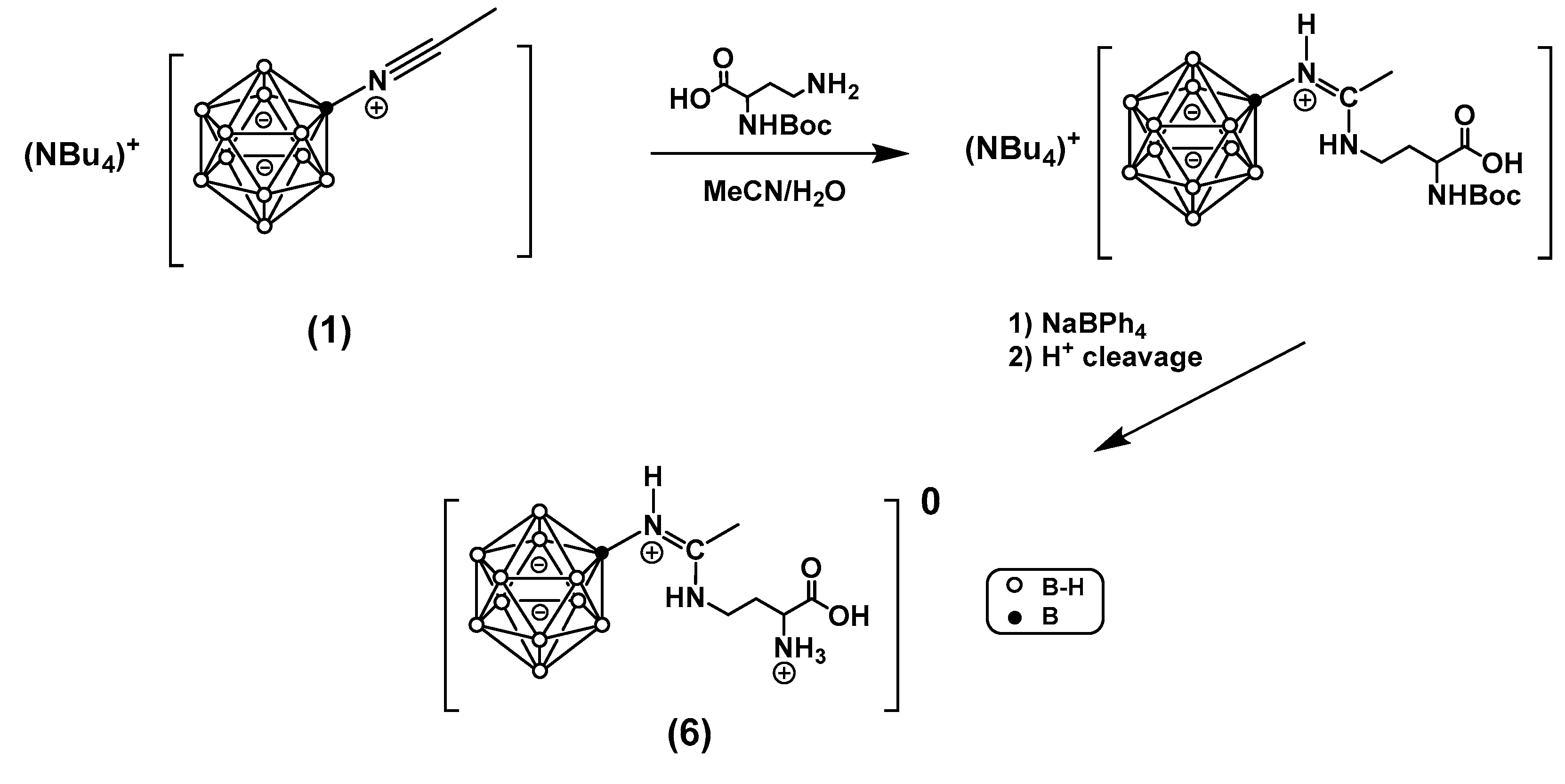
Since derivative 3 exists as a zwitterion, for deprotonation of the γ-amino group, the nucleophilic addition reaction to the nitrilium derivative (1) was conducted in an aqueous-organic medium (Figure 2). It was determined that the highest yield, as measured by 11B NMR spectroscopy, was achieved using a mixture of acetonitrile/THF/saturated aqueous Na2CO3 in a 2:1:1 ratio. For biological studies, the n-tetrabutylammonium cation was replaced by a sodium ion through reaction with sodium tetraphenylborate. After deblocking the α-amino group, the product precipitated as an internal salt of the form [B12H11NHC(NH(CH2)2CH(NH3)COOH)CH3]*3H2O (6). Recrystallization of the product from water yielded crystals suitable for structural analysis by X-ray diffraction (Figure 4), confirming the absence of sodium cations in the final derivative.
An additional experiment was conducted where the removal of the tert-butoxycarbonyl group was carried out without replacing the n-tetrabutylammonium cation. This modification required conducting the reaction in an aqueous-organic medium and produced derivative (6) with an 86% yield.
Figure 3.
General scheme for the modification of diamino acids used in this work.
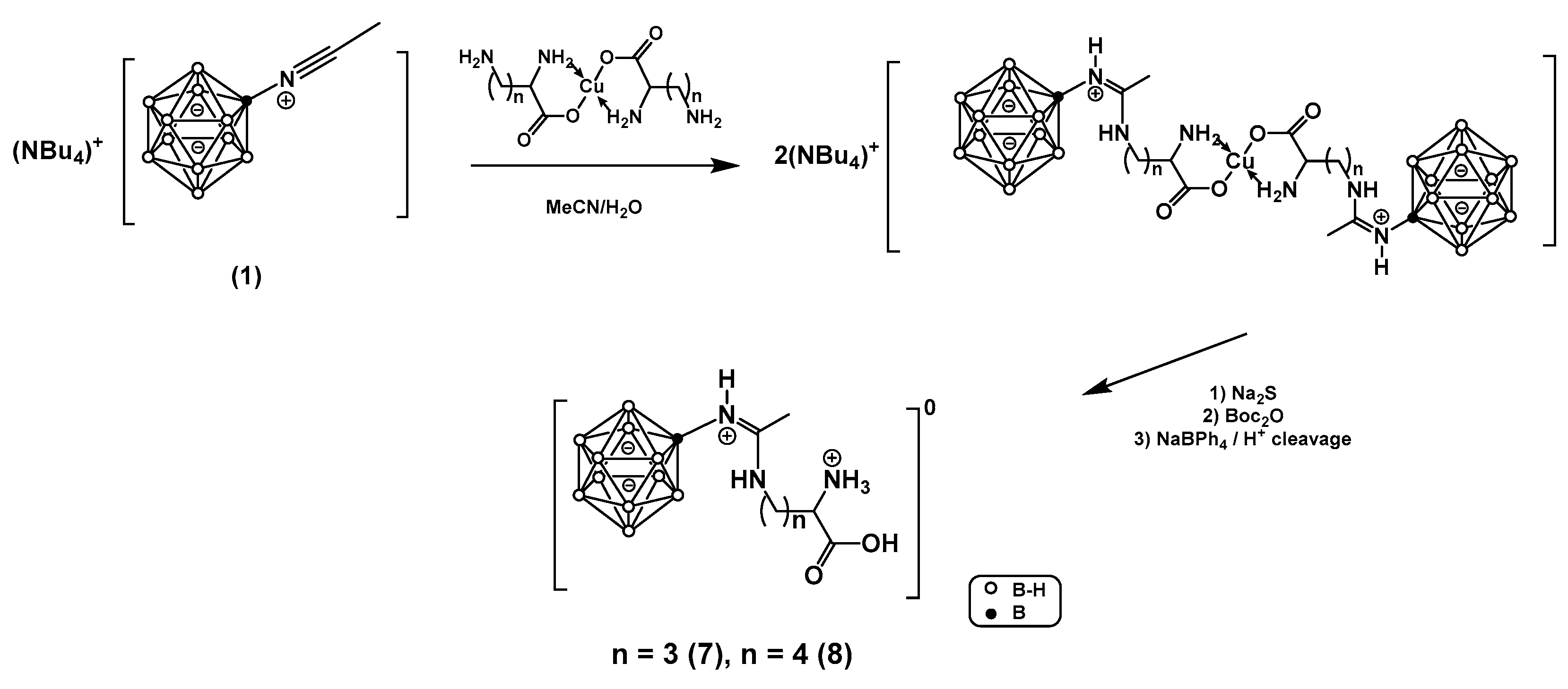
Direct interaction of the acetonitrile derivative (1) with lysine predictably led to a mixture of three regioisomers, which could not be separated using chromatographic methods. This issue could not be resolved by simply adjusting the pH of the reaction medium and required the use of an α-amino-protected amino acid [28].
For natural amino acids (Orn, Lys), obtaining Nα-Boc derivatives requires a complex multistep synthesis involving the installation of a temporary orthogonal protective group on the side-chain amino group. A simpler method for deactivating the α-amino group involves complexation with copper(II) ions. Due to the chelating properties of α-amino acids and the high stability of square-planar Cu2+ complexes with a d9 electronic configuration, it is possible to protect the α-amino group, thereby enabling selective addition reactions at the side-chain amino group of diamino acids.
The interaction of compound (1) with copper complexes of ornithine and lysine (4) and (5) in an aqueous-acetonitrile solution, followed by the decomposition of the copper complex with sodium sulfide, resulted in n-tetrabutylammonium salts (7) and (8) respectively (Figure 3). The yield of target boronated amidines, as determined by 11B NMR, was over 90%, with the main by-product being the corresponding boronated amide. Attempts to isolate these derivatives as internal salts, similar to derivative (6), were unsuccessful. Preparative flash chromatography on a reversed-phase HPLC column was proposed to isolate the target products. Using a water-acetonitrile mixture with trifluoroacetic acid as the eluent allowed for the isolation of the protonated forms (7) and (8) with yields of approximately 30%.
An attempt to replace the cation with sodium tetraphenylborate resulted in a reduced yield of 10-20% following chromatographic purification. This reduction was attributed to a side reaction during co-precipitation of the protonated form of the target derivative in an aqueous medium.
The final optimization step involved introducing Boc protection to the free amine fragment to prevent the formation of an α-ammonium cation that co-precipitates with tetrabutylammonium in the presence of sodium tetraphenylborate. After decomposing the copper complex, boron-containing derivatives were effectively separated from by-products via acetonitrile extraction. Treating derivatives (7) and (8) with Boc2O resulted in derivatives with Nα-Boc protection. Subsequent cation exchange with sodium proceeded similarly to derivative (6). Deblocking the amino group followed by chromatographic purification yielded the target derivatives (7) and (8) as internal salts with yields of 72% and 81%, respectively.
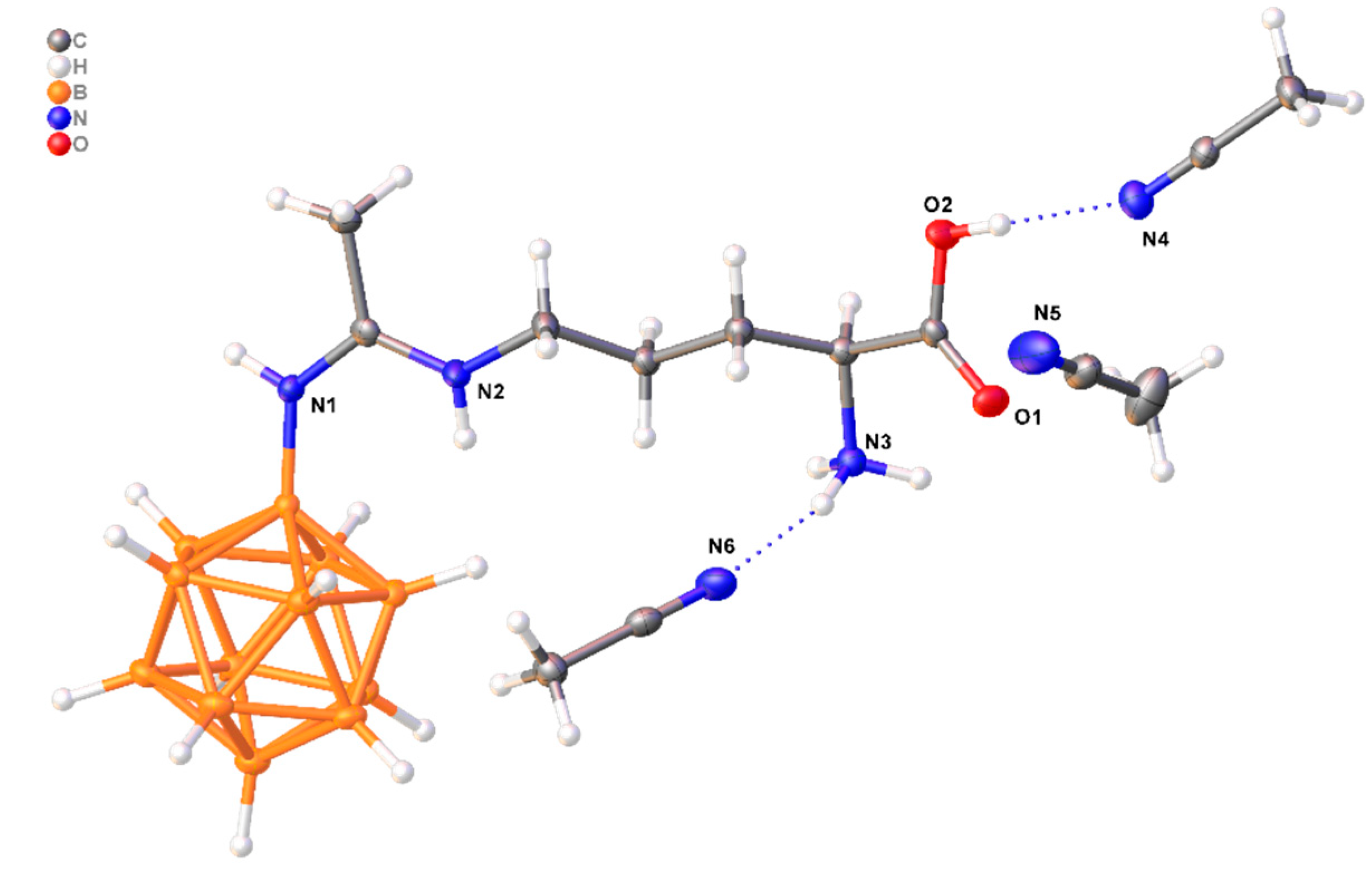
Compounds (6) and (7) crystallize in the monoclinic system (space group P21). The crystallographically independent part of compound (6) includes the zwitterionic compound B12H11NHC(Me)NHCH2CH2CH(NH3)COOH and three water molecules, while compound (7) consists of the zwitterionic B12H11NHC(Me)NHCH2CH2CH2CH(NH3)COOH and three CH3CN molecules. Solvent molecules are bound to the zwitterions in the crystals via strong OH…O(N) and NH…O(N) hydrogen bonds (Table 1 and Table 2), as well as weaker OH…HB and CH…HB contacts.
Figure 4.
Structure of compound (6).
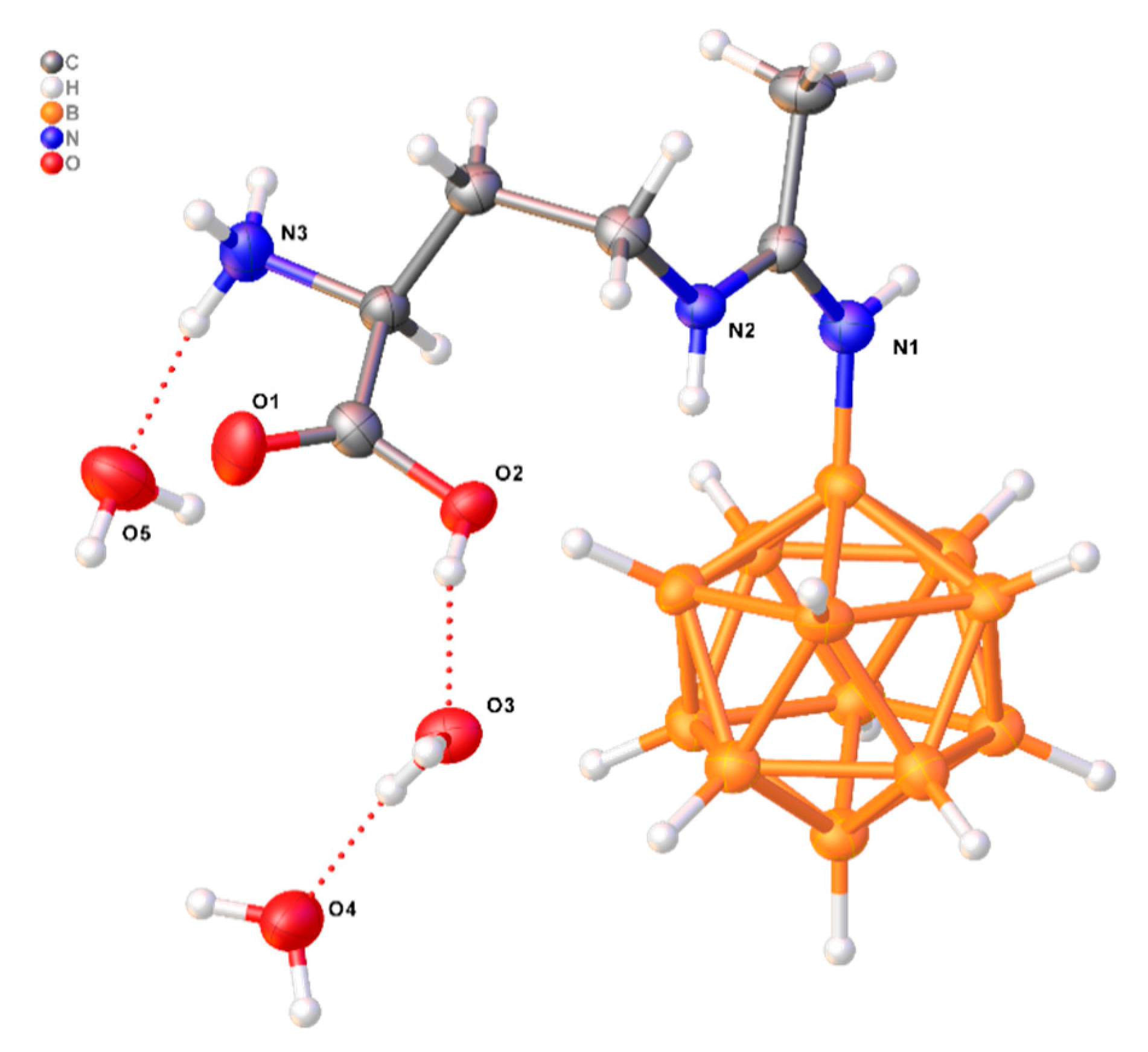
Figure 5.
Structure of compound (7).
In addition, the structures of (6) and (7) systems were analysed with the help of quantum chemistry tools (Figure 6).
Firstly, the bond order of different types of the CN bonds were estimated with the help of Wiberg approach in natural atomic orbitals basis. In our previous research it has been established that in the case of initial nitrilium derivatives of closo-dodecaborate anion [B12H11NCR]– the formal multiplicity of CN bond was 3 and Wiberg bond orders were lied in the interval of 2.62–2.66 [29]. In the systems under consideration there are three types of CN bonds. The first one is the bond between Nimine atom and C atom of the amidine group NH=C(NH2), the second one is between Namide atom and C of amidine group and the third one is the bond between Namine and α-C atom of aminoacid residue. A strong delocalization between CNimine and CNamide bonds was established. However, CNimine bond had the greatest values (1.50-1.52) of bond order comparing to CNamide (1.27-1.28). These results are very similar to analogous of amidine derivative of closo-dodecaborate anion [B12H11N=C(NH2)R]–. In the case of this derivative CNimine bond has the 1.51 value of Wiberg bond order and CNamide bond is equal to 1.31. CNamide bond of (6), (7) structures due to partial delocalization with CNimine bond has the greatest value of bond order comparing to CNamine (0.95-0.96).
The atomic charges of Namide and Nimine were close to each other and lie in interval of –0.57 to –0.61, which were lower than in case of Namine (–0.75). Carbonyl oxygen atoms hads lower atomic charges (–0.56 to –0.57) comparing to hydroxyl oxygen atoms (–0.65 to –0.66).
Non-covalent interactions play crucial role in stability and properties of closo-borate derivatives [25,26]. Analysis of non-covalent interactions was carried with the help of QTAIM formalism. The geometry parameters of optimized structure of (6) and (7) differed from analogous parameters in crystal structure. The main reason of such phenomena is the prevailing of intermolecular contacts over intramolecular contacts in crystal structure. In the crystal structures dihydrogen bonds between NHamide and BH groups were detected. For the system (7) the length of such contacts is equal to 1.82 Å and in case of an 6 the length is equal to 2.15 Å. The values of electron density at bond critical point (bcp) is equal to 0.018 e Å –3 in the case of anion (7) and 0.012 e Å–3 in the case of anion (6). In structures obtained on the basis of geometry optimization in gas phase the number of noncovalent contacts between boron cluster and exo-polyhedral substituent are increased comparing to crystal structures. In the gas phase of (6) NHamide and BH contact is shortened and equal to 1.62 Å. The value of electron density is increased comparing to crystal structure and is equal to 0.031 e Å–3. In addition to NHamide–BH contact the interactions between hydrogens of NHamine group and BH were observed. One of them has the bond length which is equal to 1.92 Å and values of electron density at the corresponding bcp is equal to 0.017 e Å–3. The second one bond length is equal to 1.46 Å and value of electron density is equal to 0.043 e Å–3. In the case of (7) NHamide–BH contact is equal to 1.92 Å and value of electron density is equal to 0.018 e Å –3. As in the case of (6) there are two contacts between hydrogens of NHamine group and BH. First of them has the bond length equal to 1.92 Å with value of electron density is equal to 0.016 e Å –3. The second one is shorter, and bond length is equal to 1.56 Å and value of electron density is equal to 0.035 e Å –3.
2.1. Safety and Tolerability of Compounds in Laboratory Animals
The safety of compound (6) was evaluated after intravenous administration to laboratory mice at three dosages: 75, 150, and 300 mg/kg of body weight. The 75 mg/kg dosage (27.5 mg of boron/kg) was chosen to correspond to the boron dosage used in studies of boronophenylalanine. Since no toxic effects were observed in mice at 75 mg/kg of compound (6), dosage levels were subsequently increased twofold and fourfold. At 75 mg/kg and 150 mg/kg, there were no cases of lethality or signs of acute toxicity (loss of consciousness, seizures, or pain syndrome). However, at a dosage of 300 mg/kg 100% lethality was observed. As LD50 value determination was out of the study scope, the dosage of 150 mg/kg was selected for subsequent tumor uptake studies as maximum tolerated dose.
When compounds (7) and (8) were administered to laboratory mice at a dosage of 75 mg/kg, all animals in the group perished. Due to the high toxicity of these compounds, further studies were discontinued. Reducing the dosage was not considered, as it would have resulted in a boron dose lower than that achieved with boronophenylalanine.
2.2. In Vivo Tumor Uptake
The in vivo tumor uptake of compound (6) was investigated in laboratory mice with two tumor models over a time range from 15 to 120 minutes following intravenous injection of 150 mg/kg of the compound.
The results of the compound (6) tumor uptake studies of in C57Bl/6 mice with subcutaneous B16F10 melanoma are presented in Figure 7.
As expected, following intravenous administration, the maximum boron concentration in blood (41±8 μg/g) was observed at the earliest time point, 15 minutes post-injection (Figure 6). Subsequently, boron levels in the blood decreased rapidly, reaching 9±3 μg/g at 45 minutes and 2.1±0.5 μg/g at 120 minutes post-injection.
In muscle tissue, the maximum boron concentration (6.3±1.4 μg/g) was only observed at the earliest time point, due to high circulating boron levels. Thereafter, boron concentrations in muscle tissue remained within a range of 0.7–2.8 μg/g.
In tumor tissue, boron concentration reached a significant level of 17±2 μg/g within 15 minutes. Although boron levels in the tumor subsequently declined, the decrease was slow, with boron concentrations remaining at 6.1±1.8 μg/g even at 60 minutes post-injection.
As a reference compound, boronophenylalanine, currently used in clinical practice, was studied (Figure 8). For tumor uptake studies, C57Bl/6 mice with subcutaneous B16F10 melanoma were administered 580 mg/kg of boronophenylalanine, equivalent to 28 mg/kg of boron. Following intravenous injection, boron levels in the blood decreased slowly, from 17±1 μg/g at 15 minutes post-injection to 6.2±0.9 μg/g at 120 minutes. Boron accumulation in muscle tissue was substantial and remained steady throughout the observation period, measuring 17±3 μg/g at 15 minutes and 12±2 μg/g at 120 minutes post-injection. Boron accumulation in tumor tissue was pronounced, with a maximum concentration of 37±3 μg/g observed at 30 minutes, followed by a subsequent decrease.
Since successful boron neutron capture therapy requires not only a high concentration of boron in the tumor but also a significant concentration gradient between the tumor and surrounding healthy tissue, we calculated the T/N ratio, which reflects the ratio of boron concentrations in the tumor to that in muscle tissue (Figure 9).
The slower clearance of compound (6) from tumor tissue resulted in a significantly higher boron concentration in the tumor compared to muscle tissue within the time range of 10 to 60 minutes. The maximum T/N ratio of 6.2±2.8 was achieved 45 minutes after intravenous injection, followed by a decrease to 2.6±1.1 at 90 minutes. In contrast, for boronophenylalanine, a T/N ratio of 2.9±0.3 was reached 30 minutes post-injection but did not persist even for 15 minutes. At all other time points within the 15–120-minute range, the T/N ratio did not exceed 2.1±0.5.
For subcutaneous 4T1 mammary carcinoma in Balb/C mice (Figure 10) similar to B16F10 melanoma, high boron concentration in blood (30±7 μg/g) was observed 15 minutes after intravenous administration of compound (6), followed by a rapid decrease. The boron concentration in muscle tissue did not exceed 2.9±0.8 μg/g during the 30-120 minute range. The maximum boron concentration in the 4T1 tumor was recorded at the earliest time point (13±3 μg/g at 15 minutes post-injection), but then decreased quickly to no more than 5.4±1.3 μg/g at 30 minutes. Thus, the dynamics of boron concentration change in 4T1 tumor tissue were characterized by significantly faster clearance compared to B16F10 melanoma. Conversely, boronophenylalanine showed gradual accumulation in the 4T1 tumor, reaching a maximum concentration of 17±1 μg/g at 60 minutes post-injection, with only a slight decrease to 15±3 μg/g at 120 minutes (Figure 11).
The dynamics of the T/N ratio reflected the rapid clearance of compound (6) from 4T1 tumor tissue (Figure 12). The T/N ratio was 3.0±0.9 at the first time point and subsequently dropped below 2, continuing to decrease throughout the observation period. In contrast, for boronophenylalanine, the T/N ratio increased over 60 minutes, reflecting drug accumulation in the tumor, and then reached a plateau. It is noteworthy that 15 minutes post-injection of compound (6), the T/N ratio was 3.0±0.9, whereas the maximum T/N for boronophenylalanine was only 2.0±0.4.
To evaluate possible excretion pathways for compound (6) in C57Bl/6 mice, excretory organs such as the kidneys and liver were also examined Figure 13.
Boron concentration in the kidneys was initially high, measuring 275±57 μg/g at 15 minutes post-injection, but then dropped rapidly to 95±17 μg/g at 30 minutes and 20±4 μg/g at 120 minutes. In contrast, boron levels in the liver were significantly lower, measuring 88±8 μg/g at 15 minutes, decreasing to 33±9 μg/g at 45 minutes, and continuing to decline. Since compound (6) is a low-molecular-weight compound, renal excretion appears to be the most likely pathway.
The dynamics of boronophenylalanine accumulation in the kidneys and liver of C57Bl/6 mice were also studied. High initial boron concentrations were observed in both organs (202±35 μg/g in the liver and 343±19 μg/g in the kidneys), with a rapid decline to 29±2 μg/g and 69±6 μg/g in the liver and kidneys, respectively, within 30 minutes (see Supplementary, Table S2).
The promising compound (6) studied in this work demonstrated significantly higher solubility, enabling administration of a boron dose twice that of boronophenylalanine. Moreover, the low toxicity of compound (6) was confirmed by the absence of any toxic manifestations even at twice the boron dose compared to boronophenylalanine.
An essential criterion for the suitability of a BNCT agent is the boron concentration ratio between tumor and healthy tissue. In this study, using the boronophenylalanine as a reference, the maximum T/N ratio of 2.9±0.3 for the B16F10 tumor model was achieved at 30 minutes post-injection, dropping below 2.6 within 15 minutes. These results align with the literature: a brief T/N ~3 ratio is characteristic for boronophenylalanine [30], while sodium borocaptate—a compound also used in clinical trials—may not even reach a T/N of 2 [31]. Compound (6) exhibited significantly more selective accumulation in melanoma tissue: throughout the 30–60-minute interval following a single injection, the T/N ratio remained >3, with a maximum T/N of 6.2±2.8 at 45 minutes. These findings suggest that it could potentially serve as the basis for a new BNCT agent suitable for single bolus administration.
It is noteworthy that in studies with 4T1 mammary adenocarcinoma, compound (6) did not demonstrate equally impressive results: the T/N ratio of 3.0±0.9 achieved at 15 minutes post-injection fell below 2 thereafter, whereas boronophenylalanine accumulation increased. These findings underscore the importance of selecting appropriate tumor models for preclinical studies and justify the need to expand the panel of available agents for radiologists to treat tumors of various localizations.
3. Materials and Methods
3.1. NMR Spectroscopy
1H, 11B, and 13C NMR spectra of the investigated compounds were recorded using a Bruker MSL-300 pulse Fourier spectrometer (Germany) at frequencies of 300.3, 96.32, and 75.49 MHz, respectively, with internal stabilization by deuterium. Tetramethylsilane or boron trifluoride etherate was used as external standards.
3.2. Analytical Reversed-Phase High-Performance Liquid Chromatography (RP-HPLC)
RP-HPLC analysis was performed on an isocratic Knauer HPLC system, consisting of a PDA Smartline 2800 detector, Smartline 1000 pump, and a NanoChrome ChromCore 120 C18 column (250×4.6 mm, 5 µm particle size of the stationary phase). Sample injection was done manually, with a loop volume of 20 µL. Eluent A was an aqueous solution of trifluoroacetic acid (0.2%), and eluent B was acetonitrile.
3.3. Preparative RP-HPLC
Preparative RP-HPLC was conducted on a Unique AutoPure100 FPLC chromatograph with a UV detector and an automatic fraction collector and a 1000 µL loop. The separation was performed on a Hawach Spherical C18 Flash Column (SLC18SP10025PF). Fractions containing the target compound (volume of 12 mL) were automatically collected based on the UV detector signal at a wavelength of 232 nm. Eluent A was 99.8/0.2 H2O/CF3COOH, and Eluent B was 100% CH3CN, with a flow rate of 35 mL/min. Elution gradient: 0-10 min 15% A, 10-20 min 35% A, 20-30 min 35% A, and 30-40 min 90% A.
3.4. High-Resolution ESI Mass Spectrometry
High-resolution ESI mass spectra of compound solutions in acetonitrile were recorded using an LCMS-IT-TOF mass spectrometer (Shimadzu, Japan) in direct injection mode, with an m/z range of 120-700 Da. Detector voltage was set at 1.55 kV, and ESI voltage at 4.50 kV. Equipment tuning (mass calibration and sensitivity check) was performed prior to analysis.
3.5. X-Ray Crystal Structure Determination
Single-crystal X-ray diffraction data for compounds 1 and 2 were collected on a three-circle Bruker D8 Venture diffractometer (T = 100 K, graphite monochromator, ω and φ scanning mode). Data were indexed and integrated using the SAINT program [32], then scaled and corrected for absorption using the SADABS program [33]. Details are provided in Table S3.
The structures were determined by direct methods and refined using the full-matrix least-squares technique on F2 with anisotropic displacement parameters for non-hydrogen atoms. Hydrogen atoms were placed in calculated positions and refined within a riding model with fixed isotropic displacement parameters [Uiso(H) = 1.5Ueq(O), 1.5Ueq(C) for CH3 groups, and 1.2Ueq(C) for other groups]. Calculations were performed using the SHELXTL program [34] and the OLEX2 program package [35].
Crystallographic data for all investigated compounds have been deposited with the Cambridge Crystallographic Data Center (CCDC) under accession numbers CCDC 2390349, 2390350. Copies of this data can be obtained free of charge from the CCDC, 12 Union Road, Cambridge CB2 1EZ, UK (Fax: +44 1223 336033; e-mail: deposit@ccdc.cam.ac.uk or via www.ccdc.cam.ac.uk).
3.6. Sample Preparation and Determination of Boron in Biological Samples
Sample digestion was performed using microwave decomposition with an Ethos Easy system (MILESTONE, Italy). Biological tissue samples were placed in a fluoropolymer autoclave, followed by the addition of 5 mL of concentrated nitric acid (HNO₃) and 1 mL of hydrogen peroxide (H₂O₂). The temperature of each autoclave was automatically controlled by an internal thermocouple in the microwave system. The decomposition protocol consisted of a linear gradual heating over 30 minutes to 190°C, followed by a 10-minute hold at 190°C. After cooling the autoclave to 50°C, it was removed from the microwave system and opened in a fume hood. The contents were transferred to boron-free plastic Falcon tubes (50 mL capacity). To each sample, 50 µL of the surfactant Triton X-100 was added, and the volume was adjusted to 10 mL with deionized water.
Quantitative boron determination in biological samples was conducted using inductively coupled plasma optical emission spectroscopy (ICP-OES). The measurements were performed on a high-resolution ICP-OES spectrometer (PlasmaQuant 9100 Series, Analytik Jena, Germany) using a standard multicalibration solution (ICP multi-element standard solution IV, Supelco Certipur, Germany). For boron concentration measurements, two emission wavelengths, 249.773 nm and 249.678 nm, were used. Each sample was measured three times at each wavelength. The obtained signal intensities were compared with a calibration curve (correlation coefficient R² > 0.9999; measurement range from 0.05 µg B/mL to 4 µg B/mL), and quantitative values of boron concentration in the sample were determined. The concentration of boron in biological tissue or organs was calculated based on the dilution factor of the sample.
3.7. Computational Details
The ORCA 5.0.4 program package [36] was used for density functional theory (DFT) calculations. The full geometry optimization of all model structures was carried out at the R2SCAN-3c level of theory. All molecular species considered had closed electron shells, and the spin restricted approximation was applied. The tight criteria of SCF convergence (Tight SCF) were employed during the calculations. For a more accurate evaluation of the electronic structures of the systems under consideration, additional single point calculations, on ωB97X-D3/TZVPP level of theory with no resolution of identity approximation (“NORI” keyword), were carried out. Symmetry operations were not applied during the geometry optimisation procedure. The Hessian matrices were calculated numerically for all model structures to prove the location of correct minima on potential energy surfaces. No imaginary frequencies were found in any of the cases. The natural bond orbital (NBO) method was employed, using the NBO7 program package [37]. Topological analysis of the electron density distribution within the Quantum Theory of Atoms in Molecules (QTAIM) formalism, developed by Bader [38] were carried out using the Multiwfn program (version 3.8) [39]. The visualisation of optimised structures was carried out with the help of the ChemCraft program (version 1.7) [40].
3.8. Reagents and Materials
The starting amino acids from Sigma Aldrich were used without additional purification. Solvents with 99% purity were used without additional purification. Trifluoroacetic acid was distilled immediately before use. The acetonitrile derivative of the closo-dodecaborate anion (1) was synthesized according to the known method [29]. Boronophenylalanine was synthesized in our in-house radiochemistry laboratory at the N.N. Blokhin National Medical Research Center of Oncology.
- Nα-(tert-Butoxycarbonyl)-L-glutamine (Boc-L-glutamine) (2)
8 g (0.2 mol, 2 equiv.) of NaOH was dissolved in 100 mL of water, cooled to 5°C, and mixed with 14.6 g (0.1 mol equiv.) of glutamine. To the resulting solution, 100 mL of dioxane and 25.2 mL (24 g, 0.11 mol, 1.1 equiv.) of di-tert-butyl dicarbonate were added. The solution was stirred for 16 hours at room temperature and concentrated using a rotary evaporator to approximately 75 mL. 1M NaHSO₄ solution was added to adjust the pH to 3, resulting in the formation of a white emulsion. The target product was extracted three times with 80 mL portions of ethyl acetate. The combined organic fraction was washed with a saturated salt solution, dried over sodium sulfate, and evaporated. The product was recrystallized from ethyl acetate. Yield: 18.2 g (74%). The spectra matched literature data [41].
1H NMR (CDCl₃, δ, ppm): 6.86 (bs, 2H, CONH₂), 5.76 (d, J = 7.7 Hz, 1H, NH), 4.28 (q, 1H, CH), 2.39 (m, 2H, CH₂), 2.25-1.95 (m, 2H, CH₂), 1.44 (s, 9H, C(CH₃)₃).
- Nα-Boc-β-amino-L-alanine (3)
Synthesized using a known method [42]. 9.85 g (0.04 mol, 1 equiv.) of compound (2) was suspended in 120 mL of an ethyl acetate-acetonitrile mixture (1:1 v/v). 15.5 g (0.048 mol, 1.2 equiv.) of (diacetoxyiodo)benzene and 30 mL of water were added to the suspension. The mixture was stirred at room temperature until completion of the reaction, as confirmed by HPLC (approximately 6 hours). The solution was concentrated, and 40 mL of ethyl acetate was added, followed by ultrasonication for 20 minutes. The precipitate was separated by centrifugation and recrystallized from methanol. Yield: 7.99 g (92%). The spectra matched literature data [43].
1H NMR (D₂O, δ, ppm): 3.97 (m, 1H, CH), 3.07 (t, J = 7.8 Hz, 2H, γ-CH₂), 2.22–1.89 (m, 2H, β-CH₂), 1.44 (s, 9H, C(CH₃)₃).
- General Method for the Synthesis of Copper Complexes of Diamino Acids
- Cu(Orn*HCl)₂ (4) and Cu(Lys*HCl)₂ (5)
10 mmol of the amino acid hydrochloride was dissolved in 20 mL of water. To the resulting solution, 20 mmol of (CuOH)₂CO₃ was added. The solution was boiled for 30 minutes. After cooling to room temperature, the solution was filtered to remove excess copper carbonate and used further without isolation or purification.
- [B₁₂H₁₁NHC(NH(CH₂)₂CH(NH₃)COOH)CH₃]*3H₂O (6)
275 mg (1.25 mmol, 1.25 equiv.) of compound (3) was dissolved in 10 mL of water, and 66 mg (0.625 mmol, 1.25 equiv.) of Na₂CO₃ was added. To the resulting clear solution, 0.424 g (1 mmol, 1 equiv.) of compound (1) in 10 mL of acetonitrile was added. The solution was stirred vigorously for 5 minutes. After completion of the reaction (as confirmed by 11B NMR), the reaction mixture was concentrated using a rotary evaporator. The residue was treated with 10 mL of 1M hydrochloric acid and extracted three times with 10 mL portions of dichloromethane. The organic phase was separated and evaporated using a rotary evaporator. The dry residue was dissolved in 10 mL of acetonitrile and 5 mL of concentrated hydrochloric acid was added. The solution was stirred for 24 hours, concentrated using a rotary evaporator, and the product was recrystallized from water. Yield: 304 mg (86%).
11B NMR (CD₃CN, δ, ppm): –8.0 (s, 1B, B(1)), –16.6 (d, J_B–H = 128 Hz, 10B, B(2-11)), –17.8 (d, 1B, B(12)). 1H NMR (CD₃CN, δ, ppm): 8.03 (br s, 1H, NH=C(NH)–CH₃), 6.92 (br s, 3H, NH₃–CH–COOH), 6.77 (br s, 1H, NH=C(NH₂)–CH₃), 4.03 (t, J = 7.0 Hz, 1H, NH₃–CH–COOH), 3.48 (m, 2H, γ-CH₂), 2.25–2.07 (m, 2H, β-CH₂), 2.11 (s, 3H, NH=C(NH)–CH₃), 2.0–0.0 (m, 10H, B₁₂H₁₁). 13C{H} NMR (CD₃CN, δ, ppm): 169.5 (NH₃–CH–COOH), 166.2 (NH=C(NH)–CH₃), 51.6 (NH₃–CH–COOH), 40.1 (γ-CH₂), 30.7 (β-CH₂), 18.9 (NH=C(NH)–CH₃). MS(ESI) m/z = 300.3073 (found for C₆H₂₄B₁₂N₃O₂); calcd. for {[A]-H⁻} 300.3058.
3.9. General Method for the Synthesis of Boronated Diamino Acid Derivatives
To a 3 mL aqueous solution of the copper complex of the corresponding amino acid (0.75 mmol, 1.5 equiv.), 250 mg (1.5 mmol, 1.5 equiv.) of NaHCO₃ dissolved in 2 mL of water was added. A solution of 424 mg (1 mmol, 1 equiv.) of compound (1) in 10 mL of acetonitrile was prepared. The copper complex solution was added to the solution of compound (1) and stirred for 5 minutes. After completion of the reaction (as confirmed by 11B NMR), 5 mL of a concentrated NaCl solution was added to the reaction mixture. The organic layer was separated, and the aqueous layer was washed twice with 10 mL portions of acetonitrile. The combined organic fractions were concentrated to a volume of approximately 10 mL, and 10 mL of water was added. A concentrated Na₂S solution was added until copper sulfide precipitated completely. The precipitate was separated by centrifugation. To the resulting orange solution, 414 µL (2 mmol, 2 equiv.) of Boc₂O was added, and the solution was stirred at room temperature for 16 hours. After completion of the reaction, acetonitrile was removed using a rotary evaporator, and the aqueous layer was acidified to pH=2 with 1M hydrochloric acid and extracted three times with 15 mL portions of dichloromethane. The combined organic layer was evaporated using a rotary evaporator and dissolved in 15 mL of methanol. A solution of 342 mg (1 mmol, 1 equiv.) of NaBPh₄ in 50 mL of water was added. The precipitate was separated, the solution was evaporated using a rotary evaporator, and the product was purified by flash chromatography.
- [B₁₂H₁₁NHC(NH(CH₂)₃CH(NH₃)COOH)CH₃]*3H₂O (7)
Yield: 266 mg (72%).
11B NMR (CD₃CN, δ, ppm): –7.1 (s, 1B, B(1)), –16.0 (d, J_B–H = 129 Hz, 10B, B(2-11)), –17.1 (d, 1B, B(12)). 1H NMR (CD₃CN, δ, ppm): 7.95 (br s, 1H, NH=C(NH)–CH₃), 6.73 (br s, 3H, NH₃–CH–COOH), 6.63 (br s, 1H, NH=C(NH₂)–CH₃), 4.04 (br s, 1H, NH₃–CH–COOH), 3.30 (q, J = 7.0 Hz, 2H, δ-CH₂), 2.11 (s, 3H, NH=C(NH)–CH₃), 1.94 (m, 2H, β-CH₂), 1.70 (m, 2H, γ-CH₂), 2.0–0.0 (m, 10H, B₁₂H₁₁). 13C{H} NMR (CD₃CN, δ, ppm): 170.0 (NH₃–CH–COOH), 165.8 (NH=C(NH)–CH₃), 54.0 (NH₃–CH–COOH), 43.3 (δ-CH₂), 27.5 (β-CH₂), 25.9 (γ-CH₂), 18.9 (NH=C(NH)–CH₃). MS(ESI) m/z = 314.3239 (found for C₆H₂₄B₁₂N₃O₂); calcd. for {[A]-H⁻} 314.3214.
- [B₁₂H₁₁NHC(NH(CH₂)₃CH(NH₃)COOH)CH₃]*3H₂O (8)
Yield: 310 mg (81%).
11B NMR (CD₃CN, δ, ppm): –7.0 (s, 1B, B(1)), –15.9 (d, J_B–H = 129 Hz, 10B, B(2-11)), –17.2 (d, 1B, B(12)). 1H NMR (CD₃CN, δ, ppm): 7.90 (br t, J = 5.8 Hz, 1H, NH=C(NH)–CH₃), 6.85 (br s, 3H, NH₃–CH–COOH), 6.58 (br s, 1H, NH=C(NH₂)–CH₃), 4.00 (m, 1H, NH₃–CH–COOH), 3.28 (q, J = 6.5 Hz, 2H, ε-CH₂), 2.08 (s, 3H, NH=C(NH)–CH₃), 2.0–1.8 (m, 2H, β-CH₂), 1.63 (m, 2H, δ-CH₂), 1.49 (m, 2H, γ-CH₂), 2.0–0.0 (m, 10H, B₁₂H₁₁). 13C{H} NMR (CD₃CN, δ, ppm): 170.4 (NH₃–CH–COOH), 165.8 (NH=C(NH)–CH₃), 54.1 (NH₃–CH–COOH), 43.6 (ε-CH₂), 30.0 (β-CH₂), 29.4 (δ-CH₂), 22.6 (γ-CH₂), 18.9 (NH=C(NH)–CH₃). MS(ESI) m/z = 328.3398 (found for C₆H₂₄B₁₂N₃O₂); calcd. for {[A]-H⁻} 328.3371.
- Animal Studies
- Preparation of solutions of compounds (6)-(8) for biological studies:
51 mg of each compound (dry weight) was dissolved in 0.5 mL of 0.1M NaOH. Distilled water (1.5 mL) was added to the solution and the pH was adjusted to 7.1 with 1M NaOH. The solution volume was adjusted to 3 mL with distilled water and filtered through a 0.22 µm syringe filter.
- Safety and Tolerability Studies
The tolerability of the synthesized compounds was evaluated in female Balb/C mice weighing 20–22 g. The compounds were administered intravenously. Compound (6) was administered at three dosages: 75, 150, and 300 mg/kg of body weight. Compounds (7) and (8) were administered at a dosage of 75 mg/kg of body weight (see Results and Discussion).
In tumor uptake studies female C57Bl/6 mice with B16F10 melanoma and female Balb/C mice with 4T1 breast adenocarcinoma were used. The animals were obtained from the breeding facility at the N.N. Blokhin National Medical Research Center of Oncology, and tumor strains were received from the Center's cryobank. To create tumor models, mice were subcutaneously injected with a freshly prepared suspension of tumor cells at a 1:6 mass/volume ratio in Hank’s solution in a volume of 60 µL into the right hind leg tibial region.
- Tumor uptake of Compounds
The dynamics of biodistribution in tumor-bearing mice were studied for compound (6) and boronophenylalanine as a reference compound.
The test compounds were administered intravenously at the following doses: compound (6) – 150 mg/kg (55 mg of boron/kg), and boronophenylalanine – 580 mg/kg (28 mg of boron/kg). At specific time intervals, animals were anesthetized by inhalation of 2.5% isoflurane air mixture, and blood samples were taken from the anterior limb venous sinus. Following euthanasia via isoflurane overdose, organs and tissues were collected, including the tumor, muscle tissue, liver, and kidneys. Subcutaneous tumors were fully excised. For muscle tissue samples, the thigh muscles of the left (healthy) hind leg were excised. Liver samples weighing 0.4 to 1.6 g were collected, and both kidneys were completely removed using a "dry" method, with vessels clamped beforehand. Mice were grouped in sets of five for each time point.
In the study of compound (6) in C57Bl/6 mice with B16F10 melanoma, organ and tissue samples were collected at 15, 30, 45, 60, 90, and 120 minutes.
In the study of compound (6) in Balb/C mice with 4T1 adenocarcinoma, organ and tissue samples were collected at 15, 30, 60, 90, and 120 minutes.
In the study of boronophenylalanine in C57Bl/6 mice with B16F10 melanoma, organ and tissue samples were collected at 15, 30, 45, 60, 90, and 120 minutes.
In the study of boronophenylalanine in Balb/C mice with 4T1 adenocarcinoma, organ and tissue samples were collected at 15, 30, 60, 90, and 120 minutes.
After removal from the animal body, all biological tissue and organ samples were placed in boron-free plastic containers. Samples were weighed on laboratory precision scales. The samples in containers were stored in a freezer at -20°C.
4. Conclusion
In this work, three new compounds were synthesized with potential for the development of new agents for BNCT. Two of these compounds exhibited high toxicity and were thus deemed unsuitable for biomedical use at this stage. The promising compound (6) demonstrated not only low toxicity and excellent solubility but also showed selective accumulation in experimental melanoma in laboratory mice. Thus, compound (6) is recommended for further in vivo BNCT studies.
Supplementary Materials
The following supporting information can be downloaded at the website of this paper posted on Preprints.org, Spectral data of synthetized compounds, crystal data for compounds (6) and (7), data of boron content in biological tests.
Author Contributions
Manuscript conception, A.P.Z., A.A.L. and K.Y.Z.; writing and original draft preparation, A.V.N., Y.A.F. and A.P.Z.; synthesis of derivatives M.N.R. and A.V.N.; N.M.R. analysis, N.A.S. and A.Y.B.; X-ray analysis, Hirshfeld analysis A.S.K.; ICP-OES analysis V.A.S.; biological tests K.E.S., A.A.K.; editing, data analysis, and interpretation, A.P.Z., Y.A.F. A.A.L., K.Y.Z. and N.T.K.; supervision, K.Y.Z., E.Y.G and N.T.K. All authors have read and agreed to the published version of the manuscript. All authors have read and agreed to the published version of the manuscript.
Funding
This work was carried out with the support of the Russian Science Foundation (project no 24-13-00295).
Institutional Review Board Statement
The study was approve by N.N. Blokhin National Medical Research Center of Oncology bioethics committee (Protocol #05b-p-2024 ) .
Informed Consent Statement
Not applicable.
Data Availability Statement
Data is contained within the article.
Acknowledgments
This research was performed using the equipment of the Center for the Collective Use of Physical Methods of Compounds and Materials of Institute of General and Inorganic Chemistry of Russian Academy of Sciences.
Conflicts of Interest
The authors declare no conflicts of interest.
References
- Barth, R.F.; Mi, P.; Yang, W. Boron Delivery Agents for Neutron Capture Therapy of Cancer. Cancer Commun 2018, 38, 1–15. [Google Scholar] [CrossRef] [PubMed]
- Zhang, Z.; Chong, Y.; Liu, Y.; Pan, J.; Huang, C.; Sun, Q.; Liu, Z.; Zhu, X.; Shao, Y.; Jin, C.; et al. A Review of Planned, Ongoing Clinical Studies and Recent Development of BNCT in Mainland of China. Cancers (Basel) 2023, 15, 4060. [Google Scholar] [CrossRef] [PubMed]
- Beck-Sickinger, A.G.; Becker, D.P.; Chepurna, O.; Das, B.; Flieger, S.; Hey-Hawkins, E.; Hosmane, N.; Jalisatgi, S.S.; Nakamura, H.; Patil, R.; et al. New Boron Delivery Agents. Cancer Biother Radiopharm 2023, 38, 160–172. [Google Scholar] [CrossRef] [PubMed]
- Li, L.; Dai, K.; Li, J.; Shi, Y.; Zhang, Z.; Liu, T.; Jun Xie; Ruiping Zhang; Liu, Z. A Boron-10 Nitride Nanosheet for Combinational Boron Neutron Capture Therapy and Chemotherapy of Tumor. Biomaterials 2021, 268. [Google Scholar] [CrossRef] [PubMed]
- Pawar, V.M.; Beck, M.; Shetgaonkar, A.D.; Pal, R.; Bakshi, A.K.; Nadkarni, V.S. Synthesis and Application of Boron Polymers for Enhanced Thermal Neutron Dosimetry. Nucl Instrum Methods Phys Res B 2020, 462, 169–176. [Google Scholar] [CrossRef]
- Kaur, M.; Singh, P.; Singh, K.; Gaharwar, U.S.; Meena, R.; Kumar, M.; Nakagawa, F.; Wu, S.; Suzuki, M.; Nakamura, H.; et al. Boron Nitride (10BN) a Prospective Material for Treatment of Cancer by Boron Neutron Capture Therapy (BNCT). Mater Lett 2020, 259. [Google Scholar] [CrossRef]
- Li, H.; Qiao, W.; Shen, Y.; Xu, H.; Fan, Y.; Liu, Y.; Lan, Y.; Gong, Y.; Chen, F.; Feng, S. Biomimetic Boron Nitride Nanoparticles for Targeted Drug Delivery and Enhanced Antitumor Activity. Pharmaceutics 2023, 15. [Google Scholar] [CrossRef]
- Soloway, A.H.; Whitman, B.; Messer, J.R. Penetration of Brain and Brain Tumor by Aromatic Compounds as a Function of Molecular Substituents. III. J Med Pharm Chem 1962, 5, 191–196. [Google Scholar] [CrossRef]
- Järvinen, J.; Pulkkinen, H.; Rautio, J.; Timonen, J.M. Amino Acid-Based Boron Carriers in Boron Neutron Capture Therapy (BNCT). Pharmaceutics 2023, 15, 2663. [Google Scholar] [CrossRef]
- Deng, J.P.; Yu, C.S. Recent Development of Radiofluorination of Boron Agents for Boron Neutron Capture Therapy of Tumor: Creation of 18F-Labeled C-F and B-F Linkages. Pharmaceuticals 2023, 16. [Google Scholar] [CrossRef]
- Uchino, H.; Kanai, Y.; Kim, D.K.; Wempe, M.F.; Chairoungdua, A.; Morimoto, E.; Anders, M.W.; Endou, H. Transport of Amino Acid-Related Compounds Mediated by L-Type Amino Acid Transporter 1 (LAT1): Insights Into the Mechanisms of Substrate Recognition. Mol Pharmacol 2002, 61, 729–737. [Google Scholar] [CrossRef] [PubMed]
- Yong, J.H.; Barth, R.F.; Wyzlic, I.M.; Soloway, A.H.; Rotaru, J.H. In Vitro and in Vivo Evaluation of O-Carboranylalanine as a Potential Boron Delivery Agent for Neutron Capture Therapy. Anticancer Res 1995, 15, 2033–2038. [Google Scholar] [PubMed]
- Gomez, F.A.; Hawthorne, M.F. A Simple Route to C-Monosubstituted Carborane Derivatives. J Org Chem 1992, 57, 1384–1390. [Google Scholar] [CrossRef]
- Wyzlic, I.M.; Soloway, A.H. A General, Convenient Way to Carborane-Containing Amino Acids for Boron Neutron Capture Therapy. Tetrahedron Lett 1992, 33, 7489–7490. [Google Scholar] [CrossRef]
- Prashar, J.K.; Lama, D.; Moore, D.E. Synthesis of Dihydroxycarboranyl Phenylalanine for Potential Use in Boron Neutron Capture Therapy or Melanoma. Tetrahedron Lett 1993, 34, 6799–6800. [Google Scholar] [CrossRef]
- Radel, P.A.; Kahl, S.B. Enantioselective Synthesis of <scp>l</Scp> - and <scp>d</Scp> -Carboranylalanine. J Org Chem 1996, 61, 4582–4588. [Google Scholar] [CrossRef]
- Barth, R.F.; Kabalka, G.W.; Yang, W.; Huo, T.; Nakkula, R.J.; Shaikh, A.L.; Haider, S.A.; Chandra, S. Evaluation of Unnatural Cyclic Amino Acids as Boron Delivery Agents for Treatment of Melanomas and Gliomas. Applied Radiation and Isotopes 2014, 88, 38–42. [Google Scholar] [CrossRef]
- Malmquist, J.; Carlsson, J.; Markides, K.E.; Pettersson, P.; Olsson, P.; Sunnerheim-Sjöberg, K.; Sjöberg, S. Asymmetric Synthesis of O- and p-Carboranyl Amino Acids. In Cancer Neutron Capture Therapy; Springer US: Boston, MA, 1996; pp. 131–136. [Google Scholar]
- Kahl, S.B.; Schaeck, J.J.; Laster, B.; Warkentien, L. Enantioselective Syntheses of 10B-Enriched L- and D-Carboranylalanine and Their Radiobiological Evaluation in V-79 Chinese Hamster Cells. In Frontiers in Neutron Capture Therapy; Springer US: Boston, MA, 2001; pp. 797–802. [Google Scholar]
- Kusaka, S.; Hattori, Y.; Uehara, K.; Asano, T.; Tanimori, S.; Kirihata, M. Synthesis of Optically Active Dodecaborate-Containing l-Amino Acids for BNCT. Applied Radiation and Isotopes 2011, 69, 1768–1770. [Google Scholar] [CrossRef]
- Hattori, Y.; Kusaka, S.; Mukumoto, M.; Ishimura, M.; Ohta, Y.; Takenaka, H.; Uehara, K.; Asano, T.; Suzuki, M.; Masunaga, S.; et al. Synthesis and in Vitro Evaluation of Thiododecaborated α, α- Cycloalkylamino Acids for the Treatment of Malignant Brain Tumors by Boron Neutron Capture Therapy. Amino Acids 2014, 46, 2715–2720. [Google Scholar] [CrossRef]
- Hattori, Y.; Kusaka, S.; Mukumoto, M.; Uehara, K.; Asano, T.; Suzuki, M.; Masunaga, S.; Ono, K.; Tanimori, S.; Kirihata, M. Biological Evaluation of Dodecaborate-Containing <scp>l</Scp> -Amino Acids for Boron Neutron Capture Therapy. J Med Chem 2012, 55, 6980–6984. [Google Scholar] [CrossRef]
- Futamura, G.; Kawabata, S.; Nonoguchi, N.; Hiramatsu, R.; Toho, T.; Tanaka, H.; Masunaga, S.-I.; Hattori, Y.; Kirihata, M.; Ono, K.; et al. Evaluation of a Novel Sodium Borocaptate-Containing Unnatural Amino Acid as a Boron Delivery Agent for Neutron Capture Therapy of the F98 Rat Glioma. Radiation Oncology 2017, 12, 26. [Google Scholar] [CrossRef] [PubMed]
- Ryabchikova, M.N.; Nelyubin, A.V.; Smirnova, A.V.; Finogenova, Yu.A.; Skribitsky, V.A.; Shpakova, K.E.; Kubasov, A.S.; Zhdanov, A.P.; Lipengolts, A.A.; Grigorieva, E.Y.; et al. Preparation of Closo-Dodecaborate Anion Conjugate with Ethyl Glycinate and Study of Its Biodistribution in Melanoma Model B16F10. Russian Journal of Inorganic Chemistry 2024. [Google Scholar] [CrossRef]
- Nelyubin, A.V.; Selivanov, N.A.; Bykov, A.Yu.; Klyukin, I.N.; Novikov, A.S.; Zhdanov, A.P.; Karpechenko, N.Yu.; Grigoriev, M.S.; Zhizhin, K.Yu.; Kuznetsov, N.T. Primary Amine Nucleophilic Addition to Nitrilium Closo-Dodecaborate [B12H11NCCH3]−: A Simple and Effective Route to the New BNCT Drug Design. Int J Mol Sci 2021, 22, 13391. [Google Scholar] [CrossRef] [PubMed]
- Laskova, J.; Ananiev, I.; Kosenko, I.; Serdyukov, A.; Stogniy, M.; Sivaev, I.; Grin, M.; Semioshkin, A.; Bregadze, V.I. Nucleophilic Addition Reactions to Nitrilium Derivatives [B 12 H 11 NCCH 3 ] − and [B 12 H 11 NCCH 2 CH 3 ] −. Synthesis and Structures of Closo -Dodecaborate-Based Iminols, Amides and Amidines. Dalton Transactions 2022, 51, 3051–3059. [Google Scholar] [CrossRef] [PubMed]
- Stogniy, M.Y.; Erokhina, S.A.; Suponitsky, K.Y.; Anisimov, A.A.; Sivaev, I.B.; Bregadze, V.I. Nucleophilic Addition Reactions to the Ethylnitrilium Derivative of: Nido -Carborane 10-EtCN-7,8-C2B9H11. New Journal of Chemistry 2018, 42, 17958–17967. [Google Scholar] [CrossRef]
- FAHRENHOLZ, F.; THIERAUCH, K. SYNTHESIS OF p -AMINO-L-PHENYLALANINE DERIVATIVES WITH PROTECTED p -AMINO GROUP FOR PREPARATION OF p -AZIDO-L-PHENYLALANINE PEPTIDES. Int J Pept Protein Res 1980, 15, 323–330. [Google Scholar] [CrossRef]
- Nelyubin, A.V.; Klyukin, I.N.; Novikov, A.S.; Zhdanov, A.P.; Selivanov, N.A.; Bykov, A.Yu.; Kubasov, A.S.; Zhizhin, K.Yu.; Kuznetsov, N.T. New Aspects of the Synthesis of Closo-Dodecaborate Nitrilium Derivatives [B12H11NCR]− (R = n-C3H7, i-C3H7, 4-C6H4CH3, 1-C10H7): Experimental and Theoretical Studies. Inorganics (Basel) 2022, 10, 196. [Google Scholar] [CrossRef]
- Kreimann, E.L.; Itoiz, M.E.; Dagrosa, A.; Garavaglia, R.; Farías, S.; Batistoni, D.; Schwint, A.E. The Hamster Cheek Pouch as a Model of Oral Cancer for Boron Neutron Capture Therapy Studies: Selective Delivery of Boron by Boronophenylalanine. Cancer Res 2001, 61, 8775–8781. [Google Scholar]
- Garabalino, M.A.; Heber, E.M.; Hughes, A.M.; González, S.J.; Molinari, A.J.; Pozzi, E.C.C.; Nievas, S.; Itoiz, M.E.; Aromando, R.F.; Nigg, D.W.; et al. Biodistribution of Sodium Borocaptate (BSH) for Boron Neutron Capture Therapy (BNCT) in an Oral Cancer Model. Radiat Environ Biophys 2013, 52, 351–361. [Google Scholar] [CrossRef]
- Bruker AXS Inc. SAINT. Version 8.40A. 2019.
- Krause, L.; Herbst-Irmer, R.; Sheldrick, G.M.; Stalke, D. Comparison of Silver and Molybdenum Microfocus X-Ray Sources for Single-Crystal Structure Determination. J Appl Crystallogr 2015, 48, 3–10. [Google Scholar] [CrossRef] [PubMed]
- Sheldrick, G.M. SHELXT - Integrated Space-Group and Crystal-Structure Determination. Acta Crystallogr A 2015, 71, 3–8. [Google Scholar] [CrossRef] [PubMed]
- Dolomanov, O.V.; Bourhis, L.J.; Gildea, R.J.; Howard, J.A.K.; Puschmann, H. OLEX2 : A Complete Structure Solution, Refinement and Analysis Program. J Appl Crystallogr 2009, 42, 339–341. [Google Scholar] [CrossRef]
- Neese, F. The ORCA Program System. WIREs Comput. Mol. Sci. 2012, 2, 73–78. [Google Scholar] [CrossRef]
- NBO 7.0. E. D. Glendening, J.K. NBO 7.0. E. D. Glendening, J.K. Badenhoop, A.E.R.; J. E. Carpenter, J.A. Bohmann, C.M. Morales, P.K.; C. R. Landis, and F. Weinhold, T.C.I.; University of Wisconsin, Madison, W. (2018) No Title.
- Bader, R.F.W. Atoms in Molecules: A Quantum Theory; Oxford University Press: Oxford, 1990. [Google Scholar]
- Lu, T.; Chen, F. Multiwfn : A Multifunctional Wavefunction Analyzer. J. Comp. Chem. 2011, 33, 580–592. [Google Scholar] [CrossRef]
- Chemcraft - Graphical Software for Visualization of Quantum Chemistry Computations. Available online: https://www.chemcraftprog.com.
- Rasheed, A.M.; Namala, R.; Manne, N.; Vanjivaka, S.; Dhamjewar, R.; Balasubramanian, G. Concise and Efficient Synthesis of Highly Potent and Selective Dipeptidyl Peptidase II Inhibitors. Synth Commun 2008, 38, 162–169. [Google Scholar] [CrossRef]
- Radić Stojković, M.; Piotrowski, P.; Schmuck, C.; Piantanida, I. A Short, Rigid Linker between Pyrene and Guanidiniocarbonyl-Pyrrole Induced a New Set of Spectroscopic Responses to the Ds-DNA Secondary Structure. Org Biomol Chem 2015, 13, 1629–1633. [Google Scholar] [CrossRef]
- Bernardes, G.J.L.; Linderoth, L.; Doores, K.J.; Boutureira, O.; Davis, B.G. Site-Selective Traceless Staudinger Ligation for Glycoprotein Synthesis Reveals Scope and Limitations. ChemBioChem 2011, 12, 1383–1386. [Google Scholar] [CrossRef]
Figure 1.
General scheme for the modification of diamino acids used in this work.
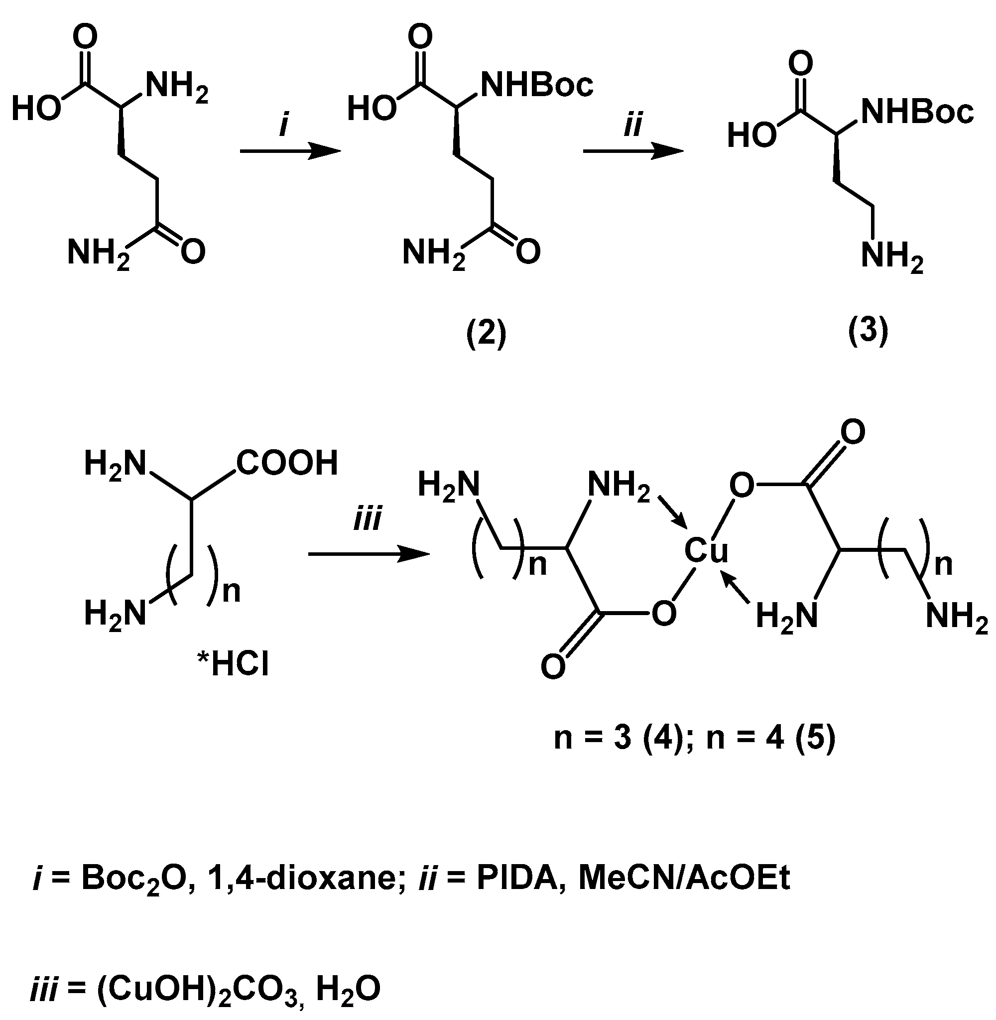
Figure 6.
Optimized structures of system (6) and (7).
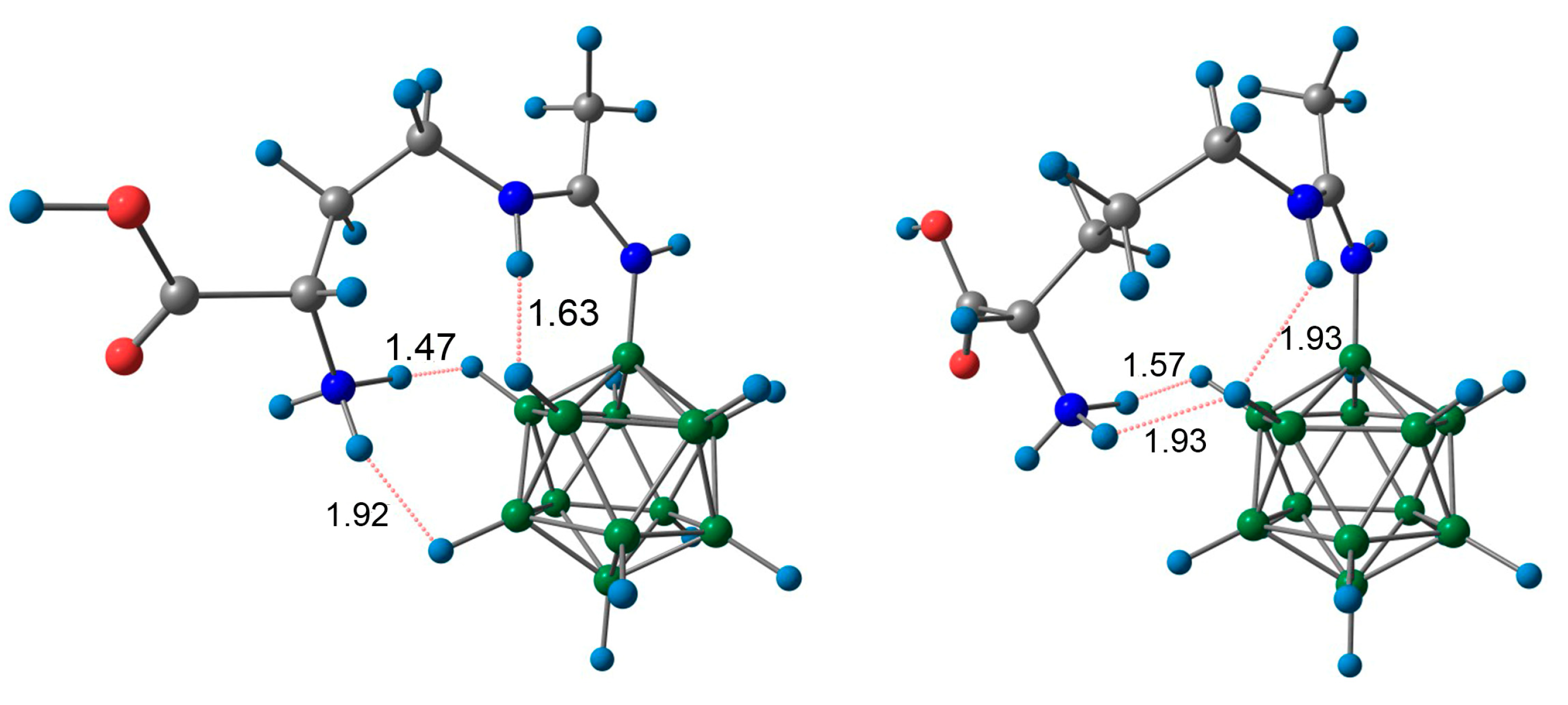
Figure 7.
Dynamics of boron content in blood (red line), muscle tissue (green line), and B16F10 melanoma tumor tissue (blue line) following intravenous administration of compound (6) in C57Bl/6 mice.
Figure 7.
Dynamics of boron content in blood (red line), muscle tissue (green line), and B16F10 melanoma tumor tissue (blue line) following intravenous administration of compound (6) in C57Bl/6 mice.

Figure 8.
Dynamics of boron content in blood (red line), muscle tissue (green line), and B16F10 melanoma tumor tissue (blue line) Following intravenous administration of boronophenylalanine to C57Bl/6 laboratory mice.
Figure 8.
Dynamics of boron content in blood (red line), muscle tissue (green line), and B16F10 melanoma tumor tissue (blue line) Following intravenous administration of boronophenylalanine to C57Bl/6 laboratory mice.
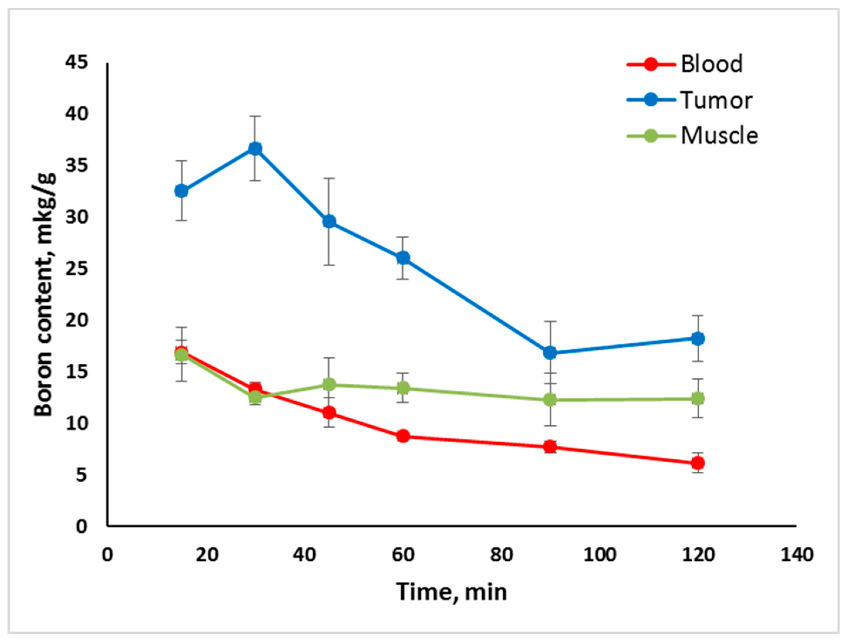
Figure 9.
Boron content ratio in tumor and muscle tissue (T/N Ratio) following intravenous administration of compound (6) (red line) and boronophenylalanine (blue line) to C57Bl/6 laboratory mice with B16F10 melanoma.
Figure 9.
Boron content ratio in tumor and muscle tissue (T/N Ratio) following intravenous administration of compound (6) (red line) and boronophenylalanine (blue line) to C57Bl/6 laboratory mice with B16F10 melanoma.
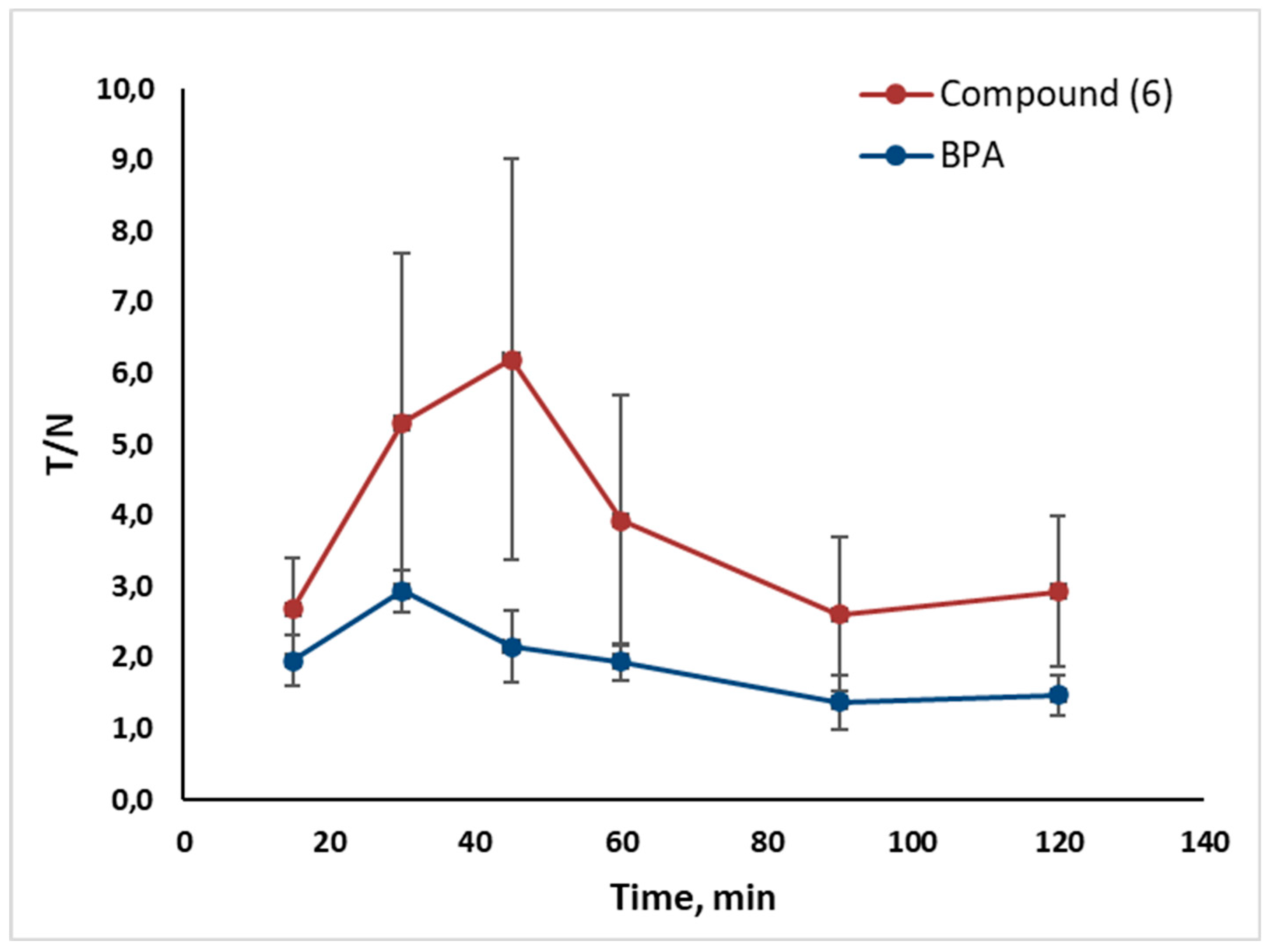
Figure 10.
Dynamics of boron content in blood (red line), muscle tissue (green line), and 4T1 adenocarcinoma tumor tissue (blue line) following intravenous administration of compound (6) to Balb/C laboratory mice.
Figure 10.
Dynamics of boron content in blood (red line), muscle tissue (green line), and 4T1 adenocarcinoma tumor tissue (blue line) following intravenous administration of compound (6) to Balb/C laboratory mice.
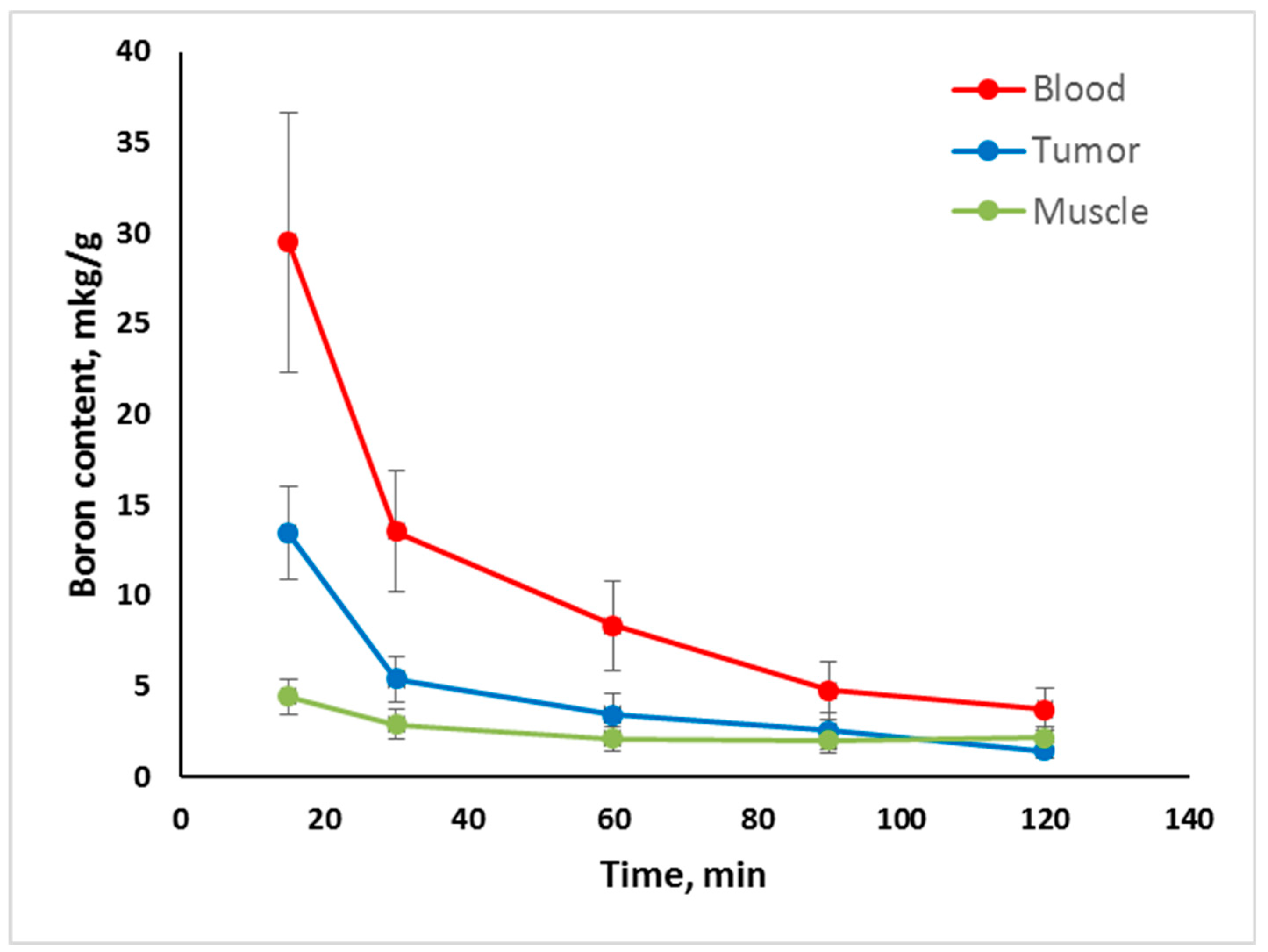
Figure 11.
Dynamics of boron content in blood (red line), muscle tissue (green line), and 4T1 adenocarcinoma tumor tissue (blue line) following intravenous administration of boronophenylalanine to Balb/C laboratory mice.
Figure 11.
Dynamics of boron content in blood (red line), muscle tissue (green line), and 4T1 adenocarcinoma tumor tissue (blue line) following intravenous administration of boronophenylalanine to Balb/C laboratory mice.
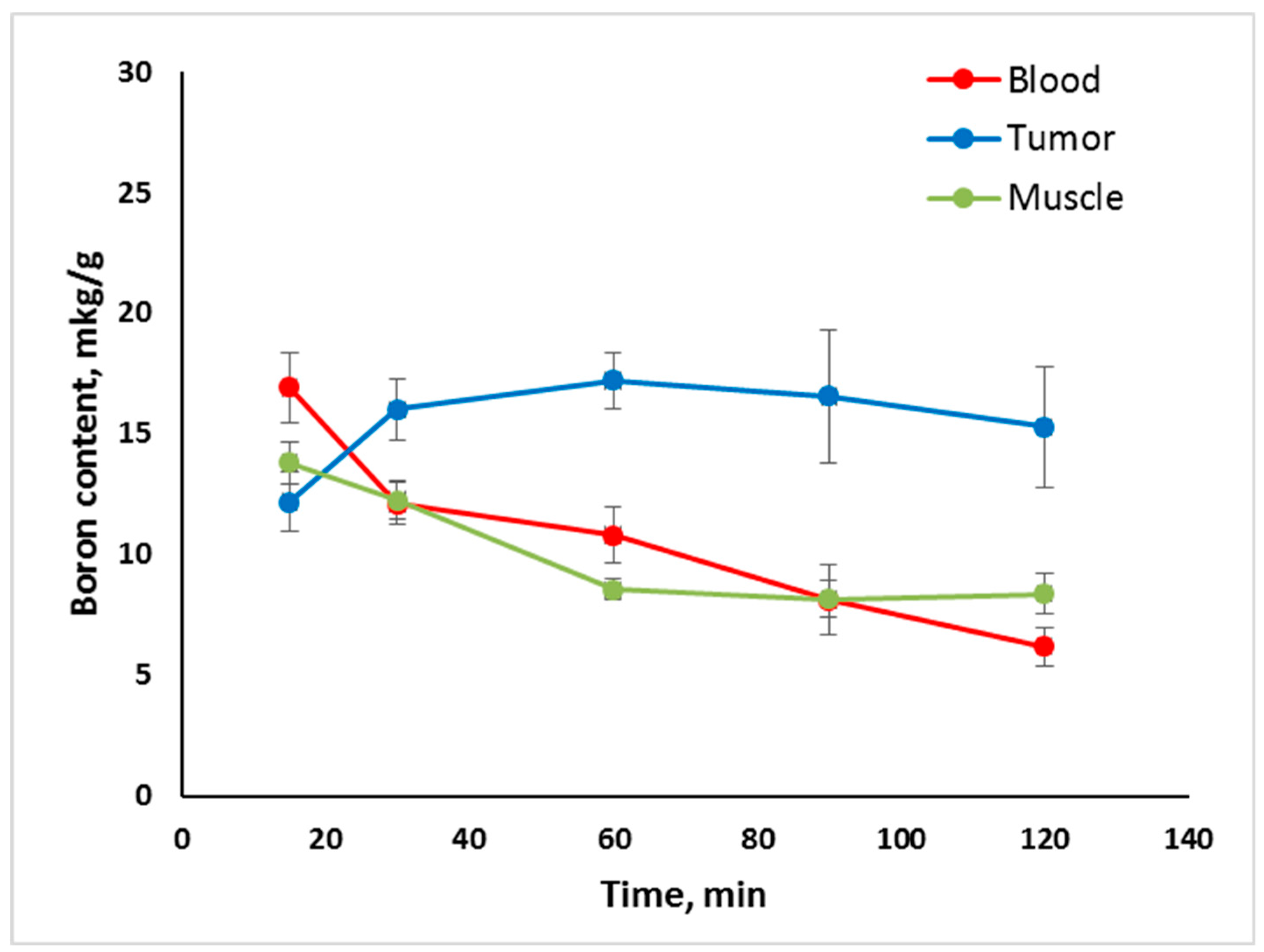
Figure 12.
Boron content ratio in tumor and muscle tissue (T/N Ratio) following intravenous administration of compound (6) (red line) and boronophenylalanine (blue line) to Balb/C laboratory mice with 4T1 adenocarcinoma.
Figure 12.
Boron content ratio in tumor and muscle tissue (T/N Ratio) following intravenous administration of compound (6) (red line) and boronophenylalanine (blue line) to Balb/C laboratory mice with 4T1 adenocarcinoma.
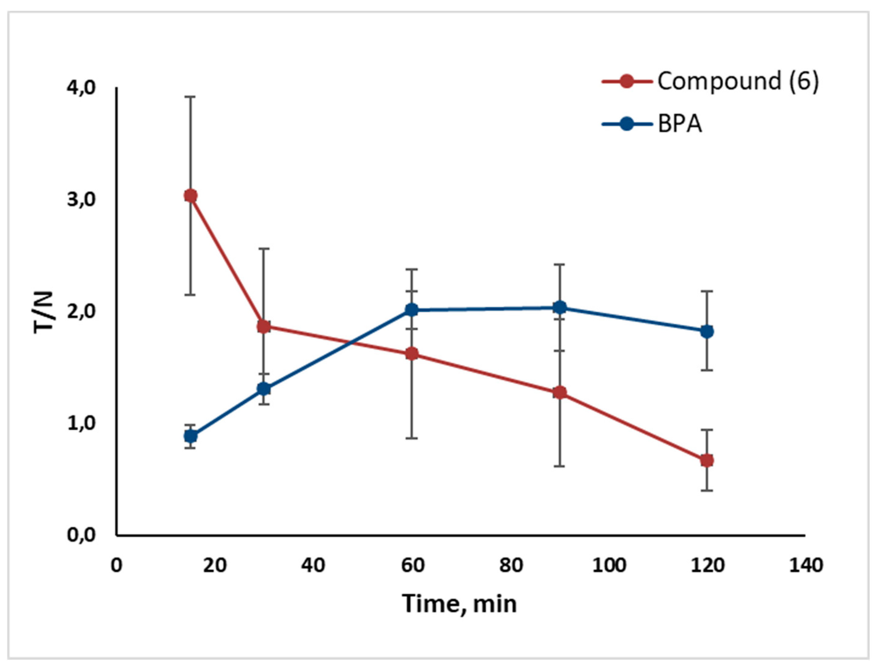
Figure 13.
Dynamics of boron content in kidneys (orange line) and liver tissue (purple line) following intravenous administration of compound (6) in C57Bl/6 laboratory mice.
Figure 13.
Dynamics of boron content in kidneys (orange line) and liver tissue (purple line) following intravenous administration of compound (6) in C57Bl/6 laboratory mice.
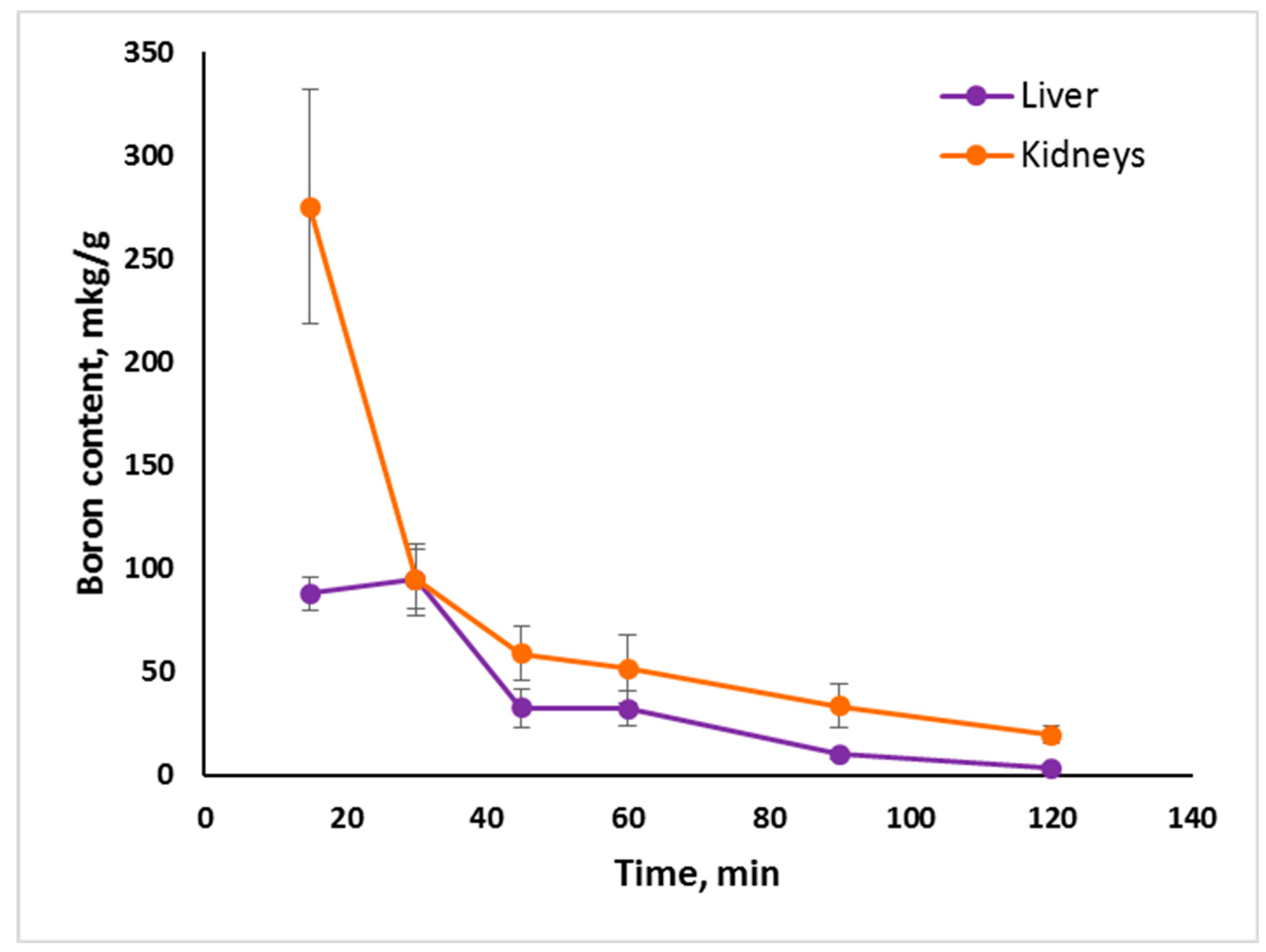

Table 1.
Hydrogen bonds in structure (6).
| D-H…A | d(D-H), Å | d(H-A), Å | d(D-A), Å | D-H-A, ° |
| O2-H2...O3 | 0.84 | 1.78 | 2.594(5) | 163.1 |
| N3-H3A...O5 | 0.91 | 1.83 | 2.735(7) | 170.3 |
| N3-H3C...O31 | 0.91 | 2.16 | 2.814(6) | 127.7 |
| O5-H5B...O42 | 0.87 | 1.94 | 2.802(8) | 169.6 |
| O3-H3F...O13 | 0.87 | 2.07 | 2.827(5) | 145.5 |
| O3-H3G...O4 | 0.87 | 1.86 | 2.726(6) | 172.3 |
1+X,1+Y,+Z; 2-1-X,1/2+Y,-1-Z; 3-X,-1/2+Y,-1-Z 1.
Table 2.
Hydrogen bonds in structure (7).
| D-H…A | d(D-H), Å | d(H-A), Å | d(D-A), Å | D-H-A, ° |
| O2-H2… N4 | 0.84 | 1.89 | 2.721(2) | 170.1 |
| N3-H3B… O11 | 0.91 | 2.18 | 2.894(2) | 135.1 |
| N3-H3B… N41 | 0.91 | 2.53 | 3.235(2) | 135.1 |
| N3-H3C… N6 | 0.91 | 2.14 | 3.024(2) | 164.4 |
1-X, -1/2 + Y, 1-Z.
Disclaimer/Publisher’s Note: The statements, opinions and data contained in all publications are solely those of the individual author(s) and contributor(s) and not of MDPI and/or the editor(s). MDPI and/or the editor(s) disclaim responsibility for any injury to people or property resulting from any ideas, methods, instructions or products referred to in the content. |
© 2024 by the authors. Licensee MDPI, Basel, Switzerland. This article is an open access article distributed under the terms and conditions of the Creative Commons Attribution (CC BY) license (http://creativecommons.org/licenses/by/4.0/).
Copyright: This open access article is published under a Creative Commons CC BY 4.0 license, which permit the free download, distribution, and reuse, provided that the author and preprint are cited in any reuse.


