

Submitted:
26 November 2024
Posted:
27 November 2024
You are already at the latest version
Abstract
In the current context, breast cancer survivors are characterized by an increased life expectancy through the introduction of screening methods but also of the multidisciplinary therapeutic approach. Thus, the appearance and persistence of adverse effects associated with cancer and its treatment, including cognitive dysfunction, can influence the quality of life in these patients. It is imperative to define the ideas that are incorporated in the vast concept of "cognitive dysfunction associated with cancer" or "oncobrain," alongside to describe the processes that lead to it. Therefore, certain physiological, but also pathological, values of the patient's profile influence the appearance and progression of chemobrain. Among the most frequently cited factors in the literature are age, disorders such as anxiety and depression, and educational level. Furthermore, based on the etiology of neurodegenerative diseases, researchers have developed multiple theories about the causes of chemobrain, many of which rely on data from clinical practice. Given the wide range of changes involved, it's crucial to identify the tests that can both subjectively and objectively demonstrate the cognitive dysfunction associated with the various targeted cognitive impairments. These observations will pave the way for progress in the therapy of these adverse effects.
Keywords:
breast cancer survivors
; cognitive dysfunction
; quality of life
; diagnosis
1. Introduction
Breast cancer survival has substantially increased in the last several years due to improved treatment approaches, development of targeted agents and a focus on personalized cancer care. However, the increasing exposure to various systemic therapies is frequently followed by long-term side effects and survivorship issues with significant impact on quality of life need to be addressed.
Early breast carcinoma management consists in a multi-modal approach that includes chemotherapy, endocrine therapy, radiation therapy and surgery. Depending on tumor stage and biology, escalation and de-escalation strategies have been proposed to decrease long term side-effects without compromising survival in these patients.
With the increasing incidence of cancer and systemic treatment use, cancer-related cognitive impairment (CRCI) is regularly expressed by between 15% and 75% of patients receiving chemotherapy, endocrine and targeted therapy for breast cancer [1,2], being an important concern for cancer survivors. The variability of CRCI incidence and patient-reported symptoms is related to a selection of attributes such as their age, intellectual ability, reproductive state, academic achievement, and differences in measurement tools or treatments [3,4]. Mental alterations may manifest either before or following the detection and management of cancer, and some studies show that they can be transient or long-lasting side effects [5,6] The causes of CRCI are still unclear, and some data suggest metabolic disorders, aging, vascular injuries, or alterations in gray matter as possible mechanisms explaining CRCI in cancer patients [7]. Effective diagnosis and management of these side effects are an important goal for both doctors and patients.
Systemic conditions that raise the potential of objective cancer-related cognitive impairment (CRCI) include healthcare conditions involving the circulatory system, advanced chronological age, and impaired mental capacity. On the other hand, risk variables associated with subjective CRCI incorporate emotional distress, elevated basal ratings for anxiety and depression, a younger lifespan, and modulation of physiological sleep characteristics [8].
Furthermore, there are certain additional factors that may influence CRCI, principally during active cancer treatment. As an example, thyroid disorders are common during cancer chemotherapy cycles, and they may lead to cognitive decline in oncologic patients. Anemia, frequently observed in this category of patients, generally associated with chemotherapy and radiation treatments, can also induce cognitive dysfunction. Most prescribed antiemetic treatments for side effects such as nausea and vomiting can also induce drowsiness. Along with the widely recognized negative implications of corticotherapy, such as sleeplessness and restlessness, it was shown that such therapy might have a minor but considerable negative influence on memory and executive functioning [9,10,11].
An analysis of the connection between psychological indicators such as depression, anxiety, stress, and worry revealed that distress plays a predictor function in the evolution of chemobrain as time passes, even though almost all of the research conducted used a cross-sectional design, limiting the perspective [12]. Examining the effect of psychological variables on subjective as well as objective CRCI is of special significance in the setting of greater incidences of depression and anxiety among cancer patients compared to everyone else. Regarding older breast carcinoma survivors, the elevated risk of CIRC appears to be a result of multiple processes, including the detrimental effects of aging on cognition, pre-morbid limited cognitive reserve, and the accumulation of effects of systemic therapy on the neurological system.
2. Mechanisms Involved in CRCI
2.1. Influencing the Permeability of the Blood-Brain Barrier and the Vascularization of the Brain
The treatment of metastatic breast cancer involves mainly agents that do not cross the blood-brain barrier; nevertheless, they have the potential to cause non-physiological changes in the brain tissue. As a result, they may be responsible for reducing the therapeutic doses used and altering the quality of life of the patients during the treatment and after its completion [13].
The blood – brain barrier represents an interface that continuously adapts to the nervous system’s needs and goes through variations in physiological and pathological situations [14]. Being involved in central nervous system homeostasis, it can be compromised by altering its permeability through different mechanisms [15]. Chemotherapy, the pro-inflammatory cytokines released by the tumor may alter blood-brain barrier.
Cancer treatments compromise the blood-brain barrier’s capacity to function by producing reactive oxygen species, which allow pro-inflammatory molecules, toxins, and the brain parenchyma to interact. Preclinical data show that IL-1 and TNF [16] and metalloproteinases play a role in this process. Environmental elements including ischemia and oxidative stress modulate the enzymatic activity of metalloproteinases like MMP 2/9, increasing the barrier’s permeability and leading to edema [17]. As part of the inflammatory response, the neuropeptide substance P and alterations in NK 1 receptor can cause early alterations in the endothelial cells of the cerebral vascular network, triggering neuronal death [18].
Proteins that play certain roles in transport and are expressed in brain tissue are among the molecules that are being targeted, such as multidrug resistance-associated protein 1 (MRP1). Although there is no correlation between the expression of the protein and the patient’s response to therapy, it did correlate with the protein’s sensitivity to certain agents as determined by the resistance genes’ polymorphism. In connection to oxidative stress, this protein is overexpressed following doxorubicin treatment [19].
In the affected areas of the brain of experimental animals, there is a tendency towards an increased rate of apoptosis and a lower rate of cell proliferation after exposure to cancer treatments. The impact persists even after the therapy is ceased. The toxicity of various cytotoxic, such as cisplatin, at relevant doses differs between neoplastic cell lines and cells of the central nervous system [20]. Research shows that delivering anthracyclines and taxanes to rodents allows a small amount of cytostatic to pass through the blood-brain barrier, which alters it’s permeability and activity [21].
Besides altering the brain-blood barrier, tumors also cause changes in cerebral vascularization such as decreased blood flow, altered cerebral glycemia, and changes in blood vessel density that result in an impairment of hippocampus cell neurogenesis [22].
2.2. The Role of Oxidative Stress and Proinflammatory Cytokines
Although oxidative stress is linked to CRCI, it is frequently present in some patients before the start of cancer treatment [23]. The severity of oxidative stress coincides with responses from immune activated cells as a boost in pro-inflammatory cytokine generation and discharge [24].
Reactive oxygen species and mediators have a central part in cell alteration, and in vascular alterations, potentially having a cumulative effect on how oncological therapy and cognitive decline interact [25].
Following oncological treatment, pro-inflammatory cytokines have higher titers and are actively involved in the configuration and function of the neural network. Accordingly, research indicates that IL1 and TNF alpha expression influence neuroplasticity and synapse-level processes, whereas IL6 affects executive function and patients’ perceptions of their cognitive decline [26].
Research shows that oxidative damage to plastic proteins leads to a spike in peripheral TNF alpha levels, which subsequently influences the central nervous system through increased protein degradation and the release of proinflammatory cytokines [27]. Plasma concentration of IL-1, IL-10, and TNF and pro-inflammatory cytokines decrease immediately after doxorubicin administration, suggesting the agent’s direct interference with their degradation [28]. The administration of paclitaxel, in contrast to doxorubicin, is associated with a high level of IL10 and a plasma lipid peroxidation condition [29].
Even though TNF-alpha-mediated oxidative stress plays a part, additional factors such as cerebral phospholipase expression, mitochondrial respiratory metabolism, and altered choline-containing components from the hippocampus additionally play an integral part in the functional degradation of neurons and their death [30].
Given its high demand for oxygen and lipid-rich structure, the brain’s normal function is negatively impacted by oxidative stress. Apolipoprotein A1, a compound involved in lipid metabolism and inflammation, plays a role in the onset of cognitive impairments linked to cancer and cancer treatment, as it regulates the generation of the cytokines IL-1 and TF-alpha by macrophages. (Figure 1)
Following chemotherapy administration, inflammation in the central nervous system is triggered via the pathway of arachidonic acid conversion into prostaglandins, which requires COX enzymes. After contact with doxorubicin, elevated levels of the COX2 isoform and PGE2 demonstrated the significant activity of immune system effectors in the brain. Alkylating agents, platinum salts, and anthracyclines are also capable of influencing the hippocampus microglia, cells crucial to synaptic transmission and other processes that affect the body’s cognitive function, leading to memory loss [31].
Preclinical data suggest the beneficial effect of certain drugs in reducing the impact on cytotoxics on cognition. Gamma-glutamyl cysteine ethyl ester administration, a precursor of glutathione, might be able to reduce oxidative alterations in proteins or lipids to a level equivalent to that of the control group unexposed to doxorubicin [32]. MESNA has been shown to possess antioxidant effects, minimize protein oxidation, and counteract the anthracycline-induced change in TNF alpha levels. Thus, the administration of MESNA might diminish the effects of doxorubicin on cognition and locomotion, which are regulated by changes in the ratios of neurochemical substances and phospholipases in the hippocampus [33].
2.3. Affecting the DNA Material and Limiting Its Repair
Cognitive dysfunction due to physiological aging process, the pathways of neurodegenerative diseases, or cancer and its treatment, is linked to oxidative stress and failures in DNA repair. DNA repair deficiencies increase a person’s likelihood of developing cognitive impairments correlated with cancers and neurodegenerative illnesses [34].
The OGG1 and APEX1 genes play an important role in neural preserving neural function and limit the negative effects of oxidative stress. The reduced activity of the OGG1 and APEX1 genes may contribute to the onset of cognitive impairments triggered by oncological treatment through disturbances in neuronal function, as these genes serve to preserve cellular genetic information against the negative consequences of oxidative stress. Their variety and poor functionality make cognitive impairment more severe [35]. Individuals exposed to prior therapy are more likely to experience this type of condition [36].
One of the crucial endonucleases identified in human cells, a protein generated by the APEX1 gene, undertakes a wide range of functions, including controlling the way the cell adapts to oxidative stress. Its roles include DNA repair and redox-mediated modulation of transcriptional factors like p53, AP-1, or HIF-1 [37].
8-oxoguanine can often be generated when reactive species interact with DNA, and it is found in parts of the genome where transcription is tightly controlled. OGG1 associates with this molecule through its enzymatic activity and can operate as a transcription regulator [36]. These shifts, which mainly impact the cortex and amygdala, are further linked to perfusion adaptations, senescence, and the neurodegenerative alterations associated with Alzheimer’s disease [38].
2.4. Telomere Length
Telomere alterations have observed in cancer patients [39].The process that illustrates how telomere length helps facilitate the progression of cognitive impairment is still not entirely known, but it has been suggested that modified telomere structure and function induced by cancer or chemotherapy produce chromosomal instability. Although the mechanisms that describe how chemotherapy shapes telomere length remain under study, the concept that the therapy directly impacts these regions’ DNA because of oxidative stress and inefficient repair actions in the terminating areas of chromosomes has been launched. Anthracyclines, which are frequently prescribed for the management of carcinoma of the breast and are also shown to cause cognitive impairment, interfere with the length of telomeres by directly altering DNA because of the synthesis of reactive compounds as well as limiting DNA repair enzymes like Topoisomerase II, resulting in the accumulation of DNA lesions [40].
Moreover, over 85% of cells that have undergone malignant transformation express telomerase, a ribonucleoprotein that extends the 3’ end of telomeres with repeated sequences and its activity helps promote the onset of the malignant phenotype in cells [41]. Currently, new medications are being explored that specifically interfere with this enzyme’s activity [42].
2.5. Genetic Predisposition
The ongoing monitoring of cancer patients who underwent oncological treatment suggests that several of them endured cognitive deficits for a longer period than others in the group. For that reason, the role of their genetic neurological vulnerability is being explored, as is the component that both serotonin and dopamine disturbance serve in the cognitive dysfunction gained due to taking any kind of oncological pharmaceutical [43].
In this context, variation in the apolipoprotein E, brain-derived neurotrophic factor (BDNF), and COMT genes may determine the way the neurological system operates [44]. The APOE E4 allele variant is one of the key variables in the onset of dementia since it plays an essential role in physiological cognitive decline and the natural aging process of humans [45]. Hippocampal volume shifts are linked to a greater limitation in the functioning of memory in breast cancer patients whose APOE E4 allele status has been determined and these individuals also experience psychomotor deficiencies, which contribute to spatial synchronization and visual memory difficulties [46].
The catechol-O-methyltransferase (COMT) enzyme has a restricted capacity for degrading the neurotransmitters tied to both the onset and sustained persistence of cognitive deficits linked to chemotherapy. Dopamine belongs to the category of these neurotransmitters that may shape executive and memory abilities, and its level is modified, particularly in the frontal cortex, which serves as an essential component in the successful execution of these CNS functions [47]. Also, neuronal transmission undergoes modification through COMT gene polymorphism. As a result, there are more functional and structural flaws in the hippocampus among people with more aggressive breast neoplasms, like those who present a high Ki67 proliferation index. Additionally, research links the COMT rs165599 and rs737865 polymorphisms to a higher risk of cognitive changes triggered by chemotherapy treatments in triple-negative and HER2-positive subtypes [48].
Proteins referred to as BDNF exert an integral part in cerebral cellular differentiation, their survival, and the growth of dendrites and axons of neurons [49]. They are prominently expressed in the cortex and hippocampus, and differences in the volumes of the brain and diminished memory with aging are both associated with the way they function [50]. The BDNF gene variation reflects how frequently patients with oncologic disease suffer from depression. Therefore, compared to carriers of the Val allele, patients with breast cancer who possess the Met allele have a lower degree of objective cognitive dysfunction in relation to the level of BDNF throughout treatment [51].
2.6. Hormonal Changes and the Influence of CDK Inhibitors
Sexual hormones can influence cognition both positively and negatively by safeguarding its function and ability to counteract oxidative stress [52]. The preferential distribution of hormone receptors and aromatase, the enzyme involved in the generation of estrogen from testosterone, in the regions of the hypothalamus, hippocampus, and prefrontal cortex compromises executive, learning, and memory abilities across the brain parenchyma [53]. The hormonal levels present within those structures have the potential to influence synaptic activity through regulating axonal and dendritic expansion as well as the expression of neurotransmitters such as norepinephrine, dopamine, and acetylcholine [53]. Comparable with the detrimental impact of psychological menopause on cognition, chemotherapy-induced menopause limits the attention and processing speed of Individuals with breast-localised malignancies [54]. Furthermore, the administration of specific treatments in hormone-sensitive cancers, such as selective estrogen receptor modulators and aromatase inhibitors, alters the physiological hormonal level, contributing to the appearance, maintenance, and accentuation of cognitive dysfunction to varying degrees depending on the chosen treatment [55]. Additionally, the negative consequences suffered by these patients, such as fatigue, sleeplessness, and mood disturbances, may exacerbate the cognitive symptoms [56].
Although both varieties of hormone therapy pharmaceuticals have access to neural tissue due to their ability to break through the blood-brain barrier, the effects on cognition can be distinguished based on the manner of action [57]. Thus, aromatase inhibitors have a universally detrimental impact on estrogen levels regardless of tissue, but tamoxifen attempts to disable the receptor in the breast and can have a stimulating effect depending on the tissue targeted. Furthermore, a different density of estrogen receptor types can be observed in the cerebral cortex, with the majority being beta-type receptors at the temporal cortex and hippocampus and alpha-type receptors at the amygdala and hippocampal levels, with implications for the mechanisms of cognitive dysfunction [58].
Multiple investigations have highlighted the significance of low-level sex hormone exposure in the rate of cognitive dysfunction related to oncological treatment with aromatase inhibitors and tamoxifen, even in disagreement with results in this direction. Furthermore, research results indicate that tamoxifen has a more potent and harmful effect on cognition if administered to individuals who have undergone previous chemotherapy [59]. The effect on cognition in chemo- and hormone-treated women is transient and fades away following therapy [60]. Thus, the effect of combining the two therapies is regarded as cumulative and more significant than in the context of monotherapy with hormone therapy in the first 6 months, with this effect dropping 12 months after chemotherapy without statistically significant variations. Furthermore, each of the kinds of aromatase inhibitors used has been linked with various levels and types of cognitive impairment. As a result, patients treated with tamoxifen exhibit a larger deficit in performing executive functions compared to those treated with exemestane, with no statistically significant discrepancies between the results of the exemestane group and the observing group [61]. In the instance of anastrozole treatment, it seems like cognitive impairment occurs in a manner like chemotherapy, with memory, focus, and executive function being acutely affected, with the possible exception that in the case of this aromatase inhibitor, a second decline in mental functioning is noted in patients who just underwent hormone therapy [62].
CDK4/6 inhibitors, which have been successfully used in the metastatic and adjuvant therapy of selected patients with Luminal and HER2-negative breast cancer, play a crucial role in improving PFS and OS [63,64,65]. The repression of cell division induced by CDK4/6 inhibitors contributes to an extended release of pro-inflammatory cytokines, which is related to cognitive decline [66). Furthermore, CDK4/6 inhibitors participate in the development and differentiation of newly created neural cells and thus influence the memorizing processes via their own mechanism of action that targets cells with a high reproducibility [67].
2.7. Direct Neurological Toxicity and the Influence on the Genesis of Neural Structures
Oncological pharmaceutical damage to the brain parenchyma has been established as an essential contributing factor to the onset and persistence of deficits in cognition in cancer patients. This injury can appear throughout the central nervous system by causing disruption with neurotransmitter levels and myelin in the neural network, as well as changing the function of cells vital to neural network preservation and repair. Even ordinary amounts of platinum derivatives and fluoropyrimidines may lead to damage in these entities, resulting in a rise in cell death rate that is inversely associated with cell division rate [68]. Animal studies demonstrate a substantial reduction in neurogenesis in the hippocampus in mice treated with cyclophosphamide and doxorubicin, which corresponds to a decline in cognitive functions specific to this structure. A closer examination of CNS cells treated with these cytostatics exposes disturbances in neuronal and glial differentiation, which establish the architecture of the neural extensions responsible for nerve impulse transmission and the density of the spinal cord. Furthermore, the hippocampus is the structural component most affected by this phenomenon [69). However, these outcomes do not fully support the influence of the alteration of the neuronal structure and physiology in hippocampus on cognitive functions such as learning ability and memory [70].
2.8. White Matter Integrity
Patients treated with cytotoxic agents exhibit progressive lesions of the central and peripheral nerve systems compared to those who are chemotherapy-naive. [71].
These lesions, which can be identified months to years after therapeutic intervention, might impact the development of cognitive deterioration associated with cancer and its treatment [72]. As a result, the studies show that anthracycline administration in conventional doses influences the integrity of the white matter as measured by fractional anisotropy in the corpus callosum of chemotherapy-treated patients, and a decrease in reaction speed can be clinically detected. On oligodendrocytes, the detrimental consequences of fluoropyrimidines on white matter integrity have been revealed, with incomplete or total demyelination of axons and punctual changes in the axonal cytoskeleton and microtubules [73]. The balance between elements that may inhibit cell apoptosis after exposure to cytostatic drugs and those that can promote it affects neurocognitive performance. A fluctuation in the titer of some of these proteins and enzymes, including p53, Bax, cytochrome c, and caspase 3, was seen after exposure to cytostatic drugs [74].
2.9. Interference with Long-Term Potentiation
Cognitive alterations reported by cancer patients undergoing treatment could be attributed to the drugs’ influence on some targets of the long-term potentiation pathway. Long-term potentiation is a key mechanism responsible for learning and memorization, in which interneural connections in the neocortex and hippocampus are potentiated through repetitive activation. Thus, their long-term effectiveness is enhanced through systematic and substantial participation in synaptic stimulation. Some receptors in this route play a role in its initiation and management. Through erroneous neuronal activation, ionotropic glutamate receptors can induce glutamate excitotoxicity, a lethal form of toxicity on neuronal tissue, and these receptors are one of the targets of medication used in psychiatric disorders to improve learning and memory in these patients [75].
Protein kinase A (PKA), Ca2+/calmodulin-dependent protein kinase II (CaMKII), and extracellular signal-regulated kinase (ERK) are all modulatory enzymes identified in this pathway. Moreover, some receptors, including NMDA and AMPA, have an excitatory role that is regulated by sodium and calcium and is involved in improving synaptic activity and neuroplasticity [76). They have a defining role in embryogenesis, causing neural tube abnormalities, but they also represent potential therapeutic targets in conditions as depression, Alzheimer’s disease, and epilepsy [77].
It is demonstrated that the interaction between these potential targets from the long-term potentiation pathway and some cytostatics can be correlated with changes in the neuro-cognitive process without being able to specify their degree or if there is the possibility of presenting a protective Influence on mental processes, or the reverse. In the therapy of breast cancer, the interactions between taxanes and NMDA, AMPA, and PKA receptors, as well as everolimus and NMDA and PKA receptors, are noteworthy [78].
2.10. Tumor Biology
A selected number of individuals diagnosed with breast cancer may exhibit shifts in cognition after receiving the diagnosis but before starting therapy, suggesting the influence of the tumor’s own biology [79]. Aside from the stressful effect of discovering the oncological diagnosis, the presence of one of the malignant phenotype’s hallmarks, chronic inflammation, has been suspected as a promoter of the development of pre-treatment cognitive deficits. This may impact not only the persistence of inflammation at the neuronal level but also the origins of neoplastic developments [80].
Anemia, probably one the most prevalent adverse complication of oncological treatments, can also be attributed to the tumor, and the symptomatology caused by the decrease in oxygen concentration in the brain is like cognitive dysfunction [81]. However, treating it with erythropoietin alpha fails to deliver consistent results in studies, with no clear and statistically significant correlation between its administration and, as a result, the correction of anemia and the significant improvement of cognition in the patients involved in these studies [82].
2.11. Tau Protein
Data from the literature describe the presence of a desirable amount of the tau microtubule-associated protein that corresponds with the microtubule organizing and constancy capacities. This is ubiquitous in nervous system neurons, and alterations to its structure create microtubular instability and axonal transport issues [83]. Because this protein participates in the etiology of neurodegenerative illnesses, its level in cerebrospinal fluid can be employed as an indicator of cerebral injury when paired with abnormal outcomes in neurocognitive testing [84]. Preclinical investigations indicate that cytostatics may accelerate the rate of tubulin deacetylation and the accumulation of hyperphosphorylated tau protein, potentially affecting the stability of microtubules in neuronal extensions. Thus, it is known that the administration of cytostatics such as cisplatin or methotrexate determines the limitation of cell proliferation in the hippocampus, an increase in tau protein in the cerebrospinal fluid, and late cognitive disorders, which persist for approximately 3 months after treatment stops [85].
Although studies involving patients with neoplasia continue to be performed to examine the relationship between tau protein levels and preclinical study outcomes, we cannot yet assume that the level of this protein in serum or CSF is a biomarker of cognitive dysfunction. Currently, studies in chemo-treated breast cancer patients display an interdependence between tau protein and beta amyloid levels, as well as neurodegeneration markers and pro-inflammatory cytokines, with the results explaining some of the treatment-related changes in cognitive function [86].
2.12. Psychological Factors
When referring to the expression of cognitive dysfunction in individuals diagnosed and treated for breast cancer, psychological factors may contribute to adjustments in attention and memory in this limited group of patients. As as a consequence, some of these patients develop the typical symptoms prior to starting oncological treatment. Furthermore, related mental health issues such as anxiety and depression, as well as the presence of stress or fatigue, can alter the patient’s clinical course [80]. The mechanisms of cognitive impairment in relation to the presence of pre-existing stressful conditions associate changes encountered in the onset of anxiety-depressive disorders and malignant diseases with induced changes in the level of pro-inflammatory cytokines [87].
3. Defining and Diagnosing Cancer-Related Cognitive Impairment (CRCI)
Current challenges remain in clearly defining and diagnosing CRCI. Research and clinical trials have failed to clearly establish tests that can identify minor alterations that define cognitive impairment associated with cancer and its treatments [88]. Also, cognition requires being perceived from both an objective and a subjective perspective, which are both interconnected and associated with the quality of life of the individuals. The potential drawback of integrating these subjective measures is represented by the impact of comorbidities such as anxiety and depression, as well as social elements related to cultural heritage and area of residence [8]. The main differences seen in trials may be attributed to the wide diversity of tests conducted and to the possibility that these tests are intended to measure either general or specific operations of the person’s cognition [89].
A neurophysiological evaluation is important in determining the presence of cognitive impairment and its severity, especially considering the discrepancies between symptom severity and results of objective tests. Additional pre-existing factors unrelated to chemotherapy should be documented, such as psychiatric conditions (e.g., anxiety, depression), sleep-related problems or other treatment-related effects (e.g., drug interactions, chronic inflammation, effects of radiation or hormonal treatment).
When discussing cognitive decline, we refer to the nervous tissue’s compromised capacity to perform its essential function of collecting, analyzing, and using data related to at least one of the fields of interest that define cognitive ability objectively via a substantial reduction in the statistical data obtained through an unbiased examination [90]. To accurately evaluate the influences of this condition, it is critical to choose resources that can assess not less than one of the following: memory, focus, attention, the cognitive aspect of the language domain, and the spatial and psychomotor orientation domain [91].
The International Cognition and Cancer Task Force provides in broad terms the main areas affected by cancer treatments and preferable tools. The certification of these tests, as they advise, necessitates the analysis of data derived through periodic interrogation at an established follow-up time interval, the use of studies contrasting the outcomes with the results obtained from a control arm formed of healthy individuals, and the detection of structural alterations through imaging of the central nervous system components [92]. (Table 1).
The TMT test, it permits evaluating not only the executive function but also the respondents’ attention [93]. It has various benefits, among them being that it is a brief examination that does not require an excess of resources. However, the outcomes might be altered by the participant’s age, which does not constitute a component of the criteria included in obtaining the score but impacts overall performance. Another test that targets the same areas of cognition is the Digit Symbol Substitution Test. It involves ordering some symbols according to the numbers that correspond to them through a predetermined legend in a given time interval [94].
The Verbal Fluency test is used for testing both language-related mental abilities and executive function, employing a design that reveals the person’s capacity to quickly but spontaneously respond with a variety of distinct words as feasible belonging to a particular category or beginning with an identical given letter, COWA being a well-known example of this type of test [95].
The Digit Span test, which consists of retaining the sequence of numbers and reproducing them in precisely the same order, also targets attention [96]. In addition, to examine the same cognition span of the individual with the Paced Auditory Serial Addition test, the participant is instructed to add the prior two numbers that are said loudly with a few seconds gap [97].
Several assessments with different designs are employed for determining higher-level cognitive processes like attention, perseverance, the capacity to learn, conceptualization, and set shifting. Therefore, since it is a set-shifting test, the Wisconsin Card Sorting Test necessitates a certain level of adaptability. The test, which includes matching specific cards without guidance provided before the activity begins but only from remarks derived after the subject’s decisions, has been applied satisfactorily in the actual situation of patients with neuropsychiatric illnesses [98].
The Stroop Color and Word Test assess flexibility and suppression for triggers to be able to name the color of a term, even though the words themselves are colored in identical shades or not [5]. Thus, it is proven that defining colors that are not in accordance with the specified word takes longer than defining colors that are the same as those specified in the word [99].
There are several tests with an extensive background in neuropsychiatric pathology that might be used such as the Montreal Cognitive Assessment and the Mini-Mental State Examination [93]. These are two of the most well-established and extensively used cognition tests. Also, other tests such as the National Adult Reading Test can be used for the initial assessment of premorbid mental status. When an exhaustive investigation is required, CANTAB and The California Verbal Learning Test, could potentially be chosen [100]. The Hopkins Verbal Learning Test measures both short-term and long-term recall and is notable for its detailed sensitivity170. To facilitate the selection of a suitable test simpler, its effectiveness was compared to that of the Rey Auditory Verbal Learning Test152. The focus was on similar cognition characteristics, and it was recognized there could possibly be inconsistent indicators due to the interpretation, which could lead to distinctive descriptions of cognitive impairment in each comparable population.
Subjective evaluations such as FACT COG [93], is a test used in descriptive and interventional research aimed at studying the cognition-related issues of adult cancer patients. It is simple to use and it scores the patient’s responses, which are divided into four main groups: perceived cognitive dysfunction and its impact on quality of life; perceived cognitive abilities; and, last but not least, remarks made by individuals with whom the person in question interacts [101].
Compared to the FACT COG, the Patient Assessment of Own Functioning Inventory (PAOFI) offers an alternative framework for neuro-psychological evaluation of patients, analyzing memory, the ability to use language and express oneself properly, performing manual tasks, but also the impairment of subjective perception of sensation, and, in addition to this, the performance of more complex intellectual functions [102]. Furthermore, this questionnaire focuses on the participant’s cognitive decline in connection with his capacity to fulfill daily duties. This questionnaire has been used in patients with oncological, mental, and nephrological conditions.
The Prospective and Retrospective Memory Questionnaire (PRMQ) examines the influence of memory on the malfunctions reported by patients throughout normal daily activities. Using an equal number of inquiries related to the two types of researched memories—the consequences of oneself, the external environment, and the long-lasting and immediate memories—on the triggering of these phenomena, this survey adds an unexpected dimension to the cognitive assessments that are employed [103].
The Measurement of Everyday Cognition (ECog) exhibits an in-depth approach to the disturbance of mental operations in everyday tasks. It evaluates memory, language-related ability, visual, and spatial abilities; however, it draws attention to certain complex functions by reviewing the ability to organize and schedule routine tasks alongside divided focus [104].
4. Conclusions
Due to improvements in cancer diagnosis and treatment with both curative and palliative intent, the number of cancer survivors is increasing, and survivorship challenges need to be better addressed. Effective patient management includes besides optimal cancer treatment, a better evaluation and management of various comorbid conditions impacting patient’s long-term quality of life, such as psychological distress, sleep disturbances, fatigue, pain. The prevalence and impact of cognitive dysfunction following cancer treatments is frequently clinically significant, as it extends beyond the cancer treatment period. The lack of a proper diagnosis and awareness among physicians leads to an insufficient support available for cancer survivors. The impact on quality of life extends from daily activities, family interactions and work, to an economic impact for both the patient and the society.
A better understanding of the underlying mechanisms and improvements in diagnosis is crucial. Various assessments are needed to objectively measure the cognitive function in addition to self-report, due to the discrepancies between the perceived symptoms and how they can be objectively assessed. Moreover, there is a current lack of clear recommendations on how to better manage patients with CRCI, although various strategies have showed improvements, such as cognitive rehabilitation, mindfulness, neuroprotective medications or exercise.
Although many tests have been evaluated in various trials, inconsistencies between the test findings and the patient’s personal perceptions are likely, either due to the accuracy of the testing in detecting slight changes or to the consistency with which the evaluation was completed.
Although the etiology of CRCI is not completely understood, a combination of both host features and biological variables plays a role in its development. Continuous efforts are being made in better diagnosing and management of CRCI, due to its increased prevalence and impact on patient’s quality of life. Behavioral and pharmacological interventions are being developed and tested to prevent and treat CRCI and further research focusing on diagnosis and treatment is needed, especially with more diverse populations in terms of race, age, cancer type and treatment.
Author Contributions
Conceptualization, D.I.P, S.R.V, C.V., C.V., M.O. and C.M.F.; Methodology, D.I.P, S.R.V, C.V., M.O. and C.M.F.; Validation, D.I.P, S.R.V, C.V., C.V., M.O. and C.M.F., Formal Analysis, D.I.P, S.R.V, C.V., C.V. and C.M.F.; Writing – Original Draft Preparation, D.I.P, S.R.V, C.V., C.V., M.O., T.A.B. and C.M.F.; Writing – Review & Editing, D.I.P, S.R.V, and C.V.; Visualization, D.I.P, S.R.V, C.V., C.V., M.O. and C.M.F.; Supervision, D.I.P, S.R.V, C.V., C.V., M.O. and C.M.F.; Project Administration, D.I.P, S.R.V, and C.V.
Funding
This research received no external funding.
Conflicts of Interest
The authors declare no conflict of interest.
References
- Bray VJ, Dhillon HM, Vardy JL. Systematic review of self-reported cognitive function in cancer patients following chemotherapy treatment. J Cancer Surviv. 2018;12(4):537-59. [CrossRef]
- Ahles TA, Saykin AJ, Furstenberg CT, Cole B, Mott LA, Skalla K, et al. Neuropsychologic impact of standard-dose systemic chemotherapy in long-term survivors of breast cancer and lymphoma. J Clin Oncol. 2002;20(2):485-93. [CrossRef]
- Edelstein K, Bernstein LJ. Cognitive dysfunction after chemotherapy for breast cancer. J Int Neuropsychol Soc. 2014;20(4):351-6. [CrossRef]
- Whittaker AL, George RP, O’Malley L. Prevalence of cognitive impairment following chemotherapy treatment for breast cancer: a systematic review and meta-analysis. Sci Rep. 2022;12(1):2135. [CrossRef]
- Koppelmans V, Breteler MM, Boogerd W, Seynaeve C, Gundy C, Schagen SB. Neuropsychological performance in survivors of breast cancer more than 20 years after adjuvant chemotherapy. J Clin Oncol. 2012;30(10):1080-6. [CrossRef]
- Reid-Arndt SA, Hsieh C, Perry MC. Neuropsychological functioning and quality of life during the first year after completing chemotherapy for breast cancer. Psychooncology. 2010;19(5):535-44. [CrossRef]
- Zimmer P, Baumann FT, Oberste M, Wright P, Garthe A, Schenk A, et al. Effects of Exercise Interventions and Physical Activity Behavior on Cancer Related Cognitive Impairments: A Systematic Review. Biomed Res Int. 2016;2016:1820954. [CrossRef]
- Janelsins MC, Heckler CE, Peppone LJ, Kamen C, Mustian KM, Mohile SG, et al. Cognitive Complaints in Survivors of Breast Cancer After Chemotherapy Compared With Age-Matched Controls: An Analysis From a Nationwide, Multicenter, Prospective Longitudinal Study. J Clin Oncol. 2017;35(5):506-14. [CrossRef]
- Cunningham RS. Anemia in the oncology patient: cognitive function and cancer. Cancer Nurs. 2003;26(6 Suppl):38S-42S. [CrossRef]
- Bower JE. Behavioral symptoms in patients with breast cancer and survivors. J Clin Oncol. 2008;26(5):768-77. [CrossRef]
- Prado CE, Crowe SF. Corticosteroids and Cognition: A Meta-Analysis. Neuropsychol Rev. 2019;29(3):288-312. [CrossRef]
- Yang Y, Hendrix CC. Cancer-Related Cognitive Impairment in Breast Cancer Patients: Influences of Psychological Variables. Asia Pac J Oncol Nurs. 2018;5(3):296-306. [CrossRef]
- Chen Y, Jungsuwadee P, Vore M, Butterfield DA, St Clair DK. Collateral damage in cancer chemotherapy: oxidative stress in nontargeted tissues. Mol Interv. 2007;7(3):147-56. [CrossRef]
- Corps KN, Roth TL, McGavern DB. Inflammation and neuroprotection in traumatic brain injury. JAMA Neurol. 2015;72(3):355-62. [CrossRef]
- Banks WA. From blood-brain barrier to blood-brain interface: new opportunities for CNS drug delivery. Nat Rev Drug Discov. 2016;15(4):275-92. [CrossRef]
- Pan W, Tu H, Yu C, Hsuchou H, Yang Y, Kastin AJ. Differential role of TNF receptors in cellular trafficking of intact TNF. Cell Physiol Biochem. 2007;20(5):559-68. [CrossRef]
- Galvez BG, Matias-Roman S, Yanez-Mo M, Vicente-Manzanares M, Sanchez-Madrid F, Arroyo AG. Caveolae are a novel pathway for membrane-type 1 matrix metalloproteinase traffic in human endothelial cells. Mol Biol Cell. 2004;15(2):678-87. [CrossRef]
- Rodriguez PL, Jiang S, Fu Y, Avraham S, Avraham HK. The proinflammatory peptide substance P promotes blood-brain barrier breaching by breast cancer cells through changes in microvascular endothelial cell tight junctions. Int J Cancer. 2014;134(5):1034-44. [CrossRef]
- Joshi G, Sultana R, Tangpong J, Cole MP, St Clair DK, Vore M, et al. Free radical mediated oxidative stress and toxic side effects in brain induced by the anti cancer drug adriamycin: insight into chemobrain. Free Radic Res. 2005;39(11):1147-54. [CrossRef]
- Dietrich J, Han R, Yang Y, Mayer-Proschel M, Noble M. CNS progenitor cells and oligodendrocytes are targets of chemotherapeutic agents in vitro and in vivo. J Biol. 2006;5(7):22. [CrossRef]
- Pieniazek A, Czepas J, Piasecka-Zelga J, Gwozdzinski K, Koceva-Chyla A. Oxidative stress induced in rat liver by anticancer drugs doxorubicin, paclitaxel and docetaxel. Adv Med Sci. 2013;58(1):104-11. [CrossRef]
- de Vos FY, Willemse PH, de Vries EG, Gietema JA. Endothelial cell effects of cytotoxics: balance between desired and unwanted effects. Cancer Treat Rev. 2004;30(6):495-513. [CrossRef]
- Cauli O. Oxidative Stress and Cognitive Alterations Induced by Cancer Chemotherapy Drugs: A Scoping Review. Antioxidants (Basel). 2021;10(7). [CrossRef]
- Bassoy EY, Walch M, Martinvalet D. Reactive Oxygen Species: Do They Play a Role in Adaptive Immunity? Front Immunol. 2021;12:755856. [CrossRef]
- Barton D, Loprinzi C. Novel approaches to preventing chemotherapy-induced cognitive dysfunction in breast cancer: the art of the possible. Clin Breast Cancer. 2002;3 Suppl 3:S121-7. [CrossRef]
- Cheung YT, Ng T, Shwe M, Ho HK, Foo KM, Cham MT, et al. Association of proinflammatory cytokines and chemotherapy-associated cognitive impairment in breast cancer patients: a multi-centered, prospective, cohort study. Ann Oncol. 2015;26(7):1446-51. [CrossRef]
- Keeney JT, Miriyala S, Noel T, Moscow JA, St Clair DK, Butterfield DA. Superoxide induces protein oxidation in plasma and TNF-alpha elevation in macrophage culture: Insights into mechanisms of neurotoxicity following doxorubicin chemotherapy. Cancer Lett. 2015;367(2):157-61. [CrossRef]
- Panis C, Lemos LG, Victorino VJ, Herrera AC, Campos FC, Colado Simao AN, et al. Immunological effects of taxol and adryamicin in breast cancer patients. Cancer Immunol Immunother. 2012;61(4):481-8. [CrossRef]
- Mencalha A, Victorino VJ, Cecchini R, Panis C. Mapping oxidative changes in breast cancer: understanding the basic to reach the clinics. Anticancer Res. 2014;34(3):1127-40.
- Ren X, Keeney JTR, Miriyala S, Noel T, Powell DK, Chaiswing L, et al. The triangle of death of neurons: Oxidative damage, mitochondrial dysfunction, and loss of choline-containing biomolecules in brains of mice treated with doxorubicin. Advanced insights into mechanisms of chemotherapy induced cognitive impairment (“chemobrain”) involving TNF-alpha. Free Radic Biol Med. 2019;134:1-8. [CrossRef]
- Christie LA, Acharya MM, Parihar VK, Nguyen A, Martirosian V, Limoli CL. Impaired cognitive function and hippocampal neurogenesis following cancer chemotherapy. Clin Cancer Res. 2012;18(7):1954-65. [CrossRef]
- Joshi G, Hardas S, Sultana R, St Clair DK, Vore M, Butterfield DA. Glutathione elevation by gamma-glutamyl cysteine ethyl ester as a potential therapeutic strategy for preventing oxidative stress in brain mediated by in vivo administration of adriamycin: Implication for chemobrain. J Neurosci Res. 2007;85(3):497-503. [CrossRef]
- Keeney JTR, Ren X, Warrier G, Noel T, Powell DK, Brelsfoard JM, et al. Doxorubicin-induced elevated oxidative stress and neurochemical alterations in brain and cognitive decline: protection by MESNA and insights into mechanisms of chemotherapy-induced cognitive impairment (“chemobrain”). Oncotarget. 2018;9(54):30324-39. [CrossRef]
- Madabhushi R, Pan L, Tsai LH. DNA damage and its links to neurodegeneration. Neuron. 2014;83(2):266-82. [CrossRef]
- Ahles TA, Saykin AJ. Candidate mechanisms for chemotherapy-induced cognitive changes. Nature Reviews Cancer. 2007;7(3):192-201. [CrossRef]
- Wang R, Hao W, Pan L, Boldogh I, Ba X. The roles of base excision repair enzyme OGG1 in gene expression. Cellular and Molecular Life Sciences. 2018;75(20):3741-50. [CrossRef]
- Kelley MR, Georgiadis MM, Fishel ML. APE1/Ref-1 role in redox signaling: translational applications of targeting the redox function of the DNA repair/redox protein APE1/Ref-1. Curr Mol Pharmacol. 2012;5(1):36-53. [CrossRef]
- Liu D, Croteau DL, Souza-Pinto N, Pitta M, Tian J, Wu C, et al. Evidence that OGG1 Glycosylase Protects Neurons against Oxidative DNA Damage and Cell Death under Ischemic Conditions. Journal of Cerebral Blood Flow & Metabolism. 2011;31(2):680-92. [CrossRef]
- Alhareeri AA, Archer KJ, Fu H, Lyon DE, Elswick RK, Kelly DL, et al. Telomere lengths in women treated for breast cancer show associations with chemotherapy, pain symptoms, and cognitive domain measures: a longitudinal study. Breast Cancer Research. 2020;22(1). [CrossRef]
- Fumagalli M, Rossiello F, Clerici M, Barozzi S, Cittaro D, Kaplunov JM, et al. Telomeric DNA damage is irreparable and causes persistent DNA-damage-response activation. Nature Cell Biology. 2012;14(4):355-65. [CrossRef]
- Artandi SE, Depinho RA. Telomeres and telomerase in cancer. Carcinogenesis. 2010;31(1):9-18. [CrossRef]
- Ivancich M, Schrank Z, Wojdyla L, Leviskas B, Kuckovic A, Sanjali A, et al. Treating Cancer by Targeting Telomeres and Telomerase. Antioxidants. 2017;6(1):15. [CrossRef]
- Argyriou AA, Assimakopoulos K, Iconomou G, Giannakopoulou F, Kalofonos HP. Either Called “Chemobrain” or “Chemofog,” the Long-Term Chemotherapy-Induced Cognitive Decline in Cancer Survivors Is Real. Journal of Pain and Symptom Management. 2011;41(1):126-39. [CrossRef]
- Sapkota S, Vergote D, Westaway D, Jhamandas J, Dixon RA. Synergistic associations of catechol-O-methyltransferase and brain-derived neurotrophic factor with executive function in aging are selective and modified by apolipoprotein E. Neurobiology of Aging. 2015;36(1):249-56. [CrossRef]
- Gharbi-Meliani A, Dugravot A, Sabia S, Regy M, Fayosse A, Schnitzler A, et al. The association of APOE ε4 with cognitive function over the adult life course and incidence of dementia: 20 years follow-up of the Whitehall II study. Alzheimer’s Research & Therapy. 2021;13(1). [CrossRef]
- Ahles TA, Saykin AJ, Noll WW, Furstenberg CT, Guerin S, Cole B, et al. The relationship of APOE genotype to neuropsychological performance in long-term cancer survivors treated with standard dose chemotherapy. Psycho-Oncology. 2003;12(6):612-9. [CrossRef]
- D’Ardenne K, Eshel N, Luka J, Lenartowicz A, Nystrom LE, Cohen JD. Role of prefrontal cortex and the midbrain dopamine system in working memory updating. Proceedings of the National Academy of Sciences. 2012;109(49):19900-9. [CrossRef]
- Li W, Yu S, Duan X, Yao S, Tang L, Cheng H. COMT rs737865 mediates chemobrain in breast cancer patients with various levels of Ki-67. Am J Cancer Res. 2022;12(7):3185-97.
- Mallei A, Ieraci A, Corna S, Tardito D, Lee FS, Popoli M. Global epigenetic analysis of BDNF Val66Met mice hippocampus reveals changes in dendrite and spine remodeling genes. Hippocampus. 2018;28(11):783-95. [CrossRef]
- Miranda M, Morici JF, Zanoni MB, Bekinschtein P. Brain-Derived Neurotrophic Factor: A Key Molecule for Memory in the Healthy and the Pathological Brain. Frontiers in Cellular Neuroscience. 2019;13. [CrossRef]
- Ng T, Teo SM, Yeo HL, Shwe M, Gan YX, Cheung YT, et al. Brain-derived neurotrophic factor genetic polymorphism (rs6265) is protective against chemotherapy-associated cognitive impairment in patients with early-stage breast cancer. Neuro-Oncology. 2016;18(2):244-51. [CrossRef]
- Ali SA, Begum T, Reza F. Hormonal Influences on Cognitive Function. Malaysian Journal of Medical Sciences. 2018;25(4):31-41. [CrossRef]
- Haggstrom LR, Vardy JL, Carson E-K, Segara D, Lim E, Kiely BE. Effects of Endocrine Therapy on Cognitive Function in Patients with Breast Cancer: A Comprehensive Review. Cancers. 2022;14(4):920. [CrossRef]
- Vega JN, Dumas J, Newhouse PA. Self-reported chemotherapy-related cognitive impairment compared with cognitive complaints following menopause. Psycho-Oncology. 2018;27(9):2198-205. [CrossRef]
- Phillips K-A, Ribi K, Sun Z, Stephens A, Thompson A, Harvey V, et al. Cognitive function in postmenopausal women receiving adjuvant letrozole or tamoxifen for breast cancer in the BIG 1-98 randomized trial. The Breast. 2010;19(5):388-95. [CrossRef]
- Cella D, Fallowfield LJ. Recognition and management of treatment-related side effects for breast cancer patients receiving adjuvant endocrine therapy. Breast Cancer Research and Treatment. 2008;107(2):167-80. [CrossRef]
- Rotheneichner P, Romanelli P, Bieler L, Pagitsch S, Zaunmair P, Kreutzer C, et al. Tamoxifen Activation of Cre-Recombinase Has No Persisting Effects on Adult Neurogenesis or Learning and Anxiety. Frontiers in Neuroscience. 2017;11. [CrossRef]
- Bean LA, Ianov L, Foster TC. Estrogen Receptors, the Hippocampus, and Memory. The Neuroscientist. 2014;20(5):534-45. [CrossRef]
- Collins B, Mackenzie J, Stewart A, Bielajew C, Verma S. Cognitive effects of hormonal therapy in early stage breast cancer patients: a prospective study. Psycho-Oncology. 2009;18(8):811-21. [CrossRef]
- Castellon SA, Ganz PA, Bower JE, Petersen L, Abraham L, Greendale GA. Neurocognitive Performance in Breast Cancer Survivors Exposed to Adjuvant Chemotherapy and Tamoxifen. Journal of Clinical and Experimental Neuropsychology. 2004;26(7):955-69. [CrossRef]
- Schilder CM, Seynaeve C, Beex LV, Boogerd W, Linn SC, Gundy CM, et al. Effects of Tamoxifen and Exemestane on Cognitive Functioning of Postmenopausal Patients With Breast Cancer: Results From the Neuropsychological Side Study of the Tamoxifen and Exemestane Adjuvant Multinational Trial. Journal of Clinical Oncology. 2010;28(8):1294-300. [CrossRef]
- Bender CM, Merriman JD, Gentry AL, Ahrendt GM, Berga SL, Brufsky AM, et al. Patterns of change in cognitive function with anastrozole therapy. Cancer. 2015;121(15):2627-36. [CrossRef]
- Cristofanilli M, Turner NC, Bondarenko I, Ro J, Im S-A, Masuda N, et al. Fulvestrant plus palbociclib versus fulvestrant plus placebo for treatment of hormone-receptor-positive, HER2-negative metastatic breast cancer that progressed on previous endocrine therapy (PALOMA-3): final analysis of the multicentre, double-blind, phas. The Lancet Oncology. 2016;17(4):425-39. [CrossRef]
- Slamon DJ, Neven P, Chia S, Fasching PA, De Laurentiis M, Im S-A, et al. Phase III Randomized Study of Ribociclib and Fulvestrant in Hormone Receptor–Positive, Human Epidermal Growth Factor Receptor 2–Negative Advanced Breast Cancer: MONALEESA-3. Journal of Clinical Oncology. 2018;36(24):2465-72. [CrossRef]
- Sledge GW, Toi M, Neven P, Sohn J, Inoue K, Pivot X, et al. The Effect of Abemaciclib Plus Fulvestrant on Overall Survival in Hormone Receptor–Positive, ERBB2-Negative Breast Cancer That Progressed on Endocrine Therapy—MONARCH 2. JAMA Oncology. 2020;6(1):116. [CrossRef]
- Piezzo M, Cocco S, Caputo R, Cianniello D, Gioia GD, Lauro VD, et al. Targeting Cell Cycle in Breast Cancer: CDK4/6 Inhibitors. International Journal of Molecular Sciences. 2020;21(18):6479. [CrossRef]
- Ferguson KL, Callaghan SM, O’Hare MJ, Park DS, Slack RS. The Rb-CDK4/6 Signaling Pathway Is Critical in Neural Precursor Cell Cycle Regulation. Journal of Biological Chemistry. 2000;275(43):33593-600. [CrossRef]
- Huo X, Reyes TM, Heijnen CJ, Kavelaars A. Cisplatin treatment induces attention deficits and impairs synaptic integrity in the prefrontal cortex in mice. Scientific Reports. 2018;8(1):17400. [CrossRef]
- Briones TL, Woods J. Chemotherapy-induced cognitive impairment is associated with decreases in cell proliferation and histone modifications. BMC Neuroscience. 2011;12(1):124. [CrossRef]
- Shors TJ, Townsend DA, Zhao M, Kozorovitskiy Y, Gould E. Neurogenesis may relate to some but not all types of hippocampal-dependent learning. Hippocampus. 2002;12(5):578-84. [CrossRef]
- Deprez S, Amant F, Yigit R, Porke K, Verhoeven J, Stock JVD, et al. Chemotherapy-induced structural changes in cerebral white matter and its correlation with impaired cognitive functioning in breast cancer patients. Human Brain Mapping. 2011;32(3):480-93. [CrossRef]
- Abraham J, Haut MW, Moran MT, Filburn S, Lemiuex S, Kuwabara H. Adjuvant Chemotherapy for Breast Cancer: Effects on Cerebral White Matter Seen in Diffusion Tensor Imaging. Clinical Breast Cancer. 2008;8(1):88-91. [CrossRef]
- Han R, Yang YM, Dietrich J, Luebke A, Mayer-Pröschel M, Noble M. Systemic 5-fluorouracil treatment causes a syndrome of delayed myelin destruction in the central nervous system. Journal of Biology. 2008;7(4):12. [CrossRef]
- John J, Kinra M, Mudgal J, Viswanatha GL, Nandakumar K. Animal models of chemotherapy-induced cognitive decline in preclinical drug development. Psychopharmacology. 2021;238(11):3025-53. [CrossRef]
- Carlsson M, Carlsson A. Interactions between glutamatergic and monoaminergic systems within the basal ganglia-implications for schizophrenia and Parkinson’s disease. Trends in Neurosciences. 1990;13(7):272-6. [CrossRef]
- Schmitt JM, Guire ES, Saneyoshi T, Soderling TR. Calmodulin-Dependent Kinase Kinase/Calmodulin Kinase I Activity Gates Extracellular-Regulated Kinase-Dependent Long-Term Potentiation. The Journal of Neuroscience. 2005;25(5):1281-90. [CrossRef]
- Moskal JR, Burch R, Burgdorf JS, Kroes RA, Stanton PK, Disterhoft JF, et al. GLYX-13, an NMDA receptor glycine site functional partial agonist enhances cognition and produces antidepressant effects without the psychotomimetic side effects of NMDA receptor antagonists. Expert Opinion on Investigational Drugs. 2014;23(2):243-54. [CrossRef]
- Fahim A, Rehman Z, Bhatti MF, Virk N, Ali A, Rashid A, et al. The Route to ‘Chemobrain’ - Computational probing of neuronal LTP pathway. Scientific Reports. 2019;9(1). [CrossRef]
- Jacobs W, Schagen SB, Brouwer SM, Kieffer JM, Baas IO, Los M, et al. The Effects of Being Informed About Chemotherapy-Related Cognitive Symptoms With And Without Self-Affirmation on Perceived Cognitive Symptoms of Breast Cancer Patients: A Randomized Prospective, Longitudinal Study. Clinical Breast Cancer. 2022;22(5):439-54. [CrossRef]
- Kaiser J, Dietrich J, Amiri M, Rüschel I, Akbaba H, Hantke N, et al. Cognitive Performance and Psychological Distress in Breast Cancer Patients at Disease Onset. Frontiers in Psychology. 2019;10. [CrossRef]
- Gattas BS, Ibetoh CN, Stratulat E, Liu F, Wuni GY, Bahuva R, et al. The Impact of Low Hemoglobin Levels on Cognitive Brain Functions. Cureus. 2020. [CrossRef]
- O’Shaughnessy, JA.Effects of Epoetin Alfa on Cognitive Function, Mood, Asthenia, and Quality of Life in Women with Breast Cancer Undergoing Adjuvant Chemotherapy. Clinical Breast Cancer. 2002;3:S116-S20. [CrossRef]
- Barbier P, Zejneli O, Martinho M, Lasorsa A, Belle V, Smet-Nocca C, et al. Role of Tau as a Microtubule-Associated Protein: Structural and Functional Aspects. Frontiers in Aging Neuroscience. 2019;11. [CrossRef]
- Qiao Z, Wang G, Zhao X, Wang K, Fan D, Chen Q, et al. Neuropsychological Performance Is Correlated With Tau Protein Deposition and Glucose Metabolism in Patients With Alzheimer’s Disease. Frontiers in Aging Neuroscience. 2022;14. [CrossRef]
- Ma J, Huo X, Jarpe MB, Kavelaars A, Heijnen CJ. Pharmacological inhibition of HDAC6 reverses cognitive impairment and tau pathology as a result of cisplatin treatment. Acta Neuropathologica Communications. 2018;6(1). [CrossRef]
- Henneghan A, Haley AP, Kesler S. Exploring Relationships Among Peripheral Amyloid Beta, Tau, Cytokines, Cognitive Function, and Psychosomatic Symptoms in Breast Cancer Survivors. Biological Research For Nursing. 2020;22(1):126-38. [CrossRef]
- Bedillion MF, Ansell EB, Thomas GA. Cancer treatment effects on cognition and depression: The moderating role of physical activity. The Breast. 2019;44:73-80. [CrossRef]
- Meyers CA, Wefel JS. The Use of the Mini-Mental State Examination to Assess Cognitive Functioning in Cancer Trials: No Ifs, Ands, Buts, or Sensitivity. Journal of Clinical Oncology. 2003;21(19):3557-8. [CrossRef]
- Whitford HS, Kalinowski P, Schembri A, Grimison P, Stockler M, Martin A, et al. The impact of chemotherapy on cognitive function: a multicentre prospective cohort study in testicular cancer. Supportive Care in Cancer. 2020;28(7):3081-91. [CrossRef]
- Baumgartner, K.Neurocognitive changes in cancer patients. Semin Oncol Nurs. 2004;20(4):284-90.
- Vardy J, Wefel JS, Ahles T, Tannock IF, Schagen SB. Cancer and cancer-therapy related cognitive dysfunction: an international perspective from the Venice cognitive workshop. Annals of Oncology. 2008;19(4):623-9. [CrossRef]
- Deprez S, Kesler SR, Saykin AJ, Silverman DHS, De Ruiter MB, McDonald BC. International Cognition and Cancer Task Force Recommendations for Neuroimaging Methods in the Study of Cognitive Impairment in Non-CNS Cancer Patients. JNCI: Journal of the National Cancer Institute. 2018;110(3):223-31. [CrossRef]
- Biglia N, Bounous VE, Malabaila A, Palmisano D, Torta DME, D’Alonzo M, et al. Objective and self-reported cognitive dysfunction in breast cancer women treated with chemotherapy: a prospective study. European Journal of Cancer Care. 2012;21(4):485-92. [CrossRef]
- Jaeger, J.Digit Symbol Substitution Test. Journal of Clinical Psychopharmacology. 2018;38(5):513-9. [CrossRef]
- Shao Z, Janse E, Visser K, Meyer AS. What do verbal fluency tasks measure? Predictors of verbal fluency performance in older adults. Frontiers in Psychology. 2014;5. [CrossRef]
- Choi HJ, Lee DY, Seo EH, Jo MK, Sohn BK, Choe YM, et al. A Normative Study of the Digit Span in an Educationally Diverse Elderly Population. Psychiatry Investigation. 2014;11(1):39. [CrossRef]
- Scherwath A, Mehnert A, Schleimer B, Schirmer L, Fehlauer F, Kreienberg R, et al. Neuropsychological function in high-risk breast cancer survivors after stem-cell supported high-dose therapy versus standard-dose chemotherapy: evaluation of long-term treatment effects. Annals of Oncology. 2006;17(3):415-23. [CrossRef]
- Chiu E-C, Wu W-C, Hung J-W, Tseng Y-H. Validity of the Wisconsin Card Sorting Test in patients with stroke. Disability and Rehabilitation. 2018;40(16):1967-71. [CrossRef]
- Scarpina F, Tagini S. The Stroop Color and Word Test. Frontiers in Psychology. 2017;8. [CrossRef]
- Magnuson A, Lei L, Gilmore N, Kleckner AS, Lin FV, Ferguson R, et al. Longitudinal Relationship Between Frailty and Cognition in Patients 50 Years and Older with Breast Cancer. Journal of the American Geriatrics Society. 2019;67(5):928-36. [CrossRef]
- Oliveira AF, Santos IM, Fernandes S, Bem-Haja P, Torres A. Validation study of the Functional Assessment of Cancer Therapy-Cognitive Function-Version 3 for the Portuguese population. BMC Psychology. 2022;10(1). [CrossRef]
- Bell MJ, Terhorst L, Bender CM. Psychometric Analysis of the Patient Assessment of Own Functioning Inventory in Women With Breast Cancer. Journal of Nursing Measurement. 2013;21(2):320-34. [CrossRef]
- Crawford J, Smith G, Maylor E, Della Sala S, Logie R. The Prospective and Retrospective Memory Questionnaire (PRMQ): Normative data and latent structure in a large non-clinical sample. Memory. 2003;11(3):261-75. [CrossRef]
- Tomaszewski Farias S, Mungas D, Harvey DJ, Simmons A, Reed BR, Decarli C. The measurement of everyday cognition: Development and validation of a short form of the Everyday Cognition scales. Alzheimer’s & Dementia. 2011;7(6):593-601. [CrossRef]
Figure 1.
ApoA1 controls the signaling pathways associated with the ABCA1 protein, which regulates macrophage production of pro-inflammatory cytokines and reduces acetylcholine synthesis in hippocampus neurons (Biorender.com).
Figure 1.
ApoA1 controls the signaling pathways associated with the ABCA1 protein, which regulates macrophage production of pro-inflammatory cytokines and reduces acetylcholine synthesis in hippocampus neurons (Biorender.com).
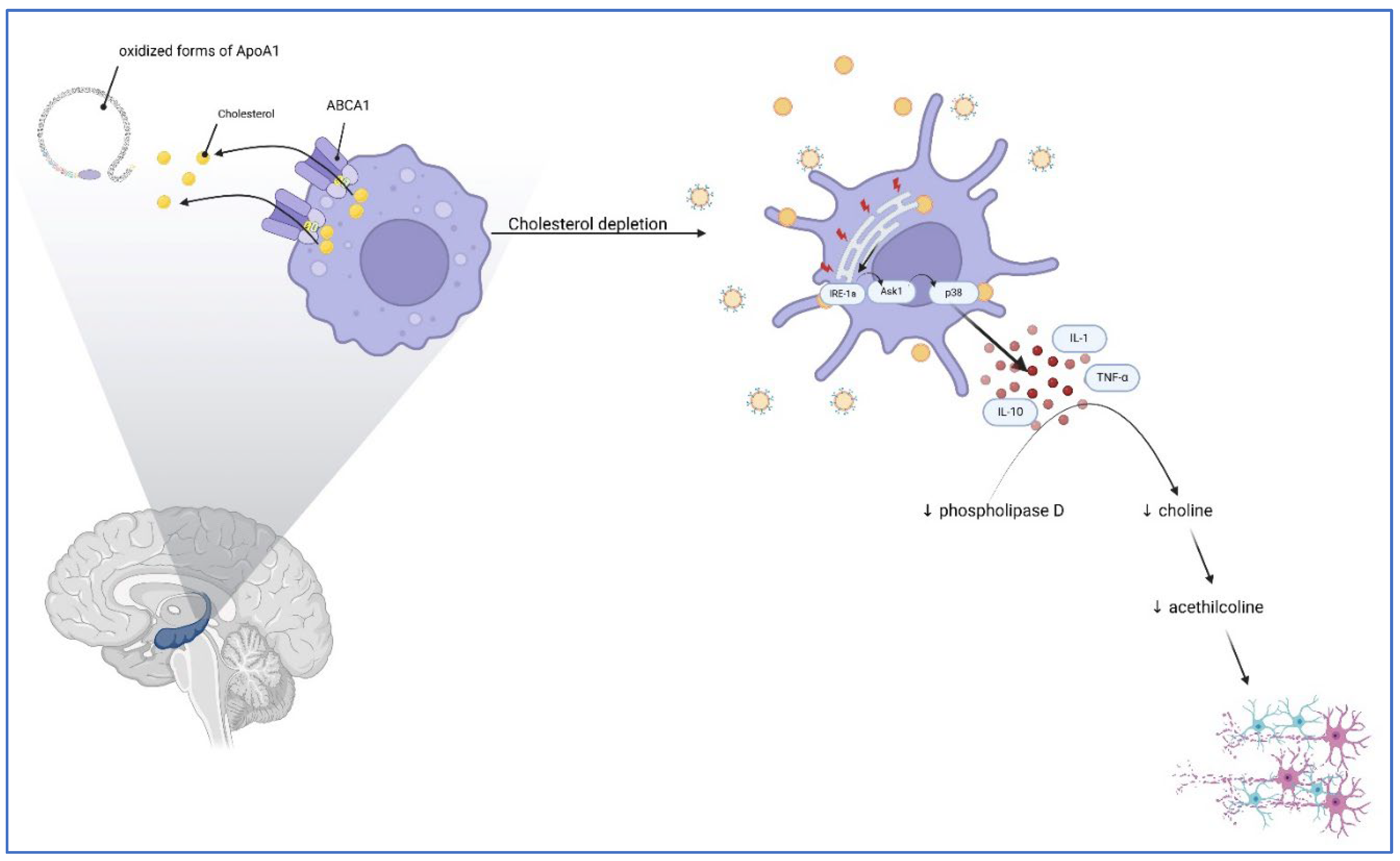

Table 1.
Recommended resources for different cognitive areas.
| Attention | High-level cognitive function | Intellectual function | Language | Memory | Psychomotor function |
|---|---|---|---|---|---|
| - Digit Span Test - Digit symbol substitution test - TMT |
- The Stroop Color and word test - Wisconsin Card Sorting Test - COWA - Verbal Fluency test - TMT |
- CANTAB - National Adult Reading Test - MMSE |
- COWA - Verbal Fluency test |
- Rey Auditory Verbal Learning Test - California Verbal Learning Test |
- Digit symbol substitution test - TMT |
Disclaimer/Publisher’s Note: The statements, opinions and data contained in all publications are solely those of the individual author(s) and contributor(s) and not of MDPI and/or the editor(s). MDPI and/or the editor(s) disclaim responsibility for any injury to people or property resulting from any ideas, methods, instructions or products referred to in the content. |
© 2024 by the authors. Licensee MDPI, Basel, Switzerland. This article is an open access article distributed under the terms and conditions of the Creative Commons Attribution (CC BY) license (https://creativecommons.org/licenses/by/4.0/).
Copyright: This open access article is published under a Creative Commons CC BY 4.0 license, which permit the free download, distribution, and reuse, provided that the author and preprint are cited in any reuse.