Preprint
Article

This version is not peer-reviewed.

Taurine and High-Intensity Interval Training Modulate TXNIP–PI3K/AKT Signaling in Male C57BL/6J Mice with High-Fat Diet-Induced Non-Alcoholic Fatty Liver Disease

Submitted:

01 August 2026

Posted:

04 August 2026

You are already at the latest version

Abstract

Introduction: Non-alcoholic fatty liver disease (NAFLD) is closely associated with insulin resistance and chronic low-grade inflammation. Exercise training and taurine supplementation have emerged as promising strategies to improve metabolic function by modulating insulin signaling and inflammatory pathways. This study investigated the effects of taurine supplementation and high-intensity interval training (HIIT), alone and in combination, on insulin signaling and inflammasome-related pathways in a high-fat diet (HFD)-induced mouse model of NAFLD. Methods: Fifty-six male C57BL/6J mice were assigned to either a control diet (CON, n = 8) or an HFD (n = 48). After 12 weeks, HFD-fed mice were randomized into six groups: HFD, HFD+HIIT (HE), HFD+2.5% taurine (HT-2.5), HFD+HIIT+2.5% taurine (HET-2.5), HFD+5% taurine (HT-5), and HFD+HIIT+5% taurine (HET-5) (n = 8/group). Interventions were performed concurrently for 10 weeks. Results: Compared with controls, HFD-fed mice exhibited increased HOMA-IR, impaired PI3K/AKT signaling, elevated PTEN expression, and increased hepatic TXNIP, NLRP3, and caspase-1 levels (all p < 0.05). All intervention groups improved HOMA-IR and reduced PTEN, TXNIP, and caspase-1 expression compared with the HFD group (p < 0.05). PI3K phosphorylation increased only in the HET-2.5 group, whereas AKT phosphorylation increased in the HE, HT-5, and HET-2.5 groups (p < 0.05). Notably, concurrent enhancement of both PI3K and AKT phosphorylation was observed exclusively in the HET-2.5 group. Conclusion: HIIT and taurine supplementation improved metabolic and inflammatory alterations associated with HFD-induced NAFLD. These findings support the potential of combined exercise and nutritional interventions as complementary strategies for mitigating metabolic dysfunction associated with NAFLD and warrant further clinical investigation.

Keywords: 
;  ;  ;  ;  ;  

Introduction

Non-alcoholic fatty liver disease (NAFLD) is one of the most prevalent chronic liver disorders worldwide and is characterized by excessive triglyceride accumulation in more than 5% of hepatocytes in the absence of significant alcohol consumption(Powell et al., 2021). It encompasses a spectrum of pathological conditions ranging from simple steatosis to non-alcoholic steatohepatitis (NASH), fibrosis, cirrhosis, and hepatocellular carcinoma (HCC), contributing substantially to liver-related morbidity and mortality(Brunt et al., 2015, Polyzos et al., 2019). Despite its rapidly increasing prevalence, driven largely by obesity and metabolic syndrome, no approved pharmacological therapy is currently available. Consequently, lifestyle modification remains the cornerstone of treatment, highlighting the need for effective interventions targeting the molecular mechanisms underlying disease progression(Powell et al., 2021, Younossi et al., 2019, Younossi et al., 2018). NAFLD development is primarily driven by hepatic insulin resistance, which disrupts glucose and lipid metabolism, promotes hepatic lipid accumulation, and accelerates disease progression (Taniguchi et al., 2006, Matsuda et al., 2013). These metabolic disturbances are further amplified by oxidative stress and chronic low-grade inflammation, indicating a close interaction between metabolic and inflammatory signaling pathways during NAFLD progression (Zhou et al., 2010).
Thioredoxin-interacting protein (TXNIP) has emerged as a central regulator of NAFLD pathogenesis by linking metabolic stress, oxidative stress, and inflammatory signaling. TXNIP promotes insulin resistance by upregulating phosphatase and tensin homolog (PTEN), thereby suppressing phosphoinositide 3-kinase/protein kinase B (PI3K/AKT) signaling, a critical pathway regulating hepatic glucose and lipid metabolism through downstream effectors such as forkhead box O1 (FOXO1), glycogen synthase kinase-3 (GSK3), and mechanistic target of rapamycin complex 1 (mTORC1) (Dai et al., 2016, Matsuda et al., 2013). In parallel, TXNIP inhibits thioredoxin (TRX), resulting in excessive reactive oxygen species (ROS) accumulation and activation of the nucleotide-binding oligomerization domain-like receptor family pyrin domain containing 3 (NLRP3) inflammasome, ultimately promoting caspase-1 activation and inflammatory cytokine production (Ding et al., 2016, Vandanmagsar et al., 2011). Collectively, these interconnected mechanisms contribute to hepatocellular injury, insulin resistance, and progression toward advanced liver disease. Accordingly, TXNIP has emerged as an attractive therapeutic target for preventing or attenuating NAFLD progression. (Ding et al., 2016).
Physical activity is a well-established and cost-effective intervention for reducing hepatic steatosis in NAFLD, with evidence demonstrating its therapeutic and preventive effects on hepatic fat accumulation as well as systemic metabolic risk factors associated with disease progression(Mahady and George, 2016, Orci et al., 2016, Keating et al., 2012). Among exercise modalities, high-intensity interval training (HIIT) is particularly effective in improving insulin sensitivity, lipid metabolism, and liver enzyme profiles(Sini et al., 2022, Ross et al., 2016). These metabolic benefits are partly attributed to improved glycemic control and reduced oxidative stress, which may suppress TXNIP expression and thereby modulate downstream metabolic and inflammatory signaling pathways (Javaid et al., 2021). Taurine, a conditionally essential amino acid, exerts antioxidant, anti-inflammatory, and metabolic regulatory effects(Chen et al., 2016). In experimental NAFLD models, taurine supplementation reduces hepatic steatosis and improves liver function. Emerging evidence suggests that taurine may further influence TXNIP-related signaling and PI3K/AKT associated pathways; however, the underlying mechanisms remain incompletely understood(Liu et al., 2019, Chen et al., 2021a). Notably, Murakami et al. demonstrated that taurine supplementation at doses of 2% and 5% attenuated hepatic injury and slowed disease progression in high-fat diet-fed rats, with greater effects observed at higher doses(Murakami et al., 2018). Collectively, these findings suggest that taurine may complement exercise-induced metabolic adaptations by targeting oxidative stress and insulin signaling pathways.
Despite these findings, it remains unclear whether combining HIIT with taurine supplementation can simultaneously target the interconnected metabolic and inflammatory pathways underlying NAFLD progression. In particular, the coordinated effects of HIIT and taurine on hepatic PI3K/AKT/PTEN and TXNIP/NLRP3/caspase-1 signaling remain incompletely understood. Therefore, this study investigated the effects of HIIT and taurine supplementation, individually and in combination, in a HFD induced NAFLD mouse model. We hypothesized that the combined intervention would exert greater protective effects than either intervention alone by improving insulin signaling and suppressing inflammatory activation, with taurine dose-dependently enhancing these responses.

Materials and Methods

Animal Models

NAFLD was induced in 56 male C57BL/6J mice (6–8 weeks old), purchased from the Pasteur Institute of Iran and transferred to the animal laboratory at the University of Tehran. After a one-week acclimation period, mice were randomly divided into a control group (CON; n = 8) and a high-fat diet group (HFD; n = 48). Sample sizes were determined based on similar previous studies investigating the effects of high-intensity interval training and dietary interventions on hepatic metabolic markers in rodent models of NAFLD The control group received standard chow containing 10% fat, whereas HFD groups were fed a high-fat diet (60% fat) for 12 weeks. Following induction, 48 HFD mice were further divided into six subgroups: HFD (control), HFD + HIIT (HE), HFD + taurine (2.5%) (HT-2.5), HFD + HIIT + taurine (2.5%) (HET-2.5), HFD + taurine (5%) (HT-5), and HFD + HIIT + taurine (5%) (HET-5). Taurine supplementation was initiated concurrently with HIIT and administered in drinking water. Animals were housed four per cage under standard laboratory conditions (12 h light/dark cycle, 22–24°C) with ad libitum access to food and water. Body weight was recorded weekly, and food intake was measured throughout the experimental period. After completion of the experimental period, animals were kept sedentary for 48 hours and fasted overnight prior to sacrifice. Blood and liver tissues were then collected. This study was approved by the Research Ethics Committee of the Faculty of Sport Sciences and Health, University of Tehran (Approval ID: IR.UT.SPORT.REC.1401.013, approved on May 30, 2022) All experimental procedures were conducted in accordance with institutional guidelines for the care and use of laboratory animals and national regulations for animal research. No animals were excluded from the study; however, mice exhibiting excessive lethargy or bleeding during exercise sessions would have been excluded per predefined criteria. No adverse events were observed in any experimental group throughout the intervention period, and no modifications to the exercise or supplementation protocols were required.

Exercise Protocol

To familiarize animals with treadmill running, exercise groups (HE, HET-2.5, and HET-5) underwent a 1-week adaptation period prior to initiation of the HIIT protocol. During this period, mice were gradually accustomed to treadmill running. Maximal running speed was assessed to estimate VO2max according to the protocol described by M.A. Høydal et al.(Høydal et al., 2007) Mice initially ran at 8 m/min on a 5° incline, and treadmill speed was increased by 1.8 m/min every 2 minutes until exhaustion. Exhaustion was defined as the inability to continue running despite gentle manual encouragement. No electrical stimulation was used during the protocol HIIT was performed five days per week for 10 weeks. Each training session lasted 30 minutes and consisted of a 6-minute warm-up at 50% VO2max, an 18-minute interval phase, and a 6-minute cool-down at 50–60% VO2max. The interval phase consisted of three high-intensity bouts at 85–90% VO2max interspersed with low-intensity recovery periods at 50–60% VO2max. Training intensity was progressively adjusted every two weeks based on reassessment of VO2max (Table 1).

Blood Analysis

Mice were anesthetized with ketamine–xylazine (37.5 and 12.5 mg/kg, respectively) and euthanized via exsanguination through cardiac puncture, in accordance with the AVMA Guidelines for the Euthanasia of Animals. Anesthesia was administered prior to all procedures to minimize animal pain and distress. For serum and plasma biochemical analyses, six mice per group were randomly selected. Serum ALT, AST, and fasting glucose levels were measured using enzymatic colorimetric assay kits based on the glucose oxidase–peroxidase (GOD–POD) method (Bio-Rex FARS, Iran). Plasma insulin concentrations were determined using an ultra-sensitive mouse insulin ELISA kit. Hepatic glycogen content was assessed using a mouse glycogen ELISA kit.

Histological Analysis of Liver Sections

Fresh liver tissues were fixed in 4% paraformaldehyde for 24 h, embedded in paraffin, sectioned at 4 μm, and stained with hematoxylin and eosin (H&E).

Western Blot Analysis

For Western blot analysis, a subset of four mice per group was randomly selected from each experimental group. Proteins were extracted from liver tissues using Pro-PRE™ lysis buffer (iNtRON Biotechnology, Korea) and centrifuged at 13,000 rpm for 15 min at 4°C. Protein concentration was determined using a BCA protein assay kit (iNtRON Biotechnology, Korea). Equal amounts of protein (20 μg) were separated by SDS–PAGE and transferred onto PVDF membranes (Bio-Rad, USA). Membranes were blocked with 5% BSA in TBST and incubated with primary antibodies against TXNIP, NLRP3, caspase-1, PI3K, phospho-PI3K, AKT2, phospho-AKT2, PTEN, and β-actin, followed by HRP-conjugated secondary antibodies. Band visualization and quantification were performed by a technician blinded to group allocation, using enhanced chemiluminescence and ImageJ software (NIH, USA). Protein expression levels were normalized to β-actin.

Assessment of Insulin Sensitivity in Mice Subjects

Insulin sensitivity was evaluated using the homeostasis model assessment-2 (HOMA2) calculator provided by the Diabetes Trials Unit, University of Oxford website (https://www.dtu.ox.ac.uk/homacalculator/). HOMA-IR values were calculated from fasting glucose and insulin concentrations

Statistical Analysis

Statistical analyses were performed using SPSS version 26.0 (IBM Corp., Armonk, NY, USA). Data normality was assessed using the Shapiro–Wilk test. Group differences were analyzed using one-way ANOVA followed by Bonferroni post hoc tests where appropriate. Data are presented as mean ± SD, and statistical significance was set at p < 0.05.

Results

Body Weight and Liver Enzymes

The weight of the HFD group increased by approximately 10–20% compared with the control group after 12 weeks of HFD feeding (Figure 1). Exercise and taurine supplementation (2.5% and 5%) attenuated this increase in body weight. A significant difference was observed between the control and HFD groups at the end of the study (p < 0.05). As shown in Table 2, hepatic enzyme levels (ALT and AST) were increased in the HFD group compared with the control group. A significant difference was observed between the control and HFD groups (p < 0.05). Notably, a significant difference in ALT levels was observed between the HFD and HET-2.5 groups (p < 0.05), whereas no significant differences were found among the other groups.

Fasting Blood Glucose, Insulin, Liver Glycogen, and Insulin Resistance

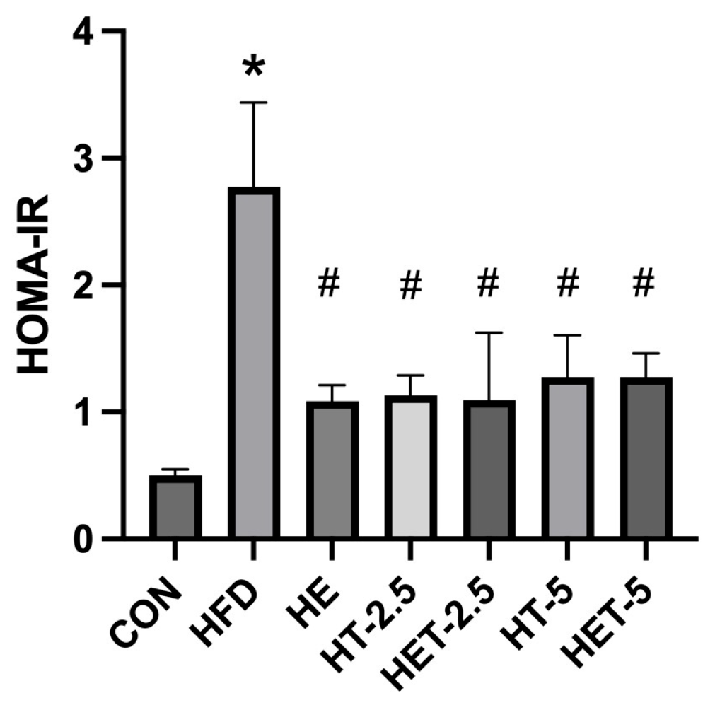
Fasting blood glucose showed significant differences between the control and HFD groups, as well as between the HFD and HET-2.5 groups (Table 2). Fasting insulin levels also differed significantly between the control and HFD groups and between the HFD and HT-5 groups. Liver glycogen content differed significantly between the control and HFD groups, as well as between the HFD group and both the HT-2.5 and HET-2.5 groups. HOMA-IR was elevated in HFD-fed mice compared with controls. All intervention groups showed significant differences compared with the HFD group (Figure 2).

Effects of Different Doses of Taurine and HIIT on PI3K, AKT, and PTEN Expression in the Liver
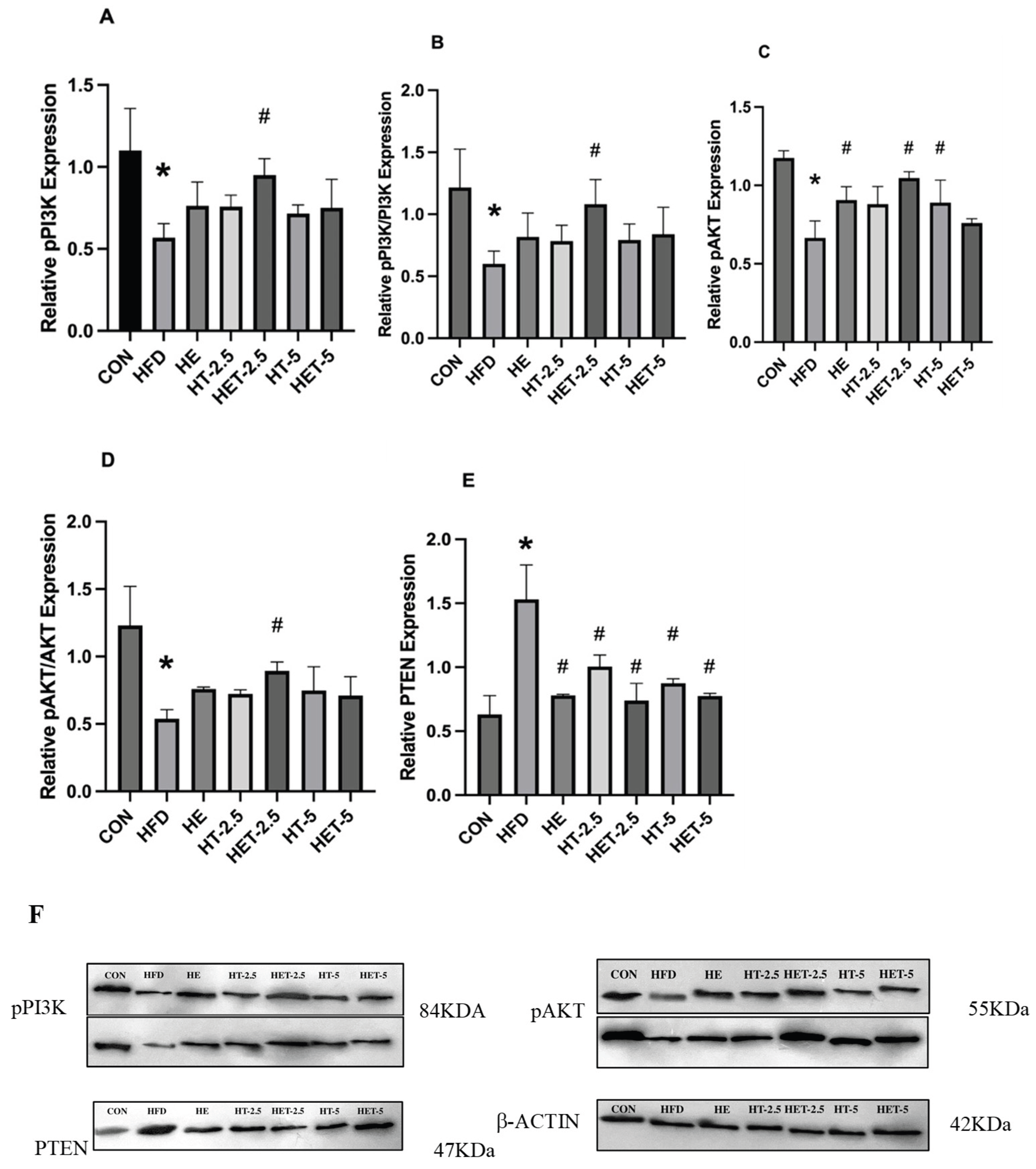
PI3K phosphorylation was reduced following high-fat diet feeding, with a significant difference observed between the CON and HFD groups (p < 0.05). Only the HET-2.5 group showed a significant increase compared with the HFD group (p < 0.05). The pPI3K/PI3K ratio differed significantly between the CON and HFD groups, as well as between the HFD and HET-2.5 groups (p < 0.05). Akt phosphorylation was reduced compared with controls, with a significant difference between the CON and HFD groups (p < 0.05). Significant differences were observed between the HFD group and the HE, HET-2.5, and HT-5 groups (p < 0.05). The p-AKT/AKT ratio differed significantly between the CON and HFD groups (p < 0.05), as well as between the HFD and HET-2.5 groups (p < 0.05). PTEN protein levels were higher in the HFD group compared with controls (p < 0.05). All intervention groups (HE, HT-2.5, HET-2.5, HT-5, and HET-5) differed significantly from the HFD group (p < 0.05).
Figure 3. Effects of HIIT and taurine supplementation on insulin signaling in HFD-induced insulin resistance. (A) PI3K phosphorylation, (B) p-PI3K/PI3K ratio, (C) p-AKT expression, (D) p-AKT/AKT ratio, and (E) PTEN expression. (F) Representative Western blot images (n = 2 per group); quantification was performed on all biological replicates (n = 4 per group). *p < 0.05 vs. CON; #p < 0.05 vs. HFD.
Preprints 226363 g003

TXNIP, NLRP3, and Caspase-1 Expression in the Liver
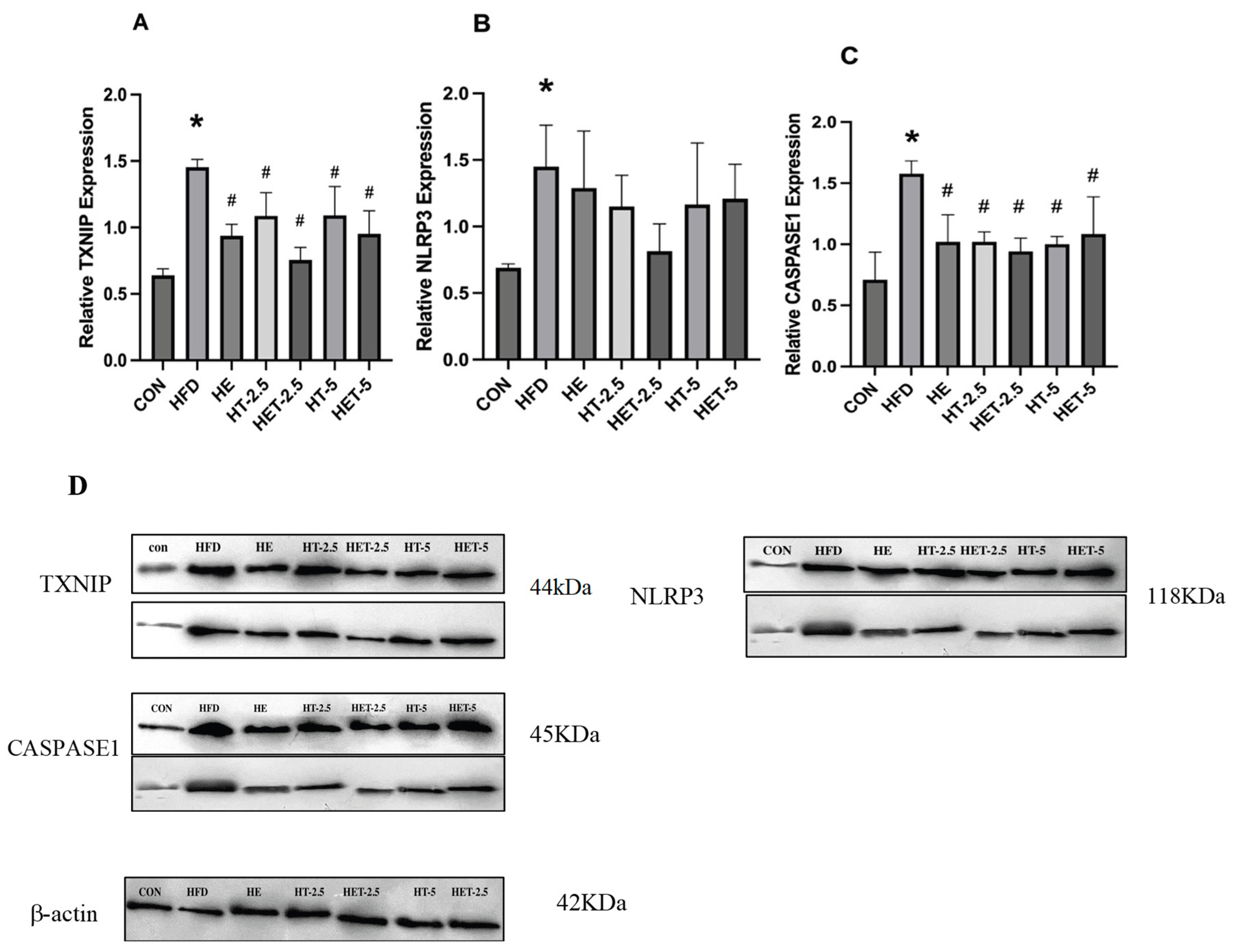
Hepatic TXNIP expression differed significantly between the HFD and CON groups (p < 0.05). Significant differences were observed between the HFD group and all intervention groups (HE, HT-2.5, HET-2.5, HT-5, and HET-5) (p < 0.05). Hepatic NLRP3 expression differed significantly between the HFD and CON groups (p < 0.05). No significant differences were observed between the HFD group and the intervention groups. Caspase-1 expression differed significantly between the HFD and CON groups (p < 0.05). Significant differences were observed between the HFD group and all intervention groups (p < 0.05).
Figure 4. Effects of HIIT and taurine on inflammatory markers in HFD-induced insulin resistance. (A) TXNIP, (B) NLRP3, and (C) Caspase-1 expression. (D) Representative Western blot images (n = 2 per group); β-actin was used as loading control. *p < 0.05 vs. CON; #p < 0.05 vs. HFD.
Preprints 226363 g004

Discussion

The present study demonstrated that 10 weeks of low-dose taurine supplementation combined with HIIT produced greater improvements in insulin resistance and inflammatory regulation in HFD-induced NAFLD mice compared with either intervention alone. In contrast, the higher taurine dose was associated with less pronounced effects on insulin-related markers, suggesting a dose-dependent response in the context of chronic metabolic stress.
TXNIP is increasingly recognized as an important regulator of glucose homeostasis in metabolic tissues, including the liver, through redox-sensitive mechanisms, and plays a central role in integrating metabolic stress and inflammatory signaling in NAFLD(Alhawiti et al., 2017). Exercise has been shown to improve hepatic insulin sensitivity and metabolic regulation through stress-adaptive mechanisms and attenuation of inflammatory pathways including NLRP3/caspase-1 signaling(Katsanos, 2004, Yang et al., 2021), with HIIT generally eliciting stronger metabolic adaptations than moderate-intensity exercise(Battista et al., 2021). In parallel, taurine has been reported to improve insulin-related signaling and antioxidant capacity by modulating redox balance, while reducing oxidative stress and inflammatory responses in diet-induced NAFLD models(Cappelli et al., 2014, Chaoyue et al., 2018). Taurine may also contribute to calcium homeostasis, and calcium-dependent signaling has been linked to regulation of TXNIP expression under metabolic stress conditions(Qayyum et al., 2021). Importantly, in the present study, the combined intervention was associated with a coordinated reduction in TXNIP and PTEN expression together with enhanced PI3K/AKT signaling, indicating improved hepatic insulin sensitivity under chronic HFD exposure. Taken together, these findings suggest that HIIT and moderate-dose taurine target interconnected metabolic and inflammatory pathways implicated in NAFLD, providing mechanistic support for their therapeutic potential.
HIIT alone induced metabolic adaptations consistent with improved hepatic energy handling and stress resilience in NAFLD(Wang et al., 2022). Collectively, previous studies have demonstrated that high-intensity exercise enhances hepatic insulin responsiveness and metabolic efficiency through stress-sensitive pathways and mitochondrial adaptations rather than isolated modulation of single signaling nodes(Hamasaki, 2019, Wang et al., 2017). In the present study, these adaptations were reflected by improved insulin-related signaling and reduced metabolic stress, suggesting a partial restoration of hepatic metabolic homeostasis under chronic dietary overload. At the molecular level, reduced TXNIP expression indicated attenuation of metabolic stress–associated signaling involved in inflammasome priming (Ding and Xu, 2021). However, unchanged NLRP3 expression suggests that upstream inflammatory signaling remained sustained under continued HFD exposure, consistent with persistent lipid-driven inflammatory pressure reported in similar models(Zhang et al., 2020) . In contrast, reduced caspase-1 levels despite unchanged NLRP3 indicate that HIIT may preferentially modulate downstream inflammatory effector activity rather than fully suppress inflammasome activation(Wang et al., 2017, Li et al., 2020). Notably, exercise intensity appears to be a critical determinant of these responses, as excessive high-intensity exercise has been associated with transient mitochondrial stress and inflammatory mediator release in specific pathological contexts (Zhang et al., 2021). Overall, HIIT induces a state of partial metabolic reprogramming characterized by improved insulin responsiveness and reduced inflammatory output, without complete normalization of hepatic inflammatory homeostasis.
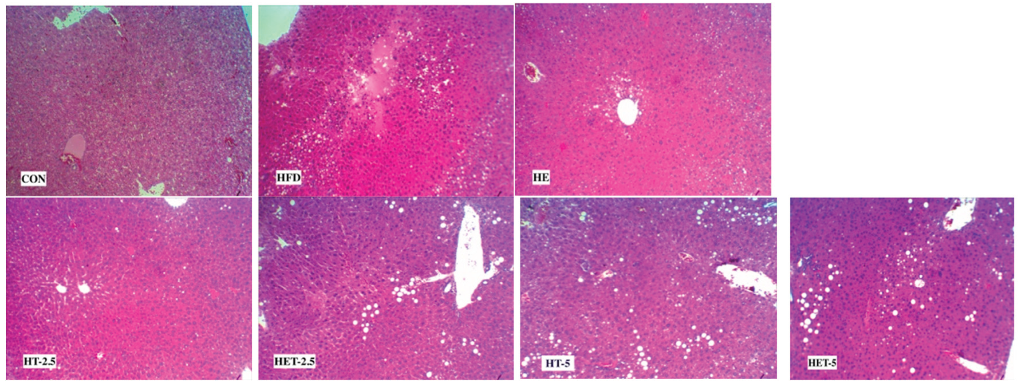
Figure 5. Histological analysis of liver tissue (H&E staining, ×20 magnification). Representative liver sections from CON, HFD, HE, HT-2.5, HET-2.5, HT-5, and HET-5 groups showing hepatic steatosis and its attenuation following HIIT and taurine interventions.
Preprints 226363 g005
Taurine supplementation alone produced dose-dependent metabolic effects in HFD-induced NAFLD mice, although these changes were less pronounced than those observed with exercise. In the present study, higher taurine dose was associated with improved insulin-related signaling, reflected by enhanced PI3K/AKT activity and reduced PTEN expression. Consistently, previous studies have reported increased AKT phosphorylation and reduced PTEN expression following 5% taurine supplementation in metabolic disease models(Cappelli et al., 2014),whereas other studies have demonstrated effective modulation of the PI3K/AKT pathway at lower doses(Chen et al., 2021a). Suggesting dose-dependent variability in signaling responsiveness. Taurine also reduced TXNIP expression, indicating partial attenuation of metabolic stress–related signaling; however, NLRP3 inflammasome activity remained largely unchanged, suggesting limited effects on upstream inflammatory activation under sustained dietary stress(Lee et al., 2021). Collectively, these findings indicate that taurine primarily supports hepatic metabolic adaptation by improving insulin resistance and reducing oxidative stress–related signaling, while exerting limited effects on inflammasome activation. Previous studies further corroborate these effects by demonstrating that taurine improves insulin sensitivity, reduces hepatic lipid accumulation, and attenuates oxidative stress through modulation of redox balance and PI3K/AKT signaling pathways(Murakami et al., 2018, Bae et al., 2022).
In contrast, the HET-5 group showed relatively smaller changes across the measured outcomes, with effects mainly observed in TXNIP/caspase-1 signaling. These findings may indicate a dose-dependent response to taurine supplementation. While taurine at low-to-moderate doses has been associated with antioxidant and regulatory effects, higher doses may not necessarily confer additional benefits under conditions of sustained metabolic stress(Chen et al., 2021b, Chaoyue et al., 2018). Given the role of reactive oxygen species in exercise-induced signaling, higher taurine doses may influence redox-sensitive pathways such as PI3K/AKT and AMPK; however, this remains speculative in the present context(Powers and Jackson, 2008). Variations in intestinal absorption at higher doses may partly contribute to differences in systemic availability. Therefore, taurine dose may represent a potential determinant of metabolic responses to HIIT in NAFLD. This study was limited to male C57BL/6J mice to eliminate the confounding effects of estrous cycle-related hormonal fluctuations on hepatic metabolic and inflammatory outcomes, consistent with standard practice in diet-induced NAFLD models. As only one sex was examined, sex-based comparisons were not possible, and whether these findings extend to female mice remains to be determined in future studies.

Conclusions

In this model of diet-induced NAFLD, combining HIIT with taurine supplementation more effectively modulated insulin signaling and inflammatory stress pathways—specifically through the TXNIP/NLRP3/CASPASE-1 and PI3K/AKT axes—than either intervention alone. These findings support the integration of structured exercise with targeted nutritional strategies as a promising approach for addressing hepatic metabolic dysfunction in NAFLD. Further studies are needed to determine optimal taurine dosing and to evaluate the translational potential of this combined approach in human populations.

Funding Statement

This research received no specific grant from any funding agency in the public, commercial, or not-for-profit sectors.

Data Availability Statement

The data that support the findings of this study are available from the corresponding author upon reasonable request.

Acknowledgments

Manuscript editing assistance was provided by Claude (Anthropic).

Conflicts of Interest

The authors declare there are no competing interests.

References

  1. ALHAWITI, N. M.; AL MAHRI, S.; AZIZ, M. A.; MALIK, S. S.; MOHAMMAD, S. TXNIP in Metabolic Regulation: Physiological Role and Therapeutic Outlook. Curr. Drug Targets 2017, 18, 1095–1103. [Google Scholar] [PubMed]
  2. BAE, M.; AHMED, K.; YIM, J. E. Beneficial Effects of Taurine on Metabolic Parameters in Animals and Humans. J. Obes. Metab. Syndr. 2022, 31, 134–146. [Google Scholar] [CrossRef] [PubMed]
  3. BATTISTA, F.; ERMOLAO, A.; VAN BAAK, M. A.; BEAULIEU, K.; BLUNDELL, J. E.; BUSETTO, L.; CARRAçA, E. V.; ENCANTADO, J.; DICKER, D.; FARPOUR-LAMBERT, N.; PRAMONO, A.; BELLICHA, A.; OPPERT, J. M. Effect of exercise on cardiometabolic health of adults with overweight or obesity: Focus on blood pressure, insulin resistance, and intrahepatic fat-A systematic review and meta-analysis. Obes. Rev. 2021, 22 Suppl 4, e13269. [Google Scholar] [CrossRef] [PubMed]
  4. BRUNT, E. M.; WONG, V. W.; NOBILI, V.; DAY, C. P.; SOOKOIAN, S.; MAHER, J. J.; BUGIANESI, E.; SIRLIN, C. B.; NEUSCHWANDER-TETRI, B. A.; RINELLA, M. E. Nonalcoholic fatty liver disease. Nat. Rev. Dis. Prim. 2015, 1, 15080. [Google Scholar] [CrossRef] [PubMed]
  5. CAPPELLI, A. P.; ZOPPI, C. C.; BARBOSA-SAMPAIO, H. C.; COSTA, J. M., JR.; PROTZEK, A. O.; MORATO, P. N.; BOSCHERO, A. C.; CARNEIRO, E. M. Taurine-induced insulin signalling improvement of obese malnourished mice is associated with redox balance and protein phosphatases activity modulation. Liver Int. 2014, 34, 771–83. [Google Scholar] [PubMed]
  6. CHAOYUE, W.; FENGNA, L.; LINGYU, Z.; YEHUI, D.; QIUPING, G.; WENLONG, W.; SHANPING, H.; JIANZHONG, L.; YULONG, Y. Taurine is Involved in Energy Metabolism in Muscles, Adipose Tissue, and the Liver. Mol. Nutr. Amp Food Res. 2018, 63. [Google Scholar]
  7. CHEN, B.; ABAYDULA, Y.; LI, D.; TAN, H.; MA, X. Taurine ameliorates oxidative stress by regulating PI3K/Akt/GLUT4 pathway in HepG2 cells and diabetic rats. J. Funct. Foods 2021a, 85, 104629. [Google Scholar] [CrossRef]
  8. CHEN, Q.; LI, Z.; PINHO, R. A.; GUPTA, R. C.; UGBOLUE, U. C.; THIRUPATHI, A.; GU, Y. The Dose Response of Taurine on Aerobic and Strength Exercises: A Systematic Review. Front Physiol. 2021b, 12, 700352. [Google Scholar] [CrossRef] [PubMed]
  9. CHEN, W.; GUO, J.; ZHANG, Y.; ZHANG, J. The beneficial effects of taurine in preventing metabolic syndrome. Food Funct. 2016, 7, 1849–63. [Google Scholar] [CrossRef] [PubMed]
  10. DAI, B.; WU, Q.; ZENG, C.; ZHANG, J.; CAO, L.; XIAO, Z.; YANG, M. The effect of Liuwei Dihuang decoction on PI3K/Akt signaling pathway in liver of type 2 diabetes mellitus (T2DM) rats with insulin resistance. J. Ethnopharmacol. 2016, 192, 382–389. [Google Scholar] [CrossRef] [PubMed]
  11. DING, C.; ZHAO, Y.; SHI, X.; ZHANG, N.; ZU, G.; LI, Z.; ZHOU, J.; GAO, D.; LV, L.; TIAN, X.; YAO, J. New insights into salvianolic acid A action: Regulation of the TXNIP/NLRP3 and TXNIP/ChREBP pathways ameliorates HFD-induced NAFLD in rats. Sci. Rep. 2016, 6, 28734. [Google Scholar] [CrossRef] [PubMed]
  12. DING, Y.; XU, X. Effects of regular exercise on inflammasome activation-related inflammatory cytokine levels in older adults: a systematic review and meta-analysis. J. Sports Sci. 2021, 39, 2338–2352. [Google Scholar] [CrossRef] [PubMed]
  13. HAMASAKI, H. Perspectives on Interval Exercise Interventions for Non-Alcoholic Fatty Liver Disease. Medicines (Basel) 2019, 6. [Google Scholar]
  14. HøYDAL, M. A.; WISLøFF, U.; KEMI, O. J.; ELLINGSEN, O. Running speed and maximal oxygen uptake in rats and mice: practical implications for exercise training. Eur. J. Cardiovasc Prev. Rehabil. 2007, 14, 753–60. [Google Scholar] [CrossRef] [PubMed]
  15. JAVAID, H. M. A.; SAHAR, N. E.; ZHUGE, D. L.; HUH, J. Y. Exercise Inhibits NLRP3 Inflammasome Activation in Obese Mice via the Anti-Inflammatory Effect of Meteorin-like. Cells 2021, 10. [Google Scholar] [CrossRef] [PubMed]
  16. KATSANOS, C. S. Lipid-induced insulin resistance in the liver: role of exercise. Sports Med. 2004, 34, 955–65. [Google Scholar] [PubMed]
  17. KEATING, S. E.; HACKETT, D. A.; GEORGE, J.; JOHNSON, N. A. Exercise and non-alcoholic fatty liver disease: a systematic review and meta-analysis. J. Hepatol. 2012, 57, 157–66. [Google Scholar] [CrossRef] [PubMed]
  18. LEE, C. C.; CHEN, W. T.; CHEN, S. Y.; LEE, T. M. Taurine Alleviates Sympathetic Innervation by Inhibiting NLRP3 Inflammasome in Postinfarcted Rats. J. Cardiovasc Pharmacol. 2021, 77, 745–755. [Google Scholar] [PubMed]
  19. LI, C.; XU, X.; WANG, Z.; WANG, Y.; LUO, L.; CHENG, J.; CHEN, S.-F.; LIU, H.; WAN, Q.; WANG, Q. Exercise ameliorates post-stroke depression by inhibiting PTEN elevation-mediated upregulation of TLR4/NF-κB/NLRP3 signaling in mice. Brain Res. 2020, 1736, 146777. [Google Scholar] [CrossRef] [PubMed]
  20. LIU, X.; ZHANG, Y. R.; CAI, C.; NI, X. Q.; ZHU, Q.; REN, J. L.; CHEN, Y.; ZHANG, L. S.; XUE, C. D.; ZHAO, J.; QI, Y. F.; YU, Y. R. Taurine Alleviates Schistosoma-Induced Liver Injury by Inhibiting the TXNIP/NLRP3 Inflammasome Signal Pathway and Pyroptosis. Infect. Immun. 2019, 87. [Google Scholar] [PubMed]
  21. MAHADY, S. E.; GEORGE, J. Exercise and diet in the management of nonalcoholic fatty liver disease. Metabolism 2016, 65, 1172–82. [Google Scholar] [CrossRef] [PubMed]
  22. MATSUDA, S.; KOBAYASHI, M.; KITAGISHI, Y. Roles for PI3K/AKT/PTEN Pathway in Cell Signaling of Nonalcoholic Fatty Liver Disease. ISRN Endocrinol. 2013, 472432. [Google Scholar] [CrossRef] [PubMed]
  23. MURAKAMI, S.; ONO, A.; KAWASAKI, A.; TAKENAGA, T.; ITO, T. Taurine attenuates the development of hepatic steatosis through the inhibition of oxidative stress in a model of nonalcoholic fatty liver disease in vivo and in vitro. Amino Acids 2018, 50, 1279–1288. [Google Scholar] [CrossRef] [PubMed]
  24. ORCI, L. A.; GARIANI, K.; OLDANI, G.; DELAUNE, V.; MOREL, P.; TOSO, C. Exercise-based Interventions for Nonalcoholic Fatty Liver Disease: A Meta-analysis and Meta-regression. Clin. Gastroenterol. Hepatol. 2016, 14, 1398–411. [Google Scholar] [CrossRef] [PubMed]
  25. POLYZOS, S. A.; KOUNTOURAS, J.; MANTZOROS, C. S. Obesity and nonalcoholic fatty liver disease: From pathophysiology to therapeutics. Metab.-Clin. Exp. 2019, 92, 82–97. [Google Scholar] [CrossRef] [PubMed]
  26. POWELL, E. E.; WONG, V. W.; RINELLA, M. Non-alcoholic fatty liver disease. Lancet 2021, 397, 2212–2224. [Google Scholar] [CrossRef] [PubMed]
  27. POWERS, S. K.; JACKSON, M. J. Exercise-induced oxidative stress: cellular mechanisms and impact on muscle force production. Physiol. Rev. 2008, 88, 1243–76. [Google Scholar] [CrossRef] [PubMed]
  28. QAYYUM, N.; HASEEB, M.; KIM, M. S.; CHOI, S. Role of Thioredoxin-Interacting Protein in Diseases and Its Therapeutic Outlook. Int. J. Mol. Sci. 2021, 22. [Google Scholar] [CrossRef] [PubMed]
  29. ROSS, L. M.; PORTER, R. R.; DURSTINE, J. L. High-intensity interval training (HIIT) for patients with chronic diseases. J. Sport Health Sci. 2016, 5, 139–144. [Google Scholar] [CrossRef] [PubMed]
  30. SINI, Z. K.; AFZALPOUR, M. E.; AHMADI, M. M.; SARDAR, M. A.; KHALEGHZADEH, H.; GORGANI-FIRUZJAEE, S.; HOFMEISTER, M.; AKARAS, E.; AZIMKHANI, A. Comparison of the effects of high-intensity interval training and moderate-intensity continuous training on indices of liver and muscle tissue in high-fat diet-induced male rats with non-alcoholic fatty liver disease. Egypt. Liver J. 2022, 12, 63. [Google Scholar] [CrossRef]
  31. TANIGUCHI, C. M.; KONDO, T.; SAJAN, M.; LUO, J.; BRONSON, R.; ASANO, T.; FARESE, R.; CANTLEY, L. C.; KAHN, C. R. Divergent regulation of hepatic glucose and lipid metabolism by phosphoinositide 3-kinase via Akt and PKClambda/zeta. Cell Metab. 2006, 3, 343–53. [Google Scholar] [CrossRef] [PubMed]
  32. VANDANMAGSAR, B.; YOUM, Y. H.; RAVUSSIN, A.; GALGANI, J. E.; STADLER, K.; MYNATT, R. L.; RAVUSSIN, E.; STEPHENS, J. M.; DIXIT, V. D. The NLRP3 inflammasome instigates obesity-induced inflammation and insulin resistance. Nat. Med. 2011, 17, 179–88. [Google Scholar] [CrossRef] [PubMed]
  33. WANG, N.; LIU, Y.; MA, Y.; WEN, D. High-intensity interval versus moderate-intensity continuous training: Superior metabolic benefits in diet-induced obesity mice. Life Sci. 2017, 191. [Google Scholar] [CrossRef] [PubMed]
  34. WANG, Y.; GUO, Y.; XU, Y.; WANG, W.; ZHUANG, S.; WANG, R.; XIAO, W. HIIT Ameliorates Inflammation and Lipid Metabolism by Regulating Macrophage Polarization and Mitochondrial Dynamics in the Liver of Type 2 Diabetes Mellitus Mice. Metabolites 2022, 13. [Google Scholar] [CrossRef] [PubMed]
  35. YANG, W.; LIU, L.; WEI, Y.; FANG, C.; LIU, S.; ZHOU, F.; LI, Y.; ZHAO, G.; GUO, Z.; LUO, Y.; LI, L. Exercise suppresses NLRP3 inflammasome activation in mice with diet-induced NASH: a plausible role of adropin. Lab Invest 2021, 101, 369–380. [Google Scholar] [CrossRef] [PubMed]
  36. YOUNOSSI, Z.; ANSTEE, Q. M.; MARIETTI, M.; HARDY, T.; HENRY, L.; ESLAM, M.; GEORGE, J.; BUGIANESI, E. Global burden of NAFLD and NASH: trends, predictions, risk factors and prevention. Nat. Rev. Gastroenterol. Hepatol. 2018, 15, 11–20. [Google Scholar] [CrossRef] [PubMed]
  37. YOUNOSSI, Z.; TACKE, F.; ARRESE, M.; CHANDER SHARMA, B.; MOSTAFA, I.; BUGIANESI, E.; WAI-SUN WONG, V.; YILMAZ, Y.; GEORGE, J.; FAN, J.; VOS, M. B. Global Perspectives on Nonalcoholic Fatty Liver Disease and Nonalcoholic Steatohepatitis. Hepatology 2019, 69, 2672–2682. [Google Scholar] [CrossRef] [PubMed]
  38. ZHANG, T.; DING, S.; WANG, R. Research Progress of Mitochondrial Mechanism in NLRP3 Inflammasome Activation and Exercise Regulation of NLRP3 Inflammasome. Int. J. Mol. Sci. 2021, 22. [Google Scholar] [CrossRef] [PubMed]
  39. ZHANG, X.; CAO, L.; JI, B.; LI, L.; QI, Z.; DING, S. Endurance training but not high-intensity interval training reduces liver carcinogenesis in mice with hepatocellular carcinogen diethylnitrosamine. Exp. Gerontol. 2020, 133, 110853. [Google Scholar] [CrossRef] [PubMed]
  40. ZHOU, R.; TARDIVEL, A.; THORENS, B.; CHOI, I.; TSCHOPP, J. Thioredoxin-interacting protein links oxidative stress to inflammasome activation. Nat. Immunol. 2010, 11, 136–40. [Google Scholar] [CrossRef] [PubMed]
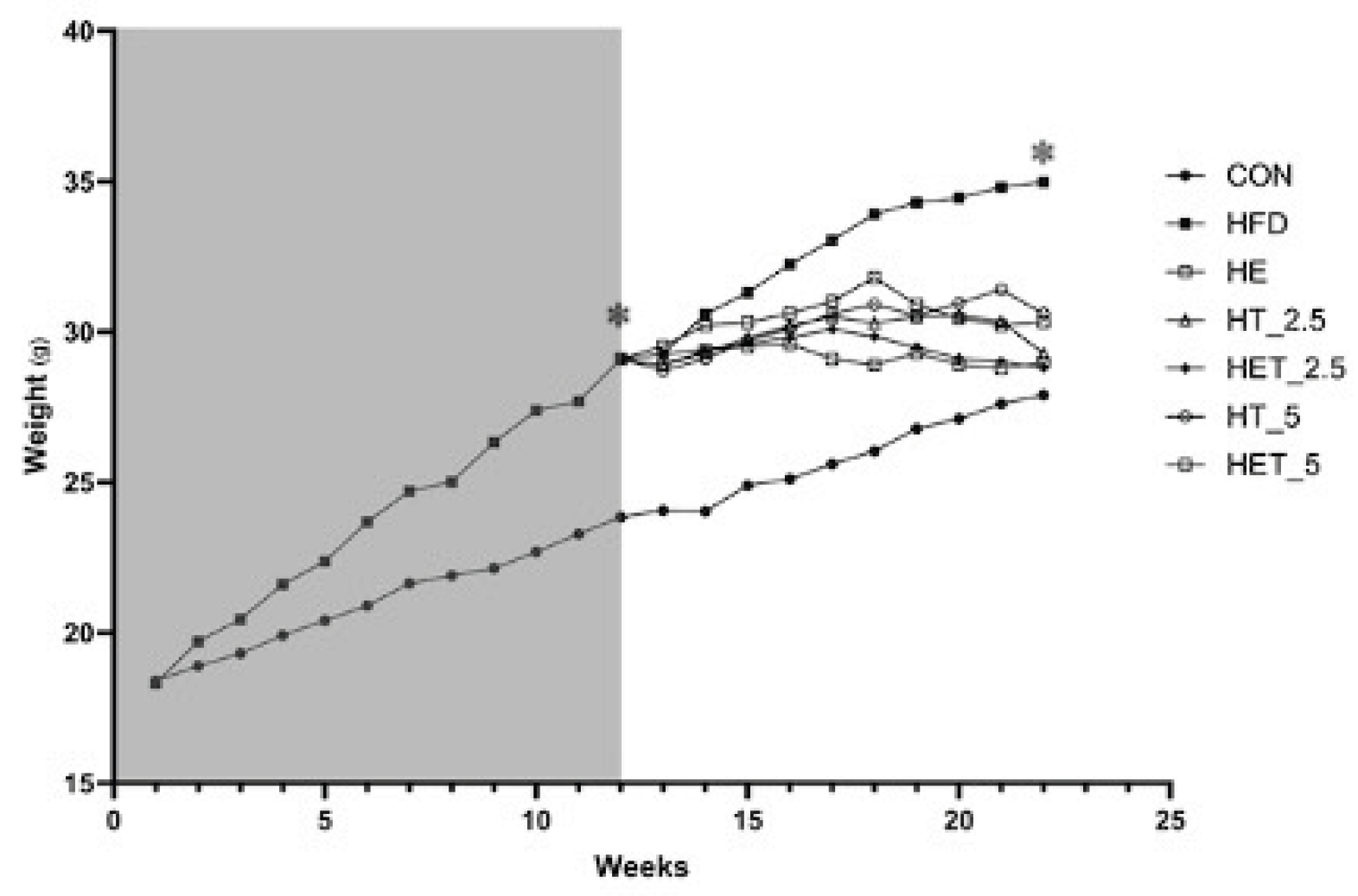
Figure 1. Body weight changes across experimental groups during two experimental phases. Phase 1 (12 weeks) included Control (CON) and high-fat diet (HFD) groups. Phase 2 (10 weeks) consisted of continued HFD feeding combined with exercise and/or taurine interventions, including: CON, HFD, HFD + high-intensity interval training (HE), HFD + 2.5% taurine (HT-2.5), HFD + HIIT + 2.5% taurine (HET-2.5), HFD + 5% taurine (HT-5), and HFD + HIIT + 5% taurine (HET-5). Data are presented as mean ± SD. *p < 0.05 vs. CON.
Preprints 226363 g001
Figure 2. HOMA-IR values across experimental groups. Data are presented as mean ± SD. *p < 0.05 vs. CON; #p < 0.05 vs. HFD. Taurine supplementation and HIIT significantly reduced HOMA-IR compared with HFD.
Preprints 226363 g002
Table 1. HIIT protocol progression during the intervention period.
Weeks Warm-up
(50% VO2max)
High-intensity intervals (85–90% VO2max) Recovery/Cool-down (50–60% VO2max)
1–2 8 m/min 16 m/min 9 m/min
3–4 10 m/min 19 m/min 11 m/min
5–6 11 m/min 22 m/min 12 m/min
7–8 14 m/min 25 m/min 15 m/min
9–10 14 m/min 25 m/min 15 m/min
Table 2. ALT, AST, Blood Glucose, Fasting Insulin, and Liver Glycogen levels in C57BL/6 Mice.
Group ALT (U/L) AST (U/L) Blood Glucose (mg/dl) Fasting Insulin (µU/mL) Liver Glycogen (mg/g)
CON 62.33±5.78 159.66±40.70 206.3±108.5 3.55±0.39 26.94±4.43
HFD 209.66±85.54* 326.16±57.56* 331.3±59.7* 6.51±.37* 11.83±3.47*
HE 134.50±30.07 253.66±90.77 263.2±53.4 6.43±0.22 15.02±1.03
HT-2.5 146.83±37.60 278.66±91.78 274.7±53.6 6.55±0.42 19.91±2.20#
HET-2.5 119.16±37.99# 217.50±84.09 210±51.1# 5.65±1.84 21.72±1.92#
HT-5 157.16±34.12 290.66±89.66 292.8±42.8 7.96±1.13# 14.11±1.35
HET-5 153.16±33.03 284.00±83.63 288.5±21.6 6.38±0.15 15.72±1.58
Values are presented as mean ±SD. *Significantly different from the CON group (p < 0.05); #Significantly different from the HFD group (p < 0.05). Groups: Control (CON), High-Fat Diet (HFD), HFD + HIIT (HE), HFD + Taurine 2.5% (HT-2.5), HFD + HIIT + Taurine 2.5% (HET-2.5), HFD + Taurine 5% (HT-5), HFD + HIIT + Taurine 5% (HET-5).
Disclaimer/Publisher’s Note: The statements, opinions and data contained in all publications are solely those of the individual author(s) and contributor(s) and not of MDPI and/or the editor(s). MDPI and/or the editor(s) disclaim responsibility for any injury to people or property resulting from any ideas, methods, instructions or products referred to in the content.
Copyright: This open access article is published under a Creative Commons CC BY 4.0 license, which permit the free download, distribution, and reuse, provided that the author and preprint are cited in any reuse.