

Submitted:
25 November 2024
Posted:
27 November 2024
You are already at the latest version
Abstract
This review examines the relationships between cholesterol and bone resorption. It seeks to elucidate the dependence of bone turnover on cholesterol metabolism by highlighting the common inhibitory effect of both statins and nitrogen-containing bisphosphonates on cholesterol biosynthesis and bone resorption as well as on bone density. Moreover, this paper also discusses the epidemiologic studies of the effects of nitrogen-containing bisphosphonates on all-cause and cardiovascular mortality using the latest publications to reinforce the relationships between bone resorption and cardiovascular disease. The review will also discuss the role of lipoproteins in supplying cholesterol to both osteoclasts and osteoblasts and the effects of doing so on both these bone cells and their precursors. As inflammation is a major factor in both bone resorption and cardiovascular calcification, this article will also discuss the role of cholesterol in triggering inflammatory responses. Finally, the paper will raise questions unanswered to date that bear on the relationship between lipid metabolism, bone resorption and cardiovascular disease.
Keywords:
cholesterol
; lipoproteins
; osteoclasts
; osteoblasts
; inflammation
; mortality
1. Introduction and Purpose
Osteoporosis, or progressive loss of bone mass, is a condition that can affect people of any age under specific circumstances, but characteristically is much more prevalent as we get older. While we tend to think that aging wears down our different organ systems independently of one another, there is increasing evidence that these changes are all linked. The purpose of this review is to demonstrate how this linkage may occur in the case of the chronic ongoing resorptive bone loss of osteoporosis and the development of hyperlipidemic cardiovascular disease, namely atherosclerosis.
The organizational plan of this review will be to introduce the topic in the same manner that I first became aware of it, by reports of the effects of statins on bone, then comparing these effects to those of nitrogen-containing bisphosphonates. Following, the epidemiological evidence linking bisphosphonates to reduced all-cause mortality in a variety of conditions will link bisphosphonates to cardiovascular disease. A discussion of the role of lipid metabolism in general and of cholesterol in particular on osteoclastic cells as well as on marrow osteoclast progenitors and osteoblasts will follow along with a discussion of the association of hypercholesterolemia with systemic bone loss. Evidence for a relationship between cholesterol and the inflammatory response will also be discussed in relation to cardiovascular disease, and by then the pathophysiologic as well as epidemiologic links between bone and the cardiovascular system should be apparent.
2. Methods
To search the literature, I used the PubMed index of the United States National Library of Medicine with key words including cholesterol, hyperlipidemia, osteoclasts, osteoblasts, lipoproteins, statins and inflammation. Articles were scanned for studies on the relationship of cholesterol to osteoclast and osteoblast function, differentiation, and metabolism, as well as the effect of hyperlipidemia on bone resorption and turnover, and the relationship of cholesterol to systemic inflammation. The most recent epidemiologic studies of the effects of bisphosphonates on all-cause and cardiovascular mortality were also investigated and related to the effects of statins on cholesterol metabolism. The effects of bisphosphonates on cholesterol biosynthesis were also the subject of the literature search. Articles duplicating findings were excluded and emphasis was placed on identifying primary sources. The specific roles of calcium and phosphate were excluded from this review as their roles have been covered in detail elsewhere.
3. Statins and Bisphosphonates
Statins are a class of drugs that inhibit 3-hydroxy-3-methylglutaryl co-enzyme A (HMG Co-A) reductase with a consequent reduction in hepatic cholesterol biosynthesis resulting in a decrease in serum cholesterol concentration and a reduced risk of heart attack. The report by Mundy et al. [1] giving statins to ovariectomized rats demonstrated that they developed an increased bone volume, increased bone formation rate, and a reduction in osteoclast numbers and increased osteoblasts on histomorphometric examination. In humans, a study by Edwards et al. [2] showed that post-menopausal women taking oral statins had greater bone density. Subsequent studies by Chuengsumarn et al. [3] and Safaei et al. [4] demonstrated that the lipophilic statin simvastatin increased bone mineral density and decreased bone resorption markers in elderly hyperlipidemic patients with osteopenia compared to a matched non-statin patient group [3] and in post-menopausal women with type 2 diabetes, lovastatin increased bone density at the lumbar spine and at Ward’s Triangle [4].
What is of interest with this use of statins is the relationship of their mechanism of action to the present generation of nitrogen-containing bisphosphonates. As shown in Figure 1, the cholesterol biosynthetic pathway in cells of liver as well as bone, as modified from Kim et al. [5] shows that HMG Co-A reductase precedes by just a few steps, the action of farnesyl pyrophosphate synthase (FPPS) in the cholesterol biosynthetic pathway. The classic mechanism of action of the anti-resorptive nitrogen-containing bisphosphonates as initially described by Rogers and his team [6] and others [7] is that an administered bisphosphonate will be preferentially taken up by bone and deposit in the hydroxyapatite matrix. As osteoclasts fix to the bone, they take up the bisphosphonate by endocytosis. Inside the osteoclast, the bisphosphonate will inhibit FPPS and thereby cholesterol biosynthesis in the cell membrane, damaging the membrane and leading to osteoclast apoptosis. As we will see in Section 5, other aspects of osteoclast function may also be affected. However, the net effect of the bisphosphonate ingestion is to inhibit osteoclast function and thereby reduce bone resorption.
One other interesting report by Tsubaki et al. [8] describes the inhibition of the chemokine macrophage inflammatory protein (MIP)-1α, or CCL3 in current nomenclature, by both the bisphosphonate minodrinate and the statins simvastatin and fluvastatin in human myeloma cells by working on the same pathways, Ras/MEK/ERK/AML-1A and Ras/PI3K/Akt/AML-1A by inhibiting Ras prenylation. This shows that the inhibition of two enzymes in the cholesterol biosynthetic pathway also can block the release of a chemokine that has been reported to induce inflammation of endovascular lipid deposits in the coronary arteries by de Jager et al. [9]. This finding also illustrates the link between cholesterol metabolism and inflammation as will be further discussed in Section 8.
4. Epidemiologic Evidence for Bisphosphonate Reduction of All-Cause Mortality in Humans
What began as isolated epidemiologic reports of an association between intake of nitrogen-containing bisphosphonates and all-cause mortality has now become too frequent to ignore. Some of the early isolated reports were summarized by Klein in 2022 [10]. However, the number of reports and the number of patients studied have increased significantly since then. Here are some of the most recent studies.
Wu et al. in 2023 [11] reviewed 46,729 subjects in the Taiwan National Health Insurance Research database between 2009 and 2018. Patients were followed for a mean of 4.7 years and those subjects with fractures receiving alendronate or risedronate, denosumab, or zoledronic acid experienced a significant reduction in all-cause mortality compared to those taking raloxifene and bazedoxifene, by 14-22%.
A study by Henney et al. [12] using the Tri Net X (Cambridge MA) database looking at denosumab, an anti-resorptive agent that blocks RANK Ligand, and its effect on all-cause including cardiovascular mortality, showed a reduction versus placebo of 21% starting with a database of over 331,000 subjects with diabetes. In another study by Alarkawi et al. [13] using a database of over 267,000 subjects in the Australian 45 and Up study, in those with fractures denosumab use was associated with 48% lower mortality in subjects with fractures while bisphosphonates were associated with a 44% reduced mortality. In those subjects who had not experienced a fracture, denosumab was associated with a 1.5-2.5-fold increase in mortality compared to oral bisphosphonates but comparable to users of intravenous zoledronic acid. However, in the fracture group, all-cause mortality from either bisphosphonate or denosumab use was lower than in subjects without treatment.
Wu et al. [14] studied the risk of all-cause mortality and cardiovascular risk among osteoporosis patients who had a previous episode of cardiovascular disease or ischemic stroke. The groups were divided into 464 patients who used bisphosphonates and 464 patients who did not. The bisphosphonate group had a 36% reduction in all-cause mortality after 8 years of follow-up compared to the group that did not take bisphosphonates, although the bisphosphonate group had a higher risk of hospitalization for atrial fibrillation than the control group with a hazard ratio of 1.76.
While these recent studies are representative of those in the literature, they are uniformly consistent with the original studies published from 2007-2016. While epidemiologic studies often expand a black box rather than contracting it, the uniform consistency of these results compels further investigation into the mechanism or mechanisms associated with these findings.
Thus, the effects of statins and bisphosphonates on the cholesterol biosynthesis pathway is a significant first step in tying skeletal and cardiovascular function together. However, as we will see, osteoclasts and their precursor cells are also heavily dependent on lipid metabolism, including, prominently, on cholesterol and low density lipoproteins. I will consider the supporting evidence for this concept in the next section.
5. Evidence Tying Cholesterol Metabolism to Osteoclasts and Their Hematopoietic Precursor Cells
Not only is cholesterol an essential part of the osteoclast cell membrane but it also functions in cell membrane signal transduction and in the interaction of the receptor activator of the nuclear transcription factor kappa B (NFκB) and its ligand(RANK-RANK ligand or RANKL) during osteoclastogenesis as discussed by Kim et al. [5] and by Luegmayr et al. [15]. Cholesterol is supplied to osteoclasts and progenitor cells by means of low density lipoproteins. As Sjogren et al. [16] demonstrated, macrophages, which have the same origin as osteoclasts have a receptor (R) for low density lipoprotein (LDL), which promotes internalization of ApoB lipoproteins, resulting in increased intracellular cholesterol. Increased intracellular cholesterol in turn downregulates the LDLR expression in macrophages to regulate the cellular intake of cholesterol. This feedback mechanism modulates the cellular content of cholesterol. LDL-induced cholesterol delivery will increase osteoclast viability while LDL depletion will suppress the formation of osteoclasts. In contrast, High-Density Lipoprotein (HDL) will carry cholesterol out of the osteoclasts as a result of membrane transporters ATP-binding cassette sub-family G member 1(ABCG1) or ABCA1, which will carry cholesterol to the HDL as noted by Huang et al. [17] and this efflux will induce osteoclast apoptosis, as discussed later.
In addition to the lipoprotein-induced delivery of cholesterol to osteoclasts and their precursors, Wei et al. [18] showed that cholesterol binds to the ligand binding domain of the estrogen-related receptor alpha (ERR α) to stimulate osteoclastogenesis while deletion of ERRα interferes with osteoclast differentiation and results in increased bone mass [19]. Use of the cholesterol binding compound cyclodextrin depleted intracellular cholesterol reduced ERRα transcriptional activity, while adding back cholesterol returned the ERRα activity [18]. This rescue action of cholesterol on ERRα was only possible in the presence of the co-activator peroxisome proliferator-activated receptor gamma co-activator -1-alpha (PGC-1 α), a co-regulator of glucose and lipid metabolism [19]. Thus there are several known factors that affect cholesterol entry into osteoclastic cells and precursors.
An additional role of cholesterol derivatives has been shown by Nevius et al. [20] to involve oxysterol, an oxidized cholesterol, which is an agonist of Epstein-Barr virus-Induced genes (EBI) 1 and 2, which stimulate osteoclast progenitors to migrate to the surfaces of bone and contribute to the bone turnover. The precise pathway is not as yet clear. They are also natural ligands of the Liver X receptors that transport cholesterol from peripheral tissues back to the liver [21].
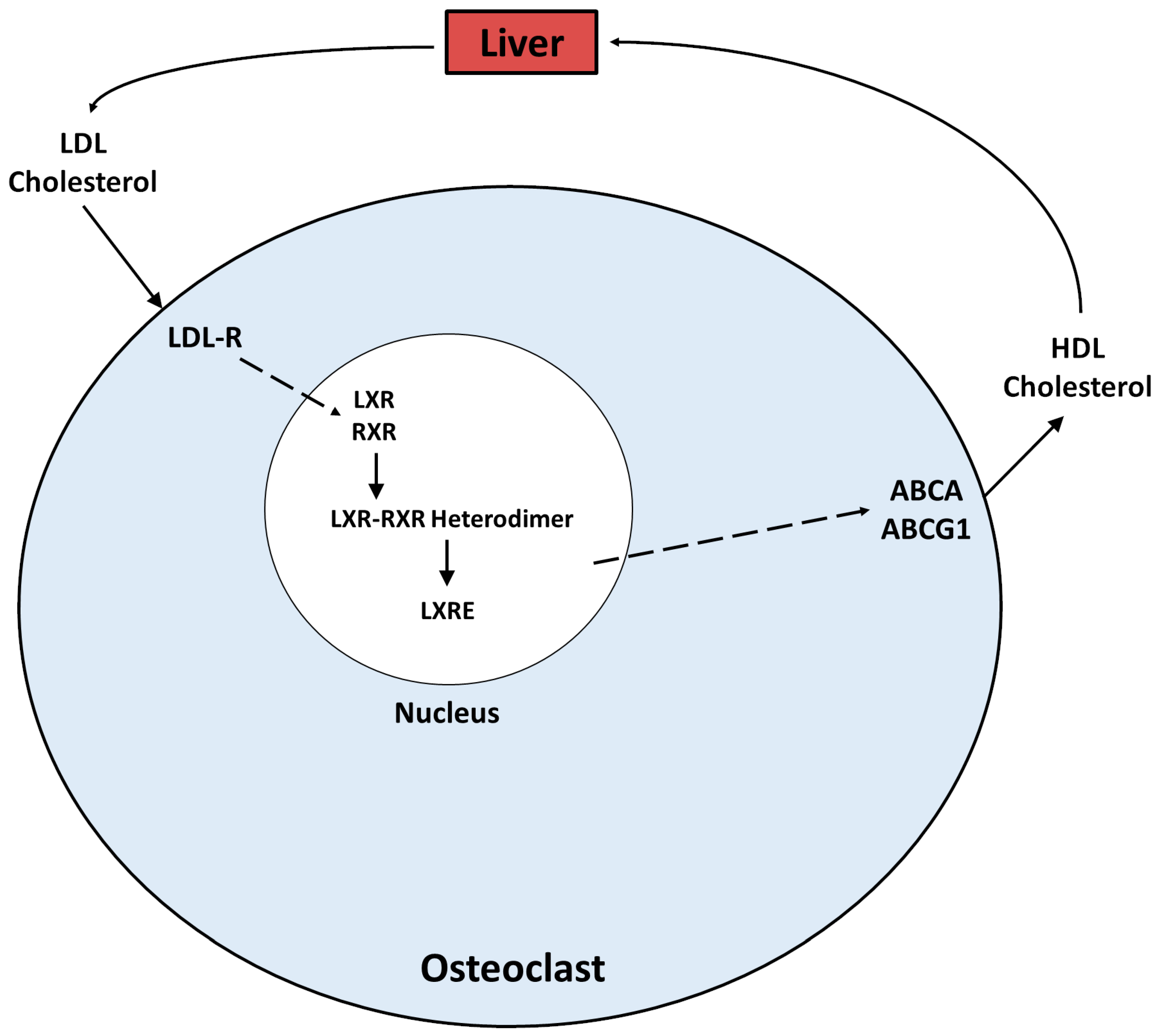
Liver X receptors (LXR) and Retinoid X receptors (RXR) are nuclear receptors that among other functions are cholesterol sensing receptors, though they also integrate metabolic and inflammatory signaling as discussed by Zelcer and Tontonoz [21]. LXR α plays a role in osteoclast function in cortical bone and LXR β does so in trabecular bone [22]. Knock-out of the LXRα in female mice resulted in increased cortical bone mass in the presence of an increased number of endosteal osteoclasts, indicating that these osteoclasts were non-functional, as evidenced by reduced serum concentrations of tartrate resistant acid phosphatase (TRAPC) and the C-telopeptide of type I collagen (CTX) [22]. Genes that are agonists of LXRα inhibit RANK ligand-induced gene c-FOS and nuclear factor of activated T cells, cytoplasmic 1 (NFAT-c1) expression, resulting in osteoclast apoptosis [23]. LXRs form heterodimers with RXRs to bind to the LXR response element (LXRE) on target genes [24]. These heterodimers stimulate the ABCA and ABCG1 membrane transporters in the osteoclast [21] to assist in the efflux of cholesterol from the cell via HDL. Thus the pathway of cholesterol efflux from the osteoclast is what ultimately contributes to apoptosis of the cell type and consequent reduction in bone resorption. This process is illustrated in Figure 2. The LXR-RXR heterodimer also appears to have pleiotropic functions as loss of RXRα and RXRβ in hematopoetic cells is associated with functionally deficient osteoclasts in a manner similar to what was reported by Joseph et al. [23] with knock out of LXRα [25]. In addition, combined knock out of RXRα and RXRβ in mice resulted in protection from ovariectomy-induced bone loss [5]. These apparently discrepant actions may depend on the different interactions of LXR and RXR with other intracellular factors.
In summary, LDL carries cholesterol into hematopoietic precursor cells and osteoclasts, where it stimulates differentiation and osteoclast function via an array of intermediary steps, including up-regulation of estrogen-related receptor α and PGC-1α. Cholesterol is also carried out of the osteoclast by a system of cholesterol sensing receptors such as LXR and RXR and membrane transport protein such as ABCG1 and ABCA, which attach the intracellular cholesterol to HDL, which carries it out of the osteoclasts, resulting in reduced osteoclast function or apoptosis. However, we also know that LXR and RXR probably have pleiotropic functions depending on which intracellular factors they interact with.
6. How Is The Cholesterol Effect on Bone Resorption Manifested at the Level of Organ Systems?
Now that we have a concept of how cholesterol affects osteoclastogenesis and function at the cellular level, we will examine how cholesterol affects bone resorption at the systemic level, examining conditions such as hypercholesterolemia and osteoporosis.
6.1. Hypercholesterolemia
There have been several reports noting that hypercholesterolemia is associated with bone loss. A study by Zhou et al. [26] showed that hypercholesterolemia in men results in high-turnover osteoporosis. Their studies showed that hypercholesterolemic men had reduced bone mineral density combined with increased serum biomarkers of bone turnover, including increased C-telopeptide of type I collagen (CTx) and increased serum type I collagen N-terminal peptide (P1NP), thus both bone formation and resorption were increased.
Pelton et al. [27] in a study of mice showed that two different strains with hypercholesterolemia lost both cortical and trabecular bone based on reduced biomechanical strength, including load to failure. In addition, histomorphometric studies demonstrated an increase in osteoclast number in the hypercholesterolemic strains.
Post-menopausal reduction in bone density accompanied by increased volume of marrow adipocytes was reported by Syed et al. [28] in a one-year randomized controlled trial of estrogen patch therapy, which treatment reduced marrow adipocyte volume and prevented an increase in adipocyte size compared to control subjects.
Finally, in a study of 6,477 Chinese and Caucasian subjects in whom bone mass was either adjusted or not adjusted for body weight, Zhao et al. [29] found that with adjustment for the effect of body weight on bone mass, there was an inverse relation between fat mass and bone mass.
All of these studies are consistent with a negative effect of fat or cholesterol on bone mass in a variety of circumstances ranging from hyperlipidemia to post-menopause and obesity, and the outcomes of these studies are consistent with the results observed from the cellular and metabolic studies in the previous section on cholesterol and osteoclast metabolism.
7. What About Osteoblasts?
As reviewed by Akhmetshina et al. [30], osteoblasts, like osteoclasts, receive cholesterol from the liver through LDL transport, cross the osteoblast cell membrane mediated by LDL receptor endocytosis. These fuse with lysosomes, which produce a lytic enzyme called lysosomal acid lipase, which hydrolyzes cholesterol esters and triglycerides to free cholesterol and free fatty acids. In addition, osteoblasts are also capable of initiating de novo cholesterol synthesis via the mevalonate pathway, as shown in Figure 1. This latter pathway appears to regulate osteoblast differentiation from stromal cell precursors as shown by Parhami et al. [31] in vitro by inhibiting the mevalonate pathway with consequent reduction in osteoblastogenesis in murine M210B4 stromal cells.
Other studies have documented that the effects of cholesterol on osteoblasts are both dose and time dependent. Li et al. [32] showed that cholesterol administration to mice resulted in increased mesenchymal cell number with increased expression of the osteoblastic genes for alkaline phosphatase (ALP), Bone Morphogenetic Protein (BMP) 1, RUNX2, and osteocalcin (OCN). However, You et al. [33] found that a high cholesterol diet inhibited bone formation in rats and that oxidized LDL, as a source of cholesterol, impaired osteoblastogenesis by reducing ALP activity, collagen synthesis and mineralization. In addition, gene mapping indicated that a high cholesterol diet may have an inhibitory effect on transforming growth factor (TGF)-β, bone morphogenetic protein-2 (BMP2), a member of the TGF-β family, and Wnt signaling. This occurs by downregulating the osteoblastic genes, specifically Wnt 5, β-catenin, TGF-βR, Smad 4, Smad 6, Smad 7, BMPR2, and BMP6, all participating in the TGF-β/BMP 2 and Wnt signaling pathways, resulting in downregulation of marrow stromal cell differentiation into osteoblasts [33]. Interestingly, oxidized LDL can also initiate atherosclerosis and can increase RANKL production in UMR 106 rat osteoblasts and in human MG63 osteosarcoma cells, an osteoblast-like cell line [34], possibly utilizing signaling pathways involving extracellular signal related kinase (ERK), NFκB and nuclear factor of activated T cells (NFAT) [34]. Also, this LDL compound appears to inhibit mineralization by inhibiting phosphate signaling in osteoblasts [35]. One component of HDL, apolipoprotein-A1 (Apo A1), when deleted in mice, shifts stromal cell differentiation into adipocytes instead of osteoblasts, as reported by Blair et al. [36]. Thus, it appears as if a certain amount of physiologic intracellular cholesterol is necessary for osteoblast function but larger amounts, such as provided by LDL-transported exogenous cholesterol lead to reduced function [30], while the reverse appears to be the case with osteoclasts. Bergstrom et al. [37] reported that osteoclasts have very low HMGCoA reductase expression, which is not up-regulated when cholesterol is depleted in the cell membrane. Therefore, exogenous cholesterol appears to be more important for osteoclast function.
On a systemic level, two Japanese studies found that hypercholesterolemic patients exhibited increased bone turnover with bone-specific alkaline phosphatase, a marker of bone formation, and N-telopeptide of type I collagen (NTx), a marker of bone resorption, being elevated, with the NTx positively correlated with serum total and LDL cholesterol [38,39]. Of additional interest, Awan et al. [40] discuss familial hypercholesterolemia associated with a mutation of the LDLR gene and a phenotype featuring serum osteocalcin, urinary calcium excretion, and glomerular filtration rate inversely correlated with the severity of aortic calcification. This condition links the LDL receptor function and hypercholesterolemia with aortic calcification, presumably by increased net bone loss.
7.1. The Effect of Statins on Osteoblasts
In addition to their inhibitory effect on HMGCoA reductase, statins appear to affect osteoblast function by stimulating or inhibiting various signaling pathways. Thus, Tsubaki et al. [41] showed in a mouse marrow stromal cell line that statins increase osteoblast-activating osteoprotegerin and inhibit RANKL expression by activating p38 MAP kinase (MAPK) and blocking the Ras/ERK pathway. In another study, Ruan et al. [42] demonstrated that statins also can activate the TGF-β/SMAD3 signaling pathway which reduces apoptosis of osteoblasts while the estrogen receptor (ER) inhibits osteoclast differentiation by stimulating osteoprotegerin.
8. Cholesterol and Inflammation
With all the focus on cholesterol, it should be noted that statin therapy, while reducing cholesterol, may have variable effects on inflammation. A study by Ridker et al. in the Lancet [43] noted that following treatment with statins the residual risk for serious cardiovascular disease depends significantly more on measures of inflammation, such as high sensitivity C-Reactive Protein than on markers of residual risk related to cholesterol, such as LDL cholesterol. That having been said, Lang et al. [44] reported that in mice, high cholesterol or inactivation of cholesterol efflux transporters stimulate hematopoietic stem cells to differentiate into monocytes and neutrophils. These cells increase levels of IL-23 and stimulate production of IL-17, resulting in increased granulocyte colony stimulating factor (G-CSF) production and secretion into the circulation, allowing these inflammatory factors to participate in the process of atherosclerosis. Thus, cholesterol also has a role in increasing inflammation and statin treatment. By decreasing cholesterol synthesis, statins may cause a reduction in inflammation, as has been evidenced by the Mendelian randomization study of Zhang et al. [45]. However, as shown in the study by Ridker et al. [43], the effects of statins on inflammation may vary. Nonetheless, these reports illustrate the interconnectedness of cholesterol with inflammation, bone, and cardiovascular disease.
9. Summary and Unanswered Questions
To summarize what we know, cholesterol deposits in the coronary vasculature but is also essential for osteoclast metabolism. Hypercholesterolemia is associated with increased bone resorption, likely due to its deposition in osteoclastic cells and their precursors. Statins, by interfering with cholesterol synthesis, can contribute to reduced cardiovascular mortality as can nitrogen-containing bisphosphonates by a related mechanism. The difference in efficacy is that statins function at highest concentration in the liver while bisphosphonates are taken up preferentially by bone matrix. The different sites of action may contribute to the relative efficacy of each class of drugs on the organs remote from their chief site of action.
Much of our knowledge about the intracellular cholesterol metabolism in osteoclasts is incomplete. Thus, while we know that low density lipoproteins help transport cholesterol into osteoclasts, the mechanisms that signal how much cholesterol to enter or leave the cell are still not understood. Further, the role of the estrogen related receptor (ERR) in coordination with the LDL receptor and the interaction of the two receptors needs further study. Moreover, the relationship between osteoclast activation, initiation of the inflammatory response, and bone resorption has yet to be worked out. Is the inflammatory response a mediator of bone resorption, can osteoclast activation work directly via intracellular cholesterol ingress, or is this function a redundancy built into the body? What are the critical roles of co-factors such as PGC-1α? On a systemic level, what is the relationship between hypercholesterolemia, inflammation, and other circulating factors such as the calcium and phosphate liberated from resorbing bone (10, 46)? Further elucidation of these intracellular metabolic processes as well as of the precise interactions of the resorptive, inflammatory and metabolic processes described herein may lead to the identification of a variety of new therapeutic targets to mitigate bone resorption-related atherosclerotic heart disease.
Acknowledgements
Randal Morris prepared Figure 1 and Figure 2 in this manuscript.
References
- Mundy G, Garrett R, Harris S, Chan J, Chen D, Rossini G, Boyce B, Zhao M, Gutierrez G Stimulation of bone formation in vitro and in rodents by statins Science 1999; 286(5446): 1946-9.
- Edwards CJ, Hart DJ, Spector TD Oral statins and increased bone mineral density in post-menopausal women Lancet 2000; 355(9222): 2218-19.
- Chuengsamarn S, Rattanamongkoulgul S. Suwanwalaikorn S, Wattanasinchaigoon S, Kaufman L Effects of statins versus non-statin lipid-lowering therapy on bone formation and bone mineal density biomarkers in patients with hyperlipidemia Bone 2010; 46: 1011-15. [CrossRef]
- Safaei H, Janghorbani M, Aminorroya A, Amini M Lovastatin effects on bone mineral density in post-menopausal women with type 2 diabetes mellitus. Acta Diabetol 2007; 44: 76-82. [CrossRef]
- Kim H, Oh, B, Park-Min K-H Regulation of osteoclast differentiation and activity by lipid metabolism Cells 2021 Jan; 10(1): 89. [CrossRef]
- Coxon FP, Helfrich MH, Van’t Hof R, Sebti S, Ralston SH, Hamilton A, Rogers MJ Protein geranylgeranylation is required for osteoclast formation, function and survival: inhibition by bisphosphonates and GGTI-298 J Bone Miner Res 2000; 15(8): 1467-76. [CrossRef]
- Ebetino FH, Sun S, Cherian P, Roshandel S, Neighbors JD, Hu E, Dunford JE, Sedghizadeh PP, McKenna CE, Srinavasan V, Boeckman RK, Russell RGG Bisphosphonates: The role of chemistry in understanding their biological actions and structural activity relationships, and new directions for their therapeutic use Bone 2022Mar: 156:116289. [CrossRef]
- Tsubaki M, Takeda T, Sakamoto K, Shimaoka H, Fujita A, Itoh T, Imano M, Mashino K, Fujiwara D, Sakaguchi K, Satou T, Nishida S Bisphosphonates and statins inhibit expression and secretion of MIP-1-alpha via suppression of Ras/MEK/ERK/AML-1A and Ras/PI3K/Akt/AML-1A pathways. Am J Cancer Res 2014 Dec 15; 5(1): 168-79, eCollection 2015.
- De Jager SCA, Bot I, Kraaijeveld AO, Korporaal SJA, Bot M, van Santbrink PJ, van Berkel TJC, Kulper J, Biessen EAL Leukocyte-specific CCL3 deficiency inhibits atherosclerotic lesion development by affecting neutrophil accumulation Atherosclerosis, Thromb Vasc Biol 2013; 33(3): e75-e83. [CrossRef]
- Klein GL Is calcium a link between inflammatory bone resorption and heart disease? eLife 2022 Dec 29; 11: e83841.
- Wu C-H, Li C-C, Hsu Y-H, Liang F-W, Chang Y-F, Hwang J-S Comparisons between different anti-osteoporosis medications on post-fracture mortality: a population based study J Clin Endocrinol Metab 2023; 108(4): 827-33. [CrossRef]
- Henney AE, Riley DR, O’Connor B, Hydes TJ, Anson M, Zhao SS, Alam U, Cuthbertson DJ Denosumab, for osteoporosis, reduces the incidence of type 2 diabetes, risk of foot ulceration and all-cause mortality in adults, compared with bisphosphonates: An analysis of real-world cohort data, with a systematic review and meta-analysis Diabetes Obes Metab 2024; 26(9): 3673-83. [CrossRef]
- Alarkawi D, Tran T, Chen W, March LM, Blyth FM, Blank RD, Bliuc D, Center JR Denosumab and mortality in a real-world setting: a comparative study J Bone Miner Res 2023; 38(12): 1757-70 10.1002/jbmr.4930.
- Wu ST, Chen JF, Tsai CJ The impact of bisphosphonates on mortality and cardiovascular risk among osteoporosis patients with cardiovascular disease J Formos Med Assoc 2021; 20(11): 1957-66. [CrossRef]
- Luegmayr E, Glantschnig H, Wesoloski GA, Gentile MA, Fisher JE, Rodan GA, Reszka AA Osteoclast formation, survival and morphology are highly dependent on exogenous cholesterol/lipoproteins Cell Death Differ 2004; 11 (Suppl 1): S108-18. [CrossRef]
- Sjogren U, Mukohyama H, Roth C, Sundquist G, Lerner UH Bone resorbing activity from cholesterol-exposed macrophages due to enhanced expression of interleukin-1-alpha J Dent Res 2002; 81: 11-16. [CrossRef]
- Huang x, Lv Y, He P, Wang Z, Xiong F, Zhang D, Cao Q, Tang C HDL impairs osteoclastogenesis and induces osteoclast apoptosis via up-regulation of ABCG1 expression. Acta Biochim Biophys Sin 2018; 50: 853-61. [CrossRef]
- Wei W, Schwaid AG, Wang X, Wang X, Chen S, Chu Q, Saghatelian A, Wan Y Ligand activation of ERR alpha by cholesterol mediates statin and bisphosphonate effects Cell Metab 2016; 23: 479-91. [CrossRef]
- Wei W, Wang X, Yang M, Smith LC, Dechow PC, Sonoda J, Evans RM, Wan Y PGC1 beta mediates PPAR gamma activation of osteoclastogenesis and rosiglitazone-induced bone loss Cell Metab 2010 Jun 9; 11(6): 503-16. [CrossRef]
- Nevius E, Pinho F, Dhodapkar M, Jin H, Nadrah K, Horowitz MC, Kikukta J, Ishii M, Pereira JP Oxysterols and EBI2 promote osteoclast precursor migration to bone surfaces and regulate bone mass homeostasis J Exp Med 2015; 212: 1931-46. [CrossRef]
- Zelcer N, Tontonoz P Liver X receptors as integrators of metabolic and inflammatory signaling. J Clin Invest 2006; 116: 607-14. [CrossRef]
- Robertson KM, Norgard M, Windahl SH, Hultenby K, Ohlsson C, Andersson G, Gustafsoson JA Cholesterol sensing receptors, liver X receptors alpha and beta, have novel and distinct roles in osteoclast differentiation and activation. J Bone Miner Res 2006; 21: 1276-87. [CrossRef]
- Joseph SB, Laffitte BA, Patel PH, Watson MA, Matsukama KE, Walczak R, Collins JL, Osborne T, Tontonoz P Direct and indirect mechanisms for regulation of fatty acid synthase gene expression by liver X receptors J Biol Chem 2002; 277: 11019-25. [CrossRef]
- Kim HJ, Yoon KA, Yoon HJ, Hong JM, Lee MJ, Lee K, Kim SY Liver X receptor activation inhibits osteoclastogenesis by suppressing NFkB activity and c-Fos induction and jprevents inflammatory bone loss in mice J Leukoc Biol 2013; 94: 99-107. [CrossRef]
- Menendez-Gutierrez MP, Roszer T, Fuentes L, Nunez V, Escolano A, Redondo JM, DeClerck N, Metzger D, Valledor AF, Ricote M Retinoid X receptors orchestrate osteoclast differentiation and postnatal bone remodeling J Clin Invest 2015; 125: 809-23. [CrossRef]
- Zhou Y, Deng T, Zhang H, Guan Q, Zhao H, Yu C, Shao S, Zhao M, Xu J Hypercholesterolemia increases the risk of high turnover osteoporosis in men Mol Med Rep 2019; 19: 4603-12. [CrossRef]
- Pelton K, Krieder J, Joiner D, Freeman MR, Goldstein SA, Solomon KR Hypercholesterolemia promotes an osteoporotic phenotype Am J Pathol 2012; 181: 928-36. [CrossRef]
- Syed FA, Oursler MJ, Hefferanm TE, Peterson JM, Riggs BL, Khosla S Effects of estrogen therapy on bone marrow adipocytes in post-menopausal osteoporotic women. Osteoporos Int 2008; 19: 1323-30. [CrossRef]
- Zhao LJ, Liu YJ, Hamilton J, Recker RR, Deng HW Relationship of obesity with osteoporosis J Clin Endocrinol Metab 2007; 92: 1640-6. [CrossRef]
- Akhmetshina A, Kratky D, Rendina-Ruedy E Influence of cholesterol on the regulation of osteoblast function Metabolites 2023 Apr 21; 13(4): 578. [CrossRef]
- Parhami F, Mody N, Gharavi N, Ballard AJ, Tintut Y, Demer LI Role of cholesterol biosynthetic pathway on osteoblastic differentiation of marrow stromal cells J Bone Miner Res 2002; 17: 1997-2003. [CrossRef]
- Li H, Guo H, Li H Cholesterol loading affects differentiation of mouse mesenchymal cells Steroids 2013; 78: 426-33. [CrossRef]
- You L, Sheng ZY, Tang CL, Chen L, Pan L, Chen JY High cholesterol diet increases osteoporosis risk via inhibiting bone formation in rats Acta Pharmacol Sin 2011; 32: 1498-504. [CrossRef]
- Maziere C, Salle V, Gomila C, Maziere JC Oxidized low density lipoproteins enhanced RANKL expression in human osteoblast-like cells: involvement of ERK, NF kappa B and NFAT. Biochim Biophys Acta 2013; 1832: 1756-64. [CrossRef]
- Maziere C, Savitsky V, Galmiche A, Gomila C, Massy Z, Maziere JC Oxidized low density lipoprotein inhibits phosphate signaling and phosphate-induced mineralization in osteoblasts: involvement of oxidative stress Biochim Biophys Acta 2010; 1802: 1013-19. [CrossRef]
- Blair HC, Kalyviot E, Papachristou NI, Tourkova IL, Syggelos SA, Deligianni D, Orkula MG, Kontyannis CG, Karavia EA, Kypreos KE, Papachristou DJ Apolipoprotein A-1 regulates osteoblast and lipoblast precursor cells in mice Lab Invest 2016; 96: 763-72. [CrossRef]
- Bergstrom JD, Bostedor RG, Masarachia PJ, Reszka AA, Rodan G Alendronate is a specific nanomolar inhibitor of farnesyl diphosphate synthase Arch Biochem Biophys 2000; 373(1): 231-41 doi. 10.1006/abbi.1999.1502.
- Majima T, Komatsu Y, Fukao A, Ninomiya K, Matsumura T, Nakao K Short-term effects of atorvastatin on bone turnover im male patients with hypercholesterolemia Endocr J 2007; 54: 145-51. [CrossRef]
- Majima T, Shimatsu A, Komatsu Y, Satoh N, Fukao A, Ninomiya K, Matsumura T, Nakao K Short-term effects of pitavastatin on biochemical markers of bone turnover in patients with hypercholesterolemia Intern Med 2007; 46: 1967-73. [CrossRef]
- Awan Z, Alwaili K, Alshahrani A, Langsetmo L, Goltzman D, Genest J Calcium homeostasis and skeletal integrity and individuals with familial hypercholesterolemia and aortic calcification Clin Chem 2010; 56: 1599-1607. [CrossRef]
- Tsubaki M, Satou T, Itoh T, Imano M, Yanae M, Kato C, Tagoshi R, Komai M, Nishida S Bisphosphonate- and statin-induced enhancement of OPG expression and inhibition of CD9, M-CSF and RANKL expressions via inhibition of the Ras/MEK/ERK pathway and activation of p38 MAPK in mouse bone marrow stromal cell line ST2 Mol Cell Endocrinol 2012; 361: 219-31. [CrossRef]
- Ruan F, Zheng Q, Wang J, Mechanisms of bone anabolism regulated by statins Biosci Rep 2012; 32: 511-19. [CrossRef]
- Ridker PM, Bhatt DL, Pradhan A, Glynn RJ, MacFadyen JG, Nissen SE on behalf of the PROMINENT, REDUCE-IT and STRENGTH investigators Inflammation and cholesterol as precitors of cardiovascular events among patients receiving statin therapy: a collaborative analysis of three randomized trials Lancet 2023; 401 (10384): 1293-301. [CrossRef]
- Lang JK, Cimato TR Cholesterol and hematopoietic stem cells: inflammatory mediators and atherosclerosis Stem Cells Transl Med 2014; 3: 549-52. [CrossRef]
- Zhang K, Liu W, Liang H Effects of statins on sepsis and inflammatory factors: A Mendelian randomization study Eur J Clin Invest 2024; 54(5): e14164. [CrossRef]
- Klein GL Phosphate as an adjunct to calcium in promoting coronary vascular calcification in chronic inflammatory states eLife 2024 June 12; 13: e91808. [CrossRef]
Figure 1.
Cholesterol biosynthetic pathway with points of enzyme inhibition for STATINS and NITROGEN-CONTAINING BISPHOSPHONATES. Steps in the pathway noted in blue while enzymes are noted in black print for each step in the pathway. Arrows point from STATINS and N-CONTAINING BISPHOSPHONATES to the specific enzymes each inhibits.
Figure 1.
Cholesterol biosynthetic pathway with points of enzyme inhibition for STATINS and NITROGEN-CONTAINING BISPHOSPHONATES. Steps in the pathway noted in blue while enzymes are noted in black print for each step in the pathway. Arrows point from STATINS and N-CONTAINING BISPHOSPHONATES to the specific enzymes each inhibits.
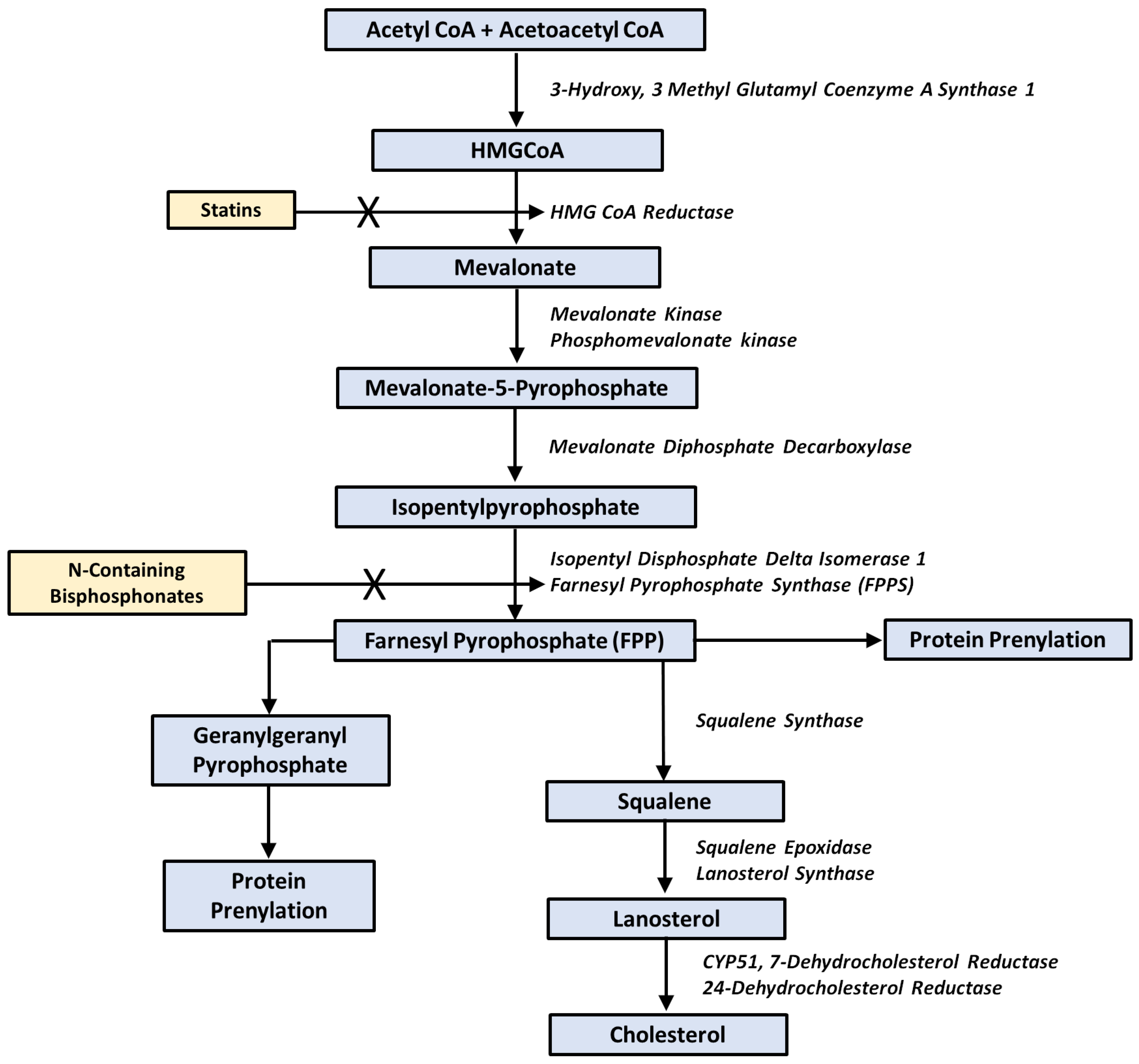
Figure 2.
The pathways for exogenous cholesterol entry and egress from the osteoclast. Note that for entry low-density lipoprotein receptor plays a role, as does the estrogen related receptor (ERR) while ATP Binding Cassette Subfamily G member 1 (ABCG1) and ABCA are membrane transporters that attach exogenous cholesterol to high density lipoprotein (HDL) on the way out of the osteoclast and back toward the liver. The process is orchestrated by the cholesterol sensing receptors in the nucleus of the cell called Liver X Receptors (LXR) and Retinoid X Receptors (RXR), which form a trimer with the LXR response element (LXRE).
Figure 2.
The pathways for exogenous cholesterol entry and egress from the osteoclast. Note that for entry low-density lipoprotein receptor plays a role, as does the estrogen related receptor (ERR) while ATP Binding Cassette Subfamily G member 1 (ABCG1) and ABCA are membrane transporters that attach exogenous cholesterol to high density lipoprotein (HDL) on the way out of the osteoclast and back toward the liver. The process is orchestrated by the cholesterol sensing receptors in the nucleus of the cell called Liver X Receptors (LXR) and Retinoid X Receptors (RXR), which form a trimer with the LXR response element (LXRE).
Disclaimer/Publisher’s Note: The statements, opinions and data contained in all publications are solely those of the individual author(s) and contributor(s) and not of MDPI and/or the editor(s). MDPI and/or the editor(s) disclaim responsibility for any injury to people or property resulting from any ideas, methods, instructions or products referred to in the content. |
© 2024 by the authors. Licensee MDPI, Basel, Switzerland. This article is an open access article distributed under the terms and conditions of the Creative Commons Attribution (CC BY) license (http://creativecommons.org/licenses/by/4.0/).
Copyright: This open access article is published under a Creative Commons CC BY 4.0 license, which permit the free download, distribution, and reuse, provided that the author and preprint are cited in any reuse.


